Purified Mouse anti-Nucleostemin — 562749 · 2016-09-12 · CRL-1821™, right blot) were probed...
2
BD Pharmingen™ Technical Data Sheet Purified Mouse anti-Nucleostemin Product Information Material Number: 562749 Alternate Name: GNL3, E2-induced gene 3 protein, NNP47, Nucleolar GTP-binding protein 3 Size: 0.1 mg Concentration: 0.5 mg/ml Clone: P22-1125 Immunogen: Human Nucleostemin Recombinant Protein Isotype: Mouse IgG1, κ Reactivity: QC Testing: Human Tested in Development: Mouse Target MW: ~ 62 kDa Storage Buffer: Aqueous buffered solution containing ≤0.09% sodium azide. Description Nucleostemin, also known as guanine nucleotide binding protein-like 3 (GNL3), was first identified in CNS stem cells and has subsequently been shown to be expressed in embryonic stem cells, bone-marrow derived stem cells, corneal epithelial cells, as well as multiple cancer cell lines and tumors. As its name suggests, nucleostemin is localized to the nucleolus and contains GTP-binding motifs. Nucleostemin has been implicated in cell cycle progression and the maintenance of proliferation. Interestingly, the depletion and the over-expression of nucleostemin cause cell cycle arrest. Both these mechanisms of cell cycle arrest are through a p53 dependant manner. The expression of nucleostemin is down-regulated during stem cell differentiation in vivo and in vitro. TOP LEFT: Western blot analysis of Nucleostemin expression in human embryonic carcinoma and mouse embryonic stem (ES) cells. Lysates from a human embryonal carcinoma cell line NCCIT (ATCC CRL-2073™, left blot) and mouse ES-E14TG2a (ATCC CRL-1821™, right blot) were probed with Purified Mouse anti-Nucleostemin monoclonal antibody at titrations of 2.0 (lane 1), 1.0 (lane 2), and 0.5 μg/ml (lane 3). Nucleostemin is identified as a band of ~62 kDa. Proteins were detected using HRP Goat Anti-Mouse Ig (Cat. No. 554002) and a chemiluminescent detection system. TOP RIGHT: Immunoflourescent staining of Nucleostemin in human embryonic stem cells. H9 human ES cells (WiCell, Madison, WI) passage 31 grown in mTESR™1 medium (StemCell Technologies) on BD Matrigel™ hESC-qualified Matrix (Cat. No. 354277) were fixed with BD Cytofix™ Buffer (Cat. No. 554655), permeabilized, and stained with Purified Mouse anti-Nucleostemin monoclonal antibody (pseudo-colored green) at 1.2 µg/ml. The second-step reagent was Alexa Fluor® 488 goat anti-mouse Ig (Life Technologies) and counter-staining was with DAPI (pseudo-colored blue). The images were captured on a BD Pathway™ 435 Cell Analyzer and merged using BD Attovision™Software. Permeabilization was with 1x BD Perm/Wash™ Buffer (Cat No. 554723); Triton™ X-100 is also suitable for permeabilization. BOTTOM ROW: Immunohistochemical staining of Nucleostemin in human colon cancer. Following antigen retrieval with BD Retrievagen A buffer (Cat. No. 550524), formalin-fixed paraffin-embedded human colon cancer was stained with either purified mouse IgG1, k isotype control (Cat. No.550878, left panel) or Purified Mouse Anti-Human Nucleostemin antibody (right panel). A three-step staining procedure that employs a Biotin Goat Anti-Mouse Ig secondary antibody, Streptavidin HRP (Cat. No. 550946) and DAB Substrate Kit (Cat. No. 550880) was used. Original magnification: 40X. Preparation and Storage The monoclonal antibody was purified from tissue culture supernatant or ascites by affinity chromatography. Store undiluted at 4°C. 562749 Rev. 1 Page 1 of 2
Transcript of Purified Mouse anti-Nucleostemin — 562749 · 2016-09-12 · CRL-1821™, right blot) were probed...
BD Pharmingen™
Technical Data Sheet
Purified Mouse anti-Nucleostemin
Product Information
Material Number: 562749
Alternate Name: GNL3, E2-induced gene 3 protein, NNP47, Nucleolar GTP-binding protein 3
Size: 0.1 mg
Concentration: 0.5 mg/ml
Clone: P22-1125
Immunogen: Human Nucleostemin Recombinant Protein
Isotype: Mouse IgG1, κ
Reactivity: QC Testing: Human
Tested in Development: Mouse
Target MW: ~ 62 kDa
Storage Buffer: Aqueous buffered solution containing ≤0.09% sodium azide.
DescriptionNucleostemin, also known as guanine nucleotide binding protein-like 3 (GNL3), was first identified in CNS stem cells and has subsequently been
shown to be expressed in embryonic stem cells, bone-marrow derived stem cells, corneal epithelial cells, as well as multiple cancer cell lines and
tumors. As its name suggests, nucleostemin is localized to the nucleolus and contains GTP-binding motifs. Nucleostemin has been implicated in cell
cycle progression and the maintenance of proliferation. Interestingly, the depletion and the over-expression of nucleostemin cause cell cycle arrest.
Both these mechanisms of cell cycle arrest are through a p53 dependant manner. The expression of nucleostemin is down-regulated during stem cell
differentiation in vivo and in vitro.
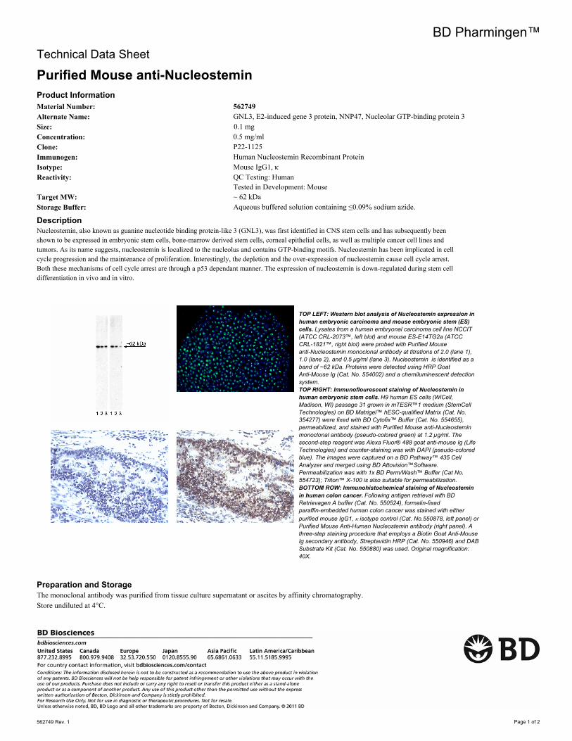
TOP LEFT: Western blot analysis of Nucleostemin expression in
human embryonic carcinoma and mouse embryonic stem (ES)
cells. Lysates from a human embryonal carcinoma cell line NCCIT
(ATCC CRL-2073™, left blot) and mouse ES-E14TG2a (ATCC
CRL-1821™, right blot) were probed with Purified Mouse
anti-Nucleostemin monoclonal antibody at titrations of 2.0 (lane 1),
1.0 (lane 2), and 0.5 μg/ml (lane 3). Nucleostemin is identified as a
band of ~62 kDa. Proteins were detected using HRP Goat
Anti-Mouse Ig (Cat. No. 554002) and a chemiluminescent detection
system.
TOP RIGHT: Immunoflourescent staining of Nucleostemin in
human embryonic stem cells. H9 human ES cells (WiCell,
Madison, WI) passage 31 grown in mTESR™1 medium (StemCell
Technologies) on BD Matrigel™ hESC-qualified Matrix (Cat. No.
354277) were fixed with BD Cytofix™ Buffer (Cat. No. 554655),
permeabilized, and stained with Purified Mouse anti-Nucleostemin
monoclonal antibody (pseudo-colored green) at 1.2 µg/ml. The
second-step reagent was Alexa Fluor® 488 goat anti-mouse Ig (Life
Technologies) and counter-staining was with DAPI (pseudo-colored
blue). The images were captured on a BD Pathway™ 435 Cell
Analyzer and merged using BD Attovision™Software.
Permeabilization was with 1x BD Perm/Wash™ Buffer (Cat No.
554723); Triton™ X-100 is also suitable for permeabilization.
BOTTOM ROW: Immunohistochemical staining of Nucleostemin
in human colon cancer. Following antigen retrieval with BD
Retrievagen A buffer (Cat. No. 550524), formalin-fixed
paraffin-embedded human colon cancer was stained with either
purified mouse IgG1, k isotype control (Cat. No.550878, left panel) or
Purified Mouse Anti-Human Nucleostemin antibody (right panel). A
three-step staining procedure that employs a Biotin Goat Anti-Mouse
Ig secondary antibody, Streptavidin HRP (Cat. No. 550946) and DAB
Substrate Kit (Cat. No. 550880) was used. Original magnification:
40X.
Preparation and StorageThe monoclonal antibody was purified from tissue culture supernatant or ascites by affinity chromatography.
Store undiluted at 4°C.
562749 Rev. 1 Page 1 of 2
Application Notes
Application
Western blot Routinely Tested
Bioimaging Tested During Development
Immunofluorescence Tested During Development
Immunohistochemistry-formalin (antigen retrieval required) Tested During Development
Intracellular staining (flow cytometry) Tested During Development
Suggested Companion Products
Catalog Number Name CloneSize
554002 HRP Goat Anti-Mouse Ig 1.0 ml (none)
354277 BD Matrigel™ hESC-qualified Matrix 5.0 ml (none)
554655 Fixation Buffer 100 ml (none)
550524 Retrievagen A (pH 6.0) 1000 ml (none)
550878 Purified Mouse IgG1 κ Isotype Control 1.0 ml MOPC-31C
550337 Biotin Goat Anti-Mouse Ig (Multiple Adsorption) 1.0 ml Polyclonal
550946 Streptavidin HRP 50 ml (none)
550880 DAB Substrate Kit 500 tests (none)
554723 Perm/Wash Buffer 100 ml (none)
353219 BD Falcon™ 96-well Imaging Plate (none)
Product NoticesSince applications vary, each investigator should titrate the reagent to obtain optimal results. 1.
Please refer to www.bdbiosciences.com/pharmingen/protocols for technical protocols. 2.
Caution: Sodium azide yields highly toxic hydrazoic acid under acidic conditions. Dilute azide compounds in running water before
discarding to avoid accumulation of potentially explosive deposits in plumbing.
3.
Sodium azide is a reversible inhibitor of oxidative metabolism; therefore, antibody preparations containing this preservative agent must not
be used in cell cultures nor injected into animals. Sodium azide may be removed by washing stained cells or plate-bound antibody or
dialyzing soluble antibody in sodium azide-free buffer. Since endotoxin may also affect the results of functional studies, we recommend the
NA/LE (No Azide/Low Endotoxin) antibody format, if available, for in vitro and in vivo use.
4.
Triton is a trademark of the Dow Chemical Company. 5.
mTESR™1 is a trademark of StemCell Technologies. 6.
ReferencesKafienah W, Mistry S, Williams C, Hollander AP. Nucleostemin is a marker of proliferating stromal stem cells in adult human bone marrow. Stem Cells. 2006;
24(4):1113-1120. (Biology)
Kawashima M, Kawakita T, Yoshida S, Shimmura S, Tsubota K. Nucleostemin as a possible progenitor marker of corneal epithelial cells. Mol Vis. 2009;
15:1162-1168. (Biology)
Ma H, Pederson T. Nucleostemin: a multiplex regulator of cell-cycle progression. Trends Cell Biol. 2008; 18(12):575-579. (Biology)
Nomura J, Maruyama M, Katano M, et al. Differential requirement for nucleostemin in embryonic stem cell and neural stem cell viability. Stem Cells. 2009;
27(5):1066-1076. (Biology)
Tsai RY, McKay RD. A nucleolar mechanism controlling cell proliferation in stem cells and cancer cells. Genes Dev. 2002; 16:2991-3003. (Biology)
562749 Rev. 1 Page 2 of 2