Protein Structure: Data Bases and Classification
57
Protein Structure: Data Bases and Classification Ingo Ruczinski Department of Biostatistics, Johns Hopkins University
Transcript of Protein Structure: Data Bases and Classification
Protein Structure: Data Bases and Classification
Ingo Ruczinski
Department of Biostatistics, Johns Hopkins University
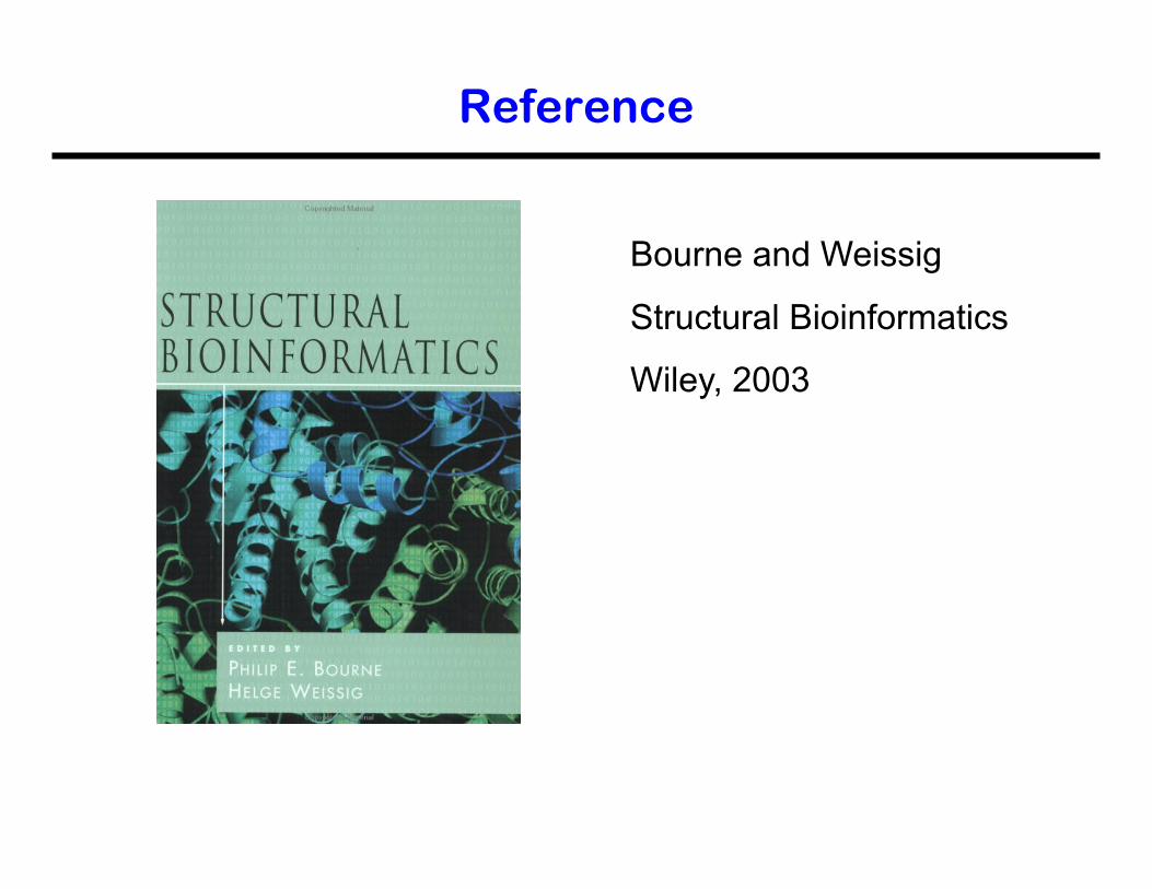
Bourne and Weissig
Structural Bioinformatics
Wiley, 2003
Reference
More References
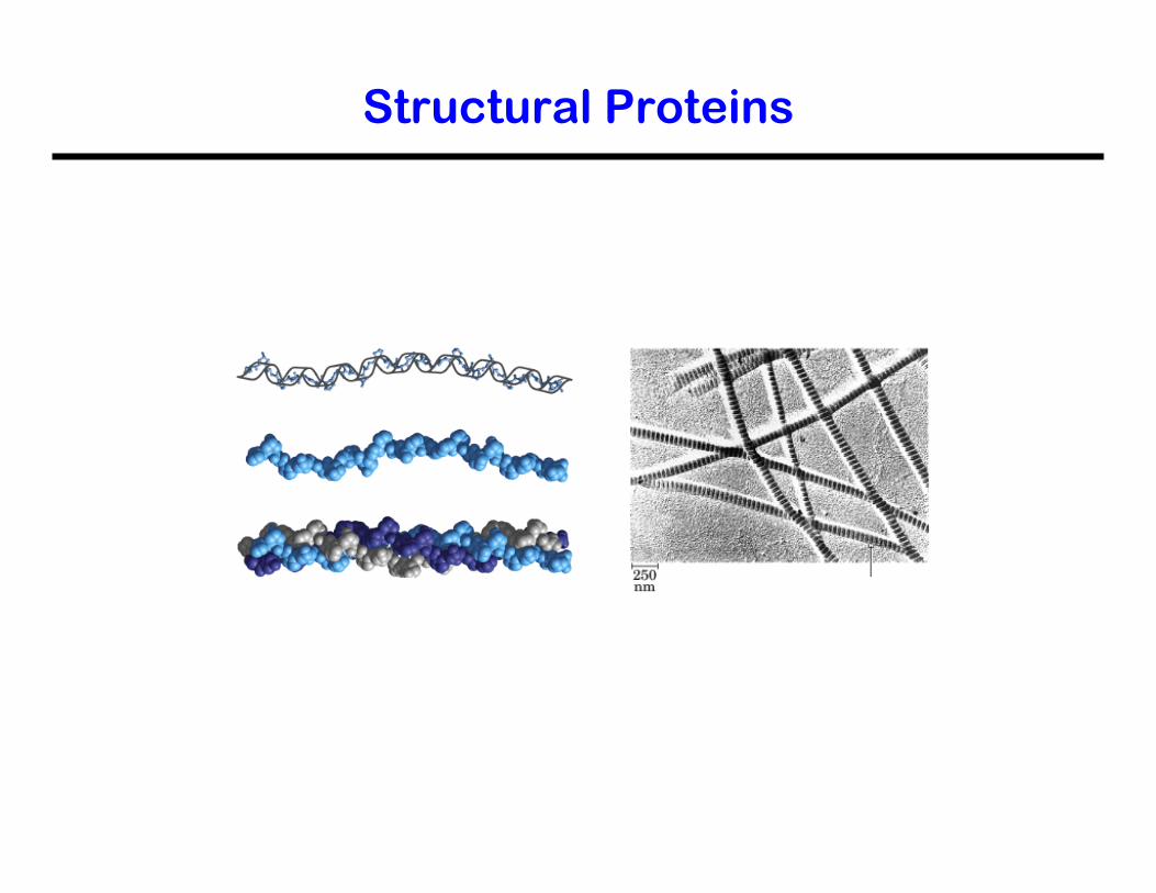
Structural Proteins
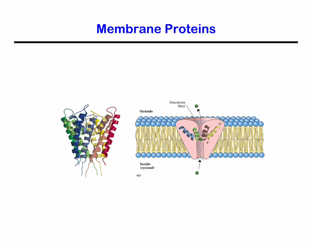
Membrane Proteins
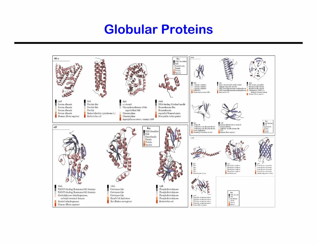
Globular Proteins
Terminology
• Primary Structure • Secondary Structure • Tertiary Structure • Quatenary Structure • Supersecondary Structure • Domain • Fold
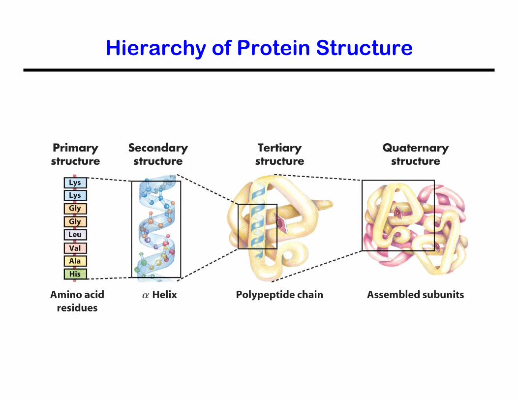
Hierarchy of Protein Structure
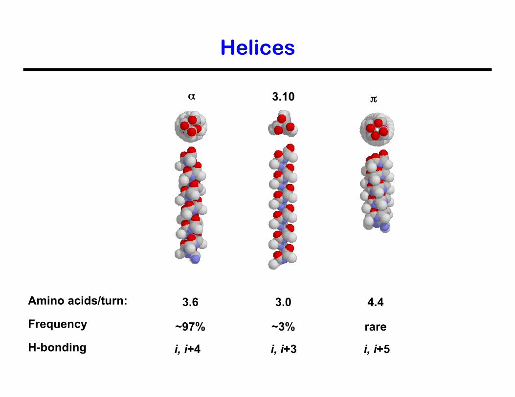
Helices
α π 3.10
3.6
~97%
4.4
rare
Amino acids/turn:
Frequency
3.0
~3%
i, i+4 i, i+5 H-bonding i, i+3
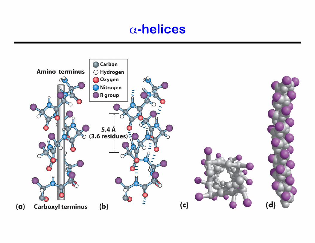
α-helices
α-helices
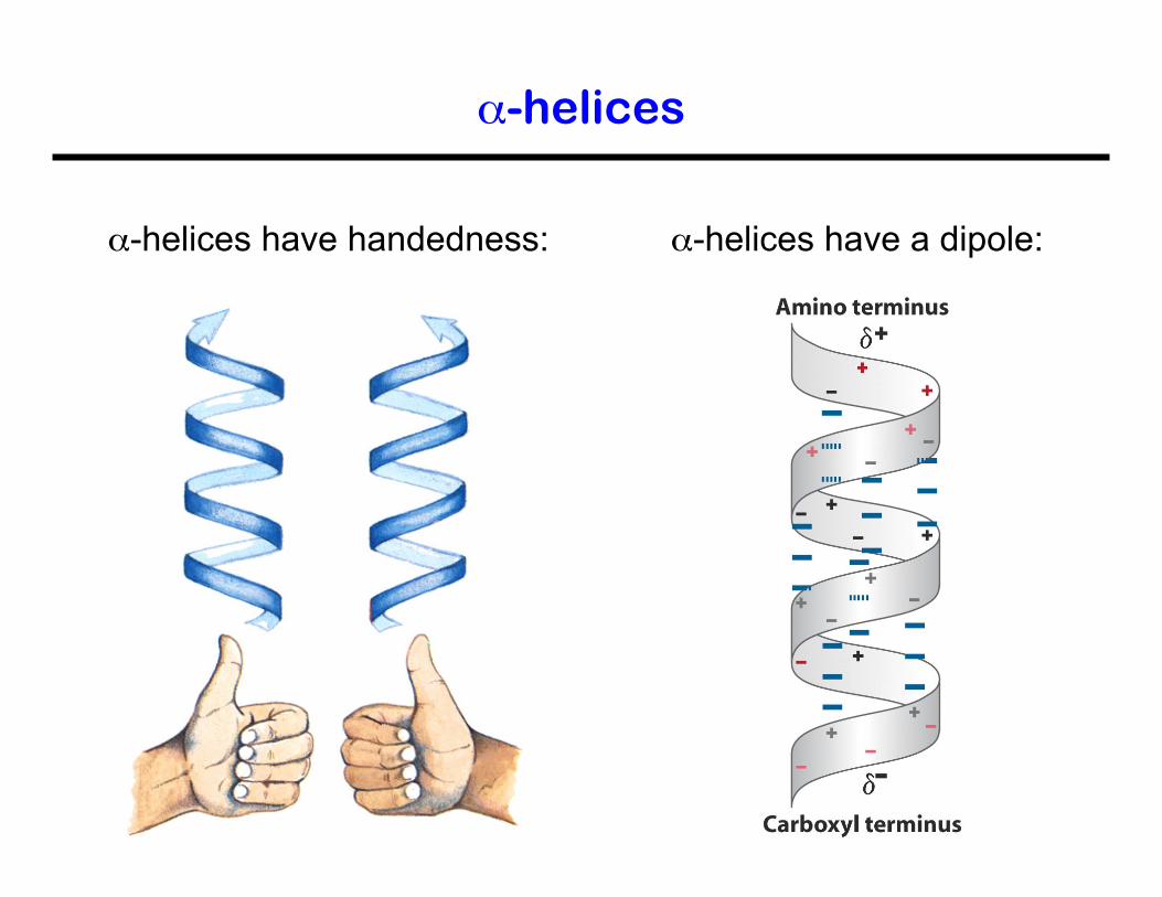
α-helices have handedness: α-helices have a dipole:
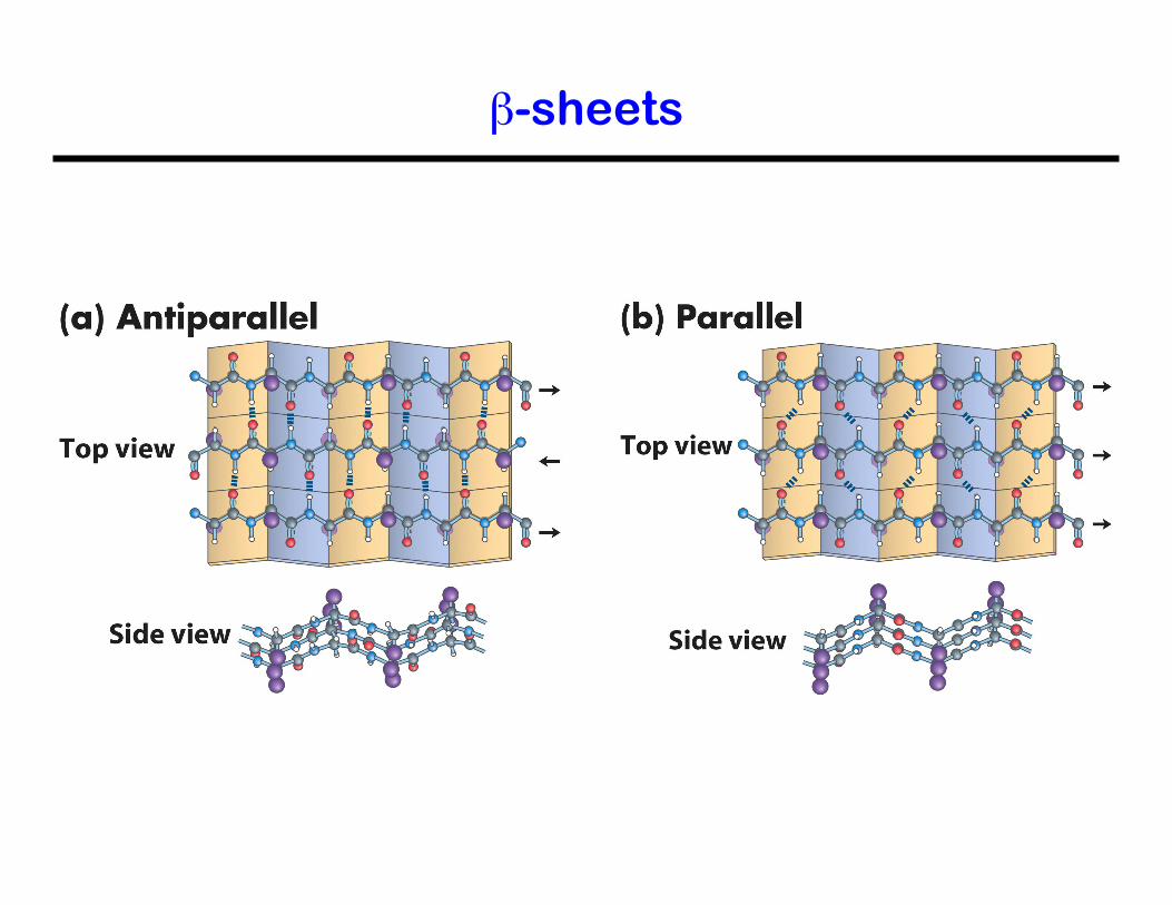
β-sheets
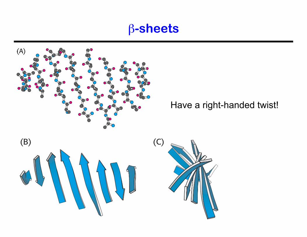
β-sheets
Have a right-handed twist!
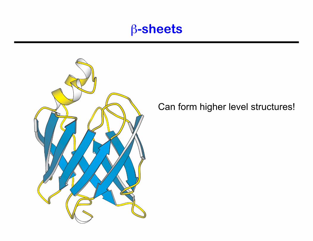
β-sheets
Can form higher level structures!
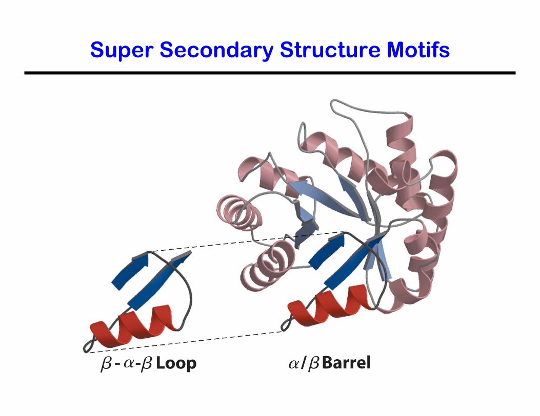
Super Secondary Structure Motifs
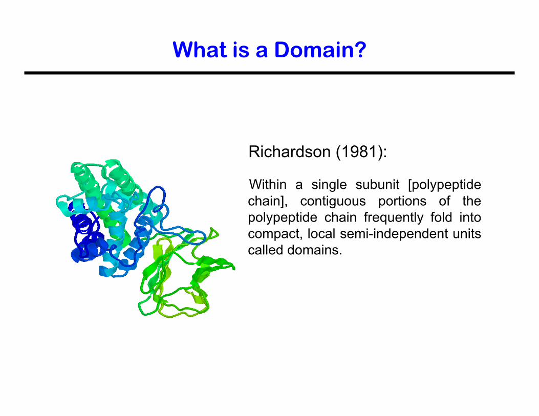
What is a Domain?
Richardson (1981):
Within a single subunit [polypeptide chain], contiguous portions of the polypeptide chain frequently fold into compact, local semi-independent units called domains.
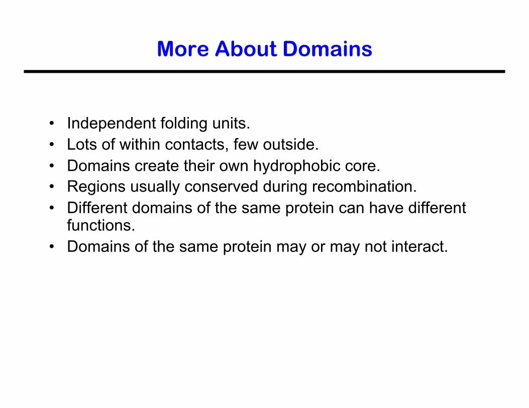
More About Domains
• Independent folding units. • Lots of within contacts, few outside. • Domains create their own hydrophobic core. • Regions usually conserved during recombination. • Different domains of the same protein can have different
functions. • Domains of the same protein may or may not interact.
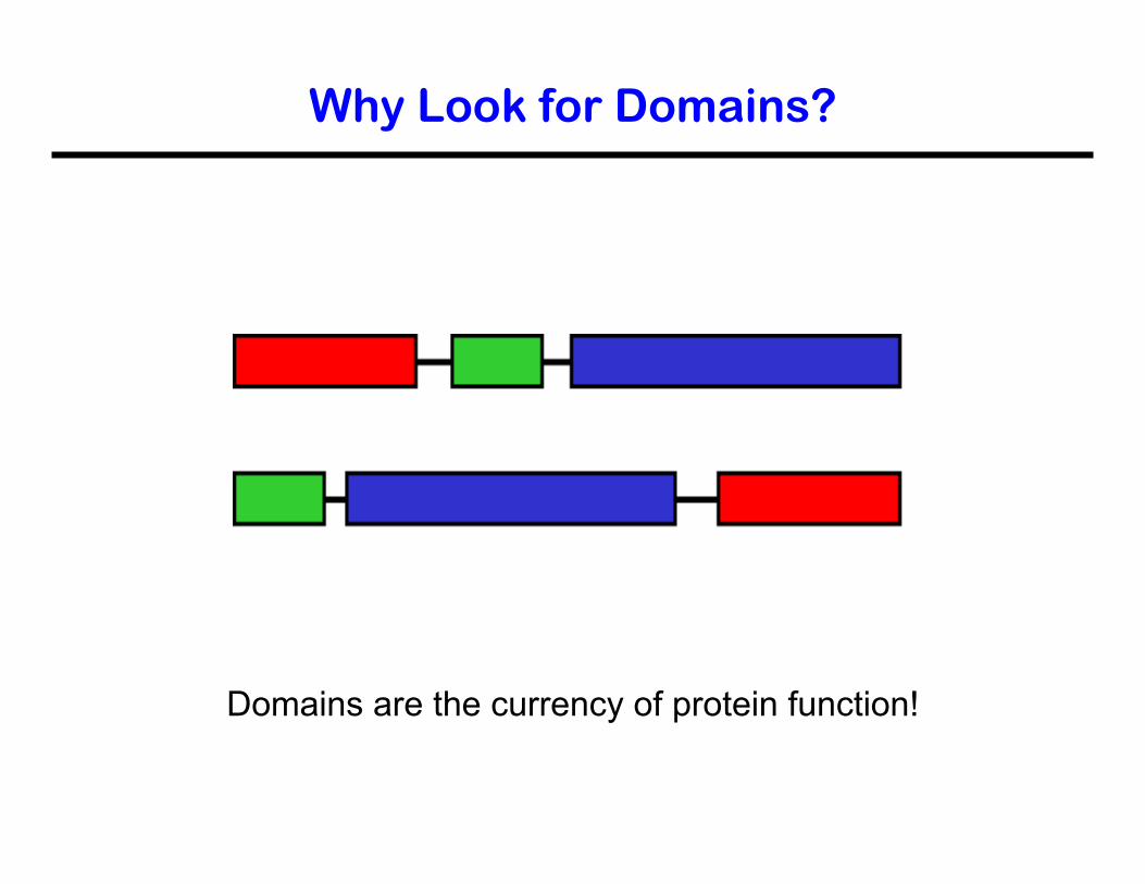
Why Look for Domains?
Domains are the currency of protein function!
Domain Size
• Domains can be between 25 and 500 residues long. • Most are less than 200 residues. • Domains can be smaller than 50 residues, but these
need to be stabilized.
Examples are the zinc finger and a scorpion toxin.
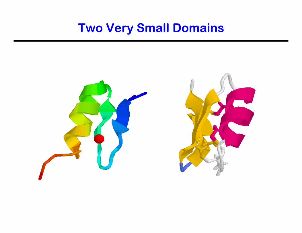
Two Very Small Domains
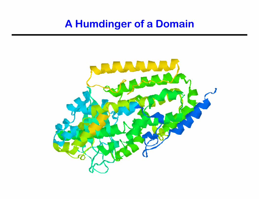
A Humdinger of a Domain
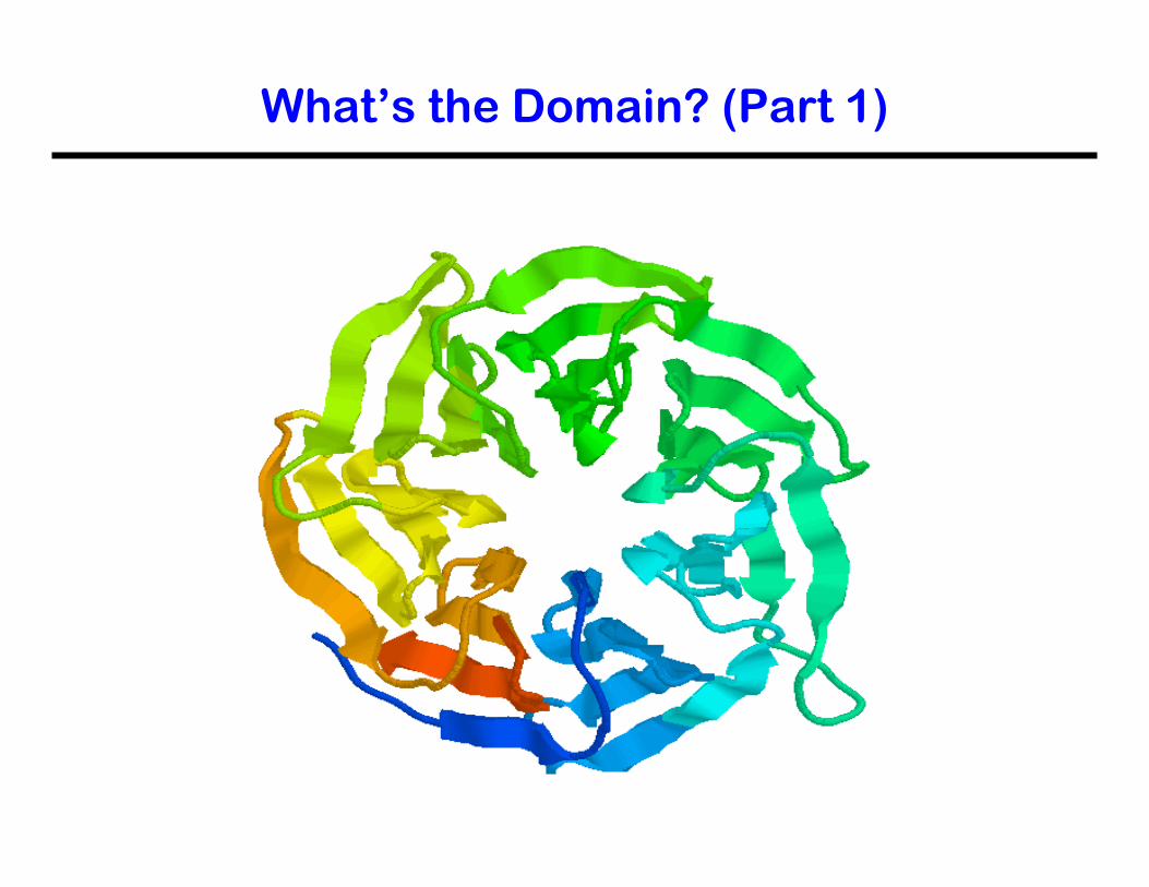
What’s the Domain? (Part 1)

What’s the Domain? (Part 2)
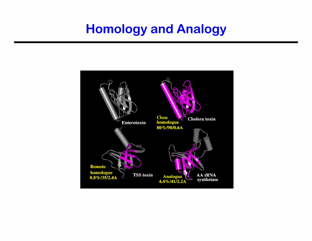
• Homology: Similarity in characteristics resulting from shared ancestry.
• Analogy: The similarity of structure between two species that are not closely related, attributable to convergent evolution.
Homology and Analogy
Homologous structures can be devided into orthologues (a result from changes in the same gene between different organisms, such as myoglobin) and paralogues (a result from gene duplication and subsequent changes within an organism and its descendents, such as hemoglobin).
Homology and Analogy
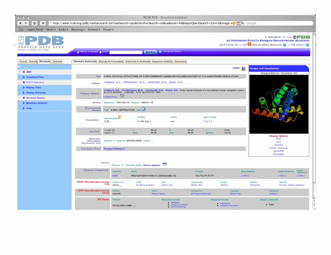
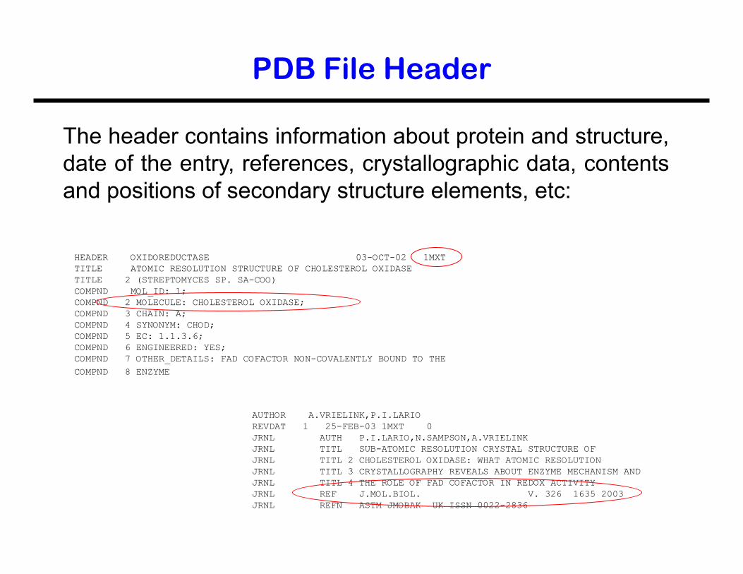
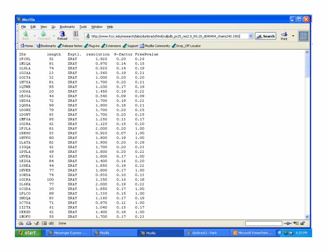
PDB File Header
HEADER OXIDOREDUCTASE 03-OCT-02 1MXT TITLE ATOMIC RESOLUTION STRUCTURE OF CHOLESTEROL OXIDASE TITLE 2 (STREPTOMYCES SP. SA-COO) COMPND MOL_ID: 1; COMPND 2 MOLECULE: CHOLESTEROL OXIDASE; COMPND 3 CHAIN: A; COMPND 4 SYNONYM: CHOD; COMPND 5 EC: 1.1.3.6; COMPND 6 ENGINEERED: YES; COMPND 7 OTHER_DETAILS: FAD COFACTOR NON-COVALENTLY BOUND TO THE
COMPND 8 ENZYME
The header contains information about protein and structure, date of the entry, references, crystallographic data, contents and positions of secondary structure elements, etc:
AUTHOR A.VRIELINK,P.I.LARIO REVDAT 1 25-FEB-03 1MXT 0 JRNL AUTH P.I.LARIO,N.SAMPSON,A.VRIELINK JRNL TITL SUB-ATOMIC RESOLUTION CRYSTAL STRUCTURE OF JRNL TITL 2 CHOLESTEROL OXIDASE: WHAT ATOMIC RESOLUTION JRNL TITL 3 CRYSTALLOGRAPHY REVEALS ABOUT ENZYME MECHANISM AND JRNL TITL 4 THE ROLE OF FAD COFACTOR IN REDOX ACTIVITY JRNL REF J.MOL.BIOL. V. 326 1635 2003 JRNL REFN ASTM JMOBAK UK ISSN 0022-2836
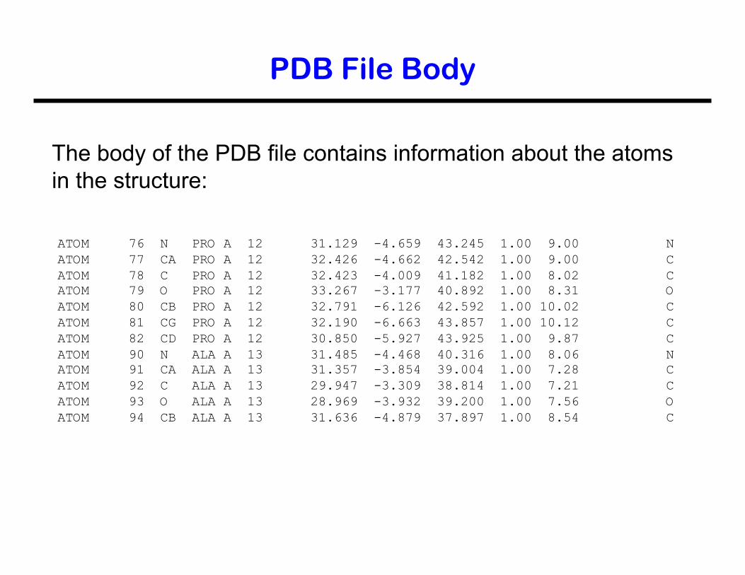
PDB File Body
The body of the PDB file contains information about the atoms in the structure:
ATOM 76 N PRO A 12 31.129 -4.659 43.245 1.00 9.00 N ATOM 77 CA PRO A 12 32.426 -4.662 42.542 1.00 9.00 C ATOM 78 C PRO A 12 32.423 -4.009 41.182 1.00 8.02 C ATOM 79 O PRO A 12 33.267 -3.177 40.892 1.00 8.31 O ATOM 80 CB PRO A 12 32.791 -6.126 42.592 1.00 10.02 C ATOM 81 CG PRO A 12 32.190 -6.663 43.857 1.00 10.12 C ATOM 82 CD PRO A 12 30.850 -5.927 43.925 1.00 9.87 C ATOM 90 N ALA A 13 31.485 -4.468 40.316 1.00 8.06 N ATOM 91 CA ALA A 13 31.357 -3.854 39.004 1.00 7.28 C ATOM 92 C ALA A 13 29.947 -3.309 38.814 1.00 7.21 C ATOM 93 O ALA A 13 28.969 -3.932 39.200 1.00 7.56 O ATOM 94 CB ALA A 13 31.636 -4.879 37.897 1.00 8.54 C
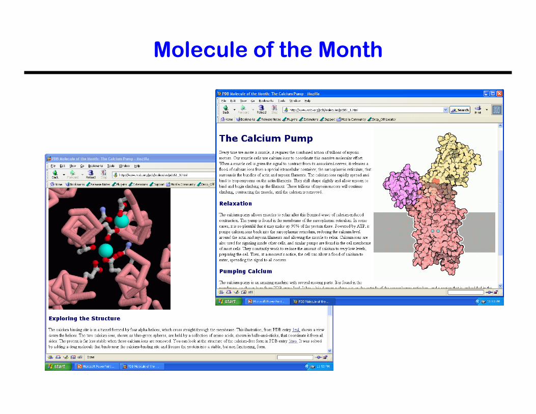
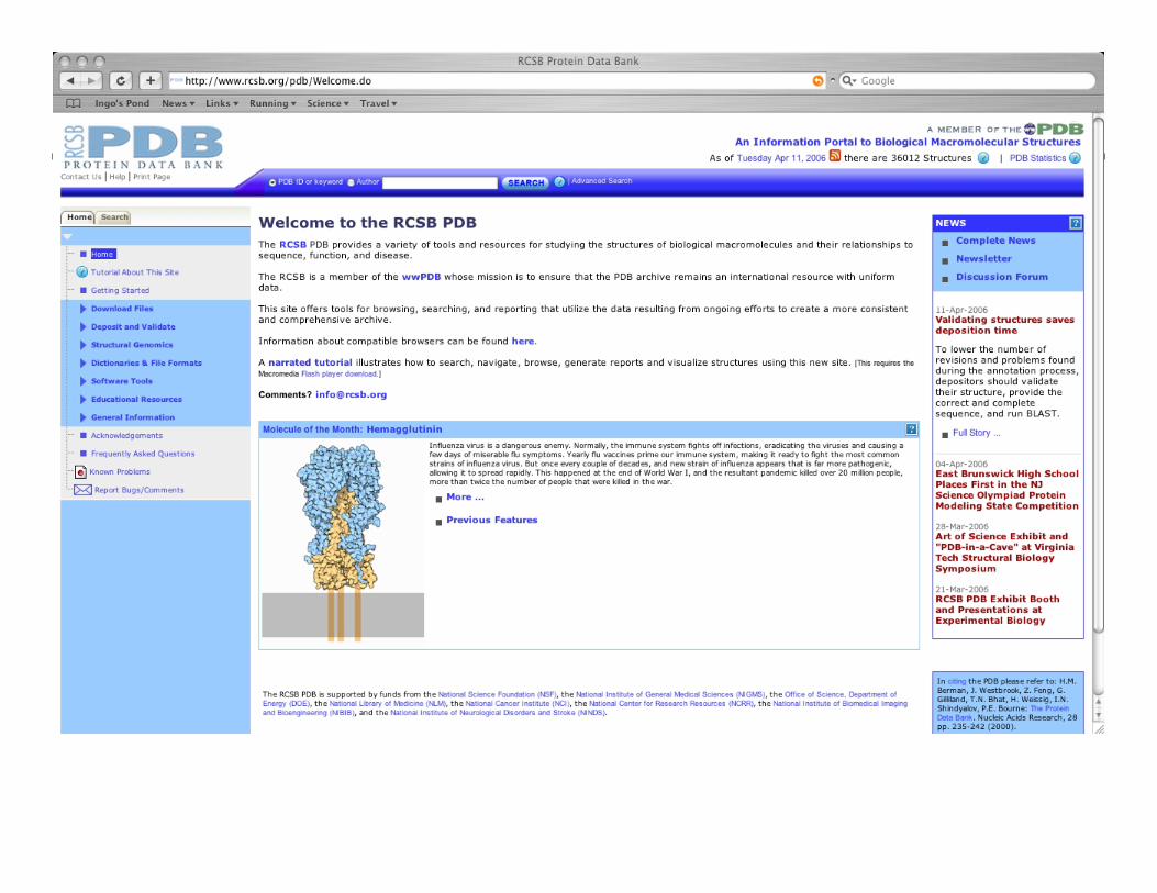
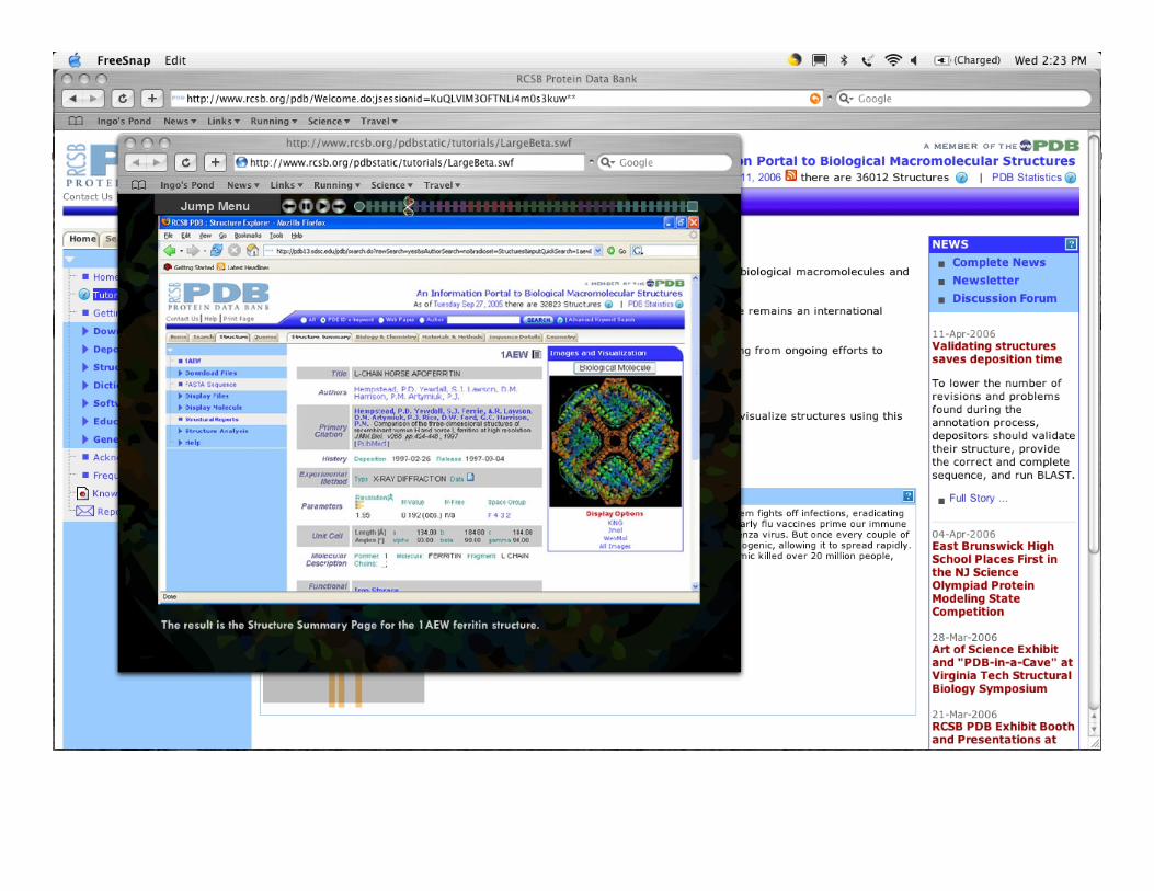
Molecule of the Month
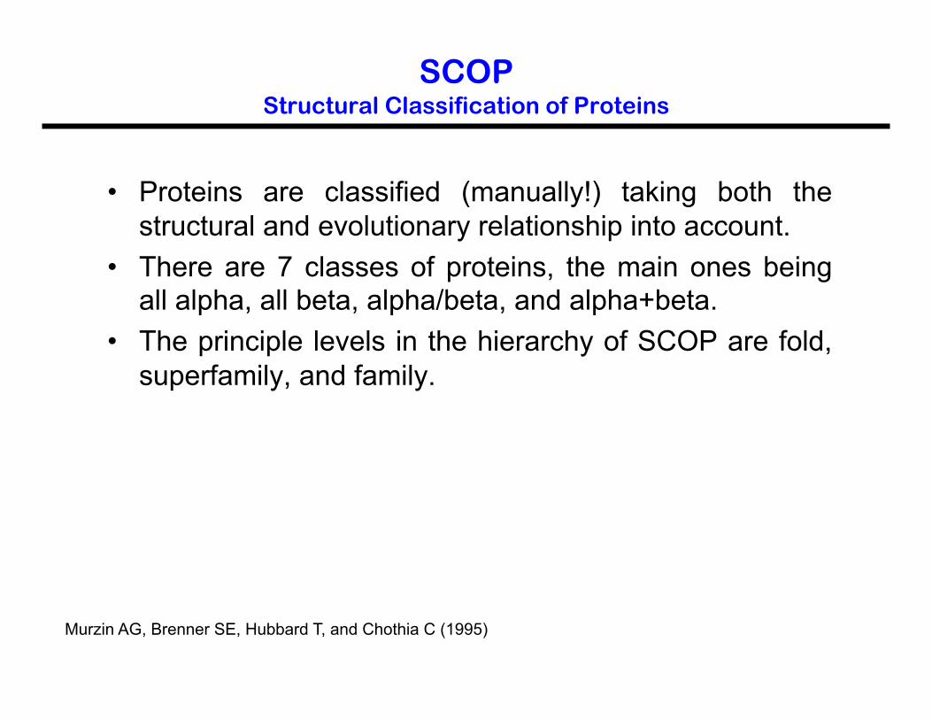
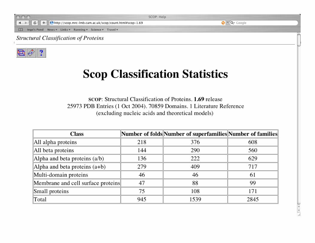
SCOP Structural Classification of Proteins
• Proteins are classified (manually!) taking both the structural and evolutionary relationship into account.
• There are 7 classes of proteins, the main ones being all alpha, all beta, alpha/beta, and alpha+beta.
• The principle levels in the hierarchy of SCOP are fold, superfamily, and family.
Murzin AG, Brenner SE, Hubbard T, and Chothia C (1995)
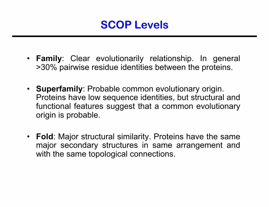
SCOP Levels
• Family: Clear evolutionarily relationship. In general >30% pairwise residue identities between the proteins.
• Superfamily: Probable common evolutionary origin. Proteins have low sequence identities, but structural and functional features suggest that a common evolutionary origin is probable.
• Fold: Major structural similarity. Proteins have the same major secondary structures in same arrangement and with the same topological connections.
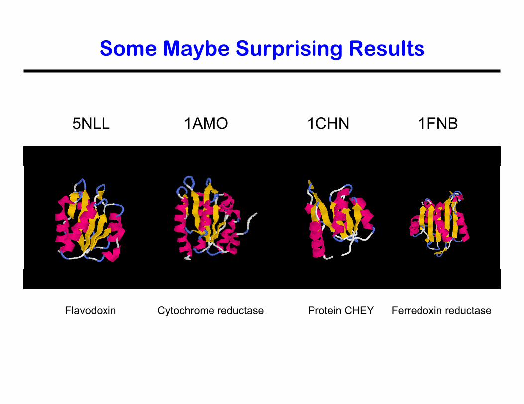
Some Maybe Surprising Results
Flavodoxin Cytochrome reductase Protein CHEY Ferredoxin reductase
5NLL 1AMO 1CHN 1FNB
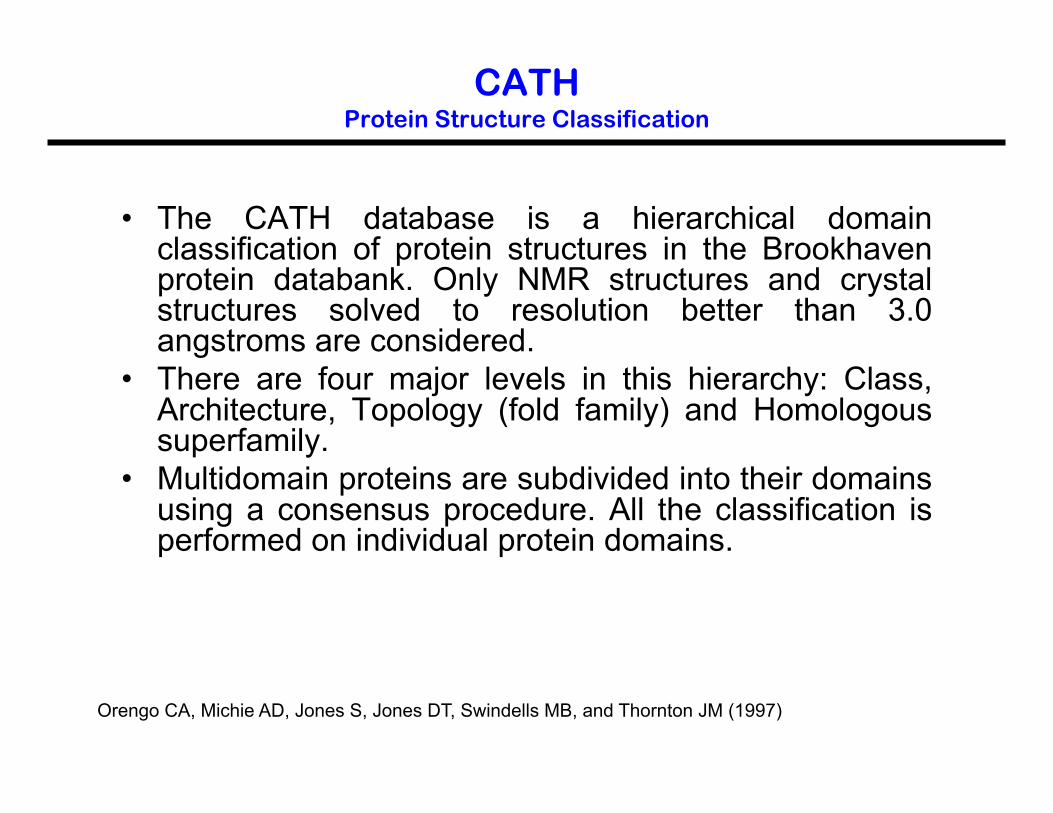
CATH Protein Structure Classification
• The CATH database is a hierarchical domain classification of protein structures in the Brookhaven protein databank. Only NMR structures and crystal structures solved to resolution better than 3.0 angstroms are considered.
• There are four major levels in this hierarchy: Class, Architecture, Topology (fold family) and Homologous superfamily.
• Multidomain proteins are subdivided into their domains using a consensus procedure. All the classification is performed on individual protein domains.
Orengo CA, Michie AD, Jones S, Jones DT, Swindells MB, and Thornton JM (1997)

The CATH Hierarchy

SCOP versus CATH
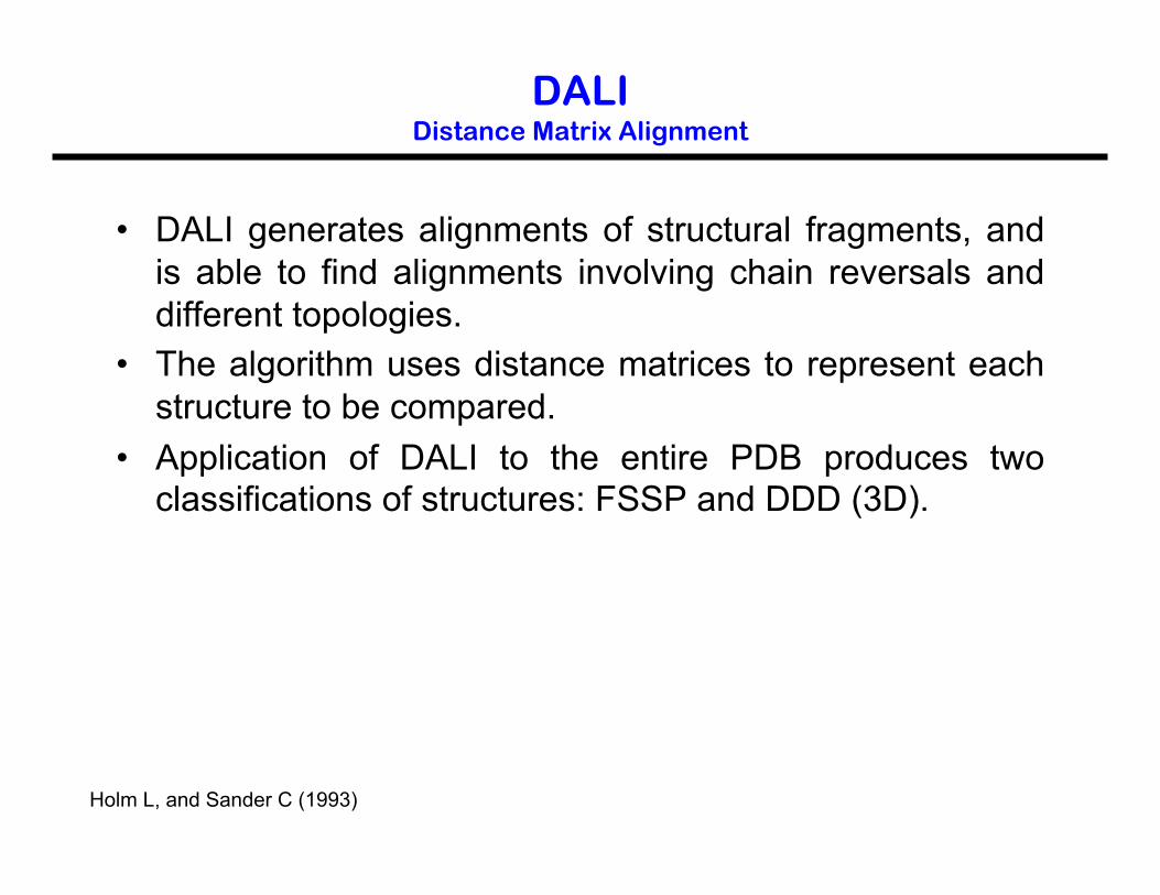
DALI Distance Matrix Alignment
• DALI generates alignments of structural fragments, and is able to find alignments involving chain reversals and different topologies.
• The algorithm uses distance matrices to represent each structure to be compared.
• Application of DALI to the entire PDB produces two classifications of structures: FSSP and DDD (3D).
Holm L, and Sander C (1993)
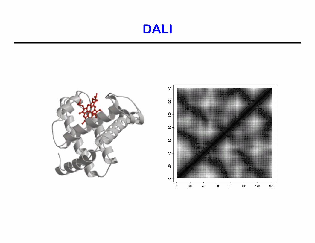
DALI
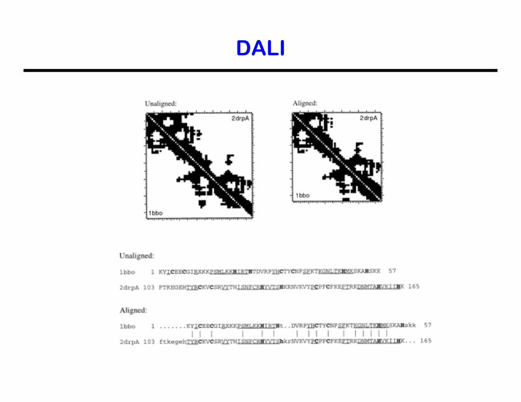
DALI
FSSP and DDD
• The families of structurally similar proteins (FSSP) is a database of structural alignments of proteins in the protein data bank (PDB). It presents the results of applying DALI to (almost) all chains of proteins in the PDB.
• The DALI domain dictionary (DDD) is a corresponding classification of recurrent domains automatically extracted from known proteins.
Other Algorithms for Domain Decomposition
• The Protein Domain Parser (PDP) uses compactness as a chief principle. http://123d.ncifcrf.gov/pdp.html
• DomainParser is graph theory based. The underlying principle used is that residue-residue contacts are denser within a domain than between domains. http://compbio.ornl.gov/structure/domainparser/
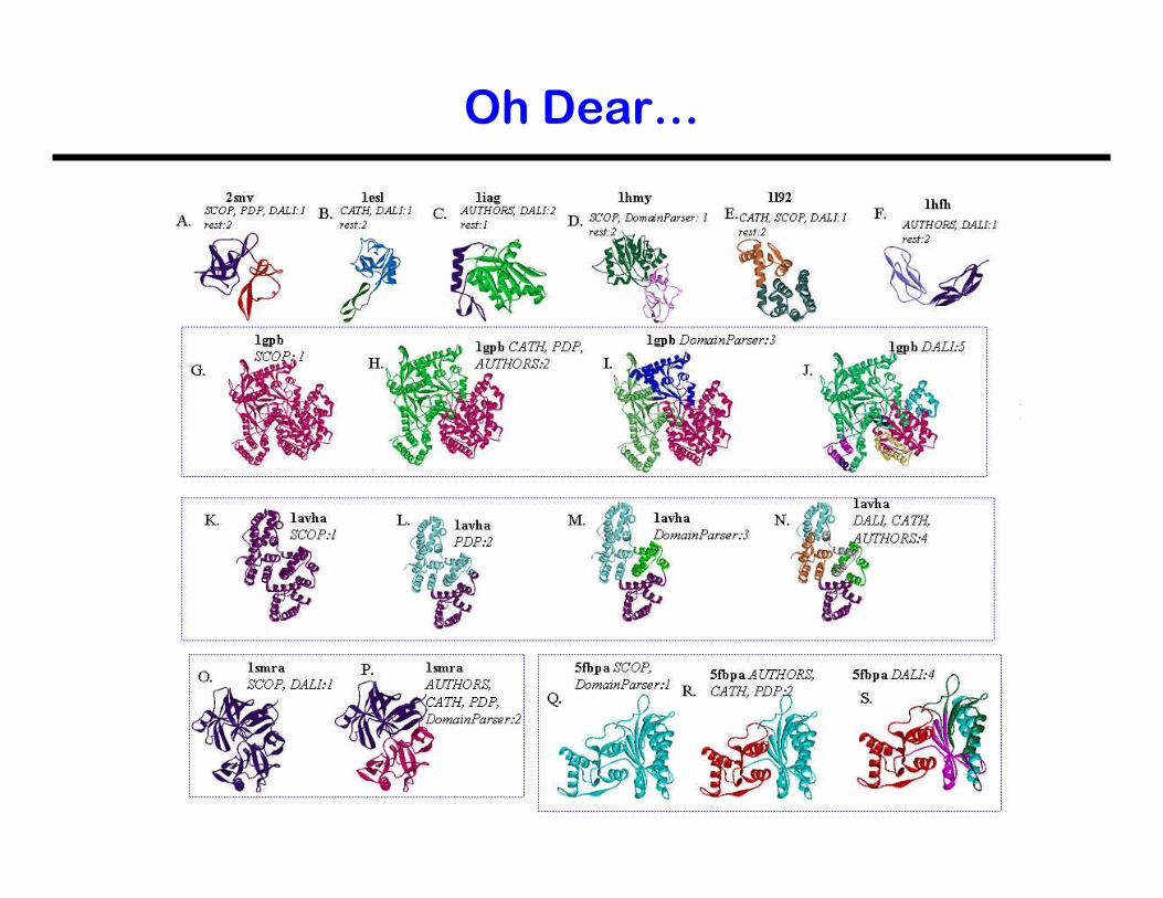
Oh Dear…
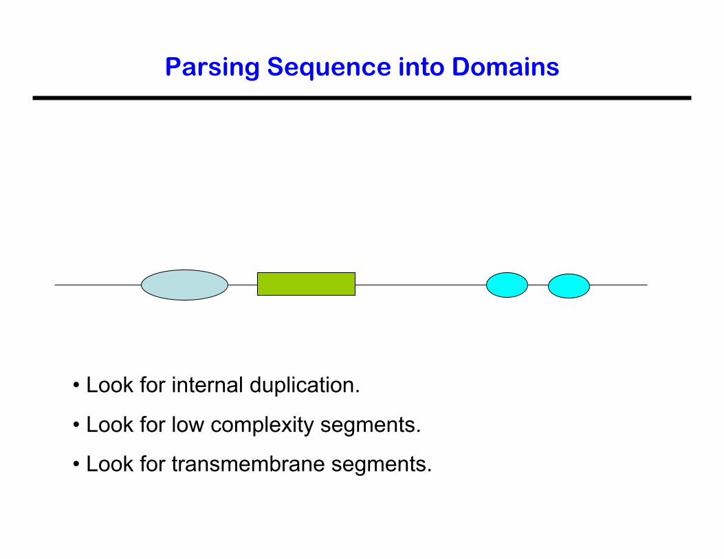
Parsing Sequence into Domains
• Look for internal duplication.
• Look for low complexity segments.
• Look for transmembrane segments.
Why is That Important?
• Functional insights. • Improved database searching. • Fold recognition. • Structure determination.
PRODOM: http://protein.toulouse.inra.fr/prodom/current/html/home.php
PFAM: http://www.sanger.ac.uk/Software/Pfam/
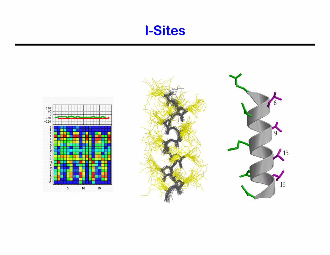
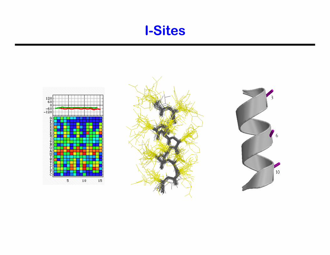
I-Sites
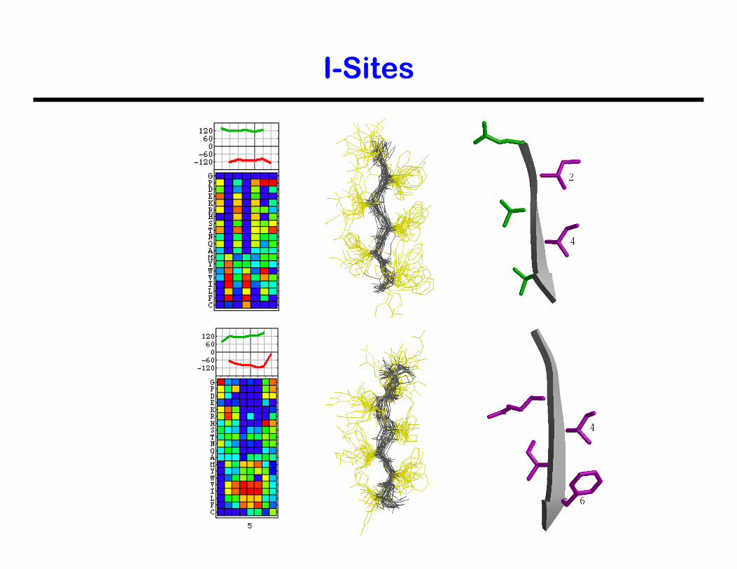
I-Sites
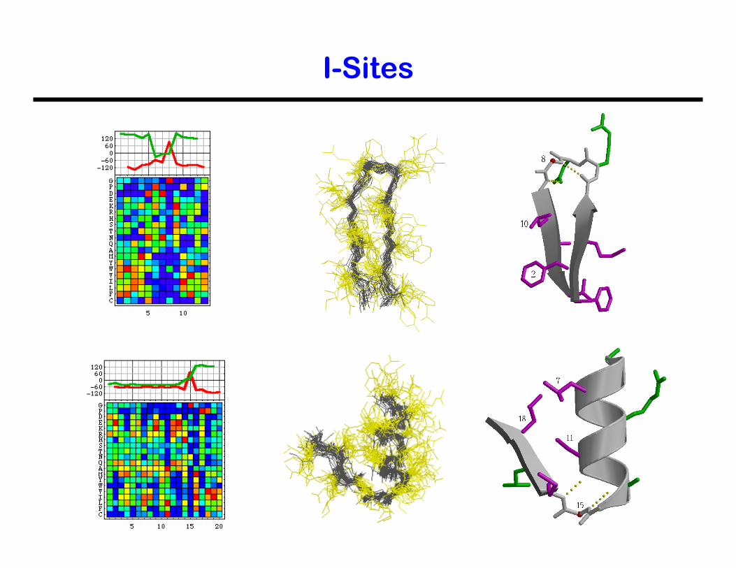
I-Sites
I-Sites