New Mechanical Features for Time-Domain WEC Modelling in ...
RESEARCH Open Access
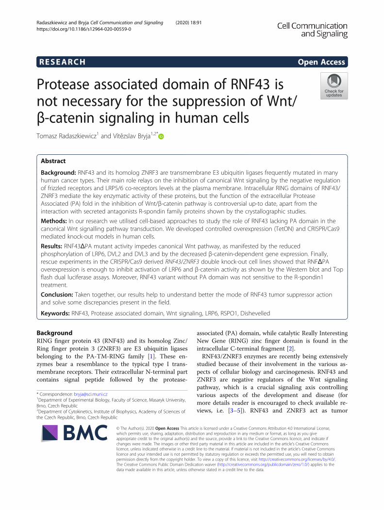
Protease associated domain of RNF43 isnot necessary for the suppression of Wnt/β-catenin signaling in human cellsTomasz Radaszkiewicz1 and Vítězslav Bryja1,2*
Abstract
Background: RNF43 and its homolog ZNRF3 are transmembrane E3 ubiquitin ligases frequently mutated in manyhuman cancer types. Their main role relays on the inhibition of canonical Wnt signaling by the negative regulationof frizzled receptors and LRP5/6 co-receptors levels at the plasma membrane. Intracellular RING domains of RNF43/ZNRF3 mediate the key enzymatic activity of these proteins, but the function of the extracellular ProteaseAssociated (PA) fold in the inhibition of Wnt/β-catenin pathway is controversial up-to date, apart from theinteraction with secreted antagonists R-spondin family proteins shown by the crystallographic studies.
Methods: In our research we utilised cell-based approaches to study the role of RNF43 lacking PA domain in thecanonical Wnt signalling pathway transduction. We developed controlled overexpression (TetON) and CRISPR/Cas9mediated knock-out models in human cells.
Results: RNF43ΔPA mutant activity impedes canonical Wnt pathway, as manifested by the reducedphosphorylation of LRP6, DVL2 and DVL3 and by the decreased β-catenin-dependent gene expression. Finally,rescue experiments in the CRISPR/Cas9 derived RNF43/ZNRF3 double knock-out cell lines showed that RNFΔPAoverexpression is enough to inhibit activation of LRP6 and β-catenin activity as shown by the Western blot and Topflash dual luciferase assays. Moreover, RNF43 variant without PA domain was not sensitive to the R-spondin1treatment.
Conclusion: Taken together, our results help to understand better the mode of RNF43 tumor suppressor actionand solve some discrepancies present in the field.
Keywords: RNF43, Protease associated domain, Wnt signaling, LRP6, RSPO1, Dishevelled
BackgroundRING finger protein 43 (RNF43) and its homolog Zinc/Ring finger protein 3 (ZNRF3) are E3 ubiquitin ligasesbelonging to the PA-TM-RING family [1]. These en-zymes bear a resemblance to the typical type I trans-membrane receptors. Their extracellular N-terminal partcontains signal peptide followed by the protease-
associated (PA) domain, while catalytic Really InterestingNew Gene (RING) zinc finger domain is found in theintracellular C-terminal fragment [2].RNF43/ZNRF3 enzymes are recently being extensively
studied because of their involvement in the various as-pects of cellular biology and carcinogenesis. RNF43 andZNRF3 are negative regulators of the Wnt signalingpathway, which is a crucial signaling axis controllingvarious aspects of the development and disease (formore details reader is encouraged to check available re-views, i.e. [3–5]). RNF43 and ZNRF3 act as tumor
© The Author(s). 2020 Open Access This article is licensed under a Creative Commons Attribution 4.0 International License,which permits use, sharing, adaptation, distribution and reproduction in any medium or format, as long as you giveappropriate credit to the original author(s) and the source, provide a link to the Creative Commons licence, and indicate ifchanges were made. The images or other third party material in this article are included in the article's Creative Commonslicence, unless indicated otherwise in a credit line to the material. If material is not included in the article's Creative Commonslicence and your intended use is not permitted by statutory regulation or exceeds the permitted use, you will need to obtainpermission directly from the copyright holder. To view a copy of this licence, visit http://creativecommons.org/licenses/by/4.0/.The Creative Commons Public Domain Dedication waiver (http://creativecommons.org/publicdomain/zero/1.0/) applies to thedata made available in this article, unless otherwise stated in a credit line to the data.
* Correspondence: [email protected] of Experimental Biology, Faculty of Science, Masaryk University,Brno, Czech Republic2Department of Cytokinetics, Institute of Biophysics, Academy of Sciences ofthe Czech Republic, Brno, Czech Republic
Radaszkiewicz and Bryja Cell Communication and Signaling (2020) 18:91 https://doi.org/10.1186/s12964-020-00559-0
suppressors and numerous mutations in the RNF43 genewere identified in cancers of various tissues, like endo-metrium, stomach, ovary, pancreases or colon [6–9].The main molecular function of RNF43/ZNRF3 in theWnt pathway is the negative regulation of the surface levelof Wnt receptors Frizzled (FZD) and co-receptor low-density lipoprotein receptor-related protein 6 (LRP6) bytheir ubiquitination and subsequent degradation [10, 11].Additionally, it was proposed that RNF43 can act down-stream from the plasma membrane receptor complexes bytethering the T-cell factor 4 (TCF4) to the nuclear mem-brane, preventing gene transcription [12].The importance of enzymatic activity of the RING do-
main for RNF43/ZNRF3 ability to inhibit Wnt pathway isundoubtful. It was proven that point mutations disruptingthis catalytic domain had dominant negative effect [10].Region adjacent to the RING domain was also shown tointeract with dishevelled (DVL) protein, mediatingRNF43/ZNRF3 dependent frizzled degradation [13]. Theseobservations underline the crucial role of the intracellularRNF43/ZNRF3 proteins regions in the facilitating signal-ing events. On the other side, the function of RNF43/ZNRF3 ectodomain, in particular PA domain, remainsunder the debate. It seems to be clear that PA domain me-diates the interaction with R-spondins (RSPO)- endogen-ous negative regulators of RNF43 and ZNRF3. RSPO 1–4are secreted proteins, which reduce plasma membranelevel of RNF43 and ZNRF3 in a leucine-rich repeat-containing G-protein coupled receptors (LGR) 4/5/6dependent (RSPO1 and 4) or independent (RSPO2 and 3)way [14]. Crystal structures revealed that R-spondins bindPA domain of RNF43 and ZNRF3 and form the ternarycomplex with LGR4/5/6 [15–20].It is currently unclear whether PA domain is required
for binding to FZD and RNF43/ZNRF3-mediated inhib-ition of Wnt/β-catenin pathway. Experiments in Caenor-habditis elegans showed that PLR-1ΔPA mutant, whichis C. elegans homolog of RNF43 and ZNRF3, was notable to reduce surface level of MIG-1/FZD and blockWnt signaling [21]. Also, MIG-1/FZD deprived ofcysteine-rich domain (CRD) was unaffected upon PLR-1overexpression. Next, experiments established in themammalian cells showed that deletion of the wholeextracellular part of the RNF43 prevented RNF43-mediated FZD5 internalization [10]. Moreover, anothergroup described that precise deletion of the PA domainof RNF43 blocked its inhibitory function on the β-catenin dependent transcription and only part of Xen-opus embryos injected with RNF43ΔPA mRNA showedphenotype similar to the observed for the wild typeRNF43 [22]. The same study demonstrated interactionbetween PA domain of RNF43 and CRD of FZD5 in theco-immunoprecipitation assay after overexpression [22].However, other researchers did not succeed to positively
verify the existence of this interaction [13]. Similarly,binding of ZNRF3 to the CRD domain of FZD8 was notdetected in a surface plasmon resonance based bindingassay [18].The existence of the above described discrepancies en-
couraged us to look at the role of RNF43 PA domain inthe negative regulation of canonical Wnt signaling in de-tail. In order to shed light on the mechanism of RNF43/ZNRF3 function and regulation, we generated severalnovel mammalian models to study consequences of PAdeletion in the cell-based assays. Our data collectivelysuggest that PA domain is dispensable for RNF43 cap-acity to block Wnt signaling pathway.
MethodsCell lines and treatmentsT-REx 293 (R71007, Thermo Fisher Scientific) cells andall their derivates were cultured at 37 °C and under con-trolled 5% (vol/vol) CO2 atmosphere in the Dulbecco’smodified Eagle’s medium (DMEM, 41966–029, Gibco,Life Technologies) supplemented with 10% fetal bovineserum (FBS, 10270–106, Gibco, Life Technologies), 2mM L-glutamine (25,030,024, Life Technologies) and 1%penicillin-streptomycin (XC-A4122/100, Biosera).Endogenous Wnt ligands secretion was blocked by the
use of 0.5 μM LGK-974 Porcupine-specific inhibitor (1,241,454, PeproTech). For the purpose of canonical Wntpathway stimulation, cells were treated with the recom-binant human WNT3A (rWNT3A) (CF 5036-WN-CF,RnD Systems) for 3 h or overnight for the Top flash dualluciferase assay in the indicated concentrations (40–100ng/ml). Recombinant RSPO1 (rRSPO1) (120–38, Pepro-Tech) in the final concentration of 25, 50 or 100 ng/mlwas employed as co-treatment with rWNT3A for the ca-nonical Wnt pathway activation enhancement. Inducibleprotein expression was activated by the overnight, if notspecified otherwise, tetracycline treatment in the dose of1 μg/ml (60–54-8, Santa Cruz Biotechnology).
Plasmids, cloning and mutagenesisFor the purpose of RNF43 and RNF43ΔPA T-REx 293inducible cell lines generation, cDNA encoding humanRNF43 fused with BirA* and HA tags was amplified bythe Phusion High Fidelity DNA polymerase (F530S,Thermo Fisher Scientific) based PCR reaction andcloned into the HindIII (ER0501, Thermo Fisher Scien-tific) and XbaI (ER0681, Thermo Fisher Scientific) line-arized pcDNA4/TO backbone (Thermo Fisher Scientific)by the In-Fusion cloning method (Takara Bio). As a re-sult, pcDNA4-TO-RNF43-BirA*-HA plasmid was ob-tained. Next, the same vector was used as a template forgeneration of the RNF43 mutant lacking PA domain -pcDNA4-TO-RNF43-ΔPA-BirA*-HA. In-frame deletionof amino acids at positions no. 87–186 was performed
Radaszkiewicz and Bryja Cell Communication and Signaling (2020) 18:91 Page 2 of 12
using QuikChange II XL site-directed mutagenesis kit(Agilent) according to the manufacturer’s instructions.All produced plasmids were validated by the Sanger se-quencing method.For targeting RNF43 and ZNRF3 in the T-REx 293, gRNAs
coding sequences: TGAGTTCCATCGTAACTGTGTGG(PAM) and AGACCCGCTCAAGAGGCCGGTGG werecloned into the gRNA-GFP-T1 backbone using previously de-scribed protocol [23].Other plasmids used in this study are: pCAG-mGFP
(Addgene #14757 [24]), pRLtkLuc (Promega), Super8XTOPFLASH (gift from Randall Moon), hCas9 (Addgene#41815 [25]), gRNA_GFP-T1 (Addgene #41819 [25]),pcDNA4-TO-RNF43Mut1-2xHA-2xFLAG [10], PiggyBack-Hygro and Transposase encoding plasmids (kindly gifted byBon-Kyoung Koo). Primers sequences are shown in theTable 1.
Transfections and stable cell lines generationT-REx 293 cells were transected using 1 μg/ml, pH 7.4polyethylenimine (PEI) and plasmid DNA in 4:1 ratio,applying already described method [26]. For the Topflash dual luciferase assay and immunofluorescence im-aging, cells growing on 24 well plate were transfected by0.4 μg of total plasmid DNA per one well. For stable celllines preparation, 6 μg of pDNA was used for plasmidsintroduction into the cells growing on 10 cm plates. 24 hpost transfection, cells were further processed and
analyzed, or culture medium was changed for the freshone, containing proper antibiotics for selection of posi-tively transfected clones. pcDNA4/TO backbone en-codes blasticidin S resistance (used 5 μg/ml, 3513-03-9,Santa Cruz Biotechnology) and PiggyBack-Hygro –hygromycin B (200 μg/ml, 31,282–04-9, Santa Cruz Bio-technology). Empty backbone pcDNA3.1 (Invitrogen)was used to equalize total plasmid DNA amount.
Top flash dual luciferase assayCanonical Wnt signaling activation leads to the β-catenin stabilization, allowing its translocation andchanges in the gene expression. To measure the tran-scriptional activity of the β-catenin, cells were trans-fected with 0.1 μg of the pRLtkLuc plasmid and 0.1 μg ofthe Super8X TopFlash and subsequently treated over-night with rWNT3A, rRSPO1 in the presence of 0.5 μMLGK-974 and tetracycline, as marked in the figures. Ap-proximately 24 h post-transfection, cells were washedwith PBS and freezed. Subsequently, assay was per-formed according to the manufacturer protocol (E1960;Promega) and luminescence was detected by the use ofHidex Bioscan Plate Chameleon Luminometer. Resultsare presented as ratios of firefly (containing Wnt-specificTCF/LEF binding sites in the promoter) to renilla (con-stitutively active promoter) luciferases measurements,RLU - relative luciferase units.
Table 1 Primers
Cloning and mutagenesis primers
Primer Sequence Purpose
RNF43 InFusion F GTTTAAACTTAAGCTTATGAGTGGTGGCCACCAG RNF43-BirA*-HA into pcDNA4
RNF43 InFusion R AAACGGGCCCTCTAGACTATGCGTAATCCGGTACA
del87–186 ATTAATGCAGTCCCACGAGCTGAAGGAGCCCC Site-directed mutagenesis, deletion of the PA domain
del87–186-antisense GGGGCTCCTTCAGCTCGTGGGACTGCATTAAT
Quantitative Polymerase Chain Reaction
GENE Forward primer seq. Reverse primer seq. Product size
B2M CACCCCCACTGAAAAAGATG ATATTAAAAAGCAAGCAAGCAGAA 167
GAPDH GACAGTCAGCCGCATCTTCT TTAAAAGCAGCCCTGGTGAC 127
RNF43 TTTCCTGCCTCCATGAGTTC CAGGGACTGGGAAAATGAATC 116
ZNRF3 GCTTTCTTCGTCGTGGTCTC GCCTGTTCATGGAATTCTGAC 91
CTNNB1 AAAGCGGCTGTTAGTCACTGG CGAGTCATTGCATACTGTCCAT 215
AXIN2 TACACTCCTTATTGGGCGATCA TTGGCTACTCGTAAAGTTTTGGT 151
CRISPR/Cas9 Genotyping
Primer Sequence Purpose
RNF43 F AGGTCTTTTACAAGTAGATAATAGCAAGG CRISRP/Cas9 RNF43 RFLP analysis
RNF43 R GCTGCAACCCTTTCCCA
ZNRF3 F TTCTCAGCCTTGCTCTTCTG CRISRP/Cas9 ZNRF3 RFLP analysis
ZNRF3 R TCACCCTGGCCTTGAGTC
Radaszkiewicz and Bryja Cell Communication and Signaling (2020) 18:91 Page 3 of 12
CRISPR/Cas9 mediated gene editingFor targeting RNF43 and ZNRF3 genes in the T-REx 293cells, gRNAs: TGAGTTCCATCGTAACTGTGTGG(PAM) and AGACCCGCTCAAGAGGCCGGTGG werecloned into the gRNA_GFP-T1 backbone and co-transfected with PiggyBack-Hygro and Transposase cod-ing plasmids using PEI. Then, cells were hygromycin Bselected and plated as single cells on 96-well plates. Foridentification of clones carrying CRISPR/Cas9-mediatedmodifications in the sequences of both alleles, RFLP ana-lysis using restriction enzymes was performed. GenomicDNA isolation from grown clonal cell lines was per-formed using DirectPCR Lysis Reagent (Cell) (ViagenBiotech), supplemented with 0.5 mg/ml Proteinase K(EO0491, Thermo Fisher Scientific). DreamTaq DNAPolymerase (EP0701, Thermo Fisher Scientific) basedPCR was done according to the manufacturer instruc-tion for amplification of gDNA fragment containing tar-geted sequence. PCR products were analyzed byrestriction digestion, using Taal (ER1361, Thermo FisherScientific) for RNF43 and HpaII (ER0511, Thermo FisherScientific) for ZNRF3, for detection of Cas9 mediateddisruption of the recognition sites nearly the PAM se-quences. Finally, all clones were sequenced using theIllumina platform and compared with the reference se-quence [27]. Predictions of amino acid sequence changesdue to the frameshift type mutations and occurrence ofpremature STOP codons were done. Primers sequencesare shown in the Table 1.
Western blotting and antibodiesWestern blot method was done accordingly to the previ-ously published protocol [28]. In brief, cells were washedwith PBS and subsequently lysed in the sample buffer(2% SDS, 10% glycerol, 5% β-mercaptoethanol, 0.002%bromphenol blue and 0.06M Tris HCl, pH 6.8), soni-cated and thermally denatured. Electrophoretically sepa-rated proteins, in the SDS-PAGE gels, were transferredonto Immobilon-P PVDF Membrane (IPVH00010,
Millipore) and detected by primary and correspondingHRP-conjugated secondary antibodies on Fusion SL im-aging system (Vibler), using Immobilon Western Chemi-luminescent HRP Substrate (Merck, WBKLS0500).Western blot signals were quantified using ImageJ soft-ware. Antibodies are listed in the Table 2. Western blotmembranes are presented together with molecular massmarkers in the Additional file 1.
Immunofluorescence and confocal microscopyFor imaging of the plasma membrane localization of ec-topically expressed RNF43 and its PA domain lackingmutant, T-REx 293-RNF43-TetON and T-REx 293-RNF43ΔPA-TetON cells were grown on coverslipscoated with 0.1% gelatin in PBS (Sigma) and 1 μg/ml hu-man fibronectin (1918-FN-02, RnD Systems). At ap-proximately 70% of confluence, they were transfectedwith plasmid pCAG-mGFP encoding membrane-boundform of GFP fused with a palmitoylation sequence ofGAP43 at its N-terminus. Approximately 8 h after trans-fection, cells were stimulated with tetracycline for trans-genes expression induction. Next day, cells were washedby PBS, fixed in 4% paraformaldehyde for 20 min,permeabilized by 0.1% Triton X-100 in PBS and blockedin the 2% solution of bovine serum albumin in PBS.Then, samples were incubated overnight at 4 °C with pri-mary anti HA tag antibodies diluted in 2% BSA in PBSand washed. Corresponding Alexa Fluor secondary anti-body (Invitrogen) was incubated with samples for 1 h atthe room temperature, along with 0.5 μM of TO-PRO-3Iodide (642/661) (T3605, Thermo Fisher Scientific) fornuclei counterstaining. After PBS washes, coverslipswere mounted in the DAKO mounting medium (S3023,DAKO). Images were taken on the confocal laser scan-ning microscopy platform Leica TCS SP8 (Leica). Histo-grams of signals in each channel were prepared in theLAS X Life Science (Leica) software and plotted in theGraphPad Prism 8 for the co-localization analysis. Anti-bodies used are listed in the Table 2.
Table 2 Antibodies
Antibody Manufacturer Dilution and application Reference
LRP6 (C47E12) cs-3395, Cell Signaling Technology 1:1000, WB [28]
LRP6 (Phospho-S1490) cs-2568, Cell Signaling Technology 1:1000, WB [28]
β-actin CS-4970, Cell Signaling Technology 1:3000, WB [26]
DVL-2 CS-3216, Cell Signaling Technology 1:1000, WB [26]
DVL-3 CS-3218, Cell Signaling Technology 1:1000, WB [26]
HA-11 MMS-101R, Covance 1:2000 WB; 1:500, IF [28]
a-mouse IgG HRP A4416 1:4000 WB –
a-rabbit IgG HRP A0545 1:4000 WB –
a-mouse Alexa Fluor™568 A10037, Thermo Fisher Scientific 1:600 IF –
Radaszkiewicz and Bryja Cell Communication and Signaling (2020) 18:91 Page 4 of 12
Quantitative real-time polymerase chain reaction (qPCR)To assess the expression level of RNF43, ZNRF3,CTNNB1 and AXIN2 (canonical Wnt signaling targetgene [29]) in the response to the various treatments(rWNT3A 100 ng/ml and rRSPO1 50 ng/ml), qPCR re-action was applied. mRNA was extracted from cellsusing RNeasy Mini Kit (74,106; Qiagen) according to themanufacturer’s instructions. One microgram of mRNAwas transcribed to the cDNA by RevertAid ReverseTranscriptase (EP0442, Thermo Fisher Scientific) andanalyzed by use of a LightCycler® 480 SYBR Green IMaster (04887352001, Roche) and Light Cycler LC480(Roche). Results are presented as 2−ΔΔCT and comparedby unpaired Student’s t test. Mean expression of B2Mand GAPDH was used as reference. Primers sequencesare shown in the Table 1.
StatisticsStatistical significance was confirmed by paired or un-paired Student’s t test. P value < 0.05 was used to deter-mine statistically significant difference. Statistical analysisand data visualization were performed in GraphPad Prism8.0 software. All graphs are presented as mean ± SD.
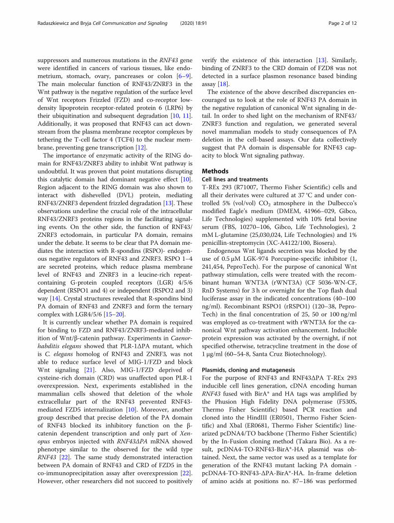
ResultsInducible RNF43 and RNF43ΔPA expression in T-REx 293cell lineIn order to address the role of PA domain of RNF43 wegenerated T-Rex 293 stable cell lines overexpressing hu-man wild type (wt) RNF43 and RNF43 variant lacking itsPA domain (Δ87–187), RNF43ΔPA, in a tetracycline(Tet)-controlled manner (TetON) (Fig. 1a). TetON sys-tem provided uniform RNF43 expression upon Tetaddition, as shown by the western blot and quantitativereal-time Polymerase Chain Reaction (qPCR) (Fig. 1a, b,c). Transgenic lines did not show altered expression ofZNRF3, encoding E3 ligase functionally redundant withRNF43, or CTNNB1 encoding β-catenin (Fig. 1c’, c”).This suggests that we have not selected cell lines func-tionally compensating for the changed levels of RNF43.Deletion of the PA domain could in principle harm pro-
tein processing, since RNF13 and RNF167 proteins lackingtheir PA fold were shown to have impaired cellularlocalization [30]. To address this issue, we performed theimmunofluorescent imaging of cells expressing RNF43variants and membrane-bound form of GFP, which servedas the plasma membrane marker. Images and signals his-tograms confirmed that both wt RNF43 as well asRNF43ΔPA showed comparable subcellular localizationand were present at the plasma membrane (Fig. 1d).After initial and positive validation of our tools, we de-
cided to move on to the functional assays. T-REx 293cells represent a suitable cell line for the analysis ofWnt/β-catenin signal transduction and the role of
RNF43 in this process. Prototypical ligand activatingWnt/β-catenin signaling cascade - rWNT3A triggeredonly a minor response when assessed by Top flash dualluciferase assay (Fig. 1e, left). However, once RNF43/ZNRF3 was inactivated by rRSPO1, rWNT3A activatedβ-catenin mediated transcription approx. 30x more effi-ciently (Fig. 1e, right). Importantly, Tet-induced exogen-ous RNF43 blocked cellular response to both rWNT3Aand rWNT3A/rRSPO1 combination (Fig. 1e). Similarmechanism could be demonstrated by the western blotanalysis of the key Wnt pathway proteins – namelyphosphorylation of LRP6 at serine (S) 1490 and phos-phorylation of DVL2 (detected as electrophoretic mobil-ity) (Fig. 1f). Phosphorylation of LRP6 and DVL proteinsmark canonical Wnt pathway activation [31, 32]. Inagreement with the Top flash dual luciferase assay results,rRSPO1 synergized with rWNT3A and RNF43 overex-pression suppressed activation of the LRP6 and DVL2after 3 h long rWNT3A stimulation (Fig. 1f). It is alsoworth noting that RNF43 TetON cells without Tet induc-tion also exhibited reduced pS1490-LRP6 signal; this isprobably due to the TetON promoter leakage that is how-ever below our detection limits (see anti-HA in Fig. 1f).
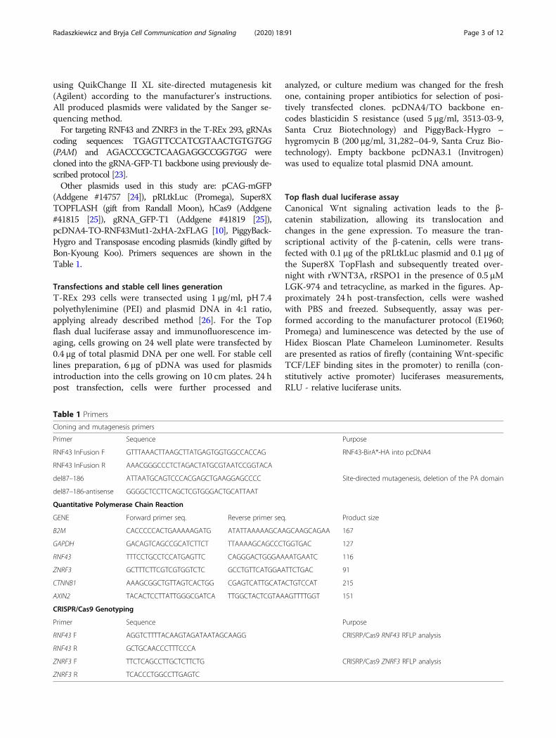
RNF43ΔPA inhibits canonical Wnt signaling pathwayAs a next step we took advantage of the model systemdescribed in Fig. 1 and started to analyze functionallythe role of RNF43 PA domain. Firstly, we comparedRNF43 ΔPA mutant with wt RNF43 in their capacity toinhibit rWNT3A and rRSPO1-induced β-catenindependent transcription. Surprisingly, deletion of PA do-main had no impact on the ability to down-regulate Topflash dual luciferase assay signal in both testedRNF43ΔPA TetON cell lines (Fig. 2a). To further backup the reporter assay results, we analyzed the expressionof the Wnt/β-catenin pathway target gene AXIN2. BothRNF43 and RNF43ΔPA overexpression reduced AXIN2expression level comparably (Fig. 2b).Finally, we have analyzed the effects of RNF43 variants
on Wnt pathway activation using the set of Western blotreadouts – namely phosphorylation of LRP6, DVL2 andDVL3. This experiment was performed in three setups:(i) RNF43 expression and Wnt pathway was induced atthe same time, (ii) RNF43 expression preceded activationof Wnt pathway and (iii) RNF43 was induced when Wntpathway was already activated (for schematics see Fig.2c). These experimental variants addressed differentproperties of RNF43 – namely the potential to preventthe switch ON of the Wnt pathway (in i and ii) and po-tential to induce switch OFF (in iii) (Fig. 2c). Neverthe-less, Tet-driven expression of RNF43 wt and RNF43ΔPAin all cases averted cellular responses to the rWNT3A/rRSPO1 and efficiently reduced phosphorylation ofLRP6 as well as of DVL2 and DVL3 (Fig. 2d, d’, d”).
Radaszkiewicz and Bryja Cell Communication and Signaling (2020) 18:91 Page 5 of 12
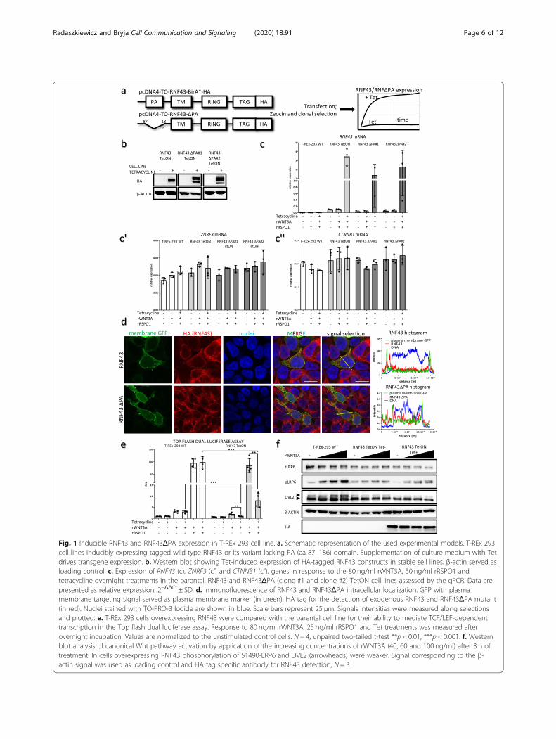
Fig. 1 Inducible RNF43 and RNF43ΔPA expression in T-REx 293 cell line. a. Schematic representation of the used experimental models. T-REx 293cell lines inducibly expressing tagged wild type RNF43 or its variant lacking PA (aa 87–186) domain. Supplementation of culture medium with Tetdrives transgene expression. b. Western blot showing Tet-induced expression of HA-tagged RNF43 constructs in stable sell lines. β-actin served asloading control. c. Expression of RNF43 (c), ZNRF3 (c’) and CTNNB1 (c”), genes in response to the 80 ng/ml rWNT3A, 50 ng/ml rRSPO1 andtetracycline overnight treatments in the parental, RNF43 and RNF43ΔPA (clone #1 and clone #2) TetON cell lines assessed by the qPCR. Data arepresented as relative expression, 2−ΔΔCt ± SD. d. Immunofluorescence of RNF43 and RNF43ΔPA intracellular localization. GFP with plasmamembrane targeting signal served as plasma membrane marker (in green), HA tag for the detection of exogenous RNF43 and RNF43ΔPA mutant(in red). Nuclei stained with TO-PRO-3 Iodide are shown in blue. Scale bars represent 25 μm. Signals intensities were measured along selectionsand plotted. e. T-REx 293 cells overexpressing RNF43 were compared with the parental cell line for their ability to mediate TCF/LEF-dependenttranscription in the Top flash dual luciferase assay. Response to 80 ng/ml rWNT3A, 25 ng/ml rRSPO1 and Tet treatments was measured afterovernight incubation. Values are normalized to the unstimulated control cells. N = 4, unpaired two-tailed t-test **p < 0.01, ***p < 0.001. f. Westernblot analysis of canonical Wnt pathway activation by application of the increasing concentrations of rWNT3A (40, 60 and 100 ng/ml) after 3 h oftreatment. In cells overexpressing RNF43 phosphorylation of S1490-LRP6 and DVL2 (arrowheads) were weaker. Signal corresponding to the β-actin signal was used as loading control and HA tag specific antibody for RNF43 detection, N = 3
Radaszkiewicz and Bryja Cell Communication and Signaling (2020) 18:91 Page 6 of 12
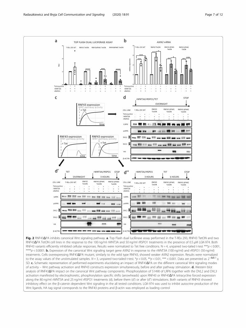
Fig. 2 RNF43ΔPA inhibits canonical Wnt signaling pathway. a. Top Flash dual luciferase assay performed in the T-REx 293, RNF43 TetON and twoRNF43ΔPA TetON cell lines in the response to the 100 ng/ml rWNT3A and 50 ng/ml rRSPO1 treatments in the presence of 0.5 μM LGK-974. BothRNF43 variants efficiently inhibited cellular responses. Results were normalized to Tet free conditions. N = 4, unpaired two-tailed t-test ***p < 0.001,****p < 0.0001. b. Expression of the canonical Wnt signaling target gene AXIN2 in response to the rWNT3A (100 ng/ml) and rRSPO1 (50 ng/ml)treatments. Cells overexpressing RNF43ΔPA mutant, similarly to the wild type RNF43, showed weaker AXIN2 expression. Results were normalizedto the assay values of the unstimulated samples. N = 3, unpaired two-tailed t-test. *p < 0.05, **p < 0.01, *** < 0.001. Data are presented as 2−ΔΔCt ±SD. c. Schematic representation of performed experiments elucidating an impact of RNF43ΔPA on the different canonical Wnt signaling modesof activity – Wnt pathway activation and RNF43 constructs expression simultaneously, before and after pathway stimulation. d. Western blotanalysis of RNF43ΔPA impact on the canonical Wnt pathway components. Phosphorylation of S1490 of LRP6 together with the DVL2 and DVL3activation manifested by electrophoretic, phosphorylation specific shifts (arrowheads) upon RNF43 or RNF43ΔPA tetracycline forced expressionalong the 80 ng/ml rWNT3A and 25 ng/ml rRSPO1 treatments (d), before them (d’) or after (d”) stimulations. Both variants of RNF43 showedinhibitory effect on the β-catenin dependent Wnt signaling in the all tested conditions. LGK-974 was used to inhibit autocrine production of theWnt ligands, HA tag signal corresponds to the RNF43 proteins and β-actin was employed as loading control
Radaszkiewicz and Bryja Cell Communication and Signaling (2020) 18:91 Page 7 of 12
These results suggest that RNF43ΔPA, similarly to thewild type RNF43, desensitized cells to the Wnt ligandstimulation and inhibited already activated receptorcomplexes.
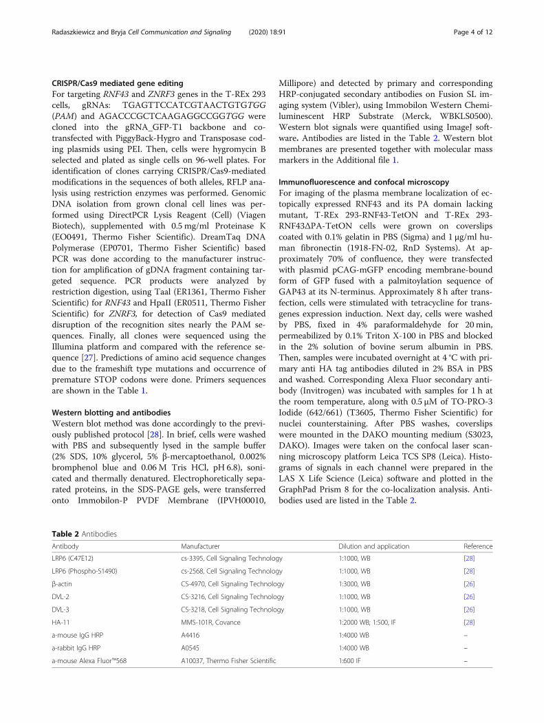
RNF43 ΔPA efficiently rescues phenotype of RNF43/ZNRF3 dKO cellsThe current experiments shown in Figs. 1 and 2 used theoverexpression system that produced high RNF43 levels(see Fig. 1c). In order to analyze RNF43 activity at thephysiological level, we generated T-REx™-293 cells lackingfunctional RNF43/ZNRF3 proteins by employing theCRISPR/Cas9 method (Clustered Regularly InterspacedShort Palindromic Repeats/Cas9). We have characterizedin detail two RNF43/ZNRF3 double knockout (dKO) celllines, which were confirmed by sequencing of targetedgenomic loci and prediction of modification consequenceson the protein level (Fig. 3a). In line with the literature,RNF43/ZNRF3 dKO cell line showed stronger response torWNT3A in the Top flash dual luciferase assay, but in therRSPO1-insensitive manner (Fig. 3b). Similar behaviorwas also confirmed by the western blot of the activationmarkers (Fig. 3c). Despite the fact that LRP6 is the re-ported target of RNF43/ZNRF3 [10, 11], our cells do notshow alteration in global LRP6 levels, a result similar tothe work of Jiang et al. [13]. It is thus possible that theconsequence of the LRP6 ubiquitination is primarily theinternalization of LRP6.RNF43/ZNRF3 dKO cells, which are hypersensitive to
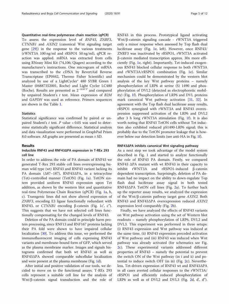
the rWNT3A treatment, allowed us to perform comple-mentation assays with RNF43 wt and RNF43ΔPA. In theserescue experiments RNF43 wt, RNF43ΔPA and enzymati-cally inactive RNF43 Mut1 carrying point mutations inthe RING domain (negative control) were introduced toboth RNF43/ZNRF3 #1 and #2 dKOs under the control oftetracycline (TetON), granting the generation of clonal in-ducible stable cell lines (Fig. 4a).Top flash dual luciferase assay performed in the TetON
RNF43/ZNRF3 #1 and #2 dKOs, confirmed that RNF43lacking PA domain efficiently inhibited canonical Wnt sig-naling (Fig. 4b and c). Induction of RNF43 as well asRNF43ΔPA mutant was also capable to efficiently sup-press WNT3A-induced S1490 phosphorylation of LRP6and activation of DVL2 and DVL3 (Fig. 4d-e). Finally, ex-pression of enzymatically dead RNF43 Mut1 variant in-capable to perform ubiquitination reaction had no effecton the Wnt signaling (Fig. 4b-e”’), which suggest that theobserved RNF43-mediated phenotypes are specific.Interestingly, treatment with recombinant R-spondin1
(RSPO1) was unable to revert the inhibitory action ofTet-induced RNF43 and RNF43 ΔPA (Fig. 4b-e). How-ever, we observed the induction of Wnt pathway byrWNT3A– determined as Top flash activity, DVL2,DVL3 and LRP6 phosphorylation - by combined
rRSPO1 treatment in the RNF43wt, but not RNF43ΔPAand RNF43 Mut1, RNF43/ZNRF3 dKO #1 and #2 cellsin the absence of Tet (Fig. 4b-e). Upon closer inspectionwe found that leakage of TetON system leads to low ex-pression of the transgene even in the absence of tetracyc-line (see HA signal in the Fig. 4d and e for RNF43 Mut1results and Additional file 1 4d and 4e long expositions ofHA signal). We propose that under these conditionsRSPO1 treatment inhibits wt RNF43 but not RNF43 ΔPA.We conclude that depletion of the PA domain produced
variant insensitive to the negative regulation by rRSPO1protein, what would be in agreement with published crys-tal structures showing direct interaction between PA do-main of RNF43 and Furin-1 fold of R-spondins, ultimatelyleading to the internalization of RNF43 [15]. In summary,using multiple sets of assays we provide evidence that theextracellular PA domain is not essential for RNF43 inhibi-tory activity in the Wnt/β-catenin pathway in human cellsbut rather it serves as a module that via action of RSPO1regulates surface levels of RNF43.
DiscussionOur experiments conclude that in mammalian cells PAdomain of RNF43 does not contribute to the ability ofRNF43 to efficiently inhibit WNT3a-induced β-catenin-dependent signal transduction. Namely, RNF43ΔPA wascomparably potent as wt RNF43 in (i) the inhibition ofTop flash dual luciferase assay and AXIN2 gene expres-sion, (ii) block of WNT3a-induced phosphorylation ofLRP6 and (iii) phosphorylation of DVL2 and DVL3. Wedemonstrated this capacity using inducible expression ofRNF43ΔPA as well as by rescue experiments in RNF43/ZNRF3 dKO cells. Notably, in the rescue experimentsRNF43ΔPA showed significantly stronger inhibitory ef-fect on the pS1490 LRP6 than wt RNF43. We proposethat in this experimental setup RNF43ΔPA variant can-not be regulated by the endogenously secreted RSPOsthat bind via PA domain [2] and as such can have en-hanced activity. This was confirmed in the Top flashdual luciferase assay, by which we showed thatRNF43ΔPA was not sensitive to the recombinant RSPO1treatment.Our key results suggest that the key function of the
PA domain of RNF43 in human cells is to mediate nega-tive regulation by R-spondin family proteins. This is incontrast to some of the previously published data thatpropose a role of PA domain in the inhibition of Wntpathway. We see multiple reasons possibly explainingexisting discrepancies. We discuss individual aspectsnamely: (i) studies in non-vertebrate models, (ii) bio-chemical studies on (RNF43) PA and (FZD) CRD do-mains, (iii) studies performed in vertebrates and (iv)presence of RNF43 PA domain mutations in Wnt-addicted tumors in detail below.
Radaszkiewicz and Bryja Cell Communication and Signaling (2020) 18:91 Page 8 of 12
One of the key studies about PA domain function wasbased on the Caenorhabditis elegans model organism.Nematode ortholog of human RNF43 and ZNRF3 pro-teins, PLR-1 blocked Wnt signaling and caused theendosomal internalization of Frizzled receptors (MIG-1in C. elegans) in a manner dependent on Frizzled’s Cyst-eine Rich Domain (CRD) and RING and PA folds ofPLR-1 [21]. This demonstrated the functional interactionbetween extracellular parts of PLR-1 and MIG-1. Inter-estingly, C. elegans lacks R-spondin related proteins. It
was shown before that RSPO/LGR regulatory axisevolved only in vertebrates, so it is not present in C. ele-gans [33]. Thus, we can speculate that the role of the PAfold changed together with the evolutionary rise of theWnt signaling complexity.The other issue that remains partially unsolved is the
physical interaction between PA domain of RNF43 andCRD of FZDs, as its existence was detected in the co-immunoprecipitation assays after overexpression PA andCRD domains [21, 22]. This still remains controversial,
Fig. 3 Preparation of cells lacking functional RNF43/ZNRF3 proteins. a. In order to generate double knockout lines, CRISPR/Cas9 method wasapplied to introduce mutations in the RNF43 and ZNRF3 genes of T-REx 293 cells. Sequencing results of loci targeted by the CRISPR/Cas9approach. PAM sequences are underlined. Modifications effects on the protein sequence were predicted. b. RNF43/ZNRF3 dKO cells had higher β-catenin dependent transcriptional activity in response to the overnight incubation with different rWNT3A doses (40, 60 and 80 ng/ml). Also, dKOcell line was insensitive to the 25 ng/ml rRSPO1 treatment. All values are normalized to the unstimulated control cells results. N = 5 (wild typecells), N = 3 (dKO), unpaired two-tailed t-test *p < 0.05, **p < 0.01, *** < 0.001. c. Western blot analysis of canonical Wnt pathway activation inresponse to the increasing doses of rWNT3A (40, 60 and 100 ng/ml) after 3 h long treatments. RNF43/ZNRF3 dKO exhibited stronger response tothe rWNT3A in comparison to the parental cells and rRSPO1 stimulation had no effect. Signal corresponding to the β-actin signal was used asloading control, N = 3
Radaszkiewicz and Bryja Cell Communication and Signaling (2020) 18:91 Page 9 of 12
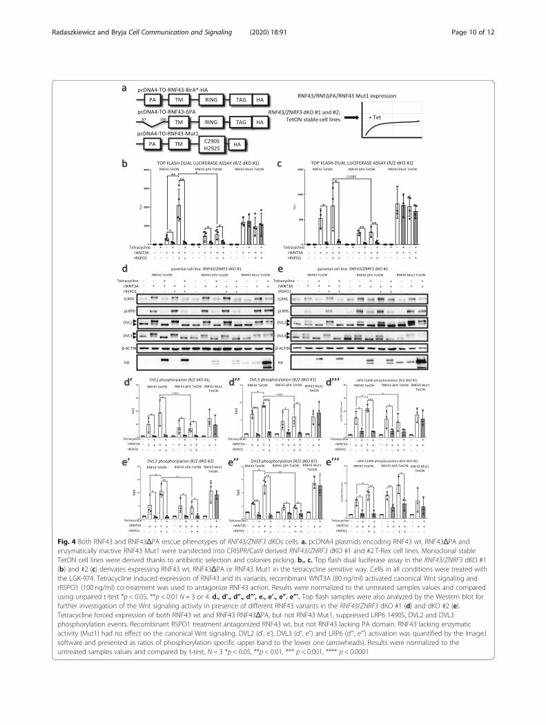
Fig. 4 Both RNF43 and RNF43ΔPA rescue phenotypes of RNF43/ZNRF3 dKOs cells. a. pcDNA4 plasmids encoding RNF43 wt, RNF43ΔPA andenzymatically inactive RNF43 Mut1 were transfected into CRISPR/Cas9 derived RNF43/ZNRF3 dKO #1 and #2 T-Rex cell lines. Monoclonal stableTetON cell lines were derived thanks to antibiotic selection and colonies picking. b., c. Top flash dual luciferase assay in the RNF43/ZNRF3 dKO #1(b) and #2 (c) derivates expressing RNF43 wt, RNF43ΔPA or RNF43 Mut1 in the tetracycline sensitive way. Cells in all conditions were treated withthe LGK-974. Tetracycline induced expression of RNF43 and its variants, recombinant WNT3A (80 ng/ml) activated canonical Wnt signaling andrRSPO1 (100 ng/ml) co-treatment was used to antagonize RNF43 action. Results were normalized to the untreated samples values and comparedusing unpaired t-test *p < 0.05, **p < 0.01 N = 3 or 4. d., d’., d”., d”’, e., e’., e”. e”’. Top flash samples were also analyzed by the Western blot forfurther investigation of the Wnt signaling activity in presence of different RNF43 variants in the RNF43/ZNRF3 dKO #1 (d) and dKO #2 (e).Tetracycline forced expression of both RNF43 wt and RNF43 RNF43ΔPA, but not RNF43 Mut1, suppressed LRP6 1490S, DVL2 and DVL3phosphorylation events. Recombinant RSPO1 treatment antagonized RNF43 wt, but not RNF43 lacking PA domain. RNF43 lacking enzymaticactivity (Mut1) had no effect on the canonical Wnt signaling. DVL2 (d’, e’), DVL3 (d”, e”) and LRP6 (d”’, e”’) activation was quantified by the ImageJsoftware and presented as ratios of phosphorylation specific upper band to the lower one (arrowheads). Results were normalized to theuntreated samples values and compared by t-test, N = 3 *p < 0.05, **p < 0.01, *** p < 0.001, **** p < 0.0001
Radaszkiewicz and Bryja Cell Communication and Signaling (2020) 18:91 Page 10 of 12
since two other groups were not able to detect directinteraction between RNF43 and Frizzled by structuralbiology approaches [13, 18]. During immunoprecipita-tion, the interaction could be indirect and could includeother proteins such as endogenously expressed R-spondins. Further report showed that RNF43 lacking thewhole extracellular part was not able to promote FZD5internalization [10]. However, it seems to us that thisparticular RNF43 mutant lacked signal peptide, and assuch could not be targeted to the plasma membrane. Inour study RNF43ΔPA contained the whole sequence be-tween signal peptide and PA part, which might be in-volved in the unknow protein-protein interactions.The solid work by Tsakayuma and others [22] sug-
gested that even in vertebrate cells the PA domain ofRNF43 is important for its function. However, still theauthors observed a weak but clear inhibitory activity to-wards Wnt/β-catenin pathway in vivo in Xenopus em-bryonic development. It suggests that PA domain is notessential but in the embryonal context RNF43ΔPA actsas a weaker inhibitor. We can only speculate if otherRNF43 functions participate on the inhibitory action ofRNF43 in this context. For example RNF43 was de-scribed to act downstream of plasma membrane receptorcomplexes and even downstream from mutated β-catenin by sequestering the Transcription factor 4(TCF4) to the nuclear membrane and thereby, leading tothe inhibition of gene expression regulated by this tran-scriptional factor [12].It was reported that missense mutations (R127P and
A169T) occurring within the PA domain suppressed in-hibitory properties of RNF43 towards the canonical Wntsignaling [12, 22]. These genetic changes, together withothers (i.e. K108E, R117H, E170K), are described incases of large intestine and pancreatic cancers (COSMICdatabase: cancer.sanger.ac.uk [34]), suggesting RNF43loss-of-function. RNF43/ZNRF3 bind its endogenouslyproduced inhibitors – R-spondins, exactly via the PAfold (residues Gln84, His86, Lys108, and Glu110) [16].Consequently, mutations disrupting these interactionsare expected to lead to the insensitivity of RNF43 to-wards R-spondins, which is counterintuitive in cases ofthese Wnt-addicted tumors. Therefore, an alternativeexplanation is that these mutations affect folding and/orlocalization of RNF43 [12, 22] or affect binding to other,not known yet RNF43 interactors. The misfolding/mis-placement hypothesis is supported by the importance ofPA domains in other PA-TM-RING E3 ubiquitin ligaseswhere PA domains serve as motives for the protein-protein interactions [35] and subcellular localization[30]. For example, PA domain of RNF13 and RNF167,from the same group of E3 ligases as RNF43/ZNRF3,was indispensable for their homodimerization andproper cellular localization [30]. In line with that R127P
variant showed impaired reticular expression in contrastto the wild type protein and possible ERAD-mediateddegradation [22]. It is thus probable that the analysis ofthe biological output of such mutants tells us only a littleabout the physiological function of the PA domain.
ConclusionIn summary, we showed by the biological assays and precisemodulation of protein levels that RNF43 lacking PA do-main can still efficiently inhibit cellular events related to thecanonical Wnt signaling pathway. In some assaysRNF43ΔPA mutant was even a better inhibitor whichpoints to the importance of PA domain as the primary re-gions required for the RNF43 regulation by R-spondin 1.Altogether, our study highlights a necessity for further workaiming to the better description of still quite poorly under-stood mechanisms of regulation and function of RNF43and ZNRF3 enzymes in the homeostasis and pathology.
Supplementary informationSupplementary information accompanies this paper at https://doi.org/10.1186/s12964-020-00559-0.
Additional file 1.
AbbreviationsCRD: Cysteine-rich domain (CRD); CRISPR/Cas9: Clustered RegularlyInterspaced Short Palindromic Repeats/Cas9; CTNNB1: Beta-catenin;DMEM: Dulbecco’s modified Eagle’s medium; DVL: Dishevelled; FBS: Fetalbovine serum; FZD: Frizzled; HA: Hemagglutinin tag; LGR4–6: Leucine-richrepeat-containing G-protein coupled receptor 4–6; LRP5/6: Low-densitylipoprotein receptor-related protein 5/6; PA: Protease-associated;PAM: Protospacer adjacent motif; PCR: Polymerase chain reaction;PEI: Polyethylenimine; qRT-PCR: Quantitative real-time polymerase chain reac-tion; RING: Really Interesting New Gene; RNF43: RING finger protein 43;RSPO1–4: R-spondin 1–4; SD: Standard deviation; TCF4: T-cell factor 4;TET: Tetracycline; TetON: Tetracycline-controlled transcriptional activation;ZNRF3: Zinc/Ring finger protein 3
AcknowledgementsWe would like to thank Katarzyna Radaszkiewicz PhD., Lucie Nesvadbová,Lenka Bryjová, Pavlína Žofka Mrhálková, Lenka Doubková and Naďa Bílá forexcellent assistance.
Authors’ contributionsT.R and V. B designed the research and wrote the original manuscript. T.R.performed all the research and data visualization. All authors read andapproved the final manuscript.
FundingThe work was supported by the Czech Science Foundation grant GX19-28347X.
Availability of data and materialsThe datasets used and/or analyzed during the current study are availablefrom the corresponding author on reasonable request.
Ethics approvalNot applicable.
Consent for publicationNot applicable.
Competing interestsThe authors declare that they have no competing interests.
Radaszkiewicz and Bryja Cell Communication and Signaling (2020) 18:91 Page 11 of 12
Received: 21 November 2019 Accepted: 23 March 2020
References1. de Lau W, Peng WC, Gros P, Clevers H. The R-spondin/Lgr5/Rnf43 module:
regulator of Wnt signal strength. Genes Dev. 2014. https://doi.org/10.1101/gad.235473.113.
2. Zebisch M, Jones EY. ZNRF3/RNF43 – a direct linkage of extracellularrecognition and E3 ligase activity to modulate cell surface signalling. ProgBiophys Mol Biol. 2015;118:112–8.
3. Wiese KE, Nusse R, van Amerongen R. Wnt signalling: conqueringcomplexity. Development. 2018;145:dev165902.
4. Nusse R, Clevers H. Wnt/β-catenin signaling, disease, and emergingtherapeutic modalities. Cell. 2017;169:985–99.
5. Grainger S, Willert K. Mechanisms of Wnt signaling and control. WileyInterdiscip Rev Syst Biol Med. 2018;10:e1422.
6. Giannakis M, Hodis E, Jasmine Mu X, et al. RNF43 is frequently mutated incolorectal and endometrial cancers. Nat Genet. 2014;46:1264–6.
7. Ryland GL, Hunter SM, Doyle MA, Rowley SM, Christie M, Allan PE, BowtellDD, Gorringe KL, Campbell IG, Campbell IG. RNF43 is a tumour suppressorgene mutated in mucinous tumours of the ovary. J Pathol. 2013;229:469–76.
8. Jiao Y, Yonescu R, Offerhaus GJA, et al. Whole-exome sequencing ofpancreatic neoplasms with acinar differentiation. J Pathol. 2014;232:428–35.
9. Wang K, Yuen ST, Xu J, et al. Whole-genome sequencing andcomprehensive molecular profiling identify new driver mutations in gastriccancer. Nat Genet. 2014;46:573–82.
10. Koo B-K, Spit M, Jordens I, et al. Tumour suppressor RNF43 is a stem-cell E3ligase that induces endocytosis of Wnt receptors. Nature. 2012;488:665–9.
11. Hao H-X, Xie Y, Zhang Y, et al. ZNRF3 promotes Wnt receptor turnover inan R-spondin-sensitive manner. Nature. 2012;485:195–200.
12. Loregger A, Grandl M, Mejías-Luque R, et al (2015) The E3 ligase RNF43inhibits Wnt signaling downstream of mutated β-catenin by sequesteringTCF4 to the nuclear membrane. Sci signal 8:ra90.
13. Jiang X, Charlat O, Zamponi R, Yang Y, Cong F. Dishevelled promotes Wntreceptor degradation through recruitment of ZNRF3/RNF43 E3 ubiquitinligases. Mol Cell. 2015;58:522–33.
14. Lebensohn AM, Rohatgi R. R-spondins can potentiate WNT signalingwithout LGRs. Elife. 2018;7:e33126.
15. Zebisch M, Xu Y, Krastev C, MacDonald BT, Chen M, Gilbert RJC, He X, JonesEY. Structural and molecular basis of ZNRF3/RNF43 transmembraneubiquitin ligase inhibition by the Wnt agonist R-spondin. Nat Commun.2013;4:2787.
16. Chen P-H, Chen X, Lin Z, Fang D, He X. The structural basis of R-spondinrecognition by LGR5 and RNF43. Genes Dev. 2013;27:1345–50.
17. Peng WCC, DeLau W, Forneris F, et al. Structure of stem cell growth factorR-spondin 1 in complex with the Ectodomain of its receptor LGR5. Cell Rep.2013;3:1885–92.
18. Peng WC, de Lau W, Madoori PK, Forneris F, Granneman JCM, Clevers H,Gros P. Structures of Wnt-antagonist ZNRF3 and its complex with R-Spondin1 and implications for signaling. PLoS One. 2013;8:e83110.
19. Wang D, Huang B, Zhang S, Yu X, Wu W, Wang X. Structural basis for R-spondin recognition by LGR4/5/6 receptors. Genes Dev. 2013;27:1339–44.
20. Xu K, Xu Y, Rajashankar KR, Robev D, Nikolov DB. Crystal structures of Lgr4and its complex with R-Spondin1. Structure. 2013;21:1683–9.
21. Moffat LL, Robinson RE, Bakoulis A, Clark SG. The conserved transmembraneRING finger protein PLR-1 downregulates Wnt signaling by reducing frizzled,Ror and Ryk cell-surface levels in C elegans. Development. 2014;141:617–28.
22. Tsukiyama T, Fukui A, Terai S, Fujioka Y, Shinada K, Takahashi H, YamaguchiTP, Ohba Y, Hatakeyama S. Molecular role of RNF43 in canonical andnoncanonical Wnt signaling. Mol Cell Biol. 2015;35:2007–23.
23. Schwank G, Koo B-K, Sasselli V, et al. Functional repair of CFTR by CRISPR/Cas9 in intestinal stem cell Organoids of cystic fibrosis patients. Cell StemCell. 2013;13:653–8.
24. Matsuda T, Cepko CL. Controlled expression of transgenes introduced byin vivo electroporation. Proc Natl Acad Sci. 2007;104:1027–32.
25. Mali P, Yang L, Esvelt KM, Aach J, Guell M, DiCarlo JE, Norville JE, Church GM(2013) RNA-guided human genome engineering via Cas9. Science (80- )339:823–826.
26. Mentink RA, Rella L, Radaszkiewicz TW, Gybel T, Betist MC, Bryja V,Korswagen HC. The planar cell polarity protein VANG-1/Vangl negatively
regulates Wnt/β-catenin signaling through a Dvl dependent mechanism.PLoS Genet. 2018;14:e1007840.
27. Malcikova J, Stano-Kozubik K, Tichy B, et al. Detailed analysis of therapy-driven clonal evolution of TP53 mutations in chronic lymphocytic leukemia.Leukemia. 2015;29:877–85.
28. Paclíková P, Bernatík O, Radaszkiewicz TW, Bryja V. The N-terminal part ofthe Dishevelled DEP domain is required for Wnt/β- catenin signaling inmammalian cells. Mol Cell Biol. 2017. https://doi.org/10.1128/MCB.00145-17.
29. Jho E, Zhang T, Domon C, Joo C-K, Freund J-N, Costantini F. Wnt/beta-catenin/Tcf signaling induces the transcription of Axin2, a negativeregulator of the signaling pathway. Mol Cell Biol. 2002;22:1172–83.
30. van Dijk JR, Yamazaki Y, Palmer RH. Tumour-associated mutations of PA-TM-RING ubiquitin ligases RNF167/RNF13 identify the PA domain as adeterminant for endosomal localization. Biochem J. 2014;459:27–36.
31. Bryja V, Schulte G, Arenas E. Wnt-3a utilizes a novel low dose and rapidpathway that does not require casein kinase 1-mediated phosphorylation ofDvl to activate β-catenin. Cell Signal. 2007;19:610–6.
32. Zeng X, Tamai K, Doble B, Li S, Huang H, Habas R, Okamura H, Woodgett J,He X. A dual-kinase mechanism for Wnt co-receptor phosphorylation andactivation. Nature. 2005;438:873–7.
33. de Lau WB, Snel B, Clevers HC. The R-spondin protein family. Genome Biol.2012;13:242.
34. Forbes SA, Beare D, Boutselakis H, et al. COSMIC: somatic cancer genetics athigh-resolution. Nucleic Acids Res. 2017;45:D777–83.
35. Lineberry N, Su L, Soares L, Fathman CG. The single subunitTransmembrane E3 ligase gene related to Anergy in lymphocytes (GRAIL)captures and then Ubiquitinates Transmembrane proteins across the cellmembrane. J Biol Chem. 2008;283:28497.
Publisher’s NoteSpringer Nature remains neutral with regard to jurisdictional claims inpublished maps and institutional affiliations.
Radaszkiewicz and Bryja Cell Communication and Signaling (2020) 18:91 Page 12 of 12


![Maroussi, 1-3-2018 DECISION: 843/2 DECISION · Domain Name that may be subject to assignment by EETT, i.e. all 2nd level [.gr] or [.ελ] Domain Names and all 3rd level [.gr] Domain](https://static.fdocument.org/doc/165x107/6003f2ec3175f641c53ed88d/maroussi-1-3-2018-decision-8432-decision-domain-name-that-may-be-subject-to-assignment.jpg)


![Domain Specific Languages [0.5ex] for Convex Optimization](https://static.fdocument.org/doc/165x107/61fb7d612e268c58cd5ec7a1/domain-specific-languages-05ex-for-convex-optimization.jpg)