Proscillaridin A exerts anti-tumor effects through GSK3β ... · Therefore, we disclosed here a...
14
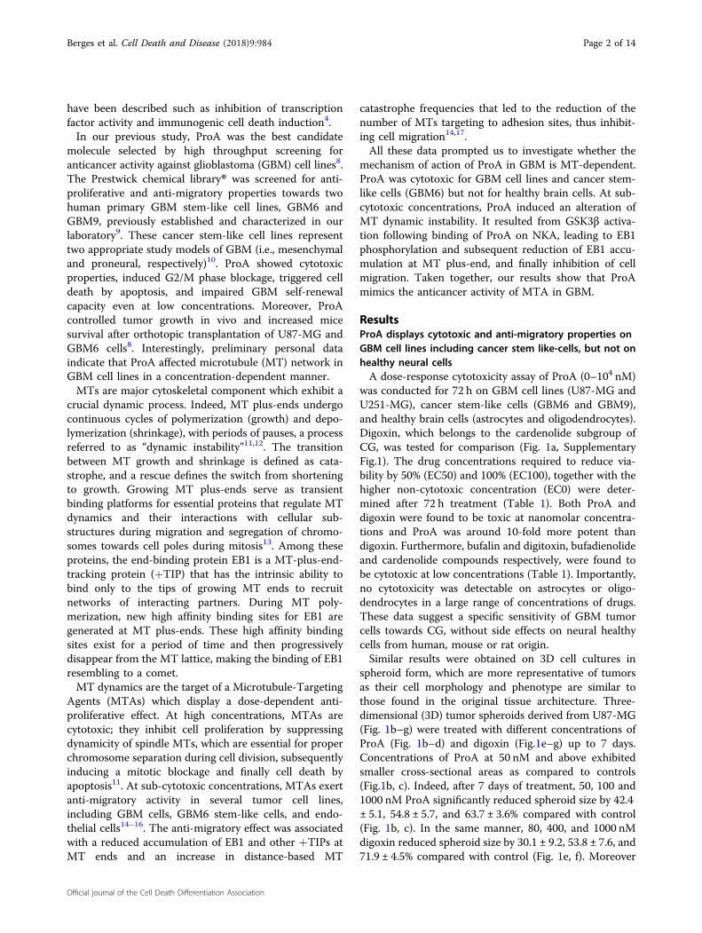
Berges et al. Cell Death and Disease (2018)9:984 DOI 10.1038/s41419-018-1018-7 Cell Death & Disease ARTICLE Open Access Proscillaridin A exerts anti-tumor effects through GSK3 β activation and alteration of microtubule dynamics in glioblastoma Raphael Berges 1 , Emilie Denicolai 1 , Aurélie Tchoghandjian 1 , Nathalie Baeza-Kallee 1 , Stephane Honore 1 , Dominique Figarella-Branger 1 and Diane Braguer 1 Abstract Glioblastoma (GBM) is characterized by highly aggressive growth and invasive behavior. Due to the highly lethal nature of GBM, new therapies are urgently needed and repositioning of existing drugs is a promising approach. We have previously shown the activity of Proscillaridin A (ProA), a cardiac glycoside inhibitor of the Na(+)/K(+) ATPase (NKA) pump, against proliferation and migration of GBM cell lines. ProA inhibited tumor growth in vivo and increased mice survival after orthotopic grafting of GBM cells. This study aims to decipher the mechanism of action of ProA in GBM tumor and stem-like cells. ProA displayed cytotoxic activity on tumor and stem-like cells grown in 2D and 3D culture, but not on healthy cells as astrocytes or oligodendrocytes. Even at sub-cytotoxic concentration, ProA impaired cell migration and disturbed EB1 accumulation at microtubule (MT) plus-ends and MT dynamics instability. ProA activates GSK3β downstream of NKA inhibition, leading to EB1 phosphorylation on S155 and T166, EB1 comet length shortening and MT dynamics alteration, and finally inhibition of cell migration and cytotoxicity. Similar results were observed with digoxin. Therefore, we disclosed here a novel pathway by which ProA and digoxin modulate MT- governed functions in GBM tumor and stem-like cells. Altogether, our results support ProA and digoxin as potent candidates for drug repositioning in GBM. Introduction Cardiac glycosides (CG) are a large family of natural compounds that are well-known drugs for increasing cardiac contractile force in cardiac diseases. Proscillaridin A (ProA) is a familiar drug that belongs to the bufadie- nolide chemical sub-group. In cardiomyocytes, CG bind and inhibit the sodium (Na + )/potassium (K + )-ATPase (NKA) transmembrane pump. The consecutive elevation of the intracellular Na + level stimulates the Na + /Ca 2+ exchanger mechanism. As a result, the intracellular Ca 2+ concentration is increased, promoting cellular events such as myocardial contractibility, leading to the positive ino- tropic effects of the CG 1 . The anticancer effects of CG were suggested in 1979 by Stenkvist in a study of women treated with digitalis in combination with chemotherapy for breast cancer 2 . A higher survival rate was also observed in a long-term follow-up study 3 . Thereafter, anticancer effects of different CG were shown on several cell lines and in various in vivo models 4 . However, sen- sitivity of CG on cell proliferation and viability depend on tumor type and CG may not be good candidates for cancer therapeutics in all tumors 5 . Hence, the mechanism of the anti-cancer activity of CG needs to be deciphered. The ability of CG to inhibit NKA pump function resulting in increased Ca 2+ concentration and subsequent apop- tosis was first suggested 6 . Furthermore, activation of NKA as a signal transducer in cell signaling pathways has been proposed to explain the anticancer activity of CG at low nanomolar concentrations, which do not lead to calcium overload 7 . More recently, additional intracellular targets for CG, whose modulation might be off-NKA targeting, © The Author(s) 2018 Open Access This article is licensed under a Creative Commons Attribution 4.0 International License, which permits use, sharing, adaptation, distribution and reproduction in any medium or format, as long as you give appropriate credit to the original author(s) and the source, provide a link to the Creative Commons license, and indicate if changes were made. The images or other third party material in this article are included in the article’ s Creative Commons license, unless indicated otherwise in a credit line to the material. If material is not included in the article’s Creative Commons license and your intended use is not permitted by statutory regulation or exceeds the permitted use, you will need to obtain permission directly from the copyright holder. To view a copy of this license, visit http://creativecommons.org/licenses/by/4.0/. Correspondence: Diane Braguer ([email protected]) 1 Aix-Marseille Univ, CNRS, INP, Inst Neurophysiopathol, Marseille, France Edited by M. L. Asselin-Labat Official journal of the Cell Death Differentiation Association 1234567890():,; 1234567890():,; 1234567890():,; 1234567890():,;
Transcript of Proscillaridin A exerts anti-tumor effects through GSK3β ... · Therefore, we disclosed here a...
Berges et al. Cell Death and Disease (2018) 9:984
DOI 10.1038/s41419-018-1018-7 Cell Death & Disease
ART ICLE Open Ac ce s s
Proscillaridin A exerts anti-tumor effectsthrough GSK3β activation and alteration ofmicrotubule dynamics in glioblastomaRaphael Berges1, Emilie Denicolai1, Aurélie Tchoghandjian1, Nathalie Baeza-Kallee1, Stephane Honore1,Dominique Figarella-Branger1 and Diane Braguer1
AbstractGlioblastoma (GBM) is characterized by highly aggressive growth and invasive behavior. Due to the highly lethalnature of GBM, new therapies are urgently needed and repositioning of existing drugs is a promising approach. Wehave previously shown the activity of Proscillaridin A (ProA), a cardiac glycoside inhibitor of the Na(+)/K(+) ATPase(NKA) pump, against proliferation and migration of GBM cell lines. ProA inhibited tumor growth in vivo and increasedmice survival after orthotopic grafting of GBM cells. This study aims to decipher the mechanism of action of ProA inGBM tumor and stem-like cells. ProA displayed cytotoxic activity on tumor and stem-like cells grown in 2D and 3Dculture, but not on healthy cells as astrocytes or oligodendrocytes. Even at sub-cytotoxic concentration, ProA impairedcell migration and disturbed EB1 accumulation at microtubule (MT) plus-ends and MT dynamics instability. ProAactivates GSK3β downstream of NKA inhibition, leading to EB1 phosphorylation on S155 and T166, EB1 comet lengthshortening and MT dynamics alteration, and finally inhibition of cell migration and cytotoxicity. Similar results wereobserved with digoxin. Therefore, we disclosed here a novel pathway by which ProA and digoxin modulate MT-governed functions in GBM tumor and stem-like cells. Altogether, our results support ProA and digoxin as potentcandidates for drug repositioning in GBM.
IntroductionCardiac glycosides (CG) are a large family of natural
compounds that are well-known drugs for increasingcardiac contractile force in cardiac diseases. ProscillaridinA (ProA) is a familiar drug that belongs to the bufadie-nolide chemical sub-group. In cardiomyocytes, CG bindand inhibit the sodium (Na+)/potassium (K+)-ATPase(NKA) transmembrane pump. The consecutive elevationof the intracellular Na+ level stimulates the Na+/Ca2+
exchanger mechanism. As a result, the intracellular Ca2+
concentration is increased, promoting cellular events suchas myocardial contractibility, leading to the positive ino-tropic effects of the CG1. The anticancer effects of CGwere suggested in 1979 by Stenkvist in a study of women
treated with digitalis in combination with chemotherapyfor breast cancer2. A higher survival rate was alsoobserved in a long-term follow-up study3. Thereafter,anticancer effects of different CG were shown on severalcell lines and in various in vivo models4. However, sen-sitivity of CG on cell proliferation and viability depend ontumor type and CG may not be good candidates forcancer therapeutics in all tumors5. Hence, the mechanismof the anti-cancer activity of CG needs to be deciphered.The ability of CG to inhibit NKA pump function resultingin increased Ca2+ concentration and subsequent apop-tosis was first suggested6. Furthermore, activation of NKAas a signal transducer in cell signaling pathways has beenproposed to explain the anticancer activity of CG at lownanomolar concentrations, which do not lead to calciumoverload7. More recently, additional intracellular targetsfor CG, whose modulation might be off-NKA targeting,
© The Author(s) 2018OpenAccessThis article is licensedunder aCreativeCommonsAttribution 4.0 International License,whichpermits use, sharing, adaptation, distribution and reproductionin any medium or format, as long as you give appropriate credit to the original author(s) and the source, provide a link to the Creative Commons license, and indicate if
changesweremade. The images or other third partymaterial in this article are included in the article’s Creative Commons license, unless indicated otherwise in a credit line to thematerial. Ifmaterial is not included in the article’s Creative Commons license and your intended use is not permitted by statutory regulation or exceeds the permitted use, you will need to obtainpermission directly from the copyright holder. To view a copy of this license, visit http://creativecommons.org/licenses/by/4.0/.
Correspondence: Diane Braguer ([email protected])1Aix-Marseille Univ, CNRS, INP, Inst Neurophysiopathol, Marseille, FranceEdited by M. L. Asselin-Labat
Official journal of the Cell Death Differentiation Association
1234
5678
90():,;
1234
5678
90():,;
1234567890():,;
1234
5678
90():,;
have been described such as inhibition of transcriptionfactor activity and immunogenic cell death induction4.In our previous study, ProA was the best candidate
molecule selected by high throughput screening foranticancer activity against glioblastoma (GBM) cell lines8.The Prestwick chemical library® was screened for anti-proliferative and anti-migratory properties towards twohuman primary GBM stem-like cell lines, GBM6 andGBM9, previously established and characterized in ourlaboratory9. These cancer stem-like cell lines representtwo appropriate study models of GBM (i.e., mesenchymaland proneural, respectively)10. ProA showed cytotoxicproperties, induced G2/M phase blockage, triggered celldeath by apoptosis, and impaired GBM self-renewalcapacity even at low concentrations. Moreover, ProAcontrolled tumor growth in vivo and increased micesurvival after orthotopic transplantation of U87-MG andGBM6 cells8. Interestingly, preliminary personal dataindicate that ProA affected microtubule (MT) network inGBM cell lines in a concentration-dependent manner.MTs are major cytoskeletal component which exhibit a
crucial dynamic process. Indeed, MT plus-ends undergocontinuous cycles of polymerization (growth) and depo-lymerization (shrinkage), with periods of pauses, a processreferred to as “dynamic instability”11,12. The transitionbetween MT growth and shrinkage is defined as cata-strophe, and a rescue defines the switch from shorteningto growth. Growing MT plus-ends serve as transientbinding platforms for essential proteins that regulate MTdynamics and their interactions with cellular sub-structures during migration and segregation of chromo-somes towards cell poles during mitosis13. Among theseproteins, the end-binding protein EB1 is a MT-plus-end-tracking protein (+TIP) that has the intrinsic ability tobind only to the tips of growing MT ends to recruitnetworks of interacting partners. During MT poly-merization, new high affinity binding sites for EB1 aregenerated at MT plus-ends. These high affinity bindingsites exist for a period of time and then progressivelydisappear from the MT lattice, making the binding of EB1resembling to a comet.MT dynamics are the target of a Microtubule-Targeting
Agents (MTAs) which display a dose-dependent anti-proliferative effect. At high concentrations, MTAs arecytotoxic; they inhibit cell proliferation by suppressingdynamicity of spindle MTs, which are essential for properchromosome separation during cell division, subsequentlyinducing a mitotic blockage and finally cell death byapoptosis11. At sub-cytotoxic concentrations, MTAs exertanti-migratory activity in several tumor cell lines,including GBM cells, GBM6 stem-like cells, and endo-thelial cells14–16. The anti-migratory effect was associatedwith a reduced accumulation of EB1 and other +TIPs atMT ends and an increase in distance-based MT
catastrophe frequencies that led to the reduction of thenumber of MTs targeting to adhesion sites, thus inhibit-ing cell migration14,17.All these data prompted us to investigate whether the
mechanism of action of ProA in GBM is MT-dependent.ProA was cytotoxic for GBM cell lines and cancer stem-like cells (GBM6) but not for healthy brain cells. At sub-cytotoxic concentrations, ProA induced an alteration ofMT dynamic instability. It resulted from GSK3β activa-tion following binding of ProA on NKA, leading to EB1phosphorylation and subsequent reduction of EB1 accu-mulation at MT plus-end, and finally inhibition of cellmigration. Taken together, our results show that ProAmimics the anticancer activity of MTA in GBM.
ResultsProA displays cytotoxic and anti-migratory properties onGBM cell lines including cancer stem like-cells, but not onhealthy neural cellsA dose-response cytotoxicity assay of ProA (0–104 nM)
was conducted for 72 h on GBM cell lines (U87-MG andU251-MG), cancer stem-like cells (GBM6 and GBM9),and healthy brain cells (astrocytes and oligodendrocytes).Digoxin, which belongs to the cardenolide subgroup ofCG, was tested for comparison (Fig. 1a, SupplementaryFig.1). The drug concentrations required to reduce via-bility by 50% (EC50) and 100% (EC100), together with thehigher non-cytotoxic concentration (EC0) were deter-mined after 72 h treatment (Table 1). Both ProA anddigoxin were found to be toxic at nanomolar concentra-tions and ProA was around 10-fold more potent thandigoxin. Furthermore, bufalin and digitoxin, bufadienolideand cardenolide compounds respectively, were found tobe cytotoxic at low concentrations (Table 1). Importantly,no cytotoxicity was detectable on astrocytes or oligo-dendrocytes in a large range of concentrations of drugs.These data suggest a specific sensitivity of GBM tumorcells towards CG, without side effects on neural healthycells from human, mouse or rat origin.Similar results were obtained on 3D cell cultures in
spheroid form, which are more representative of tumorsas their cell morphology and phenotype are similar tothose found in the original tissue architecture. Three-dimensional (3D) tumor spheroids derived from U87-MG(Fig. 1b–g) were treated with different concentrations ofProA (Fig. 1b–d) and digoxin (Fig.1e–g) up to 7 days.Concentrations of ProA at 50 nM and above exhibitedsmaller cross-sectional areas as compared to controls(Fig.1b, c). Indeed, after 7 days of treatment, 50, 100 and1000 nM ProA significantly reduced spheroid size by 42.4± 5.1, 54.8 ± 5.7, and 63.7 ± 3.6% compared with control(Fig. 1b, c). In the same manner, 80, 400, and 1000 nMdigoxin reduced spheroid size by 30.1 ± 9.2, 53.8 ± 7.6, and71.9 ± 4.5% compared with control (Fig. 1e, f). Moreover
Berges et al. Cell Death and Disease (2018) 9:984 Page 2 of 14
Official journal of the Cell Death Differentiation Association
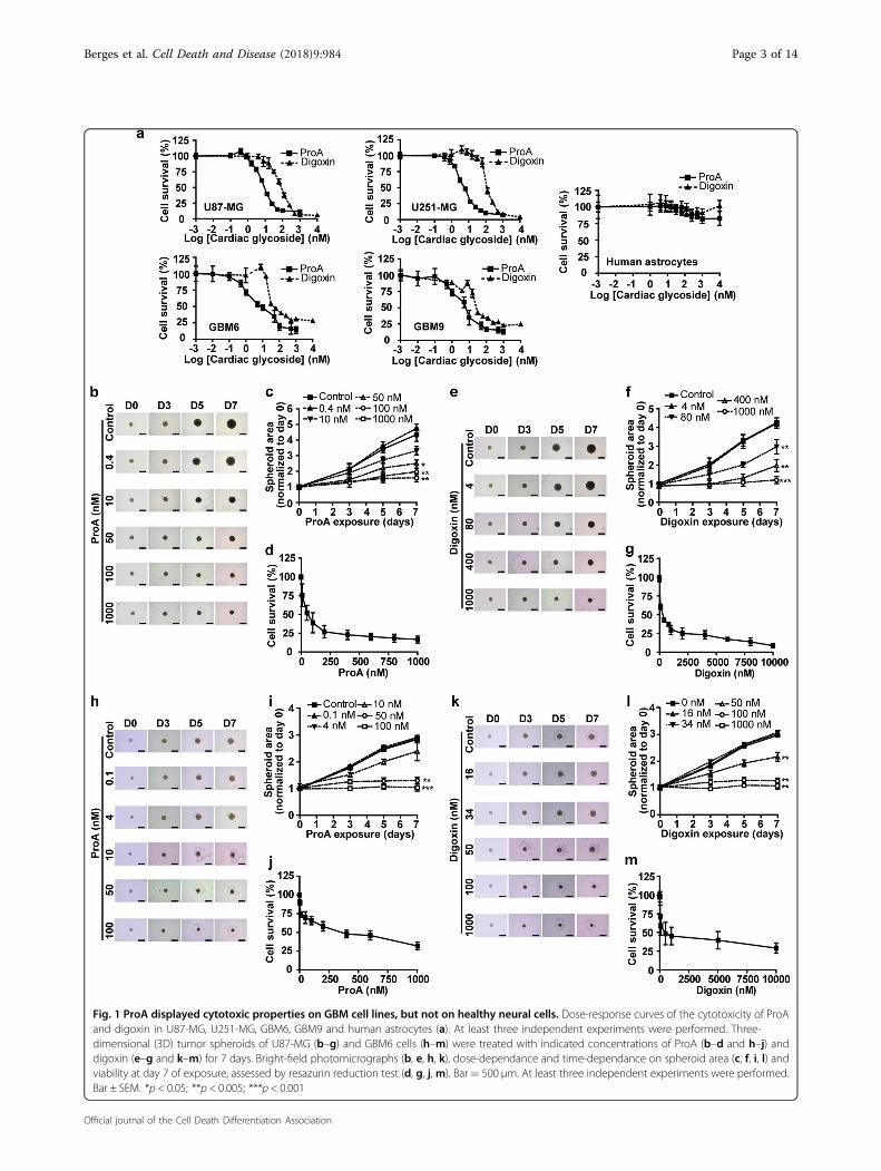
Fig. 1 ProA displayed cytotoxic properties on GBM cell lines, but not on healthy neural cells. Dose-response curves of the cytotoxicity of ProAand digoxin in U87-MG, U251-MG, GBM6, GBM9 and human astrocytes (a). At least three independent experiments were performed. Three-dimensional (3D) tumor spheroids of U87-MG (b–g) and GBM6 cells (h–m) were treated with indicated concentrations of ProA (b–d and h–j) anddigoxin (e–g and k–m) for 7 days. Bright-field photomicrographs (b, e, h, k), dose-dependance and time-dependance on spheroid area (c, f, i, l) andviability at day 7 of exposure, assessed by resazurin reduction test (d, g, j,m). Bar= 500 μm. At least three independent experiments were performed.Bar ± SEM. *p < 0.05; **p < 0.005; ***p < 0.001
Berges et al. Cell Death and Disease (2018) 9:984 Page 3 of 14
Official journal of the Cell Death Differentiation Association
ProA and digoxin decreased cell viability in a dose-dependent manner (Fig. 1d, g). The same dose-effects andtime-effects on spheroid areas and viability were obtainedusing spheroids produced from GBM6 cells (Fig. 1h–m).Together, these data highlight the potent cytotoxic effectof ProA and digoxin on GBM.
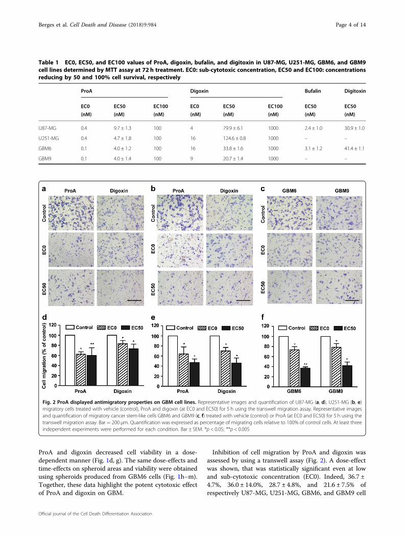
Inhibition of cell migration by ProA and digoxin wasassessed by using a transwell assay (Fig. 2). A dose-effectwas shown, that was statistically significant even at lowand sub-cytotoxic concentration (EC0). Indeed, 36.7 ±4.7%, 36.0 ± 14.0%, 28.7 ± 4.8%, and 21.6 ± 7.5% ofrespectively U87-MG, U251-MG, GBM6, and GBM9 cell
Table 1 EC0, EC50, and EC100 values of ProA, digoxin, bufalin, and digitoxin in U87-MG, U251-MG, GBM6, and GBM9cell lines determined by MTT assay at 72 h treatment. EC0: sub-cytotoxic concentration, EC50 and EC100: concentrationsreducing by 50 and 100% cell survival, respectively
ProA Digoxin Bufalin Digitoxin
EC0
(nM)
EC50
(nM)
EC100
(nM)
EC0
(nM)
EC50
(nM)
EC100
(nM)
EC50
(nM)
EC50
(nM)
U87-MG 0.4 9.7 ± 1.3 100 4 79.9 ± 6.1 1000 2.4 ± 1.0 30.9 ± 1.0
U251-MG 0.4 4.7 ± 1.8 100 16 124.6 ± 0.8 1000 – –
GBM6 0.1 4.0 ± 1.2 100 16 33.8 ± 1.6 1000 3.1 ± 1.2 41.4 ± 1.1
GBM9 0.1 4.0 ± 1.4 100 9 20.7 ± 1.4 1000 – –
Fig. 2 ProA displayed antimigratory properties on GBM cell lines. Representative images and quantification of U87-MG (a, d), U251-MG (b, e)migratory cells treated with vehicle (control), ProA and digoxin (at EC0 and EC50) for 5 h using the transwell migration assay. Representative imagesand quantification of migratory cancer stem-like cells GBM6 and GBM9 (c, f) treated with vehicle (control) or ProA (at EC0 and EC50) for 5 h using thetranswell migration assay. Bar= 200 μm. Quantification was expressed as percentage of migrating cells relative to 100% of control cells. At least threeindependent experiments were performed for each condition. Bar ± SEM. *p < 0.05; **p < 0.005
Berges et al. Cell Death and Disease (2018) 9:984 Page 4 of 14
Official journal of the Cell Death Differentiation Association
migration was inhibited at EC0. Similar effects wereobserved using digoxin at equipotent concentrations.
ProA inhibits EB1 accumulation at MT plus-ends andincreased distance-based catastrophe frequencyPreliminary experiments showed that ProA and digoxin
altered global MT cytoskeleton architecture by immuno-fluorescence staining of tubulin. At EC50, U87-MG orGBM6 cells lost their pronounced MT network whichbecame disrupted and disorganized, in comparison withcontrol untreated cells. Same results were observed withbufalin and digitoxin on U87-MG and GBM6 cells (Sup-plementary Fig. 2). Otherwise, MTAs are known to affectcell viability and migration via inhibition of EB1 accu-mulation at MT plus-ends and alteration of MT dynamicsinstability at low concentrations14,16,17. Here, we intendedto understand whether ProA and digoxin lead to suchprocesses. Then, we examined the intracellular localiza-tion of EB1, a +TIP that regulates MT dynamic instability.Immunofluorescence microscopy revealed a typical pat-tern of EB1 with comet-like structures at the plus-ends ofMTs in U87-MG and GBM6 control cells (Fig. 3a, c).Treatment with ProA at the sub-cytotoxic concentration(EC0) for 5 h significantly decreased the EB1 comet lengthfrom 1.35 ± 0.09 µm in U87-MG control cells to 0.90 ±0.05 (p < 0.005) after treatment, and from 1.67 ± 0.11 µmin GBM6 control cells to 1.16 ± 0.13 (p < 0.005) aftertreatment (Fig. 3b, d). Digoxin at equipotent concentra-tion also decreased EB1 comet length in both cell lines. Toevaluate the effect of CG on EB1 comet length and MTdynamics in cells, we used live imaging confocal micro-scopy to track all MT plus-ends positions in the cyto-plasm of U87-MG cells transfected with EB3-GFP afterProA and digoxin treatment (Fig. 3e–g). A strong increasewas reported in the distance-based catastrophe frequency(+50.1% and +83.5% for ProA and digoxin respectively,p < 0.001). Slight increase in the time-based catastrophefrequency was also observed which was statisticallysignificant due to the high number of analyzed tracks(more than 1 × 10⁴ per condition). This slight effect on thetime-based catastrophe frequency is explained by a verypoor or no effect on MT growth speed. These resultsindicate that ProA and digoxin, like MTAs, are able toalter EB comet length at growing ends of MTs, thusincreasing MT catastrophes in GBM cells.
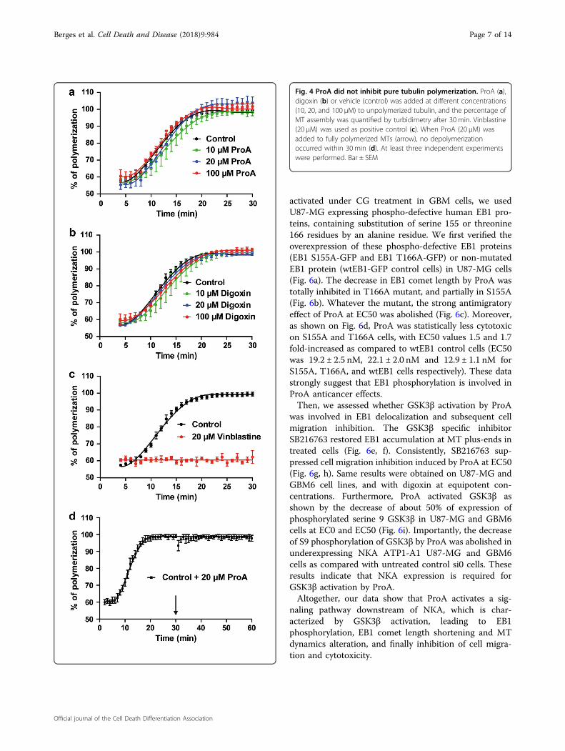
ProA does not inhibit pure tubulin polymerizationTo investigate whether ProA could directly affect MT
assembly and disassembly in a cell-free system, we testedthe effect of ProA on tubulin polymerization in vitro byturbidimetry assay (OD, 350 nm)18. As shown in Fig. 4,ProA and digoxin did not display any significant effect onMT assembly whatever the concentration tested (Fig. 4a, b).In contrast, vinblastine, a MTA used as positive control,
totally abolished polymerization at 20 µM (Fig. 4c). Fur-thermore, ProA when added to purified MTs under con-ditions favoring polymerization (37 °C and 1mM GTP)failed to disassembly MTs (Fig. 4d). Thus, the disruption ofthe MT system observed in GBM cells did not result froman effect on MT polymer mass.
NKA is required for the MT-dependent anticancer activityof Pro AConsequently, we asked whether NKA was needed to
the indirect effect of CG on MTs by using siRNA againstthe α1 isoform of NKA (ATP1-A1) which is over-expressed in GBM cells as compared with normal braintissue19. ATP1-A1 expression level (ratio to GAPDH) wasstrongly decreased in U87-MG-siATP1-A1 and in GBM6-siATP1-A1 cells (Fig. 5a) without consequence on EB1comet length and cell migration, as compared with U87-MG si0 and GBM6 si0 (p > 0.05) (Fig. 5c, d, white bars)and U87-MG and GBM6 wild-type (not shown),respectively.Interestingly, cytotoxic and antimigratory effects of
ProA are decreased in U87-MG-siATP1-A1 cells andGBM6-siATP1-A1 cells as compared to the respectivecontrol si0 cells. Indeed, in U87-MG, EC50 were 6.24 ±1.26 and 24.0 ± 1.51 nM for si0 and siATP1-A1 respec-tively and 3.28 ± 1.27 and 9.26 ± 1.16 nM for si0 andsiATP1-A1 respectively, in GBM6 (Fig. 5b). Furthermore,both the decrease in EB1 comet length (Fig. 5c) and in cellmigration (Fig. 5d) induced by ProA and digoxin at EC50were abolished in U87-MG and in GBM6 under expres-sing ATP1-A1 cells. These results allow us to concludethat the MT effects of CG required ATP1-A1.In cardiac myocytes, CG cause inhibition of NKA
ATP1-A1 pump, leading to increased intracellular Na+
and consequently an impairment of sodium-dependentcalcium transport out of cells1. Thus, we analyzed changesin 340/380 nm ratio of sodium indicator SBFI fluores-cence after drug treatment at EC0, EC50, and EC100 inU87-MG, U251-MG, and GBM6 cells. As shown inFig. 5e, ProA and digoxin activated Na+ influx in a dose-dependent manner, demonstrating that NKA was func-tional in GBM cells.
EB1 phosphorylation and GSK3β activation mediated byProA binding to NKA govern the reduction of EB1 cometlength and the inhibition of cell migrationConsequently, we hypothesize that low concentrations
of CG activate a signaling pathway that could be NKA-dependent, leading to disruption of MT functions. EBproteins are regulated by post-translational modifications.Indeed, phosphorylation on serine 155 and threonine 166of EB1 induced by GSK3β activation has been describedto modulate MT dynamics, EB1 comet length, and cellmigration20. To investigate whether this pathway is
Berges et al. Cell Death and Disease (2018) 9:984 Page 5 of 14
Official journal of the Cell Death Differentiation Association
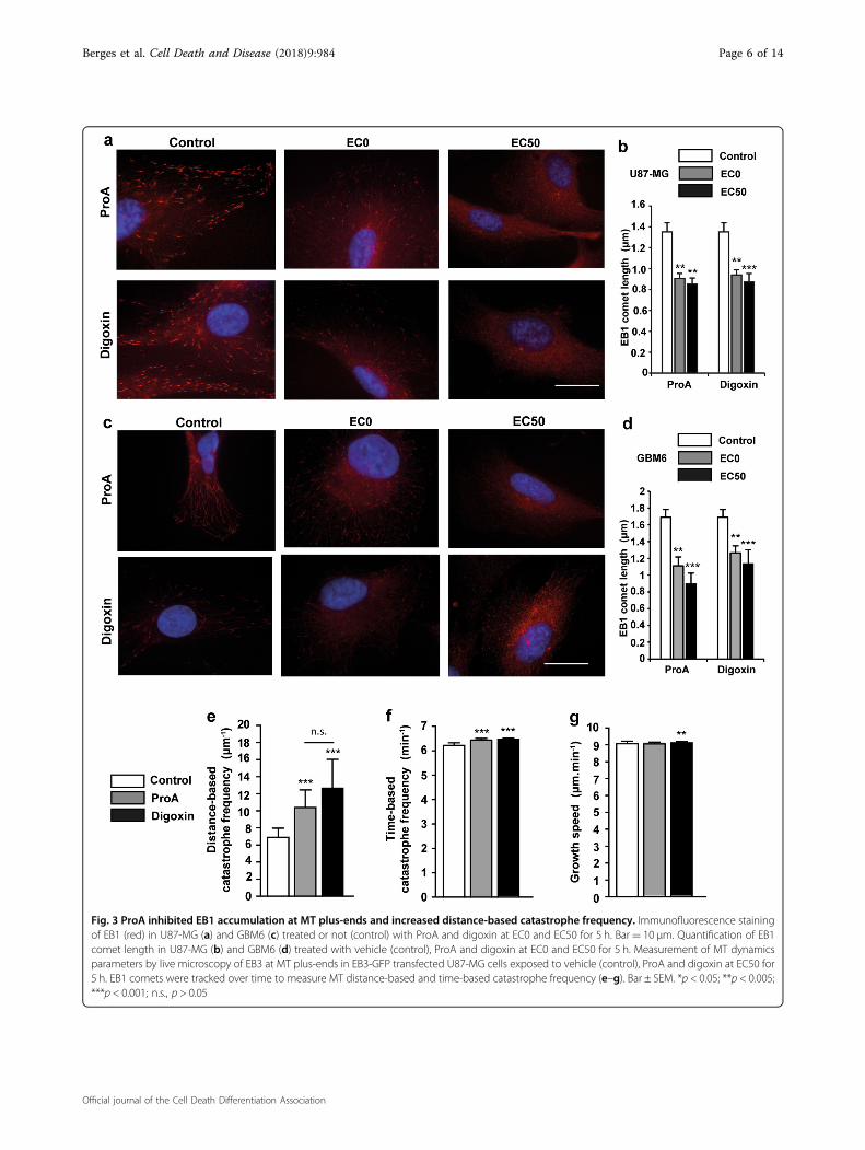
Fig. 3 ProA inhibited EB1 accumulation at MT plus-ends and increased distance-based catastrophe frequency. Immunofluorescence stainingof EB1 (red) in U87-MG (a) and GBM6 (c) treated or not (control) with ProA and digoxin at EC0 and EC50 for 5 h. Bar= 10 μm. Quantification of EB1comet length in U87-MG (b) and GBM6 (d) treated with vehicle (control), ProA and digoxin at EC0 and EC50 for 5 h. Measurement of MT dynamicsparameters by live microscopy of EB3 at MT plus-ends in EB3-GFP transfected U87-MG cells exposed to vehicle (control), ProA and digoxin at EC50 for5 h. EB1 comets were tracked over time to measure MT distance-based and time-based catastrophe frequency (e–g). Bar ± SEM. *p < 0.05; **p < 0.005;***p < 0.001; n.s., p > 0.05
Berges et al. Cell Death and Disease (2018) 9:984 Page 6 of 14
Official journal of the Cell Death Differentiation Association
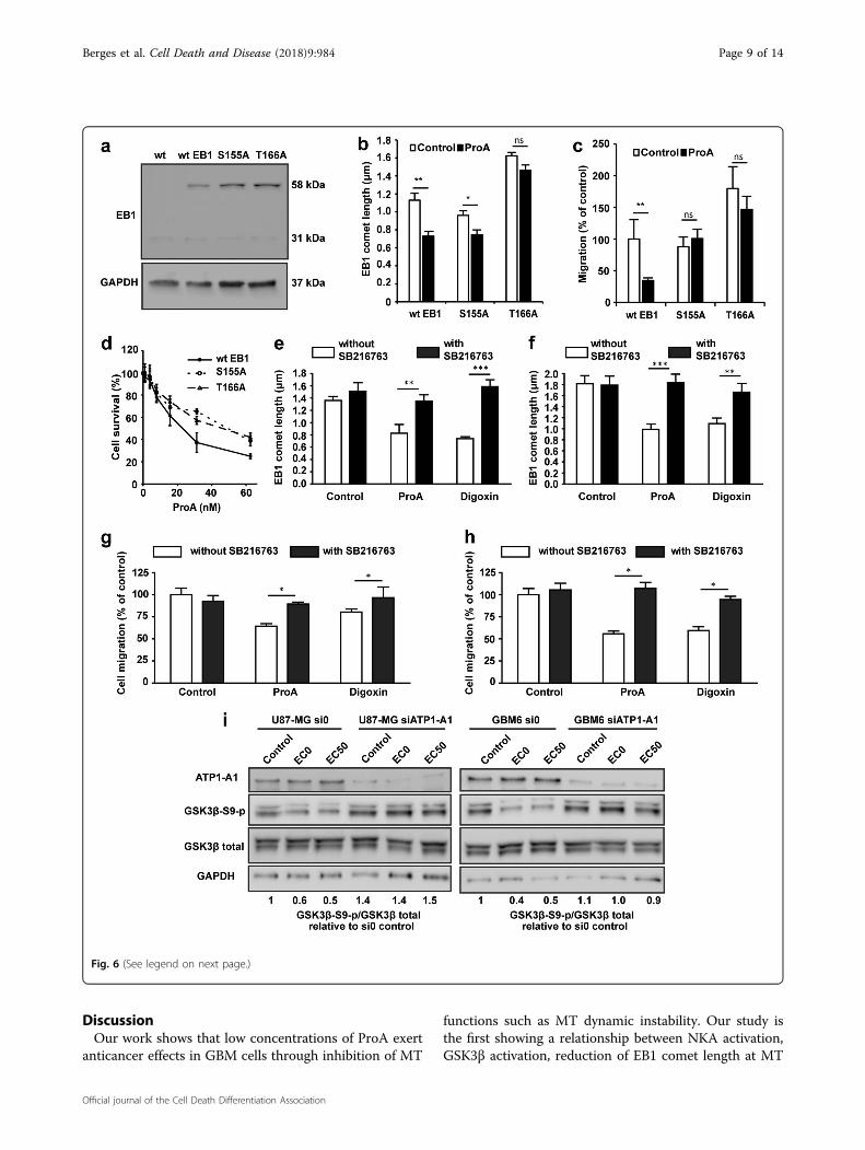
activated under CG treatment in GBM cells, we usedU87-MG expressing phospho-defective human EB1 pro-teins, containing substitution of serine 155 or threonine166 residues by an alanine residue. We first verified theoverexpression of these phospho-defective EB1 proteins(EB1 S155A-GFP and EB1 T166A-GFP) or non-mutatedEB1 protein (wtEB1-GFP control cells) in U87-MG cells(Fig. 6a). The decrease in EB1 comet length by ProA wastotally inhibited in T166A mutant, and partially in S155A(Fig. 6b). Whatever the mutant, the strong antimigratoryeffect of ProA at EC50 was abolished (Fig. 6c). Moreover,as shown on Fig. 6d, ProA was statistically less cytotoxicon S155A and T166A cells, with EC50 values 1.5 and 1.7fold-increased as compared to wtEB1 control cells (EC50was 19.2 ± 2.5 nM, 22.1 ± 2.0 nM and 12.9 ± 1.1 nM forS155A, T166A, and wtEB1 cells respectively). These datastrongly suggest that EB1 phosphorylation is involved inProA anticancer effects.Then, we assessed whether GSK3β activation by ProA
was involved in EB1 delocalization and subsequent cellmigration inhibition. The GSK3β specific inhibitorSB216763 restored EB1 accumulation at MT plus-ends intreated cells (Fig. 6e, f). Consistently, SB216763 sup-pressed cell migration inhibition induced by ProA at EC50(Fig. 6g, h). Same results were obtained on U87-MG andGBM6 cell lines, and with digoxin at equipotent con-centrations. Furthermore, ProA activated GSK3β asshown by the decrease of about 50% of expression ofphosphorylated serine 9 GSK3β in U87-MG and GBM6cells at EC0 and EC50 (Fig. 6i). Importantly, the decreaseof S9 phosphorylation of GSK3β by ProA was abolished inunderexpressing NKA ATP1-A1 U87-MG and GBM6cells as compared with untreated control si0 cells. Theseresults indicate that NKA expression is required forGSK3β activation by ProA.Altogether, our data show that ProA activates a sig-
naling pathway downstream of NKA, which is char-acterized by GSK3β activation, leading to EB1phosphorylation, EB1 comet length shortening and MTdynamics alteration, and finally inhibition of cell migra-tion and cytotoxicity.
Fig. 4 ProA did not inhibit pure tubulin polymerization. ProA (a),digoxin (b) or vehicle (control) was added at different concentrations(10, 20, and 100 µM) to unpolymerized tubulin, and the percentage ofMT assembly was quantified by turbidimetry after 30 min. Vinblastine(20 µM) was used as positive control (c). When ProA (20 µM) wasadded to fully polymerized MTs (arrow), no depolymerizationoccurred within 30 min (d). At least three independent experimentswere performed. Bar ± SEM
Berges et al. Cell Death and Disease (2018) 9:984 Page 7 of 14
Official journal of the Cell Death Differentiation Association
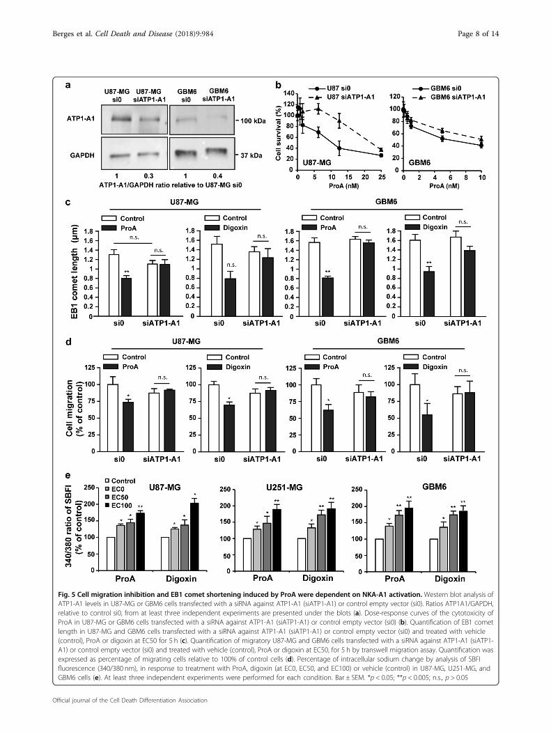
Fig. 5 Cell migration inhibition and EB1 comet shortening induced by ProA were dependent on NKA-A1 activation. Western blot analysis ofATP1-A1 levels in U87-MG or GBM6 cells transfected with a siRNA against ATP1-A1 (siATP1-A1) or control empty vector (si0). Ratios ATP1A1/GAPDH,relative to control si0, from at least three independent experiments are presented under the blots (a). Dose-response curves of the cytotoxicity ofProA in U87-MG or GBM6 cells transfected with a siRNA against ATP1-A1 (siATP1-A1) or control empty vector (si0) (b). Quantification of EB1 cometlength in U87-MG and GBM6 cells transfected with a siRNA against ATP1-A1 (siATP1-A1) or control empty vector (si0) and treated with vehicle(control), ProA or digoxin at EC50 for 5 h (c). Quantification of migratory U87-MG and GBM6 cells transfected with a siRNA against ATP1-A1 (siATP1-A1) or control empty vector (si0) and treated with vehicle (control), ProA or digoxin at EC50, for 5 h by transwell migration assay. Quantification wasexpressed as percentage of migrating cells relative to 100% of control cells (d). Percentage of intracellular sodium change by analysis of SBFIfluorescence (340/380 nm), in response to treatment with ProA, digoxin (at EC0, EC50, and EC100) or vehicle (control) in U87-MG, U251-MG, andGBM6 cells (e). At least three independent experiments were performed for each condition. Bar ± SEM. *p < 0.05; **p < 0.005; n.s., p > 0.05
Berges et al. Cell Death and Disease (2018) 9:984 Page 8 of 14
Official journal of the Cell Death Differentiation Association
DiscussionOur work shows that low concentrations of ProA exert
anticancer effects in GBM cells through inhibition of MT
functions such as MT dynamic instability. Our study isthe first showing a relationship between NKA activation,GSK3β activation, reduction of EB1 comet length at MT
Fig. 6 (See legend on next page.)
Berges et al. Cell Death and Disease (2018) 9:984 Page 9 of 14
Official journal of the Cell Death Differentiation Association
plus-end, and inhibition of GBM cell migration at sub-cytotoxic concentrations of ProA. Importantly, thismechanism of action is also demonstrated in cancer stem-like cells which are described to be at the origin of thetumor and to drive relapses due to their capacity of self-renewal and resistance to chemotherapy and radio-therapy21–24. Conversely, neural healthy cell lines such asoligodendrocytes and astrocytes were resistant to ProA.Drug specificity to cancer cells is essential for treatingbrain tumors, because maintaining a safe neurologicfunction is essential for quality of life.Alteration of MT network by CG has been described in
normal and cancer cell lines, mostly at high concentra-tions that induce G2/M arrest and apoptosis, but theunderlying mechanism is poorly understood. Digitoxinshowed a severely disruption and disorganized structureof MTs, and cell arrest in G2/M at concentrations thatinhibit growth of lung cancer cells25. On the contrary,bufalin-induced mitotic arrest has been attributed to thedownregulation of Plk1 expression, without alteration ofMT polymerization in HeLa cells26. Fridman et al.27
described a distorded MT cytoskeleton with clusters ofglycogen after treatment of human neuronal progenitorNT2 cells with bufalin, which results from a relocation ofMTs and glycogen in the cell. Our findings establish CGas MT-diruptor agents at nanomolar concentrations inGBM as well as in other solid tumor cell types such aslung, breast cancer and neuroblastoma cells (personaldata). Moreover, bufadienolides could be more potentthan cardenolides.Our study focused on low concentrations of ProA,
which is more realistic for clinical applications. We showthat ProA altered MT functions due to EB1 phosphor-ylation, subsequent inhibition of EB1 accumulation at MTplus-ends and alteration of MT dynamics. The reductionof EB1 comet length indicates a reduction of the EB1-stained MT-protective+ end cap, which results in anincrease in MT catastrophes and alteration of MTdynamics28,29. Interestingly, ProA modified MT distance-
based catastrophe frequency, which is the main MTdynamic instability parameter that correlates with theinhibition of MT targeting to the cell cortex and conse-quently the inhibition of cell migration17. EB1 binding toMT and comet length are regulated through post-translational modifications of EB proteins such as phos-phorylation15,30. Serine155 and Threonine166 are locatedin the EB1 linker region, which tightly contributes to itsMT binding by promoting the Calponin-Homologydomain binding31,32. Phosphorylation on the S155 resi-due is required for EB1 accumulation at MT plus-ends,and cancer cell migration. Phosphorylation of EB1 onT166 reduced comet length by decreasing MT growthrate and increased cancer cell migration. Here, we showthat the shortening of EB1 comet and inhibition of cellmigration by ProA were abolished in cells transfected withthe phospho-defective S155A and T166A mutants, indi-cating that phosphorylation of S155 and T166 are requi-site for antimigratory effect of ProA in cancer cells. EB1phosphorylation may result from the serine/threoninekinase GSK3β phosphorylation, leading to its activation,which is required for the antimigratory effect of ProA anddigoxin. GSK3 β activation by CG is downstream of NKAsince it is no more observed in cells underexpressingNKA-A1 pump. GSK3β activation by bufalin and otherCG such as ouabain and resibufogenin has been recentlyshown in liver, lung, and colon cell lines33–36.The ion pumping function of NKA in the anticancer
activity of CG is still under debate. Carr et al.37, haverecently shown that EB1 comet shortening induced byelectric fields was independent of calcium influx in U87-MG GBM cells. Conversely, elevation of transient spikesof calcium could activate calmodulin kinases, which inturn are known to affect GSK3β activity. At antimigratoryconcentrations, the potent anticancer MTAs also regulateMT dynamics by promoting GSK3β-mediated phos-phorylation of EB1 in several cell lines, including GBMcells. However, only phosphorylation of T166 is requiredfor MTA activity30. Moreover, MT dynamics alteration by
(see figure on previous page)Fig. 6 GSK3β was involved in the cell migration inhibition and EB1 comet shortening induced by ProA. Western blot analysis of EB1expression in wtEB1-GFP, EB1 S155A-GFP, and EB1 T166A-GFP transfected U87-MG cells. U87-MG wild type (wt) was used as a negative control. EB1expression analysis showed two protein bands for endogenous (31 kDa) and exogenous EB1-GFP (58 kDa) in the stably transfected cells. GAPDH wasused as loading control (a). Quantification of EB1 comet length in wtEB1-GFP, EB1 S155A-GFP, and EB1 T166A-GFP transfected U87-MG cells treatedwith vehicle (control) or ProA at EC50 for 5 h (b). Quantification of migratory wtEB1-GFP, EB1 S155A-GFP, and EB1 T166A-GFP transfected U87-MGcells treated with vehicle (control) or ProA at EC50 for 5 h (c). Survival of wtEB1-GFP, EB1 S155A-GFP and EB1 T166A-GFP transfected U87-MG cellsexposed to ProA, measured by the MTT test (d). Quantification of EB1 comet length in U87-MG (e) and GBM6 cells (f) treated or not with SB216763and concomitantly exposed or not (control) to ProA or digoxin at EC50 for 5 h. Quantification of migratory U87-MG (g) and GBM6 cells (h) treated ornot with SB216763 and concomitantly exposed or not (control) to ProA or digoxin at EC50 for 5 h, by transwell migration assay. Quantification wasexpressed as percentage of migrating cells relative to 100% of control cells. Western blot analysis of expression and activity of GSK3β under 5 h-treatment with ProA at EC0 or EC50 in U87-MG or GBM6 cells transfected with a siRNA against ATP1-A1 (siATP1-A1) or control empty vector (si0).Quantification of western blot bands was expressed as phospho/total GSK3β ratio. GAPDH was used as loading control (i). At least three independentexperiments were performed for each condition. Bar ± SEM. *p < 0.05; **p < 0.005; ***p < 0.001; n.s., p > 0.05
Berges et al. Cell Death and Disease (2018) 9:984 Page 10 of 14
Official journal of the Cell Death Differentiation Association
ProA did not originate from an alteration of MT polymermass contrary to classical MTAs. Some data suggest apotential link between NKA and tubulin. Indeed, tubulinin acetylated form interacts with plasma membrane NKA,resulting in inhibition of the enzyme activity, and detyr-osinated tubulin also play a role in regulation of theenzyme, suggesting that compounds that bind to tubulineither in dimer or polymeric form could alter NKAactivity38,39. Interestingly, paclitaxel, a well-known MTA,can inhibit in vitro the activity of the purified NKA in thesame range of concentrations as digitoxin. Moreover,paclitaxel and digitoxin synergize to inhibit proliferationof Her2 overexpressing breast cancer cells. However,cumulative cardiac side effects should be considered forthis drug combination40.In GBM, one of the most common found abnormalities
is the overexpression or aberrant activation of EGFR or itsconstitutively active mutant EGFRvIII41. In these tumors,the MAPK/Erk 1/2 and PI3K/Akt pathways have beenidentified as the driving forces of cellular proliferation andtumor progression. Activation of GSK3β by ProA may inpart counteracts EGFR signaling, and contribute to theanticancer effect in GBM. Digoxin and digitoxin thatbelong to the group of CG called cardenolides has shownsimilar effects to ProA and bufadienolide that belong tothe bufadienolide group, indicating that disturbance ofMTs may be a common biological effect of this class ofdrugs. Advantages of toxicity and pharmacokinetic pro-files may guide further clinical investigation.Taken together, our results sustain the anticancer
effects of ProA when administered by intra peritonealroute in mice orthotopically grafted with GBM and cancerstem-like cells8. CG at low concentrations, mimic theantimigratory and cytotoxic effects of MTAs althoughthey bind to NKA and not to tubulin. However, the use ofMTAs, which are very successful anti-cancer drugs inmany solid tumors42, is restricted for GBM treatment dueto the blood–brain barrier that blocks the crossing ofmost of the clinically relevant MTAs into the brain43. Inconclusion, our studies support CG as an alternativetreatment strategy and potent candidates for drug repo-sitioning in GBM.
Materials and methodsDrugsProA (Sigma-Aldrich, Saint-Quentin Fallavier, France),
digoxin (Sigma-Aldrich), bufalin (Merck, Fontenay-Sous-Bois, France) and digitoxin (Sigma-Aldrich) were dis-solved at 18.8, 12.8, 10 and 10mM respectively, in cellculture-grade dimethyl sulfoxide (DMSO) and stored at−20 °C until use. Vinblastine (Sigma-Aldrich) was solu-bilized in sterile distilled water. SB216763 (Sigma-Aldrich) was solubilized in DMSO. All these solutions
were freshly diluted in the culture medium forexperiments.
Cell culture and transfectionHuman GBM U87-MG, U251-MG cell lines and
C8D1A mice astrocytes were ordered from ATCC. Cellswere grown in DMEM media with glucose and L-glutamine (Lonza, Levallois-Perret, France), containing10% fetal calf serum (Lonza), 1% penicillin/streptomycin(Sigma-Aldrich). Normal human astrocytes were orderedfrom Lonza and were cultured in AGM BulletKit astro-cyte growth medium (Lonza). Oligodendroglial cell lineOLN-93 was obtained from the Richter-Landsberglaboratory. These proliferating cells were routinelymaintained as previously described44. Briefly, they weregrown in DMEM-High Glucose-GlutaMAX (Lonza)supplemented by pyruvate (Lonza), 1% penicillin andstreptomycin, 10% fetal bovine serum. GBM stem-like celllines GBM6 and GBM9 were isolated from GBM patientsand grown as previously described9. All cell types weretested weekly for the presence of mycoplasma, usingMycoalertTM Mycoplasma Detection Kit (Lonza). StableU87-MG clones overexpressing phospho-defective EB1proteins (EB1-S155A-GFP and EB1-T166A-GFP U87-MGcells) or non-mutated EB1 proteins (wtEB1-GFP U87-MGcells) were generated as previously described30. ForATP1-A1 silencing by transient transfection, cells weretransfected by lipofectamine 2000 system with siRNA forATP1-A1 (Silencer Select siRNAs s1720, Thermo FisherScientific, NY, USA and Silencer Select negative control,Thermo Fisher Scientific). ATP1-A1 down-regulation wasanalyzed 72 h later by western blotting.For analysis of MT dynamics, U87-MG cells (5 × 10³
cells/well) were grown for 24 h on 4-well Lab-tekChambered Coverglass (Thermo Fisher Scientific). U87-MG cells were then transfected with plasmid coding forgreen fluorescent protein EB3-GFP using lipofectamineTM
2000 system (Invitrogen, Villebon-sur-Yvette, France)16.
Indirect immunofluorescence analysisCells were grown on 8-well chamber slides (Labtek,
Thermo Fisher Scientific), precoated for 1 h with fibro-nectin (10 μg/ml) for U87-MG or with poly-DL-ornithine(Sigma-Aldrich) (10 µg/ml) for GBM6, to be treated for 6 hwith ProA, digoxin, bufalin or digitoxin. As previouslydescribed16, cells were incubated with the anti-EB1 (clone 5;BD Biosciences, San Jose, CA) and α-tubulin (clone DM1A;Sigma-Aldrich) primary antibodies, and then with Alexa488or 568-conjugated secondary antibodies (Invitrogen).Staining was observed using either a Leica DM-IRBEmicroscope or a Leica TCS SP5 confocal laser-scanningmicroscope (Leica, Heidelberg, Germany). Images wereacquired using Metamoph software or the Leica Confocal
Berges et al. Cell Death and Disease (2018) 9:984 Page 11 of 14
Official journal of the Cell Death Differentiation Association
software, and were processed using Image J software. Foreach experimental condition, at least 100 EB1 comets (in atleast 10 cells) were examined to measure their length.
Cytotoxicity assayFor 2D experiments, cells (5000 cells/well) were directly
seeded in 96-well plates and allowed to grow for 24 hbefore treatment with ProA, digoxin, bufalin or digitoxin.GBM6 and GBM9 were seeded on poly-DL-ornithine(Sigma-Aldrich) coated 96-well plates (10 µg/mL).Growth inhibition of cells was measured after 72 h oftreatment by using the colorimetric MTT assay (Sigma-Aldrich). Cell survival was expressed as a percentage ascompared to DMSO control cells as previouslydescribed14.To obtain U87–MG and GBM6 3D cell culture in a
form of spheroids, cells growing in regular 2D cultureswere rinsed once in phosphate buffered saline, thendetached using 0.02% Trypsin/EDTA (Gibco, UK), pel-leted by centrifugation at 200×g for 5 min and finallyresuspended in a fresh culture medium supplementedwith 20% methylcellulose and seeded on round-bottom96-well plates (Greiner Bio-one, Courtaboeuf, France) at1000 cells per well in 100 µL of medium as describedpreviously45. After 72 h of growth at 37 ◦C under 5% CO2,spheroids were formed and 100 µL of fresh mediumcontaining different amount of drugs with final con-centrations from 0.4 to 10,000 nM were added to wells.Spheroid growth was maintained for 7 days by adding10 µL of fresh medium per well three times a week andwas monitored daily by measuring spheroid area onbright-field photomicrographs (Eclipse Ts2-FL, Nikon).All images were segmented using a custom macro script,written for ImageJ software. At day 7 of drug exposure,cell viability was assessed by resazurin reduction: 20 µl ofresazurin reagent (alamarBlue, Thermo Fisher Scientific)were added to each well, and incubated for 18 h beforefluorescence was read on a microplate reader (POLARstarOmega, BMG LABTECH) according to the manu-facturer’s instructions. After subtraction of the fluores-cence signal given by blank wells containing no cells,viability was calculated as a percentage of fluorescenceobserved in control wells. These experiments were repe-ated three times independently, with four wells percondition.
Protein extraction and western blot analysisCells were lysed after 6 h treatment in RIPA buffer
(Tris-HCl 50mM pH 8.0, NaCl 250mM, Triton-X1000.1%) with a cocktail of proteases and phosphatasesinhibitors (Sigma-Aldrich) added freshly. Protein con-centrations were determined using the Bio-Rad ProteinAssay (Bio-Rad laboratories, Marnes-la-Coquette,France). Thirty micrograms of total proteins were loaded
into a 12% SDS-PAGE gel and electrotransferred onto anitrocellulose membrane. Primary antibodies used weredirected against EB1 (clone 5; BD Biosciences, San Jose,CA), ATP1-A1 (Proteintech Europe, Manchester, UK),Ser 9 phospho-GSK3β (Cell Signaling, Boston, USA), totalGSK3β (Thermo Fisher Scientific) and GAPDH (Sigma-Aldrich). Peroxydase-conjugated secondary antibodies(Jackson Immunoresearch, Baltimore, USA) and chemi-luminescence detection kit (Millipore, Molsheim, France)was used for visualization of protein bands. Chemilumi-nescent signal was acquired on a G:BOX imaging system(Syngene, Cambridge, UK) and quantification was donewith Image J software.
Transwell migration assayCells (5 × 10⁴ cells/well) were poured on the upper side
of transwell migration chamber (Becton Dickinson, LePont de Claix, France) and allowed migrating for 5 h aspreviously described16. Six fields per condition wereimaged and transmigrated cells were counted. Resultswere expressed as percent of transmigrated cells com-pared with no treatment condition. At least three inde-pendent experiments were performed for each condition.
Analysis of MT dynamics from EB3-GFP comet dataBefore time-lapse microscopy analysis, U87-MG-EB3-
GFP cells were incubated with EC50 concentration ofProA or digoxin for 5 h. Following drug incubation, cellswere maintained at 37 °C and placed in routine culturemedium supplemented with 25 mM HEPES to reducemedium pH acidity during time-lapse analysis.Time-lapse microscopy and image acquisition for MT
dynamics experiments were performed with a LEICA TCSSP5 confocal microscope equipped with a 63X objectivelens. Forty-five images per cell were acquired at 2-sintervals using a digital camera (CCD camera Hama-matsu) driven by LEICA LAS AF software.Analysis of MT dynamic instability was done by track-
ing plus-ends EB3-GFP comets of individual MTs usingthe u-track 2.2.1 package (multiple-particle trackingMATLAB software)46–48. The software package can bedownloaded from http://www.utsouthwestern.edu/labs/danuser/software/#utrack_anc. Comet detection requiresno user intervention, as the detection algorithm auto-matically estimates locally optimal thresholds. Trackingand inference of complete MT trajectories by u-trackrequires user-defined settings of several control para-meters (Movie information: Pixel size= 160 nm; Numer-ical aperture= 1,4; Camera bit depth= 8-bit; Timeinterval= 2 s. Comet detection: Watershed parameters= 3(minimum threshold) and 1 (threshold step size) standarddeviations; Difference of Gaussions filter parameters= 1(low-pass gaussian standard deviation) and 4 (high-pass)pixels. Tracking: Maximum gap to close= 15 frames;
Berges et al. Cell Death and Disease (2018) 9:984 Page 12 of 14
Official journal of the Cell Death Differentiation Association
Minimum length of track segments from first step= 4frames; Maximum forward-backward angles= 30°−10°;Maximum shrinkage factor= 3; Fluctuation radius= 2pixels; Tracking search radius range= 2–10 pixels. Trackanalysis: Forward gaps= unimodal thresholding; Back-ward= unimodal thresholding with comet latencycorrection.)Correct tracking was verified by visual inspection of
several overlay movies. The parameter settings were keptidentical for all movies. We chose 5 metrics for com-parative analyzes: growth speed, growth length, growthtime, distance-based catastrophe frequency, and time-based catastrophe frequency. Importantly, distance andtime-based catastrophe frequencies were calculated as themean of the inverse growth length and growth time ofeach track. For each experimental condition (Control,ProA and digoxin), 14 to 16 cells were analyzed(10,000–13,000 comet tracks per condition).All tracks were included in analysis of MT dynamics.
We chose to keep in growth metrics the phases of atte-nuated dynamics as the resolution of the microscope andthe automated tracking process allowed it. These phasesare characterized by decreased growth length/speed andincreased distance-based catastrophe frequencies.
Tubulin polymerization assayTubulin was extracted and purified from lamb brains by
Weisenberg procedure consisting in ammonium sulfatefractionation and ion exchange chromatography49,50. MTswere assembled by mixing 20 µM tubulin solution, 1 mMguanosine 5′-triphosphate (GTP), 4 mM MgCl2, 1 mMEthylene Glycol Tetra-acetic Acid (EGTA), 2% DMSO in80mM PIPES-KOH buffer pH 6.8 and by raising thetemperature to 37 °C. Polymerization of tubulin, whichproduced an increase of the optical density at 350 nm, wasmonitored by turbidimetry at 1 min intervals using aspectrophotometer (Polarstar omega, BMG Labtech)51.
Intracellular free sodium analysisU87-MG and U251-MG were seeded at 3 × 10⁵ cells/
well on 6-well plates. GBM6 were seeded at the samedensity on poly-DL-ornithine (Sigma-Aldrich) coated 6-well plates (10 µg/mL). One day later, cells were treatedwith ProA or digoxin for 24 h. Then, cells were loadedwith 5 µM sodium sensitive dye SBFI (Interchim, Mon-tluçon, France). The incubation continued for 1 h at 37 °C.Then, cells were trypsinized and washed in fresh culturemedium. After centrifugation (1200 rpm), cell pellets wereresuspended in PBS and used within the next 2–4 h.Fluorescence was recorded from 1mL aliquots of mag-netically stirred cellular suspension at 37 °C using a Shi-madzu spectrofluorophotometer with excitationwavelengths of 340 and 380 nm and emission at 505 nm.Changes in [Na+] were monitored by using the 340/380
nm fluorescence ratio according to the method ofGrynkiewicz52.
Statistical analysisEach experiment was performed at least in triplicate.
Data are presented as mean ± SEM. Cell counting, cellularviability data were analyzed by Student’s t test. Dynamicinstability data were analyzed with the non-parametricMann–Whitney test. Reported p-values are two-sided, andp < 0.05 was considered statistically significant. Asterisksindicate significant level vs control *p < 0.05; **p < 0.005;***p < 0.001. Statistical analyses were performed withGraphPad 5.0 statistical software.
AcknowledgementsThis work has been carried out thanks to the support of the DGOS (SIRIC label)INCa-DGOS Inserm no. 6038, the ITMO Cancer AVIESAN as part of the CancerPlan no. PC201419 and ARTC Sud. The authors wish to thank M. Clerout, Dr. S.Oddoux, and Dr. M. Carré for their technical help.
Conflict of interestThe authors declare that they have no conflict of interest.
Publisher's noteSpringer Nature remains neutral with regard to jurisdictional claims inpublished maps and institutional affiliations.
Supplementary Information accompanies this paper at (https://doi.org/10.1038/s41419-018-1018-7).
Received: 23 April 2018 Revised: 26 July 2018 Accepted: 2 August 2018
References
1. Prassas, I. & Diamandis, E. P. Novel therapeutic applications of cardiac glyco-sides. Nat. Rev. Drug. Discov. 7, 926–935 (2008).
2. Stenkvist, B. et al. Cardiac glycosides and breast cancer. Lancet Lond. Engl. 1,563 (1979).
3. Stenkvist, B. Is digitalis a therapy for breast carcinoma? Oncol. Rep. 6, 493–496(1999).
4. Diederich, M., Muller, F. & Cerella, C. Cardiac glycosides: from molecular targetsto immunogenic cell death. Biochem. Pharmacol. 125, 1–11 (2017).
5. Wolle, D. et al. Inhibition of epidermal growth factor signaling by the cardiacglycoside ouabain in medulloblastoma. Cancer Med. 3, 1146–1158 (2014).
6. McConkey, D. J., Lin, Y., Nutt, L. K., Ozel, H. Z. & Newman, R. A. Cardiacglycosides stimulate Ca2+ increases and apoptosis in androgen-independent,metastatic human prostate adenocarcinoma cells. Cancer Res. 60, 3807–3812(2000).
7. Trenti, A. et al. Therapeutic concentrations of digitoxin inhibit endothelial focaladhesion kinase and angiogenesis induced by different growth factors. Br. J.Pharmacol. 174, 3094–3106 (2017).
8. Denicolaï, E. et al. Proscillaridin A is cytotoxic for glioblastoma cell linesand controls tumor xenograft growth in vivo. Oncotarget 5, 10934–10948(2014).
9. Tchoghandjian, A. et al. A2B5 cells from human glioblastoma have cancerstem cell properties. Brain Pathol. Zur. Switz. 20, 211–221 (2010).
10. Tchoghandjian, A. et al. Cortical and subventricular zone glioblastoma-derivedstem-like cells display different molecular profiles and differential in vitro andin vivo properties. Ann. Surg. Oncol. 19(Suppl 3), S608–S619 (2012).
11. Honore, S., Pasquier, E. & Braguer, D. Understanding microtubule dynamics forimproved cancer therapy. Cell. Mol. Life Sci. 62, 3039–3056 (2005).
12. Mitchison, T. & Kirschner, M. Dynamic instability of microtubule growth. Nature312, 237–242 (1984).
Berges et al. Cell Death and Disease (2018) 9:984 Page 13 of 14
Official journal of the Cell Death Differentiation Association
13. Galjart, N. Plus-end-tracking proteins and their interactions at microtubuleends. Curr. Biol. 20, R528–R537 (2010).
14. Pagano, A. et al. Epothilone B inhibits migration of glioblastoma cells byinducing microtubule catastrophes and affecting EB1 accumulation atmicrotubule plus ends. Biochem. Pharmacol. 84, 432–443 (2012).
15. Rovini, A. et al. Anti-migratory effect of vinflunine in endothelial and glio-blastoma cells is associated with changes in EB1 C-terminal detyrosinated/tyrosinated status. PloS ONE 8, e65694 (2013).
16. Berges, R. et al. End-binding 1 protein overexpression correlates with glio-blastoma progression and sensitizes to Vinca-alkaloids in vitro and in vivo.Oncotarget 5, 12769–12787 (2014).
17. Honoré, S. et al. Antiangiogenic vinflunine affects EB1 localization andmicrotubule targeting to adhesion sites.Mol. Cancer Ther. 7, 2080–2089 (2008).
18. Timasheff, S. N. & Grisham, L. M. In vitro assembly of cytoplasmic microtubules.Annu. Rev. Biochem. 49, 565–591 (1980).
19. Lefranc, F., Mijatovic, T. & Kiss, R. The sodium pump could constitute a newtarget to combat glioblastomas. Bull. Cancer 95, 271–281 (2008).
20. Nehlig, A., Molina, A., Rodrigues-Ferreira, S., Honoré, S. & Nahmias, C. Regula-tion of end-binding protein EB1 in the control of microtubule dynamics. CellMol. Life Sci. 74, 2381–2393 (2017).
21. Binello, E. & Germano, I. M. Targeting glioma stem cells: a novel framework forbrain tumors. Cancer Sci. 102, 1958–1966 (2011).
22. Cheng, L., Bao, S. & Rich, J. N. Potential therapeutic implications of cancer stemcells in glioblastoma. Biochem. Pharmacol. 80, 654–665 (2010).
23. Tabatabai, G. & Weller, M. Glioblastoma stem cells. Cell Tissue Res. 343, 459–465(2011).
24. Venere, M., Fine, H. A., Dirks, P. B. & Rich, J. N. Cancer stem cells in gliomas:identifying and understanding the apex cell in cancer’s hierarchy. Glia 59,1148–1154 (2011).
25. Zhang, Y.-Z. et al. Compound library screening identified cardiac glycosidedigitoxin as an effective growth inhibitor of gefitinib-resistant non-small celllung cancer via downregulation of α-tubulin and inhibition of microtubuleformation. Mol. Basel Switz. 21, 374 (2016).
26. Xie, C.-M., Liu, X.-Y., Yu, S. & Cheng, C. H. K. Cardiac glycosides block cancergrowth through HIF-1α- and NF-κB-mediated Plk1. Carcinogenesis 34,1870–1880 (2013).
27. Fridman, E., Lichtstein, D. & Rosen, H. Formation of new high densityglycogen-microtubule structures is induced by cardiac steroids. J. Biol. Chem.287, 6518–6529 (2012).
28. Barlukova, A., White, D., Henry, G., Honoré, S. & Hubert, F. Mathematicalmodeling of microtubule dynamic instability: new insight into the linkbetween GTP-hydrolysis and microtubule aging. Preprint at https://www.esaim-m2an.org/component/forthcoming/ (2017).
29. White, D., Honoré, S. & Hubert, F. Exploring the effect of end-binding proteinsand microtubule targeting chemotherapy drugs on microtubule dynamicinstability. J. Theor. Biol. 429, 18–34 (2017).
30. Le Grand, M. et al. ROS-mediated EB1 phosphorylation through Akt/GSK3βpathway: implication in cancer cell response to microtubule-targeting agents.Oncotarget 5, 3408–3423 (2014).
31. Akhmanova, A. & Steinmetz, M. O. Tracking the ends: a dynamic proteinnetwork controls the fate of microtubule tips. Nat. Rev. Mol. Cell Biol. 9,309–322 (2008).
32. Komarova, Y. et al. Mammalian end binding proteins control persistentmicrotubule growth. J. Cell Biol. 184, 691–706 (2009).
33. Kang, X.-H. et al. Degradation of Mcl-1 through GSK-3β Activation RegulatesApoptosis Induced by Bufalin in Non-Small Cell Lung Cancer H1975 Cells. CellPhysiol. Biochem. Int. J. Exp. Cell Physiol. Biochem. Pharmacol. 41, 2067–2076(2017).
34. Liu, M. et al. Knockdown of apolipoprotein E enhanced sensitivity of Hep3Bcells to cardiac steroids via regulating Na+/K+-ATPase signalosome. Mol.Cancer Ther. 15, 2955–2965 (2016).
35. Gai, J. Q. et al. The effect and mechanism of bufalin on regulating hepato-cellular carcinoma cell invasion and metastasis via Wnt/β-catenin signalingpathway. Int. J. Oncol. 48, 338–348 (2016).
36. Ichikawa, M., Sowa, Y., Iizumi, Y., Aono, Y. & Sakai, T. Resibufogenin Induces G1-Phase Arrest through the Proteasomal Degradation of Cyclin D1 in HumanMalignant Tumor Cells. PLoS ONE 10, e0129851 (2015).
37. Carr, L. et al. Calcium-independent disruption of microtubule dynamics bynanosecond pulsed electric fields in U87 human glioblastoma cells. Sci. Rep. 7,41267 (2017).
38. Santander, V. S. et al. Tubulin must be acetylated in order to form a complexwith membrane Na(+),K (+)-ATPase and to inhibit its enzyme activity. Mol.Cell. Biochem. 291, 167–174 (2006).
39. Amaiden, M. R. et al. Effects of detyrosinated tubulin on Na+,K+-ATPaseactivity and erythrocyte function in hypertensive subjects. FEBS Lett. 589,364–373 (2015).
40. Einbond, L. S. et al. Digitoxin activates EGR1 and synergizes with paclitaxel onhuman breast cancer cells. J. Carcinog. 9, 10 (2010).
41. Wong, A. J. et al. Structural alterations of the epidermal growth factorreceptor gene in human gliomas. Proc. Natal Acad. Sci. USA 89, 2965–2969(1992).
42. Jordan, M. A. & Wilson, L. Microtubules as a target for anticancer drugs. Nat.Rev. Cancer 4, 253–265 (2004).
43. Nathanson, D. & Mischel, P. S. Charting the course across the blood-brainbarrier. J. Clin. Invest. 121, 31–33 (2011).
44. Richter-Landsberg, C. & Heinrich, M. OLN-93: a new permanent oligoden-droglia cell line derived from primary rat brain glial cultures. J. Neurosci. Res. 45,161–173 (1996).
45. Correard, F. et al. Delaying Anticancer Drug Delivery by Self-Assembly andBranching Effects of Minimalist Dendron-Drug Conjugates. Chem. Weinh.Bergstr. Ger. (2018).
46. Jaqaman, K. et al. Robust single particle tracking in live cell time-lapsesequences. Nat. Methods 5, 695–702 (2008).
47. Applegate, K. T. et al. plusTipTracker: Quantitative image analysis software forthe measurement of microtubule dynamics. J. Struct. Biol. 176, 168–184 (2011).
48. Stout, A., D’Amico, S., Enzenbacher, T., Ebbert, P. & Lowery, L. A. Using plus-TipTracker software to measure microtubule dynamics in Xenopus laevisgrowth cones. J. Vis. Exp. e52138, 1–9 (2014).
49. Weisenberg, R. C., Borisy, G. G. & Taylor, E. W. The colchicine-binding protein ofmammalian brain and its relation to microtubules. Biochem. 7, 4466–4479(1968).
50. Lee, J. C., Frigon, R. P. & Timasheff, S. N. The chemical characterization of calfbrain microtubule protein subunits. J. Biol. Chem. 248, 7253–7262 (1973).
51. Di Maïo, I. L., Barbier, P., Allegro, D., Brault, C. & Peyrot, V. Quantitative analysis oftau-microtubule interaction using FRET. Int. J. Mol. Sci. 15, 14697–14714 (2014).
52. Grynkiewicz, G., Poenie, M. & Tsien, R. Y. A new generation of Ca2+ indicatorswith greatly improved fluorescence properties. J. Biol. Chem. 260, 3440–3450(1985).
Berges et al. Cell Death and Disease (2018) 9:984 Page 14 of 14
Official journal of the Cell Death Differentiation Association