PROMOTION AND S T β-L A C DEPENDENT M - airccj.orgairccj.org/CSCP/vol3/csit3242.pdf · Solution...
15
Rupak Bhattacharyya et al. (Eds) : ACER 2013, pp. 445–459, 2013. © CS & IT-CSCP 2013 DOI : 10.5121/csit.2013.3242 PROMOTION AND SUPPRESSION OF THERMAL AGGREGATION OFβ-LACTOGLOBULIN BY ARGININE: A CONCENTRATION DEPENDENT MECHANISM Jishnu Chakraborty 1 and Umesh Chandra Halder 2 1 Department of Chemistry, Camellia Institute of Engineering and Technology, Budbud, Burdwan, West Bengal, India [email protected] 2 Protein Chemistry Laboratory, Department of Chemistry, Jadavpur University, 188, Raja S.C. MullikRoad,Kolkata, West Bengal, India [email protected] ABSTRACT Bovine β-lactoglobulin (β-lg), consisting of pronounced β-sheet content, have been chosen as a model protein which on prolonged thermal treatment forms large molecular aggregates similar to Alzheimer’s type amyloid fibrils. The effects of L-arginine (free base) in thermal aggregation process of β-lg were monitored at varying concentrations. Concentration dependent opposite behaviour has been reported here for the first time where 0.2-0.3 M concentration was optimized as an apparent critical concentration above which arginine acts as a suppressor and at below it behaves as a promoter of aggregation of β-lg. Solubility study and SDS-PAGE pattern followed by densitometric analysis shows this fact. Solution behaviour of arginine and its self assemblyformation were evidenced with the help of circular dichroism (CD) studies. The delocalized pi-pi (п→п) type of interaction is proposed to foster the energy stabilization during the attainment of planarity of the molecules accompanied with the self-clustering of arginine molecules. KEYWORDS Protein, Aggregation, Arginine, Aggregation & Suppression 1. INTRODUCTION L-Arginine (Arg) is one of the most commonly used suppressors of aggregation of protein and hormones in vitro [1-3]. Its general acceptability as a suppressor of protein aggregates is well known, but the mechanism of its action in molecular detail remains elusive in spite of having a series of progress with Arg and its derivatives also [4]. The effect of Arg on the in vitro refolding of protein was discovered serendipitously by a group of authors [5]. Interestingly L-arginine and its derivatives were widely used in the industrial production process for plasminogen activator and continued to be marketed as bio-pharmaceuticals to this days also. Arginine exhibited the best results in preventing the formation of aggregates of lysozyme as a model protein under
Transcript of PROMOTION AND S T β-L A C DEPENDENT M - airccj.orgairccj.org/CSCP/vol3/csit3242.pdf · Solution...
Rupak Bhattacharyya et al. (Eds) : ACER 2013,
pp. 445–459, 2013. © CS & IT-CSCP 2013 DOI : 10.5121/csit.2013.3242
PROMOTION AND SUPPRESSION OF THERMAL AGGREGATION OFβ-LACTOGLOBULIN BY
ARGININE: A CONCENTRATION DEPENDENT MECHANISM
Jishnu Chakraborty1 and Umesh Chandra Halder
2
1Department of Chemistry, Camellia Institute of Engineering and Technology,
Budbud, Burdwan, West Bengal, India [email protected]
2 Protein Chemistry Laboratory, Department of Chemistry, Jadavpur University,
188, Raja S.C. MullikRoad,Kolkata, West Bengal, India [email protected]
ABSTRACT
Bovine β-lactoglobulin (β-lg), consisting of pronounced β-sheet content, have been chosen as a
model protein which on prolonged thermal treatment forms large molecular aggregates similar
to Alzheimer’s type amyloid fibrils. The effects of L-arginine (free base) in thermal aggregation
process of β-lg were monitored at varying concentrations. Concentration dependent opposite
behaviour has been reported here for the first time where 0.2-0.3 M concentration was
optimized as an apparent critical concentration above which arginine acts as a suppressor and
at below it behaves as a promoter of aggregation of β-lg. Solubility study and SDS-PAGE
pattern followed by densitometric analysis shows this fact. Solution behaviour of arginine and
its self assemblyformation were evidenced with the help of circular dichroism (CD) studies. The
delocalized pi-pi (п→п) type of interaction is proposed to foster the energy stabilization during
the attainment of planarity of the molecules accompanied with the self-clustering of arginine
molecules.
KEYWORDS
Protein, Aggregation, Arginine, Aggregation & Suppression
1. INTRODUCTION
L-Arginine (Arg) is one of the most commonly used suppressors of aggregation of protein and
hormones in vitro [1-3]. Its general acceptability as a suppressor of protein aggregates is well
known, but the mechanism of its action in molecular detail remains elusive in spite of having a
series of progress with Arg and its derivatives also [4]. The effect of Arg on the in vitro refolding
of protein was discovered serendipitously by a group of authors [5]. Interestingly L-arginine and
its derivatives were widely used in the industrial production process for plasminogen activator
and continued to be marketed as bio-pharmaceuticals to this days also. Arginine exhibited the
best results in preventing the formation of aggregates of lysozyme as a model protein under
446 Computer Science & Information Technology (CS & IT)
thermal stress and refolding pathway from previously denatured state in comparison to other 14
amino acids [1]. The preventive actions of Arg have been also reported by Shiraki and coworkers
with other eight kinds of proteins. It was stated that the suppression effect was independent of
size and/or iso-electric point of the proteins [1]. Nonetheless, one novel derivative, arginine ethyl
ester significantly prevents thermal inactivation and aggregation of lysozyme whereas only Arg
and guanidine (Gdn.HCl) was less effective in case of heat labile proteins [6]. Though Gdn.HCl
and urea are well established as aggregation suppressor that weaken the hydrophobic
intermolecular interactions of proteins [7-8]. Arginine does not facilitate refolding, but suppresses
aggregation, with only minor effect on protein stability [9], while the solubility of aggregate –
prone molecules is enhanced. Other additives like, proline, glycerol, glycine and ethylene glycol
have also been reported which are not enough to eliminate the problems of protein aggregation
and mis-folding [8]. Recently it was also reported that di-amines [10], and poly-amines typically
spermine and spermidine, prevent heat-induced inactivation and aggregation of lysozyme [11-12].
Different mechanisms have been proposed towards the understanding of the preventive role of
Arg in protein aggregation. The influence of co-solvent additives to the aqueous protein solution
may in many cases be rationalized by the dichotomy of solubilizationvs stabilization or salting-
out and salting-in. Solubilising agents often destabilize the native conformations in solution while
stabilizing agents reduce protein solubility. The stability of the native state of dissolved proteins
is generally not affected by the presence of L-arginine. Earlier it was reported that L-arginine has
been found to destabilize RNaseA[13] and Cytochrome C [14] slightly. On the other hand, Reddy
et al (2005) hardly found any destabilizing effect of L-arginine.HCl up to concentrations level of
1.0 M in the case of hen egg white Lysozyme [15]. Arginine ethyl ester and L- Arginine amide
had been identified to destabilize the native state of protein in solution [16]. Moreover enzymatic
activity and native structure of H. Slinarum nucleoside diphosphate kinase were found to be
preserved in solution containing up to 2 M Arginine.HCl [17]. Arginine not only suppresses the
aggregation reaction but also exerts an influence on the solubility of proteins in equilibrium. It
was shown to increase the equilibrium solubility of most proteinogenic amino acids, except L-
valine and L-isolucine [18] and of aromatic molecules like, pyrene[19] and coumarine [20]. L-
tyrosine and L-tryptophan experience the greatest increase in solubility of all naturally occurring
amino acids, when L-arginine is present as a co-solvent. Based on the pronounced effects of
arginine.HCl on the solubility of model compounds, specific interaction between the guanidino
group of L-Arginine and aromatic amino acid residues have been proposed [17, 21]. The surface
tension, linked to co-solvent properties, of aqueous solutions of L-arginine.HCl significantly
increases with increasing concentration which is distinctly opposite in behaviour of Gdn.HCl that
indicates the protein stabilization and decreased protein solubility. Thus preferential binding and
preferential hydration consist of direct interactions between protein mediated by co-solvent in
solution remains enigmatic till date [13, 22].
In this work, we have selected bovine β-lactoglobulin (β-lg), consisting of pronounced β-sheet
content, as a model protein which on prolonged thermal treatment forms large molecular
aggregates similar to Alzheimer’s type amyloid fibrils. The effects of L-arginine (free base) in
thermal aggregation process of β-lg were monitored at varying concentrations. Concentration
dependent opposite behaviour has been reported here for the first time where 0.2-0.3 M
concentration was optimized as an apparent critical concentration above which arginine acts as a
suppressor and at below it behaves as a promoter of aggregation. Solubility study was indicative
of this paradoxical behaviour of arginine with the appearance of heterogeneity in solutions during
Computer Science & Information Technology (CS & IT) 447
heating process. Both native and SDS-PAGE pattern and densitometric analysis evidenced the
fact, detected preliminary on solubility studies of heat treated β-lg solutions.
Therefore our immediate interest was to study of the structural pattern of arginine in solution at
different concentration by which the suppression behaviour could be explained. The previous
knowledge was that arginine at higher concentration may form self assemblies in solution in
which the β-lg molecules can be encapsulated/ segregated and be stabilized so that thermal
exposure fails to form high molecular aggregates. Recently a group of authors have reported that
molecular clusture of arginine in solutions display hydrophobic surface by the organization of its
three methylene groups [23]. Thus hydrophobic surface inhibits protein-protein interaction by the
masking effect. Nonetheless, salt bridged stabilization of charged zwitterionic arginine aggregates
in gas phase has also been reported by another group[24]. Far UV-CD studies were of
concentration dependent arginine solutions were sufficient enough to explain the self-cluster
formation. Red shift in positive CD signals is due to facile n→п* electronic transition with
accompanied ionic association and hydrogen bond formation in between inter-playing guanidine
and carbonyl groups. Staking of Arginine molecules exhibit the clustering of hydrophobic
methylene groups. Role of hydrogen bond or the hydrophobic interactions facilitating the n→п*
energy transfer and п - п interactions in cluster stabilization have been proposed in this context.
2. MATERIALS AND METHODS:
This section contains the information about the essential reagents synthesized and/or procured
from the different available sources and from the suppliers.
2.1. Materials
Bovine β-lg used was isolated and purified from fresh cow milk following the procedure
described earlier. The final product was lyophilized and stored at 4º C [25]. The purity of the
protein was ascertained by SDS-PAGE, where a single band was observed. Concentration of β-lg
in solution was determined spectrophotometrically (ε280=17,600 M-1
cm-1
). AR grade sodium
dihydrogen phosphate and disodium hydrogen phosphate were purchased from Sisco Research
Laboratory, India. AR grade hydrochloric acid, ammonia, methanol, acetic acid, glycerol, urea,
sodium acetate (anhydrous) and ammonium sulphate were obtained from Merck, India.L-
arginine, L-lysine, acryl amide, bis- acryl amide, N, N, N, N’-tetramethylethylenediamine
(TEMED), ammonium persulphate, SDS, bromophenol blue, coomassie blue, Tris buffer, were
from Sigma Chemical Co., U.S.A. All other chemicals were of the highest purity available.
2.2. Methods:
The methodology part contain the different scientific techniques opted for better understanding
the mechanistic path of our objective and those were solubility measurements of protein solution
in presence of varying arginine concentration. Native and SDS-PAGE of heat treated solutions
were carried out to understand the aggregation profile and in presence of arginine and
consequently densitometrin analysis was performed. Circular Dichroism studies were carried out
particularly to understand the formation of higher ordered structure of choral molecules like
arginine etc.
448 Computer Science & Information Technology (CS & IT)
2.2.1. Solubility measurement of heat treated β-lg solutions
Solubility of heat treated β-lg solutions was measured according to standard procedures, with a
little modification [1]. Protein solutions of different concentrations (1 mg mL-1
, 2 mg mL-1
)
containing arginine (0 – 1.0M) in phosphate buffer (pH 6.5) were incubated at 80 ◦C for 1hour
duration followed by cooling down to room temperature. The solutions were centrifuged at 15000
×g for 15 min at 4 ◦C. Then optical densities of the aliquots of supernatant solutions were
measured at 280 nm. Inverse of optical density data signifies for the extent of aggregate formation
in percent (%) while optical density directly stands for residual soluble protein amount. The
presence of different soluble oligomers could not be discriminated through optical density
(OD280nm) measurements indeed. All the data points were an average of triplicate measurements.
2.2.2. Native and SDS-PAGE study
Native and SDS-PAGE studies were performed to characterize the monomer, dimmer, soluble
oligomers and high molecular aggregates of β-lg. Aliquots (10 µL) of heat treated β-lg solutions
containing different concentrations of arginine were loaded in the wells of the native and SDS gel
slab. The appearance of bands of protein aggregates were visible after staining with Coomassie
brilliant blue R-250 and followed by destaining the gel with methanol and acetic acid solutions in
the usual way. Both the native and SDS-PAGE were carried out to confirm the suppression
efficiency of arginine was for both the non-covalent and co-valent (disulfide linked, S-S)
aggregation of β-lg. Quantitative analysis of the aggregates was done after densitometric studies
(Quantity One software, equipped with Biorad gel documenting instrument, Universal hood II-S.
N. 76S/04602, Italy) of the band appeared in SDS-PAGE.
2.2.3. CirculalDichroism studies
All the Far UV-CD experiments were carried out in a JASCO J-815 spectropolarimeter at 25◦ C
using a cylindrical cell of path length 1mm. Solutions were made in 10 mM sodium phosphate
buffer, pH 6.5. Solution of L-arginine and L-lysine (in phosphate buffer, pH 6.5), of the
concentration range 0.03 M to 1.0 M, were used for far UV CD spectra. Typical instrumental
parameters used were: path length=1.0 mm, sensitivity=50 mdeg (for far UV) and, time constant
2 s, step resolution = 0.2 nm/data, scan speed 20 nm/min. Far UV-CD spectra were scanned
within 200-250 nm. Each spectrum was the average of three consecutive scans. The smooth
spectral profiles were achieved using the polynomial curve-fitting program. The CD results were
expressed as mean residual ellipticity (MRE) in degree cm2 dmol
-1, which can be defined as
MRE (θ) = θobs (mdeg) / 10 x n x ι x Cp
Where θobs is the elipticities in millidegrees, n is the number of amino acid residues (162), ι is the
path length of the cell in cm and Cp is the mole fraction. Far UV and near UV CD spectra of β-Lg
were recorded and the corresponding buffer spectrum was subtracted from each of them.
3. RESULTS AND DISCUSSIONS
This section contains the results obtained from the above studies which have been presented in
graphical/ image form for better understanding the exact role of arginine. This part also contain a
vivid discussion on the basis of the results obtained and for the first time we have proposed the
Computer Science & Information Technology (CS & IT) 449
delocalized pi-orbital interactions of self clustering arginine molecules actively participate in the
aggregation-inhibition process.
3.1. Solubility and aggregation studies
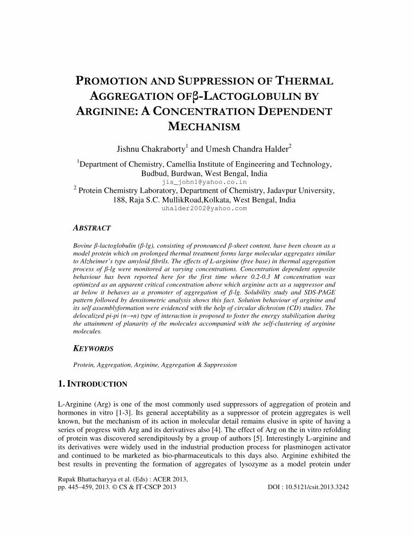
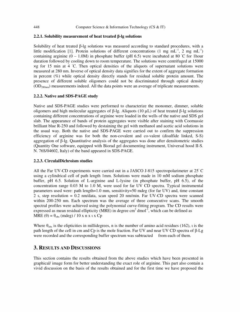
Amount of soluble β-lg present in heat treated solutions containing different concentrations of
arginine have been presented in terms of solubility percentage (%) in Figure 1. The figure shows
that there was a cut off point of arginine concentration at which the soluble protein concentrations
were minimum due to formation of large molecular weight aggregates.
0.0 0.2 0.4 0.6 0.8 1.0
0
25
50
75
100
Protein Concentration: 2mg mL-1 S
olu
bili
ty (
%)
Arginine(in Molarity)
0
25
50
75
100
Protein Concentration: 4mg ml-1
So
lub
ility
(%
)
Figure 1: Solubility profile of β-lg (protein concentration: 4 mg mL
-1 in upper panel and 2 mg mL
-1 in lower
panel) in presence of different arginine concentration in phosphate buffer (10 mM, pH 6.6-6.8).
It was necessary to mention that heterogeneities in solution were also observed with all the
solutions of β-lg (both 2mg mL-1
and 4mg mL-1
) containing arginine up to 0.25 M concentration.
Above this particular concentrations of arginine, the amount of soluble protein concentrations
increases. This clearly indicates that mechanism of suppression of aggregation became effective
after a certain concentration of arginine below which it acts as promoter of β-lg aggregation. This
concentration of arginine was optimized 0.2-0.3 M. Arginine, in the concentration range of 0.5-
1.0 M, almost suppressed the aggregation of β-lg where most of the β-lg remains in solutions.
Similar trend was also observed in the case of suppression of lysozyme aggregation by arginine
as reported elsewhere [1]. Appearance of heterogeneity/turbidity confirms the large aggregate
formation, but solubility test could not insight about formation of soluble aggregates/ oligomers.
450 Computer Science & Information Technology (CS & IT)
3.2. Native and SDS-PAGE studies: Densitometric analysis
Characterization of aggregates was done by both native and SDS
figure (2a and 2b), qualitatively it can be assumed that proportion of aggregates, particularly large
molecular aggregates were formed at arginine concentration range
where the aggregates failed to penetrate the gel (staking part) and band intensities in the
monomeric region were sufficiently less with compared to dimer/ oligomeric bands. The band
intensities in the monomeric region start
high molecular weight region ( staking gel part, after lane 4) with the gradual increase of arginine
concentrations (lane 6, 7; Arginine concentration 0.5 amd 1.0 M respectively). Interestingly, th
cause of appearance of heterogeneity during solubility studies was unfolded here and made some
visible impact as the bands appeared in different molecular weight ranges. Nonetheless the
contribution was not only because of the monomer but also the co
soluble forms was prominent even after heat treatment in all the lanes. Similar kind of
observation of the existence of soluble oligomers was also reported
dependent suppression of β-lg aggregation by arginin
mobilities and SDS-PAGE also revealed that arginine was successful in suppression of co
valently linked (di-sulfide linked, S
interactions could be discriminated.
Lane: 7 6 5 4 3 2
Figure 2: (a) Native PAGE and (b)
presence of arginine (lane1: 0 M, lane 2: 0.06m, lane 3: 0.12M, lane 4: 0.25M, lane 6: 0.5 M, lane 7: 1M)
in Tris.HCl buffered solution (pH 6.8).
Quantitative estimation of aggregate formation and arginine induced suppression was performed
with the help of densitometric analysis of the bands appeared in the SDS
estimation nicely demonstrates the monomer
transformation of β-lg depending on arginine concentrations. Partial loss of proteins due to
heterogeneity appeared in heating process may be attributed toward the erroneous density profile
(particularly in lane 3, 4, 5) but gross errors within 10
densitometric intensity profile has been presented (Fig 3b) correspondingly.
Computer Science & Information Technology (CS & IT)
PAGE studies: Densitometric analysis
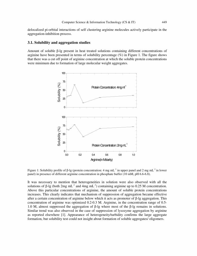
Characterization of aggregates was done by both native and SDS-PAGE techniques. Form the
figure (2a and 2b), qualitatively it can be assumed that proportion of aggregates, particularly large
molecular aggregates were formed at arginine concentration range 0.03 to 0.25 (lane 2, 3, 4, 5),
where the aggregates failed to penetrate the gel (staking part) and band intensities in the
monomeric region were sufficiently less with compared to dimer/ oligomeric bands. The band
intensities in the monomeric region starts increasing with the gradual disappearance of the band at
high molecular weight region ( staking gel part, after lane 4) with the gradual increase of arginine
concentrations (lane 6, 7; Arginine concentration 0.5 amd 1.0 M respectively). Interestingly, th
cause of appearance of heterogeneity during solubility studies was unfolded here and made some
visible impact as the bands appeared in different molecular weight ranges. Nonetheless the
contribution was not only because of the monomer but also the co-existence of oligomers in
soluble forms was prominent even after heat treatment in all the lanes. Similar kind of
observation of the existence of soluble oligomers was also reported [26]. Thus concentration
lg aggregation by arginine was nicely observed in gel-electrophoretic
PAGE also revealed that arginine was successful in suppression of co
sulfide linked, S-S) aggregation process where hydrophobic or electrostatic
iminated.
7 6 5 4 3 2 1 Lane: 7 6 5 4 3 2 1
a
b
(b) SDS-PAGE pattern of heat treated β-lactoglobulin (2mg mL
presence of arginine (lane1: 0 M, lane 2: 0.06m, lane 3: 0.12M, lane 4: 0.25M, lane 6: 0.5 M, lane 7: 1M)
in Tris.HCl buffered solution (pH 6.8).
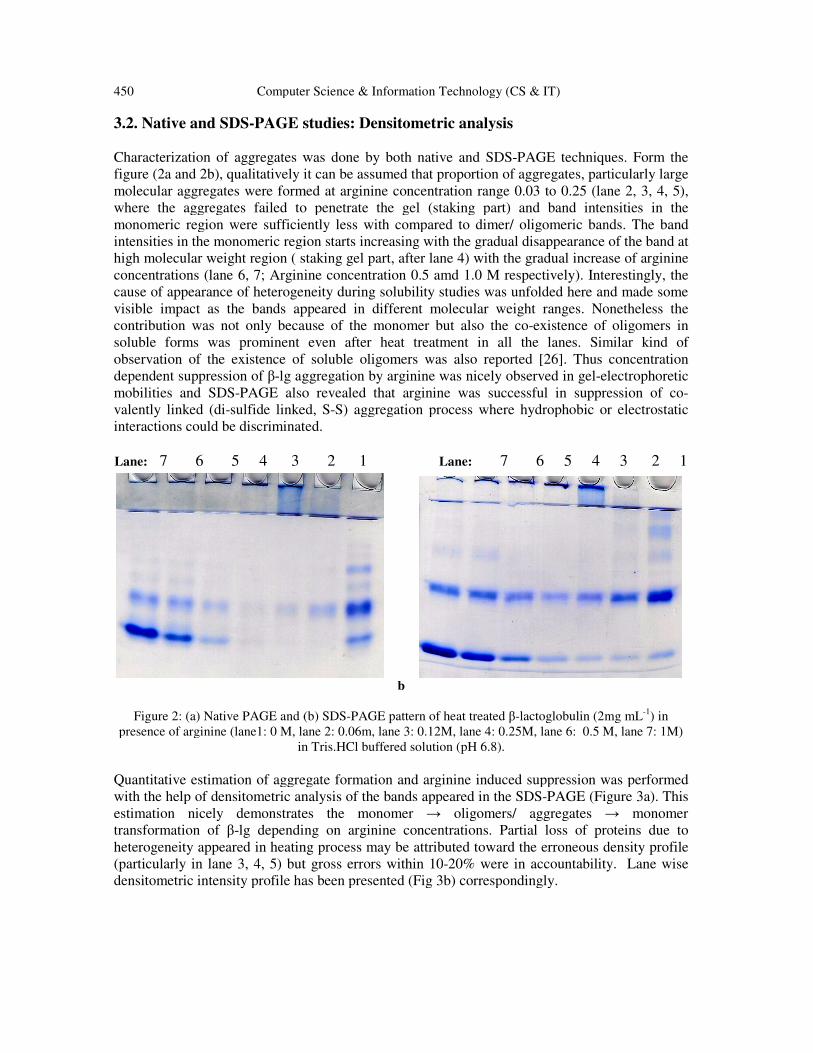
on of aggregate formation and arginine induced suppression was performed
with the help of densitometric analysis of the bands appeared in the SDS-PAGE (Figure 3a). This
estimation nicely demonstrates the monomer → oligomers/ aggregates →
lg depending on arginine concentrations. Partial loss of proteins due to
heterogeneity appeared in heating process may be attributed toward the erroneous density profile
(particularly in lane 3, 4, 5) but gross errors within 10-20% were in accountability. Lane wise
densitometric intensity profile has been presented (Fig 3b) correspondingly.
PAGE techniques. Form the
figure (2a and 2b), qualitatively it can be assumed that proportion of aggregates, particularly large
0.03 to 0.25 (lane 2, 3, 4, 5),
where the aggregates failed to penetrate the gel (staking part) and band intensities in the
monomeric region were sufficiently less with compared to dimer/ oligomeric bands. The band
s increasing with the gradual disappearance of the band at
high molecular weight region ( staking gel part, after lane 4) with the gradual increase of arginine
concentrations (lane 6, 7; Arginine concentration 0.5 amd 1.0 M respectively). Interestingly, the
cause of appearance of heterogeneity during solubility studies was unfolded here and made some
visible impact as the bands appeared in different molecular weight ranges. Nonetheless the
stence of oligomers in
soluble forms was prominent even after heat treatment in all the lanes. Similar kind of
. Thus concentration
electrophoretic
PAGE also revealed that arginine was successful in suppression of co-
S) aggregation process where hydrophobic or electrostatic
5 4 3 2 1
a
lactoglobulin (2mg mL-1
) in
presence of arginine (lane1: 0 M, lane 2: 0.06m, lane 3: 0.12M, lane 4: 0.25M, lane 6: 0.5 M, lane 7: 1M)
on of aggregate formation and arginine induced suppression was performed
PAGE (Figure 3a). This
→ monomer
lg depending on arginine concentrations. Partial loss of proteins due to
heterogeneity appeared in heating process may be attributed toward the erroneous density profile
tability. Lane wise
Computer Science & Information Technology (CS & IT) 451
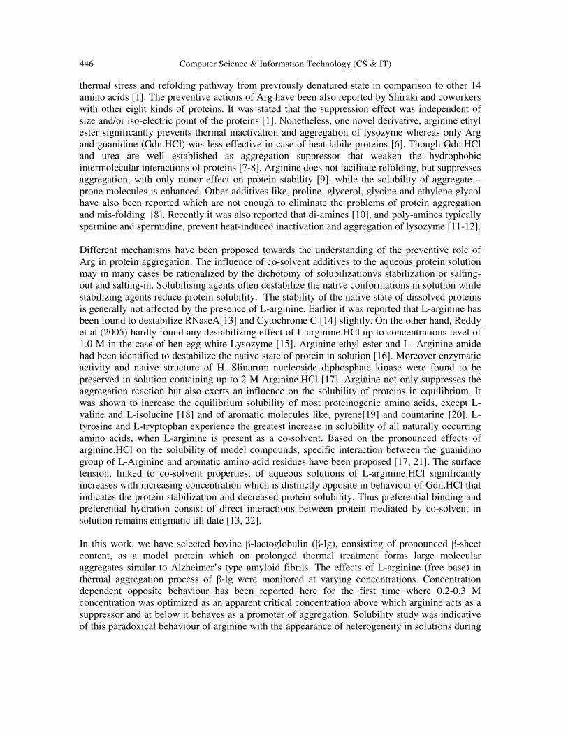
0 1 2 3 4 5 6
Monom
er
La
ne
1 L
an
e 2
La
ne
3 L
an
e 4
La
ne
5 L
an
e 6
Ba
nd
De
ns
ity
Inte
ns
ity
( in
mm
-2)
Hig
her
Aggreg
ates
Olig
omers
Dimer
La
ne
7
Figure 3a: Densitometric bar diagram of aggregation pattern and suppression in presence of L-
arginine from lane 1-7 in stacked fashion (Band density intensity mm-2
). On X-axis, Monomer
(1), Dimer (2), oligomers/ soluble aggregates (3-4) and high molecular weight aggregates (5-6)
correspond to appearance of relative proportion of the bands after coomassie blue staining.
Relative errors were within 10-20 %.
452 Computer Science & Information Technology (CS & IT)
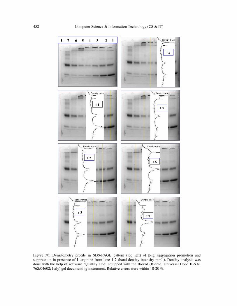
Figure 3b: Densitometry profile in SDS-PAGE pattern (top left) of β-lg aggregation promotion and
suppression in presence of L-arginine from lane 1-7 (band density intensity mm-2
). Density analysis was
done with the help of software ‘Qualtity One’ equipped with the Biorad (Biorad, Universal Hood II-S.N.
76S/04602; Italy) gel documenting instrument. Relative errors were within 10-20 %.
L2
L1
L3
L4
L5
L6
L7
L 7 6 5 4 3 2 1
Computer Science & Information Technology (CS & IT) 453
3.3. Role of Arginine: a molecular level understanding
It is evident that arginine is a novel suppressor in protein aggregation process at higher
concentration. Some detail molecular level explanation is required to justify the statement.
Several explanations have been proposed so far to support the mechanism. Earlier statement that
hydrophilicity rather than hydrophobicity of the additives is the important factor for preventing
aggregation was made by another group [1]. Okajono and co-workers have also proposed that
high hydrophobicity is not a favourable characteristic of additives for suppression considering the
hydrophobic chain length of polyamines [10]. It was also suggested that if side chain of arginine
possesses properties identical to those of Gdn.HCl, incorrect inter- or intra-molecular hydrogen
bonds may be cleaved and rearranged by arginine as in case by Gdn.HCl[27]. Even in our case it
was quite inconclusive from the results of circular dichroism studies when secondary structural
perturbation was monitored at identical Gdn.HCl and arginine concentration along with β-lg. At 1
M concentration Gdn.HCl slightly perturbed β-lg secondary structural content where as at the
same concentration, arginine signal was so prominent that fail to get any secondary
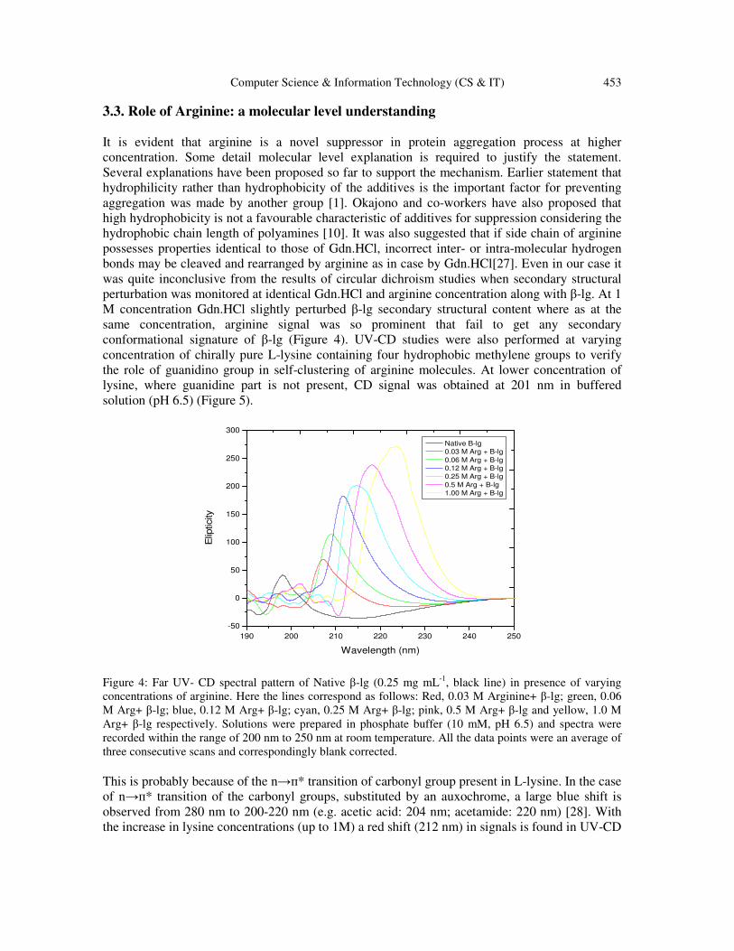
conformational signature of β-lg (Figure 4). UV-CD studies were also performed at varying
concentration of chirally pure L-lysine containing four hydrophobic methylene groups to verify
the role of guanidino group in self-clustering of arginine molecules. At lower concentration of
lysine, where guanidine part is not present, CD signal was obtained at 201 nm in buffered
solution (pH 6.5) (Figure 5).
190 200 210 220 230 240 250
-50
0
50
100
150
200
250
300
Elip
ticity
Wavelength (nm)
Native B-lg
0.03 M Arg + B-lg
0.06 M Arg + B-lg
0.12 M Arg + B-lg
0.25 M Arg + B-lg
0.5 M Arg + B-lg
1.00 M Arg + B-lg
Figure 4: Far UV- CD spectral pattern of Native β-lg (0.25 mg mL
-1, black line) in presence of varying
concentrations of arginine. Here the lines correspond as follows: Red, 0.03 M Arginine+ β-lg; green, 0.06
M Arg+ β-lg; blue, 0.12 M Arg+ β-lg; cyan, 0.25 M Arg+ β-lg; pink, 0.5 M Arg+ β-lg and yellow, 1.0 M
Arg+ β-lg respectively. Solutions were prepared in phosphate buffer (10 mM, pH 6.5) and spectra were
recorded within the range of 200 nm to 250 nm at room temperature. All the data points were an average of
three consecutive scans and correspondingly blank corrected.
This is probably because of the n→п* transition of carbonyl group present in L-lysine. In the case
of n→п* transition of the carbonyl groups, substituted by an auxochrome, a large blue shift is
observed from 280 nm to 200-220 nm (e.g. acetic acid: 204 nm; acetamide: 220 nm) [28]. With
the increase in lysine concentrations (up to 1M) a red shift (212 nm) in signals is found in UV-CD
454 Computer Science & Information Technology (CS & IT)
pattern, signifies п* energy level stabilization. Generally amino acids have a general tendency to
orient themselves in a peptide/ amide (-CONH-) like fashion with N- and C-terminal group
juxtaposition rendering side chains away on the either sides [29]. Therefore the signals lie
certainly in between 200-220 nm, which is in good agreement with the literature values. At higher
concentration, lysine form macromolecular aggregate/ micelle rather than an open ended bi-layer
structures in aqueous solution [19]. Thus lowering of energy could be due to this macromolecular
assembly of lysine residues. Therefore entire focus was directed towards the solution behavior
of arginine through circular dichroism studies. Interestingly it was observed that signals, obtained
from CD spectral patterns, of arginine put a signature of structure formation in aqueous solution
(Figure 6). The sharp positive signal (at 207-209 nm) of arginine at lower concentration (0.03 M)
was due to the n→п* electronic transition significantly contributed by the carbonyl moiety of
carboxylate anion part, which ultimately shifted toward higher wavelength (224 nm) with the
increase in arginine concentration (upto 1 M). This red shift (207 to 224 nm) in CD signals was
indicative of lowering of energy gaps accompanied with the energy stabilization up on increasing
arginine concentration. The red shifts in CD signals have been recently mentioned in
supramolecular/self-assembling peptide-amphiphilic systems [30-31].
This kind of lowering in energy may be due to self clustering of Arginine where the attainment of
the different electronic microenvironment surrounding the carbonyl moiety can not be ignored.
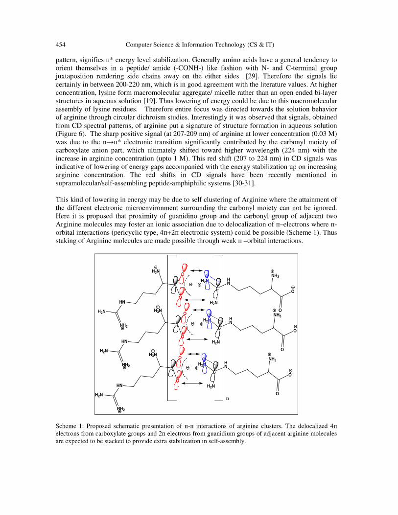
Here it is proposed that proximity of guanidino group and the carbonyl group of adjacent two
Arginine molecules may foster an ionic association due to delocalization of п–electrons where п-
orbital interactions (pericyclic type, 4п+2п electronic system) could be possible (Scheme 1). Thus
staking of Arginine molecules are made possible through weak п –orbital interactions.
O
HN
O
H3N
H2N
NH2
O
HN
O
NH3
C
H2N
H2N
O
HN
O
H3N
H2N
NH2
O
HN
O
NH3
C
H2N
H2N
O
HN
O
H3N
H2N
NH2
O
HN
O
NH3
C
H2N
H2N
n
Scheme 1: Proposed schematic presentation of п-п interactions of arginine clusters. The delocalized 4п
electrons from carboxylate groups and 2п electrons from guanidium groups of adjacent arginine molecules
are expected to be stacked to provide extra stabilization in self-assembly.
Computer Science & Information Technology (CS & IT) 455
Unlike other amino acids, Arginine stacks in head-to tail fashion and a hydrophobic column
composed of the three methylene groups is seen along one crystallographic axis [32]. Probably
the interactions involved, the strong intermolecular interactions and other less significant
secondary interactions during cluster formation was reported by a group of authors [24]. This
kind of typical orientation and close packing have also been found in many crystal structures of
arginine [33-34]. These cluster may have conformational properties as observed in crystal
patterns and supposed to be rod shaped as seen in case of chirally pure praline molecule [35]. Still
the role of intercalating water molecule might be highly significant in hydrogen bond formation
of the clustering units in aqueous solution [36]. Das and co-workers also showed that molecular
cluster of arginine in solution display a hydrophobic surface by the alignment of its three
methylene groups, which ultimately interacts with protein and manifests a masking effect toward
the protein molecules [23]. Therefore the characteristic organization of arginine leading to a
columnar or rod like cluster may be stabilized by ionic association and/ or hydrogen bond
formation and also by п-п type interactions with proximity of guanidino and/or carboxylate
groups.
190 200 210 220 230 240 250
0
200
400
600
800
1000
1200
1400
Elip
ticity
Wavelength (nm)
0.03 M Lys
0.06 M Lys
0.12 M Lys
0.25 M Lys
0.50 M Lys
1.00 M Lys
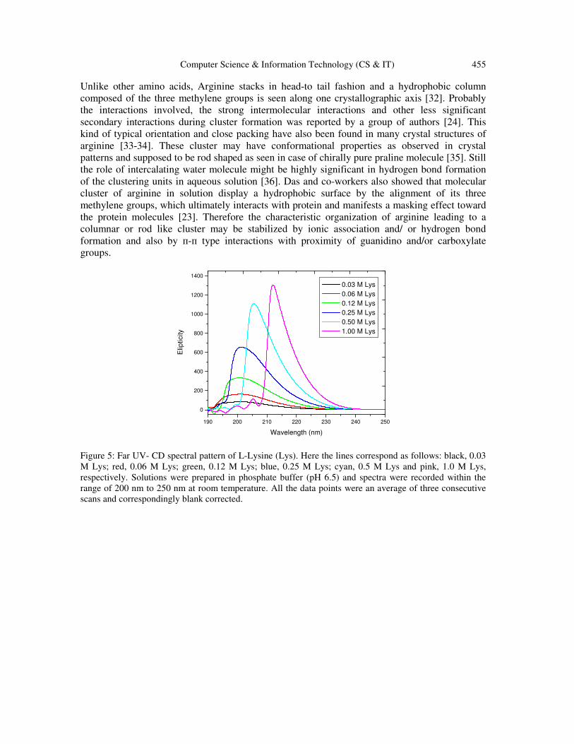
Figure 5: Far UV- CD spectral pattern of L-Lysine (Lys). Here the lines correspond as follows: black, 0.03
M Lys; red, 0.06 M Lys; green, 0.12 M Lys; blue, 0.25 M Lys; cyan, 0.5 M Lys and pink, 1.0 M Lys,
respectively. Solutions were prepared in phosphate buffer (pH 6.5) and spectra were recorded within the
range of 200 nm to 250 nm at room temperature. All the data points were an average of three consecutive
scans and correspondingly blank corrected.
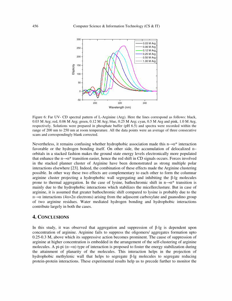
456 Computer Science & Information Technology (CS & IT)
200 220 240
-50
0
50
100
150
200
250
300
Elip
ticity
Wavelength (nm)
0.03 M Arg
0.06 M Arg
0.12 M Arg
0.25 M Arg
0.50 M Arg
1.00 M Arg
Figure 6: Far UV- CD spectral pattern of L-Arginine (Arg). Here the lines correspond as follows: black,
0.03 M Arg; red, 0.06 M Arg; green, 0.12 M Arg; blue, 0.25 M Arg; cyan, 0.5 M Arg and pink, 1.0 M Arg,
respectively. Solutions were prepared in phosphate buffer (pH 6.5) and spectra were recorded within the
range of 200 nm to 250 nm at room temperature. All the data points were an average of three consecutive
scans and correspondingly blank corrected.
Nevertheless, it remains confusing whether hydrophobic association made this n→п* interaction
favorable or the hydrogen bonding itself. On other side, the accumulation of delocalized п–
orbitals in a stacked fashion makes the ground state energy levels electronically more populated
that enhance the п→п* transition easier, hence the red shift in CD signals occurs. Forces involved
in the stacked planner cluster of Arginine have been demonstrated as strong multiple polar
interactions elsewhere [23]. Indeed, the combination of these effects made the Arginine clustering
possible. In other way these two effects are complementary to each other to form the columnar
arginine cluster projecting a hydrophobic wall segregating and inhibiting the β-lg molecules
prone to thermal aggregation. In the case of lysine, bathochromic shift in n→п* transition is
mainly due to the hydrophobic interactions which stabilizes the micellerclusture. But in case of
arginine, it is assumed that greater bathochromic shift compared to lysine is probably due to the
п→п interactions (4п+2п electrons) arising from the adjuscent carboxylate and guanodino group
of two arginine residues. Water mediated hydrogen bonding and hydrophobic interactions
contribute largely in both the cases.
4. CONCLUSIONS In this study, it was observed that aggregation and suppression of β-lg is dependent upon
concentration of arginine. Arginine fails to suppress the oligomers/ aggregates formation upto
0.25-0.3 M, above which its suppressive action becomes prominent. The cause of suppression of
arginine at higher concentration is embedded in the arrangement of the self-clustering of arginine
molecules. A pi-pi (п→п) type of interaction is proposed to foster the energy stabilization during
the attainment of planarity of the molecules. This interaction helps in the projection of
hydrophobic methylenic wall that helps to segregate β-lg molecules to segregate reducing
protein-protein interactions. These experimental results help us to precede further to monitor the
Computer Science & Information Technology (CS & IT) 457
energy stabilization in factors within the scope of computational bio-physics and/or to explore the
new horizon in some model based protein aggregation-suppression phenomena through
computational-biology.
ACKNOWLEDGEMENTS
Financial support of UGC-CAS (Govt. of INDIA), Department of Chemistry, Jadavpur
University, Kolkata is gratefully acknowledged. Authors also like to acknowledge Camellia
Institute of Engineering & technology, Budbud, Burdwan, WB, India, for providing academic and
administrative help.
REFERENCES
[1] Shiraki, K., Kudou, M., Fujiwara, S., Imanaka, T. & Takagi M. (2002) “Biophysical effect of amino
acids on the prevention of protein aggregation”, J. Biochem. (Tokyo), Vol 132, pp591-595.
[2] Bajorunait, E., Sereikaite, J. &Bumelis, V. A. (2007) “L-Arginine suppresses aggregation of
recombinant growth hormones in refolding process from E. Coli inclusion bodies”, Protein J.,Vol 26,
pp547–555.
[3] Christian, L. & Rainer, R., (2009) “Suppression of protein aggregation by L-Arginine”, Curr. Pharm.
Biotechnol.,Vol 10, pp408-414.
[4] Lange, C., Patil, G. & Rudolph, R. (2005) “Ionic liquids as refolding additives: N’-alkyl and N’-(w-
hydroxyalkyl) N-methylimidazolium chlorides”, Protein Sci.,Vol 14, pp2693-2701.
[5] Rudolph, R. & Fischer, S. (1990) US Patent 4933434.
[6] Shiraki, K., Kudou, M., Nishikori, S., Kitagawa, H., Imanaka, T. & Takagi, M. (2004) “Arginine
ethylester prevents thermal inactivation and aggregation of lysozyme”, Eur. J. Biochem.,Vol 271,
pp3242-3247
[7] Buchner,J& Rudolph, R. (1991) “Renaturation, purification and characterization of recombinant Fab-
fragments produced in Escherichia Coli”, Biotechnology,Vol 9, pp157-162.
[8] Rudolph, R. &Lilie, H. (1996) “In vitro folding of inclusion body proteins”, FASEB J.,Vol 10, pp49-
56.
[9] Arakawa, T. &Tsumoto, K. (2003) “The effects of arginine on refolding of aggregated proteins: not
facilitate refolding, but suppress aggregation”, Biochem. Biophys. Res. Commun., Vol. 304, pp148-
152.
[10] Okanojo, M., Shraki, K., Kudou, M., Nishikori, S. & Takagi, M. (2005) “Diamines prevent thermal
aggregation and inactivation of Lysozyme”, J. Biosci. Bioengg.,Vol 100, pp556-561.
[11] Kudou, M., Shiraki, K., Fujiwara, S., Imanaka, T. & Takagi, M. (2003) “Prevention of thermal
inactivation and aggregation of Lysozyme by polyamines”, Eur. J. Biochem.,Vol 270, pp4547-4554.
[12] Shiraki, K., Kudou, M., Aso, Y. & Takagi, M. (2003) “Approach for prevention of heat-induced
protein aggregation by small molecular additives”, Sci. Technol. Adv. Mat., Vol 4, pp55-59.
[13] Lin, T. Y. &Timasheff, S. N. (1996) “On the role of surface tension in the stabilization of globular
proteins”, Protein Sci.,Vol 5, pp372-381.
[14] Taneja, S. & Ahmad, F. (1994) “Increased thermal stability of proteins in the presence of amino
acids”, Biochem J.,Vol 303, pp147-153.
[15] Reddy, K. R. C., Lilie, H., Rudolph, R.& Lange, C. (2005) “L-Arginine increases the solubility of
unfolded species of hen egg white lysozyme”, Protein Sci.,Vol 14, pp929-935.
[16] Hamada, H. &Shiraki, K. (2007) “L-Argininamide improves the refolding more effectively than L-
arginine”, J. Biotechnol.,Vol 130, pp153-160.
[17] Ishibashi, M., Tsumoto, K., Tokunaga, M., Ejima, D., Kita, Y. & Arakawa, T. (2005) “Is arginine a
protein-denaturant”, Protein Expr. Purif.,Vol 42, pp1-6.
458 Computer Science & Information Technology (CS & IT)
[18] Arakawa, T., Ejima, D., Tsumoto, K., Obeyma N., Tanaka, Y., Kita, Y. &Timasheff, S. N. (2007)
“Suppression of protein interactions by arginine: A proposed mechanism of the arginine effects”,
Biophys. Chem., Vol. 127, pp1-8.
[19] Das, U., Hariprasad, G., Ethayathulla, A. S., Manral, P., Das, T. K., Pasha, S., Mann, A., Ganguli, M.,
Verma, A. K., Bhat, R., Chandryan, S. K., Ahmed, S., Sharma, S., Kaur, P., Singh, T. P. &Srinivasan,
A. (2007) “Inhibition of protein aggregation: Supramolecular assemblies of Arginine hold the key”,
pLoS ONE (2) e1176.
[20] Hirano, A., Arakawa, T. &Shiraki, K. (2008) “Arginine increases the solubility of coumarine:
Comparison with salting-in and salting-out additives”, J. Biochem., Vol 144, pp363-369.
[21] Tsumoto, K., Umetsu, M., Kumagai, I., Ejima, D., Philo, J. S. & Arakawa, T. (2004) “Role of
arginine in protein refolding, solubilisation, and purification”, Biotechnol. Prog.,Vol 20, pp1301-
1308.
[22] Kita, Y., Arakawa, T., Lin, T. Y. &Timasheff, S. N. (1994) “Contribution of the Surface Free Energy
Perturbation to Protein-Solvent Interactions”, Biochemistry,Vol 33, pp15178-15189.
[23] Das, U. &Srinivasan, A. (2008) “Protein Aggregation-Suppression: Molecular Clusters of Arginine
are responsible”, FASEB J. 2008 22:1057.2 [Meeting Abstract]
[24] Julian, R. R., Hodyss, R. & Beauchamp, J. L. (2001) “Cooperative salt bridge stabilization of gas-
phase zwitterions in neutral arginine cluster”, J. Am. Chem. Soc.,Vol 123, pp3577-3583.
[25] Chakraborty, J., Das, N., Das, K. P., Halder, U. C. (2009) “Loss of structural integrity and
hydrophobic ligand binding capacity of acetylated and succinylated bovineβ-lactoglobulin”,
International Dairy J.Vol 19, pp 43-49.
[26] Liu, Y. D., Li, J. J., Wang, F. W., Chen, J., Li, P. & Su, Z. G., (2007) “A newly proposed mechanism
for arginine-assisted protein refolding—not inhibiting soluble oligomers although promoting a correct
structure”, Protein Express Purif.,Vol 51, pp235-242.
[27] Trivedi, V. D., Raman, B., Rao, C. M. & Ramakrishna, T. (1997) “Co-refolding denatured-reduced
hen egg white lysozyme with acidic and basic proteins”, FEBS Lett.,Vol 418, pp363-366.
[28] Kalsi, P. S. (2004) Spectroscopy of Organic Compounds, 6thEdn, New Age International Publishers,
New Delhi, India, Chapter 2, pp 37.
[29] Suresh, C. G. &Vijayan, M. (1983) “X-ray studies on crystalline complexes involving amino acids
and peptides. Head-to-tail arrangement and a specific interaction in the crystal structure of L-arginine
acetate”, Int. J. Pep. Res.,Vol 21, pp223-226.
[30] Pashuck, E. T., Cui, H. &Stupp, S. I. (2010) “Tuning supramolecular rigidity of peptide fibers
through molecular structure”, J. Am. Chem. Soc.,Vol 132, pp6041-6046.
[31] Rexeisen, E. L., Fan, W., Pangburn, T. O., Taribagil, R. R., Bates, F. S., Lodge, T. P., Tsapatsis, M.
&Kokkoli, E. (2010) “Self-assembling of fibronectin mimetic peptide-amphiphilenanofibers”,
Langmuir., Vol 26, pp1953-1959.
[32] Karle, J. & Karle, I. L. (1964) “An application of the symbolic addition method to the structure of L-
argini dehydrate”, Acts Cyst., Vol 17, pp835-841.
[33] Saenger, W. & Wagner, K. G. (1972) “An X-ray study of Hydrogen Bonding in the Crystalline L-
Arginine Phosphate monohydrate complex”, ActaCrystallgr B.,Vol 28, pp2237-2244.
[34] Recacha, R., Verdeguer, N. &Subirana, J. A. (1997) “Structure and supramolecular packing features
of the dipeptide Arg-Val acetate” J. peptide Res.,Vol 50, pp388-392.
[35] Myung, S., Fioroni, M., Julian, R. R., Koeniger, S. L. &Baik, M. H. (2006) “Chirally directed
formation of nanometer-scale praline clusters”, J. Am. Chem. Soc.,Vol 128, pp10833-10839.
[36] Jensen, J. H. & Gordon, M. S., (1995) “On the number of water molecules necessary to stabilize the
glycine zwitterion”, J. Am. Chem. Soc.Vol 117, pp8159-8170.
Computer Science & Information Technology (CS & IT) 459
AUTHORS
Dr. JishnuChakraborty, currently working as an Assistant Professor,Department of
Chemistry, Camellia Institute of Engineering & Technology, Budbud, Burdwan, WB,
India, has been awarded his Ph, D. Degree in Science from Jadavpur University,
Kolkata, in the year of 2011 under the Supervision of Dr. U. C. Halder. He has
completed his Master Degree from C. S. J. M. Kanpur University, (formerly, Kanpur
University) in the year of 2005 and he is an Honoursgraduatein Chemistry from
University of Calcutta (2003).He has published several research articles in different
journals of international repute. He was selected for the “UGC-sponsored Meritorious
Fellowship in Science, 2007-2008” in the Department of Chemistry, Jadavpur
University, Kolkata. Now a Day, he is holding the position of HOD, Chemistry in CIET, Budbud,
Burdwan.His research interest is in protein chemistry, structural biology, nano-bio composite materials.
Dr. Umesh Chandra Halder, Assistant Professor,Department of Chemistry,
Jadavpur University, Kolkata, West Bengal, India, has been awarded his Ph, D.
Degree in Science from University of Calcutta, Kolkata, under the Supervision of
Prof. N. K. Sinha, in Protein Chemistry Laboratory,Bose Institute, Kolkata. He has
completed his Bachelors and Master Degree from University of Calcutta(Presidency
College). He has published many research articles in different reputed journals of
national and internationallevel.He has also run several scientific projects under DBT,
CSIR, DSTetc. since the very beginning of his career in Jadavpur University. He is
also a good scientific collaborator. His major research interest is in protein chemistry, bio-organic
chemistry, biophysical chemistry and nano biology.


![Neuartige π-Organyle der schweren Alkalimetalle und des ... · cesium compound ([CsCp(18-crown-6)CsCp]*2.75THF)n (11a) and three tetranuclear heterobimetallic alkali metal cyclopentadienide](https://static.fdocument.org/doc/165x107/5b56099a7f8b9a18618c36d6/neuartige-organyle-der-schweren-alkalimetalle-und-des-cesium-compound.jpg)