
Products for Flow Cytometry - cedarlanelabs.com
12
CF™ dyes for flow cytometer laser lines ..... p. 2 Secondary antibodies and bioconjugates ..... p. 3 Antibody labeling kits ..... p. 4 Cellular assays ..... pp. 5-8 • Cell viability ..... p. 5 • Cell proliferation and cell cycle ..... p. 6 • Mitochondrial membrane potential ..... p. 7 • Apoptosis and necrosis ..... pp. 8-10 Flow cytometry accessories ..... p. 11 Products for Flow Cytometry
Transcript of Products for Flow Cytometry - cedarlanelabs.com
CF™ dyes for flow cytometer laser lines ..... p. 2
Secondary antibodies and bioconjugates ..... p. 3
Antibody labeling kits ..... p. 4
Cellular assays ..... pp. 5-8
• Cell viability ..... p. 5
• Cell proliferation and cell cycle ..... p. 6
• Mitochondrial membrane potential ..... p. 7
• Apoptosis and necrosis ..... pp. 8-10
Flow cytometry accessories ..... p. 11
Products for Flow Cytometry
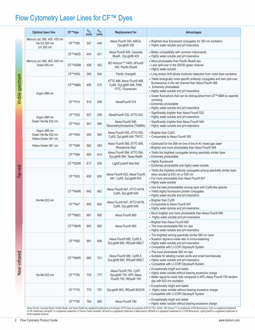
2 Flow Cytometry Product Guide www.biotium.com
Optimal laser line CF™dye λEx (nm)
λEm (nm) Replacement for Advantages
Mercury arc 366, 405, 435 nmHe-Cd 325 nm
UV 355 nmCF™350 347 448 Alexa Fluor® 350, AMCA,
DyLight® 350 • Brightest blue fluorescent conjugates for 350 nm excitation • Highly water-soluble and pH insensitive
Mercury arc 366, 405, 435 nmViolet 405 nm
CF™405S 404 431 Alexa Fluor® 405, Cascade Blue®, DyLight® 405
• Better compatibility with common instruments • Highly water-soluble and pH-insensitive
CF™405M 408 452 BD Horizon™ V450, eFluor® 450, Pacific Blue®
• More photostable than Pacific Blue® dye • Less spill-over in the 525/50 green channel • Highly water-soluble
CF™405L 395 545 Pacific Orange® • Long stokes shift allows multicolor detection from violet laser excitation
Argon 488 nm
CF™488A 490 515ATTO 488, Alexa Fluor® 488, Cy®2, DyLight® 488, FAM,
FITC, Fluorescein
• Yields biologically more specific antibody conjugates and less spill-over fluorescence in the red channel than Alexa Fluor® 488
• Extremely photostable • Highly water-soluble and pH-insensitive
CF™514 516 548 AlexaFluor® 514
• Green fluorophore that can be distinguished from CF™488A by spectral unmixing
• Extremely photostable • Highly water-soluble and pH-insensitive
Argon 488 nmGreen He-Ne 532 nm
CF™532 527 558 AlexaFluor® 532, ATTO 532 • Significantly brighter than Alexa Fluor® 532• Highly water-soluble and pH-insensitive
CF™543 541 560 Alexa Fluor® 546, Tetramethylrhodamine (TAMRA)
• Significantly brighter than Alexa Fluor® 546• Highly water-soluble and pH-insensitive
Argon 488 nmGreen He-Ne 532 nmYellow-Green 561 nm
CF™555 555 565 Alexa Fluor® 555, ATTO 550, Cy®3, DyLight® 549, TRITC
• Brighter than Cy®3 • Comparable to Alexa Fluor® 555
Yellow-Green 561 nm CF™568 562 583 Alexa Fluor® 568, ATTO 565, Rhodamine Red
• Optimized for the 568 nm line of the Ar-Kr mixed-gas laser • Brighter and more photostable than Alexa Fluor® 568
CF™594 593 614 Alexa Fluor® 594, ATTO 594, DyLight® 594, Texas Red®
• Yields the brightest conjugates among spectrally similar dyes • Extremely photostable
CF™620R 617 639 LightCycler® Red 640 • Highly fluorescent • Extremely photostable and highly water-soluble
He-Ne 633 nm
CF™633 630 650 Alexa Fluor® 633, Alexa Fluor® 647, Cy®5, DyLight® 633
• Yields the brightest antibody conjugates among spectrally similar dyes when excited at 633 nm or 635 nm
• Far more photostable than Alexa Fluor® 647 • Highly water-soluble
CF™640R 642 662 Alexa Fluor® 647, ATTO 647N, Cy®5, DyLight® 649
• Has the best photostability among dyes with Cy®5-like spectra • Yields highly fluorescent protein conjugates • Highly water-soluble and pH-insensitive
CF™647 650 665 Alexa Fluor® 647, ATTO 647N, Cy®5, DyLight® 649
• Brighter than Cy®5 • Comparable to Alexa Fluor® 647 • Highly water-soluble and pH-insensitive
CF™660C 667 685 Alexa Fluor® 660 • Much brighter and more photostable than Alexa Fluor® 660 • Highly water-soluble and pH-insensitive
CF™660R 663 682 Alexa Fluor® 660 • Brighter than Alexa Fluor® 660 • The most photostable 660 nm dye • Highly water-soluble and pH-insensitive
CF™680 681 698 Alexa Fluor® 680, Cy®5.5, DyLight® 680, IRDye® 680LT
• The brightest among spectrally similar 680 nm dyes • Superior signal-to-noise ratio in immunostaining • Highly water-soluble and pH-insensitive • Compatible with LI-COR Odyssey® System
CF™680R 680 701 Alexa Fluor® 680, Cy®5.5, DyLight® 680, IRDye® 680LT
• The most photostable 680 nm dye • Suitable for labeling nucleic acids and small biomolecules • Highly water-soluble and pH-insensitive • Compatible with LI-COR Odyssey® System
He-Ne 633 nm CF™750 755 777Alexa Fluor® 750, Cy®7, DyLight® 750, APC-Alexa Fluor® 750, IRDye® 750
• Exceptionally bright and stable • Highly water soluble without bearing excessive charge • Better signal-to-noise ratio compared to APC-Alexa Fluor® 750 tandem dye with 633 nm excitation
CF™770 770 797 DyLight® 800, IRDye® 800CW • Exceptionally bright and stable • Highly water soluble without bearing excessive charge • Compatible with LI-COR Odyssey® System
CF™790 784 806 Alexa Fluor® 790 • Exceptionally bright and stable • Highly water soluble without bearing excessive charge
Flow Cytometry Laser Lines for CF™ DyesVi
sible
spec
trum
Near
-infra
red
Far-r
ed
Alexa Fluor®, Cascade Blue®, Pacific Blue®, and Texas Red® are registered trademarks of Invitrogen; ATTO dyes are products of ATTO-TEC GmbH; BD Horizon™ is a trademark of BD Biosciences; Cy® is a registered trademark of GE Healthcare; DyLight® is a registered trademark of Thermo Fisher Scientific; eFluor® is a registered trademark of eBioscience; IRDye® is a registered trademark of LI-COR Bioscience; LightCycler® is a registered trademark of Roche Applied Science.
Flow Cytometry Product Guide 3www.biotium.com
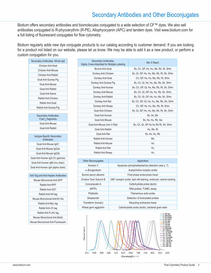
Secondary Antibodies and Other BioconjugatesBiotium offers secondary antibodies and biomolecules conjugated to a wide selection of CF™ dyes. We also sell antibodies conjugated to R-phycoerythrin (R-PE), Allophyocyanin (APC) and tandem dyes. Visit www.biotium.com for a full listing of fluorescent conjugates for flow cytometry.
Biotium regularly adds new dye conjugate products to our catalog according to customer demand. If you are looking for a product not listed on our website, please let us know. We may be able to add it as a new product, or perform a custom conjugation for you.
Secondary Antibodies, Whole IgGChicken Anti-Goat
Chicken Anti-MouseChicken Anti-Rabbit
Goat Anti-Guinea PigGoat Anti-MouseGoat Anti-RabbitGoat Anti-Swine
Rabbit Anti-ChickenRabbit Anti-Goat
Rabbit Anti-Guinea Pig
Secondary Antibodies, F(ab’)2 fragmentsGoat Anti-Mouse Goat Anti-Rabbit
Isotype-Specfic Secondary Antibodies
Goat Anti-Mouse IgG1Goat Anti-Mouse IgG2aGoat Anti-Mouse IgG2b
Goat Anti-Human IgG (Fc gamma)Goat Anti-Human IgM (mu chain)
Goat Anti-Human IgA (alpha chain)
Anti-Tag and Anti-Hapten AntibodiesMouse Monoclonal Anti-GFP
Rabbit Anti-RFPRabbit Anti-GST
Rabbit Anti-HA tagMouse Monoclonal Anti-6X His
Rabbit Anti-Myc tagRabbit Anti-V5 tag
Rabbit Anti-FLAG tagMouse Monoclonal Anti-Biotin
Mouse Monoclonal Anti-Fluorescein
Secondary Antibodies, Highly Cross-Adsorbed for Multiple Labeling Min X React
Bovine Anti-Goat Bv, Ch, GP, Hs, Hu, Ms, Rb, Rt, SHmDonkey Anti-Chicken Bv, Gt, GP, Hs, Hu, Ms, Rb, Rt, Sh, SHm
Donkey Anti-Goat Ch, GP, Hs, Hu, Ms, Rb, Rt, SHmDonkey Anti-Guinea Pig Bv, Ch, Gt, Hs, Hu, Ms, Rb, Sh, SHm
Donkey Anti-Human Bv, Ch, GP, Gt, Hs, Ms, Rb, Rt, Sh, SHmDonkey Anti-Mouse Bv, Ch, Gt, GP, Hs, Hu, Rb, Sh, SHmDonkey Anti-Rabbit Bv, Ch, Gt, GP, Hs, Hu, Ms, Sh, SHm
Donkey Anti-Rat Bv, Ch, GP, Gt, Hs, Hu, Ms, Rb, Sh, SHmDonkey Anti-Sheep Ch, GP, Hs, Hu, Ms, Rb, Rt, SHmGoat Anti-Chicken Bv, Gt, GP, Hs, Hu, Ms, Rb, Rt, Sh, SHmGoat Anti-Human Bv, Hs, Ms Goat Anti-Mouse Bv, Hs, Hu, Rb, Sw
Goat Anti-Mouse (min X Rat) Bv, Ch, Gt, GP Hs Hu Rb Rt, Sh, SHmGoat Anti-Rabbit Hu, Ms, Rt
Goat Anti-Rat Bv, Hs, Hu, RbRabbit Anti-Human MsRabbit Anti-Mouse Hu
Rabbit Anti-Rat HuRabbit Anti-Sheep Hu
Other Bioconjugates ApplicationAnnexin V Apoptosis (phosphatidylserine) detection (see p. 7)
a-Bungarotoxin Acetylcholine receptor probeBovine serum albumin Fluid phase endocytosis tracer
Cholera Toxin Subunit B GM1 receptor probe; lipid raft staining, endocytic vesicle trackingConcanavalin A Carbohydrate probe (lectin)
dNTPs DNA probes, TUNEL assayPhalloidin Filamentous actin probe
Streptavidin Detection of biotinylated probesTransferrin (human) Recycling endosome tracer
Wheat germ agglutinin Carbohydrate probe (lectin), bacterial gram stain
4 Flow Cytometry Product Guide www.biotium.com
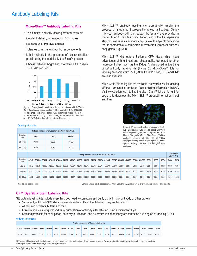
Antibody Labeling Kits
Mix-n-Stain™ antibody labeling kits dramatically simplify the process of preparing fluorescently-labeled antibodies. Simply mix your antibody with the reaction buffer and dye provided in the kit. After 30 minutes of incubation, and without a separation step, you will have an antibody conjugate of the dye of your choice that is comparable to commercially-available fluorescent antibody conjugates (Figure 1).
Mix-n-Stain™ kits feature Biotium’s CF™ dyes, which have advantages of brightness and photostability compared to other fluorescent dyes, such as the DyLight® dyes used in Lightning Link® antibody labeling kits (Figure 2). Mix-n-Stain™ kits for labeling antibodies with R-PE, APC, Per-CP, biotin, FITC and HRP are also available.
Mix-n-Stain™ labeling kits are available in several sizes for labeling different amounts of antibody (see ordering information below). Visit www.biotium.com to find the Mix-n-Stain™ kit that is right for you and to download the Mix-n-Stain™ product information sheet and flyer.
• The simplest antibody labeling protocol available• Covalently label your antibody in 30 minutes• No clean up of free dye required• Tolerates common antibody buffer components• Label antibody in the presence of excess stabilizer
protein using the modified Mix-n-Stain™ protocol• Choose between bright and photostable CF™ dyes,
R-PE, APC or Per-CP.
Figure 1. Flow cytometry analysis of Jurkat cells stained with CF™633 Mix-n-Stain labeled mouse anti-human CD3 antibodies (BD cat# 555330). For reference, cells were stained with commercial Alexa Fluor® 647 mouse anti-human CD3 (BD cat# 557706). Fluorescence was analyzed on a BD FACSCalibur flow cytometer in the FL4 channel.
Catalog numbers for CF™ Dye Mix-n-Stain™ Kits Other Mix-n-Stain™ Kits
Reaction size* CF350 CF405S CF405L CF405M CF488A CF532 CF543 CF555 CF568 CF594 CF633 CF640R CF647 CF660C CF660R CF680 CF680R CF750 CF770 CF790 Biotin FITC
5-20 ug 92270 92271 92303 92272 92273 92289 92287 92274 92275 92276 92277 92278 92279 92280 92281 92282 92283 92284 92285 92288 92286 92294
20-50 ug 92250 92251 92304 92252 92253 92290 92267 92254 92255 92256 92257 92258 92259 92260 92261 92262 92263 92264 92265 92268 92266 92295
50-100 ug 92230 92231 92305 92232 92233 92291 92247 92234 92235 92236 92237 92245 92238 92239 92243 92240 92246 92241 92242 92248 92244 92296
Catalog numbers for SE Protein Labeling Kits
CF350 CF405S CF405M CF405L CF488A CF532 CF543 CF555 CF568 CF594 CF633 CF640R CF647 CF660C CF660R CF680 CF680R CF750 CF770 biotin
92210 92211 92212 92228 92213 92208 92209 92214 92215 92216 92217 92225 92218 92219 92223 92220 92226 92221 92222 92224
CF™ Dye SE Protein Labeling KitsSE protein labeling kits include everything you need to conjugate and purify up to 1 mg of antibody or other protein:
• 3 vials of lyophilized CF™ dye succinimidyl ester, sufficient for labeling 1 mg antibody each• All required solvents, buffers and vials• Ultrafiltration vials for quick and easy purification of antibody after labeling using a microcentrifuge• Detailed protocols for conjugation, antibody purification, and determination of antibody concentration and degree of labeling (DOL)
Mix-n-Stain™ Antibody Labeling Kits
*One labeling reaction per kit.
Ordering Information
Ordering Information
Figure 2. Mouse anti-transferrin receptor antibody (BD Biosciences) was labeled using Lightning Link® Rapid DyLight® 488 Conjugation Kit from Novus Biologicals (A) or Mix-n-Stain CF488A Antibody Labeling Kit (B). The CF™488A conjugate staining shows higher signal and more specific staining compared the DyLight® 488 conjugate.
A B
Lightning Link® is registered trademark of Innova Biosciences. DyLight® is a registered trademark of Thermo Fisher Scientific.
Catalog numbers for phycobiliprotein Mix-n-Stain™ Kits
Reaction size* R-PE APC Per-CP
25-50 ug 92298 92306 92308
50-100 ug 92299 92307 92309
CF™ dyes and Mix-n-Stain antibody labeling technology are covered by granted and pending U.S. and international patents. We welcome inquiries about licensing the use of our dyes, trademarks or technologies. Please submit inquiries by e-mail to [email protected].
Flow Cytometry Product Guide 5www.biotium.com
Catalog No. Product Description Unit Size
30026 Calcein AM Cell Viability Assay Kit 1000 assays
30002-T Viability/Cytotoxicity Assay Kit for Animal Live & Dead Cells 150 assays
30002 Viability/Cytotoxicity Assay Kit for Animal Live & Dead Cells 300 assays
30027 Viability/Cytotoxicity Assay kit for Bacteria Live & Dead Cells 100-1000 assays
32001 Bacterial Viability and Gram Stain Kit 1 kit (200 assays)
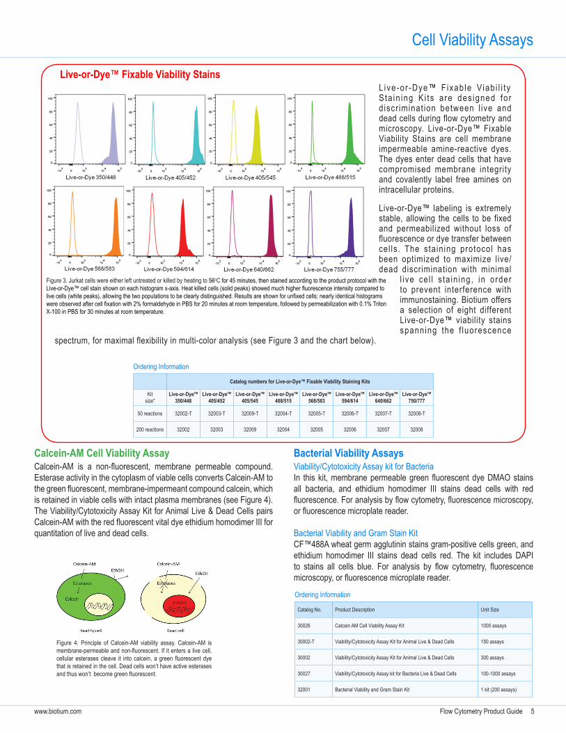
L i ve -o r -Dye™ F ixab le Viab i l i t y S ta in ing K i t s a re des igned fo r d iscr iminat ion between l ive and dead cells during flow cytometry and microscopy. Live-or-Dye™ Fixable Viability Stains are cell membrane impermeable amine-reactive dyes. The dyes enter dead cells that have compromised membrane integrity and covalently label free amines on intracellular proteins.
Live-or-Dye™ labeling is extremely stable, allowing the cells to be fixed and permeabilized without loss of fluorescence or dye transfer between cel ls . The stain ing protocol has been optimized to maximize live/dead discrimination with minimal
l i ve ce l l s ta in ing, in order to prevent interference with immunostaining. Biotium offers a selection of eight different Live-or-Dye™ viability stains spanning the f luorescence
spectrum, for maximal flexibility in multi-color analysis (see Figure 3 and the chart below).
Cell Viability Assays
Live-or-Dye™ Fixable Viability Stains
Catalog numbers for Live-or-Dye™ Fixable Viability Staining Kits
Kit size*
Live-or-DyeTM 350/448
Live-or-DyeTM 405/452
Live-or-DyeTM 405/545
Live-or-DyeTM 488/515
Live-or-DyeTM 568/583
Live-or-DyeTM 594/614
Live-or-DyeTM 640/662
Live-or-DyeTM 750/777
50 reactions 32002-T 32003-T 32009-T 32004-T 32005-T 32006-T 32007-T 32008-T
200 reactions 32002 32003 32009 32004 32005 32006 32007 32008
Calcein-AM Cell Viability AssayCalcein-AM is a non-fluorescent, membrane permeable compound. Esterase activity in the cytoplasm of viable cells converts Calcein-AM to the green fluorescent, membrane-impermeant compound calcein, which is retained in viable cells with intact plasma membranes (see Figure 4). The Viability/Cytotoxicity Assay Kit for Animal Live & Dead Cells pairs Calcein-AM with the red fluorescent vital dye ethidium homodimer III for quantitation of live and dead cells.
Ordering Information
Figure 3. Jurkat cells were either left untreated or killed by heating to 56oC for 45 minutes, then stained according to the product protocol with the Live-or-Dye™ cell stain shown on each histogram x-axis. Heat killed cells (solid peaks) showed much higher fluorescence intensity compared to live cells (white peaks), allowing the two populations to be clearly distinguished. Results are shown for unfixed cells; nearly identical histograms were observed after cell fixation with 2% formaldehyde in PBS for 20 minutes at room temperature, followed by permeabilization with 0.1% Triton X-100 in PBS for 30 minutes at room temperature.
Viability/Cytotoxicity Assay kit for BacteriaIn this kit, membrane permeable green fluorescent dye DMAO stains all bacteria, and ethidium homodimer III stains dead cells with red fluorescence. For analysis by flow cytometry, fluorescence microscopy, or fluorescence microplate reader.
Bacterial Viability and Gram Stain KitCF™488A wheat germ agglutinin stains gram-positive cells green, and ethidium homodimer III stains dead cells red. The kit includes DAPI to stains all cells blue. For analysis by flow cytometry, fluorescence microscopy, or fluorescence microplate reader.
Bacterial Viability Assays
Ordering Information
Figure 4. Principle of Calcein-AM viability assay. Calcein-AM is membrane-permeable and non-fluorescent. If it enters a live cell, cellular esterases cleave it into calcein, a green fluorescent dye that is retained in the cell. Dead cells won’t have active esterases and thus won’t become green fluorescent.
6 Flow Cytometry Product Guide www.biotium.com
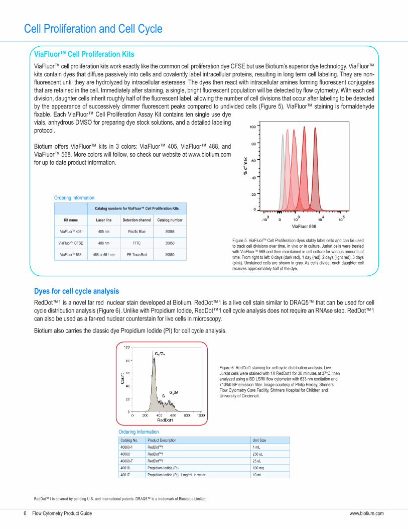
ViaFluor™ cell proliferation kits work exactly like the common cell proliferation dye CFSE but use Biotium’s superior dye technology. ViaFluor™ kits contain dyes that diffuse passively into cells and covalently label intracellular proteins, resulting in long term cell labeling. They are non-fluorescent until they are hydrolyzed by intracellular esterases. The dyes then react with intracellular amines forming fluorescent conjugates that are retained in the cell. Immediately after staining, a single, bright fluorescent population will be detected by flow cytometry. With each cell division, daughter cells inherit roughly half of the fluorescent label, allowing the number of cell divisions that occur after labeling to be detected by the appearance of successively dimmer fluorescent peaks compared to undivided cells (Figure 5). ViaFluor™ staining is formaldehyde fixable. Each ViaFluor™ Cell Proliferation Assay Kit contains ten single use dye vials, anhydrous DMSO for preparing dye stock solutions, and a detailed labeling protocol.
Biotium offers ViaFluor™ kits in 3 colors: ViaFluor™ 405, ViaFluor™ 488, and ViaFluor™ 568. More colors will follow, so check our website at www.biotium.com for up to date product information.
ViaFluorTM Cell Proliferation Kits
Cell Proliferation and Cell Cycle
Catalog numbers for ViaFluor™ Cell Proliferation Kits
Kit name Laser line Detection channel Catalog number
ViaFluorTM 405 405 nm Pacific Blue 30068
ViaFluorTM CFSE 488 nm FITC 30050
ViaFluorTM 568 488 or 561 nm PE-TexasRed 30080
Ordering Information
Figure 5. ViaFluorTM Cell Proliferation dyes stably label cells and can be used to track cell divisions over time, in vivo or in culture. Jurkat cells were treated with ViaFluorTM 568 and then maintained in cell culture for various amounts of time. From right to left: 0 days (dark red), 1 day (red), 2 days (light red), 3 days (pink). Unstained cells are shown in gray. As cells divide, each daughter cell receives approximately half of the dye.
RedDot™1 is a novel far red nuclear stain developed at Biotium. RedDot™1 is a live cell stain similar to DRAQ5™ that can be used for cell cycle distribution analysis (Figure 6). Unlike with Propidium Iodide, RedDot™1 cell cycle analysis does not require an RNAse step. RedDot™1 can also be used as a far-red nuclear counterstain for live cells in microscopy.
Biotium also carries the classic dye Propidium Iodide (PI) for cell cycle analysis.
Figure 6. RedDot1 staining for cell cycle distribution analysis. Live Jurkat cells were stained with 1X RedDot1 for 30 minutes at 37oC, then analyzed using a BD LSRII flow cytometer with 633 nm excitation and 710/50 BP emission filter. Image courtesy of Philip Hexley, Shriners Flow Cytometry Core Facility, Shriners Hospital for Children and University of Cincinnati.
Dyes for cell cycle analysis
RedDot™1 is covered by pending U.S. and international patents. DRAQ5™ is a trademark of Biostatus Limited.
Catalog No. Product Description Unit Size40060-1 RedDotTM1 1 mL40060 RedDotTM1 250 uL40060-T RedDotTM1 25 uL40016 Propidium Iodide (PI) 100 mg40017 Propidium Iodide (PI), 1 mg/mL in water 10 mL
Ordering Information
Flow Cytometry Product Guide 7www.biotium.com
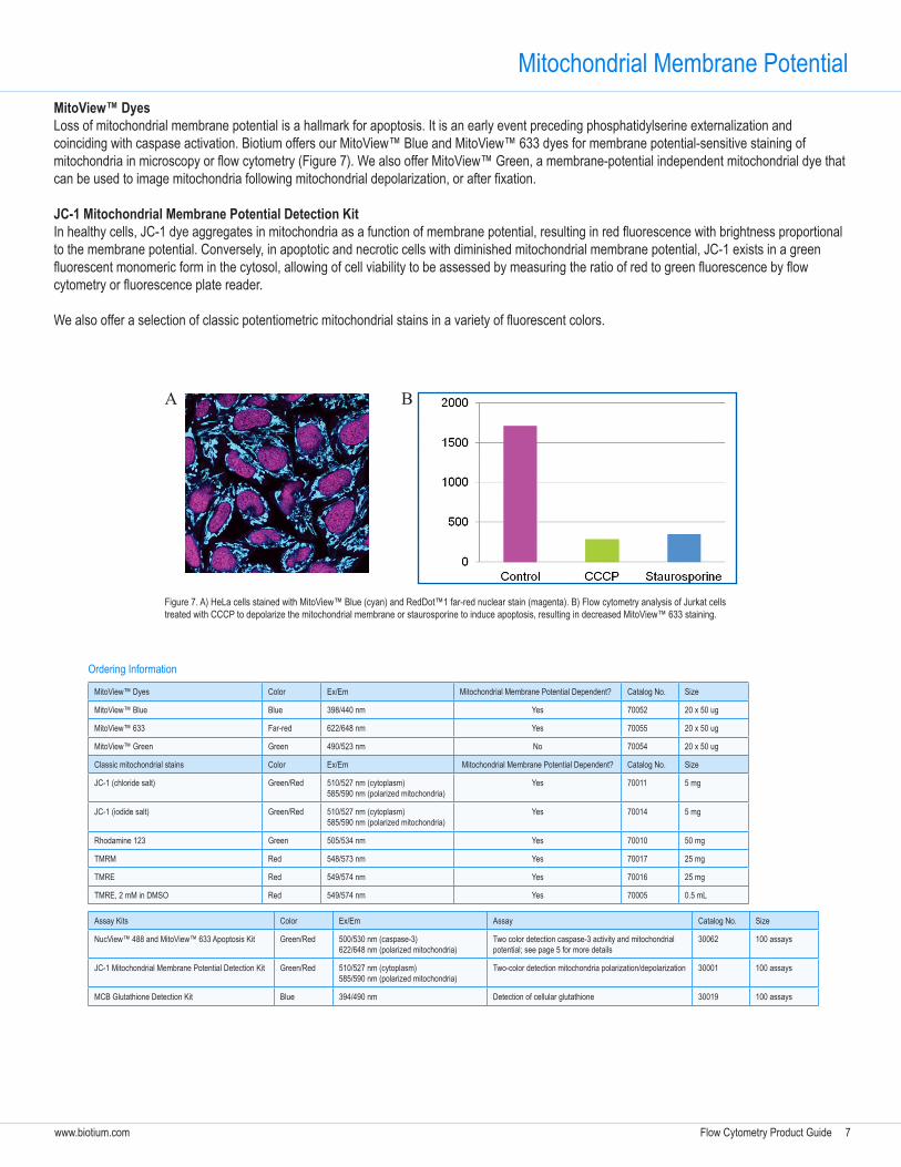
Mitochondrial Membrane PotentialMitoView™ DyesLoss of mitochondrial membrane potential is a hallmark for apoptosis. It is an early event preceding phosphatidylserine externalization and coinciding with caspase activation. Biotium offers our MitoView™ Blue and MitoView™ 633 dyes for membrane potential-sensitive staining of mitochondria in microscopy or flow cytometry (Figure 7). We also offer MitoView™ Green, a membrane-potential independent mitochondrial dye that can be used to image mitochondria following mitochondrial depolarization, or after fixation.
JC-1 Mitochondrial Membrane Potential Detection Kit In healthy cells, JC-1 dye aggregates in mitochondria as a function of membrane potential, resulting in red fluorescence with brightness proportional to the membrane potential. Conversely, in apoptotic and necrotic cells with diminished mitochondrial membrane potential, JC-1 exists in a green fluorescent monomeric form in the cytosol, allowing of cell viability to be assessed by measuring the ratio of red to green fluorescence by flow cytometry or fluorescence plate reader.
We also offer a selection of classic potentiometric mitochondrial stains in a variety of fluorescent colors.
MitoView™ Dyes Color Ex/Em Mitochondrial Membrane Potential Dependent? Catalog No. Size
MitoView™ Blue Blue 398/440 nm Yes 70052 20 x 50 ug
MitoView™ 633 Far-red 622/648 nm Yes 70055 20 x 50 ug
MitoView™ Green Green 490/523 nm No 70054 20 x 50 ug
Classic mitochondrial stains Color Ex/Em Mitochondrial Membrane Potential Dependent? Catalog No. Size
JC-1 (chloride salt) Green/Red 510/527 nm (cytoplasm)585/590 nm (polarized mitochondria)
Yes 70011 5 mg
JC-1 (iodide salt) Green/Red 510/527 nm (cytoplasm)585/590 nm (polarized mitochondria)
Yes 70014 5 mg
Rhodamine 123 Green 505/534 nm Yes 70010 50 mg
TMRM Red 548/573 nm Yes 70017 25 mg
TMRE Red 549/574 nm Yes 70016 25 mg
TMRE, 2 mM in DMSO Red 549/574 nm Yes 70005 0.5 mL
Assay Kits Color Ex/Em Assay Catalog No. Size
NucView™ 488 and MitoView™ 633 Apoptosis Kit Green/Red 500/530 nm (caspase-3)622/648 nm (polarized mitochondria)
Two color detection caspase-3 activity and mitochondrial potential; see page 5 for more details
30062 100 assays
JC-1 Mitochondrial Membrane Potential Detection Kit Green/Red 510/527 nm (cytoplasm)585/590 nm (polarized mitochondria)
Two-color detection mitochondria polarization/depolarization 30001 100 assays
MCB Glutathione Detection Kit Blue 394/490 nm Detection of cellular glutathione 30019 100 assays
Ordering Information
Figure 7. A) HeLa cells stained with MitoView™ Blue (cyan) and RedDot™1 far-red nuclear stain (magenta). B) Flow cytometry analysis of Jurkat cells treated with CCCP to depolarize the mitochondrial membrane or staurosporine to induce apoptosis, resulting in decreased MitoView™ 633 staining.
A B
8 Flow Cytometry Product Guide www.biotium.com
NucViewTM Caspase-3 Substrates
Proteolysis of cellular substrates by caspase-3 results in the morphological and biochemical features of apoptosis. NucView™ 488 Caspase-3 Substrate is a novel cell membrane-permeable fluorogenic caspase substrate designed for detecting caspase-3 activity in real time.
Traditional fluorogenic caspase substrates require cell lysis and cannot be used to measure caspase activity in live cells. Fluorescently-labeled caspase inhibitor assay (FLICA) reagents can enter live cells to detect caspase activity, but because the fluorescent probes are also irreversible caspase inhibitors, they cannot be used to follow caspase activity in real time.
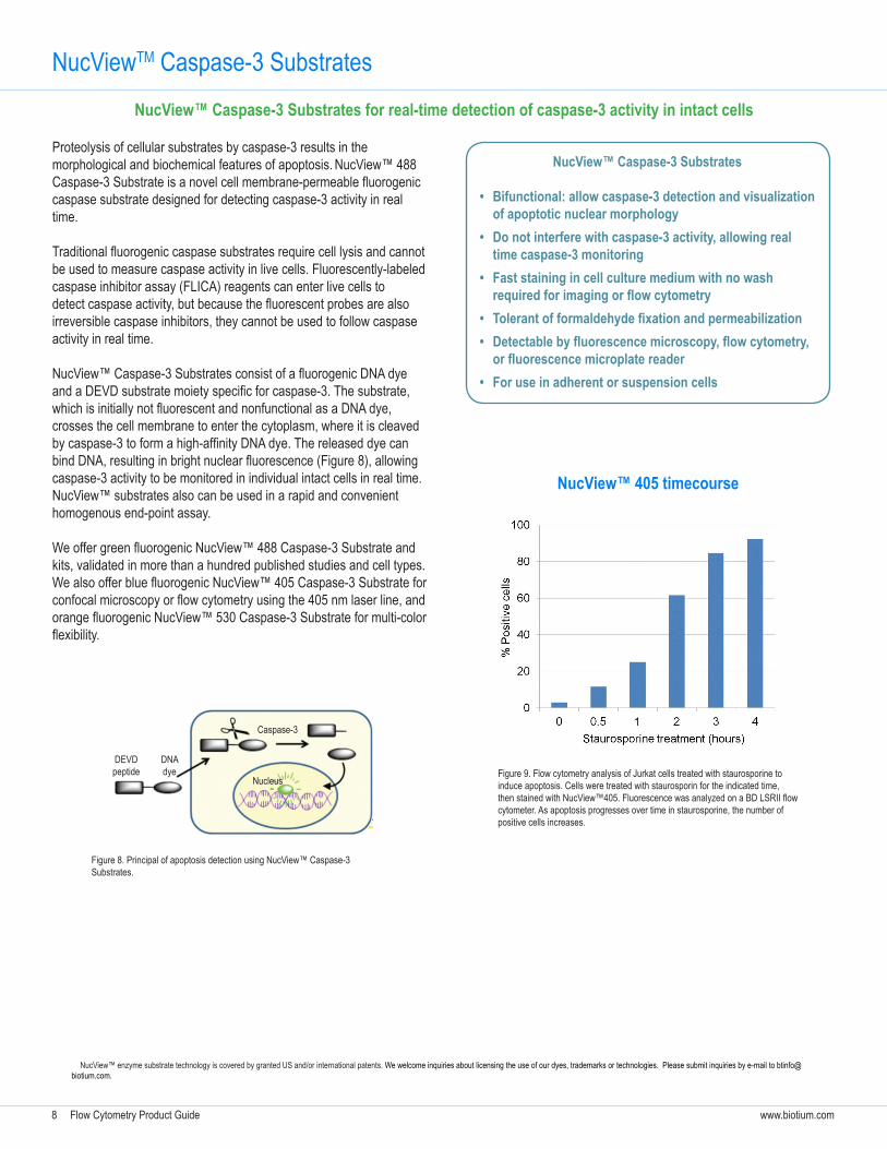
NucView™ Caspase-3 Substrates consist of a fluorogenic DNA dye and a DEVD substrate moiety specific for caspase-3. The substrate, which is initially not fluorescent and nonfunctional as a DNA dye, crosses the cell membrane to enter the cytoplasm, where it is cleaved by caspase-3 to form a high-affinity DNA dye. The released dye can bind DNA, resulting in bright nuclear fluorescence (Figure 8), allowing caspase-3 activity to be monitored in individual intact cells in real time. NucView™ substrates also can be used in a rapid and convenient homogenous end-point assay.
We offer green fluorogenic NucView™ 488 Caspase-3 Substrate and kits, validated in more than a hundred published studies and cell types. We also offer blue fluorogenic NucView™ 405 Caspase-3 Substrate for confocal microscopy or flow cytometry using the 405 nm laser line, and orange fluorogenic NucView™ 530 Caspase-3 Substrate for multi-color flexibility.
NucView™ Caspase-3 Substrates
• Bifunctional: allow caspase-3 detection and visualization of apoptotic nuclear morphology
• Do not interfere with caspase-3 activity, allowing real time caspase-3 monitoring
• Fast staining in cell culture medium with no wash requiredforimagingorflowcytometry
• Tolerantofformaldehydefixationandpermeabilization• Detectablebyfluorescencemicroscopy,flowcytometry,orfluorescencemicroplatereader
• For use in adherent or suspension cells
Figure 8. Principal of apoptosis detection using NucView™ Caspase-3 Substrates.
DEVDpeptide
DNAdye
Caspase-3
Nucleus
NucView™ enzyme substrate technology is covered by granted US and/or international patents. We welcome inquiries about licensing the use of our dyes, trademarks or technologies. Please submit inquiries by e-mail to [email protected].
NucView™ Caspase-3 Substrates for real-time detection of caspase-3 activity in intact cells
Figure 9. Flow cytometry analysis of Jurkat cells treated with staurosporine to induce apoptosis. Cells were treated with staurosporin for the indicated time, then stained with NucView™405. Fluorescence was analyzed on a BD LSRII flow cytometer. As apoptosis progresses over time in staurosporine, the number of positive cells increases.
NucView™ 405 timecourse
Flow Cytometry Product Guide 9www.biotium.com
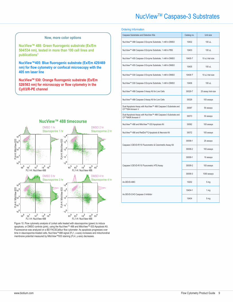
Figure 10. Flow cytometry analysis of Jurkat cells treated with staurosporine (green) to induce apoptosis, or DMSO controls (pink), using the NucView™ 488 and MitoView™ 633 Apoptosis Kit. Fluorescence was analyzed on a BD FACSCalibur flow cytometer. As apoptosis progresses over time in staurosporine-treated cells, NucView™488 signal (FL1, x-axis) increases and mitochondrial membrane potential measured by MitoView™633 staining (FL4, y-axis) decreases.
Now, more color options
NucView™488:Greenfluorogenicsubstrate(Ex/Em504/534 nm), tested in more than 100 cell lines and publications*
NucView™405:Bluefluorogenicsubstrate(Ex/Em429/469nm)forflowcytometryorconfocalmicroscopywiththe405 nm laser line
NucViewTM530:Orangefluorogenicsubstrate(Ex/Em528/563nm)formicroscopyorflowcytometryintheCy®3/R-PE channel
Caspase Substrates and Detection Kits Catalog no. Unit size
NucView™ 488 Caspase-3 Enzyme Substrate, 1 mM in DMSO 10402 100 uL
NucView™ 488 Caspase-3 Enzyme Substrate, 1 mM in PBS 10403 100 uL
NucView™ 405 Caspase-3 Enzyme Substrate, 1 mM in DMSO 10405-T 10 uL trial size
NucView™ 405 Caspase-3 Enzyme Substrate, 1 mM in DMSO 10405 100 uL
NucView™ 530 Caspase-3 Enzyme Substrate, 1 mM in DMSO 10406-T 10 uL trial size
NucView™ 530 Caspase-3 Enzyme Substrate, 1 mM in DMSO 10406 100 uL
NucView™ 488 Caspase-3 Assay Kit for Live Cells 30029-T 25 assay trial size
NucView™ 488 Caspase-3 Assay Kit for Live Cells 30029 100 assays
Dual Apoptosis Assay with NucView™ 488 Caspase-3 Substrate and CF™594 Annexin V 30067 50 assays
Dual Apoptosis Assay with NucView™ 488 Caspase-3 Substrate and CF™640R Annexin V 30073 50 assays
NucView™ 488 and MitoView™ 633 Apoptosis Kit 30062 100 assays
NucView™ 488 and RedDot™2 Apoptosis & Necrosis Kit 30072 100 assays
Caspase-3 DEVD-R110 Fluorometric & Colorimetric Assay Kit
30008-1 25 assays
30008-2 100 assays
Caspase-3 DEVD-R110 Fluorometric HTS Assay
30009-1 10 assays
30009-2 100 assays
30009-3 1000 assays
Ac-DEVD-AMC 10202 5 mg
Ac-DEVD-CHO Caspase-3 Inhibitor
10404-1 1 mg
10404 5 mg
DMSO 1 hrStaurosporine 1 hr
DMSO 2 hrStaurosporine 2 hr
DMSO 3 hrStaurosporine 3 hr
DMSO 4 hrStaurosporine 4 hr
FL1-H: NucView 488
FL4-
H: M
itoVi
ew 63
3
FL1-H: NucView 488
FL4-
H: M
itoVi
ew 63
3
FL1-H: NucView 488
FL4-
H: M
itoVi
ew 63
3
FL1-H: NucView 488
FL4-
H: M
itoVi
ew 63
3
NucView™ 488 timecourse
Ordering Information
NucViewTM Caspase-3 Substrates
10 Flow Cytometry Product Guide www.biotium.com
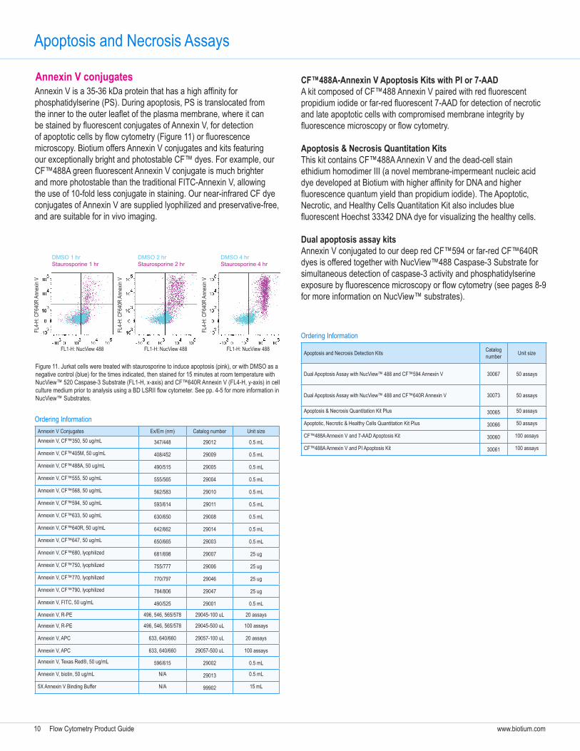
Annexin V is a 35-36 kDa protein that has a high affinity for phosphatidylserine (PS). During apoptosis, PS is translocated from the inner to the outer leaflet of the plasma membrane, where it can be stained by fluorescent conjugates of Annexin V, for detection of apoptotic cells by flow cytometry (Figure 11) or fluorescence microscopy. Biotium offers Annexin V conjugates and kits featuring our exceptionally bright and photostable CF™ dyes. For example, our CF™488A green fluorescent Annexin V conjugate is much brighter and more photostable than the traditional FITC-Annexin V, allowing the use of 10-fold less conjugate in staining. Our near-infrared CF dye conjugates of Annexin V are supplied lyophilized and preservative-free, and are suitable for in vivo imaging.
CF™488A-Annexin V Apoptosis Kits with PI or 7-AADA kit composed of CF™488 Annexin V paired with red fluorescent propidium iodide or far-red fluorescent 7-AAD for detection of necrotic and late apoptotic cells with compromised membrane integrity by fluorescence microscopy or flow cytometry.
Apoptosis & Necrosis Quantitation KitsThis kit contains CF™488A Annexin V and the dead-cell stain ethidium homodimer III (a novel membrane-impermeant nucleic acid dye developed at Biotium with higher affinity for DNA and higher fluorescence quantum yield than propidium iodide). The Apoptotic, Necrotic, and Healthy Cells Quantitation Kit also includes blue fluorescent Hoechst 33342 DNA dye for visualizing the healthy cells.
Dual apoptosis assay kitsAnnexin V conjugated to our deep red CF™594 or far-red CF™640R dyes is offered together with NucView™488 Caspase-3 Substrate for simultaneous detection of caspase-3 activity and phosphatidylserine exposure by fluorescence microscopy or flow cytometry (see pages 8-9 for more information on NucView™ substrates).
DMSO 1 hrStaurosporine 1 hr
DMSO 2 hrStaurosporine 2 hr
DMSO 4 hrStaurosporine 4 hr
FL1-H: NucView 488
FL4-
H: C
F640
R An
nexin
V
FL1-H: NucView 488
FL4-
H: C
F640
R An
nexin
V
FL1-H: NucView 488
FL4-
H: C
F640
R An
nexin
V
Annexin V Conjugates Ex/Em (nm) Catalog number Unit sizeAnnexin V, CF™350, 50 ug/mL 347/448 29012 0.5 mL
Annexin V, CF™405M, 50 ug/mL 408/452 29009 0.5 mL
Annexin V, CF™488A, 50 ug/mL 490/515 29005 0.5 mL
Annexin V, CF™555, 50 ug/mL 555/565 29004 0.5 mL
Annexin V, CF™568, 50 ug/mL 562/583 29010 0.5 mL
Annexin V, CF™594, 50 ug/mL 593/614 29011 0.5 mL
Annexin V, CF™633, 50 ug/mL 630/650 29008 0.5 mL
Annexin V, CF™640R, 50 ug/mL 642/662 29014 0.5 mL
Annexin V, CF™647, 50 ug/mL 650/665 29003 0.5 mL
Annexin V, CF™680, lyophilized 681/698 29007 25 ug
Annexin V, CF™750, lyophilized 755/777 29006 25 ug
Annexin V, CF™770, lyophilized 770/797 29046 25 ug
Annexin V, CF™790, lyophilized 784/806 29047 25 ug
Annexin V, FITC, 50 ug/mL 490/525 29001 0.5 mL
Annexin V, R-PE 496, 546, 565/578 29045-100 uL 20 assays
Annexin V, R-PE 496, 546, 565/578 29045-500 uL 100 assays
Annexin V, APC 633, 640/660 29057-100 uL 20 assays
Annexin V, APC 633, 640/660 29057-500 uL 100 assays
Annexin V, Texas Red®, 50 ug/mL 596/615 29002 0.5 mL
Annexin V, biotin, 50 ug/mL N/A 29013 0.5 mL
5X Annexin V Binding Buffer N/A 99902 15 mL
Apoptosis and Necrosis Detection Kits Catalog number Unit size
Dual Apoptosis Assay with NucView™ 488 and CF™594 Annexin V 30067 50 assays
Dual Apoptosis Assay with NucView™ 488 and CF™640R Annexin V 30073 50 assays
Apoptosis & Necrosis Quantitation Kit Plus 30065 50 assays
Apoptotic, Necrotic & Healthy Cells Quantitation Kit Plus 30066 50 assays
CF™488A Annexin V and 7-AAD Apoptosis Kit 30060 100 assays
CF™488A Annexin V and PI Apoptosis Kit 30061 100 assays
Figure 11. Jurkat cells were treated with staurosporine to induce apoptosis (pink), or with DMSO as a negative control (blue) for the times indicated, then stained for 15 minutes at room temperature with NucView™ 520 Caspase-3 Substrate (FL1-H, x-axis) and CF™640R Annexin V (FL4-H, y-axis) in cell culture medium prior to analysis using a BD LSRII flow cytometer. See pp. 4-5 for more information in NucView™ Substrates.
Apoptosis and Necrosis Assays
Annexin V conjugates
Ordering Information
Ordering Information
Flow Cytometry Product Guide 11www.biotium.com
AccuEasy™ is a novel method for staining adherent cells for flow cytometry analysis. Conventional flow cytometry staining protocols for cell surface markers on adherent cells require detachment of cells from their culture surface before performing antibody staining. Unfortunately, cell detachment introduces a significant stress to cells which can alter the expression of cell surface markers.
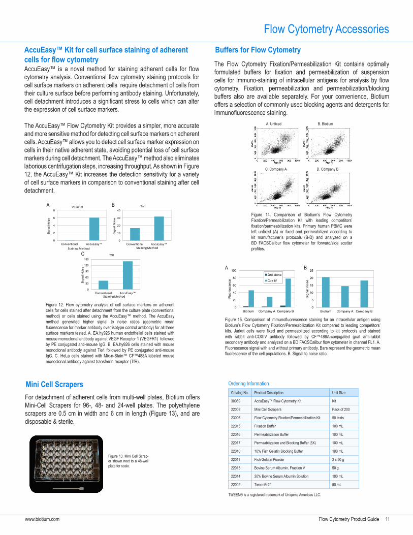
The AccuEasy™ Flow Cytometry Kit provides a simpler, more accurate and more sensitive method for detecting cell surface markers on adherent cells. AccuEasy™ allows you to detect cell surface marker expression on cells in their native adherent state, avoiding potential loss of cell surface markers during cell detachment. The AccuEasy™ method also eliminates laborious centrifugation steps, increasing throughput. As shown in Figure 12, the AccuEasy™ Kit increases the detection sensitivity for a variety of cell surface markers in comparison to conventional staining after cell detachment.
Flow Cytometry AccessoriesAccuEasy™ Kit for cell surface staining of adherent cellsforflowcytometry
For detachment of adherent cells from multi-well plates, Biotium offers Mini-Cell Scrapers for 96-, 48- and 24-well plates. The polyethylene scrapers are 0.5 cm in width and 6 cm in length (Figure 13), and are disposable & sterile.
0
2
4
6
8
Conventional AccuEasy™
Sig
nal:N
oise
VEGFR1
Staining Method
A
0
30
60
90
120
150
Conventional AccuEasy™
Sig
nal:N
oise
TfR
Staining Method
C
Figure 12. Flow cytometry analysis of cell surface markers on adherent cells for cells stained after detachment from the culture plate (conventional method) or cells stained using the AccuEasy™ method. The AccuEasy method generated higher signal to noise ratios (geometric mean fluorescence for marker antibody over isotype control antibody) for all three surface markers tested. A. EA.hy926 human endothelial cells stained with mouse monoclonal antibody against VEGF Receptor 1 (VEGFR1) followed by PE conjugated anti-mouse IgG. B. EA.hy926 cells stained with mouse monoclonal antibody against Tie1 followed by PE conjugated anti-mouse IgG. C. HeLa cells stained with Mix-n-Stain™ CF™488A labeled mouse monoclonal antibody against transferrin receptor (TfR).
Mini Cell Scrapers
Figure 13. Mini Cell Scrap-er shown next to a 48-well plate for scale.
Buffers for Flow Cytometry
C. Company A D. Company B
Figure 14. Comparison of Biotium’s Flow Cytometry Fixation/Permeabilization Kit with leading competitors’ fixation/permeabilization kits. Primary human PBMC were left unfixed (A) or fixed and permeabilized according to kit manufacturer’s protocols (B-D) and analyzed on a BD FACSCalibur flow cytometer for forward/side scatter profiles.
Figure 15. Comparison of immunofluorescence staining for an intracellular antigen using Biotium’s Flow Cytometry Fixation/Permeabilization Kit compared to leading competitors’ kits. Jurkat cells were fixed and permeabilized according to kit protocols and stained with rabbit anti-COXIV antibody followed by CF™488A-conjugated goat anti-rabbit secondary antibody and analyzed on a BD FACSCalibur flow cytometer in channel FL1. A. Fluorescence signal with and without primary antibody. Bars represent the geometric mean fluorescence of the cell populations. B. Signal to noise ratio.
0
20
40
60
80
100
Biotium Company A Company B
Fluo
resc
ence
2nd alone
Cox IV
0
5
10
15
20
25
Biotium Company A Company BS
igna
l: no
ise
A B
The Flow Cytometry Fixation/Permeabilization Kit contains optimally formulated buffers for fixation and permeabilization of suspension cells for immuno-staining of intracellular antigens for analysis by flow cytometry. Fixation, permeabilization and permeabilization/blocking buffers also are available separately. For your convenience, Biotium offers a selection of commonly used blocking agents and detergents for immunofluorescence staining.
Catalog No. Product Description Unit Size
30069 AccuEasy™ Flow Cytometry Kit Kit
22003 Mini Cell Scrapers Pack of 200
23006 Flow Cytometry Fixation/Permeabilization Kit 50 tests
22015 Fixation Buffer 100 mL
22016 Permeabilization Buffer 100 mL
22017 Permeabilization and Blocking Buffer (5X) 100 mL
22010 10% Fish Gelatin Blocking Buffer 100 mL
22011 Fish Gelatin Powder 2 x 50 g
22013 Bovine Serum Albumin, Fraction V 50 g
22014 30% Bovine Serum Albumin Solution 100 mL
22002 Tween®-20 50 mL
TWEEN® is a registered trademark of Uniqema Americas LLC.
Ordering Information
A. Unfixed B. Biotium
0
10
20
30
40
Conventional AccuEasy™
Sig
nal:N
oise
Tie1
Staining Method
B

Biotium, Inc.www.biotium.com
800-304-5357
General [email protected]
Quotes and [email protected]
Technical [email protected]
Cover art © 2013 Micah Burke
www.biotium.comGlowing Products for Science™


![Glucose Metabolism and AMPK Signaling Regulate ...bionmr.unl.edu/files/publications/128.pdf(Molecular Probes). Flow cytometry was performed as de-scribed previously [10, 30, 39, 40].](https://static.fdocument.org/doc/165x107/5f481d3d3b482616b93519a1/glucose-metabolism-and-ampk-signaling-regulate-molecular-probes-flow-cytometry.jpg)