Proceedings of the 6th Pacific Rim Conference on the ...
6
6 th Pacific Rim Conference on the Biotechnology of Bacillus thuringiensis and its Environmental Impact, Victoria BC, 2005 Côté, J.-C., Otvos, I.S., Schwartz, J.-L. and Vincent, C. (eds) Bacillus thuringiensis (Bt) is an aerobic, spore-forming bacterium that produces, during the sporulation phase, crystalline inclusions composed of proteins known as δ-endotoxins (3). The δ-endotoxins form two multigenic families, Cry and Cyt toxins. Cry proteins are specifically toxic to several orders of insects: Lepidoptera, Coleoptera, Hymenoptera, Diptera, and to nematodes. In contrast, Cyt toxins are mostly found in Bt strains active against Diptera (3). Cry and Cyt proteins are produced as protoxins that are activated by midgut proteases to release their toxin fragment (3). Cry and Cyt toxins exert their pathological effect by forming lytic pores in the membrane of midgut epithelial cells (3). Cry toxins are composed of three functional domains, an α-helical domain involved in membrane insertion (domain I) and two β-sheet domains (domains II and III) involved in receptor interaction (3). Cyt proteins, on the other hand, have a single α−β domain comprising of two outer layers of α- helix hairpins wrapped around a β-sheet (3) (Fig. 1). Cry toxins bind to specific protein receptors in the microvilli of midgut epithelial cells (3), in contrast to Cyt toxins that do not bind to protein receptors but interact directly with membrane lipids, resulting in insertion into the membrane and pore formation (32), or in membrane destruction by detergent-like interaction (6). Important Interactions with Membrane Receptors in the Mode of Action of Bacillus thuringiensis Cry Toxins * Corresponding author. Mailing address: Instituto de Biotecnología, Universidad Nacional Autónoma de México, 2001 Av. Universidad, Col. Chamilpa, Apdo. postal 510-3, Cuernavaca, Morelos, Mexico, 62250. Tel: 52 73 11-4900. Fax: 52 73 17-2388. Email: [email protected]. Mario Soberón 1* , Isabel Gómez 1 , Liliana Pardo 1 , Carlos Muñoz 1 , Luisa E. Fernandez 1 , Claudia Pérez 1 , Sarjeet S. Gill 2 , and Alejandra Bravo 1 . 1 Instituto de Biotecnología, Universidad Nacional Autónoma de México, Cuernavaca, Morelos, Mexico, 62250. 2 Department of Cell Biology and Neuroscience, University of California, Riverside, CA, U.S.A, 92506. In this review we summarize recent findings on the Cry toxin-receptor interaction and its role in the mode of action of these toxins. Cry toxins interact with multiple receptor molecules leading to the formation of membrane pores in membranes of susceptible insects. In lepidopteran insects, Cry toxins interact with a cadherin receptor and with glycosyl-phosphatidyl-inositol (GPI)- anchored proteins (aminopeptidase N and alkaline phosphatase) leading to insertion of oligomeric toxin in membrane microdomains (or lipid rafts) and inducing cell swelling. Furthermore, recent data shows that in addition to protein receptors involved in Cry toxin interaction with the membrane of susceptible organisms, certain glycolipids also play a role in toxin action. These glycolipids are specific to invertebrate organisms like insects and nematodes. Recent progress in the characterization of mosquitocidal Cry toxin GPI-anchored protein receptors will also be reviewed. The conserved structure of Cry toxins and the characterized receptor molecules tend to suggest that similar receptor molecules may be involved in Cry toxin action on different insects. Introduction FIG. 1. Three-dimensional structures of insecticidal toxins produced by Bacillus thuringiensis. A. Structure of Cry1Aa toxin showing three domains: domain I in blue and domains II and III in magenta. The loop regions involved in receptor interaction are on the apex of the structure. B. Structure of Cyt2A toxin. A B Cry toxins are being used worldwide in transgenic plants for insect pest control. They are also used to control mosquitoes, the vectors of important human diseases (2, 3). The major threat to the use of Cry toxins is the appearance of insect resistance. The most frequent mechanism of resistance to Cry toxins is receptor-binding impairment (12). For Cry1A lepidopteran toxins, at least four different protein receptors have been characterized: a cadherin-like protein (CADP), a glycosylphosphatidyl-
Transcript of Proceedings of the 6th Pacific Rim Conference on the ...
6th Pacific Rim Conference on the Biotechnology of Bacillus thuringiensis and its Environmental Impact, Victoria BC, 2005 Côté, J.-C., Otvos, I.S., Schwartz, J.-L. and Vincent, C. (eds)
4
Bacillus thuringiensis (Bt) is an aerobic, spore-forming bacterium that produces, during the sporulation phase, crystalline inclusions composed of proteins known as δ-endotoxins (3). The δ-endotoxins form two multigenic families, Cry and Cyt toxins. Cry proteins are specifically toxic to several orders of insects: Lepidoptera, Coleoptera, Hymenoptera, Diptera, and to nematodes. In contrast, Cyt toxins are mostly found in Bt strains active against Diptera (3).
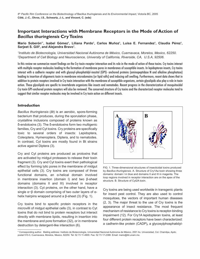
Cry and Cyt proteins are produced as protoxins that are activated by midgut proteases to release their toxin fragment (3). Cry and Cyt toxins exert their pathological effect by forming lytic pores in the membrane of midgut epithelial cells (3). Cry toxins are composed of three functional domains, an α-helical domain involved in membrane insertion (domain I) and two β-sheet domains (domains II and III) involved in receptor interaction (3). Cyt proteins, on the other hand, have a single α−β domain comprising of two outer layers of α-helix hairpins wrapped around a β-sheet (3) (Fig. 1).
Cry toxins bind to specific protein receptors in the microvilli of midgut epithelial cells (3), in contrast to Cyt toxins that do not bind to protein receptors but interact directly with membrane lipids, resulting in insertion into the membrane and pore formation (32), or in membrane destruction by detergent-like interaction (6).
Important Interactions with Membrane Receptors in the Mode of Action of Bacillus thuringiensis Cry Toxins
* Corresponding author. Mailing address: Instituto de Biotecnología, Universidad Nacional Autónoma de México, 2001 Av. Universidad, Col. Chamilpa, Apdo. postal 510-3, Cuernavaca, Morelos, Mexico, 62250. Tel: 52 73 11-4900. Fax: 52 73 17-2388. Email: [email protected].
Mario Soberón1*, Isabel Gómez1, Liliana Pardo1, Carlos Muñoz1, Luisa E. Fernandez1, Claudia Pérez1, Sarjeet S. Gill2, and Alejandra Bravo1.1Instituto de Biotecnología, Universidad Nacional Autónoma de México, Cuernavaca, Morelos, Mexico, 62250. 2Department of Cell Biology and Neuroscience, University of California, Riverside, CA, U.S.A, 92506.
In this review we summarize recent findings on the Cry toxin-receptor interaction and its role in the mode of action of these toxins. Cry toxins interact with multiple receptor molecules leading to the formation of membrane pores in membranes of susceptible insects. In lepidopteran insects, Cry toxins interact with a cadherin receptor and with glycosyl-phosphatidyl-inositol (GPI)- anchored proteins (aminopeptidase N and alkaline phosphatase) leading to insertion of oligomeric toxin in membrane microdomains (or lipid rafts) and inducing cell swelling. Furthermore, recent data shows that in addition to protein receptors involved in Cry toxin interaction with the membrane of susceptible organisms, certain glycolipids also play a role in toxin action. These glycolipids are specific to invertebrate organisms like insects and nematodes. Recent progress in the characterization of mosquitocidal Cry toxin GPI-anchored protein receptors will also be reviewed. The conserved structure of Cry toxins and the characterized receptor molecules tend to suggest that similar receptor molecules may be involved in Cry toxin action on different insects.
Introduction
FIG. 1. Three-dimensional structures of insecticidal toxins produced by Bacillus thuringiensis. A. Structure of Cry1Aa toxin showing three domains: domain I in blue and domains II and III in magenta. The loop regions involved in receptor interaction are on the apex of the structure. B. Structure of Cyt2A toxin.
A BA B
Cry toxins are being used worldwide in transgenic plants for insect pest control. They are also used to control mosquitoes, the vectors of important human diseases (2, 3). The major threat to the use of Cry toxins is the appearance of insect resistance. The most frequent mechanism of resistance to Cry toxins is receptor-binding impairment (12). For Cry1A lepidopteran toxins, at least four different protein receptors have been characterized: a cadherin-like protein (CADP), a glycosylphosphatidyl-
2
loop a 8
loop2RRPFNIGI
SFRGSAQGIEGS
HITDTNNK
IPLPASILTV
loop3GFNSNSSVSI
TGVLTLNIQ
loop a 8
loop2RRPFNIGI
SFRGSAQGIEGS
HITDTNNK
IPLPASILTV
loop3GFNSNSSVSI
TGVLTLNIQ
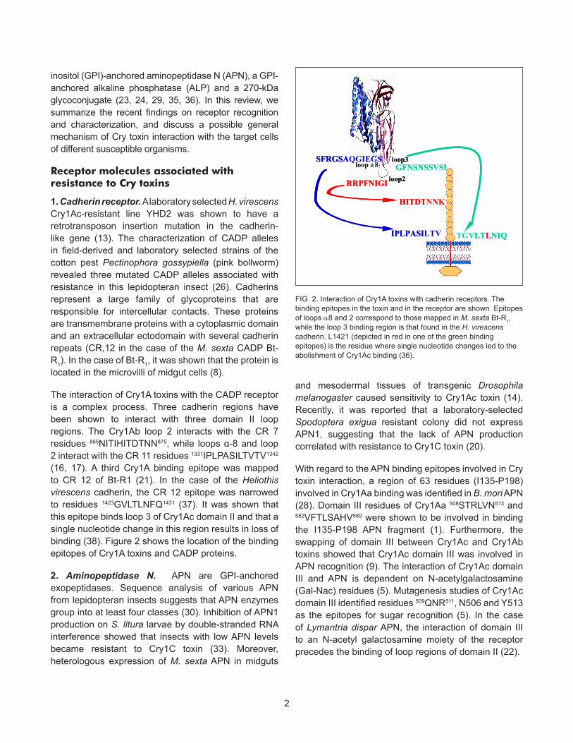
FIG. 2. Interaction of Cry1A toxins with cadherin receptors. The binding epitopes in the toxin and in the receptor are shown. Epitopes of loops α8 and 2 correspond to those mapped in M. sexta Bt-R1, while the loop 3 binding region is that found in the H. virescens cadherin. L1421 (depicted in red in one of the green binding epitopes) is the residue where single nucleotide changes led to the abolishment of Cry1Ac binding (36).
inositol (GPI)-anchored aminopeptidase N (APN), a GPI-anchored alkaline phosphatase (ALP) and a 270-kDa glycoconjugate (23, 24, 29, 35, 36). In this review, we summarize the recent findings on receptor recognition and characterization, and discuss a possible general mechanism of Cry toxin interaction with the target cells of different susceptible organisms.
Receptor molecules associated with resistance to Cry toxins
1. Cadherin receptor. A laboratory selected H. virescens Cry1Ac-resistant line YHD2 was shown to have a retrotransposon insertion mutation in the cadherin-like gene (13). The characterization of CADP alleles in field-derived and laboratory selected strains of the cotton pest Pectinophora gossypiella (pink bollworm) revealed three mutated CADP alleles associated with resistance in this lepidopteran insect (26). Cadherins represent a large family of glycoproteins that are responsible for intercellular contacts. These proteins are transmembrane proteins with a cytoplasmic domain and an extracellular ectodomain with several cadherin repeats (CR,12 in the case of the M. sexta CADP Bt-R1). In the case of Bt-R1, it was shown that the protein is located in the microvilli of midgut cells (8).
The interaction of Cry1A toxins with the CADP receptor is a complex process. Three cadherin regions have been shown to interact with three domain II loop regions. The Cry1Ab loop 2 interacts with the CR 7 residues 865NITIHITDTNN875, while loops α-8 and loop 2 interact with the CR 11 residues 1331IPLPASILTVTV1342 (16, 17). A third Cry1A binding epitope was mapped to CR 12 of Bt-R1 (21). In the case of the Heliothis virescens cadherin, the CR 12 epitope was narrowed to residues 1423GVLTLNFQ1431 (37). It was shown that this epitope binds loop 3 of Cry1Ac domain II and that a single nucleotide change in this region results in loss of binding (38). Figure 2 shows the location of the binding epitopes of Cry1A toxins and CADP proteins.
2. Aminopeptidase N. APN are GPI-anchored exopeptidases. Sequence analysis of various APN from lepidopteran insects suggests that APN enzymes group into at least four classes (30). Inhibition of APN1 production on S. litura larvae by double-stranded RNA interference showed that insects with low APN levels became resistant to Cry1C toxin (33). Moreover, heterologous expression of M. sexta APN in midguts
and mesodermal tissues of transgenic Drosophila melanogaster caused sensitivity to Cry1Ac toxin (14). Recently, it was reported that a laboratory-selected Spodoptera exigua resistant colony did not express APN1, suggesting that the lack of APN production correlated with resistance to Cry1C toxin (20).
With regard to the APN binding epitopes involved in Cry toxin interaction, a region of 63 residues (I135-P198) involved in Cry1Aa binding was identified in B. mori APN (28). Domain III residues of Cry1Aa 508STRLVN513 and 582VFTLSAHV589 were shown to be involved in binding the I135-P198 APN fragment (1). Furthermore, the swapping of domain III between Cry1Ac and Cry1Ab toxins showed that Cry1Ac domain III was involved in APN recognition (9). The interaction of Cry1Ac domain III and APN is dependent on N-acetylgalactosamine (Gal-Nac) residues (5). Mutagenesis studies of Cry1Ac domain III identified residues 509QNR511, N506 and Y513 as the epitopes for sugar recognition (5). In the case of Lymantria dispar APN, the interaction of domain III to an N-acetyl galactosamine moiety of the receptor precedes the binding of loop regions of domain II (22).
3
24 5 3
Glc
Man
GlcNac
GalNac
Gal
Fuc
2-O-Me-Fuc
24 5 3
Glc
Man
GlcNac
GalNac
Gal
Fuc
2-O-Me-Fuc
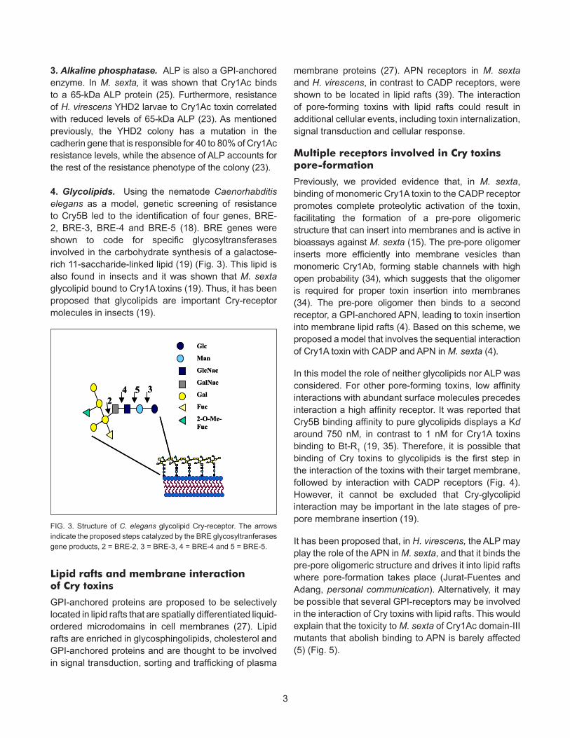
FIG. 3. Structure of C. elegans glycolipid Cry-receptor. The arrows indicate the proposed steps catalyzed by the BRE glycosyltranferases gene products, 2 = BRE-2, 3 = BRE-3, 4 = BRE-4 and 5 = BRE-5.
3. Alkaline phosphatase. ALP is also a GPI-anchored enzyme. In M. sexta, it was shown that Cry1Ac binds to a 65-kDa ALP protein (25). Furthermore, resistance of H. virescens YHD2 larvae to Cry1Ac toxin correlated with reduced levels of 65-kDa ALP (23). As mentioned previously, the YHD2 colony has a mutation in the cadherin gene that is responsible for 40 to 80% of Cry1Ac resistance levels, while the absence of ALP accounts for the rest of the resistance phenotype of the colony (23).
4. Glycolipids. Using the nematode Caenorhabditis elegans as a model, genetic screening of resistance to Cry5B led to the identification of four genes, BRE-2, BRE-3, BRE-4 and BRE-5 (18). BRE genes were shown to code for specific glycosyltransferases involved in the carbohydrate synthesis of a galactose-rich 11-saccharide-linked lipid (19) (Fig. 3). This lipid is also found in insects and it was shown that M. sexta glycolipid bound to Cry1A toxins (19). Thus, it has been proposed that glycolipids are important Cry-receptor molecules in insects (19).
Lipid rafts and membrane interaction of Cry toxins
GPI-anchored proteins are proposed to be selectively located in lipid rafts that are spatially differentiated liquid-ordered microdomains in cell membranes (27). Lipid rafts are enriched in glycosphingolipids, cholesterol and GPI-anchored proteins and are thought to be involved in signal transduction, sorting and trafficking of plasma
membrane proteins (27). APN receptors in M. sexta and H. virescens, in contrast to CADP receptors, were shown to be located in lipid rafts (39). The interaction of pore-forming toxins with lipid rafts could result in additional cellular events, including toxin internalization, signal transduction and cellular response.
Multiple receptors involved in Cry toxins pore-formation
Previously, we provided evidence that, in M. sexta, binding of monomeric Cry1A toxin to the CADP receptor promotes complete proteolytic activation of the toxin, facilitating the formation of a pre-pore oligomeric structure that can insert into membranes and is active in bioassays against M. sexta (15). The pre-pore oligomer inserts more efficiently into membrane vesicles than monomeric Cry1Ab, forming stable channels with high open probability (34), which suggests that the oligomer is required for proper toxin insertion into membranes (34). The pre-pore oligomer then binds to a second receptor, a GPI-anchored APN, leading to toxin insertion into membrane lipid rafts (4). Based on this scheme, we proposed a model that involves the sequential interaction of Cry1A toxin with CADP and APN in M. sexta (4).
In this model the role of neither glycolipids nor ALP was considered. For other pore-forming toxins, low affinity interactions with abundant surface molecules precedes interaction a high affinity receptor. It was reported that Cry5B binding affinity to pure glycolipids displays a Kd around 750 nM, in contrast to 1 nM for Cry1A toxins binding to Bt-R1 (19, 35). Therefore, it is possible that binding of Cry toxins to glycolipids is the first step in the interaction of the toxins with their target membrane, followed by interaction with CADP receptors (Fig. 4). However, it cannot be excluded that Cry-glycolipid interaction may be important in the late stages of pre-pore membrane insertion (19).
It has been proposed that, in H. virescens, the ALP may play the role of the APN in M. sexta, and that it binds the pre-pore oligomeric structure and drives it into lipid rafts where pore-formation takes place (Jurat-Fuentes and Adang, personal communication). Alternatively, it may be possible that several GPI-receptors may be involved in the interaction of Cry toxins with lipid rafts. This would explain that the toxicity to M. sexta of Cry1Ac domain-III mutants that abolish binding to APN is barely affected (5) (Fig. 5).
4
FIG. 4. Proposed mode of action of Cry1A toxins in M. sexta. Cry1A toxins are solubilized and proteolitically activated to yield monomeric toxins. Monomers interact either with glycolipids first or with cadherin receptors. The interaction with cadherins (Kd = 1 nM in contrast to Kd = 100 nM of Cry monomer interaction with APN) promotes further cleavage of helix α-1 of the monomer and formation of a pre-pore structure. The pre-pore binds then to APN with enhanced affinity (Kd= 0.75 nM) and inserts afterwards in lipid rafts.
APNCadherinManduca sexta
Multiple co-receptors ?
Heliothis virescensALPCadherin
A
B
C
APNCadherinManduca sextaAPNCadherinManduca sexta
Multiple co-receptors ?
Heliothis virescensALPCadherin
A
B
C
Kd = 1 nMKd = 0.75 nM
? ?
Kd = 730 nM
Bt-R1 = 210 kDa
APN = 120 kDaKd = 100 nM
Kd = 1 nMKd = 0.75 nM
? ?
Kd = 730 nM
Bt-R1 = 210 kDa
APN = 120 kDaKd = 100 nM
FIG. 5. Comparison of the proposed mode of action of Cry toxins in M. sexta (A) and in H. virescens (B). Alternatively, several GPI-anchored co-receptors may be involved in the late stages of pre-pore oligomer insertion (C).
FIG. 6. Proposed mode of action of Cry11Aa in Aedes aegypti. A. Binding of Cry11Aa to a 200-kDa unidentified protein and subsequent interaction with a GPI-anchored alkaline phosphatase. B. Cyt1Aa is a membrane receptor of Cry11Aa. Cyt1Aa may be involved in the first interaction of Cry11Aa or in the late stages of membrane insertion of the pre-pore oligomer.??
Aedes aegypti200 kDa,Cadherin? GPI-ALP
A
B
??
Aedes aegypti200 kDa,Cadherin? GPI-ALP
A
B
??
Aedes aegypti200 kDa,Cadherin? GPI-ALP
A
B
??
Aedes aegypti200 kDa,Cadherin? GPI-ALP
A
B
5
Receptor molecules and binding epitopes in mosquitoes
Bacillus thuringiensis subsp. israelensis (Bti) is a Bt strain used to control these disease vectors since it shows toxicity to dipteran insects (2). Bti produces crystal inclusions during sporulation that are composed principally of six toxins: Cry4Aa, Cry4Ba, Cry10Aa, Cry11Aa, Cyt1Aa and Cyt2Ba.
In the case of Cry11Aa and Cry4Ba toxins, a binding-protein of 65 kDa was identified in brush border membrane vesicles (BBMVs) from Aedes aegypti larvae (7). Data from our laboratory showed that this 65-kDa protein is a GPI-anchored ALP that binds Cry11Aa and is important for toxicity (11). Besides the GPI-ALP, a membrane-anchored 200-kDa protein was also found to bind Cry11Aa in ligand blot experiments (Fig. 6).
We identified specific regions in Cry11Aa toxin that are involved in the interaction with midgut microvilli membranes from A. aegypti (10). We isolated a peptide-displaying phage (P5.tox ) that recognized the domain II loop α-8 region of Cry11Aa and inhibited the interaction of the toxin with its receptor. Finally, mutants in loop α-8 that impaired toxicity and receptor interaction were isolated (10).
Cyt1A is a membrane receptor of cry11Aa.
Interestingly, no resistance in the field is observed for mosquito species controlled by Bti. The lack of resistance to Bti is due to the presence of the Cyt1Aa protein (37). Therefore, we explored the possibility that Cyt1Aa may function as a membrane-bound receptor of Cry11Aa. Our data shows that Cry11Aa binds membrane-bound Cyt1A and that this interaction enhances Cry11Aa insertion into the membrane. Furthermore, mapping the binding epitopes in both molecules revealed that Cry11Aa interacts with Cyt1A by means of the domain II loop regions involved in receptor interaction (31). Overall, this data points out that the interaction with Cyt1Aa may facilitate Cry11Aa toxin membrane insertion, suppressing resistance related to Cry receptor mutations. It is not known however whether Cyt1Aa interaction with Cry11Aa promotes the formation of oligomers or the insertion of the pre-pore oligomer in membrane lipid rafts. It is also possible that Cyt1Aa plays both roles. This is currently studied in our laboratory (Fig. 6).
Concluding remarks
The mode of action of Cry toxins is a multi-step process that involves the interaction of several receptor molecules leading to membrane insertion and cell lysis. The characterization of receptor molecules in other susceptible organisms will be important to fully understand the mode of action of Cry toxins. Moreover, the identification of receptor molecules and binding epitopes in the toxins and the receptors will help in the development of strategies to deal with insect resistance. This knowledge will be also critical for the development of novel toxins with novel specificities and for a better public perception of these safe toxins.
Acknowledgements
Research of our groups was supported in part by DGAPA/UNAM IN207503-3, IN206503-3 and IX217404, CONACyT 36505-N, USDA 2002-35302-12539 and NIH 1R01 AI066014-01.
References 1. Atsumi, S., E. Mizuno, H. Hara, K. Nakanishi, M. Kitami, N. Miura, H. Tabunoki, A. Watanabe, and R. Sato. 2005. Location of the Bombyx mori aminopeptidase N type I binding site on Bacillus thuringiensis Cry1Aa toxin. Appl. Environ. Microbiol. 71: 3966-3977.2. Becker N. 2000. Bacterial control of vector-mosquitoes and black flies, p. 383-396. In J. F. Charles, A. Delécluse and C. Nielsen-LeRoux (ed.), Entomopathogenic Bacteria: from Laboratory to Field Application, Kluwer Academic Publishers, Dordrecht, The Netherlands.3. Bravo, A., M. Soberón and S.S. Gill. 2005. Bacillus thuringiensis mechanisms and use, p. 175-206. In L. I. Gilbert, K. Iatrou, and S. S. Gill (ed.), Comprehensive Molecular Insect Science, Vol. 6, Elsevier, New York, NY, USA.4. Bravo, A., I. Gómez, J. Conde, C. Muñoz-Garay, J. Sánchez, M. Zhuang, S. S. Gill, and M. Soberón. 2004. Oligomerization triggers differential binding of a pore-forming toxin to a different receptor leading to efficient interaction with membrane microdomains. Biochim. Biophys. Acta 1667: 38-465. Burton, S. L., D. J. Ellar, J. Li, and D. J. Derbyshire. 1999. N-acetylgalactosamine on the putative insect receptor aminopeptidase N is recognized by a site on the domain III lectin-like fold of a Bacillus thuringiensis insecticidal toxin. J. Mol. Biol. 287: 1011-22.6. Butko, P., F. Huang, M. Pusztai-Carey, and W. K. Surewicz. 1997. Interaction of the delta-endotoxin CytA from Bacillus thuringiensis var. israelensis with lipid membranes. Biochem. 36: 12862-68.7. Buzdin, A. A., L. P. Revina, L. I. Kostina, I. A. Zalunin, and G. G. Chestukhina. 2002. Interaction of 65- and 62-kD proteins from the apical membranes of the Aedes aegypti larvae midgut epithelium with Cry4B and Cry11A endotoxins of Bacillus thuringiensis. Biochem. (Moscow). 67: 540-546.8. Chen, J., M. R. Brown, G. Hua, and M. J. Adang. 2005. Comparison of the localization of Bacillus thuringiensis Cr1A δ-endotoxins and their binding proteins in larval midgut of tobacco hornworm Manduca sexta. Cell Tissue Res. 321:123-1299. de Maagd, R. A., P. Bakker, L. Masson, M. JAdang, S. Sangadala, W. Stiekema, and D. Bosch. 1999. Domain III of the Bacillus thuringiensis delta-endotoxin Cry1Ac is involved in binding to Manduca sexta brush border membranes and to its purified aminopeptidase N. Mol. Microbiol. 31: 463-471.10. Fernández, L E., C. Pérez, L. Segovia, M. H. Rodríguez, S S. Gill, A. Bravo,, and M. Soberón. 2005. Cry11Aa toxin from Bacillus
6
thuringiensis binds its receptor in Aedes aegypti mosquito larvae trough loop α-8 of domain II. FEBS Lett. 579: 3508-3514.11. Fernandez, L. E., K. G. Aimanova, S. S. Gill, A. Bravo, and M. Soberón. 2005. A GPI-anchored alkaline phosphatase is a functional midgut receptor of Cry11Aa toxin in Aedes aegypti larvae. Biochem. J. 394:77-8412. Ferré, J., and J. van Rie. 2002. Biochemistry and genetics of insect resistance to Bacillus thuringiensis. Annu. Rev. Entomol. 47: 501-533.13. Gahan, L. J., F. Gould, and D. G. Heckel. 2001. Identification of a gene associated with Bt resistance in Heliothis virescens. Science 293: 857-860.14. Gill, M., and D. Ellar. 2002. Transgenic Drosophila reveals a functional in vivo receptor for the Bacillus thuringiensis toxin Cry1Ac1. Insect Mol. Biol. 11: 619-625.15. Gómez, I., J. Sánchez, R. Miranda, A. Bravo, and M. Soberón. 2002. Cadherin-like receptor binding facilitates proteolytic cleavage of helix α-1 in domain I and oligomer pre-pore formation of Bacillus thuringiensis Cry1Ab toxin. FEBS Lett. 513: 242-246.16. Gómez, I., J. Miranda-Rios, E. Rudiño-Piñera, D. I. Oltean, S. S. Gill, A. Bravo, and M. Soberón. 2002. Hydropathic complementarity determines interaction of epitope 869HITDTNNK876 in Manduca sexta Bt-R1 receptor with loop 2 of domain II of Bacillus thuringiensis Cry1A toxins. J. Biol. Chem. 277: 30137-30143.17. Gómez, I., D. H. Dean, A. Bravo, and M. Soberón. 2003. Molecular basis for Bacillus thuringiensis Cry1Ab toxin specificity: Two structural determinants in the Manduca sexta Bt-R1 receptor interact with loops α-8 and 2 in domain II of Cy1Ab toxin. Biochem. 42: 10482-10489.18. Griffitts, J. S., J. L. Whitacre, D. E. Stevens, and R. V. Aroian. 2001. Bacillus thuringiensis toxin resistance from loss of a putative carbohydrate-modifying enzyme. Science 293: 860-864.19. Griffits, J. S., S. M. Haslam, T. Yang, S. F. Garczynski, B. Mulloy, H. Morris, P. S. Cremer, A. Dell, M. J. Adang, and R. V. Aroian. 2005. Glycolipids as receptors for Bacillus thuringiensis crystal toxin. Science 307: 922-925.20. Herrero, S., T. Gechev, P. L. Bakker, W. J. Moar, and R. A. de Maagd. 2005. Bacillus thuringiensis Cry1Ca-resistant Spodoptera exigua lacks expression of one of four aminopeptidase N genes. BMC Genomics 6, 96.21. Hua, G., J. L. Jurat-Fuentes, and M. J. Adang. 2004. Bt-R1a extracellular cadherin repeat 12 mediates Bacillus thuringiensis binding and cytotoxicity. J. Biol. Chem. 279: 28051-28056.22. Jenkins, J. L., M. K. Lee, A. P. Valaitis, A. Curtiss, and D. H. Dean. 2000. Bivalent sequential binding model of a Bacillus thuringiensis toxin to gypsy moth aminopeptidase N receptor. J. Biol. Chem. 275: 14423-14431.23. Jurat-Fuentes, J. L., and M. J. Adang. 2004. Characterization of a Cry1Ac-receptor alkaline phosphatase in susceptible and resistant Heliothis virescens larvae. Eur. J. Biochem. 271: 3127-3135.24. Knight, P., N. Crickmore, and D. J. Ellar. 1994. The receptor for Bacillus thuringiensis CryIA(c) delta-endotoxin in the brush border membrane of the lepidopteran Manduca sexta is aminopeptidase N. Mol. Microbiol. 11: 429-436.25. McNall, R. J., and M. J. Adang. 2003. Identification of novel Bacillus thuringiensis Cry1Ac binding proteins in Manduca sexta midgut through proteomic analysis. Insect Biochem. Mol. Biol. 33: 999-1010.26. Morin, S., R. W. Biggs, M. S. Sisterson, L. Shriver, C. Ellers Kirk, D. Higginson, D. Holley, L. J. Gahan, D. G. Heckel, Y. Carrière, T. J. Dennehy, J. K. Brown, and B. E. Tabashnik. 2003. Three cadherin alleles associated with resistance to Bacillus thuringiensis in pink bollworm. Proc. Natl Acad. Sci. USA 100: 5004-5009.27. Munro, S. 2003. Lipid rafts: elusive or illusive? Cell 115: 377-38828. Nakanishi K., K. Yaoi, Y. Nagino, H. Hara, M. Kitami, S. Atsumi, N. Miura, and R. Sato. 2002. Aminopeptidase N isoforms from the midgut of Bombyx mori and Plutella xylostella- their classification and the factors that determine their binding specificity to Bacillus thuringiensis Cry1A toxin. FEBS Lett. 519: 215-220.29. Nagamatsu, Y., T. Koike, K. Sasaki, A. Yoshimoto, and Y. Furukawa. 1999. The cadherin-like protein is essential to specificity
determination and cytotoxic action of the Bacillus thuringiensis insecticidal. FEBS Lett. 460: 385-390.30. Oltean, D. I., A. K. Pullikuth, H.-K. Lee, and S. S. Gill. 1999. Partial purification and characterization of Bacillus thuringiensis Cry1A toxin receptor A from Heliothis virescens and cloning of the corresponding cDNA. Appl. Environ. Microbiol. 65: 4760-4766.31. Pérez, C., L. E. Fernandez, J. Sun, J. L. Folch, S. S. Gill, M. Soberón, and A. Bravo. 2006. Bacillus thuringiensis subsp. israelensis Cyt1Aa synergizes Cry11Aa toxin by functioning as membrane-bound receptor. Proc. Natl Acad. Sc. USA 102:18303-1830832. Promdonkoy, B., and D. J. Ellar. 2003. Investigation of the pore forming mechanism of a Cytolitic δ-endotoxin from Bacillus thuringiensis. Biochem. J. 374: 255-259.33. Rajagopal, R., S. Sivakumar, N. Agrawal, P. Malhotra, and R. K. Bhatnagar. 2002. Silencing of midgut aminopeptidase N of Spodoptera litura by double-stranded RNA establishes its role as Bacillus thuringiensis toxin receptor. J. Biol. Chem. 277: 46849-46851.34. Rausell, C., C. Muñoz-Garay, R. Miranda-CassoLuengo, I. Gómez, E. Rudiño-Piñera, M. Soberón, and A. Bravo. 2004. Tryptophan spectroscopy studies and black lipid bilayer analysis indicate that the oligomeric structure of Cry1Ab toxin from Bacillus thuringiensis is the membrane-insertion intermediate. Biochem. 43:166-174.35. Vadlamudi, R. K., E. Weber, I. H. Ji, T. H. Ji, and L. A. Jr Bulla. 1995. Cloning and expression of a receptor for an insecticidal toxin of Bacillus thuringiensis. J. Biol. Chem. 270: 5490-5494.36. Valaitis, A. P., J. L. Jenkins, M. K. Lee, D. H. Dean, and K. J. Garner. 2001. Isolation and partial characterization of Gypsy moth BTR-270 an anionic brush border membrane glycoconjugate that binds Bacillus thuringiensis Cry1A toxins with high affinity. Arch. Insect Biochem. Physiol. 46: 186-200.37. Wirth M. C., G. P. Georghiou, and B. A. Federeci. 1997. CytA enables CryIV endotoxins of Bacillus thuringiensis to overcome high levels of CryIV resistance in the mosquito, Culex quinquefasciatus. Proc. Natl Acad. Sci. USA 94: 10536-1054038. Xie, R., M. Zhuang, L. S. Ross, I. Gómez, D. I. Oltean, A. Bravo, M. Soberón, and S. S. Gill. 2005. Single amino acid mutations in the cadherin receptor from Heliothis virescens affect its toxin binding ability to Cry1A toxins. J. Biol. Chem. 280: 8416-8425.39. Zhuang, M., D.I. Oltean, I. Gómez, A.K. Pullikuth, M. Soberón, A. Bravo, and S. S. Gill. 2002. Heliothis virescens and Manduca sexta lipid rafts are involved in Cry1A toxin binding to the midgut epithelium and subsequent pore formation. J. Biol. Chem. 277: 13863-13872.


![an Ugi-azide multicomponent reaction Supporting …S1 Supporting information Novel synthesis of lower rim α-hydrazinotetrazolocalix[4]arenes via an Ugi-azide multicomponent reaction](https://static.fdocument.org/doc/165x107/5f3ff21b6dc20e37e43906a6/an-ugi-azide-multicomponent-reaction-supporting-s1-supporting-information-novel.jpg)