
Probing the N Terminal β Sheet Conversion in the Crystal … · §Department of Neuroscience,...
8
Probing the N‑Terminal β‑Sheet Conversion in the Crystal Structure of the Human Prion Protein Bound to a Nanobody Romany N. N. Abskharon, †,‡,∥,# Gabriele Giachin, §,# Alexandre Wohlkonig, †,‡,# Sameh H. Soror, †,‡,¶ Els Pardon, †,‡ Giuseppe Legname,* ,§ and Jan Steyaert* ,†,‡ † Structural Biology Brussels, Vrije Universiteit Brussel, Pleinlaan 2, 1050 Brussels, Belgium ‡ Structural Biology Research Center, VIB, Pleinlaan 2, 1050 Brussels, Belgium ∥ National Institute of Oceanography and Fisheries (NIOF), 11516 Cairo, Egypt § Department of Neuroscience, Laboratory of Prion Biology, Scuola Internazionale Superiore di Studi Avanzati (SISSA), 34136 Trieste, Italy ¶ Center of Excellence, Helwan Structure Biology Research, Faculty of Pharmacy, Helwan University, 11795 Cairo, Egypt * S Supporting Information ABSTRACT: Prions are fatal neurodegenerative transmissible agents causing several incurable illnesses in humans and animals. Prion diseases are caused by the structural conversion of the cellular prion protein, PrP C , into its misfolded oligomeric form, known as prion or PrP Sc . The canonical human PrP C (HuPrP) fold features an unstructured N-terminal part (residues 23−124) and a well-defined C-terminal globular domain (residues 125−231). Compelling evidence indicates that an evolutionary N-terminal conserved motif AGAAAAGA (residues 113−120) plays an important role in the conversion to PrP Sc . The intrinsic flexibility of the N-terminal has hampered efforts to obtain detailed atomic information on the structural features of this palindromic region. In this study, we crystallized the full-length HuPrP in complex with a nanobody (Nb484) that inhibits prion propagation. In the complex, the prion protein is unstructured from residue 23 to 116. The palindromic motif adopts a stable and fully extended configuration to form a three-stranded antiparallel β-sheet with the β1 and β2 strands, demonstrating that the full-length HuPrP C can adopt a more elaborate β0-β1-α1-β2-α2-α3 structural organization than the canonical β1-α1-β2-α2-α3 prion-like fold. From this structure, it appears that the palindromic motif mediates β-enrichment in the PrP C monomer as one of the early events in the conversion of PrP C into PrP Sc . ■ INTRODUCTION The post-translational conversion of the ubiquitously expressed cellular form of the prion protein, PrP C , into its misfolded oligomeric and pathogenic form, known as prion or PrP Sc , plays a key role in prion diseases. These maladies are denoted transmissible spongiform encephalopathies (TSEs) and affect both humans and animals. TSEs can be sporadic, inherited, or infectious. In humans, sporadic Creutzfeldt−Jakob disease (CJD) is the most common prion malady. 1,2 Structurally, PrP C shows a very similar fold and amino acid sequence among di fferent mammalian species. It is a glycosylphosphatidylinositol (GPI)-anchored glycoprotein fea- turing a long unstructured N-terminal part (from residue 23 to 124, hereinafter in human numbering) and a structured C- terminal domain (residues 125−231). The canonical PrP C globular domain contains three α-helices (α1, α2, and α3) and two very short antiparallel β-strands (β1 and β2) folding into a characteristic β1-α1-β2-α2-α3 antiparallel beta-ribbon, referred to as the PrP C -like fold in the Structural Classification of Proteins (SCOP) database. 3 A single disulfide bond bridges helix α2 to α3. 4 The flexible N-terminal moiety contains functional subdomains that are able to coordinate the binding of divalent metal ions including a conserved octarepeat region (residues 60−91) as well as a positively charged segment (residues 96−111). 5 The ability of PrP C to coordinate copper ions suggests it might also play a role in copper homeostasis. However, a unifying definition of PrP C function has not yet been found. Therefore, PrP C is often described as a pleiotropic protein involved in different physiological functions of neuronal and glial cells. 6 Unlike that of PrP C , the PrP Sc structure contains significant β-sheet and few α-helix secondary motifs. 7,8 The structural features of PrP Sc are responsible for its different physicochem- ical properties: while PrP C is monomeric, soluble in nonionic detergents, and protease-K (PK) sensitive, PrP Sc is insoluble, partially resistant to PK, and prone to aggregation. Limited digestion of PrP Sc with PK produces an infectious fragment of Received: August 3, 2013 Article pubs.acs.org/JACS © XXXX American Chemical Society A dx.doi.org/10.1021/ja407527p | J. Am. Chem. Soc. XXXX, XXX, XXX−XXX
-
Upload
truongquynh -
Category
Documents
-
view
215 -
download
0
Transcript of Probing the N Terminal β Sheet Conversion in the Crystal … · §Department of Neuroscience,...
Probing the N‑Terminal β‑Sheet Conversion in the Crystal Structureof the Human Prion Protein Bound to a NanobodyRomany N. N. Abskharon,†,‡,∥,# Gabriele Giachin,§,# Alexandre Wohlkonig,†,‡,# Sameh H. Soror,†,‡,¶
Els Pardon,†,‡ Giuseppe Legname,*,§ and Jan Steyaert*,†,‡
†Structural Biology Brussels, Vrije Universiteit Brussel, Pleinlaan 2, 1050 Brussels, Belgium‡Structural Biology Research Center, VIB, Pleinlaan 2, 1050 Brussels, Belgium∥National Institute of Oceanography and Fisheries (NIOF), 11516 Cairo, Egypt§Department of Neuroscience, Laboratory of Prion Biology, Scuola Internazionale Superiore di Studi Avanzati (SISSA), 34136Trieste, Italy¶Center of Excellence, Helwan Structure Biology Research, Faculty of Pharmacy, Helwan University, 11795 Cairo, Egypt
*S Supporting Information
ABSTRACT: Prions are fatal neurodegenerative transmissibleagents causing several incurable illnesses in humans andanimals. Prion diseases are caused by the structural conversionof the cellular prion protein, PrPC, into its misfolded oligomericform, known as prion or PrPSc. The canonical human PrPC
(HuPrP) fold features an unstructured N-terminal part(residues 23−124) and a well-defined C-terminal globulardomain (residues 125−231). Compelling evidence indicatesthat an evolutionary N-terminal conserved motif AGAAAAGA(residues 113−120) plays an important role in the conversionto PrPSc. The intrinsic flexibility of the N-terminal hashampered efforts to obtain detailed atomic information on the structural features of this palindromic region. In this study,we crystallized the full-length HuPrP in complex with a nanobody (Nb484) that inhibits prion propagation. In the complex, theprion protein is unstructured from residue 23 to 116. The palindromic motif adopts a stable and fully extended configuration toform a three-stranded antiparallel β-sheet with the β1 and β2 strands, demonstrating that the full-length HuPrPC can adopt amore elaborate β0-β1-α1-β2-α2-α3 structural organization than the canonical β1-α1-β2-α2-α3 prion-like fold. From thisstructure, it appears that the palindromic motif mediates β-enrichment in the PrPC monomer as one of the early events in theconversion of PrPC into PrPSc.
■ INTRODUCTION
The post-translational conversion of the ubiquitously expressedcellular form of the prion protein, PrPC, into its misfoldedoligomeric and pathogenic form, known as prion or PrPSc, playsa key role in prion diseases. These maladies are denotedtransmissible spongiform encephalopathies (TSEs) and affectboth humans and animals. TSEs can be sporadic, inherited, orinfectious. In humans, sporadic Creutzfeldt−Jakob disease(CJD) is the most common prion malady.1,2
Structurally, PrPC shows a very similar fold and amino acidsequence among different mammalian species. It is aglycosylphosphatidylinositol (GPI)-anchored glycoprotein fea-turing a long unstructured N-terminal part (from residue 23 to124, hereinafter in human numbering) and a structured C-terminal domain (residues 125−231). The canonical PrPC
globular domain contains three α-helices (α1, α2, and α3)and two very short antiparallel β-strands (β1 and β2) foldinginto a characteristic β1-α1-β2-α2-α3 antiparallel beta-ribbon,referred to as the PrPC-like fold in the Structural Classificationof Proteins (SCOP) database.3 A single disulfide bond bridges
helix α2 to α3.4 The flexible N-terminal moiety containsfunctional subdomains that are able to coordinate the bindingof divalent metal ions including a conserved octarepeat region(residues 60−91) as well as a positively charged segment(residues 96−111).5 The ability of PrPC to coordinate copperions suggests it might also play a role in copper homeostasis.However, a unifying definition of PrPC function has not yetbeen found. Therefore, PrPC is often described as a pleiotropicprotein involved in different physiological functions of neuronaland glial cells.6
Unlike that of PrPC, the PrPSc structure contains significantβ-sheet and few α-helix secondary motifs.7,8 The structuralfeatures of PrPSc are responsible for its different physicochem-ical properties: while PrPC is monomeric, soluble in nonionicdetergents, and protease-K (PK) sensitive, PrPSc is insoluble,partially resistant to PK, and prone to aggregation. Limiteddigestion of PrPSc with PK produces an infectious fragment of
Received: August 3, 2013
Article
pubs.acs.org/JACS
© XXXX American Chemical Society A dx.doi.org/10.1021/ja407527p | J. Am. Chem. Soc. XXXX, XXX, XXX−XXX
about 142 amino acids spanning approximately from residue 90to 231, referred to as the PK-resistant core of PrPSc.9
A prerequisite for understanding TSEs is unraveling themolecular mechanism leading to the structural conversion ofPrPC to prions. The insoluble and heterogeneous nature ofPrPSc makes its structural characterization extremely difficult.Because of the lack of atomic details for PrPSc, different prionmodels have been proposed. One such model is based on fiberX-ray diffraction and imaging simulation techniques; itproposes that the segment ∼90−175 forms a four-stranded β-sheet core organized in a β-helical configuration, whereashelices α2 and α3 retain their native conformation.10 Bycontrast, hydrogen−deuterium exchange experiments on brain-derived PrPSc showed that the region from residue ∼90 to theentire C-terminus displays slow exchange rates, which aretypical for a structure comprising a continuum of β-strands.11
The N-terminal of the PrPSc PK-resistant core features apalindromic sequence (AGAAAAGA), encompassing hydro-phobic residues from 113 to 120, and it leads to the formationof neurotoxic fibrils enriched in β-sheet motifs.12 Furtherexperimental evidence supports the idea that the palindromicsequence plays a critical role for prion generation andtransmissibility. In particular, in mature PrPSc this region isnot accessible to antibodies recognizing this epitope in PrPC,thus indicating that this segment undergoes conformationalchanges.13 The ablation of only the palindromic sequence intransgenic mice14 and murine neuroblastoma cells15 is not toxicand seems to abrogate the conversion of the deletion mutant toPrPSc. Low-resolution spectroscopy data also indicate that thehydrophobic AGAAAAGA motif may adopt multiple discreteconformations; this might imply that this region is metastableand structures upon intermolecular interactions.16,17 However,the intrinsic flexibility of this N-terminal has hampered effortsto obtain atomic information on the structural features of thepalindromic region.The solution structures of the full-length human prion
protein, HuPrP(23−230), and two C-terminal fragments,HuPrP(90−230) and HuPrP(121−230), have been solvedpreviously by NMR.4 All these structures include a globulardomain extending from residues 125−228 and an N-terminalflexibly disordered “tail.” In this study, we used nanobodies tosolve the very first structures of the full-length HuPrP(4KML.pdb) and its C-terminal truncated version (residues90−231, 4N9O.pdb) by X-ray crystallography. Nb-assistedcrystallography is a powerful tool to investigate the structure oftarget proteins that are difficult to crystallize because of theirintrinsically disordered domains.18−21 The high-resolution X-ray crystal structures of these HuPrPs in complex with aselective nanobody (Nb484) revealed a novel structural feature.While the segment from residue 128 to 225 shares a fold that isvery similar to the corresponding NMR HuPrP structures, thebinding of Nb484 to a region adjacent to the first β-sheet (β1)unveils key structural features of the hydrophobic segment fromresidue 117 to 128, which had remained unresolved in all thePrP structures published so far. In our X-ray structures, thesequence including the palindromic motif arranges in a novel β-strand we denoted as β0 (residues 118−122), which folds intoa three-stranded antiparallel β-sheet with β1 and β2. The samestructural arrangement was observed in both crystal structures,suggesting that it does not result from crystal packing but mighthave major biological implications for prion conversion. Theimplications of these findings are remarkable, as we provide a
first atomic structural view of the palindromic region adopting awell-defined β-sheet conformation.
■ EXPERIMENTAL SECTIONCloning, Expression, and Purification of Recombinant Prion
Proteins. Open reading frames encoding HuPrP(23−231),HuPrP(90−231), MoPrP(23−230), or MoPrP(89−230) were clonedin the pET-28a vector (Novagen) as an Nde1-BamH1 fragment.HuPrP(23−231), HuPrP(90−231), MoPrP(23−230), or MoPrP(89−230) were expressed and purified as soluble proteins according to theliterature.22 HuPrP(23−144) was refolded from inclusion bodiesaccording to the literature.23,24
Generation, Selection, and Purification of Anti-PrP Nano-bodies. Two llamas were immunized 6 times biweekly with 200 μg ofpurified recombinant MoPrP(23−230) or MoPrP(89−230), respec-tively. Lymphocytes were collected from the anticoagulated blood ofthe immunized llamas to prepare a cDNA library of genes coding forthe variable domains of the heavy-chain antibodies. Amplified PCRfragments were inserted into a pHEN4 phagemid vector andtransformed in E. coli TG1 cells resulting in two separate phage-display libraries against recombinant MoPrP(23−230) or MoPrP(89−230), respectively. Both libraries contained at least 2 × 10⁷ uniquetransformants with a VHH gene insert rate greater than 90%. TheVHH libraries were displayed on phage following standardprocedures,25 and phage particles expressing MoPrP(23−230) orMoPrP(89−230) specific nanobodies were selected by panning onsolid phase coated MoPrP(23−230) or MoPrP(89−230), respectively.Bound phages were recovered by incubating the antigen-coated wellswith 100 mM triethylamine pH 10 for 10 min. Optionally, theseMoPrP(23−230) or MoPrP(89−230) coated wells were neutralizedwith Tris-HCl pH 6.8 and washed several times with PBS after freshlygrown TG1 cells were added to recover noneluted phage. For all theselections performed, a clear enrichment was observed after two tothree consecutive rounds of panning. From each selection, 96randomly chosen colonies from the second and third round ofpanning were grown to express the encoded Nb as a soluble protein.Crude periplasmic extracts were tested in ELISA against solid phasecoated antigen. Positive clones were amplified by PCR and a HinfIdigestion was performed to analyze the diversity. Sequence analysisrevealed 12 different sequence families against MoPrP(89−230) andtwo sequence families against MoPrP(23−230). All nanobodies wereexpressed and purified as previously described.26
Solid-Phase ELISA. Maxisorp 96-well plates (Nunc) were coatedovernight at 4 °C with purified MoPrP(23−230) or MoPrP(89−230)at 2 μg/mL in sodium bicarbonate buffer pH 8.2. Residual proteinbinding sites in the wells were blocked with 2% milk in PBS for twohours at room temperature. Antigen-bound nanobodies were detectedwith a mouse anti-Hemagglutinin-alkaline phosphatase conjugatedmonoclonal antibody (clone 16B12, BAbCO) that binds theHemagglutin in decapeptide-tag fused at the C-terminus of allnanobodies. Absorption at 405 nm was measured 20 min after addingthe phosphatase substrate p-nitrophenyl phosphate.
Determination of Nb-PrP Affinities Using Surface PlasmonResonance (SPR). Binding isotherms were determined on a Biacore3000 (GE Healthcare). CM5 chips were activated with N-hydroxysuccinimide and N-ethyl-N-(3-dimethylaminopropyl) carbo-diimide. For each PrP variant, 2 μg was diluted in 10 mM NaOAC atpH 5.2 and immobilized on the activated CM5 chip at a flow rate of 5μL/min. Next, chips were blocked with ethanolamine. All binding anddissociation experiments were run at 30 μL/min flow rate in PBS,0.05% Tween 20, and 3 mM EDTA at 25 °C. After each cycle, thesurface was regenerated with a 60 s pulse of 100 mM glycine, pH 1.5.Association rates (Kon) and dissociation rates (Koff) were obtainedusing a simple 1:1 Langmiur binding model (Biacore evaluationsoftware version 4.1). Equilibrium dissociation constant was calculatedfrom the ratio Koff/Kon.
In Vitro Characterization of Nb484 in the Amyloid SeedingAssay. Amyloid seeding assays (ASA) were performed according toColby et al., 2007 with minor modifications.27 Briefly, a stock of 5 mg/mL of MoPrP(23−230) was diluted to 0.1 mg/mL (corresponding to
Journal of the American Chemical Society Article
dx.doi.org/10.1021/ja407527p | J. Am. Chem. Soc. XXXX, XXX, XXX−XXXB
4.3 μM) in PBS containing 0.4 M GndHCl, 10 mM Thioflavin T(ThT), preformed mouse PrPSc seeds, and nanobody. All ASAs wereperformed in a final volume of 200 μL in 96-well plate (BD Falcon,BD Bioscience). For agitation, each well contained one 3-mm glassbead (Sigma-Aldrich). Nb484 was added to each well at a finalconcentration of 5 μg/mL (corresponding to 0.3 μM). Our controlsincluded wells containing only the seed in the presence or absence ofNb. Each condition was performed in four replicates. The plate wasincubated at 37 °C with continuous shaking on a plate reader(Spectramax M5, Molecular Device). All seeds were purified from ahypothalamic ScGT1 murine cell line chronically infected by prions.Infected cells were lysed by adding 500 μL of PBS containing 4%sarkosyl, protease inhibitor (Complete, Roche), and 0.5% of theprecipitant phosphotungstic acid (PTA) under continuous shaking(350 rpm) for 1 h at 37 °C. Seeds were recovered by centrifugation(14000g for 30 min), and pellets were washed, resuspended in lysisbuffer, and then centrifuged again and resuspended in 150 μL of steriledouble-distilled H2O. Four μL of the resuspended PTA pellet wasdiluted in 400 μL of water, and 20 μL of this dilution was added toeach well as the PrPSc seed. The kinetics of fibril formation wasmonitored by top reading of the fluorescence intensity every 5 min at444 nm excitation and 485 nm emission. The lag phase was estimatedon the basis of 10% of the ThT fluorescence increase.28
Treatment of ScGT1 Cells with Nb484. Scrapie-infected GT1mouse hypothalamic (ScGT1) cells were seeded in 10-cm cell culturedish containing 10 mL of Dulbecco’s modified Eagle’s medium(DMEM) supplemented with 10% fetal bovine serum (FBS) and 1%penicillin−streptomycin. The cells were grown at 37 °C in 5% CO2 to95% confluence for 1 week before splitting at 1:10 for furthercultivation. After splitting, cells were treated with increasingconcentrations of Nb484 (0.75, 1.75, 3.5 μM) and incubated for 7more days. After this treatment, the accumulation of PrPSc wasdetected by PK digestion followed by immunoblotting of lysed cells.
One mL of lysis buffer (10 mM Tris-HCl pH 8.0, 150 mM NaCl, 0.5%Nonidet P-40, 0.5% deoxycholic acid sodium salt) was added to thecell plates, and the cell lysates were collected after centrifugation at1800g for 5 min. The total protein amount of the samples wasmeasured by BCA (Pierce), and 250 μg of total protein was digestedby PK (10 μg/mL) for 1 h at 37 °C. The reaction was stopped with 2mM phenylmethylsulfonylfluoride (PMSF), and the PK-digested celllysates were centrifuged at 150000g for 1 h at 4 °C in anultracentrifuge (Beckman Coulter). The pellets were resuspended in1× sample loading buffer. For the non-PK digested sample, 50 μg ofcell lysates for ScGT1 was used and 2× loading buffer (125 mM Tris-HCl, pH 6.8, 10% 2-mercapethanol, 4% SDS, 0.2% bromophenol blue,20% glycerol) was added in a 1:1 ratio. The samples were boiled for 5min at 100 °C, loaded onto 15% Tris-Glycine SDS-PAGE gel, andtransferred overnight onto Immobilon PVDF membranes (Millipore).Membranes were blocked by 5% nonfat milk, incubated with 1 μg/mLanti-PrP Fab D18 (InPro Biotechnology, South San Francisco, CA;ABR-0D18), followed by incubation with goat antihuman IgG F(ab)2fragment conjugated with horseradish peroxidase. Blots weredeveloped with the enhanced chemiluminescent system (ECL,Amersham Biosciences) and visualized on Hyperfilm (AmershamBiosciences). In another experiment, cells were incubated for 1 weekin the presence of Nb484 as described above and next passaged twicefor 7 days in Nb-free medium before they were lysed and analyzed forthe presence of PrPSc.
Crystallization and Data Collection. Nb484 and HuPrP(23−231) or HuPrP(90−231) were mixed in an equimolar ratio to formprotein complexes. Presence of a stable complex was monitored byanalytical SEC using a Superdex 75 HR 10/30 column (GE HealthcareLife Sciences) using 20 mM Tris-HCl pH 7.5, 150 mM NaCl as therunning buffer. HuPrP(23−231)·Nb484 crystals were grown at 20 °Cin condition A9 of the MD-proplex screen (0.2 M sodium chloride, 0.1M MES pH 6.0, 20% w/v PEG 2000 MME) at a final concentration of
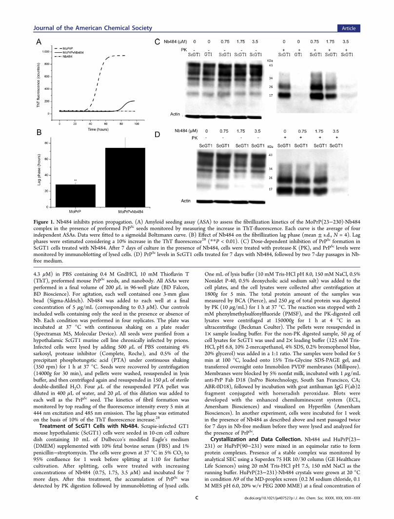
Figure 1. Nb484 inhibits prion propagation. (A) Amyloid seeding assay (ASA) to assess the fibrillization kinetics of the MoPrP(23−230)·Nb484complex in the presence of preformed PrPSc seeds monitored by measuring the increase in ThT-fluorescence. Each curve is the average of fourindependent ASAs. Data were fitted to a sigmoidal Boltzmann curve. (B) Effect of Nb484 on the fibrillization lag phase (mean ± s.d., N = 4). Lagphases were estimated considering a 10% increase in the ThT fluorescence28 (**P < 0.01). (C) Dose-dependent inhibition of PrPSc formation inScGT1 cells treated with Nb484. After 7 days of culture in the presence of Nb484, cells were treated with protease-K (PK), and PrPSc levels weremonitored by immunoblotting of lysed cells. (D) PrPSc levels in ScGT1 cells treated for 7 days with Nb484, followed by two 7-day passages in Nb-free medium.
Journal of the American Chemical Society Article
dx.doi.org/10.1021/ja407527p | J. Am. Chem. Soc. XXXX, XXX, XXX−XXXC
34 mg/mL. A high-resolution data set of 1.5 Å was collected at theX06DA beamline at SLS (Villigen, Switzerland). HuPrP(90−231)·Nb484 crystals were grown at 20 °C in condition F6 of MD-proplexscreen (0.1 M HEPES-Na pH 7, 15% PEG20000) at a finalconcentration of 53 mg/mL within 3−4 weeks as described.29 Ahigh-resolution data set of 1.5 Å was collected on BM30 at theEuropean Synchrotron Radiation Facility (ESRF, Grenoble, France).All crystals were cryoprotected using 15% glycerol.Data Collection, Structure Determination, Refinement, and
Model Building. Data were processed with XDS.30 Table S1(Supporting Information) summarizes the data collection details andrefinement statics. The structures of the different HuPrP·Nb484complexes were determined by molecular replacement (PHASER)31
using the HuPrP crystal structure (pdb entry 2W9E)32 and thestructure of Nb48426 as search models. Models were built manuallyusing the Crystallographic Object-Oriented Toolkit (Coot),33 andmultiple refinement rounds were performed using Refmac5.34
Structural analyses were performed using Ligplot,35 Promotif,36
DaliLite37 or Pisa.38
■ RESULTS
Generation and Characterization of Nb484. Mono-clonal antibodies with high affinity for PrPC were generated ascandidates for therapeutic approaches aimed to inhibit prionreplication.32,39,40 We selected 14 Nbs displaying high affinityfor both human and mouse (Mo) PrPs (Table S2, SupportingInformation). Nb484 displayed the highest affinity forHuPrP(23−231) and HuPrP(90−231) (Table S3, Figure S1,Supporting Information). To characterize Nb484 in the contextof prion replication, we evaluated its effectiveness in inhibitingprion propagation using fibrillization and cell-based assays. We
assessed the fibrillization kinetics of the MoPrP(23−230)·Nb484 complex in the presence of a preformed PrPSc seedpurified from ScGT1 cells. The addition of Nb484 toMoPrP(23−230) extended the lag phase of fibrillization byabout 40 h in amyloid seeding assay (ASA).27 This indicatesthat the interaction of Nb484 with the full-length MoPrPinhibits the formation of PrPSc-like aggregates (Figure 1A,B).To further confirm that Nb484 inhibits prion propagation, wetreated scrapie infected murine cells (ScGT1) with differentconcentrations of Nb484. We evaluated the effect of thetreatment measuring PrPSc level by PK assay and Westernblotting. We observed that, compared with nontreated cells, thePrPSc levels in the ScGT1 cells treated with Nb484 weredramatically reduced in a dose-dependent manner. After adding3.5 μM of Nb484, PrPSc levels were no longer detectable(Figure 1C). We also tested whether PrPSc remainedundetectable after removing Nb484 from the medium. Cellspreviously treated with 3.5 μM of Nb484 exhibited PrPSc
clearance upon its withdrawal (Figure 1D).Nanobody-Assisted Crystallization of the Full-Length
and Truncated HuPrPs. Atomic-level structural investigationof the events leading to conformational conversion of PrPC toPrPSc has been challenging, because of the dynamic equilibriumamong different structural species. The pathological process ofamyloid formation observed in prion diseases usually takesseveral years in vivo, and the intermediate species are highlyunstable. Using specific antibodies targeting proteins linked toneurodegenerative diseases (such as PrPC, α-synuclein, or β-amyloid) may be a promising strategy for probing the amyloidformation process by biophysical methods including X-ray
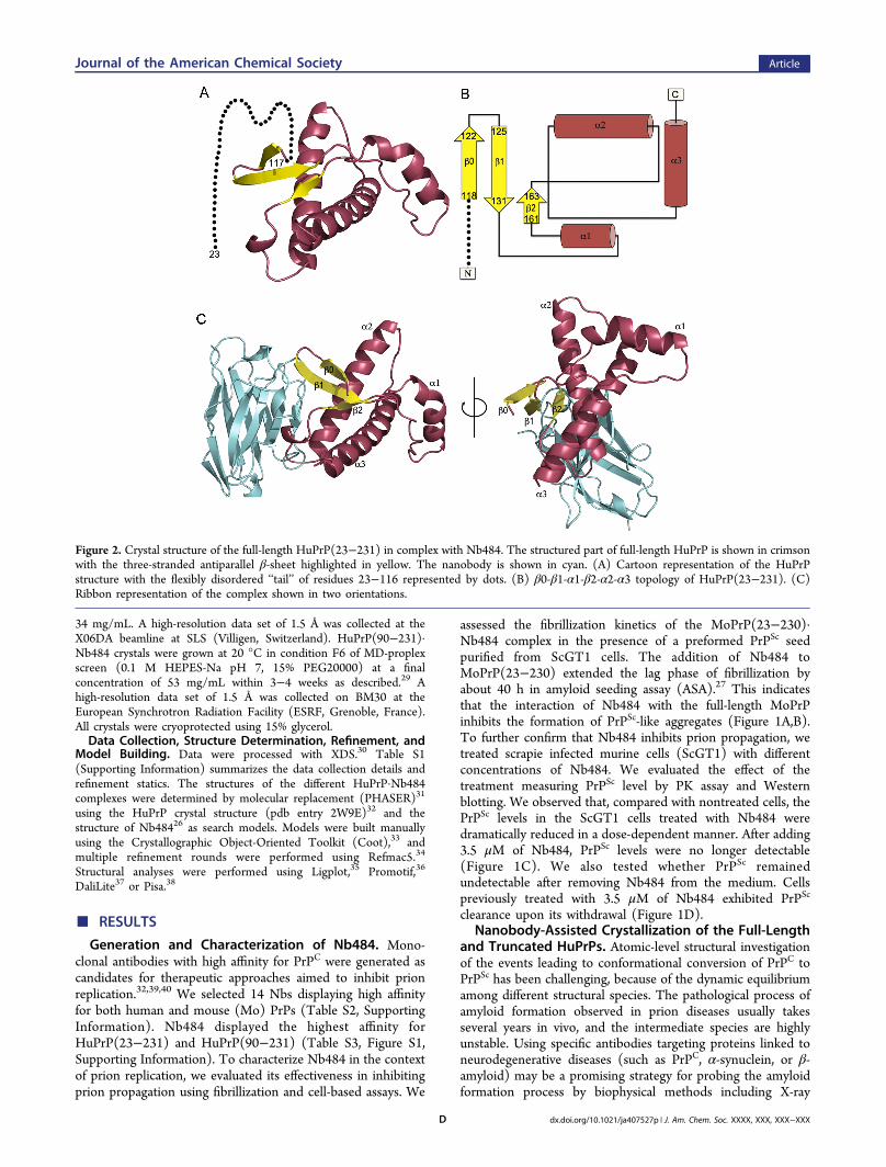
Figure 2. Crystal structure of the full-length HuPrP(23−231) in complex with Nb484. The structured part of full-length HuPrP is shown in crimsonwith the three-stranded antiparallel β-sheet highlighted in yellow. The nanobody is shown in cyan. (A) Cartoon representation of the HuPrPstructure with the flexibly disordered ‘‘tail’’ of residues 23−116 represented by dots. (B) β0-β1-α1-β2-α2-α3 topology of HuPrP(23−231). (C)Ribbon representation of the complex shown in two orientations.
Journal of the American Chemical Society Article
dx.doi.org/10.1021/ja407527p | J. Am. Chem. Soc. XXXX, XXX, XXX−XXXD
crystallography.41,42 We used Nb484 as crystallization chaper-one to obtain high-resolution crystal structures of HuPrP(23−231) and HuPrP(90−231). Crystal structures were refined to1.5 Å resolutions (Table S1, Supporting Information). Each cellunit was composed of a monomer of the HuPrP·Nb484complex. Remarkably, clear electron density was available toconfidently model half of the palindromic motif (residues 117−120) of both HuPrPs. The N-terminal downstream regionremained highly unstructured in both crystals. We were able torefine the structure of the HuPrP from residues 117 to 225 and118 to 224 for the full-length and truncated HuPrP,respectively. The average backbone root-mean-square distancebetween the refined structures is only 0.16 Å, indicating thatboth structures are identical. For clarity, hereinafter we willdiscuss the features of the HuPrP(23−231)·Nb484 complex.The segment from residue 128 to 225 of the HuPrP bound toNb484 shares a fold that is very similar to that observed for theX-ray and NMR HuPrP structures (Figure S3, SupportingInformation). HuPrP interacts with Nb484 through adiscontinuous epitope including residues 123−125 in the β0-β1 loop, residues 164−170 in the β2-α2 loop, and residues174−185 in the α2-helix. Table S4 (Supporting Information)shows a detailed overview of all the interactions between thefolded domain of HuPrP and Nb484.Remarkably, solving the full-length structure of the HuPrP
using Nb-assisted crystallography revealed unprecedentedstructural features at the N-terminal palindromic sequence,which is close to the canonical fold. Although this segment hasbeen reported as largely unfolded in several NMR HuPrPstructures,4,43,44 we identified the palindromic motif to be partof an extended three-stranded antiparallel β-sheet. Thisrevealed a previously unresolved β-sheet secondary element(residues 118−122, denoted as β0) followed by two antiparallelβ-strands comprising residues 125−131 (β1) and 161−163(β2) (Figure 2). It thus appears that full-length HuPrP canadopt a more elaborate β0-β1-α1-β2-α2-α3 fold than thecanonical prion-like β1-α1-β2-α2-α3 antiparallel beta-ribbon asdefined in the SCOP classification.3
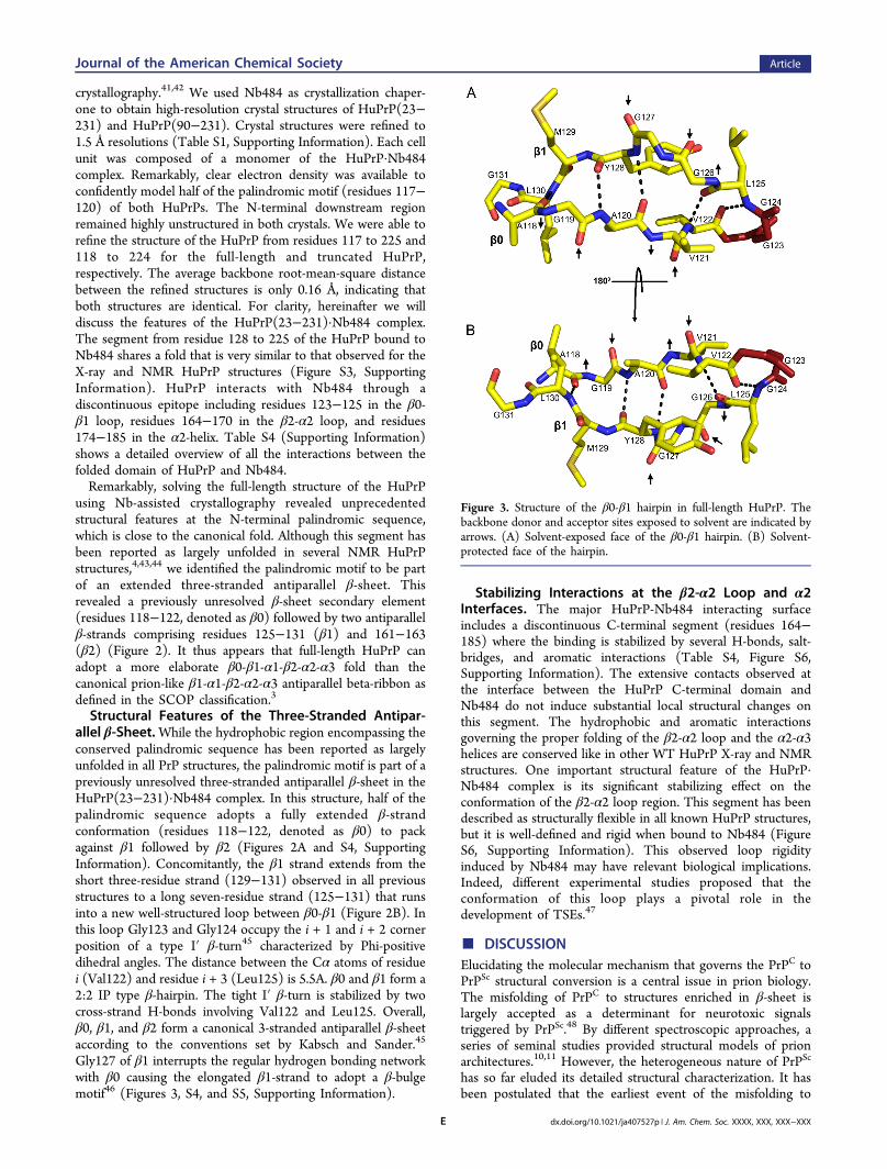
Structural Features of the Three-Stranded Antipar-allel β-Sheet.While the hydrophobic region encompassing theconserved palindromic sequence has been reported as largelyunfolded in all PrP structures, the palindromic motif is part of apreviously unresolved three-stranded antiparallel β-sheet in theHuPrP(23−231)·Nb484 complex. In this structure, half of thepalindromic sequence adopts a fully extended β-strandconformation (residues 118−122, denoted as β0) to packagainst β1 followed by β2 (Figures 2A and S4, SupportingInformation). Concomitantly, the β1 strand extends from theshort three-residue strand (129−131) observed in all previousstructures to a long seven-residue strand (125−131) that runsinto a new well-structured loop between β0-β1 (Figure 2B). Inthis loop Gly123 and Gly124 occupy the i + 1 and i + 2 cornerposition of a type I′ β-turn45 characterized by Phi-positivedihedral angles. The distance between the Cα atoms of residuei (Val122) and residue i + 3 (Leu125) is 5.5A. β0 and β1 form a2:2 IP type β-hairpin. The tight I′ β-turn is stabilized by twocross-strand H-bonds involving Val122 and Leu125. Overall,β0, β1, and β2 form a canonical 3-stranded antiparallel β-sheetaccording to the conventions set by Kabsch and Sander.45
Gly127 of β1 interrupts the regular hydrogen bonding networkwith β0 causing the elongated β1-strand to adopt a β-bulgemotif46 (Figures 3, S4, and S5, Supporting Information).
Stabilizing Interactions at the β2-α2 Loop and α2Interfaces. The major HuPrP-Nb484 interacting surfaceincludes a discontinuous C-terminal segment (residues 164−185) where the binding is stabilized by several H-bonds, salt-bridges, and aromatic interactions (Table S4, Figure S6,Supporting Information). The extensive contacts observed atthe interface between the HuPrP C-terminal domain andNb484 do not induce substantial local structural changes onthis segment. The hydrophobic and aromatic interactionsgoverning the proper folding of the β2-α2 loop and the α2-α3helices are conserved like in other WT HuPrP X-ray and NMRstructures. One important structural feature of the HuPrP·Nb484 complex is its significant stabilizing effect on theconformation of the β2-α2 loop region. This segment has beendescribed as structurally flexible in all known HuPrP structures,but it is well-defined and rigid when bound to Nb484 (FigureS6, Supporting Information). This observed loop rigidityinduced by Nb484 may have relevant biological implications.Indeed, different experimental studies proposed that theconformation of this loop plays a pivotal role in thedevelopment of TSEs.47
■ DISCUSSIONElucidating the molecular mechanism that governs the PrPC toPrPSc structural conversion is a central issue in prion biology.The misfolding of PrPC to structures enriched in β-sheet islargely accepted as a determinant for neurotoxic signalstriggered by PrPSc.48 By different spectroscopic approaches, aseries of seminal studies provided structural models of prionarchitectures.10,11 However, the heterogeneous nature of PrPSc
has so far eluded its detailed structural characterization. It hasbeen postulated that the earliest event of the misfolding to
Figure 3. Structure of the β0-β1 hairpin in full-length HuPrP. Thebackbone donor and acceptor sites exposed to solvent are indicated byarrows. (A) Solvent-exposed face of the β0-β1 hairpin. (B) Solvent-protected face of the hairpin.
Journal of the American Chemical Society Article
dx.doi.org/10.1021/ja407527p | J. Am. Chem. Soc. XXXX, XXX, XXX−XXXE
prions involves the formation of a metastable intermediate,denoted PrP*, which displays an aggregation-prone, β-sheetenriched structure.49,50 Recent NMR studies on pointmutations in HuPrP linked to genetic forms of human priondiseases43,51,52 provided preliminary hints on the structuraleffect of the mutations clustered in the globular domain, butthey have not yet clarified the structural rearrangementsoccurring at the N-terminal region, because of its flexibility.Here we used Nb-assisted crystallography as a strategy toobtain structural insights into HuPrP segments that areintrinsically disordered. In complex with the nanobody, thetotal amount of structured polypeptide (125 amino acids ofantibody and 108 amino acids of HuPrP) rises to 71% asagainst 52% for free HuPrP, thus providing a much betterstarting point for crystallization. The nanobody contributes togenerate a crystal lattice that leaves enough space for the N-terminal moiety (Figure S2, Supporting Information). Thestructure of the full-length HuPrP in complex with thenanobody reveals unprecedented structural features of thehydrophobic region encompassing the conserved palindromicsequence. This region first attracted interest because of itsability to form neurotoxic species12,53 and inspired several insilico and in vitro studies, which characterized the structuralproperties of short peptides carrying the AGAAAAGAmotif.54−58
The high resolution crystal structure of full-length HuPrPprovides a first atomic structural view of the palindromic regionadopting a well-defined β-sheet conformation. Edges of regularβ-sheets are inherently aggregation-prone because the motif forH-bonding with any other β-strand is available.59 From ourstructure, it appears that the exposed edge of the short β1strand observed in all previous PrP structures serves as anintramolecular nucleus for edge-to-edge β aggregation of part ofthe palindromic sequence to form the β0 strand. Thepropensity of the palindromic sequence to engage in such β-structures strongly indicates that this motif mediates β-enrichment in the PrPC monomer as one of the early eventsin the PrPC to PrPSc conversion.The observation that the conserved GGLGG sequence is
contained in the β0-β1 hairpin may be particularly relevant tothe structural conversion from PrPC to PrPSc. GGX motives arecommon in fibrillogenic proteins like spider silk proteins.60 Thefinding that Nb484 interacts with the GGLGG motif (Table S4,Supporting Information) is consistent with conversionmechanisms whereby the formation of the β0-β1 hairpin isfacilitated either by intermolecular contacts with other PrPs orby yet unknown cellular cofactors. Possible concerns that theantibody has trapped the HuPrP in a non-native conformationare groundless. First, Nb484 does not interact with β0. Moreimportantly, Nb484 is an in vivo matured conformationalantibody that has been cloned from the blood of an immunizedllama. In living animals, immature B cells expressing antibodiesthat have to pay a substantial energetic penalty for distortingthe prion structure would not undergo clonal expansion todifferentiate into mature B lymphocytes that circulate in theblood.61
At this stage it is still unclear how this early event of β-enrichment may drive a complete structural conversion of PrPC
to PrPSc. We found that the formation of the β0-β1 hairpin,including the GGLGG sequence, exposes several new backboneH-bond donor and acceptor sites to solvent (Figure 3). Thesesites are prone to stacking with other β-strands in a parallel orantiparallel configuration. Indeed, other strands may associate
perpendicularly to build large intermolecular β-sheets. It thusappears that the β-hairpin (β0-β1) can serve as a structuralnucleus for the growth of intermolecular β-sheets. Remarkably,the hairpin configuration we observe in both structures alsoexposes to the solvent several hydrophobic side-chainsincluding Ala118, Gly119, Val121, Gly126, and Gly127. This“dry surface” may provide a driving force for β-sheets ofgrowing oligomers to associate and interdigitate to form stericzippers.24
The question remains: why is Nb484 able to halt prionreplication in ScGT1 cells and in ASA acting as a molecularchaperone to stabilize PrPC? Steric hindrance, whereby theantibody prevents the association of aggregation-pronemetastable intermediates, may account for slowing downamyloidogenic conversion. Alternatively, conformational anti-bodies may prevent structural rearrangements that are pivotalfor the formation of early intermediates. Nb484 binds andstabilizes a discontinuous epitope that includes the β2-α2 loopand half of the α2-helix (Figure S6, Table S4, SupportingInformation). The b-factors indicate that the β2-α2 loop issignificantly more rigid in the antibody complex as compared toPrPC alone. The structural flexibility of this loop in PrPC
attracted particular interest in prion biology because itmodulates the susceptibility of a given species to TSEs (FigureS7, Supporting Information). It has been observed thatmammals (such as humans and mice) that express PrP with aflexible β2-α2 loop can be easily infected by prions, whereasspecies encoding PrP with a rigid loop (e.g., horse, rabbit, andmarsupials)62−64 do not develop spontaneous prion diseasesunder natural conditions. The structural stabilization of thiscritical epitope may represent an effective mechanism forNb484 to inhibit prion formation.Here we show that nanobody-assisted crystallography is a
powerful tool to unveil local structural features of intrinsicallydisordered proteins. These data provide structural evidence thatthe palindromic motif is important as dynamic site for β-sheetstructural conversion in prion formation. The structures wesolved feed the hypothesis that the conserved palindromicsequence mediates β-enrichment in the PrPC monomer as oneof the early events in prion formation. Crystals of the full-lengthhuman PrP in complex with Nb484 are amenable to soakingexperiments to study the interactions of small molecules withthe flexible part of HuPrP.
■ ASSOCIATED CONTENT
*S Supporting InformationDetails on the biophysical properties of the nanobodies,additional figures and tables, and tables of crystallographicdata collection and refinement statistics. This material isavailable free of charge via the Internet at http://pubs.acs.org.The atomic coordinates have been deposited in the ProteinData Bank (PDB) with the following accession codes: 4KMLand 4N9O.
■ AUTHOR INFORMATION
Corresponding [email protected]; [email protected]
Author Contributions#R.N.N.A, G.G., and A.W. contributed equally.
NotesThe authors declare no competing financial interest.
Journal of the American Chemical Society Article
dx.doi.org/10.1021/ja407527p | J. Am. Chem. Soc. XXXX, XXX, XXX−XXXF
■ ACKNOWLEDGMENTS
We acknowledge the Fund For Research Vlaanderen (FWO-Vlaanderen), the Interuniversity Attraction Poles (BELSPO),project P6/19, and the use of the beamlines at the SLS and theESRF.
■ REFERENCES(1) Mastrianni, J. A. Genet. Med. 2010, 12, 187.(2) Parchi, P.; Saverioni, D. Folia Neuropathol. 2012, 50, 20.(3) Andreeva, A.; Howorth, D.; Chandonia, J. M.; Brenner, S. E.;Hubbard, T. J.; Chothia, C.; Murzin, A. G. Nucleic Acids Res. 2008, 36,D419.(4) Zahn, R.; Liu, A.; Luhrs, T.; Riek, R.; von Schroetter, C.; LopezGarcia, F.; Billeter, M.; Calzolai, L.; Wider, G.; Wuthrich, K. Proc. Natl.Acad. Sci. U. S. A. 2000, 97, 145.(5) Walter, E. D.; Stevens, D. J.; Visconte, M. P.; Millhauser, G. L. J.Am. Chem. Soc. 2007, 129, 15440.(6) Aguzzi, A.; Baumann, F.; Bremer, J. Annu. Rev. Neurosci. 2008, 31,439.(7) Gong, B.; Ramos, A.; Vazquez-Fernandez, E.; Silva, C. J.; Alonso,J.; Liu, Z.; Requena, J. R. Biochemistry 2011, 50, 4963.(8) Pan, K. M.; Baldwin, M.; Nguyen, J.; Gasset, M.; Serban, A.;Groth, D.; Mehlhorn, I.; Huang, Z.; Fletterick, R. J.; Cohen, F. E.; et al.Proc. Natl. Acad. Sci. U. S. A. 1993, 90, 10962.(9) Colby, D. W.; Prusiner, S. B. Cold Spring Harbor Perspect. Biol.2011, 3, a006833.(10) Wille, H.; Bian, W.; McDonald, M.; Kendall, A.; Colby, D. W.;Bloch, L.; Ollesch, J.; Borovinskiy, A. L.; Cohen, F. E.; Prusiner, S. B.;Stubbs, G. Proc. Natl. Acad. Sci. U. S. A. 2009, 106, 16990.(11) Smirnovas, V.; Baron, G. S.; Offerdahl, D. K.; Raymond, G. J.;Caughey, B.; Surewicz, W. K. Nat. Struct. Mol. Biol. 2011, 18, 504.(12) Forloni, G.; Angeretti, N.; Chiesa, R.; Monzani, E.; Salmona, M.;Bugiani, O.; Tagliavini, F. Nature 1993, 362, 543.(13) Peretz, D.; Williamson, R. A.; Matsunaga, Y.; Serban, H.; Pinilla,C.; Bastidas, R. B.; Rozenshteyn, R.; James, T. L.; Houghten, R. A.;Cohen, F. E.; Prusiner, S. B.; Burton, D. R. J. Mol. Biol. 1997, 273, 614.(14) Baumann, F.; Tolnay, M.; Brabeck, C.; Pahnke, J.; Kloz, U.;Niemann, H. H.; Heikenwalder, M.; Rulicke, T.; Burkle, A.; Aguzzi, A.EMBO J. 2007, 26, 538.(15) Holscher, C.; Delius, H.; Burkle, A. J. virol. 1998, 72, 1153.(16) Kuwata, K.; Matumoto, T.; Cheng, H.; Nagayama, K.; James, T.L.; Roder, H. Proc. Natl. Acad. Sci. U. S. A. 2003, 100, 14790.(17) Lim, K. H.; Nguyen, T. N.; Damo, S. M.; Mazur, T.; Ball, H. L.;Prusiner, S. B.; Pines, A.; Wemmer, D. E. Solid State Nucl. Magn. Reson.2006, 29, 183.(18) De Genst, E. J.; Guilliams, T.; Wellens, J.; O’Day, E. M.;Waudby, C. A.; Meehan, S.; Dumoulin, M.; Hsu, S. T.; Cremades, N.;Verschueren, K. H.; Pardon, E.; Wyns, L.; Steyaert, J.; Christodoulou,J.; Dobson, C. M. J. Mol. Biol. 2010, 402, 326.(19) Loris, R.; Marianovsky, I.; Lah, J.; Laeremans, T.; Engelberg-Kulka, H.; Glaser, G.; Muyldermans, S.; Wyns, L. J. Biol. Chem. 2003,278, 28252.(20) Rasmussen, S. G.; Choi, H. J.; Fung, J. J.; Pardon, E.; Casarosa,P.; Chae, P. S.; Devree, B. T.; Rosenbaum, D. M.; Thian, F. S.;Kobilka, T. S.; Schnapp, A.; Konetzki, I.; Sunahara, R. K.; Gellman, S.H.; Pautsch, A.; Steyaert, J.; Weis, W. I.; Kobilka, B. K. Nature 2011,469, 175.(21) Rasmussen, S. G.; DeVree, B. T.; Zou, Y.; Kruse, A. C.; Chung,K. Y.; Kobilka, T. S.; Thian, F. S.; Chae, P. S.; Pardon, E.; Calinski, D.;Mathiesen, J. M.; Shah, S. T.; Lyons, J. A.; Caffrey, M.; Gellman, S. H.;Steyaert, J.; Skiniotis, G.; Weis, W. I.; Sunahara, R. K.; Kobilka, B. K.Nature 2011, 477, 549.(22) Abskharon, R. N.; Ramboarina, S.; El Hassan, H.; Gad, W.;Apostol, M. I.; Giachin, G.; Legname, G.; Steyaert, J.; Messens, J.;Soror, S. H.; Wohlkonig, A. Microb. Cell Fact. 2012, 11, 6.(23) Zahn, R.; von Schroetter, C.; Wuthrich, K. FEBS Lett. 1997, 417,400.
(24) Morillas, M.; Swietnicki, W.; Gambetti, P.; Surewicz, W. K. J.Biol. Chem. 1999, 274, 36859.(25) Arbabi Ghahroudi, M.; Desmyter, A.; Wyns, L.; Hamers, R.;Muyldermans, S. FEBS Lett. 1997, 414, 521.(26) Abskharon, R. N.; Soror, S. H.; Pardon, E.; El Hassan, H.;Legname, G.; Steyaert, J.; Wohlkonig, A. Acta Crystallogr., Sect. F:Struct. Biol. Cryst. Commun. 2010, 66, 1644.(27) Colby, D. W.; Zhang, Q.; Wang, S.; Groth, D.; Legname, G.;Riesner, D.; Prusiner, S. B. Proc. Natl. Acad. Sci. U. S. A. 2007, 104,20914.(28) Polano, M.; Bek, A.; Benetti, F.; Lazzarino, M.; Legname, G. J.Mol. Biol. 2009, 393, 1033.(29) Abskharon, R. N.; Soror, S. H.; Pardon, E.; El Hassan, H.;Legname, G.; Steyaert, J.; Wohlkonig, A. Protein Eng., Des. Sel. 2011,24, 737.(30) Kabsch, W. Acta Crystallogr., Sect. D: Biol. Crystallogr. 2010, 66,133.(31) McCoy, A. J. Acta Crystallogr., Sect. D: Biol. Crystallogr. 2007, 63,32.(32) Antonyuk, S. V.; Trevitt, C. R.; Strange, R. W.; Jackson, G. S.;Sangar, D.; Batchelor, M.; Cooper, S.; Fraser, C.; Jones, S.; Georgiou,T.; Khalili-Shirazi, A.; Clarke, A. R.; Hasnain, S. S.; Collinge, J. Proc.Natl. Acad. Sci. U. S. A. 2009, 106, 2554.(33) Emsley, P.; Cowtan, K. Acta Crystallogr., Sect. D: Biol. Crystallogr.2004, 60, 2126.(34) Vagin, A. A.; Steiner, R. A.; Lebedev, A. A.; Potterton, L.;McNicholas, S.; Long, F.; Murshudov, G. N. Acta Crystallogr., Sect. D:Biol. Crystallogr. 2004, 60, 2184.(35) Wallace, A. C.; Laskowski, R. A.; Thornton, J. M. Protein Eng.1995, 8, 127.(36) Hutchinson, E. G.; Thornton, J. M. Protein Sci. 1996, 5, 212.(37) Dietmann, S.; Park, J.; Notredame, C.; Heger, A.; Lappe, M.;Holm, L. Nucleic Acids Res. 2001, 29, 55.(38) Krissinel, E.; Henrick, K. J. Mol. Biol. 2007, 372, 774.(39) Jones, D. R.; Taylor, W. A.; Bate, C.; David, M.; Tayebi, M.PLoS One 2010, 5, e9804.(40) Peretz, D.; Williamson, R. A.; Kaneko, K.; Vergara, J.; Leclerc,E.; Schmitt-Ulms, G.; Mehlhorn, I. R.; Legname, G.; Wormald, M. R.;Rudd, P. M.; Dwek, R. A.; Burton, D. R.; Prusiner, S. B. Nature 2001,412, 739.(41) Domanska, K.; Vanderhaegen, S.; Srinivasan, V.; Pardon, E.;Dupeux, F.; Marquez, J. A.; Giorgetti, S.; Stoppini, M.; Wyns, L.;Bellotti, V.; Steyaert, J. Proc. Natl. Acad. Sci. U. S. A. 2011, 108, 1314.(42) Dumoulin, M.; Dobson, C. M. Biochimie 2004, 86, 589.(43) Biljan, I.; Ilc, G.; Giachin, G.; Plavec, J.; Legname, G.Biochemistry 2012, 51, 7465.(44) Calzolai, L.; Zahn, R. J. Biol. Chem. 2003, 278, 35592.(45) Venkatachalam, C. M. Biopolymers 1968, 6, 1425.(46) Chan, A. W.; Hutchinson, E. G.; Harris, D.; Thornton, J. M.Protein Sci. 1993, 2, 1574.(47) Sigurdson, C. J.; Nilsson, K. P.; Hornemann, S.; Manco, G.;Fernandez-Borges, N.; Schwarz, P.; Castilla, J.; Wuthrich, K.; Aguzzi,A. J. Clin. Invest. 2010, 120, 2590.(48) Aguzzi, A.; Falsig, J. Nat. Neurosci. 2012, 15, 936.(49) Khan, M. Q.; Sweeting, B.; Mulligan, V. K.; Arslan, P. E.;Cashman, N. R.; Pai, E. F.; Chakrabartty, A. Proc. Natl. Acad. Sci. U. S.A. 2010, 107, 19808.(50) Kuwata, K.; Li, H.; Yamada, H.; Legname, G.; Prusiner, S. B.;Akasaka, K.; James, T. L. Biochemistry 2002, 41, 12277.(51) Biljan, I.; Ilc, G.; Giachin, G.; Raspadori, A.; Zhukov, I.; Plavec,J.; Legname, G. J. Mol. Biol. 2011, 412, 660.(52) Ilc, G.; Giachin, G.; Jaremko, M.; Jaremko, L.; Benetti, F.;Plavec, J.; Zhukov, I.; Legname, G. PLoS One 2010, 5, e11715.(53) Jobling, M. F.; Stewart, L. R.; White, A. R.; McLean, C.;Friedhuber, A.; Maher, F.; Beyreuther, K.; Masters, C. L.; Barrow, C.J.; Collins, S. J.; Cappai, R. J. Neurochem. 1999, 73, 1557.(54) Walsh, P.; Neudecker, P.; Sharpe, S. J. Am. Chem. Soc. 2010,132, 7684.
Journal of the American Chemical Society Article
dx.doi.org/10.1021/ja407527p | J. Am. Chem. Soc. XXXX, XXX, XXX−XXXG
(55) Cheng, H. M.; Tsai, T. W.; Huang, W. Y.; Lee, H. K.; Lian, H.Y.; Chou, F. C.; Mou, Y.; Chan, J. C. Biochemistry 2011, 50, 6815.(56) Norstrom, E. M.; Mastrianni, J. A. J. Biol. Chem. 2005, 280,27236.(57) Silva, R. A.; Barber-Armstrong, W.; Decatur, S. M. J. Am. Chem.Soc. 2003, 125, 13674.(58) Lee, S. W.; Mou, Y.; Lin, S. Y.; Chou, F. C.; Tseng, W. H.;Chen, C. H.; Lu, C. Y.; Yu, S. S.; Chan, J. C. J. Mol. Biol. 2008, 378,1142.(59) Richardson, J. S.; Richardson, D. C. Proc. Natl. Acad. Sci. U. S. A.2002, 99, 2754.(60) Gatesy, J.; Hayashi, C.; Motriuk, D.; Woods, J.; Lewis, R. Science2001, 291, 2603.(61) Steyaert, J.; Kobilka, B. K. Curr. Opin. Struct. Biol. 2011, 21, 567.(62) Wen, Y.; Li, J.; Yao, W.; Xiong, M.; Hong, J.; Peng, Y.; Xiao, G.;Lin, D. J. Biol. Chem. 2010, 285, 31682.(63) Christen, B.; Hornemann, S.; Damberger, F. F.; Wuthrich, K. J.Mol. Biol. 2009, 389, 833.(64) Perez, D. R.; Damberger, F. F.; Wuthrich, K. J. Mol. Biol. 2010,400, 121.
Journal of the American Chemical Society Article
dx.doi.org/10.1021/ja407527p | J. Am. Chem. Soc. XXXX, XXX, XXX−XXXH


![b,d) 1 arXiv:1007.2378v1 [hep-ph] 14 Jul 2010 · 2018. 10. 29. · arXiv:1007.2378v1 [hep-ph] 14 Jul 2010 Ref. SISSA 36/2010/EP Ref. TUM-HEP 763/10 Ref. IPPP/10/42, DCTP/10/84 TeVScaleSee-Saw](https://static.fdocument.org/doc/165x107/60ac6dec85c9d80e5a0c5430/bd-1-arxiv10072378v1-hep-ph-14-jul-2010-2018-10-29-arxiv10072378v1.jpg)