Probing the Binding Site in the Antibiotic Resistance ...
2
Grand Valley State University ScholarWorks@GVSU Honors Projects Undergraduate Research and Creative Practice 2013 Probing the Binding Site in the Antibiotic Resistance Enzyme, AmpC β-lactamase Brianne Docter Grand Valley State University Mujahid Anwar Grand Valley State University David Leonard Grand Valley State University Rachel Powers Grand Valley State University Bradley Wallar Grand Valley State University Follow this and additional works at: hp://scholarworks.gvsu.edu/honorsprojects is Open Access is brought to you for free and open access by the Undergraduate Research and Creative Practice at ScholarWorks@GVSU. It has been accepted for inclusion in Honors Projects by an authorized administrator of ScholarWorks@GVSU. For more information, please contact [email protected]. Recommended Citation Docter, Brianne; Anwar, Mujahid; Leonard, David; Powers, Rachel; and Wallar, Bradley, "Probing the Binding Site in the Antibiotic Resistance Enzyme, AmpC β-lactamase" (2013). Honors Projects. 217. hp://scholarworks.gvsu.edu/honorsprojects/217
Transcript of Probing the Binding Site in the Antibiotic Resistance ...
Grand Valley State UniversityScholarWorks@GVSU
Honors Projects Undergraduate Research and Creative Practice
2013
Probing the Binding Site in the AntibioticResistance Enzyme, AmpC β-lactamaseBrianne DocterGrand Valley State University
Mujahid AnwarGrand Valley State University
David LeonardGrand Valley State University
Rachel PowersGrand Valley State University
Bradley WallarGrand Valley State University
Follow this and additional works at: http://scholarworks.gvsu.edu/honorsprojects
This Open Access is brought to you for free and open access by the Undergraduate Research and Creative Practice at ScholarWorks@GVSU. It hasbeen accepted for inclusion in Honors Projects by an authorized administrator of ScholarWorks@GVSU. For more information, please [email protected].
Recommended CitationDocter, Brianne; Anwar, Mujahid; Leonard, David; Powers, Rachel; and Wallar, Bradley, "Probing the Binding Site in the AntibioticResistance Enzyme, AmpC β-lactamase" (2013). Honors Projects. 217.http://scholarworks.gvsu.edu/honorsprojects/217
Cefotaxime Km (μM) kcat (s-1) kcat/Km (μM-1s-1)
WT 1.32‡ 0.012 0.009 N152G 455 1.09 0.002 N152S 47 0.56 0.012 N152T 800 1.85 0.002
Cefoxitin Km (μM) kcat (s-1) kcat/Km (μM-1s-1)
WT 0.99‡ 0.11 0.11 N152G 56 0.050 0.00090 N152S 1.16‡ 0.0083 0.0071 N152T 1.12‡ 0.0042 0.0038
Oxacillin Km (μM) kcat (s-1) kcat/Km (μM-1s-1)
WT 0.053‡ ND* ND*
N152G 0.27‡ 0.41 1.52 N152S 0.068‡ 0.21 3.17 N152T 1.2‡ 1.38 1.15
β-lactam drugs, such as penicillins and cephalosporins, are widely used to treat bacterial infections, but resistance to these drugs is increasingly becoming a problem. One of the main causes of resistance to these β-lactam drugs is the bacterial production of β-lactamase enzymes, such as AmpC. These enzymes are capable of breaking down the drug within their active sites, rendering the antibiotic unable to harm the bacteria. The exact roles that the active site amino acid residues play in the recognition and breakdown of the drug are not fully understood. Here, we investigate the role of the active site residue asparagine-152 (Asn152) in AmpC by mutating it to a glycine, serine, or threonine residue and examining the effect that these mutations have on kinetic and structural properties. Uncovering the specific role of Asn152 in the function of AmpC will be useful in the development of inhibitors to these enzymes in order to combat bacterial resistance.
Probing the Binding Site in the Antibiotic Resistance Enzyme, AmpC β-lactamase
Brianne Docter1, Mujahid Anwar2, David Leonard1, Rachel Powers1, and Bradley Wallar1
1Department of Chemistry and 2Cell and Molecular Biology Program, Grand Valley State University, 558 Cook-DeVos Center for Health Sciences, 301 Michigan Ave NE, Grand Rapids, MI 49503
Abstract
Introduction
Results and Discussion
Conclusions and Future Work
Acknowledgments
The Michaelis-Menten kinetic constants were determined using UV-Vis spectroscopy to follow the hydrolysis of the β-lactam amide bond. The values for the wild type AmpC enzyme and the N152G/S/T mutants with 3 different β-lactam drugs are shown below.
Summarizing the kinetics: • Cefotaxime – the mutants are faster than the wild type enzyme, but do not bind as well to the drug • Cefoxitin – the N152S and N152T mutants bind the substrate as well as the wild type enzyme but are slower to break it down. • Oxacillin – the wild type enzyme does not have measureable activity against this drug, but all three mutants show significant activity. The mutants do not all bind oxacillin as well as wild type, but they bind oxacillin significantly better than cefoxitin or cefotaxime, producing high kcat/Km values.
The mutations created a substrate specificity switch—allowing the enzyme to now break down oxacillin, at the cost of an activity reduction against cefoxitin, as well as affinity for cefoxitin and cefotaxime.
Deacylation NHO
O
Ser
Enzyme
O HH
Acylation OH
Ser
Enzyme
NHO
O-
OH
Ser
Enzyme
NO
Acyl intermediate Figure 1: Proposed mechanism of β-lactam hydrolysis for AmpC
H
NH3+
O
O-
CH2
C
NH2
O
Asparagine (Asn) (N)
Serine (Ser) (S)
Threonine (Thr) (T)
H
NH3+
O
O-
H
Glycine (Gly) (G)
Figure 2: Selected amino acids
H
NH3+
O
O-
CH2
OH
H
NH3+
O
O-
CH
CH3
OH
In order to compare the effectiveness of each of the mutants, we determined three Michaelis-Menten kinetic constants: • Km – “pseudo”-affinity constant – how well the enzyme binds to the substrate, a low value indicates tight binding • kcat – the “turnover number” – the total number of products that are formed by each enzyme active site per unit of time • kcat/Km – overall efficiency – taking binding and rate of turnover into account
β-lactam drugs, named for their β-lactam ring, have been used to treat bacterial infections for decades; however, bacteria are continually becoming resistant to these drugs, creating a growing problem in today’s treatment of bacterial infections. Resistance requires the need for stronger, more potent antibiotics and these may not even be able to combat the infection. One of the primary mechanisms of resistance to these β-lactam drugs is the production of β-lactamases by the bacteria. These enzymes are able to hydrolyze the β-lactam ring, turning the drug into a molecule that is harmless to the bacteria1.
One important β-lactamase that is a problem in many pathogens is AmpC2. This class C β-lactamase is able to hydrolyze extended-spectrum antibiotics, which are usually active against a wide range of bacteria3. The proposed mechanism of action of AmpC is shown in figure 1 and involves an acyl-intermediate where the substrate is covalently bound to the enzyme4.
Although the general mechanism is understood, it is unknown how specific amino acid residues of the enzyme active site contribute to the catalysis, or how they are involved in recognition of the substrate. Elucidating the roles of each of the key residues would not only provide a clearer picture of the mechanism of hydrolysis of the drugs, but could also provide details of substrate recognition and binding for the development of new drugs that might combat antibiotic resistance.
One of the key residues in the active site is asparagine-152. The Asn152 residue is highly conserved in class C β-lactamases and takes part in the active site hydrogen-bonding network5,6. This residue appears to play a role in substrate selectivity, as a study done on P99 cephalosporinase, another class C β-lactamase, revealed that certain mutations at this site led to a switch in substrate selectivity, causing some substrates to be hydrolyzed more quickly than the wild type enzyme but others to be hydrolyzed more slowly6. Understanding the exact role of this residue could help determine the reason for the substrate specificity of AmpC. This information could be used to develop inhibitors that bind very well to the active site but are not hydrolyzed (released), thereby inactivating the β-lactamase and helping to kill the infectious bacteria.
1. Neu, H. C. (1992). Science, 257(5073), 1064-1073. 2. Choi, S., Lee, J. E., Park, S. J., Choi, S., Lee, S., Jeong, J., & Kim, M. (2008). Antimicrobial Agents and Chemotherapy, 52(3), 995-1000. 3. Thomson, K. S. (2010). Journal of Clinical Microbiology, 48(4), 1019-1025. 4. Trehan, I., Beadle, B. M., & Shoichet, B. K. (2001). Biochemistry, 40, 7992-7999. 5. Goldberg, S. D., Iannuccilli, W., Nguyen, T., Ju, J., & Cornish, V. W. (2003). Protein Science, 12, 1633-1645. 6. Lefurgy, S. T., De Jong, R. M., & Cornish, V. W. (2007). Protein Science, 16, 2636-2646.
Figure 4: Active site of AmpC N152G in an acyl-intermediate complex with cefoxitin. Blue cages represent electron density contoured at 1σ. Image and subsequent images were produced with Pymol 1.3.
Figure 5: Active site of AmpC N152G in an acyl-intermediate complex with cefotaxime. Blue cages represent electron density contoured at 1σ.
S
N
OO-
O
NH
ON
S
O
O
NH2
NO
S
N
OO-
O
NH
O
O
S
O NH2
O
S
N
OO
-
O
NH
O
NO
Oxacillin Cefoxitin
Cefotaxime
Figure 3: β-lactam drugs used in this study, with the β-lactam ring in red, C4 carboxylate in green, and the amide of the R1 sidechain in blue.
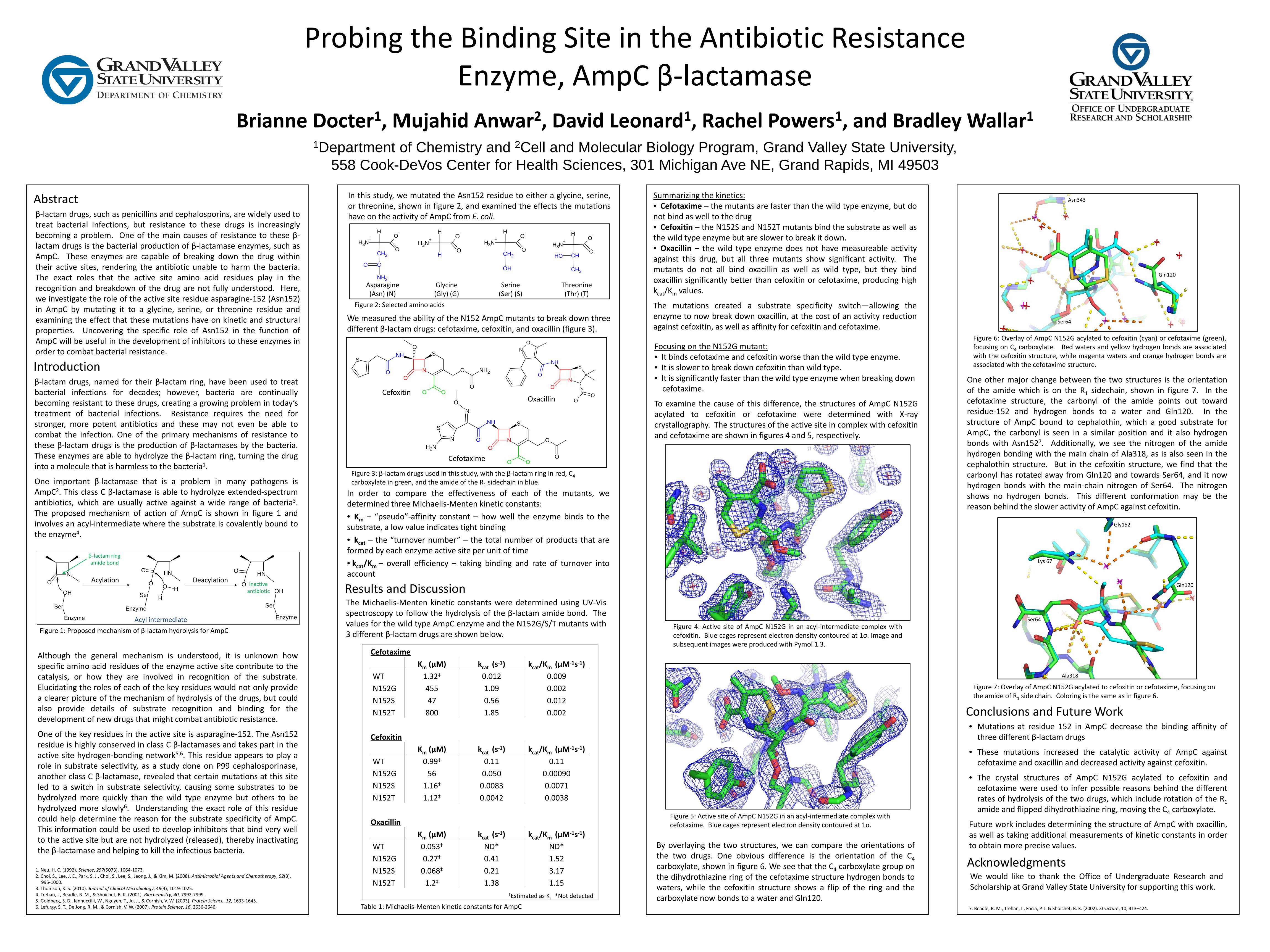
One other major change between the two structures is the orientation of the amide which is on the R1 sidechain, shown in figure 7. In the cefotaxime structure, the carbonyl of the amide points out toward residue-152 and hydrogen bonds to a water and Gln120. In the structure of AmpC bound to cephalothin, which a good substrate for AmpC, the carbonyl is seen in a similar position and it also hydrogen bonds with Asn1527. Additionally, we see the nitrogen of the amide hydrogen bonding with the main chain of Ala318, as is also seen in the cephalothin structure. But in the cefoxitin structure, we find that the carbonyl has rotated away from Gln120 and towards Ser64, and it now hydrogen bonds with the main-chain nitrogen of Ser64. The nitrogen shows no hydrogen bonds. This different conformation may be the reason behind the slower activity of AmpC against cefoxitin.
We would like to thank the Office of Undergraduate Research and Scholarship at Grand Valley State University for supporting this work.
Figure 6: Overlay of AmpC N152G acylated to cefoxitin (cyan) or cefotaxime (green), focusing on C4 carboxylate. Red waters and yellow hydrogen bonds are associated with the cefoxitin structure, while magenta waters and orange hydrogen bonds are associated with the cefotaxime structure.
Gln120
Asn343
Ser64
By overlaying the two structures, we can compare the orientations of the two drugs. One obvious difference is the orientation of the C4 carboxylate, shown in figure 6. We see that the C4 carboxylate group on the dihydrothiazine ring of the cefotaxime structure hydrogen bonds to waters, while the cefoxitin structure shows a flip of the ring and the carboxylate now bonds to a water and Gln120.
Figure 7: Overlay of AmpC N152G acylated to cefoxitin or cefotaxime, focusing on the amide of R1 side chain. Coloring is the same as in figure 6.
Gln120
Ser64
Lys 67
Gly152
Ala318
7. Beadle, B. M., Trehan, I., Focia, P. J. & Shoichet, B. K. (2002). Structure, 10, 413–424.
In this study, we mutated the Asn152 residue to either a glycine, serine, or threonine, shown in figure 2, and examined the effects the mutations have on the activity of AmpC from E. coli.
We measured the ability of the N152 AmpC mutants to break down three different β-lactam drugs: cefotaxime, cefoxitin, and oxacillin (figure 3).
Focusing on the N152G mutant: • It binds cefotaxime and cefoxitin worse than the wild type enzyme. • It is slower to break down cefoxitin than wild type. • It is significantly faster than the wild type enzyme when breaking down cefotaxime.
To examine the cause of this difference, the structures of AmpC N152G acylated to cefoxitin or cefotaxime were determined with X-ray crystallography. The structures of the active site in complex with cefoxitin and cefotaxime are shown in figures 4 and 5, respectively.
• Mutations at residue 152 in AmpC decrease the binding affinity of three different β-lactam drugs
• These mutations increased the catalytic activity of AmpC against cefotaxime and oxacillin and decreased activity against cefoxitin.
• The crystal structures of AmpC N152G acylated to cefoxitin and cefotaxime were used to infer possible reasons behind the different rates of hydrolysis of the two drugs, which include rotation of the R1 amide and flipped dihydrothiazine ring, moving the C4 carboxylate.
Future work includes determining the structure of AmpC with oxacillin, as well as taking additional measurements of kinetic constants in order to obtain more precise values.
β-lactam ring amide bond
inactive antibiotic
‡Estimated as Ki *Not detected
Table 1: Michaelis-Menten kinetic constants for AmpC