Preferred Conformers and Photochemical ( > 200 nm ... · Preferred Conformers and Photochemical ......
19
Preferred Conformers and Photochemical (λ > 200 nm) Reactivity of Serine and 3,3-Dideutero-Serine In the Neutral Form S. Jarmelo, ² L. Lapinski, ‡ M. J. Nowak, ‡ P. R. Carey, § and R. Fausto* ,² Department of Chemistry, UniVersity of Coimbra, 3004-535 Coimbra, Portugal, Institute of Physics, Polish Academy of Sciences, Al. Lotnikow 32/46, 02-668 Warsaw, Poland, and Department of Biochemistry, Case Western ReserVe UniVersity, 10900 Euclid AVe., CleVeland Ohio 44106-4935 ReceiVed: March 4, 2005; In Final Form: April 22, 2005 A systematic investigation of the conformational potential energy surface of neutral serine [HOCH 2 CHNH 2 - COOH] and 3,3-dideutero-serine [HOCD 2 CHNH 2 COOH] was undertaken, revealing the existence of 61 different minima. The structures and vibrational spectra of the most stable conformers, which were estimated to have relative energies within 7 kJ mol -1 and account for ca. 93% of the total conformational population at room temperature, were calculated at both the MP2 and DFT/BLYP levels of theory with the 6-311++G- (d,p) basis-set and used to interpret the spectroscopic data obtained for the compounds isolated in low- temperature inert matrixes. The assignment of the main spectral infrared features observed in the range 4000- 400 cm -1 to the most stable conformers of serine was undertaken. In addition, UV irradiation (λ > 200 nm) of the matrix-isolated compounds was also performed, leading to decarboxylation, which was found to be strongly dependent on the conformation assumed by the reactant molecule. Introduction Organic compounds observed in the interstellar medium and in solar system bodies are of particular importance for revealing the chemistry that may have led to life’s origin and evolution. 1-5 Among these compounds, amino acids may have a crucial importance, since they are the basic components of proteins, which are the essential constituents of all organisms. Under- standing the details of the structure of amino acids, under different experimental conditions, is then a fundamental problem in chemistry. On the other hand, the knowledge of the fundamental photochemical behavior of this type of compounds does also appear relevant to biochemistry and prebiotic chem- istry. Amino acids are well-known to exist in the zwitterionic form in the liquid or solid phases. Only very recently the neutral form of simple amino acids [glycine, sarcosine and N,N-dimethyl- glycine (DMG)] could be observed for the pure solid com- pounds, as a metastable species resulting from fast deposition of the gaseous compound onto a suitable substrate cooled at ca. 10 K. 6 The stabilization of the zwitterionic form in the condensed phases is essentially due to dipolar interactions and intermolecular H-bonds. On the contrary, in the gaseous phase, as well as for the compound isolated in low-temperature inert matrixes, amino acids exist in the neutral form. This has been confirmed for several amino acids, such as glycine, R- and -alanine, proline, sarcosine, DMG, and γ-aminobutyric acid (GABA), by experimental studies based on microwave spec- troscopy, electron diffraction and matrix-isolation infrared spectroscopy techniques. 6-14 In fact, for the isolated molecule situation, the zwitterionic species of simple amino acids have been shown not to constitute minimum energy structures, as revealed by high-level computational studies. 15,16 The main obstacle to investigation of the neutral forms of amino acids is due to the difficulty of promoting sublimation of this kind of compound without significant decomposition. This results from the low vapor pressure shown by most of the amino acids at room temperature, due to the presence of very strong intermolecular H-bonds in the solid phase. Serine is, among the simplest amino acids, one of those exhibiting lower vapor pressure and it decomposes extensively below its melting point, at normal pressure. Such behavior justifies the absence of any experimental study dealing with this amino acid in the gaseous phase. On the other hand, serine in the neutral form is a very interesting and challenging molec- ular system for structural research, because it contains a variety of intramolecular interactions. In particular, different types of H-bonds can be established between the various substituents in the molecule, leading to a large number of low energy conformers, stabilized by different intramolecular interactions. Despite the relative complexity of serine, which is a confor- mationally flexible molecule with five internal rotation axes, several computational studies on this molecule have been reported previously. 17-29 However, with a single exception, 30 all studies have only considered a small subset of possible conformers. Experimentally, amino acids have been extensively studied in their easily accessible zwitterionic form, but as mentioned above, they have been considerably less studied as neutral species. For instance, the photo- and thermal-decomposition behavior of neutral amino acid forms is a relatively recent topic of research. 31-33 In the case of serine, Zubavichus et al. 34 focused their attention on photodecomposition of the compound, as a solid layer adsorbed over indium, upon X-ray irradiation in ultrahigh vacuum. The experimental results indicated that the molecule decomposes by several pathways, such as dehydration, decarboxylation, decarbonylation, and deamination. On the other hand, Sato et al. 35 investigated the thermal decomposition of zwitterionic serine in water solution at high temperature (200- 340 °C) and high pressure (MPa), concluding that the general * Corresponding author. E-mail: [email protected]. ² University of Coimbra. ‡ Polish Academy of Sciences. § Case Western Reserve University. 5689 J. Phys. Chem. A 2005, 109, 5689-5707 10.1021/jp0511202 CCC: $30.25 © 2005 American Chemical Society Published on Web 06/09/2005
Transcript of Preferred Conformers and Photochemical ( > 200 nm ... · Preferred Conformers and Photochemical ......
Preferred Conformers and Photochemical (λ > 200 nm) Reactivity of Serine and3,3-Dideutero-Serine In the Neutral Form
S. Jarmelo,† L. Lapinski, ‡ M. J. Nowak,‡ P. R. Carey,§ and R. Fausto*,†
Department of Chemistry, UniVersity of Coimbra, 3004-535 Coimbra, Portugal, Institute of Physics, PolishAcademy of Sciences, Al. Lotnikow 32/46, 02-668 Warsaw, Poland, and Department of Biochemistry, CaseWestern ReserVe UniVersity, 10900 Euclid AVe., CleVeland Ohio 44106-4935
ReceiVed: March 4, 2005; In Final Form: April 22, 2005
A systematic investigation of the conformational potential energy surface of neutral serine [HOCH2CHNH2-COOH] and 3,3-dideutero-serine [HOCD2CHNH2COOH] was undertaken, revealing the existence of 61different minima. The structures and vibrational spectra of the most stable conformers, which were estimatedto have relative energies within 7 kJ mol-1 and account for ca. 93% of the total conformational populationat room temperature, were calculated at both the MP2 and DFT/BLYP levels of theory with the 6-311++G-(d,p) basis-set and used to interpret the spectroscopic data obtained for the compounds isolated in low-temperature inert matrixes. The assignment of the main spectral infrared features observed in the range 4000-400 cm-1 to the most stable conformers of serine was undertaken. In addition, UV irradiation (λ > 200 nm)of the matrix-isolated compounds was also performed, leading to decarboxylation, which was found to bestrongly dependent on the conformation assumed by the reactant molecule.
Introduction
Organic compounds observed in the interstellar medium andin solar system bodies are of particular importance for revealingthe chemistry that may have led to life’s origin and evolution.1-5
Among these compounds, amino acids may have a crucialimportance, since they are the basic components of proteins,which are the essential constituents of all organisms. Under-standing the details of the structure of amino acids, underdifferent experimental conditions, is then a fundamental problemin chemistry. On the other hand, the knowledge of thefundamental photochemical behavior of this type of compoundsdoes also appear relevant to biochemistry and prebiotic chem-istry.
Amino acids are well-known to exist in the zwitterionic formin the liquid or solid phases. Only very recently the neutral formof simple amino acids [glycine, sarcosine andN,N-dimethyl-glycine (DMG)] could be observed for the pure solid com-pounds, as a metastable species resulting from fast depositionof the gaseous compound onto a suitable substrate cooled atca. 10 K.6 The stabilization of the zwitterionic form in thecondensed phases is essentially due to dipolar interactions andintermolecular H-bonds. On the contrary, in the gaseous phase,as well as for the compound isolated in low-temperature inertmatrixes, amino acids exist in the neutral form. This has beenconfirmed for several amino acids, such as glycine,R- andâ-alanine, proline, sarcosine, DMG, andγ-aminobutyric acid(GABA), by experimental studies based on microwave spec-troscopy, electron diffraction and matrix-isolation infraredspectroscopy techniques.6-14 In fact, for the isolated moleculesituation, the zwitterionic species of simple amino acids havebeen shown not to constitute minimum energy structures, asrevealed by high-level computational studies.15,16
The main obstacle to investigation of the neutral forms ofamino acids is due to the difficulty of promoting sublimationof this kind of compound without significant decomposition.This results from the low vapor pressure shown by most of theamino acids at room temperature, due to the presence of verystrong intermolecular H-bonds in the solid phase.
Serine is, among the simplest amino acids, one of thoseexhibiting lower vapor pressure and it decomposes extensivelybelow its melting point, at normal pressure. Such behaviorjustifies the absence of any experimental study dealing withthis amino acid in the gaseous phase. On the other hand, serinein the neutral form is a very interesting and challenging molec-ular system for structural research, because it contains a varietyof intramolecular interactions. In particular, different types ofH-bonds can be established between the various substituents inthe molecule, leading to a large number of low energyconformers, stabilized by different intramolecular interactions.
Despite the relative complexity of serine, which is a confor-mationally flexible molecule with five internal rotation axes,several computational studies on this molecule have beenreported previously.17-29 However, with a single exception,30
all studies have only considered a small subset of possibleconformers.
Experimentally, amino acids have been extensively studiedin their easily accessible zwitterionic form, but as mentionedabove, they have been considerably less studied as neutralspecies. For instance, the photo- and thermal-decompositionbehavior of neutral amino acid forms is a relatively recent topicof research.31-33 In the case of serine, Zubavichus et al.34 focusedtheir attention on photodecomposition of the compound, as asolid layer adsorbed over indium, upon X-ray irradiation inultrahigh vacuum. The experimental results indicated that themolecule decomposes by several pathways, such as dehydration,decarboxylation, decarbonylation, and deamination. On the otherhand, Sato et al.35 investigated the thermal decomposition ofzwitterionic serine in water solution at high temperature (200-340 °C) and high pressure (MPa), concluding that the general
* Corresponding author. E-mail: [email protected].† University of Coimbra.‡ Polish Academy of Sciences.§ Case Western Reserve University.
5689J. Phys. Chem. A2005,109,5689-5707
10.1021/jp0511202 CCC: $30.25 © 2005 American Chemical SocietyPublished on Web 06/09/2005
reaction network of amino acids under hydrothermal conditionstakes two main paths: deamination to produce ammonia andorganic acids and decarboxylation to produce carbonic acid andamines.
Neutral serine was for the first time successfully isolated ina low-temperature matrix in our laboratory, and preliminaryresults on the conformational preferences of this species werereported.36,37 To sublimate efficiently the necessary amount ofthe compound without appreciable decomposition, we tookadvantage of the capabilities of the specially designed internalminifurnace developed in our laboratories for preparation ofcryogenic matrixes of thermolabile low-vapor-pressure com-pounds.38 Our experimental approach has been used later onby Lambie et al.,29 to investigate the infrared spectrum of neutralserine isolated in solid argon. In that study, an assignment ofthe observed spectrum was proposed, which assumed significantcontributions to the observed spectral features from the fourconformers we had previously identified experimentally (Figure1).36,37 However, to undertake the band assignments, Lambieet al. relied only on the comparison between the experimentaldata and theoretical predictions of the vibrational spectra of thefour conformers obtained at the DFT/B3LYP/6-31++G(d,p)level, not attempting to use any further information, such as,for example, those obtained by annealing or in situ irradiationof the matrix. These procedures can be expected to change therelative conformational populations and then enable a morereliable identification of bands due to each experimentallyobservable conformer. Furthermore, the possibility of contribu-tion to the observed spectra of more than 4 conformers was nottaken into consideration in that study.29 As it will be shown inthe present paper, this last assumption is in fact not supportedby the experimental data.
In this work, the conformational preferences of neutral serinewere studied, starting by a full systematic investigation of itspotential energy surface. The optimized geometries and vibra-tional spectra of the 61 different conformers of serine and its3,3-dideutero isotopologue were calculated, indicating that 9conformers lay within 7 kJ mol-1 and account for ca. 93% ofthe total conformational population at room temperature.Theoretical calculations, undertaken at both the MP2 and DFT/B3LYP levels of theory with the 6-311++G(d,p) basis-set,together with temperature variation studies and in situ irradiationexperiments were then used to interpret the spectroscopic dataobtained for the compounds isolated in low-temperature inertmatrixes. The assignment of the main spectral infrared featuresobserved in the range 4000-400 cm-1 to the most stableconformers of serine was undertaken and the photochemicaldecarboxylation of both serine isotopologues, resulting from insitu UV irradiation (λ> 200 nm) of the matrixes, was alsoinvestigated.
Materials and Methods
Computational Details.The theoretical calculations on serineand 3,3-dideutero-serine were performed with the Gaussian98program package39 at Hartree-Fock (HF), density functionaltheory (DFT), and second-order Møller-Plesset (MP2) levelsof theory, with the standard split-valence 3-21G(d)40-42 andextended 6-311++G(d,p)43 basis sets. The DFT calculationswere carried out with the three-parameter density functionalabbreviated as B3LYP, which includes Becke’s gradient ex-change correction44 and the Lee, Yang, and Parr correlationfunctional.45 All structures were subjected to optimizationwithout any geometrical constraints, and the resulting optimized
geometries were characterized by inspection of the correspond-ing Hessian matrixes. Optimizations were followed by frequencycalculations, performed at the same level of theory. Thecalculated frequencies were used to assist the analysis of theexperimental spectra and to account for the zero-point vibrationalenergy (ZPVE) corrections. Due to the general good cor-respondence between the experimentally observed and the DFT/B3LYP/6-311++G(d,p) predicted spectra, these calculationswere preferentially used both to assign the main featuresobserved in the spectra of matrix-isolated monomers and tocalculate energy barriers between the experimentally relevantconformers of serine. The DFT/B3LYP/6-311++G(d,p) har-monic frequencies and ZPVE were scaled using a single factorof 0.978,46-48 except forνOH stretching modes, which werefound to require the use of different scaling factors to describeproperly the experimental spectra. For these vibrations, twoscaling factors were used (0.953 or 0.913) depending on thestrength of the H-bond interactions in which the correspondinghydroxyl group is involved.
Experimental Details. Both D,L-serine and its 3,3-dideuter-ated (>98% D) isotopologue used in this study were commercialproducts obtained from Aldrich and Icon Stable Isotopes,respectively. Matrixes were prepared by deposition of the vaporof the compounds onto a cold CsI (10 K) window directlyassembled to the cold tip of a continuous flow liquid heliumcryostat. The compounds were sublimated by electrical heating(470 K) in a specially designed minifurnace,38 placed insidethe vacuum chamber of the cryostat. The vapors of thecompounds were deposited during ca. 70 min, together with alarge excess of argon by Linde AG. T. These experimentalconditions allowed minimization of trace amounts of thermaldecomposition of the compound during sublimation. Argon ofspectral purity (6.0) was supplied by Linde AG. The infraredspectra were recorded in the range 4000-400 cm-1, with 0.5cm-1 resolution and 600 scans, using a Thermo Nicolet Nexus670 FTIR spectrometer equipped with KBr beam splitter andDTGS detector. Integral intensities of the IR absorption bandswere measured by numerical integration. The matrixes wereirradiated through the outer quartz window of the cryostat, usinga high-pressure mercury lamp (HBO200) fitted with a waterfilter and a cutoff filter transmitting light withλ > 200 nm.The time of irradiation was 30 min. After UV irradiation, thematrixes were annealed to 30 K.
Results and Discussion
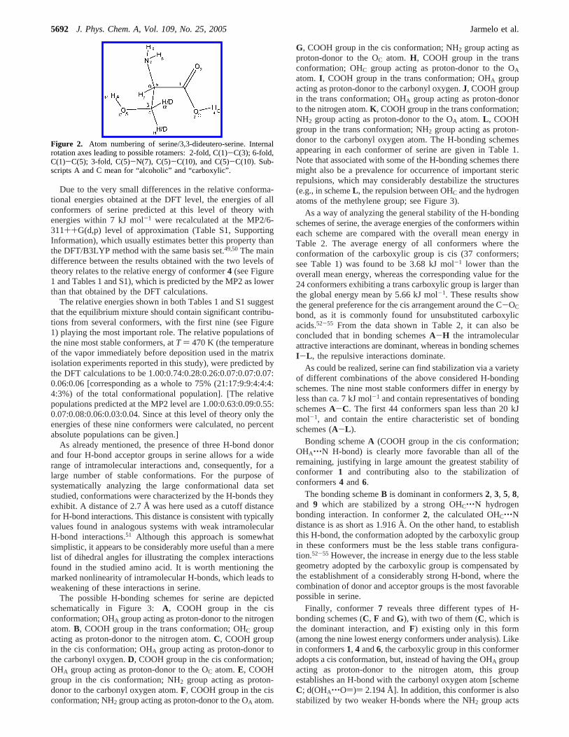
Computational Results (Structures and Relative Confor-mational Energies).To truly characterize the conformationsof serine, the full ensemble of possible conformations wasconsidered. Preliminary calculations were undertaken at the lessexpensive HF level of theory, using the split-valence 3-21G(d)basis set. The starting set of serine conformations was chosenby allowing all possible combinations of single-bond putativerotamers (see Figure 2). There are five different internal rotationaxes in serine that can give rise to conformational isomers. Forthe carboxyl group, internal rotation around the C(1)-O(3) bondleads to two possible minimum-energy arrangements: syn(τ(O(2)dC(1)O(3)H(4))) 0°) and trans (τ(O(2)dC(1)O(3)H-(4) ) 180°). The orientation of theR-carbon substituents (NH2and CH2OH) relative to the COOH group (internal rotationaround the CR-C(1) bond) can lead to six possible minimum-energy structures (corresponding to 3 staggered and 3 eclipsedgeometries relative to the CdO bond). Both the NH2 and CH2-OH groups may adopt three different staggered arrangements(defined by the CR-N and CR-Câ internal rotations). Finally,
5690 J. Phys. Chem. A, Vol. 109, No. 25, 2005 Jarmelo et al.
the Câ-O(13) bond represents a 3-fold rotor (Figure 2), definingthe possible conformations of the OHA group (throughout thisarticle, in references to the OH groups, the subscript A standsfor alcohol, whereas the subscript C stands for carboxylic group).All possible rotations around the axes described above lead toa total of 324 trial structures for the conformational states ofserine, which were submitted to optimization. From the HF/3-21G(d) optimizations, 71 unique conformers were identified.
These structures were afterward submitted to reoptimization atthe DFT/B3LYP/6-311++G(d,p) level of theory.
At the DFT/B3LYP/6-311++G(d,p) level of theory, only61 unique conformations were located, with energies varyingby =47 kJ mol-1 (Table 1). Ten structures predicted at theHF/3-21G(d) level as minimum-energy conformations bythe lower-level theory used were found to converge to otherforms.
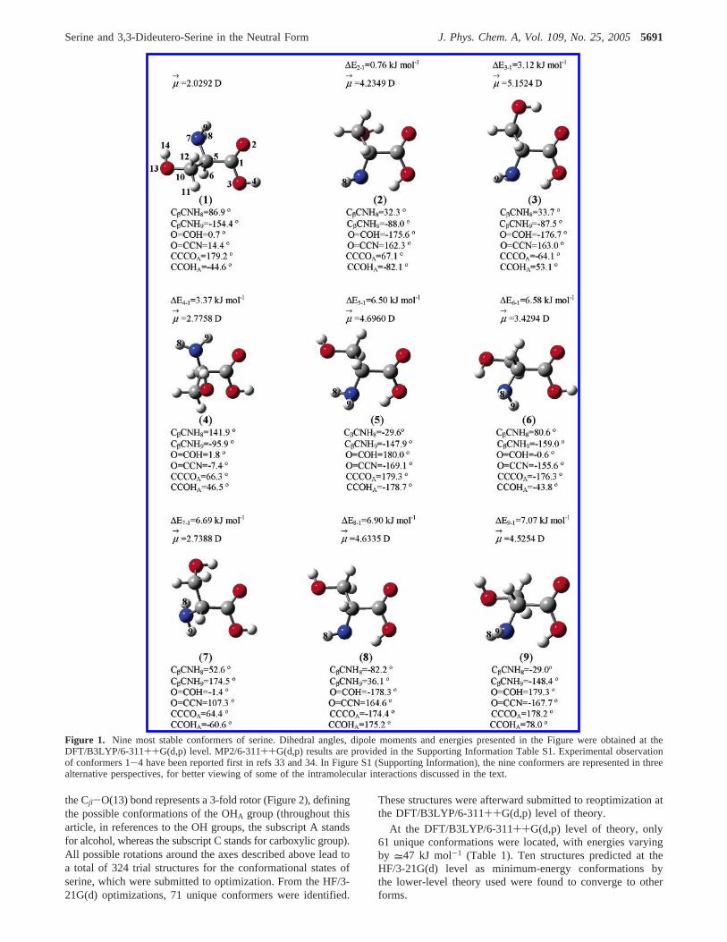
Figure 1. Nine most stable conformers of serine. Dihedral angles, dipole moments and energies presented in the Figure were obtained at theDFT/B3LYP/6-311++G(d,p) level. MP2/6-311++G(d,p) results are provided in the Supporting Information Table S1. Experimental observationof conformers 1-4 have been reported first in refs 33 and 34. In Figure S1 (Supporting Information), the nine conformers are represented in threealternative perspectives, for better viewing of some of the intramolecular interactions discussed in the text.
Serine and 3,3-Dideutero-Serine in the Neutral Form J. Phys. Chem. A, Vol. 109, No. 25, 20055691
Due to the very small differences in the relative conforma-tional energies obtained at the DFT level, the energies of allconformers of serine predicted at this level of theory withenergies within 7 kJ mol-1 were recalculated at the MP2/6-311++G(d,p) level of approximation (Table S1, SupportingInformation), which usually estimates better this property thanthe DFT/B3LYP method with the same basis set.49,50The maindifference between the results obtained with the two levels oftheory relates to the relative energy of conformer4 (see Figure1 and Tables 1 and S1), which is predicted by the MP2 as lowerthan that obtained by the DFT calculations.
The relative energies shown in both Tables 1 and S1 suggestthat the equilibrium mixture should contain significant contribu-tions from several conformers, with the first nine (see Figure1) playing the most important role. The relative populations ofthe nine most stable conformers, atT ) 470 K (the temperatureof the vapor immediately before deposition used in the matrixisolation experiments reported in this study), were predicted bythe DFT calculations to be 1.00:0.74:0.28:0.26:0.07:0.07:0.07:0.06:0.06 [corresponding as a whole to 75% (21:17:9:9:4:4:4:4:3%) of the total conformational population]. [The relativepopulations predicted at the MP2 level are 1.00:0.63:0.09:0.55:0.07:0.08:0.06:0.03:0.04. Since at this level of theory only theenergies of these nine conformers were calculated, no percentabsolute populations can be given.]
As already mentioned, the presence of three H-bond donorand four H-bond acceptor groups in serine allows for a widerange of intramolecular interactions and, consequently, for alarge number of stable conformations. For the purpose ofsystematically analyzing the large conformational data setstudied, conformations were characterized by the H-bonds theyexhibit. A distance of 2.7 Å was here used as a cutoff distancefor H-bond interactions. This distance is consistent with typicallyvalues found in analogous systems with weak intramolecularH-bond interactions.51 Although this approach is somewhatsimplistic, it appears to be considerably more useful than a merelist of dihedral angles for illustrating the complex interactionsfound in the studied amino acid. It is worth mentioning themarked nonlinearity of intramolecular H-bonds, which leads toweakening of these interactions in serine.
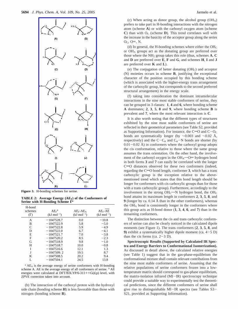
The possible H-bonding schemes for serine are depictedschematically in Figure 3: A, COOH group in the cisconformation; OHA group acting as proton-donor to the nitrogenatom.B, COOH group in the trans conformation; OHC groupacting as proton-donor to the nitrogen atom.C, COOH groupin the cis conformation; OHA group acting as proton-donor tothe carbonyl oxygen.D, COOH group in the cis conformation;OHA group acting as proton-donor to the OC atom.E, COOHgroup in the cis conformation; NH2 group acting as proton-donor to the carbonyl oxygen atom.F, COOH group in the cisconformation; NH2 group acting as proton-donor to the OA atom.
G, COOH group in the cis conformation; NH2 group acting asproton-donor to the OC atom. H, COOH group in the transconformation; OHC group acting as proton-donor to the OA
atom. I , COOH group in the trans conformation; OHA groupacting as proton-donor to the carbonyl oxygen.J, COOH groupin the trans conformation; OHA group acting as proton-donorto the nitrogen atom.K , COOH group in the trans conformation;NH2 group acting as proton-donor to the OA atom.L , COOHgroup in the trans conformation; NH2 group acting as proton-donor to the carbonyl oxygen atom. The H-bonding schemesappearing in each conformer of serine are given in Table 1.Note that associated with some of the H-bonding schemes theremight also be a prevalence for occurrence of important stericrepulsions, which may considerably destabilize the structures(e.g., in schemeL , the repulsion between OHC and the hydrogenatoms of the methylene group; see Figure 3).
As a way of analyzing the general stability of the H-bondingschemes of serine, the average energies of the conformers withineach scheme are compared with the overall mean energy inTable 2. The average energy of all conformers where theconformation of the carboxylic group is cis (37 conformers;see Table 1) was found to be 3.68 kJ mol-1 lower than theoverall mean energy, whereas the corresponding value for the24 conformers exhibiting a trans carboxylic group is larger thanthe global energy mean by 5.66 kJ mol-1. These results showthe general preference for the cis arrangement around the C-OC
bond, as it is commonly found for unsubstituted carboxylicacids.52-55 From the data shown in Table 2, it can also beconcluded that in bonding schemesA-H the intramolecularattractive interactions are dominant, whereas in bonding schemesI-L , the repulsive interactions dominate.
As could be realized, serine can find stabilization via a varietyof different combinations of the above considered H-bondingschemes. The nine most stable conformers differ in energy byless than ca. 7 kJ mol-1 and contain representatives of bondingschemesA-C. The first 44 conformers span less than 20 kJmol-1, and contain the entire characteristic set of bondingschemes (A-L ).
Bonding schemeA (COOH group in the cis conformation;OHA‚‚‚N H-bond) is clearly more favorable than all of theremaining, justifying in large amount the greatest stability ofconformer 1 and contributing also to the stabilization ofconformers4 and6.
The bonding schemeB is dominant in conformers2, 3, 5, 8,and 9 which are stabilized by a strong OHC‚‚‚N hydrogenbonding interaction. In conformer2, the calculated OHC‚‚‚Ndistance is as short as 1.916 Å. On the other hand, to establishthis H-bond, the conformation adopted by the carboxylic groupin these conformers must be the less stable trans configura-tion.52-55 However, the increase in energy due to the less stablegeometry adopted by the carboxylic group is compensated bythe establishment of a considerably strong H-bond, where thecombination of donor and acceptor groups is the most favorablepossible in serine.
Finally, conformer7 reveals three different types of H-bonding schemes (C, F andG), with two of them (C, which isthe dominant interaction, andF) existing only in this form(among the nine lowest energy conformers under analysis). Likein conformers1, 4 and6, the carboxylic group in this conformeradopts a cis conformation, but, instead of having the OHA groupacting as proton-donor to the nitrogen atom, this groupestablishes an H-bond with the carbonyl oxygen atom [schemeC; d(OHA‚‚‚O))) 2.194 Å]. In addition, this conformer is alsostabilized by two weaker H-bonds where the NH2 group acts
Figure 2. Atom numbering of serine/3,3-dideutero-serine. Internalrotation axes leading to possible rotamers: 2-fold, C(1)-C(3); 6-fold,C(1)-C(5); 3-fold, C(5)-N(7), C(5)-C(10), and C(5)-C(10). Sub-scripts A and C mean for “alcoholic” and “carboxylic”.
5692 J. Phys. Chem. A, Vol. 109, No. 25, 2005 Jarmelo et al.
as proton-donor: NH(8)‚‚‚OA (schemeF) and NH(9)‚‚‚OC (bond-ing schemeG).
As general conclusions, from the data shown in Table 2 (andalso in Table 1), the following can be stated:
(a) The cis arrangement of the carboxylic group is preferredover the trans configuration (with a single exception,B, allpreferred bonding schemes have the carboxylic group in thecis conformation).
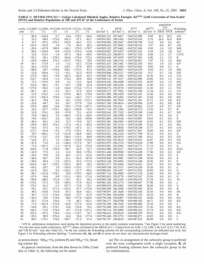
TABLE 1: DFT/B3LYP/6-311++G(d,p) Calculated Dihedral Angles, Relative Energies∆EZPVE [with Correction of Non-ScaledZPVE] and Relative Populations at 298 and 470 Ka of the Conformers of Serine
population(%)confor
merCâCNH8
b
(°)CâCNH9
(°)OdCOH
(°)OdCCN
(°)CCCOA
(°)CCOHA
(°)E
(kJ mol-1)ZPVE
(kJ mol-1)EZPVE
(kJ mol-1)∆EZPVE
(kJ mol-1)c 298 K 470 KH-bondingschemesd
1 86.9 -154.4 0.7 14.4 179.2 -44.6 -1047831.551 297.6457-1047533.906 0.00 36.1 20.5 A/E2 32.3 -88.0 -175.6 162.3 67.1 -82.1 -1047831.691 298.5456-1047533.145 0.76 26.4 16.9 B/K3 33.7 -87.5 -176.7 163.0 -64.1 53.1-1047830.085 299.2947-1047530.790 3.12 9.9 9.2 B/I4 141.9 -95.9 1.8 -7.4 66.3 46.5 -1047828.035 297.4995-1047530.535 3.37 8.9 8.7 A/E5 -29.6 -147.9 180.0 -169.1 179.3 -178.7 -1047825.352 297.9462-1047527.406 6.50 2.4 3.9 B/K6 80.6 -159.0 -0.6 -155.6 -176.3 -43.8 -1047824.967 297.6457-1047527.322 6.58 2.4 3.8 A/G7 52.6 174.5 -1.4 107.3 64.4 -60.6 -1047825.222 298.0013-1047527.220 6.69 2.3 3.7 C/F/G8 -82.2 36.1 -178.3 164.6 -174.4 175.2 -1047824.449 297.4478-1047527.001 6.90 2.1 3.5 B9 -29.0 -148.4 179.3 -167.7 178.2 78.0 -1047825.105 298.2742-1047526.831 7.07 1.9 3.4 B/K
10 56.7 173.9 1.4 -3.2 63.1 173.8 -1047821.437 296.1441-1047525.293 8.61 1.0 2.3 E/F11 66.4 -172.9 0.1 120.1 -57.9 69.9-1047822.424 297.3426-1047525.082 8.82 0.9 2.1 G12 -88.9 29.7 -179.6 169.1 -175.3 87.4 -1047821.344 296.8667-1047524.478 9.43 0.7 1.8 B/K13 51.8 169.4 -2.2 -16.1 62.3 -83.9 -1047820.846 296.5127-1047524.333 9.57 0.7 1.8 D/E14 171.8 -68.2 -0.8 142.5 -66.4 65.5 --1047820.736 297.1945-1047523.541 10.36 0.5 1.4 C/G15 152.7 -85.4 -1.3 174.5 69.9 44.6 -1047820.151 297.1617-1047522.989 10.92 0.4 1.3 A/G16 72.5 -170.0 0.0 2.2 -72.2 56.8 -1047819.435 296.4588-1047522.976 10.93 0.4 1.3 D/E17 152.0 -86.1 -0.1 -16.7 -74.8 63.4 -1047819.549 296.6436-1047522.906 11.00 0.4 1.2 D/E18 -175.9 -56.5 -1.8 134.2 -174.4 -171.3 -1047818.575 295.9719-1047522.603 11.30 0.3 1.1 F/G19 -83.1 36.1 -3.3 91.5 67.9 -62.4 -1047820.137 297.7695-1047522.368 11.54 0.3 1.1 C/F20 57.0 176.1 -1.4 -179.4 68.3 179.9 -1047817.893 296.3459-1047521.547 12.36 0.2 0.9 F/G21 137.2 -102.3 174.9 -14.3 -64.7 172.7 -1047818.496 297.2282-1047521.267 12.64 0.2 0.8 H/L22 -42.3 78.4 0.4 26.1 -179.0 -173.1 -1047816.962 295.7810-1047521.181 12.72 0.2 0.8 E/F23 -35.6 84.7 0.5 24.7 177.8 75.8-1047817.396 296.4014-1047520.994 12.91 0.2 0.8 E/F24 170.0 -68.0 -0.6 -20.3 -173.6 -167.2 -1047816.336 295.534 -1047520.802 13.10 0.2 0.7 E/F25 35.6 -87.7 -179.1 166.2 -169.8 -76.5 -1047817.714 297.1392-1047520.575 13.33 0.1 0.7 B26 52.8 176.1 0.6 -97.8 72.1 -65.0 -1047818.241 297.7322-1047520.508 13.40 0.1 0.7 D/F27 74.8 -166.5 3.5 -140.3 -51.6 -66.0 -1047816.547 296.3509-1047520.196 13.71 0.1 0.6 E28 74.9 -165.7 2.6 54.2 -58.0 169.8 -1047815.802 295.9148-1047519.887 14.02 0.1 0.6 E29 -40.4 83.1 -0.4 15.3 -74.4 53.7 -1047815.381 296.0383-1047519.343 14.56 0.1 0.5 D/E30 139.9 -99.3 174.5 -17.4 -61.1 -85.9 -1047815.501 297.2116-1047518.289 15.62 0.1 0.4 H/L31 -54.3 67.5 0.5 -179.5 -67.5 51.7 -1047815.022 296.8826-1047518.140 15.77 0.1 0.4 C/G32 173.7 -63.0 -0.2 -17.0 -176.1 81.5 -1047813.311 295.4638-1047517.847 16.06 0.0 0.3 E/F33 70.5 -169.1 -1.9 -121.8 -48.8 -66.5 -1047814.021 296.2316-1047517.789 16.12 0.0 0.3 G34 -173.4 -52.7 -2.0 136.6 -176.5 80.9 -1047813.096 295.6976-1047517.399 16.51 0.0 0.3 F/G35 -52.0 67.1 1.9 -146.2 -174.8 -176.0 -1047812.592 295.6610-1047516.931 16.97 0.0 0.3 F/G36 -47.0 71.6 1.6 -148.4 -177.0 78.7 -1047812.975 296.1574-1047516.818 17.09 0.0 0.3 F/G37 71.9 -167.5 -1.3 -107.9 -55.2 173.0 -1047812.601 295.8302-1047516.771 17.13 0.0 0.3 G38 164.0 -77.8 176.0 -86.7 62.0 52.7 -1047815.990 299.3557-1047516.635 17.27 0.0 0.2 H/J/L39 -42.9 77.0 1.5 14.3 60.6-179.2 -1047811.444 294.9948-1047516.449 17.46 0.0 0.2 E/F40 -50.0 70.9 2.4 44.1 -57.3 -61.6 -1047811.609 295.9102-1047515.699 18.21 0.0 0.2 E41 -60.6 60.7 0.8 61.1 -63.2 167.0 -1047810.846 295.4981-1047515.348 18.56 0.0 0.2 E42 -58.6 60.4 -1.0 -167.5 67.5-173.5 -1047810.148 295.6005-1047514.548 19.36 0.0 0.1 F43 -163.6 -44.7 -178.5 -162.5 60.0 59.3 -1047812.077 297.6456-1047514.431 19.47 0.0 0.1 B/J44 -64.2 59.7 1.9 -143.6 -176.1 -77.7 -1047809.706 295.3557-1047514.350 19.56 0.0 0.1 e45 -61.8 59.2 0.2 -126.3 -51.7 -60.2 -1047808.461 295.5457-1047512.916 20.99 0.0 0.1 G46 86.7 -155.4 -178.1 16.8 -179.5 -46.0 -1047807.716 296.4969-1047511.219 22.69 0.0 0.1 J/L47 -67.0 54.6 0.8 -115.1 -58.3 171.6 -1047805.625 295.0778-1047510.547 23.36 0.0 0.1 G48 -90.0 148.5 172.8 -19.1 62.1 46.3 -1047803.188 296.5185-1047506.670 27.24 0.0 0.1 J/L49 -176.2 -57.3 -0.9 -109.3 -57.9 -65.3 -1047801.585 295.5773-1047506.007 27.90 0.0 0.0 e50 179.2 -61.5 -1.2 107.5 -72.6 -70.1 -1047800.876 295.4302-1047505.446 28.46 0.0 0.0 G51 -78.1 43.1 -171.4 -103.2 67.7 -175.0 -1047800.389 296.3958-1047503.994 29.91 0.0 0.0 K52 49.3 170.8 175.8 91.3 66.0-63.1 -1047799.607 297.2640-1047502.343 31.56 0.0 0.0 I/K53 43.7 -77.2 -174.4 156.0 -75.7 -67.0 -1047797.804 296.4284-1047501.376 32.53 0.0 0.0 B54 -42.3 78.0 -174.6 38.0 -177.6 -169.0 -1047794.188 294.9467-1047499.241 34.66 0.0 0.0 K/L55 59.2 175.4 176.6 -7.0 60.1 165.1 -1047794.171 294.9798-1047499.192 34.71 0.0 0.0 K/L56 77.4 -161.8 -175.6 -93.6 -177.0 -41.6 -1047793.509 296.7026-1047496.806 37.10 0.0 0.0 J57 -34.8 85.1 -174.1 33.8 176.4 76.3-1047791.608 295.1318-1047496.476 37.43 0.0 0.0 K/L58 160.5 -75.9 -178.4 -8.7 -171.8 -157.1 -1047790.114 293.9490-1047496.165 37.74 0.0 0.0 K/L59 167.5 -67.5 179.4 -13.4 -176.7 74.7 -1047784.412 294.0524-1047490.359 43.55 0.0 0.0 K/L60 -29.5 90.9 -176.4 16.6 59.4 177.4-1047783.446 293.3753-1047490.071 43.83 0.0 0.0 L61 68.3 -170.1 179.0 -100.7 -57.3 67.4 -1047781.707 294.6814-1047487.025 46.88 0.0 0.0 e
a 470 K, sublimation temperature used during the deposition process in the matrix isolation experiments.b See Figure 2 for atom numbering.c For the nine most stable conformers,∆EZPVE values calculated at the MP2/6-311++G(d,p) level are 0.00, 1.13, 5.88, 1.46, 6.47, 6.13, 7.03, 8.43,and 7.98 kJ mol-1 (see also Table S1).d In the last column the H-bonding schemes for the corresponding conformer are indicated (see text). SeeFigure 3 for H-bonding schemes labeling.e Conformers61, 44, and49 of serine do not show any intramolecular hydrogen bond.
Serine and 3,3-Dideutero-Serine in the Neutral Form J. Phys. Chem. A, Vol. 109, No. 25, 20055693
(b) The interaction of the carboxyl proton with the hydroxylside chain (bonding schemeH) is less favorable than those withnitrogen (bonding schemeB).
(c) When acting as donor group, the alcohol group (OHA)prefers to take part in H-bonding interactions with the nitrogenatom (schemeA) or with the carbonyl oxygen atom (schemeC) than with OC (schemeD). This trend correlates well withthe increase in the basicity of the acceptor group along the seriesOC, O), N.
(d) In general, the H-bonding schemes where either the OHC
or OHA groups act as the donating group are preferred overthose where the NH2 group takes this role (thus, schemesA, CandD are preferred overE, F andG, and schemesH, I andJare preferred overK andL ).
(e) The conjugation of better donating (OHC) and acceptor(N) moieties occurs in schemeB, justifying the exceptionalcharacter of the position occupied by this bonding scheme(which is associated with the higher-energy trans arrangementof the carboxylic group, but corresponds to the second preferredstructural arrangement) in the energy scale.
(f) taking into consideration the dominant intramolecularinteractions in the nine most stable conformers of serine, theycan be grouped in 3 classes:1, 4 and6, where bonding schemeA dominates;2, 3, 5, 8 and 9, where bonding schemeB isprevalent and7, where the most relevant interaction isC.
It is also worth noting that the different types of structuresexhibited by the nine most stable conformers of serine arereflected in their geometrical parameters (see Table S2, providedas Supporting Information). For instance, the CdO and C-OC
bonds are systematically longer (by∼0.003 and∼0.02 Å,respectively) and the C-CR and CR-N bonds are shorter (by0.01-0.02 Å) in conformers where the carboxyl group adoptsthe cis conformation, relative to those where the same groupassumes the trans orientation. On the other hand, the involve-ment of the carbonyl oxygen in the OHA‚‚‚Od hydrogen bondin both forms3 and7 can easily be correlated with the longerCdO distances observed for these two conformers (indeed,regarding the CdO bond length, conformer3, which has a transcarboxylic group is the exception relative to the above-mentioned trend which states that this bond length should belonger for conformers with cis carboxylic groups than for thosewith a trans carboxylic group). Furthermore, accordingly to theinvolvement in the strong OHC‚‚‚N hydrogen bond, the OHCbond attains its maximum length in conformers2, 3, 5, 8, and9 (longer by ca. 0.14 Å than in the other conformers), whereasthe OHA bond is consistently longer in the conformers wherethis group acts as H-bond donor (1, 3, 4, 6, and7) than in theremaining conformers.
The distinction between the cis and trans carboxylic conform-ers of serine can also be clearly noticed in the calculated dipolemoments (see Figure 1). The trans conformers (2, 3, 5, 8, and9) exhibit a systematically higher dipole moment (ca. 4-5 D)than the cis forms (ca. 2-3 D).
Spectroscopic Results (Supported by Calculated IR Spec-tra and Energy Barriers to Conformational Isomerization).As discussed in detail above, the calculated relative energies(see Table 1) suggest that in the gas-phase-equilibrium theconformational mixture shall contain relevant contributions fromthe nine most stable conformers of serine. Assuming that therelative populations of serine conformers frozen into a low-temperature matrix should correspond to gas-phase equilibrium,the matrix-isolation infrared (MI-IR) spectroscopy techniquecould provide a suitable way to experimentally test the theoreti-cal predictions, since the different conformers of serine shallgive rise to distinguishable MI-IR spectra (see Tables S3-S21, provided as Supporting Information).
Figure 3. H-bonding schemes for serine.
TABLE 2: Average Energy (AEΓ) of the Conformers ofSerine with H-Bonding SchemeΓa
H-bondschemes
(Γ)AEΓ
b
(kJ mol-1)AEΓ-AEA
(kJ mol-1)AEΓ-AE
(kJ mol-1)
A -1047528.7 0.0 -10.8B -1047522.9 5.8 -5.0C -1047522.8 5.9 -4.9D -1047522.0 6.7 -4.1E -1047521.7 7.0 -3.8F -1047520.2 8.5 -2.3G -1047518.9 9.8 -1.0H -1047518.7 10.0 -0.8I -1047516.6 12.1 1.3J -1047509. 2 19.5 8.7K -1047508.5 20.2 9.4L -1047504.1 24.5 13.8
a AEA is the average energy of serine conformers with H-bondingscheme A. AE is the average energy of all conformers of serine.b Allenergies were calculated at DFT/B3LYP/6-311++G(d,p) level, withZPVE correction taken into account.
5694 J. Phys. Chem. A, Vol. 109, No. 25, 2005 Jarmelo et al.
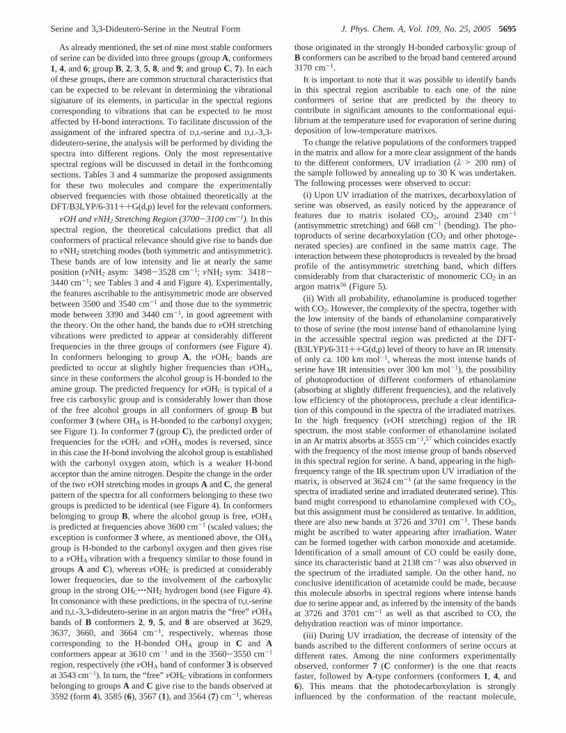
As already mentioned, the set of nine most stable conformersof serine can be divided into three groups (groupA, conformers1, 4, and6; groupB, 2, 3, 5, 8, and9; and groupC, 7). In eachof these groups, there are common structural characteristics thatcan be expected to be relevant in determining the vibrationalsignature of its elements, in particular in the spectral regionscorresponding to vibrations that can be expected to be mostaffected by H-bond interactions. To facilitate discussion of theassignment of the infrared spectra ofD,L-serine andD,L-3,3-dideutero-serine, the analysis will be performed by dividing thespectra into different regions. Only the most representativespectral regions will be discussed in detail in the forthcomingsections. Tables 3 and 4 summarize the proposed assignmentsfor these two molecules and compare the experimentallyobserved frequencies with those obtained theoretically at theDFT/B3LYP/6-311++G(d,p) level for the relevant conformers.
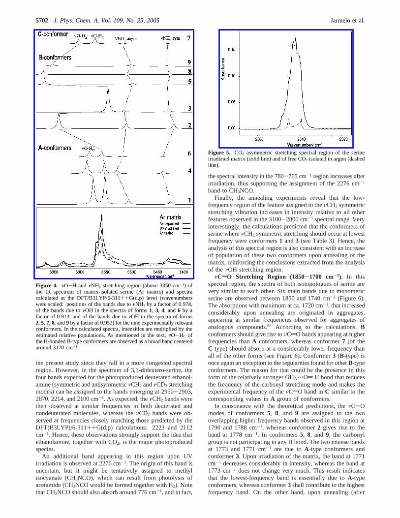
νOH andνNH2 Stretching Region (3700-3100 cm-1). In thisspectral region, the theoretical calculations predict that allconformers of practical relevance should give rise to bands dueto νNH2 stretching modes (both symmetric and antisymmetric).These bands are of low intensity and lie at nearly the sameposition (νNH2 asym: 3498-3528 cm-1; νNH2 sym: 3418-3440 cm-1; see Tables 3 and 4 and Figure 4). Experimentally,the features ascribable to the antisymmetric mode are observedbetween 3500 and 3540 cm-1 and those due to the symmetricmode between 3390 and 3440 cm-1, in good agreement withthe theory. On the other hand, the bands due toνOH stretchingvibrations were predicted to appear at considerably differentfrequencies in the three groups of conformers (see Figure 4).In conformers belonging to groupA, the νOHC bands arepredicted to occur at slightly higher frequencies thanνOHA,since in these conformers the alcohol group is H-bonded to theamine group. The predicted frequency forνOHC is typical of afree cis carboxylic group and is considerably lower than thoseof the free alcohol groups in all conformers of groupB butconformer3 (where OHA is H-bonded to the carbonyl oxygen;see Figure 1). In conformer7 (groupC), the predicted order offrequencies for theνOHC andνOHA modes is reversed, sincein this case the H-bond involving the alcohol group is establishedwith the carbonyl oxygen atom, which is a weaker H-bondacceptor than the amine nitrogen. Despite the change in the orderof the twoνOH stretching modes in groupsA andC, the generalpattern of the spectra for all conformers belonging to these twogroups is predicted to be identical (see Figure 4). In conformersbelonging to groupB, where the alcohol group is free,νOHA
is predicted at frequencies above 3600 cm-1 (scaled values; theexception is conformer3 where, as mentioned above, the OHA
group is H-bonded to the carbonyl oxygen and then gives riseto aνOHA vibration with a frequency similar to those found ingroupsA andC), whereasνOHC is predicted at considerablylower frequencies, due to the involvement of the carboxylicgroup in the strong OHC‚‚‚NH2 hydrogen bond (see Figure 4).In consonance with these predictions, in the spectra ofD,L-serineandD,L-3,3-dideutero-serine in an argon matrix the “free”νOHA
bands ofB conformers2, 9, 5, and 8 are observed at 3629,3637, 3660, and 3664 cm-1, respectively, whereas thosecorresponding to the H-bonded OHA group in C and Aconformers appear at 3610 cm-1 and in the 3560-3550 cm-1
region, respectively (theνOHA band of conformer3 is observedat 3543 cm-1). In turn, the “free”νOHC vibrations in conformersbelonging to groupsA andC give rise to the bands observed at3592 (form4), 3585 (6), 3567 (1), and 3564 (7) cm-1, whereas
those originated in the strongly H-bonded carboxylic group ofB conformers can be ascribed to the broad band centered around3170 cm-1.
It is important to note that it was possible to identify bandsin this spectral region ascribable to each one of the nineconformers of serine that are predicted by the theory tocontribute in significant amounts to the conformational equi-librium at the temperature used for evaporation of serine duringdeposition of low-temperature matrixes.
To change the relative populations of the conformers trappedin the matrix and allow for a more clear assignment of the bandsto the different conformers, UV irradiation (λ > 200 nm) ofthe sample followed by annealing up to 30 K was undertaken.The following processes were observed to occur:
(i) Upon UV irradiation of the matrixes, decarboxylation ofserine was observed, as easily noticed by the appearance offeatures due to matrix isolated CO2, around 2340 cm-1
(antisymmetric stretching) and 668 cm-1 (bending). The pho-toproducts of serine decarboxylation (CO2 and other photoge-nerated species) are confined in the same matrix cage. Theinteraction between these photoproducts is revealed by the broadprofile of the antisymmetric stretching band, which differsconsiderably from that characteristic of monomeric CO2 in anargon matrix56 (Figure 5).
(ii) With all probability, ethanolamine is produced togetherwith CO2. However, the complexity of the spectra, together withthe low intensity of the bands of ethanolamine comparativelyto those of serine (the most intense band of ethanolamine lyingin the accessible spectral region was predicted at the DFT-(B3LYP)/6-311++G(d,p) level of theory to have an IR intensityof only ca. 100 km mol-1, whereas the most intense bands ofserine have IR intensities over 300 km mol-1), the possibilityof photoproduction of different conformers of ethanolamine(absorbing at slightly different frequencies), and the relativelylow efficiency of the photoprocess, preclude a clear identifica-tion of this compound in the spectra of the irradiated matrixes.In the high frequency (νOH stretching) region of the IRspectrum, the most stable conformer of ethanolamine isolatedin an Ar matrix absorbs at 3555 cm-1,57 which coincides exactlywith the frequency of the most intense group of bands observedin this spectral region for serine. A band, appearing in the high-frequency range of the IR spectrum upon UV irradiation of thematrix, is observed at 3624 cm-1 (at the same frequency in thespectra of irradiated serine and irradiated deuterated serine). Thisband might correspond to ethanolamine complexed with CO2,but this assignment must be considered as tentative. In addition,there are also new bands at 3726 and 3701 cm-1. These bandsmight be ascribed to water appearing after irradiation. Watercan be formed together with carbon monoxide and acetamide.Identification of a small amount of CO could be easily done,since its characteristic band at 2138 cm-1 was also observed inthe spectrum of the irradiated sample. On the other hand, noconclusive identification of acetamide could be made, becausethis molecule absorbs in spectral regions where intense bandsdue to serine appear and, as inferred by the intensity of the bandsat 3726 and 3701 cm-1 as well as that ascribed to CO, thedehydration reaction was of minor importance.
(iii) During UV irradiation, the decrease of intensity of thebands ascribed to the different conformers of serine occurs atdifferent rates. Among the nine conformers experimentallyobserved, conformer7 (C conformer) is the one that reactsfaster, followed byA-type conformers (conformers1, 4, and6). This means that the photodecarboxylation is stronglyinfluenced by the conformation of the reactant molecule,
Serine and 3,3-Dideutero-Serine in the Neutral Form J. Phys. Chem. A, Vol. 109, No. 25, 20055695
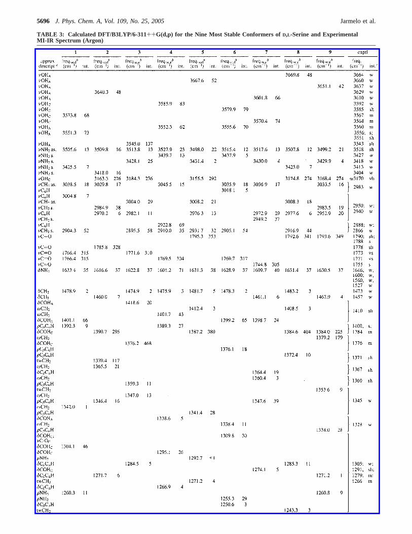
TABLE 3: Calculated DFT/B3LYP/6-311++G(d,p) for the Nine Most Stable Conformers ofD,L-Serine and ExperimentalMI-IR Spectrum (Argon)
5696 J. Phys. Chem. A, Vol. 109, No. 25, 2005 Jarmelo et al.
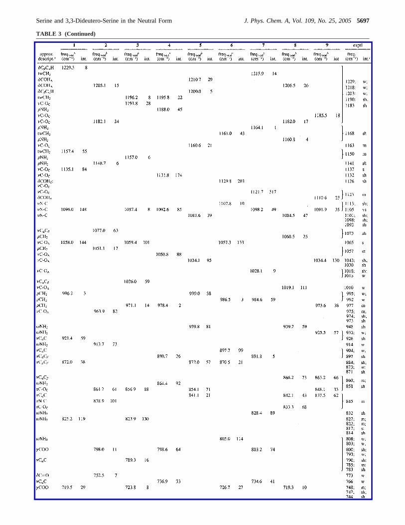
TABLE 3 (Continued)
Serine and 3,3-Dideutero-Serine in the Neutral Form J. Phys. Chem. A, Vol. 109, No. 25, 20055697
occurring preferentially for those conformers where the car-boxylic group is not engaged in H-bond as proton donor. Thereason for the observed specific preference for theC conformerrelative to theA conformers can be related with the presencein the C conformer of the stronger O-HA‚‚‚O) bond insteadof the considerably weaker N-H‚‚‚O) bond present in theAconformers. The different rates of decarboxylation associatedwith the three groups of serine conformers strongly facilitatedthe assignment of the experimental spectra. This is clearlydemonstrated in the case of theνO-H stretching spectral regionin Figure 4, but it does also apply to the other regions of thespectra, as will be stressed further below.
(iv) Besides aggregation, which can be noticed by appearanceof characteristic bands of aggregates throughout the spectra,annealing of the matrixes led only to minor spectral changes,from which the most relevant is the increase of the intensity ofthe bands with predominant contribution from conformers1 and3 relative to bands due to other conformers. This is illustratedin Figure 4 by the relative increase of intensity of the bands at3567 and 3556 cm-1, due to conformer1 and at 3543 cm-1,ascribed to conformer3. When annealing of the matrix is done,conversion from the less stable conformers into an energeticallyaccessible lower energy form shall take place. The energybarriers separating the reactant conformers from the productsshall be low enough to be overcome at the low work temper-ature. As a rough estimate of the barriers of interconversionbetween the different experimentally observed conformers of
serine in the low-temperature matrixes, the DFT/B3LYP/6-311++G(d,p) barriers for the molecule in a vacuum werecalculated (see Table S22 of the Supporting Information; theCOOH cisT COOH trans interconversion is well-known tohave associated barriers of more than ca. 40 kJ mol-1 58 andwas not here subjected to calculation). The calculated barriersare, in general, predicted to be relatively high. Indeed, in manycases, the simultaneous rearrangement of several groups, as wellas breaking of H bonds, are required for the molecular systemto undergo a transition from one conformer to other of lowerenergy. However, taking into account the margin of errorassociated with the unavoidable intrinsic errors in the theoreticalestimations as well as the effects of the environment, thecalculated values for the energy barriers are compatible withobservation of, at least, some conformational rearrangementsupon annealing of the matrixes. If we took into considerationthe theoretical results just as an indicator of the relativeaccessibility of the various processes (by looking at the relativevalues of the barriers), then, in consonance with the experimentalobservations, for cis carboxylic conformers (A andC conform-ers), one could expect an increase of population of conformer1, particularly at expenses of both forms4 and7 (conformer6might also be converted to1 in a two step process6 f 4 f1;see Table S22). In the case of trans carboxylic conformers (Bconformers), the theoretically predicted energy barriers areconsistent with conversion from conformers5, 8, and9 eitherin conformer2 or/and3 (energy barriers leading to production
TABLE 3 (Continued)
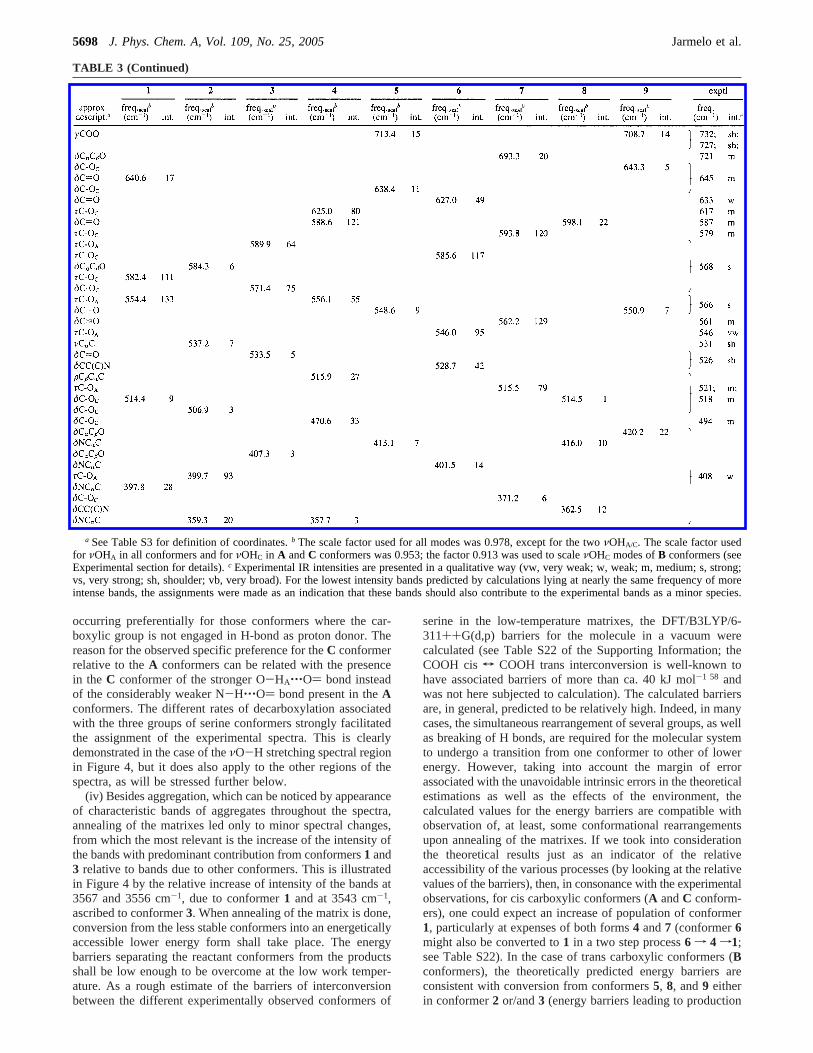
a See Table S3 for definition of coordinates.b The scale factor used for all modes was 0.978, except for the twoνOHA/C. The scale factor usedfor νOHA in all conformers and forνOHC in A andC conformers was 0.953; the factor 0.913 was used to scaleνOHC modes ofB conformers (seeExperimental section for details).c Experimental IR intensities are presented in a qualitative way (vw, very weak; w, weak; m, medium; s, strong;vs, very strong; sh, shoulder; vb, very broad). For the lowest intensity bands predicted by calculations lying at nearly the same frequency of moreintense bands, the assignments were made as an indication that these bands should also contribute to the experimental bands as a minor species.
5698 J. Phys. Chem. A, Vol. 109, No. 25, 2005 Jarmelo et al.
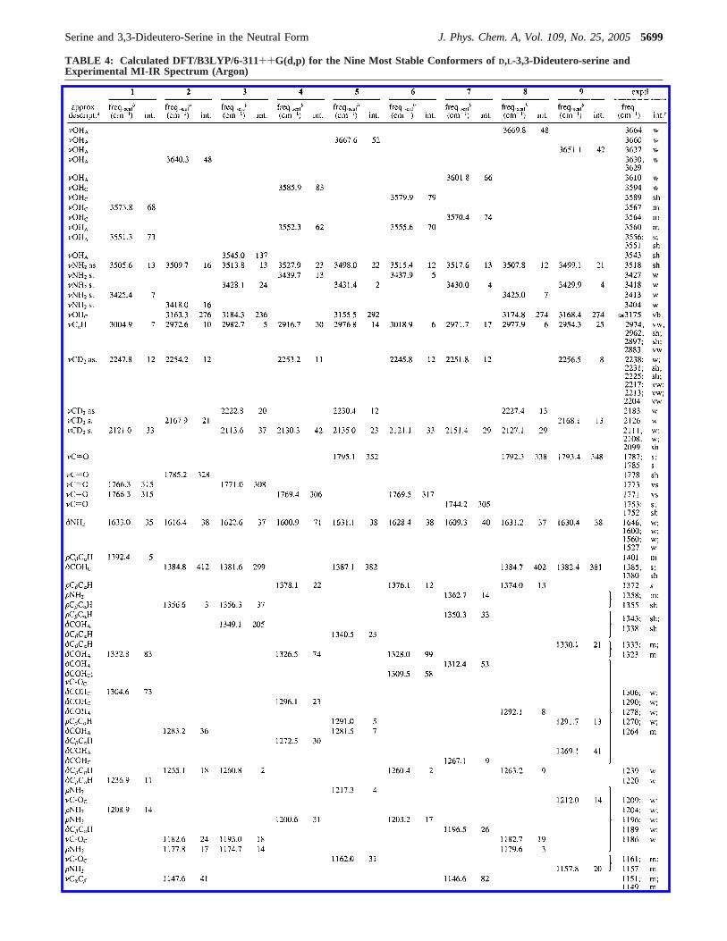
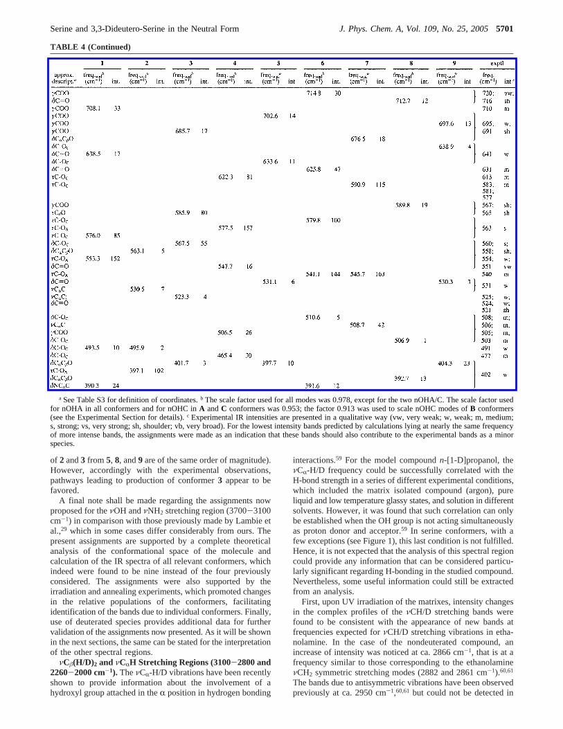
TABLE 4: Calculated DFT/B3LYP/6-311++G(d,p) for the Nine Most Stable Conformers ofD,L-3,3-Dideutero-serine andExperimental MI-IR Spectrum (Argon)
Serine and 3,3-Dideutero-Serine in the Neutral Form J. Phys. Chem. A, Vol. 109, No. 25, 20055699
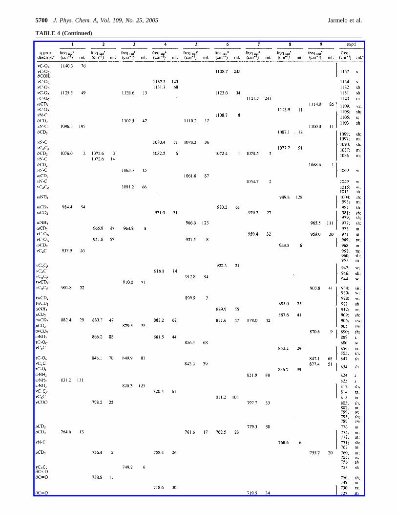
TABLE 4 (Continued)
5700 J. Phys. Chem. A, Vol. 109, No. 25, 2005 Jarmelo et al.
of 2 and3 from 5, 8, and9 are of the same order of magnitude).However, accordingly with the experimental observations,pathways leading to production of conformer3 appear to befavored.
A final note shall be made regarding the assignments nowproposed for theνOH andνNH2 stretching region (3700-3100cm-1) in comparison with those previously made by Lambie etal.,29 which in some cases differ considerably from ours. Thepresent assignments are supported by a complete theoreticalanalysis of the conformational space of the molecule andcalculation of the IR spectra of all relevant conformers, whichindeed were found to be nine instead of the four previouslyconsidered. The assignments were also supported by theirradiation and annealing experiments, which promoted changesin the relative populations of the conformers, facilitatingidentification of the bands due to individual conformers. Finally,use of deuterated species provides additional data for furthervalidation of the assignments now presented. As it will be shownin the next sections, the same can be stated for the interpretationof the other spectral regions.
νCâ(H/D)2 and νCRH Stretching Regions (3100-2800 and2260-2000 cm-1). TheνCR-H/D vibrations have been recentlyshown to provide information about the involvement of ahydroxyl group attached in theR position in hydrogen bonding
interactions.59 For the model compoundn-[1-D]propanol, theνCR-H/D frequency could be successfully correlated with theH-bond strength in a series of different experimental conditions,which included the matrix isolated compound (argon), pureliquid and low temperature glassy states, and solution in differentsolvents. However, it was found that such correlation can onlybe established when the OH group is not acting simultaneouslyas proton donor and acceptor.59 In serine conformers, with afew exceptions (see Figure 1), this last condition is not fulfilled.Hence, it is not expected that the analysis of this spectral regioncould provide any information that can be considered particu-larly significant regarding H-bonding in the studied compound.Nevertheless, some useful information could still be extractedfrom an analysis.
First, upon UV irradiation of the matrixes, intensity changesin the complex profiles of theνCH/D stretching bands werefound to be consistent with the appearance of new bands atfrequencies expected forνCH/D stretching vibrations in etha-nolamine. In the case of the nondeuterated compound, anincrease of intensity was noticed at ca. 2866 cm-1, that is at afrequency similar to those corresponding to the ethanolamineνCH2 symmetric stretching modes (2882 and 2861 cm-1).60,61
The bands due to antisymmetric vibrations have been observedpreviously at ca. 2950 cm-1,60,61 but could not be detected in
TABLE 4 (Continued)
a See Table S3 for definition of coordinates.b The scale factor used for all modes was 0.978, except for the two nOHA/C. The scale factor usedfor nOHA in all conformers and for nOHC inA andC conformers was 0.953; the factor 0.913 was used to scale nOHC modes ofB conformers(see the Experimental Section for details).c Experimental IR intensities are presented in a qualitative way (vw, very weak; w, weak; m, medium;s, strong; vs, very strong; sh, shoulder; vb, very broad). For the lowest intensity bands predicted by calculations lying at nearly the same frequencyof more intense bands, the assignments were made as an indication that these bands should also contribute to the experimental bands as a minorspecies.
Serine and 3,3-Dideutero-Serine in the Neutral Form J. Phys. Chem. A, Vol. 109, No. 25, 20055701
the present study since they fall in a more congested spectralregion. However, in the spectrum of 3,3-dideutero-serine, thefour bands expected for the photoproduced deuterated ethanol-amine (symmetric and antisymmetricνCH2 andνCD2 stretchingmodes) can be assigned to the bands emerging at 2950-2903,2870, 2214, and 2100 cm-1. As expected, theνCH2 bands werethen observed at similar frequencies in both deuterated andnondeuterated molecules, whereas theνCD2 bands were ob-served at frequencies closely matching those predicted by theDFT(B3LYP)/6-311++G(d,p) calculations: 2223 and 2112cm-1. Hence, these observations strongly support the idea thatethanolamine, together with CO2, is the major photoproducedspecies.
An additional band appearing in this region upon UVirradiation is observed at 2276 cm-1. The origin of this band isuncertain, but it might be tentatively assigned to methylisocyanate (CH3NCO), which can result from photolysis ofacetamide (CH3NCO would be formed together with H2). Notethat CH3NCO should also absorb around 776 cm-1, and in fact,
the spectral intensity in the 780-765 cm-1 region increases afterirradiation, thus supporting the assignment of the 2276 cm-1
band to CH3NCO.Finally, the annealing experiments reveal that the low-
frequency region of the feature assigned to theνCH2 symmetricstretching vibration increases in intensity relative to all otherfeatures observed in the 3100-2800 cm-1 spectral range. Veryinterestingly, the calculations predicted that the conformers ofserine whereνCH2 symmetric stretching should occur at lowestfrequency were conformers1 and3 (see Table 3). Hence, theanalysis of this spectral region is also consistent with an increaseof population of these two conformers upon annealing of thematrix, reinforcing the conclusions extracted from the analysisof the νOH stretching region.
νCdO Stretching Region (1850-1700 cm-1). In thisspectral region, the spectra of both isotopologues of serine arevery similar to each other. Six main bands due to monomericserine are observed between 1850 and 1740 cm-1 (Figure 6).The absorptions with maximum at ca. 1720 cm-1, that increasedconsiderably upon annealing are originated in aggregates,appearing at similar frequencies observed for aggregates ofanalogous compounds.62 According to the calculations,Bconformers should give rise toνCdO bands appearing at higherfrequencies thanA conformers, whereas conformer7 (of theC-type) should absorb at a considerably lower frequency thanall of the other forms (see Figure 6). Conformer3 (B-type) isonce again an exception to the regularities found for otherB-typeconformers. The reason for that could be the presence in thisform of the relatively stronger OHA‚‚‚Od H bond that reducesthe frequency of the carbonyl stretching mode and makes theexperimental frequency of theνCdO band inC similar to thecorresponding values inA group of conformers.
In consonance with the theoretical predictions, theνCdOmodes of conformers5, 8, and 9 are assigned to the twooverlapping higher frequency bands observed in this region at1790 and 1788 cm-1, whereas conformer2 gives rise to theband at 1778 cm-1. In conformers5, 8, and 9, the carbonylgroup is not participating in any H bond. The two intense bandsat 1773 and 1771 cm-1 are due toA-type conformers andconformer3. Upon irradiation of the matrix, the band at 1771cm-1 decreases considerably in intensity, whereas the band at1773 cm-1 does not change very much. This result indicatesthat the lowest-frequency band is essentially due toA-typeconformers, whereas conformer3 shall contribute to the highestfrequency band. On the other hand, upon annealing (after
Figure 4. νO-H andνNH2 stretching region (above 3350 cm-1) ofthe IR spectrum of matrix-isolated serine (Ar matrix) and spectracalculated at the DFT/B3LYP/6-311++G(d,p) level (wavenumberswere scaled: positions of the bands due toνNH2 by a factor of 0.978,of the bands due toνOH in the spectra of forms1, 3, 4, and6 by afactor of 0.913, and of the bands due toνOH in the spectra of forms2, 5, 7, 8, and9 by a factor of 0.953) for the nine experimentally relevantconformers. In the calculated spectra, intensities are multiplied by theestimated relative populations. As mentioned in the text,νO-HC ofthe H-bonded B-type conformers are observed as a broad band centeredaround 3170 cm-1.
Figure 5. CO2 asymmetric stretching spectral region of the serineirradiated matrix (solid line) and of free CO2 isolated in argon (dashedline).
5702 J. Phys. Chem. A, Vol. 109, No. 25, 2005 Jarmelo et al.
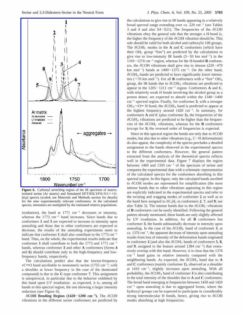
irradiation), the band at 1771 cm-1 decreases in intensity,whereas the 1773 cm-1 band increases. Since bands due toconformers1 and3 are expected to increase in intensity uponannealing and those due to other conformers are expected todecrease, the results of the annealing experiments seem toindicate that conformer1 shall also contribute to the 1773 cm-1
band. Thus, on the whole, the experimental results indicate thatconformer1 shall contribute to both the 1773 and 1771 cm-1
bands, whereas conformer3 and otherA conformers (forms4and6) should contribute only to the high-frequency and low-frequency bands, respectively.
The calculations predict also that the lowest-frequencyνCdO band ascribable to monomeric serine (1755 cm-1; witha shoulder at lower frequency in the case of the deuteratedcompound) is due to theC-type conformer7. This assignmentis unequivocal, in particular due to the behavior exhibited bythis band upon UV irradiation: as expected, it is, among allbands in this spectral region, the one showing a larger intensityreduction (see Figure 6).
δCOH Bending Region (1420-1200 cm-1). The δCOHvibrations in the different serine conformers are predicted by
the calculations to give rise to IR bands appearing in a relativelybroad spectral range extending over ca. 220 cm-1 (see Tables3 and 4 and also S4-S21). The frequencies of theδCOHvibrations obey the general rule that the stronger a H-bond is,the higher the frequency of theδCOH vibration should be. Thisrule should be valid for both alcohol and carboxylic OH groups.The δCOHC modes in theA and C conformers (which havetheir OHC group “free”) are predicted by the calculations togive rise to low-intensity IR bands (5-50 km mol-1) in the1310-1274 cm-1 region, whereas for the H-bondedB conform-ers, theδCOH vibrations shall give rise to intense (220-470km mol-1) bands at 1400-1375 cm-1. On the other hand,δCOHA bands are predicted to have significantly lower intensi-ties (<70 km mol-1). For all B conformers with a “free” OHAgroup, the IR bands due toδCOHA vibrations are predicted toappear in the 1205-1211 cm-1 region. ConformersA andC,with relatively weak H bonds involving the alcohol group as aproton donor, are expected to absorb within the 1340-1400cm-1 spectral region. Finally, for conformer3, with a strongerOHA‚‚‚Od H bond, theδCOHA band is predicted to appear atthe highest frequency around 1420 cm-1. In summary, forconformersA andC (plus conformer3), the frequencies of theδCOHA vibrations are predicted to be higher than the frequen-cies of theδCOHC vibrations, whereas for theB conformers(except for3) the reversed order of frequencies is expected.
Since in this spectral region the bands not only due toδCOHmodes, but also due to other vibrations (e.g., C-H deformations)do also appear, the complexity of the spectra precludes a detailedassignment to the bands observed in the experimental spectrato the different conformers. However, the general patternextracted from the analysis of the theoretical spectra reflectswell in the experimental data. Figure 7 displays the regionbetween 1460 and 1350 cm-1 of the spectrum of serine andcompares the experimental data with a schematic representationof the calculated spectra for the conformers absorbing in thisspectral region. In this figure, only the calculated bands ascribedto δCOH modes are represented for simplification (the mostintense bands due to other vibrations appearing in this regionare explicitly indicated in the experimental spectra and refer tothe twisting and wagging modes of conformer2 as well as tothe band here assigned toδCâH2 in conformers2, 7, and9; seealso Table 3). The intense bands due to theδCOHC vibrationsof B conformers can be easily identified. Following the generalpattern already mentioned, these bands are only slightly affectedby UV irradiation. In addition, for allB conformers butconformer3, the bands substantially decrease in intensity uponannealing. In the case of theδCOHC band of conformer3, atca. 1376 cm-1, the apparent decrease of intensity upon annealingresults from loss of intensity of the deformation bands originatedin conformer2 (and also theδCOHC bands of conformers5, 8,and 9, assigned to the feature around 1384 cm-1) that exten-sively overlap with this band. However, it is clear that the 1376cm-1 band gains in relative intensity compared with theneighboring bands. As expected, theδCOHA band due toAandC conformers (mainly conformer1), observed as a shoulderat 1410 cm-1, slightly increases upon annealing. With allprobability, theδCOHA band of conformer3 is also contributingto the total intensity of the shoulder due toA andC conformers.The broad band emerging at frequencies between 1450 and 1420cm-1 upon annealing is due to aggregated forms, where thehydroxyl groups can be expected to participate in considerablystrong intermolecular H bonds, hence, giving rise toδCOHmodes absorbing at high frequencies.
Figure 6. Carbonyl stretching region of the IR spectrum of matrix-isolated serine (Ar matrix) and Simulated DFT/B3LYP/6-311++G-(d,p) spectra (scaled; see Materials and Methods section for details)for the nine experimentally relevant conformers. In the calculatedspectra, intensities are multiplied by the estimated relative populations.
Serine and 3,3-Dideutero-Serine in the Neutral Form J. Phys. Chem. A, Vol. 109, No. 25, 20055703
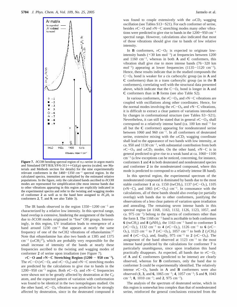
The IR bands observed in the region 1350-1200 cm-1 arecharacterized by a relative low intensity. In this spectral range,band overlap is extensive, hindering the assignment of the bandsdue toδCOH modes originated in “free” OH groups. Interest-ingly, in this region, UV irradiation leads to emergence of aband around 1230 cm-1 that appears at nearly the samefrequency of one of the twCH2 vibrations of ethanolamine.57
Note that ethanolamine has also two bands at 1385 and 1375cm-1 (ωCH2
57), which are probably very responsible for thesmall increase of intensity of the bands at nearly thesefrequencies ascribed to the twisting and wagging modes ofconformer2 of serine upon irradiation (see Figure 7).
νC-O and νN-C Stretching Region (1200- 950 cm-1).TheνC-O (νC-OC andνC-OA) andνN-C stretching modesare predicted by the calculations to give rise to bands in the1200-950 cm-1 region. BothνC-OC andνN-C frequencieswere shown not to be greatly affected by deuteration at the Câatom, and the expected pattern of variation with conformationwas found to be identical in the two isotopologues studied. Onthe other hand,νC-OA vibration was predicted to be stronglyaffected by deuteration, since in the deuterated compound it
was found to couple extensively with theωCD2 waggingoscillation (see Tables S13-S21). For each conformer of serine,besidesνC-O andνN-C stretching modes many other vibra-tions were predicted to give rise to bands in the 1200-950 cm-1
spectral range. However, calculations also indicated that mostof those vibrations should give rise to bands of low relativeintensity.
In B conformers,νC-OC is expected to originate low-intensity bands (<30 km mol-1) at frequencies between 1200and 1160 cm-1, whereas in bothA and C conformers, thisvibration shall give rise to more intense bands (76-320 kmmol-1) appearing at lower frequencies (1135-1120 cm-1).Hence, these results indicate that in the studied compounds theC-OC bond is weaker for a cis carboxylic group (as inA andC conformers) than in a trans carboxylic group (as in theBconformers), correlating well with the structural data presentedabove, which indicate that the C-OC bond is longer inA andC conformers than inB forms (see also Table S2).
In various conformers, theνC-OA andνN-C vibrations arecoupled with oscillations along other coordinates. Hence, forthe normal modes involving theνC-OA andνN-C vibrations,it is difficult to extract a clear pattern of variations introducedby changes in conformational structure (see Tables S3-S21).Nevertheless, it can still be stated that in generalνC-OA shallcorrespond to a relatively intense band (ca. 100 km mol-1 forall but theC conformer) appearing for nondeuterated serinebetween 1060 and 960 cm-1. In all conformers of deuteratedserine, extensive mixing with theωCD2 wagging coordinateshall lead to the appearance of two bands with low intensity, atca. 950 and 1130 cm-1, with substantial contribution from bothνC-OA and ωCD2 modes. On the other hand,νN-C is ingeneral predicted to give rise to a weak band at ca. 1060-1100cm-1 (a few exceptions can be noticed, concerning, for instance,conformers1 and4 in both deuterated and nondeuterated speciesand conformer2 in the nondeuterated compound, where thismode is predicted to correspond to a relatively intense IR band).
In this spectral region, the experimental spectrum of thenondeuterated compound is dominated by bands due to the moststable conformer1 at ca. 1150 (twCH2), 1137 (νC-OC), 1105(νN-C), and 1065 (νC-OA) cm-1. In consonance with thecalculations, all of these bands should also overlap (or partiallyoverlap) with bands due to other conformers, justifying theobservations of a less clear pattern of variation upon irradiationand annealing. The remaining seven intense bands in thisspectral region (at 1168, 1163, 1132, 1126, 1123, 1057, andca. 975 cm-1) belong to the spectra of conformers other thanthe form1. The 1168 cm-1 band is ascribable to both conformers6 (twCH2) and8 (FNH2), the 1163 cm-1 band to conformer5(νC-OC), 1132 cm-1 to 4 (νC-OC), 1126 cm-1 to 6 (νC-OC), 1123 cm-1 to 7 (νC-OC), 1057 cm-1 to both2 (γCH2)and 4 (νC-OA), and, finally, 975 cm-1 to 2 (νC-OA). Theassignment of the band observed at 1123 cm-1 to the mostintense band predicted by the calculations for conformer7 isparticularly unambiguous, since upon irradiation this bandpractically disappears. As expected, all bands due toνC-OC
of A and C conformers (predicted to be intense) are clearlyobserved, whereas forB conformers, only the band due toconformer5 could be experimentally identified. The relativelyintense νC-OA bands in A and B conformers were alsoobserved (1, 3, and6, 1065 cm-1; 4, 1057 cm-1; 5 and9, 1043cm-1; 8, 1010 cm-1; and2, 975 cm-1).
The analysis of the spectrum of deuterated serine, which inthis region is somewhat less complex than that of nondeuteratedserine, reinforced the general conclusions extracted from the
Figure 7. δCOH bending spectral region ofD,L-serine in argon matrixand Simulated DFT/B3LYP/6-311++G(d,p) spectra (scaled; see Ma-terials and Methods section for details) for the nine experimentallyrelevant conformers in the 1460-1350 cm-1 spectral region. In thecalculated spectra, intensities are multiplied by the estimated relativepopulations. In the figure, only the calculated bands ascribed toδCOHmodes are represented for simplification (the most intense bands dueto other vibrations appearing in this region are explicitly indicated inthe experimental spectra and refer to the twisting and wagging modesof conformer2 as well as to the band here assigned toδCâH2 inconformers2, 7, and9; see also Table 3).
5704 J. Phys. Chem. A, Vol. 109, No. 25, 2005 Jarmelo et al.
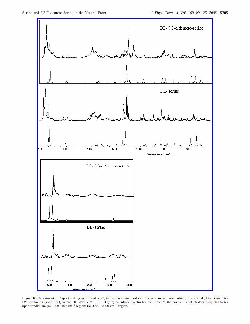
Figure 8. Experimental IR spectra ofD,L-serine andD,L-3,3-dideutero-serine molecules isolated in an argon matrix [as deposited (dotted) and afterUV irradiation (solid line)] versus DFT/B3LYP/6-311++G(d,p) calculated spectra for conformer7, the conformer which decarboxylates fasterupon irradiation. (a) 1800-400 cm-1 region; (b) 3700-2800 cm-1 region.
Serine and 3,3-Dideutero-Serine in the Neutral Form J. Phys. Chem. A, Vol. 109, No. 25, 20055705
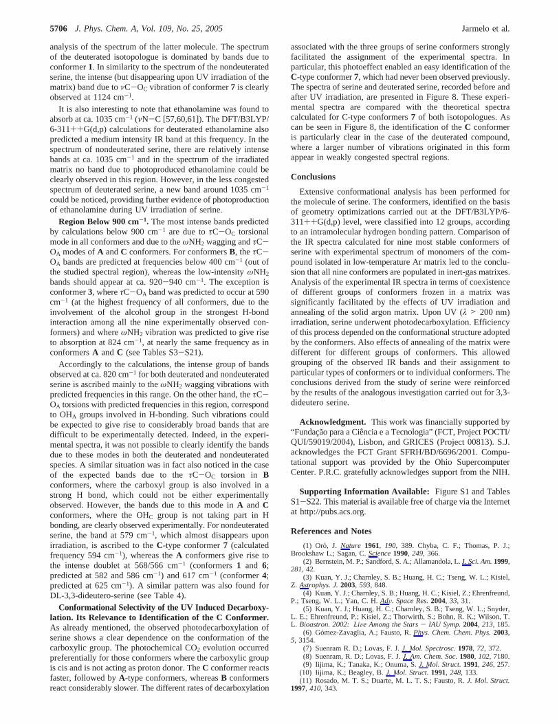
analysis of the spectrum of the latter molecule. The spectrumof the deuterated isotopologue is dominated by bands due toconformer1. In similarity to the spectrum of the nondeuteratedserine, the intense (but disappearing upon UV irradiation of thematrix) band due toνC-OC vibration of conformer7 is clearlyobserved at 1124 cm-1.
It is also interesting to note that ethanolamine was found toabsorb at ca. 1035 cm-1 (νN-C [57,60,61]). The DFT/B3LYP/6-311++G(d,p) calculations for deuterated ethanolamine alsopredicted a medium intensity IR band at this frequency. In thespectrum of nondeuterated serine, there are relatively intensebands at ca. 1035 cm-1 and in the spectrum of the irradiatedmatrix no band due to photoproduced ethanolamine could beclearly observed in this region. However, in the less congestedspectrum of deuterated serine, a new band around 1035 cm-1
could be noticed, providing further evidence of photoproductionof ethanolamine during UV irradiation of serine.
Region Below 900 cm-1. The most intense bands predictedby calculations below 900 cm-1 are due toτC-OC torsionalmode in all conformers and due to theωNH2 wagging andτC-OA modes ofA andC conformers. For conformersB, theτC-OA bands are predicted at frequencies below 400 cm-1 (out ofthe studied spectral region), whereas the low-intensityωNH2
bands should appear at ca. 920-940 cm-1. The exception isconformer3, whereτC-OA band was predicted to occur at 590cm-1 (at the highest frequency of all conformers, due to theinvolvement of the alcohol group in the strongest H-bondinteraction among all the nine experimentally observed con-formers) and whereωNH2 vibration was predicted to give riseto absorption at 824 cm-1, at nearly the same frequency as inconformersA andC (see Tables S3-S21).
Accordingly to the calculations, the intense group of bandsobserved at ca. 820 cm-1 for both deuterated and nondeuteratedserine is ascribed mainly to theωNH2 wagging vibrations withpredicted frequencies in this range. On the other hand, theτC-OA torsions with predicted frequencies in this region, correspondto OHA groups involved in H-bonding. Such vibrations couldbe expected to give rise to considerably broad bands that aredifficult to be experimentally detected. Indeed, in the experi-mental spectra, it was not possible to clearly identify the bandsdue to these modes in both the deuterated and nondeuteratedspecies. A similar situation was in fact also noticed in the caseof the expected bands due to theτC-OC torsion in Bconformers, where the carboxyl group is also involved in astrong H bond, which could not be either experimentallyobserved. However, the bands due to this mode inA and Cconformers, where the OHC group is not taking part in Hbonding, are clearly observed experimentally. For nondeuteratedserine, the band at 579 cm-1, which almost disappears uponirradiation, is ascribed to theC-type conformer7 (calculatedfrequency 594 cm-1), whereas theA conformers give rise tothe intense doublet at 568/566 cm-1 (conformers1 and 6;predicted at 582 and 586 cm-1) and 617 cm-1 (conformer4;predicted at 625 cm-1). A similar pattern was also found forDL-3,3-dideutero-serine (see Table 4).
Conformational Selectivity of the UV Induced Decarboxy-lation. Its Relevance to Identification of the C Conformer.As already mentioned, the observed photodecarboxylation ofserine shows a clear dependence on the conformation of thecarboxylic group. The photochemical CO2 evolution occurredpreferentially for those conformers where the carboxylic groupis cis and is not acting as proton donor. TheC conformer reactsfaster, followed byA-type conformers, whereasB conformersreact considerably slower. The different rates of decarboxylation
associated with the three groups of serine conformers stronglyfacilitated the assignment of the experimental spectra. Inparticular, this photoeffect enabled an easy identification of theC-type conformer7, which had never been observed previously.The spectra of serine and deuterated serine, recorded before andafter UV irradiation, are presented in Figure 8. These experi-mental spectra are compared with the theoretical spectracalculated for C-type conformers7 of both isotopologues. Ascan be seen in Figure 8, the identification of theC conformeris particularly clear in the case of the deuterated compound,where a larger number of vibrations originated in this formappear in weakly congested spectral regions.
Conclusions
Extensive conformational analysis has been performed forthe molecule of serine. The conformers, identified on the basisof geometry optimizations carried out at the DFT/B3LYP/6-311++G(d,p) level, were classified into 12 groups, accordingto an intramolecular hydrogen bonding pattern. Comparison ofthe IR spectra calculated for nine most stable conformers ofserine with experimental spectrum of monomers of the com-pound isolated in low-temperature Ar matrix led to the conclu-sion that all nine conformers are populated in inert-gas matrixes.Analysis of the experimental IR spectra in terms of coexistenceof different groups of conformers frozen in a matrix wassignificantly facilitated by the effects of UV irradiation andannealing of the solid argon matrix. Upon UV (λ > 200 nm)irradiation, serine underwent photodecarboxylation. Efficiencyof this process depended on the conformational structure adoptedby the conformers. Also effects of annealing of the matrix weredifferent for different groups of conformers. This allowedgrouping of the observed IR bands and their assignment toparticular types of conformers or to individual conformers. Theconclusions derived from the study of serine were reinforcedby the results of the analogous investigation carried out for 3,3-dideutero serine.
Acknowledgment. This work was financially supported by“Fundacao para a Cieˆncia e a Tecnologia” (FCT, Project POCTI/QUI/59019/2004), Lisbon, and GRICES (Project 00813). S.J.acknowledges the FCT Grant SFRH/BD/6696/2001. Compu-tational support was provided by the Ohio SupercomputerCenter. P.R.C. gratefully acknowledges support from the NIH.
Supporting Information Available: Figure S1 and TablesS1-S22. This material is available free of charge via the Internetat http://pubs.acs.org.
References and Notes
(1) Oro, J. Nature 1961, 190, 389. Chyba, C. F.; Thomas, P. J.;Brookshaw L.; Sagan, C.Science1990, 249, 366.
(2) Bernstein, M. P.; Sandford, S. A.; Allamandola, L. J.Sci. Am.1999,281, 42.
(3) Kuan, Y. J.; Charnley, S. B.; Huang, H. C.; Tseng, W. L.; Kisiel,Z. Astrophys. J.2003, 593, 848.
(4) Kuan, Y. J.; Charnley, S. B.; Huang, H. C.; Kisiel, Z.; Ehrenfreund,P.; Tseng, W. L.; Yan, C. H.AdV. Space Res.2004, 33, 31.
(5) Kuan, Y. J.; Huang, H. C.; Charnley, S. B.; Tseng, W. L.; Snyder,L. E.; Ehrenfreund, P.; Kisiel, Z.; Thorwirth, S.; Bohn, R. K.; Wilson, T.L. Bioastron. 2002: LiVe Among the Stars- IAU Symp.2004, 213, 185.
(6) Gomez-Zavaglia, A.; Fausto, R.Phys. Chem. Chem. Phys.2003,5, 3154.
(7) Suenram R. D.; Lovas, F. J.J. Mol. Spectrosc.1978, 72, 372.(8) Suenram, R. D.; Lovas, F. J.J. Am. Chem. Soc.1980, 102, 7180.(9) Iijima, K.; Tanaka, K.; Onuma, S.J. Mol. Struct.1991, 246, 257.
(10) Iijima, K.; Beagley, B.J. Mol. Struct.1991, 248, 133.(11) Rosado, M. T. S.; Duarte, M. L. T. S.; Fausto, R.J. Mol. Struct.
1997, 410, 343.
5706 J. Phys. Chem. A, Vol. 109, No. 25, 2005 Jarmelo et al.
(12) Stepanian, S. G.; Reva, I. D.; Radchenko, E. D.; Rosado, M. T. S.;Duarte, M. L. T. S.; Fausto, R.; Adamowicz, L.J. Phys. Chem.1998, 102,1041.
(13) Stepanian, S. G.; Reva, I. D.; Radchenko, E. D.; Adamowicz, L.J.Phys. Chem.2001, 105A, 10664.
(14) Rosado, M. T. S.Estrutura Molecular e Espectros Vibracionaisde n- e R-Aminoacidos Simples, Ph.D. Thesis, University of Lisbon,Portugal, 2004.
(15) Kikuchi, O.; Natsui, T.; Kozaki, T.J. Mol. Struct. (THEOCHEM)1990, 207, 103.
(16) Ding, Y.; Krogh-Jespersen, K.Chem. Phys. Lett.1992, 199, 261.(17) Dixon, D. A.; Lipscomb, W. N.J. Biol. Chem.1976, 251, 5992.(18) Clementi, E.; Cavallone, F.; Scordamaglia, R.J. Am. Chem. Soc.
1997, 99, 5531.(19) Wright, L. R.; Borkman, R. F.J. Am. Chem. Soc.1980, 102,
6207.(20) Alsenoy, C. V.; Scarsdale, J. N.; Sellers, H. L.; Scha¨fer, L. Chem.
Phys. Lett.1981, 80, 124.(21) Espinosa-Muller, A. W.; Bravo, A. N.J. Mol. Struct. (THEOCHEM)
1982, 90, 203.(22) Masamura, M.J. Mol. Struct. (THEOCHEM)1987, 152, 293.(23) Alsenoy, C. V.; Kulp, S.; Siam, K.; Klimkowski, V. J.; Ewbank, J.
D.; Schafer, L. J. Mol. Struct. (THEOCHEM)1988, 181, 169.(24) Masamura, M.J. Mol. Struct. (THEOCHEM)1988, 164, 299.(25) Tarakeshwar, P.; Manogaran, S.J. Mol. Struct. (THEOCHEM)
1994, 305, 205.(26) Tortonda, F. R.; Silla, E.; Tuno´n, I.; Rinaldi, D.; Ruiz-Lopez, M.
F. Theor. Chem. Acc.2000, 104, 89.(27) Noguera, M.; Rodrı´guez-Santiago, L.; Sodupe, M.; Bertran, J.J.
Mol. Struct. (THEOCHEM)2001, 537, 307.(28) Lakard, B.J. Mol. Struct. (THEOCHEM)2004, 681, 183.(29) Lambie, B.; Ramaekers, R.; Maes, G.J. Phys. Chem.2004, 108A,
10426.(30) Gronert, S.; O’Hair, R. A. J.J. Am. Chem. Soc. 1995, 117, 2071.(31) Pierazzo, E.; Chyba, C. F.Meteorit. Planet. Sci.1999, 34, 909.(32) Blank, J. G.; Miller, G. H.; Ahrens, M. J.; Winans, R. E.Origin
Life EVol. Biosphere2001, 31, 15.(33) Ehrenfreund, P.; Bernstein, M. P.; Dworkin, J. P.; Sandford, S. A.;
Allamandola, L. J.Astrophys. J.2001, 550, L95.(34) Zubavichus, Y.; Fuchs, O.; Weinhardt, L.; Heske, C.; Umbach, E.;
Denlinger, J. D.; Grunze, M.Radiat. Res.2004, 161, 346.(35) Sato, N.; Quitain, A. T.; Kang, K.; Daimon, H.; Fujie, K.Ind. Eng.
Chem. Res.2004, 43, 3217.(36) Jarmelo, S.; Lapinski, L.; Fausto, R.Proceedings of the Annual
Meeting of the Cryobiology and Low Temperature Biology Societies:Cryobiomol 2003, From the Low-Temperature Physics and Chemistry ofBiological Molecules to Life in Extreme Low-Temperature Conditions,Coimbra, Portugal, 2003; p 142.
(37) Jarmelo, S.; Lapinski, L.; Nowak, M. J.; Fausto, R.Proceedingsof the XX IUPAC Symposium on Photochemistry, Granada, Spain, 2004; p355.
(38) Szczesniak, M.; Nowak, M. J.; Rostkowska, H.; Szczepaniak, K.;Person, W. B.; Shugar, D.J. Am. Chem. Soc.1983, 105, 5969.
(39) Frisch, M. J.; Trucks, G. W.; Schlegel, H. B.; Scuseria, G. E.; Robb,M. A.; Cheeseman, J. R.; Zakrzewski, V. G.; Montgomery, J. A., Jr.;Stratmann, R. E.; Burant, J. C.; Dapprich, S.; Millam, J. M.; Daniels, A.D.; Kudin, K. N.; Strain, M. C.; Farkas, O.; Tomasi, J.; Barone, V.; Cossi,M.; Cammi, R.; Mennucci, B.; Pomelli, C.; Adamo, C.; Clifford, S.;Ochterski, J.; Petersson, G. A.; Ayala, P. Y.; Cui, Q.; Morokuma, K.; Malick,D. K.; Rabuck, A. D.; Raghavachari, K.; Foresman, J. B.; Cioslowski, J.;Ortiz, J. V.; Stefanov, B. B.; Liu, G.; Liashenko, A.; Piskorz, P.; Komaromi,I.; Gomperts, R.; Martin, R. L.; Fox, D. J.; Keith, T.; Al-Laham, M. A.;Peng, C. Y.; Nanayakkara, A.; Gonzalez, C.; Challacombe, M.; Gill, P. M.W.; Johnson, B. G.; Chen, W.; Wong, M. W.; Andres, J. L.; Head-Gordon,M.; Replogle, E. S.; Pople, J. A.Gaussian 98, revision A.9; Gaussian,Inc.: Pittsburgh, PA, 1998.
(40) Binkley, J. S.; Pople, J. A.; Hehre, W. J.J. Am. Chem. Soc.1980,102, 939.
(41) Pietro, W. J.; Francl, M. M.; Hehre, W. J.; Defrees, D. J.; Pople,J. A.; Binkley, J. S.J. Am. Chem. Soc. 1982, 104, 5039.
(42) Frisch, M. J.; Pople, J. A.; Binkley, J. S.J. Chem. Phys.1984, 80,3265.
(43) McLean, A. D.; Chandler, G. S.J. Chem. Phys.1980, 72, 5639.(44) Becke, A. D.Phys. ReV. 1988, 38A, 3098.(45) Lee, C. T.; Yang, W. T.; Parr, R. G.Phys. ReV. 1988, 37B, 785.(46) Gomez-Zavaglia, A.; Fausto, R.Vibrat. Spectrosc. 2003, 33, 105.(47) Borba, A.; Go´mez-Zavaglia, A.; Lapinski, L.; Fausto, R.Phys.
Chem. Chem. Phys. 2004, 6, 2101.(48) De Frees, D.; McLean, A.J. Chem. Phys.1985, 82, 333.(49) Stepanian, S. G.; Reva, I. D.; Radchenko, E. D.; Adamowicz, L.J.
Phys. Chem.1998, 102A, 4623.(50) Reva, I. D.; Stepanian, S. G.; Adamowicz, L.; Fausto, R.J. Phys.
Chem.2001, 105A, 4773.(51) Csaszar, A. G. J. Am. Chem. Soc.1992, 114, 9568.(52) Owen, N. L.; Sheppard, N.Proc. Chem. Soc., London1963, 264.(53) Piercy, J. E.; Subrahmanyan, S. V.J. Chem. Phys.1965, 42, 1475.(54) Bayley, J.; North, A. M.Trans. Faraday Soc.1968, 64, 1499.(55) Fausto, R.; Teixeira-Dias, J. J. C.J. Mol. Struct. (THEOCHEM)
1987, 150, 381.(56) Svensson, T.; Nelander, B.; Karlstro¨m, G.Chem. Phys.2001, 265,
323.(57) Cacela, C.; Duarte, M. L.; Fausto, R.J. Mol. Struct.1999, 482/
483, 591.(58) Fausto, R.; Batista de Carvalho, L. A. E.; Teixeira-Dias, J. J. C.;
Ramos, M. N.J. Chem. Soc. Faraday Trans.2 1989, 85, 1945.(59) Jarmelo, S.; Maiti, N.; Anderson, V.; Carey, P.; Fausto, R.J. Phys.
Chem.2005, 109A, 2069.(60) Rasanen, M.; Aspiala, A.; Homanen, L.; Murto, J.J. Mol. Struct.
1982, 96, 81.(61) EPA Vapor Phase Library, CAS Number 141-43-5, 2001 Thermo
Galactic.(62) Jarmelo, S.; Fausto, R.Phys. Chem. Chem. Phys.2002, 4, 1555.
Serine and 3,3-Dideutero-Serine in the Neutral Form J. Phys. Chem. A, Vol. 109, No. 25, 20055707