Potential Use of Cyclodextrins for the Improvement of … hydroxyl propyl-β-cyclodextrin...
18
IOSR Journal of Pharmacy and Biological Sciences (IOSR-JPBS) e-ISSN:2278-3008, p-ISSN:2319-7676. Volume 12, Issue 5 Ver. I (Sep. – Oct. 2017), PP 79-96 www.iosrjournals.org DOI: 10.9790/3008-1205017996 www.iosrjournals.org 79 | Page Potential Use of Cyclodextrins for the Improvement of Ocular Bioavailability of Meloxicam Abd-Elgawad H. Abd-Elgawad, Osama A. Soliman, Marwa S. El-Dahan, Ghada A. A. El-Emam * (Pharmaceutical department, Pharmacy College, Mansoura University, Mansoura, Egypt) Abstract: Meloxicam (MX) is a nonsteroidal anti-inflammatory drug. Also, it is a more distinct COX-2 inhibitor than diclofenac with less secondary effects. Yet, its usage for the treatment of ophthalmic inflammations has retarded due to its low aqueous solubility and dissolution rate. The aim of this study was to prepare the inclusion complexes of MX with cyclodextrins to enhance its aqueous solubility, dissolution rate, and ocular bioavailability. Formulation of those complexes into different ocular delivery systems and also in-vitro and in- vivo assessments has not been accomplished in the earlier attempts. In this study, MX was complexed with each of hydroxyl propyl-β-cyclodextrin (HP-β-CyD) and β-cyclodextrin (β-CyD) utilizing kneading, co-evaporation, and freeze drying techniques. Moreover, the prepared complexes were discriminated in the solid state by Fourier transform infrared spectroscopy (FT-IR), differential scanning calorimetry (DSC), and powder X-ray diffraction (PXRD). These complexes were incorporated into eye drops, eye gels, and ocuserts using hydroxypropyl methylcellulose (HPMC), sodium carboxy methylcellulose (Sod. CMC), carbopol 940 (CP 940 ), and sodium alginate (Sod. ALG). These formulae were characterized on the subject of drug content, pH, viscosity, in-vitro release characteristics. Kinetic analysis of the release data was done. Real inclusion complexes were obtained with co-evaporation method that confirmed by DSC investigations. The MX release was enhanced by complexation especially with HP-β-CyD. Ocular bioavailability of meloxicam from the selected formulations was investigated. In-vivo study proved that, ocuserts containing MX-HP-CyD complexes showed pronounced ocular bioavailability than MX alone which indicated by higher AUC, C max and relative bioavailability values. Keywords: cyclodextrins, inclusion complexes, meloxicam, ocular bioavailability. --------------------------------------------------------------------------------------------------------------------------------------- Date of Submission: 28-08-2017 Date of acceptance: 13-09-2017 --------------------------------------------------------------------------------------------------------------------------------------- I. Introduction Topically applied nonsteroidal anti-inflammatory drugs (NSAIDs) are used in the control of post- operative ocular inflammation, as well as for the remedy of intraoperative miosis during cataract surgery [1]. The reduction of symptoms associated to seasonal allergic conjunctivitis and the relief of ocular discomfort after refractive surgery are the target for the use of NSAIDs [2]. NSAIDs produce analgesic and anti-inflammatory effects by inhibiting prostaglandin synthesis by the cyclooxygenase (COX) enzyme. During the ocular injury, both COX-1 and COX-2 are expressed, but there is an evidence that, an increase in COX-2 expression by inflammation is occured [3]. So that, the use of a selective COX-2 inhibitor appears to be more preferable. Meloxicam is a NSAID derived from enolic acid that inhibits particularly, COX-2 enzyme with additional mechanisms of analgesia, such as activation of the nitric oxide- cyclic GMPK+ channel pathway in peripheral tissues [4]. It is a more specific COX-2 inhibitor than diclofenac with less secondary effects due to the constitutive COX-1 inhibition. Meloxicam is named chemically; 4-hydroxy-2-methyl-N-(5-methyl-2-thiazolyl)-2H-1,2-benzothiazine- 3-carboxamide-1,1- dioxide [5]. The impaired bioavailability of drugs from ocular dosage forms is mainly due to several protective mechanisms of the eye including; blinking, the rapid turnover of lachrymal fluid, short pre- corneal residence time, as well as the relative poor permeability of the corneal epithelial membrane. Among the several techniques that increasing the apparent aqueous solubility of poorly soluble drugs without lowering their lipophilicity for enhancing their absorption through biological membrane is the complexation with cyclodextrins [6]. Cyclodextrins (CyDs) are cyclic oligosaccharides composed of D-glucopyranoside units linked by glycosidic bonds. They are obtained from biotechnological processes involving the enzymatic degradation of corn starch and offer greater yield with 6, 7 and 8 units of glucose, known as α -CyD, β-CyD and γ-CyD, respectively [7]. CyDs are characterized by their ability to modify the physicochemical characteristics of molecules that are accommodated within their internal cavity to form the so-called inclusion complexes [8].
Transcript of Potential Use of Cyclodextrins for the Improvement of … hydroxyl propyl-β-cyclodextrin...

IOSR Journal of Pharmacy and Biological Sciences (IOSR-JPBS)
e-ISSN:2278-3008, p-ISSN:2319-7676. Volume 12, Issue 5 Ver. I (Sep. – Oct. 2017), PP 79-96
www.iosrjournals.org
DOI: 10.9790/3008-1205017996 www.iosrjournals.org 79 | Page
Potential Use of Cyclodextrins for the Improvement of Ocular
Bioavailability of Meloxicam
Abd-Elgawad H. Abd-Elgawad, Osama A. Soliman, Marwa S. El-Dahan,
Ghada A. A. El-Emam*
(Pharmaceutical department, Pharmacy College, Mansoura University, Mansoura, Egypt)
Abstract: Meloxicam (MX) is a nonsteroidal anti-inflammatory drug. Also, it is a more distinct COX-2 inhibitor
than diclofenac with less secondary effects. Yet, its usage for the treatment of ophthalmic inflammations has
retarded due to its low aqueous solubility and dissolution rate. The aim of this study was to prepare the
inclusion complexes of MX with cyclodextrins to enhance its aqueous solubility, dissolution rate, and ocular
bioavailability. Formulation of those complexes into different ocular delivery systems and also in-vitro and in-
vivo assessments has not been accomplished in the earlier attempts. In this study, MX was complexed with each
of hydroxyl propyl-β-cyclodextrin (HP-β-CyD) and β-cyclodextrin (β-CyD) utilizing kneading, co-evaporation,
and freeze drying techniques. Moreover, the prepared complexes were discriminated in the solid state by
Fourier transform infrared spectroscopy (FT-IR), differential scanning calorimetry (DSC), and powder X-ray
diffraction (PXRD). These complexes were incorporated into eye drops, eye gels, and ocuserts using
hydroxypropyl methylcellulose (HPMC), sodium carboxy methylcellulose (Sod. CMC), carbopol 940 (CP940),
and sodium alginate (Sod. ALG). These formulae were characterized on the subject of drug content, pH,
viscosity, in-vitro release characteristics. Kinetic analysis of the release data was done. Real inclusion
complexes were obtained with co-evaporation method that confirmed by DSC investigations. The MX release
was enhanced by complexation especially with HP-β-CyD. Ocular bioavailability of meloxicam from the
selected formulations was investigated. In-vivo study proved that, ocuserts containing MX-HP-CyD complexes
showed pronounced ocular bioavailability than MX alone which indicated by higher AUC, Cmax and relative
bioavailability values.
Keywords: cyclodextrins, inclusion complexes, meloxicam, ocular bioavailability. ---------------------------------------------------------------------------------------------------------------------------------------
Date of Submission: 28-08-2017 Date of acceptance: 13-09-2017
---------------------------------------------------------------------------------------------------------------------------------------
I. Introduction Topically applied nonsteroidal anti-inflammatory drugs (NSAIDs) are used in the control of post-
operative ocular inflammation, as well as for the remedy of intraoperative miosis during cataract surgery [1].
The reduction of symptoms associated to seasonal allergic conjunctivitis and the relief of ocular discomfort after
refractive surgery are the target for the use of NSAIDs [2].
NSAIDs produce analgesic and anti-inflammatory effects by inhibiting prostaglandin synthesis by the
cyclooxygenase (COX) enzyme. During the ocular injury, both COX-1 and COX-2 are expressed, but there is an
evidence that, an increase in COX-2 expression by inflammation is occured [3]. So that, the use of a selective
COX-2 inhibitor appears to be more preferable. Meloxicam is a NSAID derived from enolic acid that inhibits
particularly, COX-2 enzyme with additional mechanisms of analgesia, such as activation of the nitric oxide-
cyclic GMPK+ channel pathway in peripheral tissues [4]. It is a more specific COX-2 inhibitor than diclofenac
with less secondary effects due to the constitutive COX-1 inhibition.
Meloxicam is named chemically; 4-hydroxy-2-methyl-N-(5-methyl-2-thiazolyl)-2H-1,2-benzothiazine-
3-carboxamide-1,1- dioxide [5]. The impaired bioavailability of drugs from ocular dosage forms is mainly due
to several protective mechanisms of the eye including; blinking, the rapid turnover of lachrymal fluid, short pre-
corneal residence time, as well as the relative poor permeability of the corneal epithelial membrane. Among the
several techniques that increasing the apparent aqueous solubility of poorly soluble drugs without lowering their
lipophilicity for enhancing their absorption through biological membrane is the complexation with cyclodextrins
[6].
Cyclodextrins (CyDs) are cyclic oligosaccharides composed of D-glucopyranoside units linked by
glycosidic bonds. They are obtained from biotechnological processes involving the enzymatic degradation of
corn starch and offer greater yield with 6, 7 and 8 units of glucose, known as α-CyD, β-CyD and γ-CyD,
respectively [7]. CyDs are characterized by their ability to modify the physicochemical characteristics of
molecules that are accommodated within their internal cavity to form the so-called inclusion complexes [8].

Potential Use of Cyclodextrins for the Improvement of Ocular Bioavailability of Meloxicam
DOI: 10.9790/3008-1205017996 www.iosrjournals.org 80 | Page
The cavity size is more appropriate for common pharmaceutical drugs with molecular weights between
200 and 800 g/mol [9]. Beta-cyclodextrin (β-CyD) was chosen to enhance the solubility of meloxicam because
of its central cavity diameter (6-6.5 Aº) which is suitable to accommodate most aromatic rings, its efficiency in
producing stable drug complexes, low toxicity and relatively low cost [10]. Hydroxyalkyl derivatives such as
hydroxypropyl-β-cyclodextrin have high water solubility and low hygroscopicity compared to the original β-
cyclodextrin [11].
The main target of this study was to enhance aqueous solubility of MX by complexation with CyDs
using different techniques. Formulation of MX-CyD complexes in different ophthalmic preparations was also of
our goal. In addition, inspection of the influence of inclusion complexation of MX with β-CyD and HP-β-CyD
on its solubility was of prime interest. Characterization of the complexes will be accomplished by Fourier
transform infrared spectroscopy (FT-IR), differential scanning calorimetry (DSC) and powder X-ray diffraction
(PXRD). Moreover, all the formulations were examined for their physical properties and in-vitro release
characteristics. In addition, the ocular bioavailability of MX from the selected formulations were studied.
II. Materials And Methods Meloxicam (MX) was provided by Zydus Pharms, USA. Hydroxypropyl methylcellulose (4000 cP)
was provided by Dow chemical company, USA. Potassium dihydrogen orthophosphate, disodium hydrogen
phosphate, propylene glycol, ammonia solution 25%, methanol and sodium carboxymethylcellulose (Sod. CMC)
were supplied from Adwic, El Nasr, Pharmaceutical Chemicals Co., Egypt. Sodium alginate (Sod. ALG), beta-
cyclodextrin (β-CyD) (M.W = 1135.12), hydroxypropyl-β-cyclodextrin (HP-β-CyD) (M.W = 1375.35) were
provided from Saito Trading Co., Ltd, Japan. Triethanolamine was purchased by Provizer Pharma, India. Propyl
paraben and methyl paraben were supplied by AppliChem GmbH, Germany. Acetonitril and methanol (HPLC
grade) were supplied from Fisher scientific, Fair Lawn, New Jersy, UK. Piroxicam was supplied from Sigma-
Aldrich Co. LLC, St. Louis, Missouri, USA.
Methodology
2.1. Phase solubility study
The solubility assessments were performed according to the method described by Higuchi and
Connors, [12]. An excess amount of MX (10 mg) was added to aqueous solutions of β-CyD or HP-β-CyD in
separate screw capped glass bottles. Solutions were shaken on thermostatically controlled water bath (Grant
Instrument Cambridge Ltd., Barrington Cambridge (B2, 5002, England) at 37±0.5oC at 50 rpm. After
equilibrium was reached (72 hrs), the solutions were filtered through membrane filter (with 10 mm diameter and
membrane filter Corporation, Bedford, MA 01730 of pore size 0.22 μm and 0.45μm, Berlin, Germany) and then
one ml from each filtrate was diluted and analyzed spectrophotometrically at 362 nm for total drug content. The
experiments were done in triplicate, the mean and SD were calculated. Then, the determination of an apparent
1:1 stability constant of the complexes (K1:1) from the initial straight-line portion of the phase solubility
diagrams were performed using the following equation:
K1:1 = Slope / So (1 - slope) (1) Where; So is the free MX aqueous solubility
2. 2. Formulation of solid meloxicam-cyclodextrin complexes
Inclusion complexes of 1:1 molar ratio of MX with β-CyD / HP-β-CyD were performed by the
kneading, co-evaporation, and freeze drying methods [13]. For comparative study, physical mixtures (PMs) of
MX with either CyDs were prepared by simple thoroughly mixing in a morter then, seived. The three
complexation methods are further discussed as follows:
2.2.1. Kneading method (Kn) Triturate a physical mixture of drug and CyDs in 1:1 molar ratio in a mortar with a small amount of
water-methanol (1:1) solution [13]. Then the formed thick slurry was kneaded for 45 mins. after that, dried then
sieved through (75-150 µm) seive and stored in desiccators, until use.
2.2.2. Co-evaporation method (Co)
An aqueous solution of either CyDs was added to the equivalent weight of the drug dissolved in (1 N)
liquid ammonia (25%), then stirred for 1 hr and evaporated at 45oC until dried after that, sieved through (75-150
µm) seive and stored in airtight vessels.
2.2.3. Freeze- drying method (FD) In this method, dissolve stoichiometric amount of drug and CyDs in distilled water and shaken for 24
hrs. (1 N) ammonia solution (25% v/v) was added to the mixture drop wise till a clear solution was obtained.
The solution was frozen overnight in stainless steel dishes then lyophilized in freeze dryer (FD8-8 with T

Potential Use of Cyclodextrins for the Improvement of Ocular Bioavailability of Meloxicam
DOI: 10.9790/3008-1205017996 www.iosrjournals.org 81 | Page
controller, Model " FD8-8B ", SIM-USA) at -45oc for 48 hrs [14].The obtained product was kept at room
temperature for one day. Finally, the lyophilized powder was sieved and kept in a sealed vial.
2.3. Characterization of inclusion complexes
2.3.1. Fourier Transform Infrared Spectroscopy (FT-IR)
FT-IR spectra of MX, β-CyD, HP-β-CyD, PMs and the prepared complexes by different methods were
determined using Fourier transform infrared spectroscopy (Thermo Fisher Scientific, Inc., Waltham, MA, USA).
Each sample was mixed with potassium bromide (KBr). These mixtures were ground into fine powder and then
compressed into KBr discs using a hydraulic press. Each KBr disc was scanned over a wavenumber region of
400-4000 cm–1
at a resolution of 1 cm–1
. Characteristic bands were determined for all samples.
2.3.2. Differential scanning calorimetry (DSC)
The thermograms of MX, β-CyD, HP-β-CyD, PMs and the prepared complexes were analyzed using
differential scanning calorimeter (Perkin Elmer DSC 7, USA). The powdered sample (10 mg) was sealed in
aluminum pans and an empty aluminum pan was used as a reference, then covered by lids and scanned under
nitrogen flow rate of 4oC/min over the temperature range of 30-300
oC.
2.3.3. Powder X-ray diffraction (PXRD)
Powder X-ray diffraction patterns of MX, β-CyD, HP-β-CyD, PMs and the prepared complexes by
different methods were performed using X-ray diffractometer (Rigaku Denki, Rint-2500 VL, Tokyo, Japan) over
the range of 10-30o (2θ) angles and operated at the conditions of Ni-filtered Cu-kα radiation. The voltage of 40
KV and a current of 40 mA, were used. The time constant was 1.25 sec, and a scanning speed was adjusted to
0.02oC / min.
2.4. Preparation of different formulations
2.4.1. Preparation of eye drops
Sodium carboxy methylcellulose (Sod. CMC), hydroxylpropyl methylcellulose (HPMC) and sodium
alginate (Sod. ALG) in concentrations of 1, 0.5 and 0.7% w/w respectively, were dissolved in distilled water
containing 10% propylene glycol at which , MX 0.03% w/w or its equivalent weights of MX-β-CyD or MX-HP-
β-CyD complexes were previously dissolved in it "Table 1". 0.05% w/w methyl paraben, 0.01% w/w propyl
paraben were added as preservatives [15]. The mixtures were completed to final weight, and then filled in clean,
dry and sterile glass containers for further studies.
2.4.2. Preparation of eye gels MX (0.03% w/w) or its equivalent weights of MX-β-CyD or MX-HP-β-CyD complexes were dissolved
separately in 10% propylene glycol and added to aqueous solution of different polymers namely Sod. CMC,
HPMC and Sod. ALG in concentrations of 3.5, 3 and 3% w/w, respectively "Table 1". Methyl and propyl
parabens were used as preservatives in concentration of 0.05% and 0.01% w/w, respectively [15]. The dispersion
was mixed until a clear transparent gel free from air bubbles was obtained. The weight of gel was adjusted and
then packaged in clean, dry and sterile glass containers until used.
2.4.3. Preparation of ocuserts
Ocuserts containing MX or its complexes were prepared according to the film casting method [16].
Carbopol 940 (CP940) (0.1% w/w) was used in combination with Sod. CMC, HPMC and Sod. ALG to enhance
the elasticity, film properties, and bioadhesion. Meloxicam (0.03%) or its equivalent weights of MX-β-CyD or
MX-HP-β-CyD complexes were dissolved in distilled water containing 10% propylene glycol which acted as a
plasticizer to aid the formation of flexible films as well as to guard the polymeric inserts against being brittle
upon storage [17]. Then, the solution was added to the different polymeric solutions as illustrated in "Table 1".
Methyl and propyl parabens were used as preservatives in concentrations of 0.05% and 0.01% (w/w),
respectively [15]. Triethanolamine (TEA) was added to allow CP940 gelation since this polymer is a polyacrylic
acid which undergoes a sol to gel transition in aqueous solution at pH above its pKa (5.5) [18]. Sonication for 1
hr for all the prepared polymeric solutions in an ultrasonic water bath (Saris- USA) were done to remove
entrapped air and then, stored for 24 hrs at ambient temperature to allow total hydration of the used polymers.
Then, equal volumes of the prepared solutions were transferred into teflon plates. The solvent was allowed to
evaporate for 3 days at ambient temperature. The formed films were weighed and transferred to a desiccator
containing silica gel, where it was stored for another 24 hrs before use. The prepared ocuserts were cut in the
form of circular discs having thickness of 0.4-0.5 mm and diameter of 5 mm. Then, individually sealed in foil
sachets until use.

Potential Use of Cyclodextrins for the Improvement of Ocular Bioavailability of Meloxicam
DOI: 10.9790/3008-1205017996 www.iosrjournals.org 82 | Page
Table 1: Composition of MX-CyDs complexes ophthalmic formulations
Formulation code
Type of complex Type of polymer
Type of
Formulation
Concentration% (w/w)
CT
Control
Dβ 1a MX-β-CyD 1% Sod.CMC
Eye drops
DH-β11a
MX-HP-β-CyD
Dβ 2a
MX-β-CyD
0.5% HPMC
DH-β12a MX-HP-β-CyD
Dβ 3a
MX-β-CyD
0.7% Sod. ALG DH-β13a MX-HP-β-CD
Gβ 4a
MX-β-CyD
3.5% Sod. CMC
Eye gels
GH-β14a
MX-HP-β-CyD
Gβ 5a MX-β-CyD
3% HPMC
GH-β15a
MX-HP-β-CyD
Gβ 6a MX-β-CyD
3% Sod. ALG GH-β16a
MX-HP-β-CyD
Oβ 7a
MX-β-CyD
1.5% Sod. CMC + 0.1% CP940
Ocuserts
OH-β17a
MX-HP-β-CyD
Oβ 8a
A
MX-β-CyD
1% HPMC + 0.1% CP940
OH-β18a
MX-HP-β-CyD
Oβ 9a MX-β-CyD
2% Sod. ALG + 0.1% CP940 OH-β19a
MX-HP-β-CyD
Where; All formulae contain 0.03% w/w meloxicam or its equivalent weights of complexes, 10% propylene
glycol, 0.05% w/w methyl paraben and 0.01% w/w propyl paraben.
CT: Control, Sod. CMC: sodium carboxy methylcellulose, HPMC: hydroxypropyl methylcellulose, Sod. ALG:
sodium alginate, β-CyD: β-cyclodextrin, HP-β-CyD: hydroxypropyl β-cyclodextrin, MX: meloxicam and a: is
Kn (kneading method), Co (co-evaporation method) or FD (freeze-drying method).
2.5. Physicochemical characterization of the formulations
2.5.1. The drug content
Drug content was determined by weighing one gram from each formulation accurately and dissolved in
50 ml phosphate buffer pH 7.4 and shaken in thermostatically controlled water bath at 37±0.5oC for 1 hr then by
using membrane filters of 0.45 µm the solutions were filtered and assayed at 362 nm.
2.5.2. The formulations pH
One gram of each formula was dissolved in 20 ml of distilled water, and then the pH was measured
using pH-meter (Beckman Instruments Fullerton, CA 92634, Germany) [19].
2.5.3. The formulations viscosity
The viscosity measurement was performed by the use of a cone and plate rotary viscometer (Haake
Inc., Germany). One gram of each formulation was spreaded on the stationary plate of the viscometer and
permitted to equilibrate for 5 min. to reach the running temperature before each measurement. The rotary
viscometer was thermostatically adjusted to 37±0.5oC. Viscosity values were calculated using the following
equation;
𝜼 = G. S / N (2) Where:
η = Viscosity in mPa.s (mPa. S = 1 centipoise, cP).
G = Instrumental factor = 14200 (mPa.s / scalagrad.min).
S = Torque (scale grad).
N =Speed (256 rpm).
2.5.4. In-vitro release study
The drug release from different formulations, in phosphate buffer solution of pH 7.4 was established
according to the method assumed by Shastri et al., [20]. The in- vitro release of MX from different formulations
was performed using the dialysis method. Dialysis membrane (12000–14000 molecular weight Cutoffs) was
Potential Use of Cyclodextrins for the Improvement of Ocular Bioavailability of Meloxicam
DOI: 10.9790/3008-1205017996 www.iosrjournals.org 83 | Page
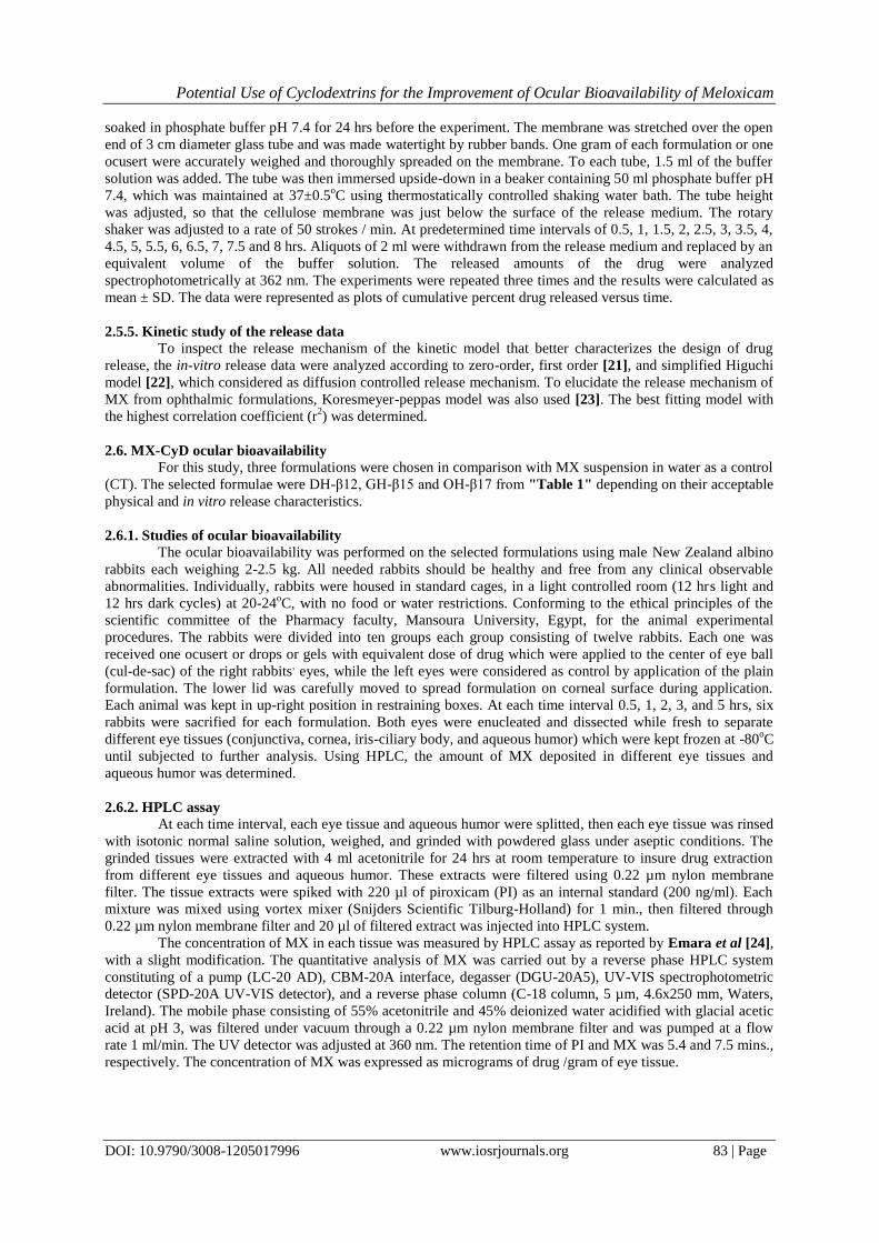
soaked in phosphate buffer pH 7.4 for 24 hrs before the experiment. The membrane was stretched over the open
end of 3 cm diameter glass tube and was made watertight by rubber bands. One gram of each formulation or one
ocusert were accurately weighed and thoroughly spreaded on the membrane. To each tube, 1.5 ml of the buffer
solution was added. The tube was then immersed upside-down in a beaker containing 50 ml phosphate buffer pH
7.4, which was maintained at 37±0.5oC using thermostatically controlled shaking water bath. The tube height
was adjusted, so that the cellulose membrane was just below the surface of the release medium. The rotary
shaker was adjusted to a rate of 50 strokes / min. At predetermined time intervals of 0.5, 1, 1.5, 2, 2.5, 3, 3.5, 4,
4.5, 5, 5.5, 6, 6.5, 7, 7.5 and 8 hrs. Aliquots of 2 ml were withdrawn from the release medium and replaced by an
equivalent volume of the buffer solution. The released amounts of the drug were analyzed
spectrophotometrically at 362 nm. The experiments were repeated three times and the results were calculated as
mean ± SD. The data were represented as plots of cumulative percent drug released versus time.
2.5.5. Kinetic study of the release data To inspect the release mechanism of the kinetic model that better characterizes the design of drug
release, the in-vitro release data were analyzed according to zero-order, first order [21], and simplified Higuchi
model [22], which considered as diffusion controlled release mechanism. To elucidate the release mechanism of
MX from ophthalmic formulations, Koresmeyer-peppas model was also used [23]. The best fitting model with
the highest correlation coefficient (r2) was determined.
2.6. MX-CyD ocular bioavailability
For this study, three formulations were chosen in comparison with MX suspension in water as a control
(CT). The selected formulae were DH-β12, GH-β15 and OH-β17 from "Table 1" depending on their acceptable
physical and in vitro release characteristics.
2.6.1. Studies of ocular bioavailability
The ocular bioavailability was performed on the selected formulations using male New Zealand albino
rabbits each weighing 2-2.5 kg. All needed rabbits should be healthy and free from any clinical observable
abnormalities. Individually, rabbits were housed in standard cages, in a light controlled room (12 hrs light and
12 hrs dark cycles) at 20-24oC, with no food or water restrictions. Conforming to the ethical principles of the
scientific committee of the Pharmacy faculty, Mansoura University, Egypt, for the animal experimental
procedures. The rabbits were divided into ten groups each group consisting of twelve rabbits. Each one was
received one ocusert or drops or gels with equivalent dose of drug which were applied to the center of eye ball
(cul-de-sac) of the right rabbits, eyes, while the left eyes were considered as control by application of the plain
formulation. The lower lid was carefully moved to spread formulation on corneal surface during application.
Each animal was kept in up-right position in restraining boxes. At each time interval 0.5, 1, 2, 3, and 5 hrs, six
rabbits were sacrified for each formulation. Both eyes were enucleated and dissected while fresh to separate
different eye tissues (conjunctiva, cornea, iris-ciliary body, and aqueous humor) which were kept frozen at -80oC
until subjected to further analysis. Using HPLC, the amount of MX deposited in different eye tissues and
aqueous humor was determined.
2.6.2. HPLC assay
At each time interval, each eye tissue and aqueous humor were splitted, then each eye tissue was rinsed
with isotonic normal saline solution, weighed, and grinded with powdered glass under aseptic conditions. The
grinded tissues were extracted with 4 ml acetonitrile for 24 hrs at room temperature to insure drug extraction
from different eye tissues and aqueous humor. These extracts were filtered using 0.22 µm nylon membrane
filter. The tissue extracts were spiked with 220 µl of piroxicam (PI) as an internal standard (200 ng/ml). Each
mixture was mixed using vortex mixer (Snijders Scientific Tilburg-Holland) for 1 min., then filtered through
0.22 µm nylon membrane filter and 20 µl of filtered extract was injected into HPLC system.
The concentration of MX in each tissue was measured by HPLC assay as reported by Emara et al [24],
with a slight modification. The quantitative analysis of MX was carried out by a reverse phase HPLC system
constituting of a pump (LC-20 AD), CBM-20A interface, degasser (DGU-20A5), UV-VIS spectrophotometric
detector (SPD-20A UV-VIS detector), and a reverse phase column (C-18 column, 5 µm, 4.6x250 mm, Waters,
Ireland). The mobile phase consisting of 55% acetonitrile and 45% deionized water acidified with glacial acetic
acid at pH 3, was filtered under vacuum through a 0.22 µm nylon membrane filter and was pumped at a flow
rate 1 ml/min. The UV detector was adjusted at 360 nm. The retention time of PI and MX was 5.4 and 7.5 mins.,
respectively. The concentration of MX was expressed as micrograms of drug /gram of eye tissue.
Potential Use of Cyclodextrins for the Improvement of Ocular Bioavailability of Meloxicam
DOI: 10.9790/3008-1205017996 www.iosrjournals.org 84 | Page
6.3. Pharmacokinetic parameters
According to Cheruvu et al [25] method, the pharmacokinetic parameters were calculated. Direct
estimation, from the eye tissue concentration-time curves, the maximum meloxicam concentration in eye tissues
(Cmax) and the time required to attain the maximum eye tissue concentration (tmax) were determined. As well, the
elimination rate constant (Ke) was calculated from the linear portion of the plot by linear regression analysis.
The biological half-life (t1/2) was estimated as 0.693/Ke. Additionally, by using the trapezoidal rule, the area
under eye tissue concentration-time curve from 0 to 5 hrs (AUC0-5) was calculated. The AUC was extrapolated
to infinity (AUC0-∞) and calculated according to;
AUC0-∞ = AUC0-5 + Clast / Ke (3) Where; Clast was the last measurable drug concentration after 5 hrs
The ratio between AUC0-∞ of the tested formulation to that of the control was the relative
bioavailability of MX.
7. Statistical analysis
The resulting data of in-vitro dissolution study were expressed as mean ± SD, while ocular
bioavailability studies results were represented as mean ± SEM. Multiple groups comparisons were performed
using one-way analysis of variance (ANOVA) followed by the Tukey-Kramer multiple comparisons test.
Statistical calculations were carried out by using Instate Graphpad prism software (version 6.00 Graphpad
software, San Diego, CA, USA) [26].
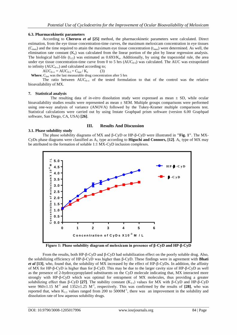
III. Results And Discussion 3.1. Phase solubility study
The phase solubility diagrams of MX and β-CyD or HP-β-CyD were illustrated in "Fig. 1". The MX-
CyDs phase diagrams were classified as AL type according to Higuchi and Connors, [12]. AL type of MX may
be attributed to the formation of soluble 1:1 MX-CyD inclusion complexes.
C o n c e n t r a t io n o f C y D s X 1 0- 3
M / L
Co
nc
en
tra
tio
n o
f m
elo
xic
am
X 1
0-5
M /
L
0 1 2 3 4 5 6
0 .0
0 .5
1 .0
1 .5
2 .0
2 .5
3 .0
3 .5
4 .0
4 .5
5 .0
H P - -C y D
-C y D
Figure 1: Phase solubility diagram of meloxicam in presence of β-CyD and HP-β-CyD
From the results, both HP-β-CyD and β-CyD had solubilization effect on the poorly soluble drug. Also,
the solubilizing efficiency of HP-β-CyD was higher than β-CyD. These findings were in agreement with Bhati
et al [13], who, found that, the solubility of MX increased by the effect of HP-β-CyDs. In addition, the affinity
of MX for HP-β-CyD is higher than for β-CyD. This may be due to the larger cavity size of HP-β-CyD as well
as the presence of 2-hydroxypropylated substituents on the CyD molecule indicating that, MX interacted more
strongly with HP-β-CyD which was optimal for entrapment of MX molecules, thus providing a greater
solubilizing effect than β-CyD [27]. The stability constant (K1:1) values for MX with β-CyD and HP-β-CyD
were 960±1.15 M-1
and 1352±1.25 M-1
, respectively. This was confirmed by the results of [28], who was
reported that, when K1:1 values ranged from 200 to 5000M-1
, there was an improvement in the solubility and
dissolution rate of low aqueous solubility drugs.

Potential Use of Cyclodextrins for the Improvement of Ocular Bioavailability of Meloxicam
DOI: 10.9790/3008-1205017996 www.iosrjournals.org 85 | Page
3.2. Characterization of inclusion complexes
Meloxicam inclusion complexes with CyDs were prepared and characterized in the solid state using FT-IR,
DSC, and PXRD.
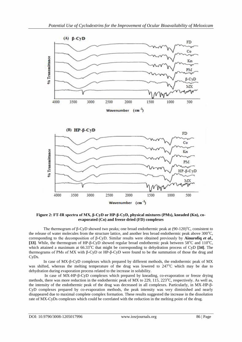
3.2.1. Fourier transform infrared spectroscopy
The FT-IR spectra of MX, β-CyD, HP-β-CyD, the PMs and inclusion complexes prepared by different
methods were analyzed using FT-IR spectrophotometer for characteristic bands as shown in "Fig. 2". The
characteristic absorption peaks of MX appeared at 3288 cm−1
denoting stretching vibration of –NH, 1549 cm-1
together with 1183 cm−1
denoting stretching vibration of the thiazole ring, 1346 cm-1
for the asymmetry
stretching vibration of sulfone, 1526 cm-1
for amide II band of –CO–NH–C and 1263 cm-1
for amide III band of
–CO–NH–C.
The FT-IR spectrum of β-CyD was characterized by intense bands at 3200-3600 cm-1
due to O-H
stretching vibration bands. The vibration bands of the C-H and CH2 groups appear in 2800-3000 cm-1
region, a
broad band appeared at 1649 cm-1
due to adsorbed water [29]. While, that of HP-β-CyD, showed prominent
absorbance bands at 3417 cm-1
and 2930 cm-1
for O-H stretching vibrations. Also, two stretching vibration bands
were obtained at 1421 cm-1
and 1083 cm-1
for C-H and C-O, respectively [30]. In both PMs spectra, the
characteristic peaks of both MX and β-CyD or HP-β-CyD can be observed, and the spectra can be regarded as
simple superimposition of the individual components.
However, obvious changes occurred in the feature and fingerprint region of the FT-IR spectra of MX-β-
CyD and MX-HP-β-CyD inclusion complexes prepared either by kneading, co-evaporation or freeze drying
methods. In the feature region, the band at 3288 cm−1
for –NH stretching vibration peak of MX disappeared in
all inclusion complexes. It seemed that intermolecular hydrogen bond between–NH of MX and –OH of the
carrier or inclusion complexes have been formed [31 & 32].
Similarly, almost all of the peaks of the functional groups containing electronegative atoms underwent
changes: for the amide groups, the amide II (1526 cm−1
) and amide III (1263 cm−1
) bands were shifted to lower
wavenumber band bands; (1516 and 1239 cm-1
), respectively. The asymmetry stretching vibration of sulfone at
(1346 cm−1
) shifted to lower wavenumber band too (1326 cm-1
).
Interestingly, both of the two thiazole rings characteristic peaks simultaneously disappeared in the FT-
IR spectra of all inclusion complexes. This is a direct evidence indicating a possible inclusion of the thiazole
ring into CyD cavities as for the amide groups, which are adjacent to the thiazole ring, the disappearance and the
shifting of the absorption peaks were also attributable to the inclusion into the CyDs cavity. All of the above
evidences implied possible intermolecular interaction between MX and both cyclodextrins.
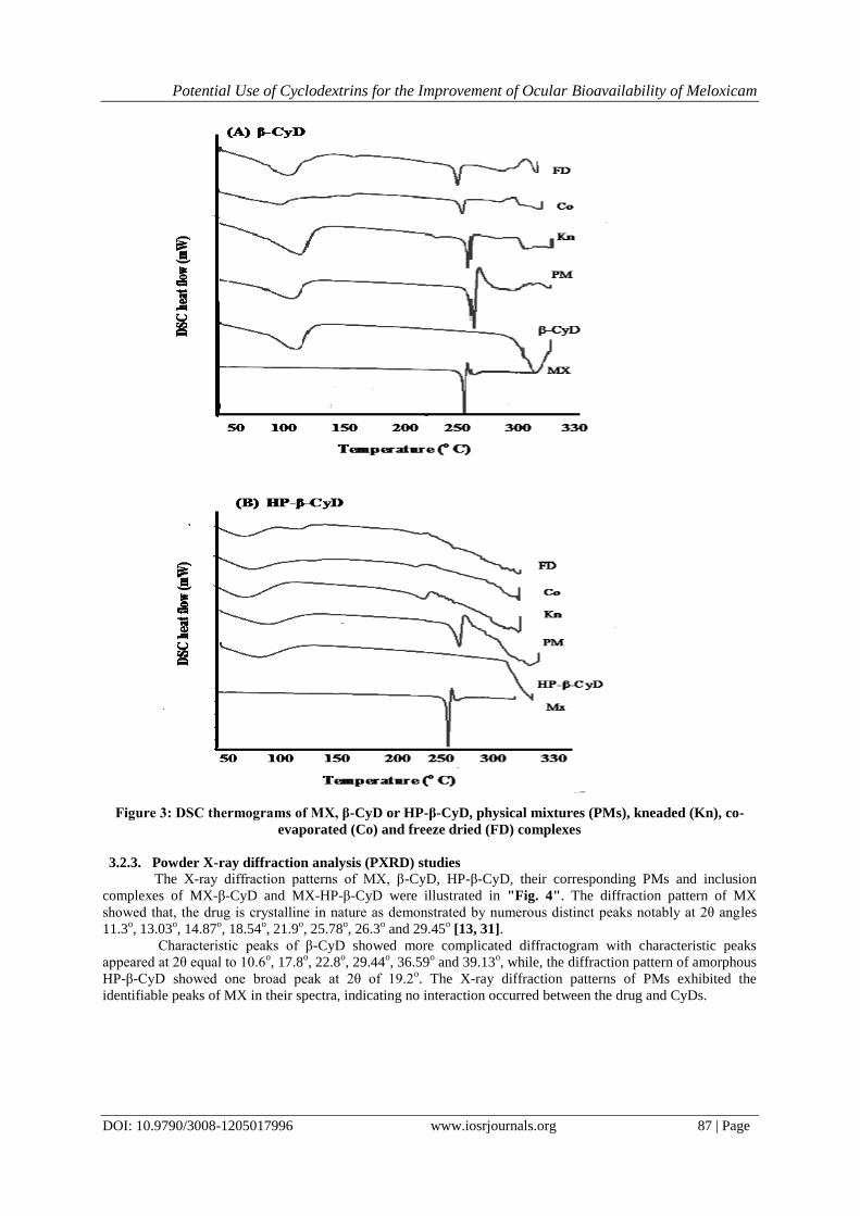
3.2.2 Differential scanning calorimetry
When guest molecules were included inside CyDs cavities, the peak corresponding to their melting point
was generally shifted to another degree. Also, the peak intensity may be decreased or disappeared. Therefore,
the thermal behavior of MX-β-CyD and MX-HP-β-CyD solid complexes and PMs were studied using DSC in
order to characterize the complex formation process.
As shown in "Fig. 3", the thermograms of MX and its PMs indicated a sharp endothermic peak at
254oC attributable to the melting process of the anhydrous crystalline form of the drug, followed by its thermal
decomposition [13].
Potential Use of Cyclodextrins for the Improvement of Ocular Bioavailability of Meloxicam
DOI: 10.9790/3008-1205017996 www.iosrjournals.org 86 | Page
Figure 2: FT-IR spectra of MX, β-CyD or HP-β-CyD, physical mixtures (PMs), kneaded (Kn), co-
evaporated (Co) and freeze dried (FD) complexes
The thermogram of β-CyD showed two peaks; one broad endothermic peak at (90-120)oC, consistent to
the release of water molecules from the structure lattice, and another less broad endothermic peak above 300oC,
corresponding to the decomposition of β-CyD. Similar results were obtained previously by Ainurofiq et al.,
[33]. While, the thermogram of HP-β-CyD showed regular broad endothermic peak between 58oC and 110
oC,
which attained a maximum at 66.33oC that might be corresponding to dehydration process of CyD [34]. The
thermograms of PMs of MX with β-CyD or HP-β-CyD were found to be the summation of those the drug and
CyDs.
In case of MX-β-CyD complexes which prepared by different methods, the endothermic peak of MX
was shifted, whereas the melting temperature of the drug was lowered to 247oC which may be due to
dehydration during evaporation process related to the increase in solubility.
In case of MX-HP-β-CyD complexes which prepared by kneading, co-evaporation or freeze drying
methods, there was more reduction in the endothermic peak of MX to 229, 115, 223oC, respectively. As well as,
the intensity of the endothermic peak of the drug was decreased in all complexes. Particularly, in MX-HP-β-
CyD complexes prepared by co-evaporation methods, the peak intensity was very diminished and nearly
disappeared due to maximal complete complex formation. These results suggested the increase in the dissolution
rate of MX-CyDs complexes which could be correlated with the reduction in the melting point of the drug.
Potential Use of Cyclodextrins for the Improvement of Ocular Bioavailability of Meloxicam
DOI: 10.9790/3008-1205017996 www.iosrjournals.org 87 | Page
Figure 3: DSC thermograms of MX, β-CyD or HP-β-CyD, physical mixtures (PMs), kneaded (Kn), co-
evaporated (Co) and freeze dried (FD) complexes
3.2.3. Powder X-ray diffraction analysis (PXRD) studies The X-ray diffraction patterns of MX, β-CyD, HP-β-CyD, their corresponding PMs and inclusion
complexes of MX-β-CyD and MX-HP-β-CyD were illustrated in "Fig. 4". The diffraction pattern of MX
showed that, the drug is crystalline in nature as demonstrated by numerous distinct peaks notably at 2θ angles
11.3o, 13.03
o, 14.87
o, 18.54
o, 21.9
o, 25.78
o, 26.3
o and 29.45
o [13, 31].
Characteristic peaks of β-CyD showed more complicated diffractogram with characteristic peaks
appeared at 2θ equal to 10.6o, 17.8
o, 22.8
o, 29.44
o, 36.59
o and 39.13
o, while, the diffraction pattern of amorphous
HP-β-CyD showed one broad peak at 2θ of 19.2o. The X-ray diffraction patterns of PMs exhibited the
identifiable peaks of MX in their spectra, indicating no interaction occurred between the drug and CyDs.

Potential Use of Cyclodextrins for the Improvement of Ocular Bioavailability of Meloxicam
DOI: 10.9790/3008-1205017996 www.iosrjournals.org 88 | Page
Figure 4: Powder X – ray diffraction patterns of MX, β-CyD or HP- β-CyD, Physical mixtures (PMs),
kneaded (Kn), co-evaporated (Co) and freeze dried (FD) complexes
The X-ray diffraction patterns of MX-β-CyD complex showed diffuse peaks with low intensities,
indicating that, the crystallinity of the drug was remarkably reduced leading to the formation of a new solid state
due to inclusion complex formation between MX and β-CyD prepared by different techniques [13].
The X-ray diffraction patterns of MX-HP-β-CyD complex showed broad and diffuse peaks with low
intensities, indicating an amorphous solid state was performed may be due to inclusion complex formation
between MX and HP-β-CyD prepared by different techniques. Similar findings have been reported by other
authors [35].
Based on these findings, a decrease in the drug crystallinity with subsequent increase in the surface area
of the drug exposed to the dissolution medium might be responsible for the improved dissolution of MX.
3.3. Physicochemical characterization of different formulations
The superiority of co-evaporation technique was confirmed from the results of FT-IR, DSC and PXRD
due to the combined effect of complexation and crystallinity reduction; hence, the formulations prepared by this
method were further investigated.
Potential Use of Cyclodextrins for the Improvement of Ocular Bioavailability of Meloxicam
DOI: 10.9790/3008-1205017996 www.iosrjournals.org 89 | Page
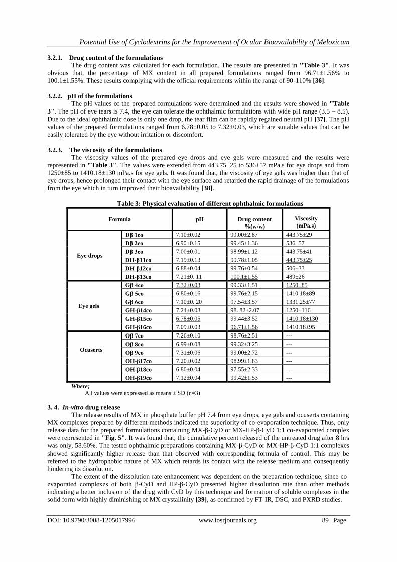
3.2.1. Drug content of the formulations
The drug content was calculated for each formulation. The results are presented in "Table 3". It was
obvious that, the percentage of MX content in all prepared formulations ranged from 96.71±1.56% to
100.1±1.55%. These results complying with the official requirements within the range of 90-110% [36].
3.2.2. pH of the formulations
The pH values of the prepared formulations were determined and the results were showed in "Table
3". The pH of eye tears is 7.4, the eye can tolerate the ophthalmic formulations with wide pH range (3.5 – 8.5).
Due to the ideal ophthalmic dose is only one drop, the tear film can be rapidly regained neutral pH [37]. The pH
values of the prepared formulations ranged from 6.78±0.05 to 7.32±0.03, which are suitable values that can be
easily tolerated by the eye without irritation or discomfort.
3.2.3. The viscosity of the formulations
The viscosity values of the prepared eye drops and eye gels were measured and the results were
represented in "Table 3". The values were extended from 443.75±25 to 536±57 mPa.s for eye drops and from
1250±85 to 1410.18±130 mPa.s for eye gels. It was found that, the viscosity of eye gels was higher than that of
eye drops, hence prolonged their contact with the eye surface and retarded the rapid drainage of the formulations
from the eye which in turn improved their bioavailability [38].
Table 3: Physical evaluation of different ophthalmic formulations
Viscosity
(mPa.s)
Drug content
%(w/w)
pH
Formula
443.75±29 99.00±2.87 7.10±0.02 Dβ 1co
Eye drops
536±57 99.45±1.36 6.90±0.15 Dβ 2co
443.75±41 98.99±1.12 7.00±0.01 Dβ 3co
443.75±25 99.78±1.05 7.19±0.13 DH-β11co
506±33 99.76±0.54 6.88±0.04 DH-β12co
489±26 100.1±1.55 7.21±0. 11 DH-β13co
1250±85 99.33±1.51 7.32±0.03 Gβ 4co
Eye gels
1410.18±89 99.76±2.15 6.80±0.16 Gβ 5co
1331.25±77 97.54±3.57 7.10±0. 20 Gβ 6co
1250±116 98. 82±2.07 7.24±0.03 GH-β14co
1410.18±130 99.44±3.52 6.78±0.05 GH-β15co
1410.18±95 96.71±1.56 7.09±0.03 GH-β16co
--- 98.76±2.51 7.26±0.10 Oβ 7co
Ocuserts
--- 99.32±3.25 6.99±0.08 Oβ 8co
--- 99.00±2.72 7.31±0.06 Oβ 9co
--- 98.99±1.83 7.20±0.02 OH-β17co
--- 97.55±2.33 6.80±0.04 OH-β18co
--- 99.42±1.53 7.12±0.04 OH-β19co
Where; All values were expressed as means ± SD (n=3)
3. 4. In-vitro drug release
The release results of MX in phosphate buffer pH 7.4 from eye drops, eye gels and ocuserts containing
MX complexes prepared by different methods indicated the superiority of co-evaporation technique. Thus, only
release data for the prepared formulations containing MX-β-CyD or MX-HP-β-CyD 1:1 co-evaporated complex
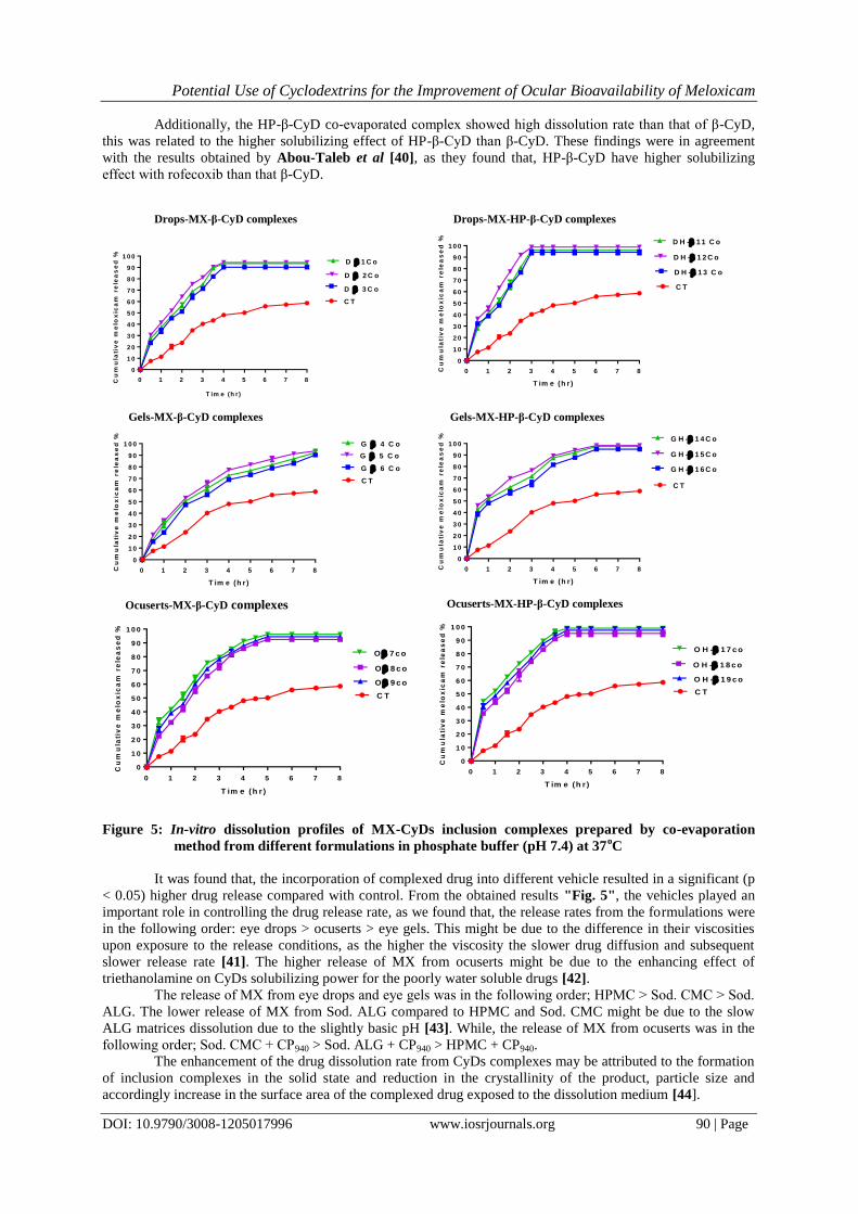
were represented in "Fig. 5". It was found that, the cumulative percent released of the untreated drug after 8 hrs
was only, 58.60%. The tested ophthalmic preparations containing MX-β-CyD or MX-HP-β-CyD 1:1 complexes
showed significantly higher release than that observed with corresponding formula of control. This may be
referred to the hydrophobic nature of MX which retards its contact with the release medium and consequently
hindering its dissolution.
The extent of the dissolution rate enhancement was dependent on the preparation technique, since co-
evaporated complexes of both β-CyD and HP-β-CyD presented higher dissolution rate than other methods
indicating a better inclusion of the drug with CyD by this technique and formation of soluble complexes in the
solid form with highly diminishing of MX crystallinity [39], as confirmed by FT-IR, DSC, and PXRD studies.
Potential Use of Cyclodextrins for the Improvement of Ocular Bioavailability of Meloxicam
DOI: 10.9790/3008-1205017996 www.iosrjournals.org 90 | Page
Additionally, the HP-β-CyD co-evaporated complex showed high dissolution rate than that of β-CyD,
this was related to the higher solubilizing effect of HP-β-CyD than β-CyD. These findings were in agreement
with the results obtained by Abou-Taleb et al [40], as they found that, HP-β-CyD have higher solubilizing
effect with rofecoxib than that β-CyD.
Drops-MX-β-CyD complexes
T im e (h r)
0 1 2 3 4 5 6 7 8
0
1 0
2 0
3 0
4 0
5 0
6 0
7 0
8 0
9 0
1 0 0
C T
D 1 C o
D 2 C o
D 3 C o
Cu
mu
lati
ve
me
lox
ica
m r
ele
as
ed
%
Drops-MX-HP-β-CyD complexes
T im e (h r )
Cu
mu
lati
ve
me
lox
ica
m r
ele
as
ed
%
0 1 2 3 4 5 6 7 8
0
1 0
2 0
3 0
4 0
5 0
6 0
7 0
8 0
9 0
1 0 0
C T
D H - 1 3 C o
D H - 1 1 C o
D H - 1 2 C o
Gels-MX-β-CyD complexes
T im e (h r )
Cu
mu
lati
ve
me
lox
ica
m r
ele
as
ed
%
0 1 2 3 4 5 6 7 8
0
1 0
2 0
3 0
4 0
5 0
6 0
7 0
8 0
9 0
1 0 0
C T
G 6 C o
G 4 C o
G 5 C o
Gels-MX-HP-β-CyD complexes
T im e (h r )
Cu
mu
lati
ve
me
lox
ica
m r
ele
as
ed
%
0 1 2 3 4 5 6 7 8
0
1 0
2 0
3 0
4 0
5 0
6 0
7 0
8 0
9 0
1 0 0
C T
G H - 1 6 C o
G H - 1 4 C o
G H - 1 5 C o
Ocuserts-MX-β-CyD complexes
T im e (h r )
Cu
mu
lati
ve
me
lox
ica
m r
ele
as
ed
%
0 1 2 3 4 5 6 7 8
0
1 0
2 0
3 0
4 0
5 0
6 0
7 0
8 0
9 0
1 0 0
C T
O 8 c o
O 9 c o
O 7 c o
Ocuserts-MX-HP-β-CyD complexes
T im e (h r )
Cu
mu
lati
ve
me
lox
ica
m r
ele
as
ed
%
0 1 2 3 4 5 6 7 8
0
1 0
2 0
3 0
4 0
5 0
6 0
7 0
8 0
9 0
1 0 0
O H - 1 8 c o
O H - 1 9 c o
O H - 1 7 c o
C T
Figure 5: In-vitro dissolution profiles of MX-CyDs inclusion complexes prepared by co-evaporation
method from different formulations in phosphate buffer (pH 7.4) at 37oC
It was found that, the incorporation of complexed drug into different vehicle resulted in a significant (p
< 0.05) higher drug release compared with control. From the obtained results "Fig. 5", the vehicles played an
important role in controlling the drug release rate, as we found that, the release rates from the formulations were
in the following order: eye drops > ocuserts > eye gels. This might be due to the difference in their viscosities
upon exposure to the release conditions, as the higher the viscosity the slower drug diffusion and subsequent
slower release rate [41]. The higher release of MX from ocuserts might be due to the enhancing effect of
triethanolamine on CyDs solubilizing power for the poorly water soluble drugs [42].
The release of MX from eye drops and eye gels was in the following order; HPMC > Sod. CMC > Sod.
ALG. The lower release of MX from Sod. ALG compared to HPMC and Sod. CMC might be due to the slow
ALG matrices dissolution due to the slightly basic pH [43]. While, the release of MX from ocuserts was in the
following order; Sod. CMC + CP940 > Sod. ALG + CP940 > HPMC + CP940.
The enhancement of the drug dissolution rate from CyDs complexes may be attributed to the formation
of inclusion complexes in the solid state and reduction in the crystallinity of the product, particle size and
accordingly increase in the surface area of the complexed drug exposed to the dissolution medium [44].
Potential Use of Cyclodextrins for the Improvement of Ocular Bioavailability of Meloxicam
DOI: 10.9790/3008-1205017996 www.iosrjournals.org 91 | Page
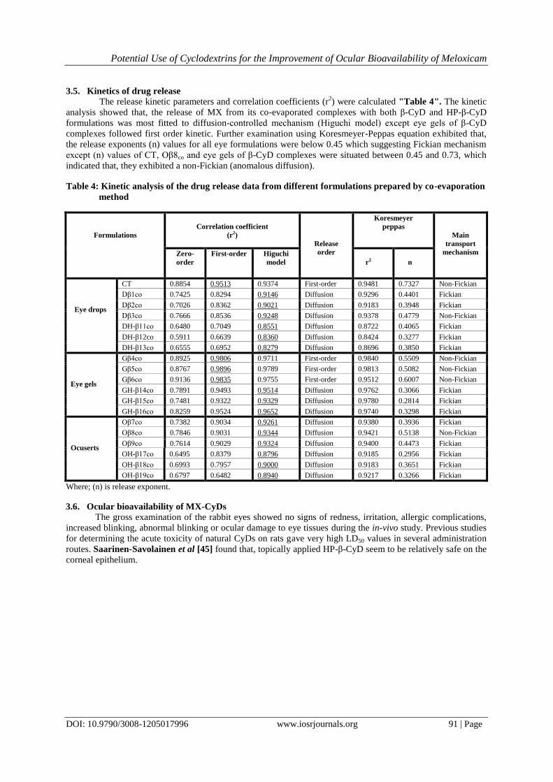
3.5. Kinetics of drug release
The release kinetic parameters and correlation coefficients (r2) were calculated "Table 4". The kinetic
analysis showed that, the release of MX from its co-evaporated complexes with both β-CyD and HP-β-CyD
formulations was most fitted to diffusion-controlled mechanism (Higuchi model) except eye gels of β-CyD
complexes followed first order kinetic. Further examination using Koresmeyer-Peppas equation exhibited that,
the release exponents (n) values for all eye formulations were below 0.45 which suggesting Fickian mechanism
except (n) values of CT, Oβ8co and eye gels of β-CyD complexes were situated between 0.45 and 0.73, which
indicated that, they exhibited a non-Fickian (anomalous diffusion).
Table 4: Kinetic analysis of the drug release data from different formulations prepared by co-evaporation
method
Main
transport
mechanism
Koresmeyer
peppas
Release
order
Correlation coefficient
(r2)
Formulations
n
r2
Higuchi
model
First-order Zero-
order
Non-Fickian 0.7327 0.9481 First-order 0.9374 0.9513 0.8854 CT
Eye drops
Fickian 0.4401 0.9296 Diffusion 0.9146 0.8294 0.7425 Dβ1co
Fickian 0.3948 0.9183 Diffusion 0.9021 0.8362 0.7026 Dβ2co
Non-Fickian 0.4779 0.9378 Diffusion 0.9248 0.8536 0.7666 Dβ3co
Fickian 0.4065 0.8722 Diffusion 0.8551 0.7049 0.6480 DH-β11co
Fickian 0.3277 0.8424 Diffusion 0.8360 0.6639 0.5911 DH-β12co
Fickian 0.3850 0.8696 Diffusion 0.8279 0.6952 0.6555 DH-β13co
Non-Fickian 0.5509 0.9840 First-order 0.9711 0.9806 0.8925 Gβ4co
Eye gels
Non-Fickian 0.5082 0.9813 First-order 0.9789 0.9896 0.8767 Gβ5co
Non-Fickian 0.6007 0.9512 First-order 0.9755 0.9835 0.9136 Gβ6co
Fickian 0.3066 0.9762 Diffusion 0.9514 0.9493 0.7891 GH-β14co
Fickian 0.2814 0.9780 Diffusion 0.9329 0.9322 0.7481 GH-β15co
Fickian 0.3298 0.9740 Diffusion 0.9652 0.9524 0.8259 GH-β16co
Fickian 0.3936 0.9380 Diffusion 0.9261 0.9034 0.7382 Oβ7co
Ocuserts
Non-Fickian 0.5138 0.9421 Diffusion 0.9344 0.9031 0.7846 Oβ8co
Fickian 0.4473 0.9400 Diffusion 0.9324 0.9029 0.7614 Oβ9co
Fickian 0.2956 0.9185 Diffusion 0.8796 0.8379 0.6495 OH-β17co
Fickian 0.3651 0.9183 Diffusion 0.9000 0.7957 0.6993 OH-β18co
Fickian 0.3266 0.9217 Diffusion 0.8940 0.6482 0.6797 OH-β19co
Where; (n) is release exponent.
3.6. Ocular bioavailability of MX-CyDs
The gross examination of the rabbit eyes showed no signs of redness, irritation, allergic complications,
increased blinking, abnormal blinking or ocular damage to eye tissues during the in-vivo study. Previous studies
for determining the acute toxicity of natural CyDs on rats gave very high LD50 values in several administration
routes. Saarinen-Savolainen et al [45] found that, topically applied HP-β-CyD seem to be relatively safe on the
corneal epithelium.

Potential Use of Cyclodextrins for the Improvement of Ocular Bioavailability of Meloxicam
DOI: 10.9790/3008-1205017996 www.iosrjournals.org 92 | Page
T im e (h r )
Tis
su
e c
on
ce
ntra
tio
n
of m
elo
xic
am
(
g/g
)
0 1 2 3 4 5 6
0
1 0
2 0
3 0
4 0
5 0
6 0
D H - 1 2 c o
G H - 1 5 C o
O H - 1 7 c o
C T
(a ) C o n ju n c t iv a
T im e (h r )
Tis
su
e c
on
ce
ntra
tio
n o
f m
elo
xic
am
(
g/g
)
0 1 2 3 4 5 6
0
5
1 0
1 5
2 0
2 5
3 0
3 5
4 0
D H - 1 2 c o
G H - 1 5 c o
O H - 1 7 c o
C T
(b ) C o rn e a
T im e (h r )
Tis
su
e c
on
ce
ntra
tio
no
f m
elo
xic
am
(
g/g
)
0 1 2 3 4 5 6
0
5
1 0
1 5
2 0
2 5
D H - 1 2 c o
G H - 1 5 c o
O H - 1 7 c o
C T
( c ) I r i s - c i l i a r y b o d y
T im e (h r )
Tis
su
e c
on
ce
ntr
atio
n o
f m
elo
xic
am
(µ
g/m
l)
0 1 2 3 4 5 6
0
2
4
6
8
1 0
1 2
1 4
D H - 1 2 c o
G H - 1 5 c o
O H - 1 7 c o
C T
(d ) A q u e o u s h u m o r
Figure 6: The eye tissue concentration-time profiles of MX-HP-β-CyD inclusion complexes following
topical application of the selected formulations
"Fig. 6" showed that, the concentration of MX after a single application of the selected formulations or
CT in the eye tissues and the aqueous humor. The pharmacokinetic parameters of MX were shown in "Table
5". It was clear that, MX bioavailability in eye tissues and aqueous humor were arranged in the order of;
conjunctiva > cornea > iris-ciliary body > aqueous humor which confirmed by the Cmax, AUC0-5, AUC0-∞ and the
relative bioavailability values. The higher MX bioavailability in conjunctiva and cornea may be related to the
direct contact of these tissues with the tear film which containing the drug. These results were in agreement with
those adopted by Soliman et al [46], as they reported that, there is a higher concentration of loteprednol
etabonate in cornea than in aqueous humor. The ocular bioavailability of the prepared formulations were
arranged in the following order; ocuserts > eye gels > eye drops [47], who found that, the total bioavailability of
ciprofloxacin hydrochloride from the tested formulations was in the same order.

Potential Use of Cyclodextrins for the Improvement of Ocular Bioavailability of Meloxicam
DOI: 10.9790/3008-1205017996 www.iosrjournals.org 93 | Page
Table 5: Pharmacokinetic parameters of selected formulations containing MX-HP-β-CyD complex
Tissues Pharmacokinetic
parameters DH-β12co GH-β15co OH-β17co CT
Conjunctiva Cmax (µg/g) 25.39±3.33 47.40±1.76* 52.46±1.39* 21.20±1.10
tmax(hr) 1* 1* 1* 0.5
Ke (hr-1)
0.468±0.035 0.359±0.014* 0.356±0.014* 0.432±0.03
t1/2(hr) 1.48±0.11 1.93±0.08* 1.94±0.08* 1.60±0.09
AUC0-5
(µg.hr/g) 51.93±2.46* 126.37±2.34*, a 147.1±2.52*, a 42.36±2.36
AUC0-∞
(µg.hr/g) 60.02±3.55 158.18±5.52*,a 184.12±4.20*,a 49.44±3.46
Relative
Bioavailability 1.22±0.05 3.21±0.17a 3.74±0.32a ------------
Cornea Cmax (µg/g) 18.85±0.9 27.77±1.85*,a 33.37±1.17*, a 17.3±0.92
tmax(hr) 1* 1* 1* 0.5
Ke (hr-1)
0.479±0.03 0.295±0.03* 0.302±0.02* 0.483±0.05
t1/2(hr) 1.45±0.09 2.36±0.24* 2.29±0.13* 1.44±0.14
AUC0-5
(µg.hr/g) 48.03±1.34* 82.18±5.8*,a 101.38±2.71*,a 33.30±1.14
AUC0-∞
(µg.hr/g) 54.01±2.22 112.24±11.57*, a 135.87±6.94* 37.15±1.90
Relative
Bioavailability 1.46±0.04 3.02±0.16a 3.66±0.30a ------------
Iris-ciliary
body
Cmax (µg/g) 6.73±0.67 15.73±1.01* 18.40±1.14* 6.00±0.8
tmax(hr) 2* 2* 2* 0.5
Ke (hr-1)
0.458±0.01 0.531±0.043* 0.375±0.014 0.38±0.049
t1/2(hr) 1.51±0.02 1.31±0.11* 1.85±0.07 1.84±0.22
AUC0-5
(µg.hr/g) 15.99±1.33* 43.42±0.87*,a 54.58±1.78*,a 8.89±0.39
AUC0-∞
(µg.hr/g) 19.09±1.53* 49.33±1.86*,a 69.85±2.30*,a 11.14±0.36
Relative
Bioavailability 1.71±0.10 4.43±0.05a 6.28±0.38a ----------
Aqueous
humor
Cmax (µg/g) 3.00±0.40 7.87±0.75* 13.23±0.85* 3.77±0.65
tmax(hr) 2* 2* 2* 0.5
Ke (hr-1)
0.33±0.045 0.43±0.07 0.42±0.043 0.34±0.04
t1/2(hr) 2.06±0.26 1.64±0.26 1.67±0.18 2.04±0.24
AUC0-5
(µg.hr/g) 8.11±0.23* 18.02±1.0*,a 36.76±0.39*,a 5.23±0.23
AUC0-∞
(µg.hr/g)
11.04±0.81* 22.57±0.20*,a 45.53±1.72*,a 7.00±0.12
Relative
Bioavailability 1.58±0.11 3.22±0.08a 6.50±0.14a -----------
All values are expressed as means ± SEM (n=6)
Cmax the maximum concentration of drug in eye tissues, tmax time required to reach the maximum eye tissue concentration, Ke
the elimination rate constant, t1/2 the biological half-life, AUC0-5 the area under eye tissue concentration-time curve from 0 to
5 hrs, AUC0-∞ the area under eye tissue concentration-time curve from 0 to infinity. (*) considered significant compared to control (p < 0.05) and (a) considered significant compared to DH-β12co
The time required to reach maximum concentration values tmax, for all the investigated formulations,
gave extended values which reached up to 2 hrs in iris- ciliary body and aqueous humor, while in conjunctiva
and cornea, tmax reached up to 1 hr. It was clear that, the tmax of all formulations were significantly (p < 0.05)
superior to that of control in different eye tissues. Also, t1/2 values for all tested formulations had prolonged t1/2

Potential Use of Cyclodextrins for the Improvement of Ocular Bioavailability of Meloxicam
DOI: 10.9790/3008-1205017996 www.iosrjournals.org 94 | Page
that ranged from 1.31±0.11 hrs to 2.36±0.24 hrs in different eye tissues compared to 1.44±0.14 hrs to 2.04±0.24
hrs for the control.
From Cmax and tmax results, the data showed that, after single administration of 0.03% MX ophthalmic
formulae, permeated poorly into aqueous humor, iris and ciliary body, whereas it permeated well into the cornea
and the conjunctiva. These results were parallel to that revealed by [48], who study the ophthalmic composition
containing fourth generation flouroquinolones for the treatment of eye infections and with [49], who studied the
distribution of amefenac to the posterior segment of the eye. The concentration gradient between the tear film
and the drug concentration in the cornea and conjunctival epithelium was the driving force for passive diffusion
across these tissues [50].
Furthermore, the relative bioavailability was generally increased from 1.22 to 6.50 folds vs. control for
all tested formulations in different eye tissues and aqueous humor. The higher bioavailability of MX from
ocuserts that contain CP 940 can be explained by the enhancing effect of triethanolamine on CyDs solubilizing
power for the poorly water soluble drugs by forming drug-CyD-TEA multicomponent system improving the
ocular delivery of the drug [42].
The enhanced bioavailability of the eye gels and ocuserts containing MX-CyDs complexes than eye
drops may be due to the high viscosity and bioadhesiveness properties of the polymers which forms the eye gels
and ocuserts. These properties prevented the rapid drainage of the formulations from the eye and so increased
their contact time with the eye surface which in turn improved their bioavailability. These results are in line with
Budai et al [51], as they reported that, the high viscosity and the bioadhesive properties of the gel formulations
improved the ocular bioavailability of ciprofloxacin.
It was obvious that, the Cmax, AUC0-5 and AUC0-∞ of all formulations were significantly (p < 0.05)
superior to that of CT in cornea, conjunctiva and iris-ciliary body. While, in aqueous humor, all formulations
were significantly different at (p < 0.05) versus that of CT except eye drops were non-significant. This might be
due to rapid turnover of the eye drops from the eye surface [19], as they found that, gels containing celecoxib
nanoparticles showed a higher ocular bioavailability which indicated by higher AUC and Cmax values.
IV. Conclusion Inclusion complexes of MX with β-CyD or HP-β-CyD have been successfully prepared. The higher
degree of amorphous entities has been yielded by the co-evaporation method ensuring the formation of true MX-
β-CyD/MX-HP-β-CyD inclusion complexes. Thus, the complex preparation methods played an important role in
improving the dissolution rate. Also, the MX ocular bioavailability was improved by complexation of it with
HP-β-CyD more than β-CyD. Ocuserts and eye gels containing MX-CyD have a higher bioavailability and more
extended action than eye drops and control. Accordingly, the complexation of MX with CyDs provided a
promising mean for enhancement the dissolution rate and the ocular bioavailability of MX.
References [1] C P Herbort, A Jauch, P Othenin-Girard, J J Tritten, and M Fsadni, Diclofenac drops to treat inflammation surgery after
cataract inflammation, Acta Ophthalmologica Scandinavica, 78, 2000, 421-424.
[2] S Sivaprasad, C Bunce, and R Wormald, Nonsteroidal anti-inflammatory agents for cystoid macular edema following
cataract surgery: a systematic review, British Journal of Ophthalmology, 89, 2005, 1420-1422.
[3] C Maihofner, U Schlotzer-Schrehardt, H Guhring, H U Zeilhofer, G O Naumann, A Pahl, C Mardin, E R Tamm, and K
Brune, Expression of cyclooxgynase-1 and -2 in normal and glaumatous human eyes, Investigative Ophthalmology and
Visual Science, 42, 2001, 2616-2624.
[4] M I Ortiz, G Castaneda-Hernandez, and V Granados-Soto, Pharmacological evidence for the activation of Ca+2–activated
K+ channels by meloxicam in the formalin test, Pharmacology Biochemistry and Behavior, 81, 2005, 725-731.
[5] Martindale, The Complete Drug Reference, 36th Ed., Sweetman, S. C. ed., The pharmaceutical press, London, U. K.,
2009, P. 531.
[6] J P Kaur, and M Kanwar, Ocular preparations: the formulation approach, Drug Development and industrial pharmacy,
28, 2002, 473-493.
[7] H M Heise, R Kucker, A Bereck and D Riegel, Infrared spectroscopy and Raman spectroscopy of cyclodextrin derivatives
and their ferrocene inclusion complexes, Vibrational Spectroscopy, 53, 2010, 19-23.
[8] Y Tsai, H Tsai, C Wu and F Tsai, Preparation, characterization and activity of the inclusion complex of paeonol with β-
cyclodextrin, Food Chemistry, 120, 2010, 837-841.
[9] M M Doile, K A Fortunato, I S Schmücker, S K Schucko, M A S Silva, and P O Rodrigues, Physicochemical properties
and dissolution studies of dexamethasone acetate-β-cyclodextrin inclusion complexes produced by different methods,
AAPS Pharmaceutical Science and Technology, 9, 2008, 314-321.
[10] F S Bandarkar and P R Vavia, Physico-chemical characterisation and invivo pharmacodynamics evaluation of lyophilized
meloxicam: β-cyclodextrin inclusion complexes, International Journal of Pharmacy and Pharmaceutical Sciences, 5(3),
2013, 159-165.
[11] F Veiga, C Pecorelli and L Ribeiro, As ciclodextrinas em tecnologia farmacêutica. Coimbra: Minerva Coimbra,
9789727981748, 2006, 9-33.
[12] T Higuchi, and K A Connors, Phase solubility techniques, Adv. Anal. Chem. Instrum.; 4, 1965, 117-122.

Potential Use of Cyclodextrins for the Improvement of Ocular Bioavailability of Meloxicam
DOI: 10.9790/3008-1205017996 www.iosrjournals.org 95 | Page
[13] L K Bhati, G Tiwari, R Tiwari, and V Kumar, Enhancement of complexation efficiency of meloxicam using binary and
ternary solid systems; formulation consideration, American Journal of Drug Discovery and Development, 2 (1), 2012, 17-
31.
[14] R Govindarajan, and M S Nagarsenkar, Influence of preparation methodology on solid-state properties of an acidic drug
–cyclodextrin system, Journal of Pharmacy and Pharmacology, 56, 2004, 725-733.
[15] S P Epstein, M Ahdoot, E Marcus, and P A Asbell, Comparative toxicity of preservatives on immortalized corneal and
conjunctival epithelial Cells, Journal of Ocular Pharmacology and Therapeutics, 25 (2), 2009, 113-119.
[16] R M Gilhotra, N Gilhotra, and D N Mishra, Piroxicam bioadhesive ocular inserts: physicochemical characterization and
evaluation in prostaglandin-induced inflammation, Current Eye Research, 34(12), 2009, 1065-1073.
[17] M H Aburahma, and A A Mahmoud, Biodegradable ocular inserts for sustained delivery of brimonidine tartarate:
Preparation and in-vitro/in-vivo evaluation, AAPS Pharmaceutical Science and Technology, 12 (4), 2011, 1335-1347.
[18] B Abrar, S Anis, B Tanu and S Singh, formulation and in vitro evaluation of NSAIDs gel, International Journal of
current Pharmaceutical Research, 4 (3), 2012, 56-58.
[19] M M Ibrahim, A H Abd-Elgawad, O A Soliman, and M M Jablonski, Natural bioadhesive biodegradable nanoparticle-
based topical ophthalmic formulations for sustained celecoxib release: in vitro study, Journal of Pharmaceutical
Technology and Drug Research, 2 (7), 2013, 1-15.
[20] D H Shastri, L D Patel, and R K Parikh, Studies on in situ hydrogel: a smart way for safe and sustained ocular drug
delivery, Journal of Young Pharmaceutics, 2(2), 2010, 116-120.
[21] A Martin, P Bustamaante, and A H C Chun: "Kinetics", Chapter 12, in: "Physical Pharmacy", 4th Ed., lea and Febiger,
Pheladelphia, U.S.A., 1993, 284-323.
[22] WI Higuchi, and A Suzuki, Theoretical model studies of drug absorption and transport in the gastrointestinal tract II,
Journal of Pharmaceutical Sciences, 59, 1970, 651-659.
[23] R W Koresmeyer, R Gurny, E Doelker, P Buri, and N A peppas, Mechanisms of solute release from porous hydrophilic
polymers, International Journal of pharmaceutics, 15, 1983, 25-35.
[24] L H Emara, M F Emam, N F Taha, H M Raslan, and A A El-shmawy, A simple and sensitive HPLC/UV method for
determination of meloxicam in human plasma for bioavailability and bioequivalence studies, Journal of Applied
Pharmaceutical Science, 7, 2016, 012-019.
[25] N P S Cheruvu, A C Amrite, and U P Kompella, Effect of eye pigmentation on transscleral drug delivery, Investigative
Ophthalmology and Visual Science, 49, 2008, 333-341.
[26] J E De Muth, Basic statistics and pharmaceutical statistical applications. 2nd
ed., New York: Chapman & Hall/CRC,
Taylor & Francis Group, 2006, 201-243.
[27] V R Yadav, S Suresh, K Devi, and S Yadav, Effect of cyclodextrin complexation of curcumin on its solubility and anti-
angiogenic and anti-inflammatory activity in rat colitis model, AAPS Pharmaceutical Science and Technology, 10, 2009,
752-762.
[28] R P Patel, and M M Patel, Preparation and evaluation of inclusion complex of the lipid lowering drug lovastatin with β-
cyclodextrin, Dhaka University Journal of Pharmaceutical Sciences, 6 (1), 2007, 25-36.
[29] A A Mahmoud, G S El-Feky, R Kamal, and G E A Awad, Chitosan / sufobutylether–β-cyclodextrin nanoparticles as a
potential approach for ocular drug delivery, International Journal of pharmaceutics, 413 (1-2), 2011, 229-236.
[30] M M Al-Omaria, M H Daraghmha, M I El-Barghouthib, M B Zughulc, B Z Chowdhryd, S A Leharned, and A A
Badwana, Noval inclusion complex of ibuprofen tromethamine with cyclodextrins: physicochemical characterization,
Journal of Pharmaceutical and Biomedical Analysis, 50, 2009, 449-458.
[31] Y Lu, X Zhaang, J Lai, Z Yin, and W Wu, Physical characterization of meloxicam-β-cyclodextrin inclusion complex
pellets prepared by a fluid–bed coating method, Particuology, 7, 2009, 1-8.
[32] S Baboota, and S P Agarwal, Preparation and characterisation of meloxicam hydroxyl propyl β-cyclodextrin inclusion
complex, Journal of Inclusion Phenomena and Macrocyclic Chemistry, 51, 2005, 219-224.
[33] A Ainurofiq, S Choiri1, M A Azhari1, C R Siagian, B B Suryadi, F Prihapsara, and S Rohmani, Improvement of
meloxicam solubility using a β-cyclodextrin complex prepared via the kneading method and incorporated into an orally
disintegrating tablet, Advanced Pharmaceutical Bulletin, 6(3), 2016, 399-406.
[34] J Wang, Y Cao, B Sun, and C Wang, Characterization of inclusion complex of trans-ferulic acid and hydroxypropyl-β-
cyclodextrin, Food Chemistry, 124 (3), 2011, 1069-1075.
[35] H A El-maradny, S A Mortada, O A Kamel and A H Hikal, Characterization of ternary complexes of meloxicam-HP-CD
and PVP or L-arginine prepared by the spray-drying technique, Acta Pharmaceutica, 58, 2008, 455-466.
[36] B. P., "British Pharmacopeia", Vol. III, 6th Ed., The Council of Europe, The Stationary office, London, U.K., 2010, PP.,
3155-3157.
[37] USP 34-NF 29, “The United States Pharmacopeia”, Pharmaceutical dosage forms, ophthalmic preparation and uniformity
of dosage units, United States Pharmacopeial Convention, Inc. MD, The National formulary 29th, 2011, P. 700-701.
[38] M M Ibrahim, A H Abd-Elgawad, O A Soliman, and M M Jablonski, Natural bioadhesive biodegradable nanoparticle-
based topical ophthalmic formulations for management of glaucoma, Translational Vision Science and Technology, 4 (3),
2015, 1-13.
[39] A M Yousaf, D W Kim, K H Cho, J O Kim, and C S Yong, Effect of the preparation method on crystallinity, particle size,
aqueous solubility and dissolution of different samples of the poorly water-soluble fenofibrate with HP-β-CD, Journal of
Inclusion Phenomena and Macrocyclic Chemistry, 81(3-4), 2015, 347-356.
[40] A E Abou-Taleb, A A Abdel-Rhman, E M Samy, and H M Tawfeek, Interaction of rofecoxib with β-cyclodextrin and
HP-β-cyclodextrin in aqueous solution and in solid state, Bulletin of Pharmaceutical Sciences of Assiut University, 29
(2), 2006, 236-252.
[41] K Stamatopoulos, H K Batchelor, F Alberini, J Ramsay, M J H Simmons, Understanding the impact of media viscosity on
dissolution of a highly water soluble drug within a USP 2 mini vessel dissolution apparatus using an optical planar
induced fluorescence (PLIF) method, International Journal of Pharmaceutics, 495(1), 2015, 362-373.
[42] G E Granero, M M Maitre, C Garnero, and M R Longhi, Synthesis, characterization and in-vitro release studies of a new
Acetazolamide-HP-β-CyD TEA inclusion complex, European Journal of Medicinal Chemistry, 43, 2008, 464-470.

Potential Use of Cyclodextrins for the Improvement of Ocular Bioavailability of Meloxicam
DOI: 10.9790/3008-1205017996 www.iosrjournals.org 96 | Page
[43] A Oh, D H Jin, J Yun, Y S Lee, and H I Kim, Effect of pH-dependent solubility on release behavior of alginate-chitosan
blend containing activated carbon, Carbon Letters, 10, 2009, 208-212.
[44] R Chadha, N Kashid, and A Sain, Acount of analytical techniques employed for the determination of thermodynamic of
inclusion complexation of drugs with cyclodextrins, Journal of Scientific and Industrial Research, 63, 2004, 211-229.
[45] P Saarinen-Savolainen, T Jarvinen, K Araki-Sasaki, H Watanabe, and A Urtti. Evaluation of cytotoxicity of various
ophthalmic drugs, eye drops excipients and cyclodextrins in an immortalized human corneal epithelial cell line, Pharmacy
Research, 15, 1998, 1275-80. [46] O A Soliman, E A Mohamed, S M El-dahan and N A A Khatera, Potential use of cyclodextrin complexes for enhanced
stability, anti-inflammatory efficacy, and ocular bioavailability of loteprednol etabonate, AAPS Pharmaceutical Science
and Technology, 18(4), 2016, 1228-1241.
[47] D Shaker, "Formulation and Stability of Some Topical Systems Containing Certain Drugs ","Master Thesis", Faculty of
Pharmacy, pharmaceutics Department, Helwan University, Egypt (2000).
[48] A R Blanco, R A Sudano, and C Civiale, Ophthalmic compositions containing gemifloxacin for the treatment of ocular
infections, European patent, 2010, 143, 422A1.
[49] J E Chastain, M E Sanders, M A Curtis, N V Chemuturi, M E Gadd, M A Kapin, K L Markwardt, and D C Dahlin,
Distribution of topical ocular nepafenac and its active metabolite amefenac to the posterior segment of the eye,
Experimental Eye Research, 145, 2016, 58-67.
[50] S B Koevary, Pharmacokinetics of topical ocular drug delivery: potential uses for the treatment of diseases of the
posterior segment and beyond, Current Drug Metabolism, 4, 2003, 213-222.
[51] L Budai, M Hajdu, M Budai, P Grof, S Beni, B Noszal, I Klebovich, and I Antal, Gels and liposomes in optimized ocular
drug delivery: studies on ciprofloxacin formulations, International Journal of Pharmaceutics, 343, 2007, 34-40.