Potential for Dietary ω3 Fatty Acids to Prevent ...
25
1 Potential for Dietary ω3 Fatty Acids to Prevent 1 Nonalcoholic Fatty Liver Disease and Reduce the Risk of 2 Primary Liver Cancer 3 4 5 Donald B. Jump, Christopher M. Depner, Sasmita Tripathy and Kelli A. Lytle 6 7 Nutrition Program 8 School of Biological and Population Health Sciences 9 Linus Pauling Institute 10 Oregon State University 11 Corvallis Oregon, 97331 12 13 14 15 16 Corresponding Author: 17 Donald B. Jump, Ph.D. 18 107A Milam Hall 19 School of Biological and Population Health Sciences 20 Oregon State University 21 Corvallis, OR 97370 22 Phone: 541-737-4007 23 Email: [email protected] 24 25 26 Pubmed indexing: Jump; Depner; Tripathy; Lytle 27 Running Title: NASH, a potential risk factor for HCC 28 29 30 31
Transcript of Potential for Dietary ω3 Fatty Acids to Prevent ...
1
Potential for Dietary ω3 Fatty Acids to Prevent 1
Nonalcoholic Fatty Liver Disease and Reduce the Risk of 2
Primary Liver Cancer 3
4
5
Donald B. Jump, Christopher M. Depner, Sasmita Tripathy and Kelli A. Lytle 6 7
Nutrition Program 8 School of Biological and Population Health Sciences 9
Linus Pauling Institute 10 Oregon State University 11 Corvallis Oregon, 97331 12
13 14 15 16 Corresponding Author: 17 Donald B. Jump, Ph.D. 18 107A Milam Hall 19 School of Biological and Population Health Sciences 20 Oregon State University 21 Corvallis, OR 97370 22 Phone: 541-737-4007 23 Email: [email protected] 24 25 26
Pubmed indexing: Jump; Depner; Tripathy; Lytle 27
Running Title: NASH, a potential risk factor for HCC 28
29
30
31
2
ABSTRACT 32
Nonalcoholic fatty liver disease (NAFLD) has increased in parallel with central obesity and its prevalence 33
is anticipated to increase as the obesity epidemic remains unabated. NAFLD is now the most common 34
cause of chronic liver disease in developed countries and is defined as excessive lipid accumulation in 35
the liver, i.e., hepatosteatosis. NAFLD ranges in severity from benign fatty liver to nonalcoholic 36
steatohepatitis (NASH), where NASH is characterized by hepatic injury, inflammation, oxidative stress 37
and fibrosis. NASH can progress to cirrhosis; and cirrhosis is a risk factor for primary hepatocellular 38
carcinoma (HCC). The prevention of NASH will lower the risk of cirrhosis and NASH-associated HCC. 39
Our studies have focused on NASH prevention. We developed a model of NASH using Ldlr-/- mice fed the 40
western diet (WD). The WD induces a NASH phenotype in these mice that is similar to that seen in 41
humans; and includes robust induction of hepatic steatosis, inflammation, oxidative stress and fibrosis. 42
Using transcriptomic, lipidomic and metabolomic approaches, we examined the capacity of 2 dietary ω3 43
polyunsaturated fatty acids, eicosapentaenoic acid (20:5ω-3; EPA) and docosahexaenoic acid (22:6ω-3; 44
DHA), to prevent WD-induced NASH. Dietary DHA was superior to EPA at attenuating WD-induced 45
changes in plasma lipids and hepatic injury; and reversing WD effects on hepatic metabolism, oxidative 46
stress, and fibrosis. The outcome of these studies suggests that DHA may be useful in the prevention of 47
NASH and reducing the risk of HCC. 48
49
Key words: 50
Fatty liver disease, liver cancer, inflammation, oxidative stress, fibrosis, metabolomics, ω3 PUFAs 51

3
Introduction. 52
Primary hepatocellular carcinoma (HCC) is the 5th most common human cancer in men and the 53
7th most common cancer in women in the western societies; and HCC represents the 3rd most frequent 54
cause of cancer deaths worldwide (1-3). High rates of HCC are seen in eastern and southeastern 55
Africa and Asia and lower levels in western countries. Risk factors for HCC include age and gender 56
(male), hepatitis virus infection (HBV, HCV), exposure to toxins (aflatoxin), chronic alcohol abuse, 57
cirrhosis, tobacco, and genetic disorders (hereditary hemochromatosis, α1-antitrypsin deficiency and 58
primary biliary cirrhosis) (1, 2). 59
The unabated increase in the incidence of obesity, type 2 diabetes and non-alcoholic fatty liver 60
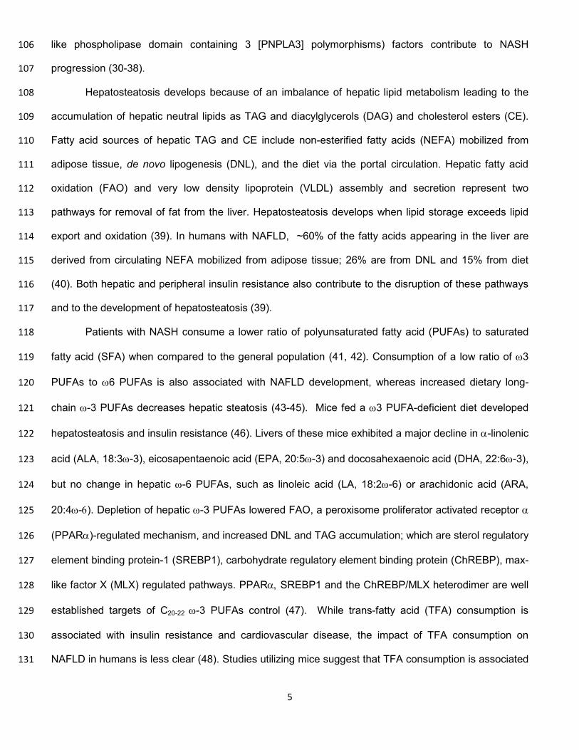
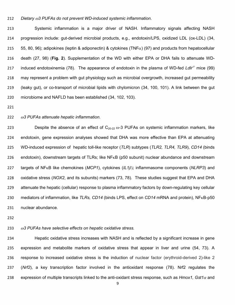
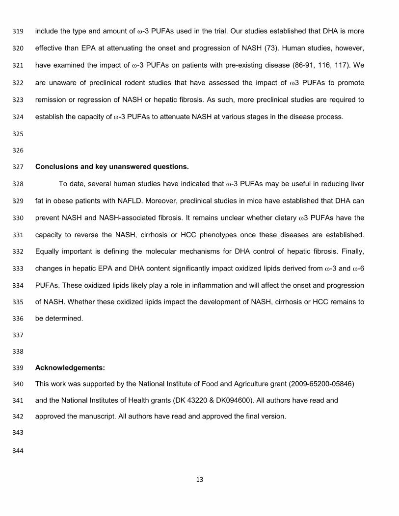
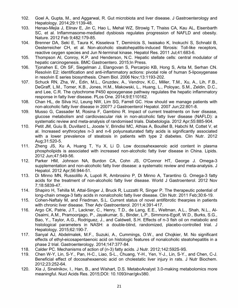
disease (NAFLD) (Fig. 1) is driving the concern for an increased HCC incidence in western societies 61
(4). This is because NAFLD can progress to non-alcoholic steatohepatitis (NASH) and cirrhosis; 62
cirrhosis is a risk factor for HCC. Chronic fatty liver disease sets the stage for poorly regulated 63
regeneration of hepatic parenchymal cells resulting from hepatic inflammation, parenchymal cell death 64
and fibrosis; thus increasing HCC risk. Current treatment options for HCC are limited to surgery and 65
drugs like the multi-kinase inhibitor, sorafenib. Since diet is a major driver of NAFLD and NASH 66
progression, our focus has been on developing nutritional strategies to prevent NASH. This report 67
focuses on the use of dietary C20-22 ω-3 polyunsaturated fatty acids (PUFAs) to prevent NASH. 68
69
NAFLD and NASH. 70
Current data from the CDC estimates that nearly 78.6 million obese adults and 12.7 million obese 71
children (ages 2-19) are in the US (5, 6). Obesity is a risk factor for developing NAFLD and NASH. As 72
such, the prevalence of NAFLD and NASH has increased in parallel with the incidence of central 73
obesity in western societies (7, 8). NAFLD is the most common fatty liver disease in developed 74
countries (9) and is defined as excessive lipid accumulation in the liver, i.e., hepatosteatosis (10, 11). 75
NAFLD is the hepatic manifestation of metabolic syndrome (MetS) (12); and MetS risk factors include 76
obesity, elevated plasma triacylglycerols (TAG) and LDL cholesterol, reduced HDL cholesterol, high 77
blood pressure and fasting hyperglycemia (13). The prevalence of NAFLD in the general population is 78
4
estimated to range from 6% to 30% depending on the method of analysis and population studied (14) 79
(Fig. 1). 80
NAFLD ranges from benign hepatosteatosis to NASH (15), which is defined as hepatosteatosis 81
with inflammation and hepatic injury (16). Approximately 30-40% of patients with steatosis develop 82
NASH (17); representing ~3% to 5% in the general population (14). NAFLD and NASH have high 83
prevalence (>60%) in the type 2 diabetic (T2D) population (18). The level of NAFLD and NASH in 84
patients undergoing bariatric surgery is 93% and 26%, respectively (19). NASH patients have higher 85
mortality rates than NAFLD patients; and both are higher than in the general population (20-22). Over a 86
10 year period, cirrhosis and liver related death occurs in 20% and 12% of NASH patients, respectively 87
(23). Given the increasing prevalence of NASH and its adverse clinical outcome, NASH is rapidly 88
becoming a significant public health burden. NASH can progress to cirrhosis and HCC (8, 17). By the 89
year 2020, cirrhosis resulting from NASH is projected to be the leading cause of liver transplantation in 90
the United States (24). 91
92
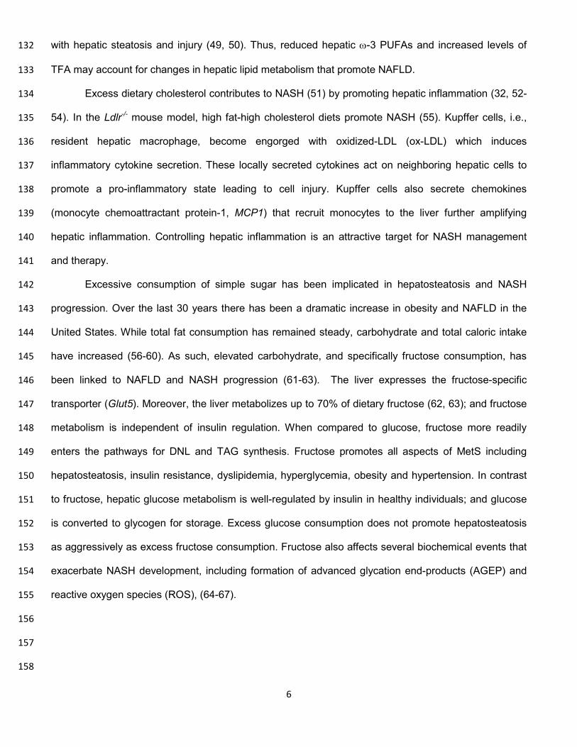
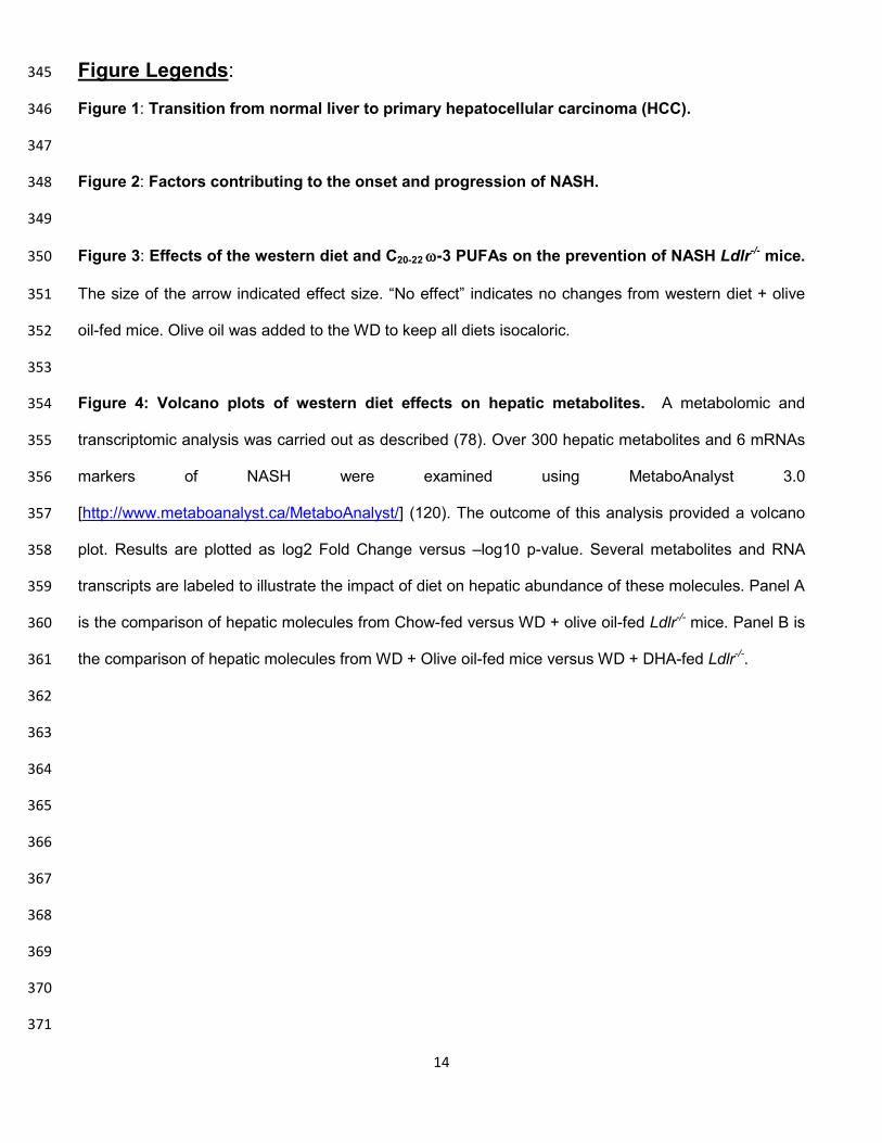
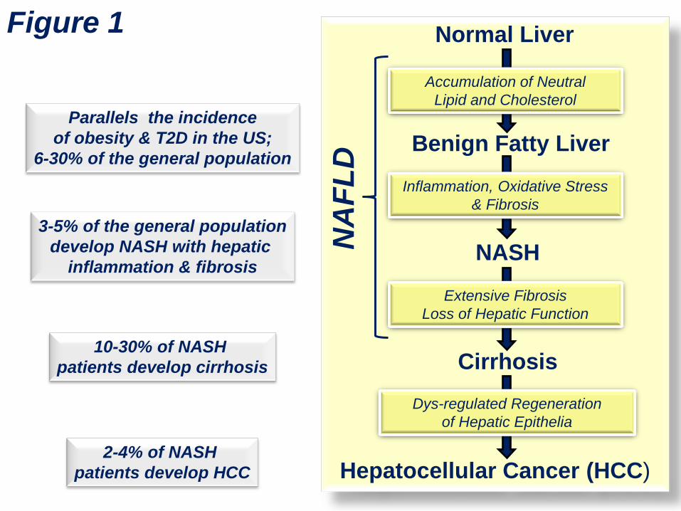
Multi-hit hypotheses for NASH development. 93
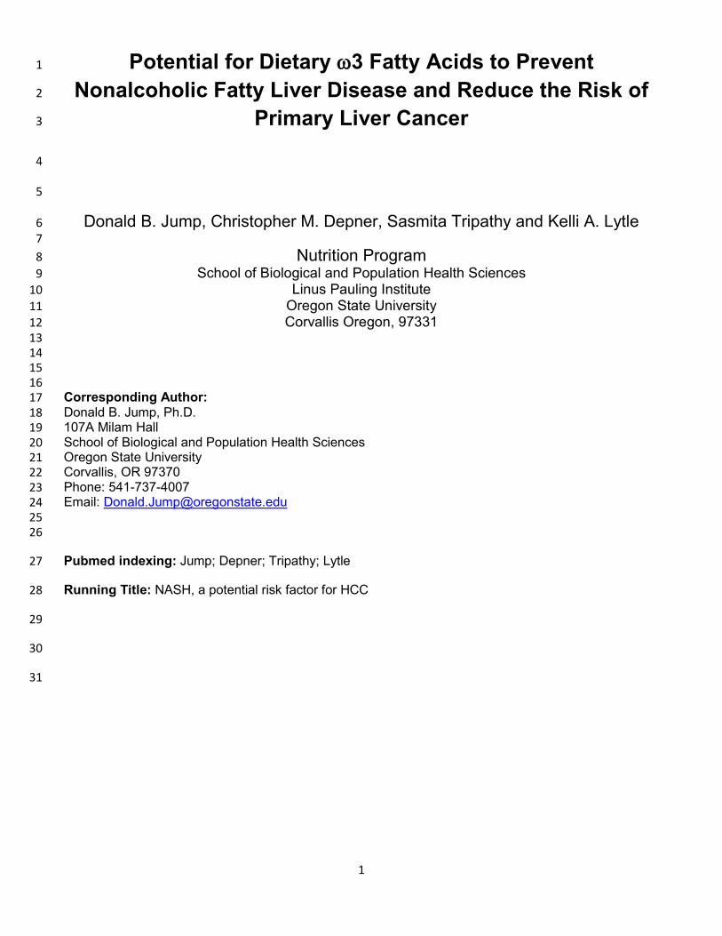
The development of NASH has been proposed to follow a multi-hit model (25-27). The “1st Hit” 94
involves excessive neutral lipid accumulation in the liver which sensitizes the liver to the “2nd Hit” (26) 95
(Fig. 2). The “2nd Hit” is characterized by hepatic inflammation, oxidative stress and hepatic insulin 96
resistance. These events promote hepatic damage which is associated with increased blood levels of 97
hepatic enzymes/proteins (alanine aminotransferase [ALT], aspartate aminotransferase (AST), C-98
reactive protein, serum amyloid A1 and plasminogen activator inhibitor-1 (PIA1) (7, 8, 28). This pro-99
inflammatory state leads to hepatocellular death & necrosis (necroinflammation); and cell death 100
promotes fibrosis, i.e., the “3rd Hit”. Fibrosis is mediated by activation of hepatic stellate cells and 101
myofibrillar cells; these cells produce extracellular matrix (ECM) proteins, such as collagen (collagen 102
1A1, Col1A1) and smooth muscle α2 actin (29). Dietary (excess fat, cholesterol, glucose and fructose), 103
metabolic (plasma and hepatic fatty acid profiles, hepatic ceramide, oxidized LDL), endocrine/paracrine 104
(insulin, leptin, adiponectin & TGFβ), gut (endotoxin, microbial metabolites) and genetic (e.g., patatin-105
5
like phospholipase domain containing 3 [PNPLA3] polymorphisms) factors contribute to NASH 106
progression (30-38). 107
Hepatosteatosis develops because of an imbalance of hepatic lipid metabolism leading to the 108
accumulation of hepatic neutral lipids as TAG and diacylglycerols (DAG) and cholesterol esters (CE). 109
Fatty acid sources of hepatic TAG and CE include non-esterified fatty acids (NEFA) mobilized from 110
adipose tissue, de novo lipogenesis (DNL), and the diet via the portal circulation. Hepatic fatty acid 111
oxidation (FAO) and very low density lipoprotein (VLDL) assembly and secretion represent two 112
pathways for removal of fat from the liver. Hepatosteatosis develops when lipid storage exceeds lipid 113
export and oxidation (39). In humans with NAFLD, ~60% of the fatty acids appearing in the liver are 114
derived from circulating NEFA mobilized from adipose tissue; 26% are from DNL and 15% from diet 115
(40). Both hepatic and peripheral insulin resistance also contribute to the disruption of these pathways 116
and to the development of hepatosteatosis (39). 117
Patients with NASH consume a lower ratio of polyunsaturated fatty acid (PUFAs) to saturated 118
fatty acid (SFA) when compared to the general population (41, 42). Consumption of a low ratio of ω3 119
PUFAs to ω6 PUFAs is also associated with NAFLD development, whereas increased dietary long-120
chain ω-3 PUFAs decreases hepatic steatosis (43-45). Mice fed a ω3 PUFA-deficient diet developed 121
hepatosteatosis and insulin resistance (46). Livers of these mice exhibited a major decline in α-linolenic 122
acid (ALA, 18:3ω-3), eicosapentaenoic acid (EPA, 20:5ω-3) and docosahexaenoic acid (DHA, 22:6ω-3), 123
but no change in hepatic ω-6 PUFAs, such as linoleic acid (LA, 18:2ω-6) or arachidonic acid (ARA, 124
20:4ω-6). Depletion of hepatic ω-3 PUFAs lowered FAO, a peroxisome proliferator activated receptor α 125
(PPARα)-regulated mechanism, and increased DNL and TAG accumulation; which are sterol regulatory 126
element binding protein-1 (SREBP1), carbohydrate regulatory element binding protein (ChREBP), max-127
like factor X (MLX) regulated pathways. PPARα, SREBP1 and the ChREBP/MLX heterodimer are well 128
established targets of C20-22 ω-3 PUFAs control (47). While trans-fatty acid (TFA) consumption is 129
associated with insulin resistance and cardiovascular disease, the impact of TFA consumption on 130
NAFLD in humans is less clear (48). Studies utilizing mice suggest that TFA consumption is associated 131
6
with hepatic steatosis and injury (49, 50). Thus, reduced hepatic ω-3 PUFAs and increased levels of 132
TFA may account for changes in hepatic lipid metabolism that promote NAFLD. 133
Excess dietary cholesterol contributes to NASH (51) by promoting hepatic inflammation (32, 52-134
54). In the Ldlr-/- mouse model, high fat-high cholesterol diets promote NASH (55). Kupffer cells, i.e., 135
resident hepatic macrophage, become engorged with oxidized-LDL (ox-LDL) which induces 136
inflammatory cytokine secretion. These locally secreted cytokines act on neighboring hepatic cells to 137
promote a pro-inflammatory state leading to cell injury. Kupffer cells also secrete chemokines 138
(monocyte chemoattractant protein-1, MCP1) that recruit monocytes to the liver further amplifying 139
hepatic inflammation. Controlling hepatic inflammation is an attractive target for NASH management 140
and therapy. 141
Excessive consumption of simple sugar has been implicated in hepatosteatosis and NASH 142
progression. Over the last 30 years there has been a dramatic increase in obesity and NAFLD in the 143
United States. While total fat consumption has remained steady, carbohydrate and total caloric intake 144
have increased (56-60). As such, elevated carbohydrate, and specifically fructose consumption, has 145
been linked to NAFLD and NASH progression (61-63). The liver expresses the fructose-specific 146
transporter (Glut5). Moreover, the liver metabolizes up to 70% of dietary fructose (62, 63); and fructose 147
metabolism is independent of insulin regulation. When compared to glucose, fructose more readily 148
enters the pathways for DNL and TAG synthesis. Fructose promotes all aspects of MetS including 149
hepatosteatosis, insulin resistance, dyslipidemia, hyperglycemia, obesity and hypertension. In contrast 150
to fructose, hepatic glucose metabolism is well-regulated by insulin in healthy individuals; and glucose 151
is converted to glycogen for storage. Excess glucose consumption does not promote hepatosteatosis 152
as aggressively as excess fructose consumption. Fructose also affects several biochemical events that 153
exacerbate NASH development, including formation of advanced glycation end-products (AGEP) and 154
reactive oxygen species (ROS), (64-67). 155
156
157
158
7
Development of mouse models of NASH. 159
Several mouse models of NAFLD and NASH have been developed. Four such models include the 160
genetic models (ob/ob and db/db mice), a dietary model (methionine-choline deficient diets) and 161
chemically-induced model (intraperitoneal carbon tetrachloride) (68, 69). These models recapitulate 162
some aspects of human NAFLD/NASH, but not other aspects of the disease. Mice with global ablation 163
of the low density lipoprotein receptor (Ldlr-/-) develop hypercholesteremia due to elevated plasma 164
VLDL and LDL when fed a high cholesterol diet (70). While Ldlr-/- mice have been used to study 165
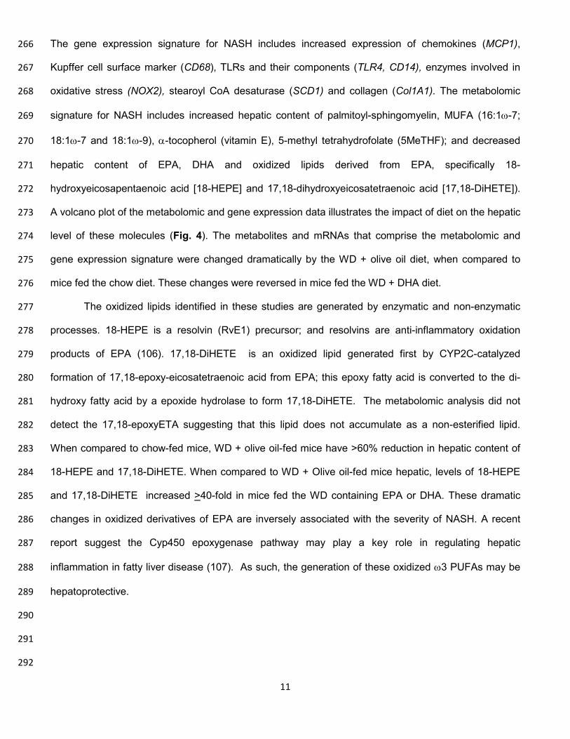
atherosclerosis, we and others observed that when Ldlr-/- mice are fed high fat-high cholesterol diet, like 166
the western diet, mice develop a NASH phenotype similar to that seen in humans (32, 36, 54, 71-74). 167
Since humans and Ldlr-/- mice develop NAFLD and NASH in a context of obesity and insulin resistance, 168
these mice appear to be a useful preclinical model to investigate the development, progression and 169
remission of NASH. 170
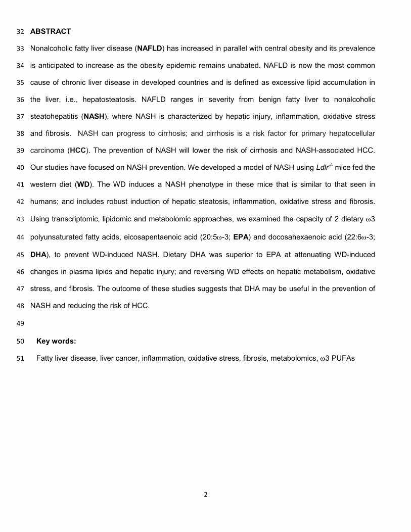
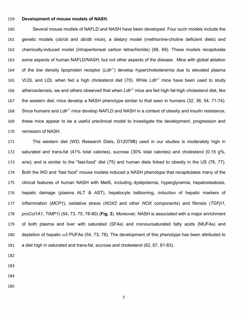
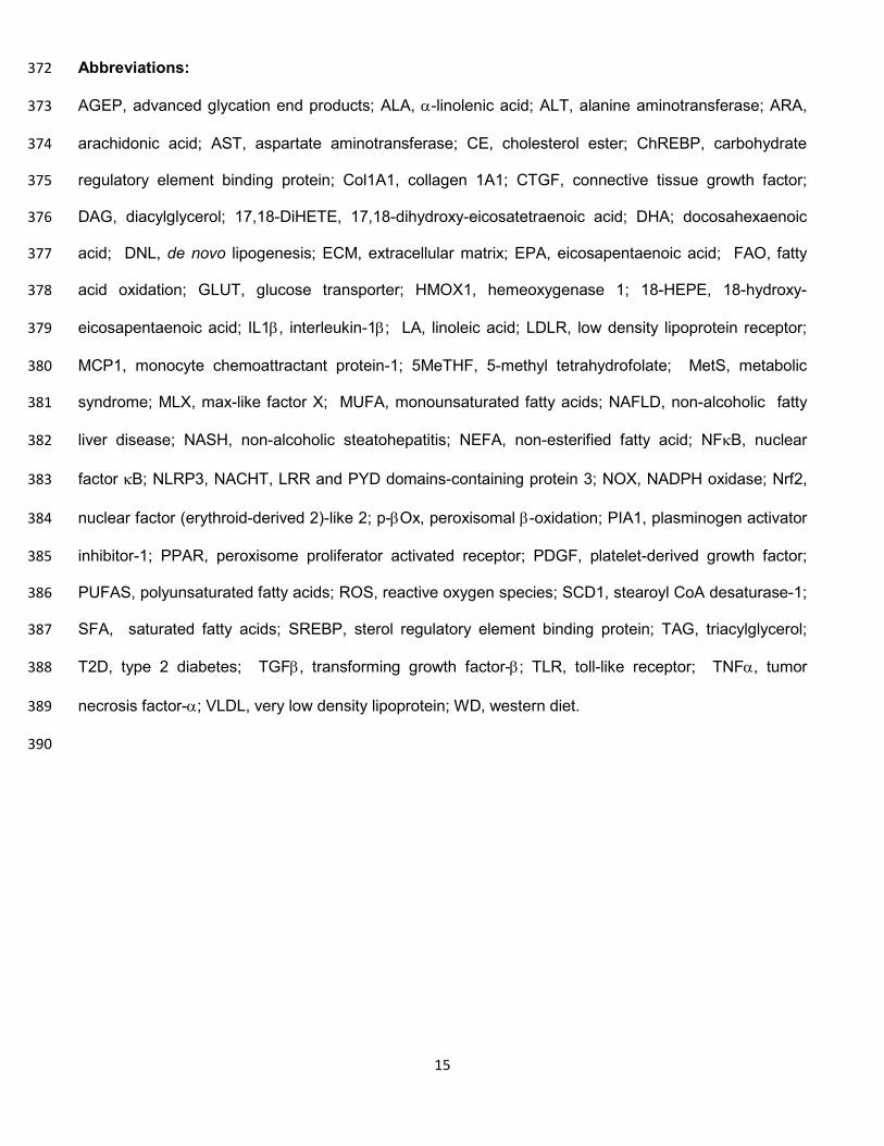
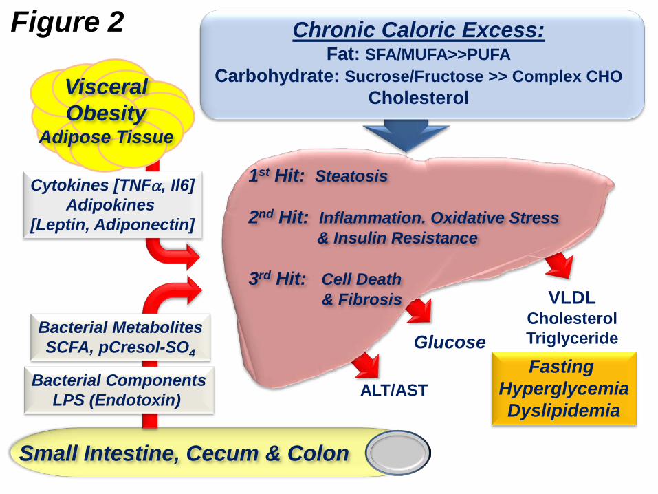
The western diet (WD; Research Diets, D12079B) used in our studies is moderately high in 171
saturated and trans-fat (41% total calories), sucrose (30% total calories) and cholesterol (0.15 g%, 172
w/w); and is similar to the “fast-food” diet (75) and human diets linked to obesity in the US (76, 77). 173
Both the WD and “fast food” mouse models induced a NASH phenotype that recapitulates many of the 174
clinical features of human NASH with MetS, including dyslipidemia, hyperglycemia, hepatosteatosis, 175
hepatic damage (plasma ALT & AST), hepatocyte ballooning, induction of hepatic markers of 176
inflammation (MCP1), oxidative stress (NOX2 and other NOX components) and fibrosis (TGFβ1, 177
proCol1A1, TIMP1) (54, 73, 75, 78-80) (Fig. 3). Moreover, NASH is associated with a major enrichment 178
of both plasma and liver with saturated (SFAs) and monounsaturated fatty acids (MUFAs) and 179
depletion of hepatic ω3 PUFAs (54, 73, 78). The development of this phenotype has been attributed to 180
a diet high in saturated and trans-fat, sucrose and cholesterol (62, 67, 81-83). 181
182
183
184
185
8
Potential for dietary C20-22 ω3 PUFAs to prevent NASH. 186
C20-22 ω3 PUFAs are pleiotropic regulators of cell function; they have well established effects on 187
membrane structure, cell signaling, gene expression, lipid and carbohydrate metabolism and 188
inflammation (84). As such, these fatty acids appear to be an ideal bioactive nutrient to combat NASH. 189
A meta-analysis of 9 clinical studies indicated that dietary supplementation with C20-22 ω-3 PUFAs 190
decreased liver fat (85) and clinical trials suggest C20-22 ω-3 PUFAs may lower liver fat in children and 191
adults with NAFLD (86-91). Of 235 clinical trials (119) assessing NASH and NASH therapies, 23 trials 192
used C20-22 ω3 PUFAs as a treatment strategy. In most trials, diets were supplemented with fish oil or a 193
combination of EPA + DHA; few studies used EPA or DHA alone. 194
195
Preclinical assessment of the efficacy of ω3 PUFA supplementation to prevent NASH in Ldlr-/- mice. 196
Diets supplemented with fish oil, EPA or DHA prevent high fat diet-induced NASH to varying 197
degrees (54, 73, 78, 84). The level of EPA and DHA in these high fat diets was at ~2% of total calories. 198
This dose of C20-22 ω-3 PUFAs is comparable to the dose consumed by patients taking LovazaTM 199
(GlaxoSmithKline) for the treatment of dyslipidemia (92). Humans consuming EPA + DHA ethyl esters 200
(4 g/d for 12 wks) exhibited increased plasma EPA + DHA from 5.5 mol% before treatment to 16.2 201
mol% after treatment (93). Supplementing human diets with a DHA-enriched fish oil (6 g/day for 8 wks) 202
increased plasma DHA from 4 mol% before treatment to 8 mol% after treatment (94, 95). Plasma 203
levels of DHA and total C20-22 ω-3 PUFA [EPA, docosapentaenoic acid (DPA, 22:5ω-3) and DHA] in Ldlr-204
/- mice fed a western diet for 16 wks was 4.3 and 6.7 mol%, respectively. Feeding Ldlr-/- mice a western 205
diet containing DHA (at 2% total calories) for 16 wks increased plasma DHA and total C20-22 ω-3 PUFA 206
to 9 and 15.2 mol%, respectively. Our protocol for C20-22 ω-3 PUFA supplementation of diets yields a 207
change in blood C20-22 ω3 PUFAs that is comparable to that seen in humans consuming 4-6 g/d of C20-22 208
ω-3 PUFA. 209
210
211
9
Dietary ω3 PUFAs do not prevent WD-induced systemic inflammation. 212
Systemic inflammation is a major driver of NASH. Inflammatory signals affecting NASH 213
progression include: gut-derived microbial products, e.g., endotoxin/LPS, oxidized LDL (ox-LDL) (34, 214
55, 80, 96); adipokines (leptin & adiponectin) & cytokines (TNFα) (97) and products from hepatocellular 215
death (27, 98) (Fig. 2). Supplementation of the WD with either EPA or DHA fails to attenuate WD-216
induced endotoxinemia (78). The appearance of endotoxin in the plasma of WD-fed Ldlr-/- mice (99) 217
may represent a problem with gut physiology such as microbial overgrowth, increased gut permeability 218
(leaky gut), or co-transport of microbial lipids with chylomicron (34, 100, 101). A link between the gut 219
microbiome and NAFLD has been established (34, 102, 103). 220
221
ω3 PUFAs attenuate hepatic inflammation. 222
Despite the absence of an effect of C20-22 ω-3 PUFAs on systemic inflammation markers, like 223
endotoxin, gene expression analyses showed that DHA was more effective than EPA at attenuating 224
WD-induced expression of hepatic toll-like receptor (TLR) subtypes (TLR2, TLR4, TLR9), CD14 (binds 225
endotoxin), downstream targets of TLRs; like NFκB (p50 subunit) nuclear abundance and downstream 226
targets of NFκB like chemokines (MCP1), cytokines (IL1β), inflammasome components (NLRP3) and 227
oxidative stress (NOX2, and its subunits) markers (73, 78). These studies suggest that EPA and DHA 228
attenuate the hepatic (cellular) response to plasma inflammatory factors by down-regulating key cellular 229
mediators of inflammation, like TLRs, CD14 (binds LPS, effect on CD14 mRNA and protein), NFκB-p50 230
nuclear abundance. 231
232
ω3 PUFAs have selective effects on hepatic oxidative stress. 233
Hepatic oxidative stress increases with NASH and is reflected by a significant increase in gene 234
expression and metabolite markers of oxidative stress that appear in liver and urine (54, 73). A 235
response to increased oxidative stress is the induction of nuclear factor (erythroid-derived 2)-like 2 236
(Nrf2), a key transcription factor involved in the antioxidant response (78). Nrf2 regulates the 237
expression of multiple transcripts linked to the anti-oxidant stress response, such as Hmox1, Gst1α and 238
10
several NOX subunits. Adding EPA or DHA to the WD did not prevent the WD-mediated increase in 239
hepatic nuclear content of Nrf2 or expression of Hmox1 or Gst1α. The EPA- and DHA-containing diets, 240
however, significantly lowered WD-mediated induction of multiple NOX subunits [Nox2, P22phox, 241
P40phox and P67phox] (73). NOX subtypes are a major source of superoxide and hydrogen peroxide. 242
As such, the NOX pathway is a major target of WD and C20-22 ω3 PUFAs. 243
244
245
ω3 PUFAs attenuate hepatic fibrosis. 246
Hepatic fibrosis (scarring) develops as a result of cell death and activation of hepatic stellate 247
cells and myofibrillar cells to produce extracellular matrix (ECM) proteins. Key regulators of fibrosis 248
include transforming growth factor (TGFβ), connective tissue growth factor (CTGF), platelet-derived 249
growth factor (PDGF), NOX, inflammatory mediators (endotoxin, TLR agonist), and leptin (38, 80, 104). 250
A fibrotic liver can progress to a cirrhotic liver (Fig. 1); and 90% of HCCs arise from cirrhotic livers 251
(105). 252
Addition of DHA to the WD attenuated the WD-mediated fibrosis as quantified by suppression of 253
expression of Col1A1, tissue inhibitor of metalloprotease-1 (TIMP1), TGFβ1, plasminogen activated 254
inhibitor-1 (PIA1) and staining of liver for fibrosis using trichrome, a collagen stain (54, 73). 255
Interestingly, EPA did not prevent WD-induced fibrosis. Based on these studies, DHA is the preferred 256
ω-3 PUFA to prevent NASH-associated fibrosis. 257
258
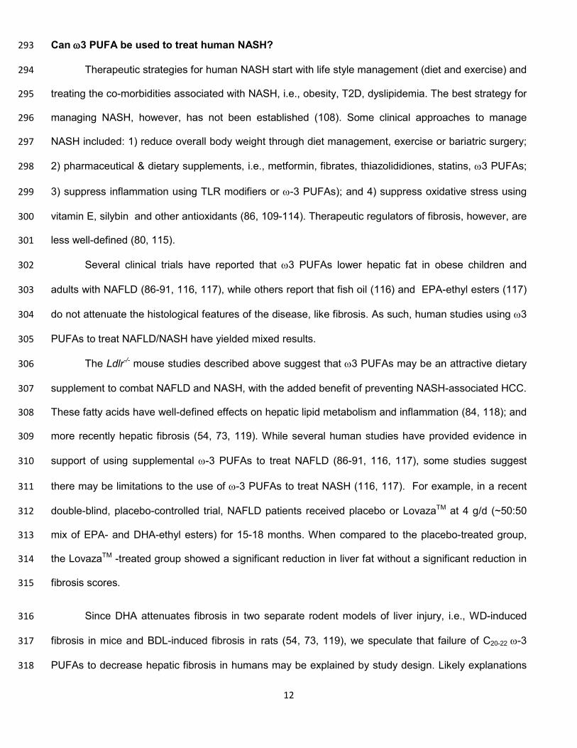
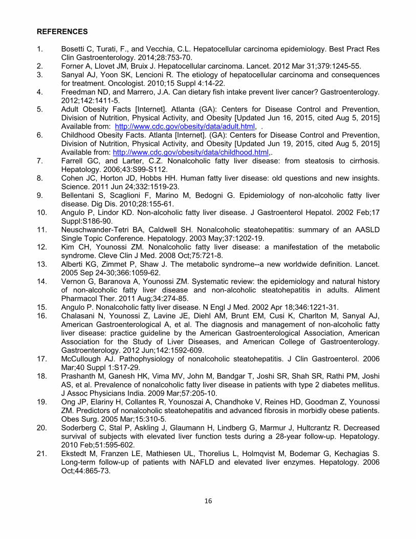
The WD and C20-22 ω3 PUFAs affect all major hepatic metabolic pathways. 259
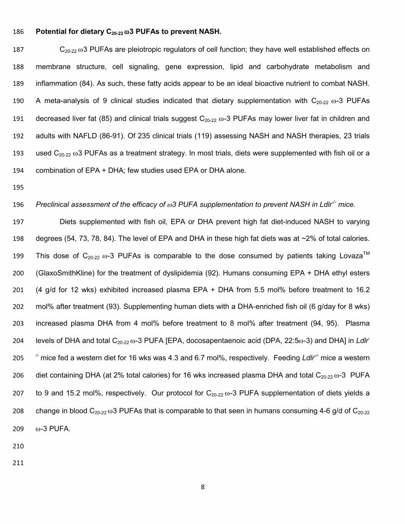
Additional insight into the impact of the WD and C20-22 ω-3 PUFAs on liver metabolism was 260
gained by using a global non-targeted metabolomic approach. The analysis identified 320 known 261
biochemicals (78). When compared to chow-fed mice, both the WD + olive oil- and WD + DHA-262
containing diets significantly affected the abundance of metabolites in all major hepatic metabolic 263
pathways including amino acids & peptides, carbohydrate and energy, lipid, nucleotide and vitamins & 264
cofactors. Our studies have identified gene expression and metabolite signatures for NASH (73, 78). 265
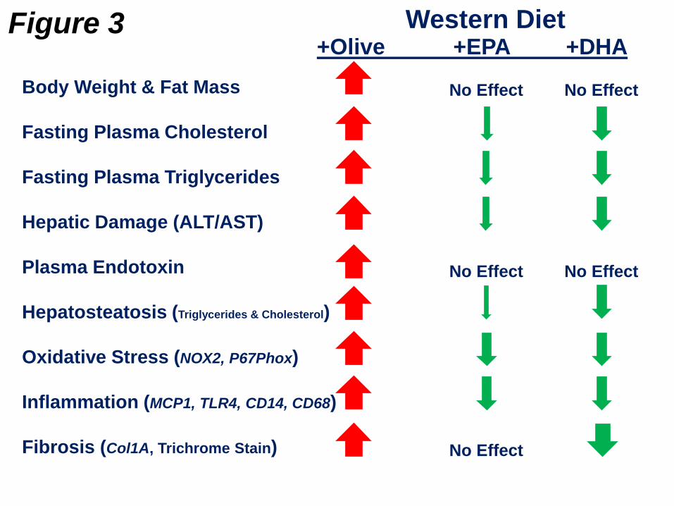
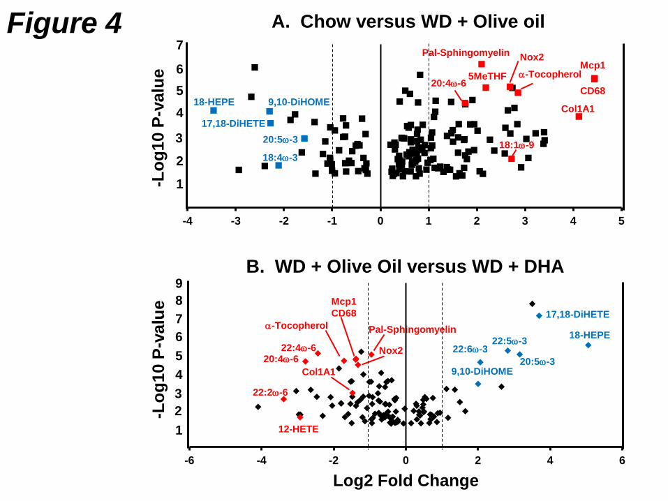
11
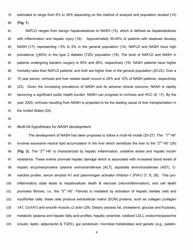
The gene expression signature for NASH includes increased expression of chemokines (MCP1), 266
Kupffer cell surface marker (CD68), TLRs and their components (TLR4, CD14), enzymes involved in 267
oxidative stress (NOX2), stearoyl CoA desaturase (SCD1) and collagen (Col1A1). The metabolomic 268
signature for NASH includes increased hepatic content of palmitoyl-sphingomyelin, MUFA (16:1ω-7; 269
18:1ω-7 and 18:1ω-9), α-tocopherol (vitamin E), 5-methyl tetrahydrofolate (5MeTHF); and decreased 270
hepatic content of EPA, DHA and oxidized lipids derived from EPA, specifically 18-271
hydroxyeicosapentaenoic acid [18-HEPE] and 17,18-dihydroxyeicosatetraenoic acid [17,18-DiHETE]). 272
A volcano plot of the metabolomic and gene expression data illustrates the impact of diet on the hepatic 273
level of these molecules (Fig. 4). The metabolites and mRNAs that comprise the metabolomic and 274
gene expression signature were changed dramatically by the WD + olive oil diet, when compared to 275
mice fed the chow diet. These changes were reversed in mice fed the WD + DHA diet. 276
The oxidized lipids identified in these studies are generated by enzymatic and non-enzymatic 277
processes. 18-HEPE is a resolvin (RvE1) precursor; and resolvins are anti-inflammatory oxidation 278
products of EPA (106). 17,18-DiHETE is an oxidized lipid generated first by CYP2C-catalyzed 279
formation of 17,18-epoxy-eicosatetraenoic acid from EPA; this epoxy fatty acid is converted to the di-280
hydroxy fatty acid by a epoxide hydrolase to form 17,18-DiHETE. The metabolomic analysis did not 281
detect the 17,18-epoxyETA suggesting that this lipid does not accumulate as a non-esterified lipid. 282
When compared to chow-fed mice, WD + olive oil-fed mice have >60% reduction in hepatic content of 283
18-HEPE and 17,18-DiHETE. When compared to WD + Olive oil-fed mice hepatic, levels of 18-HEPE 284
and 17,18-DiHETE increased >40-fold in mice fed the WD containing EPA or DHA. These dramatic 285
changes in oxidized derivatives of EPA are inversely associated with the severity of NASH. A recent 286
report suggest the Cyp450 epoxygenase pathway may play a key role in regulating hepatic 287
inflammation in fatty liver disease (107). As such, the generation of these oxidized ω3 PUFAs may be 288
hepatoprotective. 289
290
291
292
12
Can ω3 PUFA be used to treat human NASH? 293
Therapeutic strategies for human NASH start with life style management (diet and exercise) and 294
treating the co-morbidities associated with NASH, i.e., obesity, T2D, dyslipidemia. The best strategy for 295
managing NASH, however, has not been established (108). Some clinical approaches to manage 296
NASH included: 1) reduce overall body weight through diet management, exercise or bariatric surgery; 297
2) pharmaceutical & dietary supplements, i.e., metformin, fibrates, thiazolididiones, statins, ω3 PUFAs; 298
3) suppress inflammation using TLR modifiers or ω-3 PUFAs); and 4) suppress oxidative stress using 299
vitamin E, silybin and other antioxidants (86, 109-114). Therapeutic regulators of fibrosis, however, are 300
less well-defined (80, 115). 301
Several clinical trials have reported that ω3 PUFAs lower hepatic fat in obese children and 302
adults with NAFLD (86-91, 116, 117), while others report that fish oil (116) and EPA-ethyl esters (117) 303
do not attenuate the histological features of the disease, like fibrosis. As such, human studies using ω3 304
PUFAs to treat NAFLD/NASH have yielded mixed results. 305
The Ldlr-/- mouse studies described above suggest that ω3 PUFAs may be an attractive dietary 306
supplement to combat NAFLD and NASH, with the added benefit of preventing NASH-associated HCC. 307
These fatty acids have well-defined effects on hepatic lipid metabolism and inflammation (84, 118); and 308
more recently hepatic fibrosis (54, 73, 119). While several human studies have provided evidence in 309
support of using supplemental ω-3 PUFAs to treat NAFLD (86-91, 116, 117), some studies suggest 310
there may be limitations to the use of ω-3 PUFAs to treat NASH (116, 117). For example, in a recent 311
double-blind, placebo-controlled trial, NAFLD patients received placebo or LovazaTM at 4 g/d (~50:50 312
mix of EPA- and DHA-ethyl esters) for 15-18 months. When compared to the placebo-treated group, 313
the LovazaTM -treated group showed a significant reduction in liver fat without a significant reduction in 314
fibrosis scores. 315
Since DHA attenuates fibrosis in two separate rodent models of liver injury, i.e., WD-induced 316
fibrosis in mice and BDL-induced fibrosis in rats (54, 73, 119), we speculate that failure of C20-22 ω-3 317
PUFAs to decrease hepatic fibrosis in humans may be explained by study design. Likely explanations 318
13
include the type and amount of ω-3 PUFAs used in the trial. Our studies established that DHA is more 319
effective than EPA at attenuating the onset and progression of NASH (73). Human studies, however, 320
have examined the impact of ω-3 PUFAs on patients with pre-existing disease (86-91, 116, 117). We 321
are unaware of preclinical rodent studies that have assessed the impact of ω3 PUFAs to promote 322
remission or regression of NASH or hepatic fibrosis. As such, more preclinical studies are required to 323
establish the capacity of ω-3 PUFAs to attenuate NASH at various stages in the disease process. 324
325
326
Conclusions and key unanswered questions. 327
To date, several human studies have indicated that ω-3 PUFAs may be useful in reducing liver 328
fat in obese patients with NAFLD. Moreover, preclinical studies in mice have established that DHA can 329
prevent NASH and NASH-associated fibrosis. It remains unclear whether dietary ω3 PUFAs have the 330
capacity to reverse the NASH, cirrhosis or HCC phenotypes once these diseases are established. 331
Equally important is defining the molecular mechanisms for DHA control of hepatic fibrosis. Finally, 332
changes in hepatic EPA and DHA content significantly impact oxidized lipids derived from ω-3 and ω-6 333
PUFAs. These oxidized lipids likely play a role in inflammation and will affect the onset and progression 334
of NASH. Whether these oxidized lipids impact the development of NASH, cirrhosis or HCC remains to 335
be determined. 336
337
338
Acknowledgements: 339
This work was supported by the National Institute of Food and Agriculture grant (2009-65200-05846) 340
and the National Institutes of Health grants (DK 43220 & DK094600). All authors have read and 341
approved the manuscript. All authors have read and approved the final version. 342
343
344
14
Figure Legends: 345
Figure 1: Transition from normal liver to primary hepatocellular carcinoma (HCC). 346
347
Figure 2: Factors contributing to the onset and progression of NASH. 348
349
Figure 3: Effects of the western diet and C20-22 ω-3 PUFAs on the prevention of NASH Ldlr-/- mice. 350
The size of the arrow indicated effect size. “No effect” indicates no changes from western diet + olive 351
oil-fed mice. Olive oil was added to the WD to keep all diets isocaloric. 352
353
Figure 4: Volcano plots of western diet effects on hepatic metabolites. A metabolomic and 354
transcriptomic analysis was carried out as described (78). Over 300 hepatic metabolites and 6 mRNAs 355
markers of NASH were examined using MetaboAnalyst 3.0 356
[http://www.metaboanalyst.ca/MetaboAnalyst/] (120). The outcome of this analysis provided a volcano 357
plot. Results are plotted as log2 Fold Change versus –log10 p-value. Several metabolites and RNA 358
transcripts are labeled to illustrate the impact of diet on hepatic abundance of these molecules. Panel A 359
is the comparison of hepatic molecules from Chow-fed versus WD + olive oil-fed Ldlr-/- mice. Panel B is 360
the comparison of hepatic molecules from WD + Olive oil-fed mice versus WD + DHA-fed Ldlr-/-. 361
362
363
364
365
366
367
368
369
370
371
15
Abbreviations: 372
AGEP, advanced glycation end products; ALA, α-linolenic acid; ALT, alanine aminotransferase; ARA, 373
arachidonic acid; AST, aspartate aminotransferase; CE, cholesterol ester; ChREBP, carbohydrate 374
regulatory element binding protein; Col1A1, collagen 1A1; CTGF, connective tissue growth factor; 375
DAG, diacylglycerol; 17,18-DiHETE, 17,18-dihydroxy-eicosatetraenoic acid; DHA; docosahexaenoic 376
acid; DNL, de novo lipogenesis; ECM, extracellular matrix; EPA, eicosapentaenoic acid; FAO, fatty 377
acid oxidation; GLUT, glucose transporter; HMOX1, hemeoxygenase 1; 18-HEPE, 18-hydroxy-378
eicosapentaenoic acid; IL1β, interleukin-1β; LA, linoleic acid; LDLR, low density lipoprotein receptor; 379
MCP1, monocyte chemoattractant protein-1; 5MeTHF, 5-methyl tetrahydrofolate; MetS, metabolic 380
syndrome; MLX, max-like factor X; MUFA, monounsaturated fatty acids; NAFLD, non-alcoholic fatty 381
liver disease; NASH, non-alcoholic steatohepatitis; NEFA, non-esterified fatty acid; NFκB, nuclear 382
factor κB; NLRP3, NACHT, LRR and PYD domains-containing protein 3; NOX, NADPH oxidase; Nrf2, 383
nuclear factor (erythroid-derived 2)-like 2; p-βOx, peroxisomal β-oxidation; PIA1, plasminogen activator 384
inhibitor-1; PPAR, peroxisome proliferator activated receptor; PDGF, platelet-derived growth factor; 385
PUFAS, polyunsaturated fatty acids; ROS, reactive oxygen species; SCD1, stearoyl CoA desaturase-1; 386
SFA, saturated fatty acids; SREBP, sterol regulatory element binding protein; TAG, triacylglycerol; 387
T2D, type 2 diabetes; TGFβ, transforming growth factor-β; TLR, toll-like receptor; TNFα, tumor 388
necrosis factor-α; VLDL, very low density lipoprotein; WD, western diet. 389
390
16
REFERENCES
1. Bosetti C, Turati, F., and Vecchia, C.L. Hepatocellular carcinoma epidemiology. Best Pract Res Clin Gastroenterology. 2014;28:753-70.
2. Forner A, Llovet JM, Bruix J. Hepatocellular carcinoma. Lancet. 2012 Mar 31;379:1245-55. 3. Sanyal AJ, Yoon SK, Lencioni R. The etiology of hepatocellular carcinoma and consequences
for treatment. Oncologist. 2010;15 Suppl 4:14-22. 4. Freedman ND, and Marrero, J.A. Can dietary fish intake prevent liver cancer? Gastroenterology.
2012;142:1411-5. 5. Adult Obesity Facts [Internet]. Atlanta (GA): Centers for Disease Control and Prevention,
Division of Nutrition, Physical Activity, and Obesity [Updated Jun 16, 2015, cited Aug 5, 2015] Available from: http://www.cdc.gov/obesity/data/adult.html, .
6. Childhood Obesity Facts. Atlanta [Internet]. (GA): Centers for Disease Control and Prevention, Division of Nutrition, Physical Activity, and Obesity [Updated Jun 19, 2015, cited Aug 5, 2015] Available from: http://www.cdc.gov/obesity/data/childhood.html,.
7. Farrell GC, and Larter, C.Z. Nonalcoholic fatty liver disease: from steatosis to cirrhosis. Hepatology. 2006;43:S99-S112.
8. Cohen JC, Horton JD, Hobbs HH. Human fatty liver disease: old questions and new insights. Science. 2011 Jun 24;332:1519-23.
9. Bellentani S, Scaglioni F, Marino M, Bedogni G. Epidemiology of non-alcoholic fatty liver disease. Dig Dis. 2010;28:155-61.
10. Angulo P, Lindor KD. Non-alcoholic fatty liver disease. J Gastroenterol Hepatol. 2002 Feb;17 Suppl:S186-90.
11. Neuschwander-Tetri BA, Caldwell SH. Nonalcoholic steatohepatitis: summary of an AASLD Single Topic Conference. Hepatology. 2003 May;37:1202-19.
12. Kim CH, Younossi ZM. Nonalcoholic fatty liver disease: a manifestation of the metabolic syndrome. Cleve Clin J Med. 2008 Oct;75:721-8.
13. Alberti KG, Zimmet P, Shaw J. The metabolic syndrome--a new worldwide definition. Lancet. 2005 Sep 24-30;366:1059-62.
14. Vernon G, Baranova A, Younossi ZM. Systematic review: the epidemiology and natural history of non-alcoholic fatty liver disease and non-alcoholic steatohepatitis in adults. Aliment Pharmacol Ther. 2011 Aug;34:274-85.
15. Angulo P. Nonalcoholic fatty liver disease. N Engl J Med. 2002 Apr 18;346:1221-31. 16. Chalasani N, Younossi Z, Lavine JE, Diehl AM, Brunt EM, Cusi K, Charlton M, Sanyal AJ,
American Gastroenterological A, et al. The diagnosis and management of non-alcoholic fatty liver disease: practice guideline by the American Gastroenterological Association, American Association for the Study of Liver Diseases, and American College of Gastroenterology. Gastroenterology. 2012 Jun;142:1592-609.
17. McCullough AJ. Pathophysiology of nonalcoholic steatohepatitis. J Clin Gastroenterol. 2006 Mar;40 Suppl 1:S17-29.
18. Prashanth M, Ganesh HK, Vima MV, John M, Bandgar T, Joshi SR, Shah SR, Rathi PM, Joshi AS, et al. Prevalence of nonalcoholic fatty liver disease in patients with type 2 diabetes mellitus. J Assoc Physicians India. 2009 Mar;57:205-10.
19. Ong JP, Elariny H, Collantes R, Younoszai A, Chandhoke V, Reines HD, Goodman Z, Younossi ZM. Predictors of nonalcoholic steatohepatitis and advanced fibrosis in morbidly obese patients. Obes Surg. 2005 Mar;15:310-5.
20. Soderberg C, Stal P, Askling J, Glaumann H, Lindberg G, Marmur J, Hultcrantz R. Decreased survival of subjects with elevated liver function tests during a 28-year follow-up. Hepatology. 2010 Feb;51:595-602.
21. Ekstedt M, Franzen LE, Mathiesen UL, Thorelius L, Holmqvist M, Bodemar G, Kechagias S. Long-term follow-up of patients with NAFLD and elevated liver enzymes. Hepatology. 2006 Oct;44:865-73.
17
22. Adams LA, Lymp JF, St Sauver J, Sanderson SO, Lindor KD, Feldstein A, Angulo P. The natural history of nonalcoholic fatty liver disease: a population-based cohort study. Gastroenterology. 2005 Jul;129:113-21.
23. McCullough AJ. The clinical features, diagnosis and natural history of nonalcoholic fatty liver disease. Clin Liver Dis. 2004 Aug;8:521-33, viii.
24. McCollough AJ. Epidemiology of the metabolic syndrome in the USA. J Dig Dis. 2011;12:333-40.
25. Day CP, James OF. Steatohepatitis: a tale of two "hits"? Gastroenterology. 1998 Apr;114:842-5. 26. LaBrecque D, Abbas, Z., Anania, F., Ferenci, P., Gahafoor Kahn, A., Goh, K-L., Hamid, S.S.,
Isakov, V., Lizarzabal, M., Mojica Pernaranda, M., Rivera Ramos, J.F., Sarin, S., Stimak, D., Thomson, A.B.R., Umar, Muhammed., Krabshuis, J., LeMair, A. Nonalcoholic fatty liver disease and nonalcoholic steatohepatitis. World Gastroentrology Organization Global Guidelines. 2012;June:1-29.
27. Tilg H, and Moschen, A.R. Evolution of inflammation in nonalcoholic fatty liver disease: the multiple parallel hits hypothesis. Hepatology. 2010;52:1836-46.
28. Hashimoto E, Tokushige K, Farrell GC. Histological features of non-alcoholic fatty liver disease: What is important? J Gastroenterol Hepatol. 2011 Jan;27:5-7.
29. Friedman SL. Mechanisms of hepatic fibrogenesis. Gastroenterology. 2008 May;134:1655-69. 30. Abdelmalek MF, Suzuki A, Guy C, Unalp-Arida A, Colvin R, Johnson RJ, Diehl AM. Increased
fructose consumption is associated with fibrosis severity in patients with nonalcoholic fatty liver disease. Hepatology. 2010 Jun;51:1961-71.
31. Guturu P, Duchini A. Etiopathogenesis of nonalcoholic steatohepatitis: role of obesity, insulin resistance and mechanisms of hepatotoxicity. Int J Hepatol. 2012;2012:212865.
32. Wouters K, van Gorp PJ, Bieghs V, Gijbels MJ, Duimel H, Lutjohann D, Kerksiek A, van Kruchten R, Maeda N, et al. Dietary cholesterol, rather than liver steatosis, leads to hepatic inflammation in hyperlipidemic mouse models of nonalcoholic steatohepatitis. Hepatology. 2008 Aug;48:474-86.
33. Pagadala M, Kasumov T, McCullough AJ, Zein NN, Kirwan JP. Role of ceramides in nonalcoholic fatty liver disease. Trends Endocrinol Metab. 2012 Aug;23:365-71.
34. Harte AL, da Silva NF, Creely SJ, McGee KC, Billyard T, Youssef-Elabd EM, Tripathi G, Ashour E, Abdalla MS, et al. Elevated endotoxin levels in non-alcoholic fatty liver disease. Journal of inflammation. 2010;7:15.
35. Hooper AJ, Adams LA, Burnett JR. Genetic determinants of hepatic steatosis in man. J Lipid Res. 2011 Apr;52:593-617.
36. Bieghs V, Van Gorp PJ, Wouters K, Hendrikx T, Gijbels MJ, van Bilsen M, Bakker J, Binder CJ, Lutjohann D, et al. LDL receptor knock-out mice are a physiological model particularly vulnerable to study the onset of inflammation in non-alcoholic fatty liver disease. PLoS One. 2012;7:e30668.
37. Joyce SA, MacSharry, J., Casey, P.G., Kinsella, M., Murphy, E.F., Shanahan, F., Hill, C., and Gahan, C.G.M. Regulation of host weight gain and lipid metabolism by bacterial bile acid modification in the gut. Proc Natl Acad Sci, USA. 2014;111:7421-6.
38. Elinav E, Ali, M., Bruck, R., Brazowski, E., Phillips, A., Shapira, Y., Katz, M., Solomon, G., Halpern, Z., and Gertler, A. Competitive in hibition of leptin signaling results in amelioration of liver fibrosis through modulation of stellate cell function. Hepatology. 2009;49:278-86.
39. Matherly SC, Puri P. Mechanisms of simple hepatic steatosis: not so simple after all. Clin Liver Dis. 2012 Aug;16:505-24.
40. Donnelly KL, Smith CI, Schwarzenberg SJ, Jessurun J, Boldt MD, Parks EJ. Sources of fatty acids stored in liver and secreted via lipoproteins in patients with nonalcoholic fatty liver disease. J Clin Invest. 2005 May;115:1343-51.
41. Toshimitsu K, Matsuura B, Ohkubo I, Niiya T, Furukawa S, Hiasa Y, Kawamura M, Ebihara K, Onji M. Dietary habits and nutrient intake in non-alcoholic steatohepatitis. Nutrition. 2007 Jan;23:46-52.
18
42. Musso G, Gambino R, De Michieli F, Cassader M, Rizzetto M, Durazzo M, Faga E, Silli B, Pagano G. Dietary habits and their relations to insulin resistance and postprandial lipemia in nonalcoholic steatohepatitis. Hepatology. 2003 Apr;37:909-16.
43. Capanni M, Calella F, Biagini MR, Genise S, Raimondi L, Bedogni G, Svegliati-Baroni G, Sofi F, Milani S, et al. Prolonged n-3 polyunsaturated fatty acid supplementation ameliorates hepatic steatosis in patients with non-alcoholic fatty liver disease: a pilot study. Aliment Pharmacol Ther. 2006 Apr 15;23:1143-51.
44. Cortez-Pinto H, Jesus L, Barros H, Lopes C, Moura MC, Camilo ME. How different is the dietary pattern in non-alcoholic steatohepatitis patients? Clin Nutr. 2006 Oct;25:816-23.
45. Levy JR, Clore JN, Stevens W. Dietary n-3 polyunsaturated fatty acids decrease hepatic triglycerides in Fischer 344 rats. Hepatology. 2004 Mar;39:608-16.
46. Pachikian BD, Essaghir A, Demoulin JB, Neyrinck AM, Catry E, De Backer FC, Dejeans N, Dewulf EM, Sohet FM, et al. Hepatic n-3 polyunsaturated fatty acid depletion promotes steatosis and insulin resistance in mice: genomic analysis of cellular targets. PLoS One.6:e23365.
47. Jump DB, Tripathy, S. and Depner, C.M. Fatty acid-regulated transcription factors in the liver. Annu Rev Nutr. 2013;33:249-69.
48. Zelber-Sagi S, Ratziu V, Oren R. Nutrition and physical activity in NAFLD: an overview of the epidemiological evidence. World J Gastroenterol. 2011 Aug 7;17:3377-89.
49. Tetri LH, Basaranoglu M, Brunt EM, Yerian LM, Neuschwander-Tetri BA. Severe NAFLD with hepatic necroinflammatory changes in mice fed trans fats and a high-fructose corn syrup equivalent. Am J Physiol Gastrointest Liver Physiol. 2008 Nov;295:G987-95.
50. Lottenberg AM, Afonso Mda S, Lavrador MS, Machado RM, Nakandakare ER. The role of dietary fatty acids in the pathology of metabolic syndrome. J Nutr Biochem. 2012 Sep;23:1027-40.
51. Yasutake K, Nakamuta M, Shima Y, Ohyama A, Masuda K, Haruta N, Fujino T, Aoyagi Y, Fukuizumi K, et al. Nutritional investigation of non-obese patients with non-alcoholic fatty liver disease: the significance of dietary cholesterol. Scand J Gastroenterol. 2009;44:471-7.
52. Wouters K, van Bilsen M, van Gorp PJ, Bieghs V, Lutjohann D, Kerksiek A, Staels B, Hofker MH, Shiri-Sverdlov R. Intrahepatic cholesterol influences progression, inhibition and reversal of non-alcoholic steatohepatitis in hyperlipidemic mice. FEBS Lett. 2010 Mar 5;584:1001-5.
53. Teratani T, Tomita K, Suzuki T, Oshikawa T, Yokoyama H, Shimamura K, Tominaga S, Hiroi S, Irie R, et al. A high-cholesterol diet exacerbates liver fibrosis in mice via accumulation of free cholesterol in hepatic stellate cells. Gastroenterology. 2012 Jan;142:152-64 e10.
54. Depner CM, Torres-Gonzalez M, Tripathy S, Milne G, Jump DB. Menhaden oil decreases high-fat diet-induced markers of hepatic damage, steatosis, inflammation, and fibrosis in obese Ldlr-/- mice. J Nutr. 2012 Aug;142:1495-503.
55. Walenbergh SMA, Koek, G.H., Bieghs, V., and Shiri-Sverdlov, R. Non-alcoholic steatohepatitis: the role of oxidized low-density lipoproteins. J Hepatology. 2013;58:801-20.
56. Marriott BP, Olsho L, Hadden L, Connor P. Intake of added sugars in the United States: what is the measure? Am J Clin Nutr. 2010 Dec;94:1652-3; author reply 3.
57. Chun OK, Chung CE, Wang Y, Padgitt A, Song WO. Changes in intakes of total and added sugar and their contribution to energy intake in the U.S. Nutrients. 2010 Aug;2:834-54.
58. Chanmugam P, Guthrie JF, Cecilio S, Morton JF, Basiotis PP, Anand R. Did fat intake in the United States really decline between 1989-1991 and 1994-1996? J Am Diet Assoc. 2003 Jul;103:867-72.
59. Lee S, Harnack L, Jacobs DR, Jr., Steffen LM, Luepker RV, Arnett DK. Trends in diet quality for coronary heart disease prevention between 1980-1982 and 2000-2002: The Minnesota Heart Survey. J Am Diet Assoc. 2007 Feb;107:213-22.
60. Marriott BP, Olsho L, Hadden L, Connor P. Intake of added sugars and selected nutrients in the United States, National Health and Nutrition Examination Survey (NHANES) 2003-2006. Crit Rev Food Sci Nutr. 2010 Mar;50:228-58.
61. Vos MB, Kimmons JE, Gillespie C, Welsh J, Blanck HM. Dietary fructose consumption among US children and adults: the Third National Health and Nutrition Examination Survey. Medscape J Med. 2008;10:160.
19
62. Lim JS, Mietus-Snyder M, Valente A, Schwarz JM, Lustig RH. The role of fructose in the pathogenesis of NAFLD and the metabolic syndrome. Nat Rev Gastroenterol Hepatol. 2010 May;7:251-64.
63. Bizeau ME, Pagliassotti MJ. Hepatic adaptations to sucrose and fructose. Metabolism. 2005 Sep;54:1189-201.
64. Schalkwijk CG, Stehouwer CD, van Hinsbergh VW. Fructose-mediated non-enzymatic glycation: sweet coupling or bad modification. Diabetes Metab Res Rev. 2004 Sep-Oct;20:369-82.
65. Bunn HF, Higgins PJ. Reaction of monosaccharides with proteins: possible evolutionary significance. Science. 1981 Jul 10;213:222-4.
66. Bose T, Chakraborti AS. Fructose-induced structural and functional modifications of hemoglobin: implication for oxidative stress in diabetes mellitus. Biochim Biophys Acta. 2008 May;1780:800-8.
67. Wei Y, Wang, D., Moran, G., Estrada, A., Pagliassotti, M.J. Fructose-induced stress signaling in the liver involves methylglyoxal. Nutr Meta (Lond). 2013;10:32-8.
68. Kucera O, and Cervinkova, Z. Experimental models of non-alcholic fatty liver disease in rats. World journal of gastroenterology : WJG. 2014;20:8364-76.
69. Takahashi Y, Soejima, Y., and Fukusato, T. Animal models of nonalcoholic fatty liver disease and nonalcoholic steatohepatitis. World journal of gastroenterology : WJG. 2012;18.
70. Ishibashi S, Brown MS, Goldstein JL, Gerard RD, Hammer RE, Herz J. Hypercholesterolemia in low density lipoprotein receptor knockout mice and its reversal by adenovirus-mediated gene delivery. J Clin Invest. 1993 Aug;92:883-93.
71. Saraswathi V, Gao L, Morrow JD, Chait A, Niswender KD, Hasty AH. Fish oil increases cholesterol storage in white adipose tissue with concomitant decreases in inflammation, hepatic steatosis, and atherosclerosis in mice. J Nutr. 2007 Jul;137:1776-82.
72. Saraswathi V, Morrow, J.D., and Hasty, A.H. Dietary fish oil exerts hypolipidemic effects in lean and insulin sensitizing effect in obese LDLR-/- mice. J Nutr. 2009;139:2380-6.
73. Depner CM, Philbrick KA, Jump DB. Docosahexaenoic acid attenuates hepatic inflammation, oxidative stress, and fibrosis without decreasing hepatosteatosis in a Ldlr(-/-) mouse model of western diet-induced nonalcoholic steatohepatitis. J Nutr. 2013 Mar;143:315-23.
74. Subramanian S, Goodspeed L, Wang S, Kim J, Zeng L, Ioannou GN, Haigh WG, Yeh MM, Kowdley KV, et al. Dietary cholesterol exacerbates hepatic steatosis and inflammation in obese LDL receptor-deficient mice. J Lipid Res. 2011 Sep;52:1626-35.
75. Charlton M, Krishnan, A., Viker, K., Sanderson, S., Cazanave., McConico, A., Masuoko, H., and Gores, G. Fast food diet mouse: novel small animal model of NASH with balloning, progressive fibrosis and high physiological facelity to the human condition. Am J Physiol Gastrointest Liver Physiol. 2011;301:G825-G34.
76. Cordain L, Eaton, S.B., Sebastian, A., Mann, N., Lindeberg, S., Watkins, B.A., O'Keefe, J.H., and Brand-Miller, J. Orgins and evolution of the western diet: health implications for the 21st century. Am J Clin Nutr. 2005;81:341-54.
77. Ishimoto T, Lanaspa MA, Rivard CJ, Roncal-Jimenez CA, Orlicky DJ, Cicerchi C, McMahan RH, Abdelmalek MF, Rosen HR, et al. High-fat and high-sucrose (western) diet induces steatohepatitis that is dependent on fructokinase. Hepatology. 2013 Nov;58:1632-43.
78. Depner CM, Traber, M.G., Bobe, G., Bohren, K.M., Morin-Kensicki, E., Milne, G., Jump, D.B. A metabolomic analysis of omega-3 fatty acid mediated attenuation of western diet-induced non-alcoholic steatohepatitis in LDLR-/- mice. Plos One. 2013;8 (12): e83756.
79. Depner CM, Lytle, K., Tripathy, S. and Jump, D.B. Omega-3 fatty acids and nonalcoholic fatty liver disease. In: Fatty Liver Diseases and Consequences, CRC Press, Francis & Taylor; ed O Tirosh. 2014;Chapt. 13:247-80.
80. Schuppan DaK, Y.O. Evolving therapies for liver fibrosis. J Clin Invest. 2013;123:1887-901. 81. Nomura K, Yamanouchi T. The role of fructose-enriched diets in mechanisms of nonalcoholic
fatty liver disease. J Nutr Biochem. 2012 Mar;23:203-8.
20
82. Lim JS, Mietus-Snyder, M., Valente, A. Schwarz, J.M. and Lustig, R.H. The role of fructose in the pathogenesis of NAFLD and the metabolic syndrome. Nat Rev Gastroenterol Hepatol. 2010;7:251-64.
83. Nomura KaY, T. The role of fructose-enriched diets in mechanisms of nonalcoholic fatty liver disease. J Nutr Biochem. 2012;23:203-8.
84. Jump DB, Tripathy S, Depner CM. Fatty Acid-regulated transcription factors in the liver. Annu Rev Nutr. 2013 Jul 17;33:249-69.
85. Parker HM, Johnson NA, Burdon CA, Cohn JS, O'Connor HT, George J. Omega-3 supplementation and non-alcoholic fatty liver disease: a systematic review and meta-analysis. J Hepatol. Apr;56:944-51.
86. Nobili V, Bedogni G, Alisi A, Pietrobattista A, Rise P, Galli C, Agostoni C. Docosahexaenoic acid supplementation decreases liver fat content in children with non-alcoholic fatty liver disease: double-blind randomised controlled clinical trial. Arch Dis Child. 2011 Apr;96:350-3.
87. Sofi F, Giangrandi I, Cesari F, Corsani I, Abbate R, Gensini GF, Casini A. Effects of a 1-year dietary intervention with n-3 polyunsaturated fatty acid-enriched olive oil on non-alcoholic fatty liver disease patients: a preliminary study. Int J Food Sci Nutr. 2011 Dec;61:792-802.
88. Bulchandani DG, Nachnani JS, Nookala A, Naumovitch C, Herndon B, Molteni A, Quinn T, Alba LM. Treatment with omega-3 fatty acids but not exendin-4 improves hepatic steatosis. Eur J Gastroenterol Hepatol. 2011 Oct;22:1245-52.
89. Ishikawa Y, Yokoyama M, Saito Y, Matsuzaki M, Origasa H, Oikawa S, Sasaki J, Hishida H, Itakura H, et al. Preventive effects of eicosapentaenoic acid on coronary artery disease in patients with peripheral artery disease. Circ J. 2011 Jul;74:1451-7.
90. Kishino T, Ohnishi H, Ohtsuka K, Matsushima S, Urata T, Watanebe K, Honda Y, Mine Y, Matsumoto M, et al. Low concentrations of serum n-3 polyunsaturated fatty acids in non-alcoholic fatty liver disease patients with liver injury. Clin Chem Lab Med. 2011 Jan;49:159-62.
91. Scorletti E, Bhatia L, McCormick KG, Clough GF, Nash K, Hodson L, Moyses HE, Calder PC, Byrne CD, on behalf of the WSI. Effects of purified eicosapentaenoic and docosahexaenoic acids in non-alcoholic fatty liver disease: Results from the *WELCOME study. Hepatology. 2014 Jul 4;60:1211-21.
92. Barter P, Ginsberg HN. Effectiveness of combined statin plus omega-3 fatty acid therapy for mixed dyslipidemia. Am J Cardiol. 2008 Oct 15;102:1040-5.
93. Di Stasi D, Bernasconi, R., Marchioli, R., Marfisi, R.M., Rossi, G., Tognoni, G., Tacconi, M.T. Early modification of fatty acid composition in plasma phospholipids, platelets and mononucleates of healthy volunteers after low doses of n-3 PUFA. Eur J Clin Pharmacol. 2004;60:183-90.
94. Superko HR, Superko, S.M., Nasir, K., Agatston, A., Garrett, B.C. Omega-3 fatty acid blood levels. Clinical significance and controversy. Circulaton. 2013;128:2154-61.
95. Lockyer S, Tzanetou, M., Carvalho-Wells, A.L., Jackson, J.G., Minihane, A.M., and Lovegrove, J.A. STAT gene dietary model to implement diets of differing fat composition in prospectively genotyped groups (apoE) using commercially available foods. Br J Nutr. 2012;108:1705-13.
96. Cani PD, Amar J, Iglesias MA, Poggi M, Knauf C, Bastelica D, Neyrinck AM, Fava F, Tuohy KM, et al. Metabolic endotoxemia initiates obesity and insulin resistance. Diabetes. 2007 Jul;56:1761-72.
97. Leclercq IA, Farrell GC, Schriemer R, Robertson GR. Leptin is essential for the hepatic fibrogenic response to chronic liver injury. J Hepatol. 2002 Aug;37:206-13.
98. Marra F, Gastaldelli, A., Baroni, G.S., Tell, G., and Tiribelli, C. . Molecular basis and mechanisms of progression of non-alcoholic steatohepatitis. Trends Mol Med. 2008;14:72-81.
99. Akira S, Takeda K. Toll-like receptor signalling. Nat Rev Immunol. 2004 Jul;4:499-511. 100. Erridge C, Attina T, Spickett CM, Webb DJ. A high-fat meal induces low-grade endotoxemia:
evidence of a novel mechanism of postprandial inflammation. The American journal of clinical nutrition. 2007 Nov;86:1286-92.
101. Laugerette F, Vors C, Geloen A, Chauvin MA, Soulage C, Lambert-Porcheron S, Peretti N, Alligier M, Burcelin R, et al. Emulsified lipids increase endotoxemia: possible role in early postprandial low-grade inflammation. The Journal of nutritional biochemistry. 2011 Jan;22:53-9.
21
102. Goel A, Gupta, M., and Aggarwal, R. Gut microbiota and liver disease. J Gastroenterology and Hepatology. 2014;29:1139-48.
103. Henao-Mejia J, Elinav E, Jin C, Hao L, Mehal WZ, Strowig T, Thaiss CA, Kau AL, Eisenbarth SC, et al. Inflammasome-mediated dysbiosis regulates progression of NAFLD and obesity. Nature. 2012 Feb 9;482:179-85.
104. Brenner DA, Seki E, Taura K, Kisseleva T, Deminicis S, Iwaisako K, Inokuchi S, Schnabl B, Oesterreicher CH, et al. Non-alcoholic steatohepatitis-induced fibrosis: Toll-like receptors, reactive oxygen species and Jun N-terminal kinase. Hepatol Res. 2011 Jul;41:683-6.
105. Thompson AI, Conroy, K.P. and Henderson, N.C. Hepatic stellate cells: central modulator of hepatic carcinogenesis. BMC Gastroentero. 2015;In Press.
106. Tjonahen E, Oh SF, Siegelman J, Elangovan S, Percarpio KB, Hong S, Arita M, Serhan CN. Resolvin E2: identification and anti-inflammatory actions: pivotal role of human 5-lipoxygenase in resolvin E series biosynthesis. Chem Biol. 2006 Nov;13:1193-202.
107. Schuck RN, Zha, W., Edin, M.L., Gruzdev, A., Vendrov, K.C., Miller, T.M., Xu, A., Lih, F.B., DeGraff, L.M., Tomer, K.B., Jones, H.M., Makowski, L., Huang, L., Poloyac, S.M., Zeldin, D.C., and Lee, C.R. The cytochrome P450 epoxygenase pathway regulates the hepatic inflammatory response in fatty liver disease. PLos One. 2014;9:E110162.
108. Chan HL, de Silva HJ, Leung NW, Lim SG, Farrell GC. How should we manage patients with non-alcoholic fatty liver disease in 2007? J Gastroenterol Hepatol. 2007 Jun;22:801-8.
109. Musso G, Cassader M, Rosina F, Gambino R. Impact of current treatments on liver disease, glucose metabolism and cardiovascular risk in non-alcoholic fatty liver disease (NAFLD): a systematic review and meta-analysis of randomised trials. Diabetologia. 2012 Apr;55:885-904.
110. Petit JM, Guiu B, Duvillard L, Jooste V, Brindisi MC, Athias A, Bouillet B, Habchi M, Cottet V, et al. Increased erythrocytes n-3 and n-6 polyunsaturated fatty acids is significantly associated with a lower prevalence of steatosis in patients with type 2 diabetes. Clin Nutr. 2012 Aug;31:520-5.
111. Zheng JS, Xu A, Huang T, Yu X, Li D. Low docosahexaenoic acid content in plasma phospholipids is associated with increased non-alcoholic fatty liver disease in China. Lipids. 2012 Jun;47:549-56.
112. Parker HM, Johnson NA, Burdon CA, Cohn JS, O'Connor HT, George J. Omega-3 supplementation and non-alcoholic fatty liver disease: a systematic review and meta-analysis. J Hepatol. 2012 Apr;56:944-51.
113. Di Minno MN, Russolillo A, Lupoli R, Ambrosino P, Di Minno A, Tarantino G. Omega-3 fatty acids for the treatment of non-alcoholic fatty liver disease. World J Gastroenterol. 2012 Nov 7;18:5839-47.
114. Shapiro H, Tehilla M, Attal-Singer J, Bruck R, Luzzatti R, Singer P. The therapeutic potential of long-chain omega-3 fatty acids in nonalcoholic fatty liver disease. Clin Nutr. 2011 Feb;30:6-19.
115. Cohen-Naftaly M, and Friedman, S.L. Current status of novel antifibrotic thearpies in patients with chronic liver disease. Ther Adv Gastroenterol. 2011;4:391-417.
116. Argo CK, Patrie, J.T., Lackner, C., Henry, T.D., de Lang, E.E., Weltman, A.L., Shah, N.L., Al-Osaimi, A.M., Pramoonjago, P., Jayakumar, S., Binder, L.P., Simmons-Egolf, W.D., Burks, S.G., Bao, Y., Taylor, A.G., Rodriguez, J., and Caldwell, S.H. Effects of n-3 fish oil on metabolic and histological parameters in NASH: a double-blind, randomized, placebo-controlled trial. J Hepatology. 2015;62:190-7.
117. Sanyal AJ, Abdelmalek, M.F., Suzuki, A., Cummings, O.W., and Chojkier, M. No significant effects of ethyl-eicosapentaenoic acid on histologic features of nonalcoholic steatohepatitis in a phase 2 trial. Gastroenterology. 2014;147:377-84.
118. Calder PC. Mechanisms of action of (n-3) fatty acids. J Nutr. 2012;142:592S-9S. 119. Chen W-Y, Lin, S-Y., Pan, H-C., Liao, S-L., Chuang, Y-H., Yen, Y-J., Lin, S-Y., and Chen, C-J.
Beneficial effect of docosahexaenoic acid on cholestatic liver injury in rats. J Nutr Biochem. 2012;23:252-64.
120. Xia J, Sinelnikov, I., Han, B., and Wishart, D.S. MetaboAnalyst 3.0-making metabolomics more meaningful. Nucl Acids Res. 2015;DOI: 10.1093/nar/gkv380.
Normal Liver
Benign Fatty Liver
NASH
Cirrhosis
Hepatocellular Cancer (HCC) 2-4% of NASH
patients develop HCC
Parallels the incidence of obesity & T2D in the US;
6-30% of the general population
3-5% of the general population develop NASH with hepatic
inflammation & fibrosis
10-30% of NASH patients develop cirrhosis
Figure 1
Dys-regulated Regeneration of Hepatic Epithelia
Accumulation of Neutral Lipid and Cholesterol
Inflammation, Oxidative Stress & Fibrosis
Extensive Fibrosis Loss of Hepatic Function
NA
FLD
Chronic Caloric Excess: Fat: SFA/MUFA>>PUFA
Carbohydrate: Sucrose/Fructose >> Complex CHO Cholesterol
Small Intestine, Cecum & Colon
Cytokines [TNFα, Il6] Adipokines
[Leptin, Adiponectin]
Bacterial Components LPS (Endotoxin)
Bacterial Metabolites SCFA, pCresol-SO4
Visceral Obesity
Adipose Tissue
Fasting Hyperglycemia Dyslipidemia
VLDL Cholesterol Triglyceride Glucose
Figure 2
1st Hit: Steatosis 2nd Hit: Inflammation. Oxidative Stress & Insulin Resistance 3rd Hit: Cell Death & Fibrosis
ALT/AST
Western Diet
Body Weight & Fat Mass Fasting Plasma Cholesterol Fasting Plasma Triglycerides Hepatic Damage (ALT/AST) Plasma Endotoxin Hepatosteatosis (Triglycerides & Cholesterol) Oxidative Stress (NOX2, P67Phox) Inflammation (MCP1, TLR4, CD14, CD68) Fibrosis (Col1A, Trichrome Stain)
No Effect
No Effect
+Olive +EPA +DHA
No Effect
No Effect
Figure 3
No Effect
-4 -3 -2 -1 0 1 2 3 4 5
5 4 3 2 1
-Log
10 P
-val
ue
Log2 Fold Change
CD68
18:1ω-9
Col1A1
5MeTHF
Pal-Sphingomyelin
22:4ω-6
9,10-DiHOME
B. WD + Olive Oil versus WD + DHA
A. Chow versus WD + Olive oil
-6 -4 -2 0 2 4 6
6 5 4
3
2
1
7
-Log
10 P
-val
ue
17,18-DiHETE
12-HETE
20:4ω-6
20:4ω-6
22:2ω-6
18:4ω-3
22:6ω-3
9,10-DiHOME
α-Tocopherol
18-HEPE
Col1A1
Nox2 Mcp1
Nox2
20:5ω-3
Pal-Sphingomyelin
6
8
α-Tocopherol
Mcp1 CD68
9
7 18-HEPE
17,18-DiHETE
20:5ω-3
22:5ω-3
Figure 4