Potential application of β-1, 3 glucanase from an ...
8
Indian Journal of Experimental Biology Vol.52, January 2014, pp. 89-96 Potential application of β-1, 3 glucanase from an environmental isolate of Pseudomonas aeruginosa MCCB 123 in fungal DNA extraction Divya Jose, P Jayesh, Prem Gopinath, A Mohandas & I S Bright Singh* National Centre for Aquatic Animal Health, Cochin University of Science and Technology, Lakeside Campus, Fine Arts Avenue, Cochin 682 016, India Received 7 March 2013; revised 8 August 2013 Pseudomonas aeruginosa MCCB 123 was grown in a synthetic medium for β-1,3 glucanase production. From the culture filtrate, β-1,3 glucanase was purified with a molecular mass of 45 kDa. The enzyme was a metallozyme as its β-1,3 glucanase activity got inhibited by the metal chelator EDTA. Optimum pH and temperature for β-1,3 glucanase activity on laminarin was found to be 7 and 50 °C respectively. The MCCB 123 β-1,3 glucanase was found to have good lytic action on a wide range of fungal isolates, and hence its application in fungal DNA extraction was evaluated. β-1,3 glucanase purified from the culture supernatant of P. aeruginosa MCCB 123 could be used for the extraction of fungal DNA without the addition of any other reagents generally used. Optimum pH and temperature of enzyme for fungal DNA extraction was found to be 7 and 65 °C respectively. This is the first report on β-1,3 glucanase employed in fungal DNA extraction. Keywords: β-1,3 glucanase, DNA extraction, Fungus, Pseudomonas aeruginosa β-1,3 glucanases represent a well-known class of enzymes widespread in bacteria and fungi, and they are hydrolases specific to O-glycoside bonds between 1,3-linked glucopyranose residues found in a variety of β-glucans 1 . Exo-β-1,3 glucanases cleave glucose residues from non-reducing ends degrading the polysaccharides completely and releasing monosaccharide residues, while, endo-β-1,3 glucanases cleave β-linkages at random sites along polysaccharide chain releasing short oligosaccharides 2,3 . Bacterial and fungal β-1,3 glucanases are involved in the degradation of polysaccharides present in their natural environment and used as an energy source 4 . Several strains of bacteria are able to lyse and grow on viable yeast and fungal cells by producing a variety of cell wall degrading enzymes such as endo- β-1,3 glucanases, β-1,6 glucanases, mannanases and chitinases 5 . A glucanase producing strain of Pseudomonas aeruginosa was used in the biological control of cyst forming nematode Heterodera cajani on sesame 6 . β-1,3 glucanase produced by Pseudomonas aeruginosa PN1 is reported to cause mycelial lysis, vacuolation and granulation of cytoplasm, hyphal deformities and branching in polyphagous fungus Macrophomina phaseolina 7 . Mycolytic enzymes produced by antagonistic microorganisms are very important in bio-control technology 8 . Jose 9 found P.aeruginosa MCCB 123 to be a potential producer of β-1,3 glucanase having lytic action on the cell wall of a wide range of fungi and therefore an evaluation has been made in the application of this enzyme in fungal DNA extraction. Materials and Methods Enzyme production—Absorbance of an 18 h old broth culture of Pseudomonas aeruginosa MCCB 123 was adjusted to 0.1 at Abs 600 . The flasks were inoculated with 1% v/v (final) of the culture to get cell count of 1×10 7 CFU/mL. β-1,3 glucanase was produced in a synthetic medium composed of (gL -1 distilled water) glucose,7.5; yeast extract, 2.5; NH 4 H 2 PO 4 , 10.04; Na 2 HPO 4 , 0.5; KH 2 PO 4 , 3.0; MgSO4.7H 2 O, 0.2; CaCl 2 , 0.000625; ZnCl 2 , 0.01; casein, 10.0; pH, 7.0 in a 5-L fermenter (Biostat-B-Lite, Sartorius, Germany). Fermentation was carried out at 25 °C, pH 7.0±0.05, 300 rpm supplied with sterile air at the rate 2.5 L min -1 . For enzyme extraction the culture was centrifuged at 8260 g for 15 min at 4 °C and the supernatant stored in 300 mL aliquots at -20 °C, and used for further purification and characterization. Purification of β-1, 3 glucanase—Partial purification of the enzyme was carried out by ————— *Correspondent author Telephone: 91- 0484-2381120 Fax: 91- 0484-2381120 E-mail: [email protected]
Transcript of Potential application of β-1, 3 glucanase from an ...

Indian Journal of Experimental Biology
Vol.52, January 2014, pp. 89-96
Potential application of β-1, 3 glucanase from an environmental isolate of
Pseudomonas aeruginosa MCCB 123 in fungal DNA extraction
Divya Jose, P Jayesh, Prem Gopinath, A Mohandas & I S Bright Singh*
National Centre for Aquatic Animal Health, Cochin University of Science and Technology, Lakeside Campus,
Fine Arts Avenue, Cochin 682 016, India
Received 7 March 2013; revised 8 August 2013
Pseudomonas aeruginosa MCCB 123 was grown in a synthetic medium for β-1,3 glucanase production. From the
culture filtrate, β-1,3 glucanase was purified with a molecular mass of 45 kDa. The enzyme was a metallozyme as its β-1,3
glucanase activity got inhibited by the metal chelator EDTA. Optimum pH and temperature for β-1,3 glucanase activity on
laminarin was found to be 7 and 50 °C respectively. The MCCB 123 β-1,3 glucanase was found to have good lytic action on
a wide range of fungal isolates, and hence its application in fungal DNA extraction was evaluated. β-1,3 glucanase purified
from the culture supernatant of P. aeruginosa MCCB 123 could be used for the extraction of fungal DNA without the
addition of any other reagents generally used. Optimum pH and temperature of enzyme for fungal DNA extraction was
found to be 7 and 65 °C respectively. This is the first report on β-1,3 glucanase employed in fungal DNA extraction.
Keywords: β-1,3 glucanase, DNA extraction, Fungus, Pseudomonas aeruginosa
β-1,3 glucanases represent a well-known class of
enzymes widespread in bacteria and fungi, and they
are hydrolases specific to O-glycoside bonds between
1,3-linked glucopyranose residues found in a variety
of β-glucans1. Exo-β-1,3 glucanases cleave glucose
residues from non-reducing ends degrading the
polysaccharides completely and releasing monosaccharide
residues, while, endo-β-1,3 glucanases cleave
β-linkages at random sites along polysaccharide chain
releasing short oligosaccharides2,3
.
Bacterial and fungal β-1,3 glucanases are involved
in the degradation of polysaccharides present in their
natural environment and used as an energy source4.
Several strains of bacteria are able to lyse and grow
on viable yeast and fungal cells by producing a
variety of cell wall degrading enzymes such as endo-
β-1,3 glucanases, β-1,6 glucanases, mannanases and
chitinases5. A glucanase producing strain of
Pseudomonas aeruginosa was used in the biological
control of cyst forming nematode Heterodera cajani
on sesame6. β-1,3 glucanase produced by Pseudomonas
aeruginosa PN1 is reported to cause mycelial lysis,
vacuolation and granulation of cytoplasm, hyphal
deformities and branching in polyphagous fungus
Macrophomina phaseolina7. Mycolytic enzymes
produced by antagonistic microorganisms are very
important in bio-control technology8.
Jose9 found P.aeruginosa MCCB 123
to be a
potential producer of β-1,3 glucanase having lytic
action on the cell wall of a wide range of fungi and
therefore an evaluation has been made in the
application of this enzyme in fungal DNA extraction.
Materials and Methods Enzyme production—Absorbance of an 18 h old
broth culture of Pseudomonas aeruginosa MCCB 123
was adjusted to 0.1 at Abs600. The flasks were inoculated
with 1% v/v (final) of the culture to get cell count of
1×10⁷CFU/mL. β-1,3 glucanase was produced in a
synthetic medium composed of (gL-1
distilled water)
glucose,7.5; yeast extract, 2.5; NH4H2PO4, 10.04;
Na2HPO4, 0.5; KH2PO4, 3.0; MgSO4.7H2O, 0.2;
CaCl2, 0.000625; ZnCl2, 0.01; casein, 10.0; pH, 7.0 in
a 5-L fermenter (Biostat-B-Lite, Sartorius, Germany).
Fermentation was carried out at 25 °C, pH 7.0±0.05,
300 rpm supplied with sterile air at the rate 2.5 L min-1
.
For enzyme extraction the culture was centrifuged at
8260 g for 15 min at 4 °C and the supernatant stored
in 300 mL aliquots at -20 °C, and used for further
purification and characterization.
Purification of β-1, 3 glucanase—Partial
purification of the enzyme was carried out by
—————
*Correspondent author
Telephone: 91- 0484-2381120
Fax: 91- 0484-2381120
E-mail: [email protected]

INDIAN J EXP BIOL, JANUARY 2014
90
precipitation of the cell-free culture supernatant with
ammonium sulphate between 30-80% saturation. The
precipitates were collected by centrifugation at 8260 g
for 15 min at 4 °C and the active fractions were
pooled and resuspended in 20 mM Tris-Cl buffer, at
pH 8.5. The partially purified enzyme was dialyzed
against 20 mM Tris-Cl buffer, at pH 8.5 using
Amicon UF stirred cell (Millipore Corporation, USA,
Model 8010), with a 10 kDa cut off membrane
(Omega, 25 MM, 10K, Pall life sciences) and used for
further purification. The enzyme was then loaded on
an AKTA Prime protein purification system equipped
with a C16/40 (16 mm×40 cm) (GE Healthcare
Biosciences, Uppsala) DEAE cellulose (Sigma–
Aldrich Co.) column equilibrated with 20 mM Tris-Cl
buffer, at pH 8.5. The column was washed with the
same buffer to remove the unbound proteins, and
the enzyme was eluted by applying a linear gradient
of NaCl from 0–1000 mM at a flow rate of
0.5 mL/min, and fractions of 2 mL were collected.
β-1,3 glucanases fractions were pooled and
concentrated by lyophilization.
β-1,3-glucanase assay—β-1,3 glucanase activity
was measured by using laminarin from Laminaria
digita (Sigma–Aldrich Co.) as substrate according to
the modified protocol of Zhu et al1. The reaction
mixture consisting of 1mg mL-1
laminarin and 0.5 mL
of 5 mg mL-1
β-1,3 glucanase (dissolved in 50 mM
sodium-phosphate buffer, pH 6.0) and incubated at
50 °C for 30 min. After incubation, 1 mL DNS
reagent was added and tubes were placed in boiling
water bath for 10 min, cooled and 4 mL of distilled
water was added and the amount of reducing sugar
liberated was measured at 540 nm. Assays were
carried out in triplicates. One unit of enzyme activity
is defined as the amount of enzyme that catalyzed
the liberation of reducing sugar equivalent to 1 µg
D-glucose per minute under standard assay conditions.
Characterization of β -1, 3 glucanase
Determination of molecular weight—The
lyophilized active fractions of the enzyme were
subjected to reducing sodium dodecyl sulphate
polyacrylamide gel electrophoresis (SDS–PAGE) as
per Laemmli10
.
Protein assay—Quantification of protein was
carried out according to the method of Hatree11
using
bovine serum albumin as standard.
Specific activity—Specific activity was calculated
by dividing the enzyme units with the protein
content.
Effect of pH on β-1,3 glucanase activity—Effect of
pH on β-1,3 glucanase activity was determined over a
pH range of 3-10 using the buffers of 50 mM
concentrations: sodium–phosphate (6,7), Tris-Cl
(8 and 9), glycine-NaOH (9, 10, 11 and 12) for
30 min at 37 °C.
Effect of temperature on β-1,3 glucanase activity—
Effect of temperature on β-1,3 glucanase activity
was tested by carrying out the assay at temperature
ranges of 30, 40, 50, 60, 70 and 80 °C for 30 min in
50 mM Tris-Cl buffer (pH 9.0).
Effect of inhibitors on β-1,3 glucanase activity—
Inhibitory action of 5 mM phenyl methyl sulphonyl
fluoride (PMSF), EDTA, 1,10 phenanthroline,
leupeptin, pepstatin, phosphoramidon and TLCK was
investigated by including them in the β-1,3 glucanase
assay mixture, and the relative activity measured under
standard assay conditions. Untreated enzyme was taken
as the control (100% activity).
Cytotoxicity analysis of purified β-1,3-glucanase—
HeLa cells were seeded in 96 well plates
(Greiner Bio-One) containing 82 mM glutamine,
1.5 g L-1
sodium bicarbonate and 10% fetal
bovine serum. Purified enzyme in concentrations of
0-250 µg mL-1
(v/v) was added to the wells in
triplicates. A control was kept without the enzyme
addition. After 14 h incubation MTT assay was
performed and the percentage of inhibited cells at
each concentration of the protease was calculated
using SPSS software (SPSS package for Windows).
MTT assay—After replacing the medium,
50 µL MTT (3-(4,5-dimethylthiazol-2-yl)-2,5-diphenyl
tetrazolium bromide) (Sigma-Aldrich Co.) having a
strength of 5 mg mL-1 in PBS was added to each well
and incubated for 5 h in dark. MTT was added to the
control wells with the medium alone. The medium was
removed and MTT-formazan crystals were dissolved in
200 µL dimethylsulfoxide. Absorbance was recorded
immediately at 570 nm in micro plate reader (TECAN
Infinite Tm, Austria). Probit analysis for percentage
cell inhibition was done with SPSS software package
(version 17).
Application of MCCB 123 β-1, 3 glucanase in fungal
DNA extraction
Standardization of pH, temperature and incubation
time for cell lysis by β-1,3 glucanase on fungus
Saccharomyces cervisiae MTCC 1766 as the
reference strain—Lytic activity was carried out
according to the modified method of Niwa et al12
. Saccharomyces cervisiae MTCC 1722 was grown for
JOSE et al.: β-1,3 GLUCANASE FROM PSEUDOMONAS AERUGINOSA
91
48 h at 28 °C. The absorbance of cell suspension was
adjusted to 1.0 at Abs600, centrifuged at 15,000 g at
4 °C for 15 min and the pellets recovered. For pH
optimization, cells were suspended in 1 mL of β-1,3
glucanase (10 mg enzyme suspended in 50 mM
sodium acetate of pH 5-6, 50 mM Tris-Cl of pH 7-10)
and incubated for 30 min at 25 °C. For temperature
optimization, cells were suspended in 1 mL purified
β-1,3 glucanase (10 mg enzyme suspended in 1 mL
50 mM Tris-Cl, pH 7.0) and incubated for
30 min at various temperatures ranging from
25-75 °C. To determine optimum incubation time for cell
lysis, the cells were suspended in 1 mL of β-1,3 glucanase
(10 mg enzyme suspended 1 mL 50 mM Tris-Cl, pH 7.0 )
at 35 °C and incubated up to 60 min, drawing samples for
DNA extraction at every 10 min interval.
After each experiment, un-lysed cells were
removed by centrifugation at 15,000 g for 15 min at
4 °C. Into the supernatant equal volume of absolute
ethanol was added, kept for 30 min and the pellet was
recovered by centrifugation at 15,000 g for 15 min at
4 °C and dissolved in 100 µL sterile Milli Q, and the
presence of DNA was confirmed on 1% agarose gel
and the DNA yield was determined by triplicate
measurements at 260 nm. Reactions without enzyme
were included as controls. Optimum was determined
based on band intensity and DNA yield. The band
intensity was calculated using Quantity one software,
BioRad, USA.
DNA extraction from fungal cultures—Fungal
cultures used for DNA extraction and their culture
conditions are listed in Table 3. Fungal cultures were
grown until enough fungal mycelia could be
generated. Aliquots of 1mL each of the cultures were
centrifuged at 15,000 g at 4 °C for 15 min and
mycelia having uniform quantity were treated with
1 mL (10 mg enzyme mL-1
) column purified
glucanase resuspended in 50 mM Tris-Cl having
pH 7.0 and incubated at 65 °C for 60 min. DNA
extraction and yield determination were carried out by
the method as described earlier.
Nucleic acid yield and purity—Nucleic acid
extracted from fungal isolates was quantified using
UV-visible spectrophotometer (UV-1601,
Shaimadzu). The absorbance at 260 nm (Abs260) was
measured for each sample and used to calculate the
average total nucleic acid yield for each set of
triplicate samples. To estimate the purity of extracted
nucleic acid, the absorbance at 280 nm (Abs280) was
measured and the average ratio between the Abs260 nm
and Abs280 nm (Abs260/Abs280) was calculated for each
set of triplicate samples.
PCR amplification of ITS region—PCR
amplification of ITS region of the extracted DNA
from fungi consisting of ITS 1 and ITS 2 was
performed according to White et al.13
using primers
ITS1 (5’ TCC GTA GGT GAA CCT GCGG-3’) and
ITS4 (5’ TCC TCC GCT TAT TGA TAT GC-3’).
The amplification profile consisted of initial
denaturation at 95 °C for 5 min followed by 30 cycles
of denaturation at 94 °C for 1 min, annealing at 56 °C
for 45 sec and extension at 72 °C for 1 min followed
by final extension at 72 °C for 10 min, and the PCR
products were separated on 1% agarose gel.
Microscopic examination of β-1,3 glucanase
treated fungal hyphae and yeast cells—For the
examination of cell rupture, lysed cells were observed
under phase contrast microscope (Olympus) and
compared with those of the controls (untreated cells).
Statistical analysis—Data were analyzed using
one-way Analysis of Variance (ANOVA) with post-
hoc multiple comparison analysis performed using
Tukey’s HSD. Mean of the results was compared
using SPSS 17.0 package for Windows at a
significance level of P<0.05. Data are presented as
mean± SD.
Results
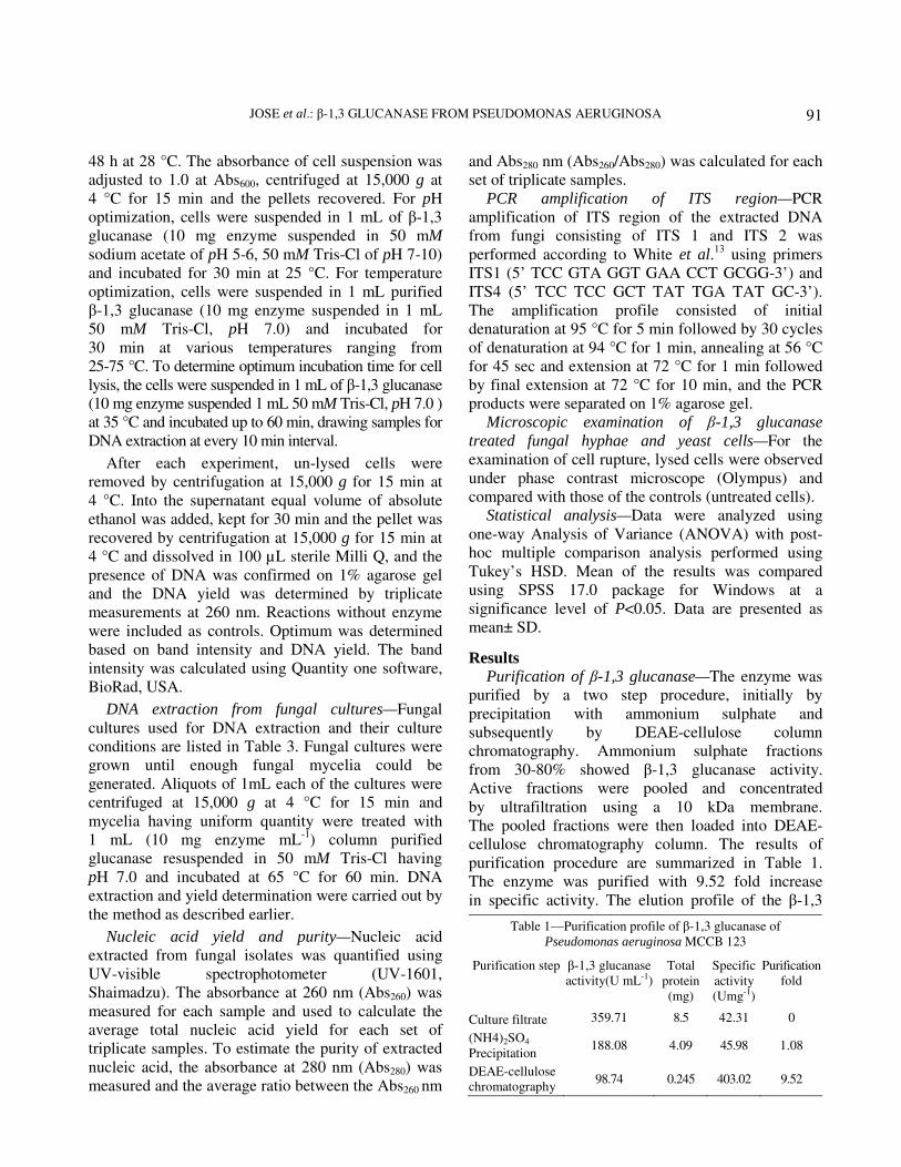
Purification of β-1,3 glucanase—The enzyme was
purified by a two step procedure, initially by
precipitation with ammonium sulphate and
subsequently by DEAE-cellulose column
chromatography. Ammonium sulphate fractions
from 30-80% showed β-1,3 glucanase activity.
Active fractions were pooled and concentrated
by ultrafiltration using a 10 kDa membrane.
The pooled fractions were then loaded into DEAE-
cellulose chromatography column. The results of
purification procedure are summarized in Table 1.
The enzyme was purified with 9.52 fold increase
in specific activity. The elution profile of the β-1,3
Table 1—Purification profile of β-1,3 glucanase of
Pseudomonas aeruginosa MCCB 123
Purification step β-1,3 glucanase
activity(U mL-1)
Total
protein
(mg)
Specific
activity
(Umg-1)
Purification
fold
Culture filtrate 359.71 8.5 42.31 0
(NH4)2SO4
Precipitation 188.08 4.09 45.98 1.08
DEAE-cellulose
chromatography 98.74 0.245 403.02 9.52

INDIAN J EXP BIOL, JANUARY 2014
92
glucanase on DEAE-cellulose column is shown in
Fig. 1. The enzyme eluted between 0.70 M - 0.81 M
NaCl (fractions, 70 to 81) contained β-1,3
glucanase.
Characterization of β-1,3 glucanase—The purified
MCCB 123 β-1,3 glucanase was homogenous on
SDS-PAGE and its molecular weight was estimated to
be 45 kDa by reducing SDS-PAGE (Fig. 2).
Effect of pH on activity of β-1,3 glucanase—The
effect of pH on β-1,3 glucanase activity was
determined using buffers in the pH range of 6-12 at
50 °C. The enzyme was found to exhibit activity from
pH 3-10 with its optimum at pH 7.0. Statistical
analysis by One-way ANOVA indicated that there
was a significant (P<0.05) difference in the β-1,3
glucanase activity between pH 3-7.
Effect of temperature on activity of β-1,3
glucanase—The enzyme was found to exhibit activity
from 30 to 80 °C with its optimum at 50 °C.
There was a significant (P<0.05) difference in the
β-1,3 glucanase activity in temperature ranges
between 30–50 °C.
Effect of inhibitors on activity of β-1, 3
glucanase—There was a partial inhibition (42.98%) of
enzyme activity by metalloprotease inhibitor EDTA
thus proving to be metalloprotease. The enzyme
retained 83.34, 84.22, 81.51 and 95.62 % activity in
presence of 5 mM 1, 10 phenanthroline, 50 µM
leupeptin, 10 µM pepstatin and 0.1 mM
phosphoramidon, respectively, confirming that the
enzyme did not belong to the class of serine and
cysteine protease, respectively (Table 2).
Cytotoxicity analysis of purified β-1, 3-glucanase—
Cytotoxicity analysis revealed that 236.87±1.89 µg mL-1
was the LD50 dose (50 % inhibition).
Application of β-1, 3 glucanase in fungal DNA
extraction—Optimization of pH for DNA extraction was accomplished over a pH range of 5-10 using 50 mM sodium acetate for pH 5-6, 50 mM Tris-Cl for pH 7-10 at 25 °C for 30 min. The enzyme exhibited
good lytic activity on cells of Saccharomyces
cervisiae from pH 5-10 with its optimum at 7.0 with a DNA yield of 231.66±5.20 µg mL
-1. The statistical
analysis revealed that pH imposed a significant (P<0.05) difference in the DNA yield between pH 8-9. However, there was no significant difference
in the DNA yield between pH 5-10. The enzyme was found to have good cell lysis from 25-75 °C with its optimum at 65 °C with a DNA yield of 310±2.5 µg mL
-1. There was a significant (P<0.05)
Fig.1—Elution profile of β-1,3 glucanase on DEAE-cellulose
C16/40 column.
Fig. 2—SDS-PAGE profile of purified LasA protease.
Lane 1, Molecular weight marker; lane 2, 45 kDa β-1,3 glucanase
enzyme.
Table 2—Effect of inhibitors on β-1,3 glucanase activity
Inhibitors Concentration Relative activity (%)
Control 100
EDTA 10 mM 57.02
PMSF 2 mM 100
1, 10 phenanthroline 5 mM 83.34
TLCK 0.1 mM 100
Leupeptin 50 µM 84.22
Pepstatin 10 µM 81.51
phosphoramidon 0.1 mM 95.62
JOSE et al.: β-1,3 GLUCANASE FROM PSEUDOMONAS AERUGINOSA
93
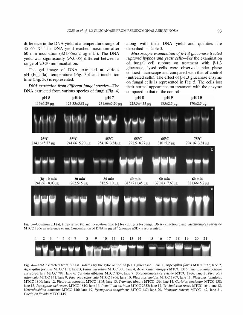
difference in the DNA yield at a temperature range of 45–65 °C. The DNA yield reached maximum after 60 min incubation (321.66±5.2 µg mL
1). The DNA
yield was significantly (P<0.05) different between a range of 20-30 min incubation.
The gel image of DNA extracted at various pH (Fig. 3a), temperature (Fig. 3b) and incubation time (Fig. 3c) is represented.
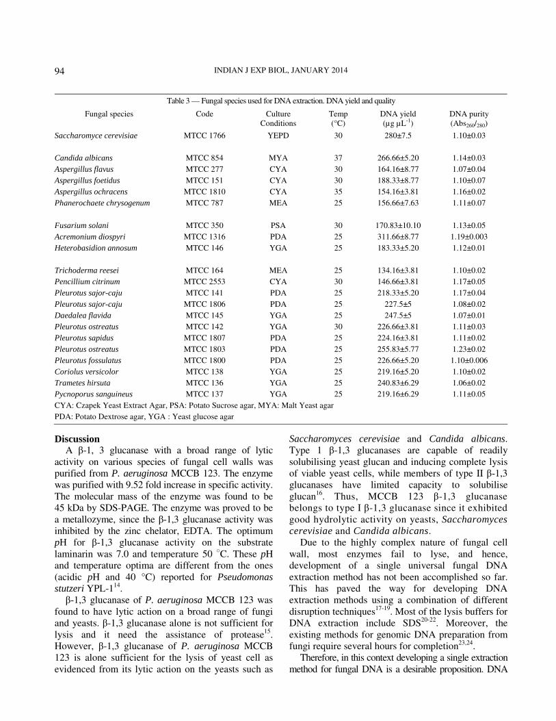
DNA extraction from different fungal species—The
DNA extracted from various species of fungi (Fig. 4)
along with their DNA yield and qualities are
described in Table 3. Microscopic examination of β-1,3 glucanase treated
ruptured hyphae and yeast cells—For the examination of fungal cell rupture on treatment with β-1,3 glucanase, lysed cells were observed under phase contrast microscope and compared with that of control (untreated cells). The effect of β-1,3 glucanase enzyme on fungal cells is represented in Fig. 5. The cells lost their normal appearance on treatment with the enzyme compared to that of the control.
Fig. 3—Optimum pH (a), temperature (b) and incubation time (c) for cell lysis for fungal DNA extraction using Sacchromyces cervisiae
MTCC 1766 as reference strain. Concentration of DNA in µg µl-1 (average ±SD) is represented.
Fig. 4—DNA extracted from fungal isolates by the lytic action of β-1,3 glucanase. Lane 1, Aspergillus flavus MTCC 277; lane 2,
Aspergillus foetidus MTCC 151; lane 3, Fusarium solani MTCC 350; lane 4, Acremonium diospyri MTCC 1316; lane 5, Phanerochaete
chrysosporium MTCC 787; lane 6, Candida albicans MTCC 854; lane 7, Saccharomyces cerevisiae MTCC 1766; lane 8, Pleurotus
sajor-caju MTCC 141; lane 9, Pleurotus sajor-caju MTCC 1806; lane 10, Pleurotus sapidus MTCC 1807; lane 11, Pleurotus fossulatus
MTCC 1800; lane 12, Pleurotus ostreatus MTCC 1803; lane 13, Trametes hirsute MTCC 136; lane 14, Coriolus versicolor MTCC 138;
lane 15, Aspergillus ochracens MTCC 1810; lane 16, Pencillium citrinum MTCC 2553; lane 17, Trichoderma reesei MTCC 164; lane 18,
Heterobasidion annosum MTCC 146; lane 19, Pycnoporus sanguineus MTCC 137; lane 20, Pleurotus osterus MTCC 142; lane 21,
Daedalea flavida MTCC 145.
INDIAN J EXP BIOL, JANUARY 2014
94
Discussion A β-1, 3 glucanase with a broad range of lytic
activity on various species of fungal cell walls was
purified from P. aeruginosa MCCB 123. The enzyme
was purified with 9.52 fold increase in specific activity.
The molecular mass of the enzyme was found to be
45 kDa by SDS-PAGE. The enzyme was proved to be
a metallozyme, since the β-1,3 glucanase activity was
inhibited by the zinc chelator, EDTA. The optimum
pH for β-1,3 glucanase activity on the substrate
laminarin was 7.0 and temperature 50 ○C. These pH
and temperature optima are different from the ones
(acidic pH and 40 °C) reported for Pseudomonas
stutzeri YPL-114
.
β-1,3 glucanase of P. aeruginosa MCCB 123 was
found to have lytic action on a broad range of fungi
and yeasts. β-1,3 glucanase alone is not sufficient for
lysis and it need the assistance of protease15
.
However, β-1,3 glucanase of P. aeruginosa MCCB
123 is alone sufficient for the lysis of yeast cell as
evidenced from its lytic action on the yeasts such as
Saccharomyces cerevisiae and Candida albicans.
Type 1 β-1,3 glucanases are capable of readily
solubilising yeast glucan and inducing complete lysis
of viable yeast cells, while members of type II β-1,3
glucanases have limited capacity to solubilise
glucan16
. Thus, MCCB 123 β-1,3 glucanase
belongs to type I β-1,3 glucanase since it exhibited
good hydrolytic activity on yeasts, Saccharomyces
cerevisiae and Candida albicans.
Due to the highly complex nature of fungal cell
wall, most enzymes fail to lyse, and hence,
development of a single universal fungal DNA
extraction method has not been accomplished so far.
This has paved the way for developing DNA
extraction methods using a combination of different
disruption techniques17-19
. Most of the lysis buffers for
DNA extraction include SDS20-22
. Moreover, the
existing methods for genomic DNA preparation from
fungi require several hours for completion23,24
.
Therefore, in this context developing a single extraction
method for fungal DNA is a desirable proposition. DNA
Table 3 — Fungal species used for DNA extraction. DNA yield and quality
Fungal species Code Culture
Conditions
Temp
(°C)
DNA yield
(µg µL-1)
DNA purity
(Abs260/280)
Saccharomyce cerevisiae MTCC 1766 YEPD 30 280±7.5 1.10±0.03
Candida albicans MTCC 854 MYA 37 266.66±5.20 1.14±0.03
Aspergillus flavus MTCC 277 CYA 30 164.16±8.77 1.07±0.04
Aspergillus foetidus MTCC 151 CYA 30 188.33±8.77 1.10±0.07
Aspergillus ochracens MTCC 1810 CYA 35 154.16±3.81 1.16±0.02
Phanerochaete chrysogenum MTCC 787 MEA 25 156.66±7.63 1.11±0.07
Fusarium solani MTCC 350 PSA 30 170.83±10.10 1.13±0.05
Acremonium diospyri MTCC 1316 PDA 25 311.66±8.77 1.19±0.003
Heterobasidion annosum MTCC 146 YGA 25 183.33±5.20 1.12±0.01
Trichoderma reesei MTCC 164 MEA 25 134.16±3.81 1.10±0.02
Pencillium citrinum MTCC 2553 CYA 30 146.66±3.81 1.17±0.05
Pleurotus sajor-caju MTCC 141 PDA 25 218.33±5.20 1.17±0.04
Pleurotus sajor-caju MTCC 1806 PDA 25 227.5±5 1.08±0.02
Daedalea flavida MTCC 145 YGA 25 247.5±5 1.07±0.01
Pleurotus ostreatus MTCC 142 YGA 30 226.66±3.81 1.11±0.03
Pleurotus sapidus MTCC 1807 PDA 25 224.16±3.81 1.11±0.02
Pleurotus ostreatus MTCC 1803 PDA 25 255.83±5.77 1.23±0.02
Pleurotus fossulatus MTCC 1800 PDA 25 226.66±5.20 1.10±0.006
Coriolus versicolor MTCC 138 YGA 25 219.16±5.20 1.10±0.02
Trametes hirsuta MTCC 136 YGA 25 240.83±6.29 1.06±0.02
Pycnoporus sanguineus MTCC 137 YGA 25 219.16±6.29 1.11±0.05
CYA: Czapek Yeast Extract Agar, PSA: Potato Sucrose agar, MYA: Malt Yeast agar
PDA: Potato Dextrose agar, YGA : Yeast glucose agar

JOSE et al.: β-1,3 GLUCANASE FROM PSEUDOMONAS AERUGINOSA
95
could be extracted from 21 fungal species by the lytic
action of the purified β-1,3 glucanase from P. aeruginosa
MCCB 123 without the addition of any other reagent and
implementation of mechanical treatments as well,
transforming this method unique among the ones
reported. The extracted DNA could be directly used for
PCR amplification without further purification. Moreover,
the method is less expensive as it employs only β-1,3
glucanase as the sole reagent, the quality and quantity of
DNA obtained is suitable for molecular assays and it
doesn’t require the use of specialised equipments or
hazardous reagents.
The quality of the extracted nucleic acid is
important for further processing. Samples with mean
A260/A280 ratios below 1.8 were presumed to contain
protein or other contaminants, whereas samples with
ratios above 2.0 were presumed to be due to the
presence of RNA. However, nucleic acids
preparations free of phenol should have Abs 260/280
ratios near 1.224,25
. In the case of DNA extracted
with β-1,3 glucanase from various fungal species,
Abs260/280 ratios were with in the range of 1.0 to 1.1.
As phenol had not been used in the process it could be
concluded that the DNA extracted using this method
was free of contamination and was suitable for PCR.
Fungal DNA extraction using MCCB 123 β-1,3
glucanase has several advantages. The number of steps
in DNA extraction procedure was minimized by
replacing phenol chloroform extraction method and it
also did not involve the addition of any detergent,
lytic agents and implementation of other mechanical
lytic methods such as grinding with sand, repeated
freeze thaw cycles in liquid nitrogen etc. Several
samples could be processed within a short time period of
30 min. The method yielded high quality DNA suitable
for PCR. This method is likely to be cost-effective since
β-1,3 glucanase alone needs to be used as the sole
reagent to a broad range of fungi. These properties
qualify the enzyme unique over all other lytic
enzymes used for DNA extraction from fungi.
This is the first report of a lytic enzyme being
employed solely in fungal DNA extraction without
any additives. The broad range of lytic activity of
β-1,3 glucanase on a wide range of fungi has immense
benefits in DNA extraction in commercial point of
view.
Fig. 5—Rupture of fungal hyphe on treatment with β-1,3 glucanase of Pseudomonas aeruginosa MCCB 123. Control (c) represents
untreated fungal hyphae and test (t) represents the changes in fungal hyphae on treatment with 10 mg mL-1 of purified β-1,3 glucanase
[A= Aspergillus foetidus MTCC 151, B= Aspergillus flavus MTCC 277, C= Acremonium dyosprii MTCC 1316 , D= Fusarium solani
MTCC 350, E= Phanerocheate chrysogenum MTCC 787, F= Pleurotus fossulatus MTCC 1800] Magnification 600X.

INDIAN J EXP BIOL, JANUARY 2014
96
Conclusion Development of a single universal fungal
DNA extraction method has significance and
has not been accomplished so far. There is no single
protocol appropriate for cell lysis for all fungi
and each species requires a specific method for
efficient DNA extraction. β-1,3 glucanase from
P. aeruginosa MCCB 123 was found to have lytic action
on a broad range of fungal and yeast strains. This
could be exploited in DNA extraction from fungi
without the addition of other reagents, and by
incorporating other mechanical lytic steps. Therefore,
broad range of lytic action of MCCB 123 β-1,3
glucanase has immense benefits in fungal DNA
extraction for the development of a single universal
protocol.
Acknowledgement This work was supported by Cochin University of
Science and Technology, Kerala, India.
References 1 Zhu BW, Zhao JG, Yang JF, Mikiro T, Zhang ZS & Zhou
DY, 2008. Purification and partial characterization of a novel
β-1,3-glucanase from the gut of sea cucumber Stichopus
japonicas, Process Biochem, 43 (2008) 1102.
2 Garciduenas SV, Morales CAL & Estrella AH, Analysis of
β-1, 3 glucanolytic system of the biocontrol agent
Trichoderma harzianum, Appl Environ Microbiol,
64 (1998) 1442.
3 Vijayendra SVN & Kashiwagi Y, Characterization of a new acid
stable exo-β-1, 3-glucanase of Rhizoctonia solani and its action on
microbial polysaccharides, Int J Biol Macromol, 44 (2009) 92.
4 Planas A, Bacterial 1, 3-1, 4-β-glucanases: structure, function and protein engineering, Biochim Biophys Acta, 1543 (2000) 361.
5 Ferrer P, Revisiting the Cellulosimicrobium cellulans yeast-lytic β-1,3-glucanases toolbox: A review, Microb Cell Fact, 5 (2006) 10.
6 Kumar T, Wahla V, Pande P, Dubey RC & Maheshwari DK, Rhizosphere competent Pseudomonas aeruginosa in the management of Heterodera cajani on sesame, World J Microbiol Biotechnol, 25 (2009) 277.
7 Singh N, Kumar S, Bajpai VK, Dubey RC, Maheshwari DK
& Kang SC, Biological control of Macrophomina phaseolina
by chemotactic fluorescent Pseudomonas aeruginosa PN1
and its plant growth promotory activity in chir-pine,
Crop prot, 29 (2010) 1142.
8 Diby P, Saju KA, Jisha PJ, Sarma YR, Kumar A &
Anandaraj M, Mycolytic enzymes produced by
Pseudomonas fluorescens and Trichoderma spp. against
Phytopthora capsici, the foot rot pathogen of black pepper
(Piper nigrum L.), Ann Microbiol, 55 (2005) 129.
9 Jose D, Proteases from an environmental isolate of
Pseudomonas aeruginosa MCCB 123 and their applications,
Ph.D. Thesis, Cochin University of Science and Technology,
Cochin, India, 2012.
10 Laemmli UK, Cleavage of structural proteins during the
assembly of the head of bacteriophage T4, Nature,
227 (1970) 680.
11 Hatree E, Determination of protein; a modification of the Lowry
method that gives linear photometric response, Analytical
Biochem, 48 (1972) 422.
12 Niwa T, Kawamura Y, Katagiri Y & EZAKI T, Lytic enzyme,
labiase for a broad range of gram positive bacteria and its
application to analyze functional DNA/RNA, J Microbiol
Methods, 61 (2005) 251.
13 White TJ, Burns T, Lee S & Tayler J, Amplification and
direct sequencing of fungal ribosomal RNA genes for
phylogenetics. A guide to methods and applications, in PCR
protocols (Academic Press, San Diago, CA) 1990, 322.
14 Lim HS & Kim SD, The role of and characterization of β-1,
3 glucanase in the biocontrol of Fusarium solani by
Pseudomonas stutzeri. YPL-1, J Microbiol, 33 (1995) 295.
15 Salazar O & Asenjo A, Enzymatic lysis of bacterial cells,
Biotechnol Lett, 29 (2007) 985.
16 Doi K & Doi A, Cloning and expression in Escherichia coli of
the gene for Arthrobacter β-1,3-glucanase, J Bacteriol,
168 (1986) 1272.
17 Manian, S, Sreenivasaprasad S & Mills PR, DNA extraction
method for PCR in mycorrhizal fungi, Lett Appl Microbiol,
33 (2001) 307.
18 Kuhad RC, Kapoor RK & LAL R, Improving the yield and
quality of DNA isolated from white-rot fungi, Folia Microbiol,
49 (2004) 112 .
19 Karakousis A, Tan L, Ellis D, Alexiou H & WORMALD PJ,
An assessment of the efficiency of fungal DNA extraction
methods for maximizing the detection of medically important
fungi using PCR, J Microbiol Methods, 65 (2006) 38.
20 Erland S, Henrion B, Martin F, Glover LA & Alexander IJ,
Identification of the ectomycorrhizal basidiomycete
Tylospora fibrillosa Donk by RFLP analysis of the PCR-
amplified ITS and IGS regions of ribosomal DNA,
New Phytol, 126 (1994) 525.
21 Haugland RA, Heckman JL & WYMER LJ, Evaluation of
different methods for the extraction of DNA from fungal
conidia by quantitative competitive PCR analysis,
J Microbiol Methods, 37 (1999) 165.
22 Plaza GA, Upchurch R, Brigmon RL, Whitman WB &
ULFIG K, Rapid DNA extraction for screening soil
filamentous fungi using PCR amplification, Pol J Environ
Stud, 13 (2004) 315.
23 Muller FM, Werner KE, Kasai M, Francesconi A, Chanock SJ
&Walsh TJ, Rapid extraction of genomic DNA from medically
important yeasts and filamentous fungi by highspeed cell
disruption, J Clinical Microbiol, 36 (1998) 1625.
24 Sambrook J & Russell DW, Rapid isolation of yeast DNA,
in Molecular cloning, a laboratory manual, (Cold Spring
Harbor Laboratory Press, New York) 2001, 631.
25 Lemarchand K, Berthiaume F, Maynard C, Harel J, Payment
P, Bayardelle P, Masson L & Brousseau R, Optimization of
microbial DNA extraction and purification from raw
wastewater samples for downstream pathogen detection by
microarrays, J Microbiol Methods, 63 (2005) 115.