Physiology of the Blood II. Red Blood Cells(Erythrocytes) · PDF filePhysiology of the Blood...
28
Physiology of the Blood II. Red Blood Cells (Erythrocytes) Prof. Szabolcs Kéri University of Szeged, Faculty of Medicine, Department of Physiology 2017
Transcript of Physiology of the Blood II. Red Blood Cells(Erythrocytes) · PDF filePhysiology of the Blood...
Physiology of the Blood II. Red Blood Cells (Erythrocytes)
Prof. Szabolcs Kéri
University of Szeged, Faculty of Medicine, Department of Physiology
2017
1. Number, structure, physical properties
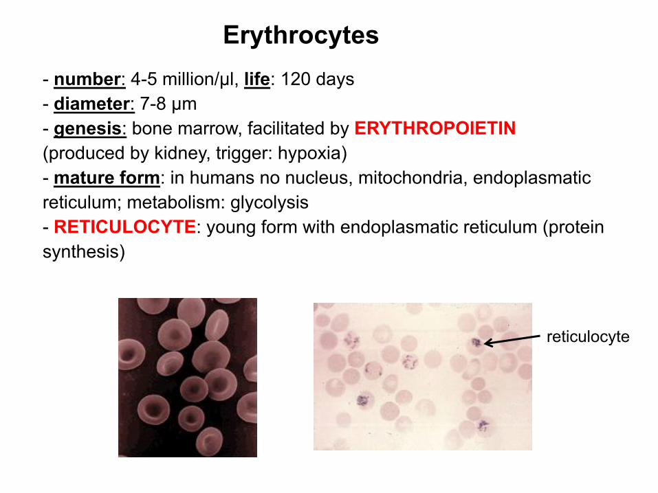
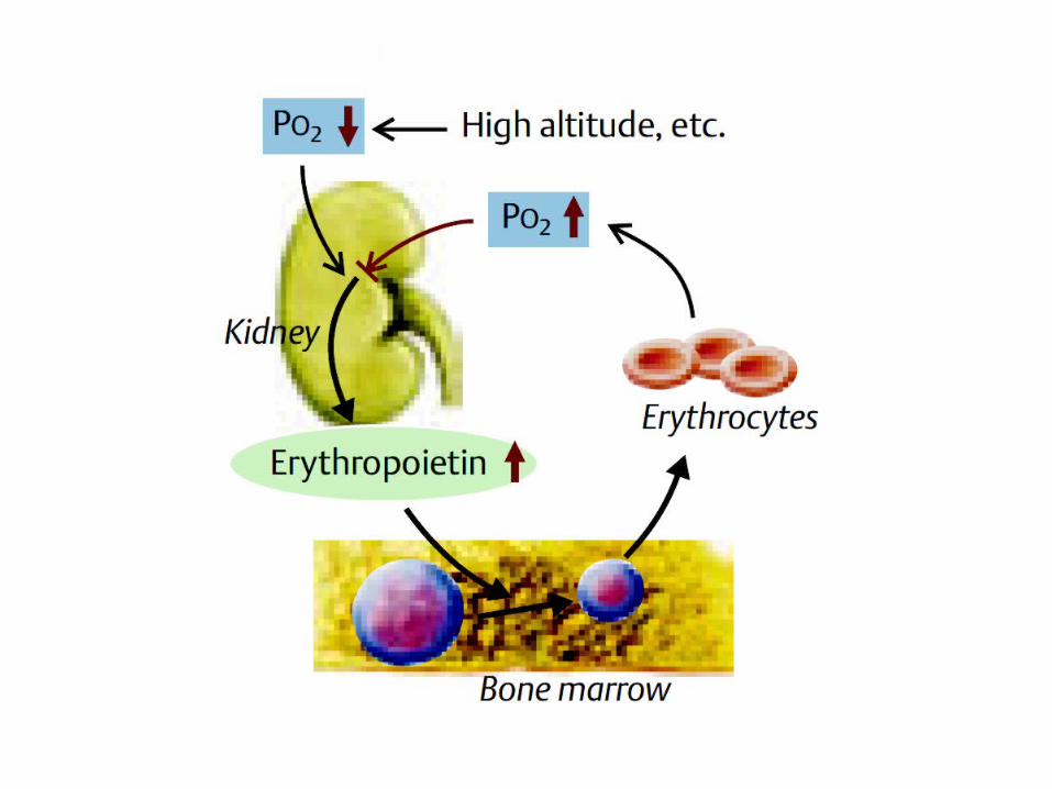
- number: 4-5 million/μl, life: 120 days- diameter: 7-8 μm- genesis: bone marrow, facilitated by ERYTHROPOIETIN(produced by kidney, trigger: hypoxia)- mature form: in humans no nucleus, mitochondria, endoplasmatic reticulum; metabolism: glycolysis- RETICULOCYTE: young form with endoplasmatic reticulum (protein synthesis)
reticulocyte
Erythrocytes
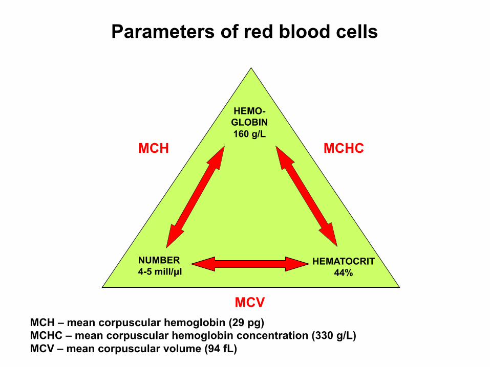
Parameters of red blood cells
MCH – mean corpuscular hemoglobin (29 pg)MCHC – mean corpuscular hemoglobin concentration (330 g/L)MCV – mean corpuscular volume (94 fL)
HEMATOCRIT44%
HEMO-GLOBIN160 g/L
NUMBER4-5 mill/μl
MCHC
MCV
MCH
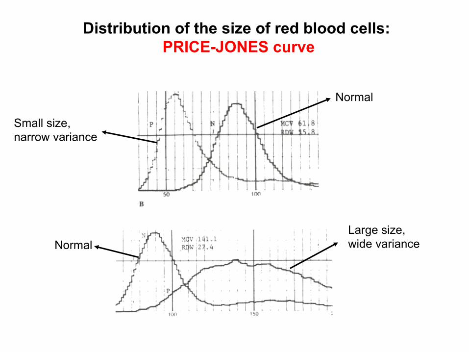
Distribution of the size of red blood cells: PRICE-JONES curve
Normal
Small size, narrow variance
NormalLarge size, wide variance
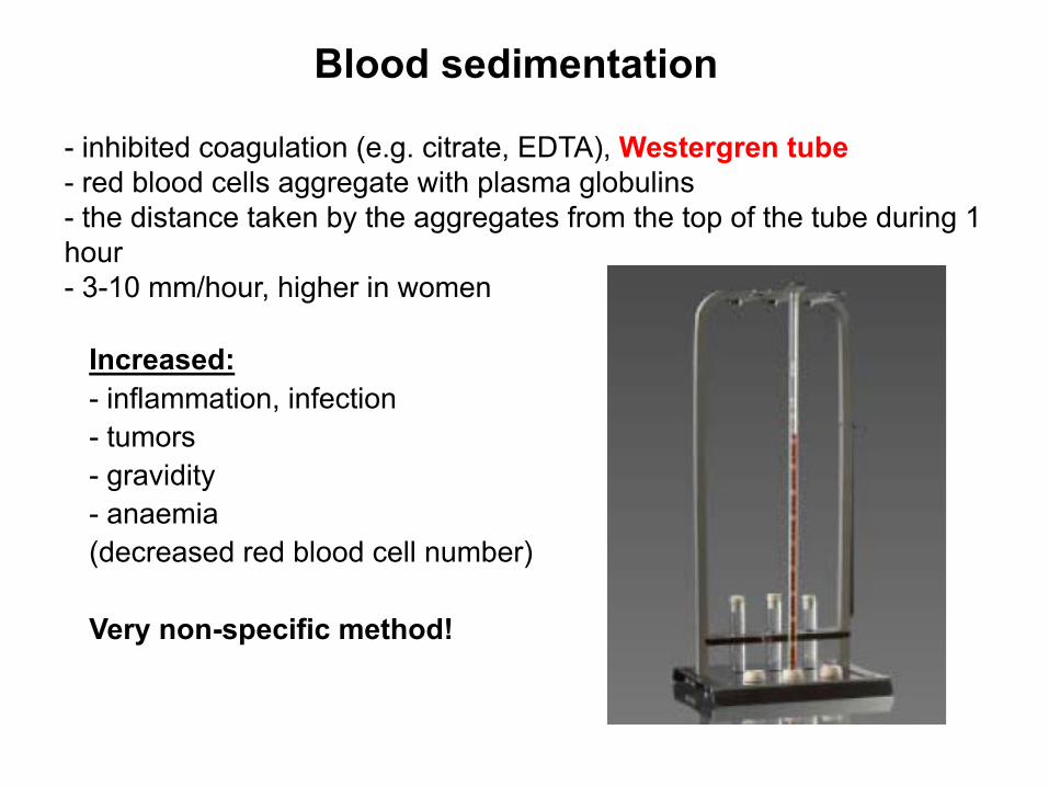
Blood sedimentation
- inhibited coagulation (e.g. citrate, EDTA), Westergren tube- red blood cells aggregate with plasma globulins- the distance taken by the aggregates from the top of the tube during 1 hour - 3-10 mm/hour, higher in women
Increased:- inflammation, infection- tumors- gravidity- anaemia (decreased red blood cell number)
Very non-specific method!
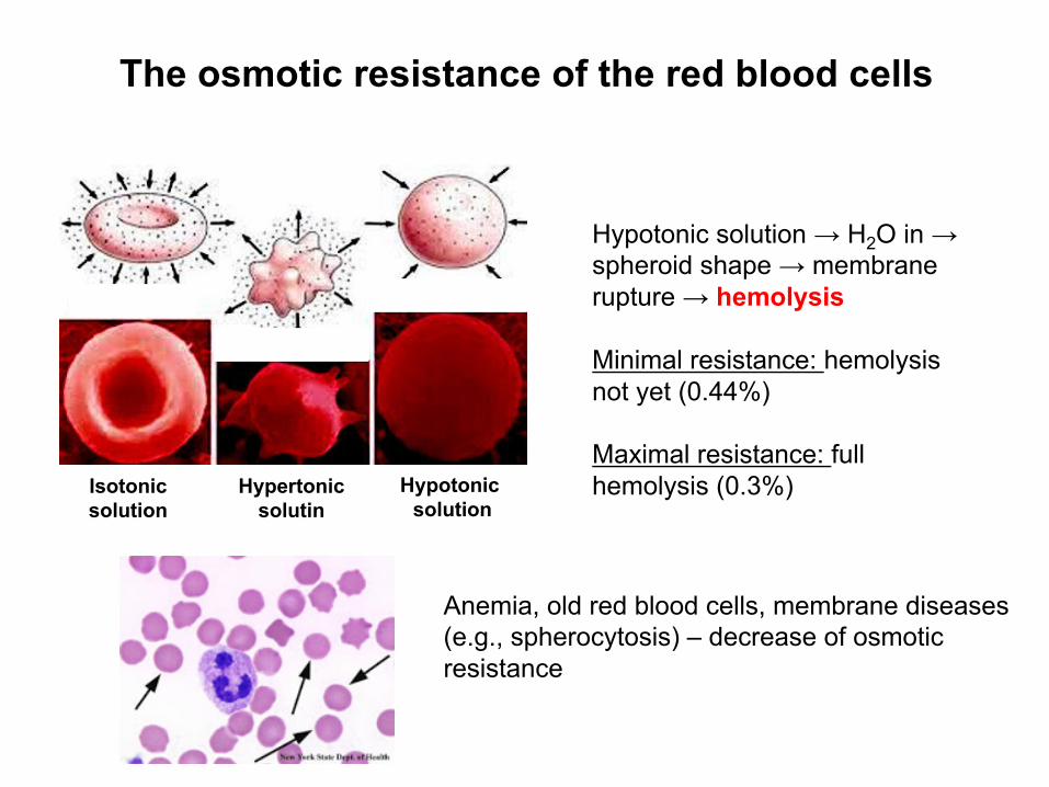
The osmotic resistance of the red blood cells
Isotonicsolution
Hypertonic solutin
Hypotonic solution
Hypotonic solution → H2O in →spheroid shape → membranerupture → hemolysis
Minimal resistance: hemolysisnot yet (0.44%)
Maximal resistance: fullhemolysis (0.3%)
Anemia, old red blood cells, membrane diseases(e.g., spherocytosis) – decrease of osmoticresistance
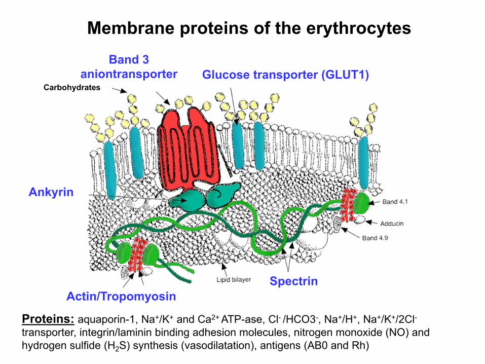
Glucose transporter (GLUT1)Carbohydrates
Ankyrin
SpectrinActin/Tropomyosin
Band 3 aniontransporter
Proteins: aquaporin-1, Na+/K+ and Ca2+ ATP-ase, Cl- /HCO3-, Na+/H+, Na+/K+/2Cl-transporter, integrin/laminin binding adhesion molecules, nitrogen monoxide (NO) andhydrogen sulfide (H2S) synthesis (vasodilatation), antigens (AB0 and Rh)
Membrane proteins of the erythrocytes
2. Genesis: iron and vitamins
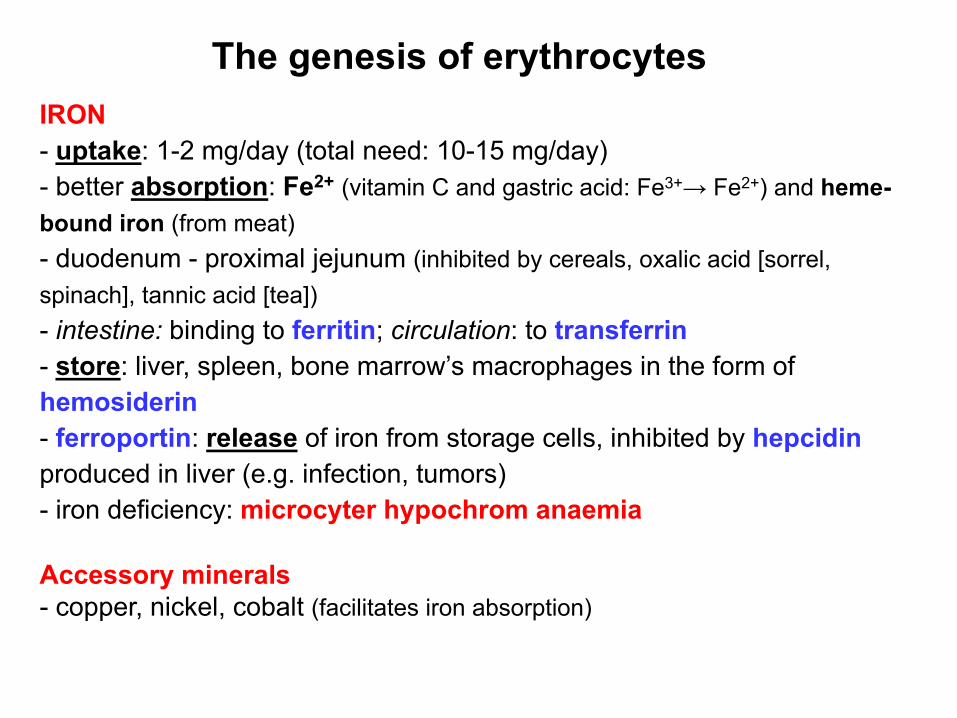
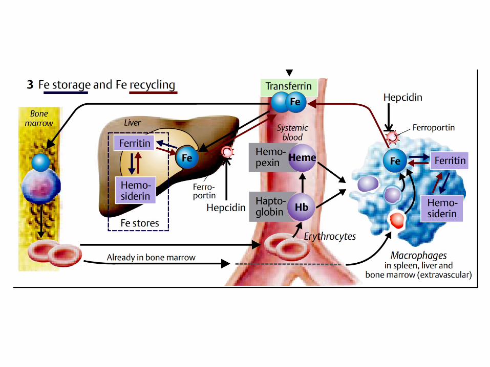
The genesis of erythrocytesIRON- uptake: 1-2 mg/day (total need: 10-15 mg/day)- better absorption: Fe2+ (vitamin C and gastric acid: Fe3+→ Fe2+) and heme-bound iron (from meat)- duodenum - proximal jejunum (inhibited by cereals, oxalic acid [sorrel, spinach], tannic acid [tea]) - intestine: binding to ferritin; circulation: to transferrin- store: liver, spleen, bone marrow’s macrophages in the form of hemosiderin- ferroportin: release of iron from storage cells, inhibited by hepcidinproduced in liver (e.g. infection, tumors)- iron deficiency: microcyter hypochrom anaemia
Accessory minerals- copper, nickel, cobalt (facilitates iron absorption)
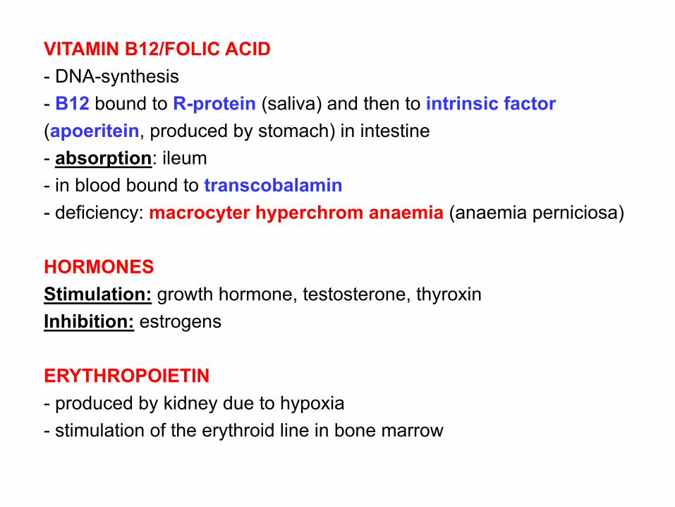
VITAMIN B12/FOLIC ACID- DNA-synthesis- B12 bound to R-protein (saliva) and then to intrinsic factor (apoeritein, produced by stomach) in intestine- absorption: ileum- in blood bound to transcobalamin- deficiency: macrocyter hyperchrom anaemia (anaemia perniciosa)
HORMONESStimulation: growth hormone, testosterone, thyroxinInhibition: estrogens
ERYTHROPOIETIN- produced by kidney due to hypoxia- stimulation of the erythroid line in bone marrow
3. Hemoglobin structure and function: gas transport
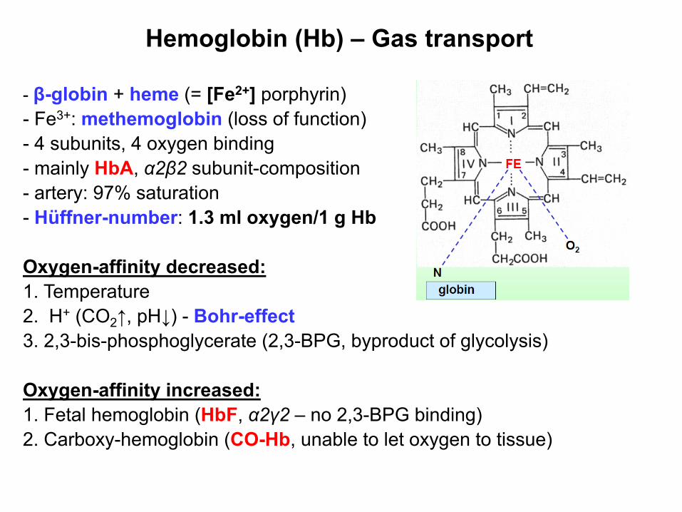
Hemoglobin (Hb) – Gas transport
- β-globin + heme (= [Fe2+] porphyrin)- Fe3+: methemoglobin (loss of function)- 4 subunits, 4 oxygen binding- mainly HbA, α2β2 subunit-composition- artery: 97% saturation- Hüffner-number: 1.3 ml oxygen/1 g Hb
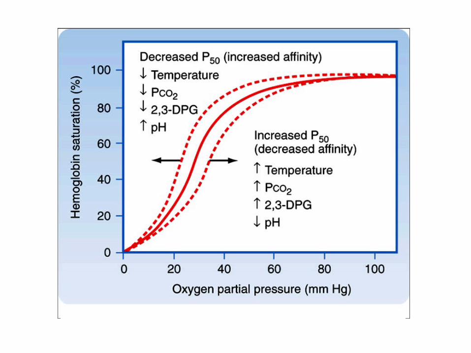
Oxygen-affinity decreased:1. Temperature2. H+ (CO2↑, pH↓) - Bohr-effect3. 2,3-bis-phosphoglycerate (2,3-BPG, byproduct of glycolysis)
Oxygen-affinity increased:1. Fetal hemoglobin (HbF, α2γ2 – no 2,3-BPG binding)2. Carboxy-hemoglobin (CO-Hb, unable to let oxygen to tissue)

Changes of globin-chains with age
%
Gamma-chain (fetal)
Epsilon-chain(embrional)
Beta-chain(adult)
Delta-chain
Alpha-chain
Pregnancy (months) Age (months)BIRTH
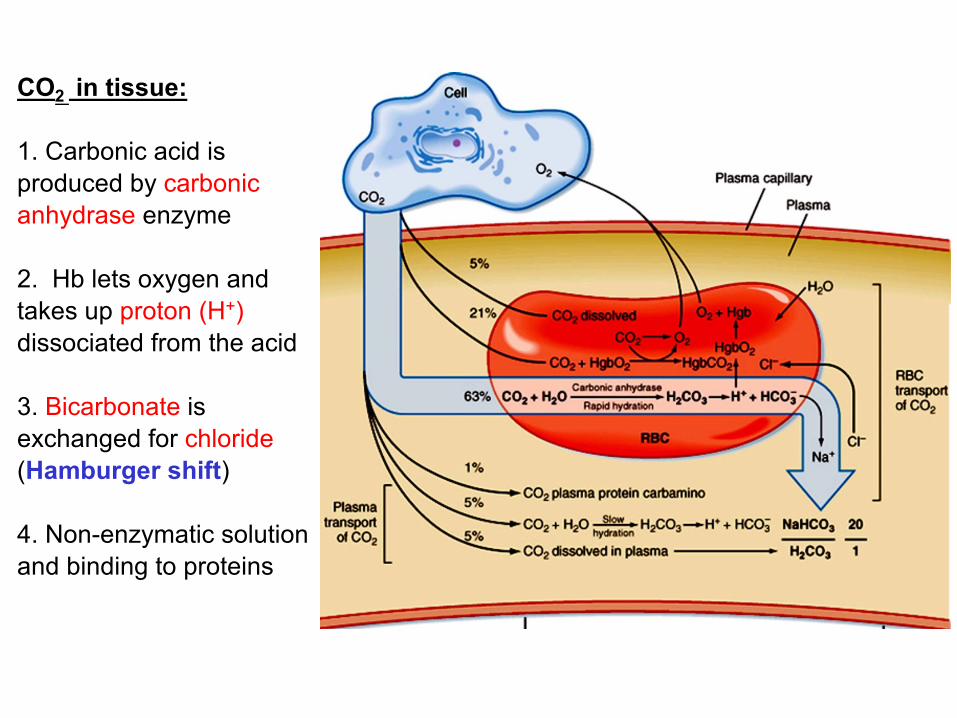
CO2 in tissue:
1. Carbonic acid is produced by carbonic anhydrase enzyme
2. Hb lets oxygen and takes up proton (H+)dissociated from the acid
3. Bicarbonate is exchanged for chloride(Hamburger shift)
4. Non-enzymatic solution and binding to proteins
4. Degradation of hemoglobin: the question of bilirubin
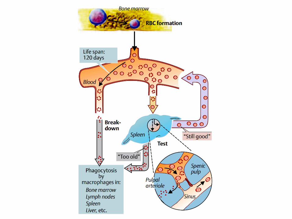
Degradation of hemoglobin 1.
1. Old erythrocytes: extraction from blood by macrophages (liver, spleen)
2. Haptoglobin transiently binds hemoglobin in circulation(hemopexin: heme-binding protein in blood)
3. Fe2+ dissociation (used again or stored) & proteolysis of β-globin
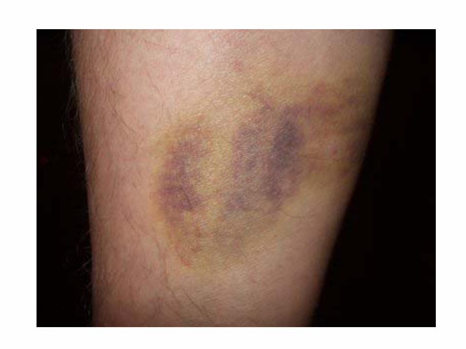
4. Porphyrin degradation: CO + biliverdin (green), then bilirubin (yellow)
Circulation: bilirubin binds to albumin – indirect bilirubin
6. Liver takes up bilirubin and conjugates that with glucuronide –direct bilirubin
7. From liver to gut with bile where further transformation occurs (urobilinogen - urobilin, stercobilinogen - stercobilin; oxidoreductive process mediated by bacteria)
8. Some of them are reabsorbed to liver with bile acids via the portal vein: Enterohepatic circulation
9. Secretion with faces (gives its color) and urine
Degradation of hemoglobin 2.
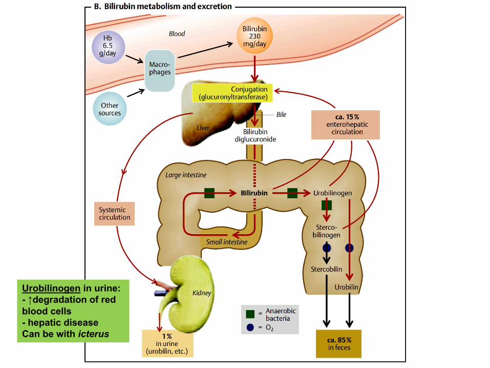
Urobilinogen in urine:- ↑degradation of redblood cells- hepatic diseaseCan be with icterus
5. Blood groups
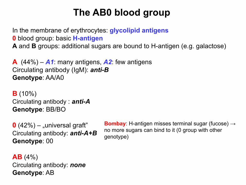
The AB0 blood groupIn the membrane of erythrocytes: glycolipid antigens0 blood group: basic H-antigenA and B groups: additional sugars are bound to H-antigen (e.g. galactose)
A (44%) – A1: many antigens, A2: few antigens Circulating antibody (IgM): anti-BGenotype: AA/A0
B (10%)Circulating antibody : anti-AGenotype: BB/BO
0 (42%) – „universal graft”Circulating antibody: anti-A+BGenotype: 00
AB (4%)Circulating antibody: noneGenotype: AB
Bombay: H-antigen misses terminal sugar (fucose) → no more sugars can bind to it (0 group with othergenotype)
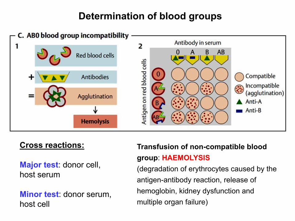
Cross reactions:
Major test: donor cell, host serum
Minor test: donor serum, host cell
Determination of blood groups
Transfusion of non-compatible blood group: HAEMOLYSIS(degradation of erythrocytes caused by the antigen-antibody reaction, release of hemoglobin, kidney dysfunction and multiple organ failure)
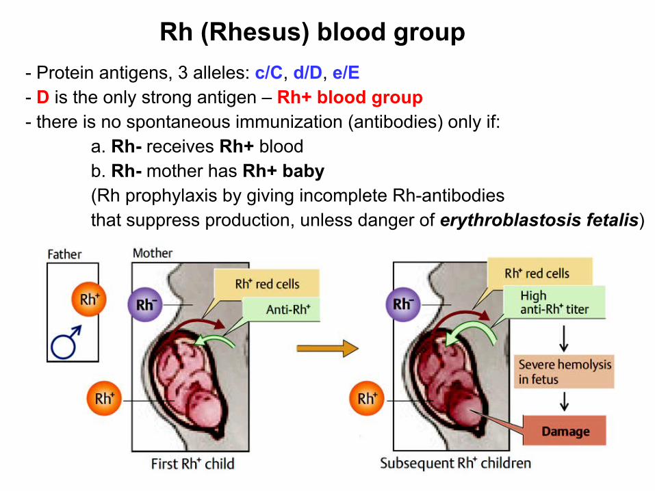
Rh (Rhesus) blood group- Protein antigens, 3 alleles: c/C, d/D, e/E- D is the only strong antigen – Rh+ blood group- there is no spontaneous immunization (antibodies) only if:
a. Rh- receives Rh+ bloodb. Rh- mother has Rh+ baby(Rh prophylaxis by giving incomplete Rh-antibodiesthat suppress production, unless danger of erythroblastosis fetalis)