physical interaction of x ray with matter
53
PRESENTATION BY:DR. CHARUSMITA CHAUDHARY Physical interaction of x ray with matter
-
Upload
charusmita-chaudhary -
Category
Health & Medicine
-
view
8.622 -
download
7
Transcript of physical interaction of x ray with matter
PRESENTATION BY: DR. CHARUSMITA CHAUDHARY
Physical interaction of x ray with matter
Dual characteristics of X-raysX-rays belong to a group of radiation called
electromagnetic radiation . Electromagnetic radiation has dual
characteristic, comprises of both WaveParticle
Wave concept : Propagated through space in the form of waves. Waves of all types have associated wavelength and frequency Relationship : c=λν.
c=velocity of light λ=wavelength ν=frequency The wavelength of diagnostic X-rays is very short around
0.1 to 1A. Wave concept explains why it can be reflected.
Particle concept of EM radiation: Short EM waves such as X-rays predominantly react with matter as if they were particles rather than waves. At high frequencies electrons interact with EM radiation as if the EM radiation were an energy bundle. These particles are actually discrete bundles of energy and each of these bundles is called a quantum or photon.
Particle concept used to describe interaction between radiation & matter
.The amount of energy carried by each quantum is given by
E=h ν E=photon’s energy h=planck’s constant ν =frequency
c= νλ or ν=c/λ so substituting c/ λ for ν we get E=hc/ λ h=4.13x 10⁻¹⁸ keV/sec c=3 x 10⁸ m/sec E=12.4 E=Energy in keV. λ=wavelength in A0
METHODS OF INTERACTIONS
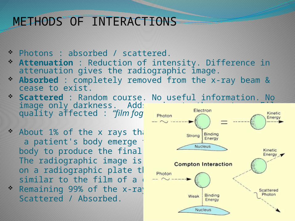
Photons : absorbed / scattered. Attenuation : Reduction of intensity. Difference in
attenuation gives the radiographic image. Absorbed : completely removed from the x-ray beam & cease
to exist. Scattered : Random course. No useful information. No image
only darkness. Adds noise to the system. Film quality affected : “film fog”.
About 1% of the x rays that strike a patient's body emerge from the body to produce the final image. The radiographic image is formed on a radiographic plate that is similar to the film of a camera.
Remaining 99% of the x-rays --- Scattered / Absorbed.
ATOMIC STRUCTUREX-ray photons may interact either with orbital
electrons or with the nucleus. In the diagnostic energy range, the interactions are always with orbital electrons.
The molecular bonding energies ,however are too small to influence the type and number of interactions .
The most important factor is the atomic make up of a tissue and not its molecular structure.
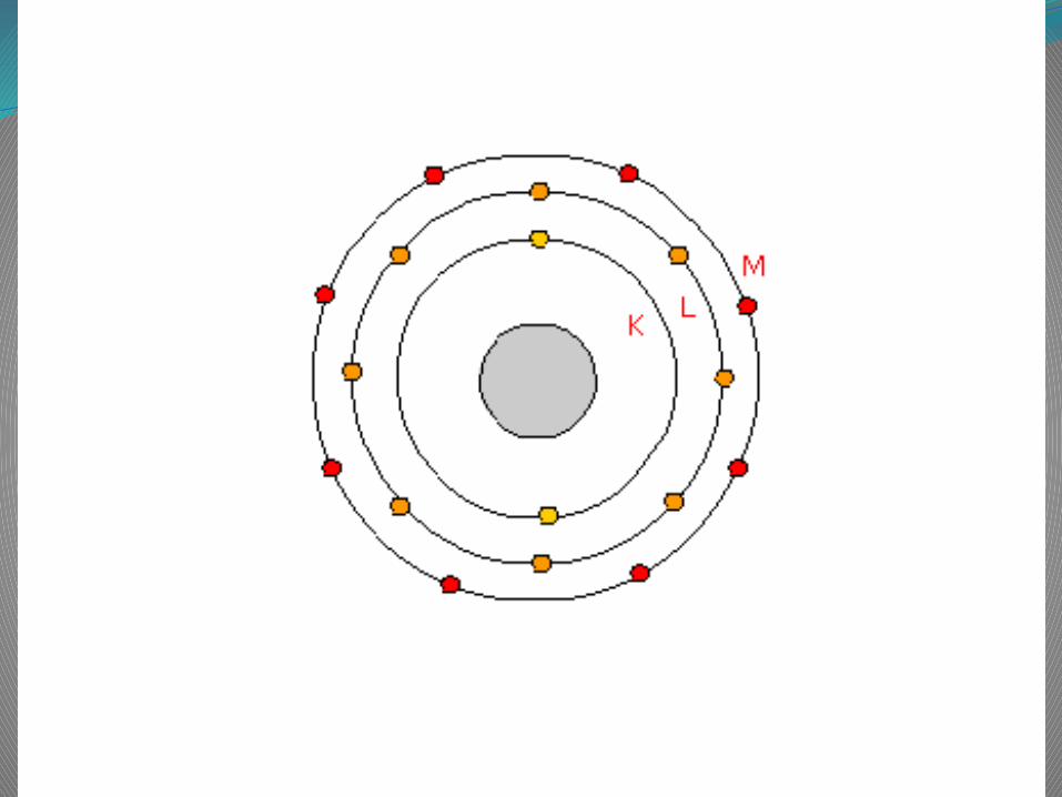
Atomic structure
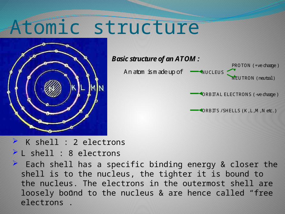
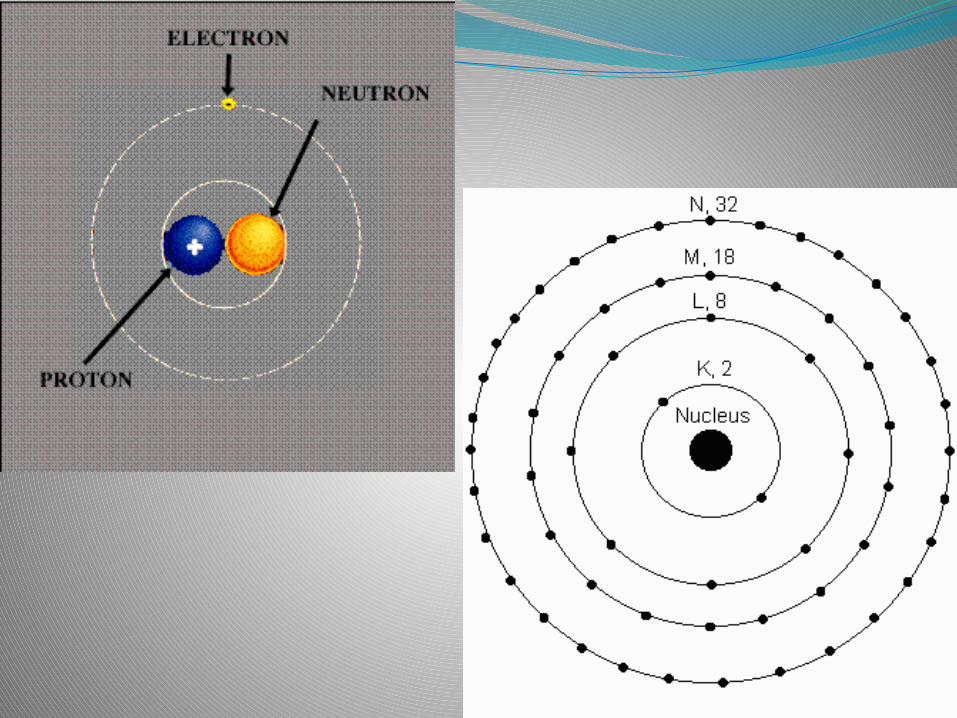
K shell : 2 electrons L shell : 8 electrons Each shell has a specific binding energy & closer the shell is
to the nucleus, the tighter it is bound to the nucleus. The electrons in the outermost shell are loosely bound to the nucleus & are hence called “free electrons”.
Basic structure of an ATOM : PROTON ( +ve charge )
An atom is made up of NUCLEUS NEUTRON ( neutral ) ORBITAL ELECTRONS ( -ve charge ) ORBITS / SHELLS ( K, L, M, N etc. )
Energy value of electronic shells is also determined by the atomic number of the atom.
K-shell electron are more tightly bound in elements of high atomic number. Pb : 88keV while Ca : 4keV.
Electrons in the K -shell are at a lower energy level than electrons in the L-shell. If we consider the outermost electrons as free ,than inner shell electrons are in energy debt. The energy debt is greatest when they are close to nucleus in an element with a high atomic number.
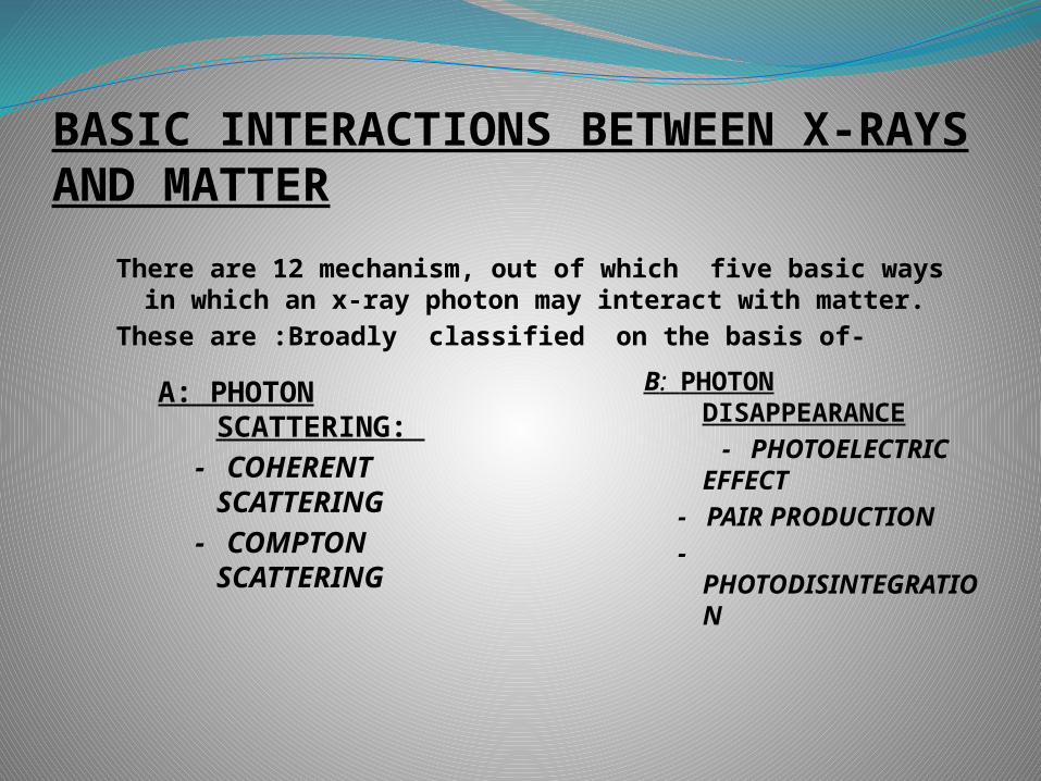
BASIC INTERACTIONS BETWEEN X-RAYS AND MATTER
There are 12 mechanism, out of which five basic ways in which an x-ray photon may interact with matter.
These are :Broadly classified on the basis of-
A: PHOTON SCATTERING:
- COHERENT SCATTERING
- COMPTON SCATTERING
B: PHOTON DISAPPEARANCE
- PHOTOELECTRIC EFFECT
- PAIR PRODUCTION
- PHOTODISINTEGRATION
1. COHERENT SCATTERING
Radiation undergoes Only Change in direction. No change in wavelengththats why sometime called “ unmodified scattering”
Coherent scattering of X-rays is an interaction of the wavetype in which the X-ray is deflected.
Coherent Scattering occurs mainly at low energies.
It is of two types :Both type described in terms of “ wave Particle
Interaction” ( also called “ Classical scattering”)
Thomson scattering : Single electron involved in the interaction.
Rayleigh scattering : Co-operative interaction of all the electrons.
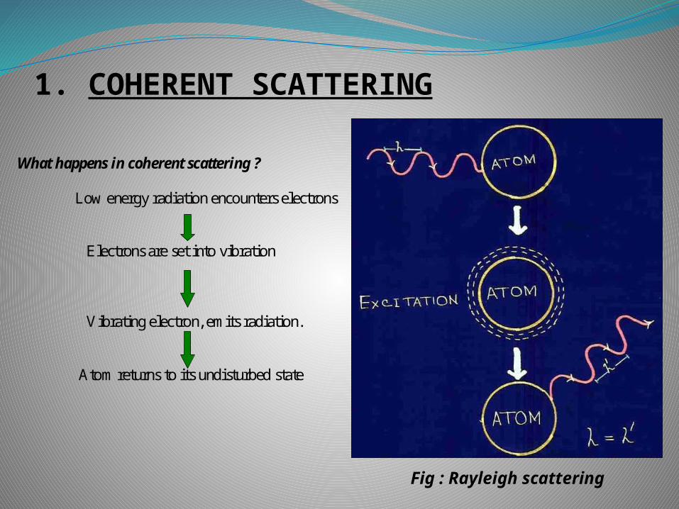
1. COHERENT SCATTERING
What happens in coherent scattering ? Low energy radiation encounters electrons
Electrons are set into vibration Vibrating electron, emits radiation. Atom returns to its undisturbed state
Fig : Rayleigh scattering
1. COHERENT SCATTERING
No ionization --- why??? because, no energy transfer. Only change of direction.
Only effect is to change direction of incident photon.
Less than 5%. Not important in diagnostic radiology. Produces scattered radiation but of negligible quantity.
2. PHOTOELECTRIC EFFECT
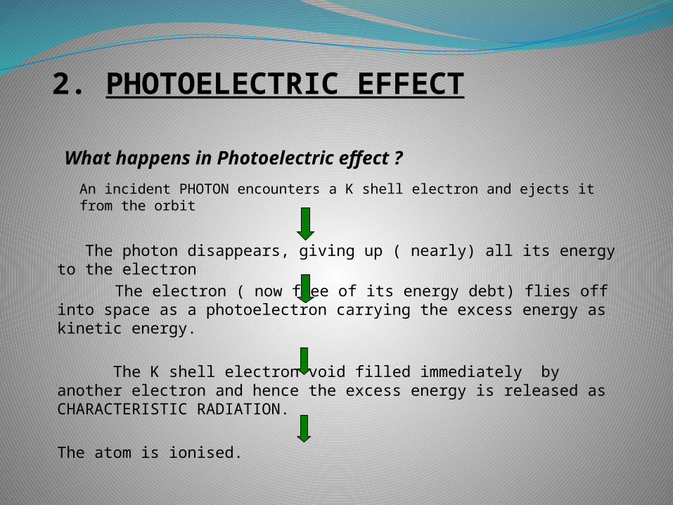
What happens in Photoelectric effect ?
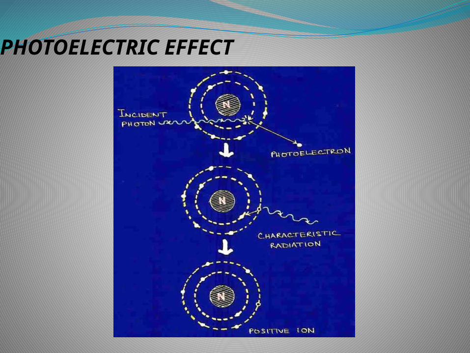
An incident PHOTON encounters a K shell electron and ejects it from the orbit
The photon disappears, giving up ( nearly) all its energy to the electron
The electron ( now free of its energy debt) flies off into space as a photoelectron carrying the excess energy as kinetic energy.
The K shell electron void filled immediately by another electron and hence the excess energy is released as CHARACTERISTIC RADIATION.
The atom is ionised.
PHOTOELECTRIC EFFECT
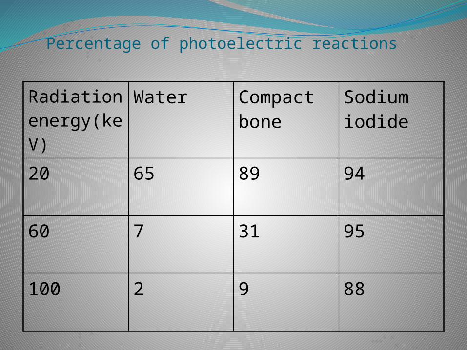
Percentage of photoelectric reactions
Radiation energy(keV)
Water Compact bone
Sodium iodide
20 65 89 94
60 7 31 95
100 2 9 88
CHARACTERISTIC RADIATIONCharacteristic radiation generated by the photoelectric effect is exactly the same The only difference in the modality used to eject the inner shell electron.
In x ray tube a high speed electron ejects the bound electron, while In photoelectric effect an X ray photon does the trick. In both cases the atom is left with an excess of energy = the binding energy of an ejected electron
Usually referred to as Secondary Radiation to differentiate It from scatter radiation……End result is same for both,“A Photon that is deflected from its original path”
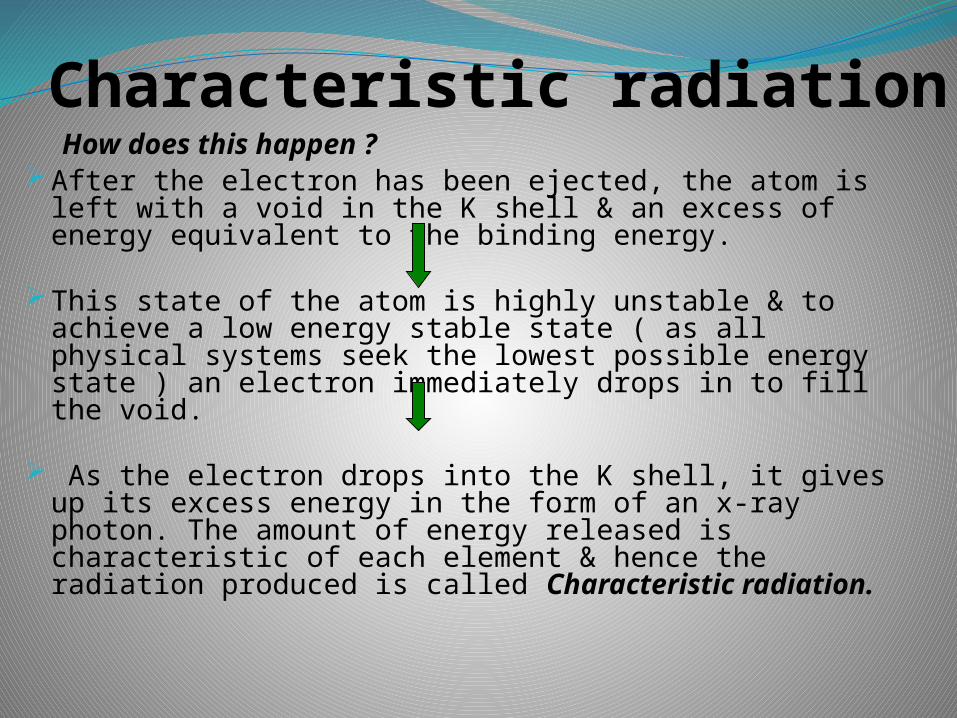
Characteristic radiationHow does this happen ?
After the electron has been ejected, the atom is left with a void in the K shell & an excess of energy equivalent to the binding energy.
This state of the atom is highly unstable & to achieve a low energy stable state ( as all physical systems seek the lowest possible energy state ) an electron immediately drops in to fill the void.
As the electron drops into the K shell, it gives up its excess energy in the form of an x-ray photon. The amount of energy released is characteristic of each element & hence the radiation produced is called Characteristic radiation.
2. PHOTOELECTRIC EFFECT
Thus the Photoelectric effect yields three end products :
Characteristic radiation A -ve ion ( photoelectron ) A+ve ion (atom deficient in one electron )
2. PHOTOELECTRIC EFFECT
Probability of occurrence : The incident photon energy > binding energy of the
electron.
Photon energy similar to electron binding energy
Photoelectric effect 1
(energy)³
The probability of a reaction increases sharply as the atomic no. increases
Photoelectric effect (atomic no.)³
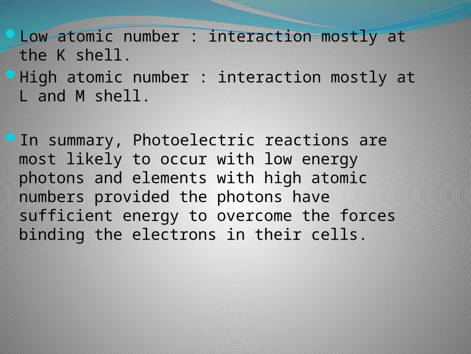
Low atomic number : interaction mostly at the K shell.
High atomic number : interaction mostly at L and M shell.
In summary, Photoelectric reactions are most likely to occur with low energy photons and elements with high atomic numbers provided the photons have sufficient energy to overcome the forces binding the electrons in their cells.
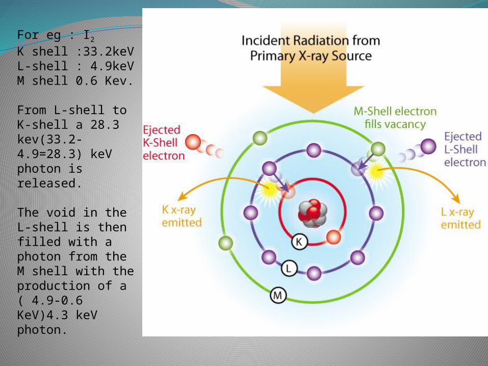
For eg : I2
K shell :33.2keVL-shell : 4.9keVM shell 0.6 Kev.
From L-shell to K-shell a 28.3 kev(33.2-4.9=28.3) keV photon is released.
The void in the L-shell is then filled with a photon from the M shell with the production of a ( 4.9-0.6 KeV)4.3 keV photon.
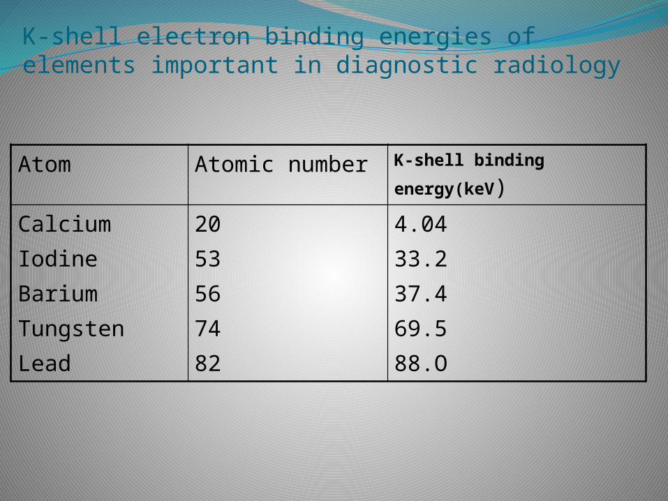
K-shell electron binding energies of elements important in diagnostic radiology
Atom Atomic number K-shell binding
energy(keV)
CalciumIodineBariumTungstenLead
2053567482
4.0433.237.469.588.O
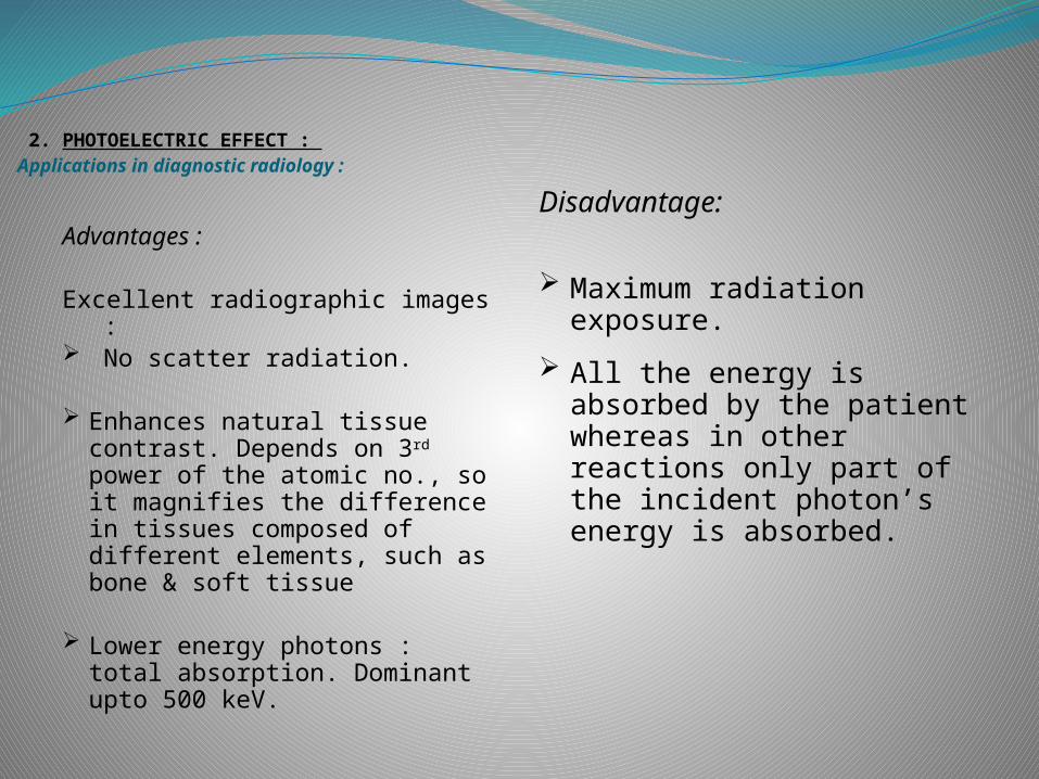
2. PHOTOELECTRIC EFFECT : Applications in diagnostic radiology :
Advantages :
Excellent radiographic images :
No scatter radiation.
Enhances natural tissue contrast. Depends on 3rd power of the atomic no., so it magnifies the difference in tissues composed of different elements, such as bone & soft tissue
Lower energy photons : total absorption. Dominant upto 500 keV.
Disadvantage:
Maximum radiation exposure.
All the energy is absorbed by the patient whereas in other reactions only part of the incident photon’s energy is absorbed.
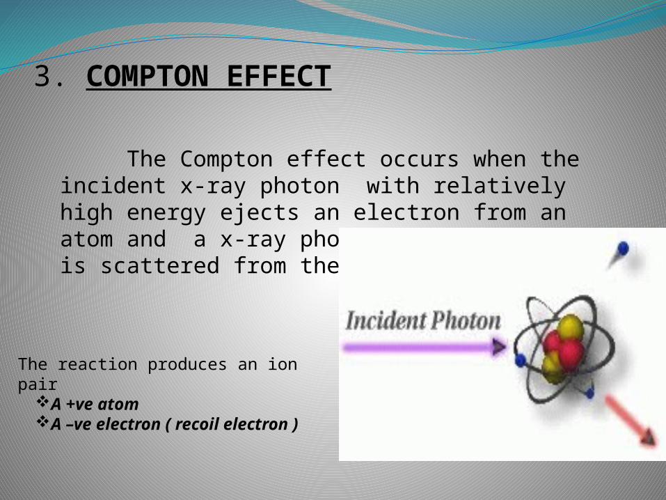
3. COMPTON EFFECT
The Compton effect occurs when the incident x-ray photon with relatively high energy ejects an electron from an atom and a x-ray photon of lower energy is scattered from the atom.
The reaction produces an ion pair
A +ve atomA –ve electron ( recoil
electron )
COMPTON SCATTERING
Almost all the scatter radiation that we encountered In diagnostic radiology comes from Compton Scattering
Kinetic energy of recoil electron
Energy of photon distributed
Retained by the deflected photon.
Two factors determine the amount of energy the photon transmits : The initial energy of the photon. Its angle of deflection.
1.Initial energy :- Higher the energy more difficult to deflect.High energy : Travel straight retaining most of the energy. Low energy : Most scatter back at angle of 180º
2. Angle of deflection :- Greater the angle, lesser the energy trasmitted. With a direct hit, maximum energy is transferred to the recoil electron. The photon retains some energy & deflects back along its original path at an angle of 180º.
3. COMPTON EFFECT
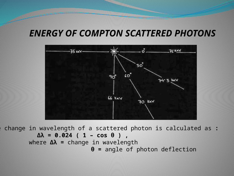
ENERGY OF COMPTON SCATTERED PHOTONS
· The change in wavelength of a scattered photon is calculated as :Δλ = 0.024 ( 1 – cos θ ) ,
where Δλ = change in wavelength θ = angle of photon deflection
3. COMPTON EFFECTProbability of occurence :
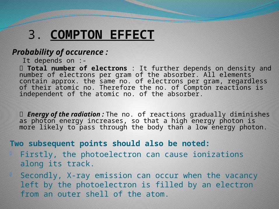
It depends on :- Total number of electrons : It further depends on density and number of electrons per gram of the absorber. All elements contain approx. the same no. of electrons per gram, regardless of their atomic no. Therefore the no. of Compton reactions is independent of the atomic no. of the absorber.
Energy of the radiation : The no. of reactions gradually diminishes as photon energy increases, so that a high energy photon is more likely to pass through the body than a low energy photon.
Two subsequent points should also be noted: Firstly, the photoelectron can cause ionizations along its track. Secondly, X-ray emission can occur when the vacancy left by
the photoelectron is filled by an electron from an outer shell of the atom.
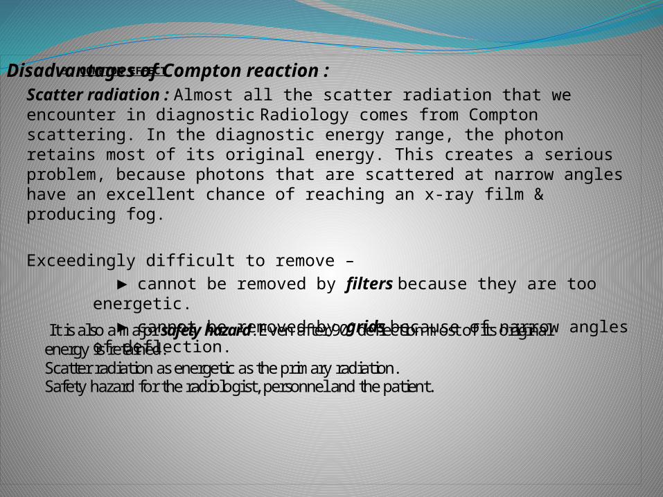
3. COMPTON EFFECTDisadvantages of Compton reaction :Scatter radiation : Almost all the scatter radiation that we encounter in diagnostic Radiology comes from Compton scattering. In the diagnostic energy range, the photon retains most of its original energy. This creates a serious problem, because photons that are scattered at narrow angles have an excellent chance of reaching an x-ray film & producing fog.
Exceedingly difficult to remove – ► cannot be removed by filters because they are too
energetic. ► cannot be removed by grids because of narrow angles of
deflection.
It is also a major safety hazard. Even after 90˚ deflection most of its original energy is retained. Scatter radiation as energetic as the primary radiation. Safety hazard for the radiologist, personnel and the patient.
4. PAIR PRODUCTION
No importance in diagnostic radiology.
What happens in Pair production ?A high energy photon interacts with the nucleus of an atom.
The photon disappears & its energy is converted into matter in the form
of two particles An electron A positron (particle with same mass as electron, but with
+ve charge.)
Mass of one electron is 0.51 MeV.2 electron masses are produced.So the interaction cannot take place with photon energy less
than 1.02 MeV.
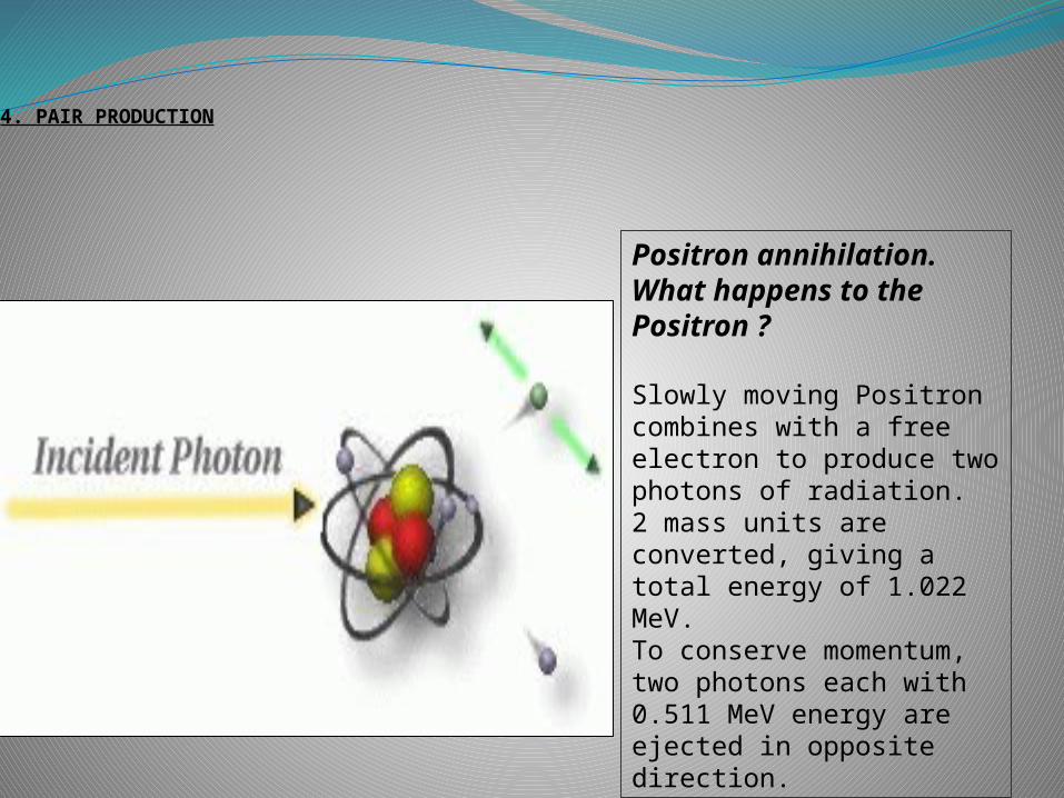
4. PAIR PRODUCTION
Positron annihilation. What happens to the Positron ?
Slowly moving Positron combines with a free electron to produce two photons of radiation. 2 mass units are converted, giving a total energy of 1.022 MeV. To conserve momentum, two photons each with 0.511 MeV energy are ejected in opposite direction.
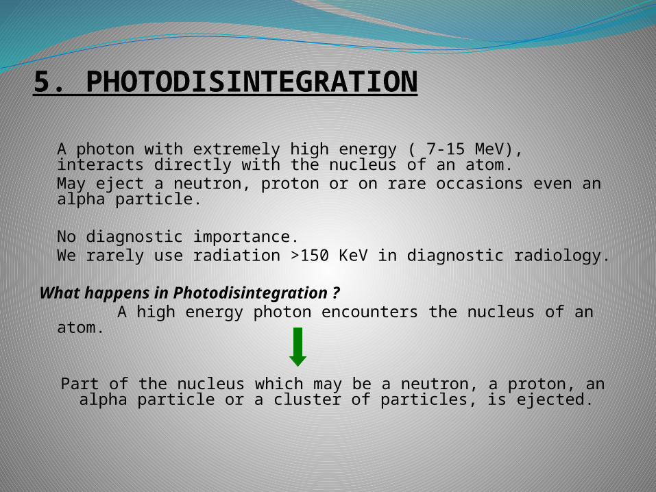
5. PHOTODISINTEGRATION
A photon with extremely high energy ( 7-15 MeV), interacts directly with the nucleus of an atom.May eject a neutron, proton or on rare occasions even an alpha particle.
No diagnostic importance. We rarely use radiation >150 KeV in diagnostic radiology.
What happens in Photodisintegration ? A high energy photon encounters the nucleus of an atom.
Part of the nucleus which may be a neutron, a proton, an alpha particle or a cluster of particles, is ejected.
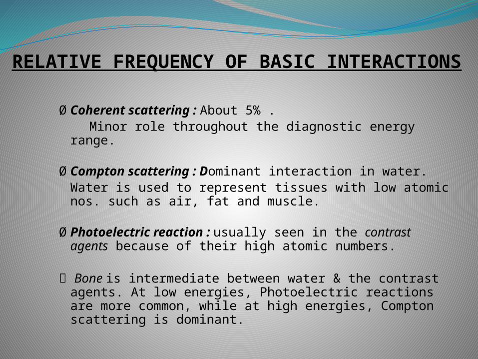
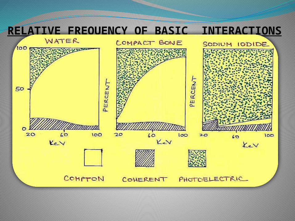
RELATIVE FREQUENCY OF BASIC INTERACTIONS
ØCoherent scattering : About 5% . Minor role throughout the diagnostic energy range.
ØCompton scattering : Dominant interaction in water.Water is used to represent tissues with low atomic nos. such as air, fat and muscle.
ØPhotoelectric reaction : usually seen in the contrast agents because of their high atomic numbers.
Bone is intermediate between water & the contrast agents. At low energies, Photoelectric reactions are more common, while at high energies, Compton scattering is dominant.
RELATIVE FREQUENCY OF BASIC INTERACTIONS
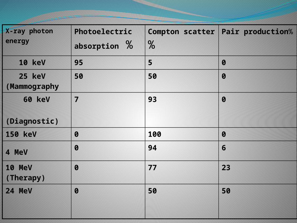
X-ray photon energy
Photoelectric
absorption %Compton
scatter %Pair production%
10 keV 95 5 0
25 keV (Mammography
50 50 0
60 keV (Diagnostic)
7 93 0
150 keV 0 100 0
4 MeV 0 94 6
10 MeV (Therapy)
0 77 23
24 MeV 0 50 50
Scatter Radiation
Scatter Radiation
Definition
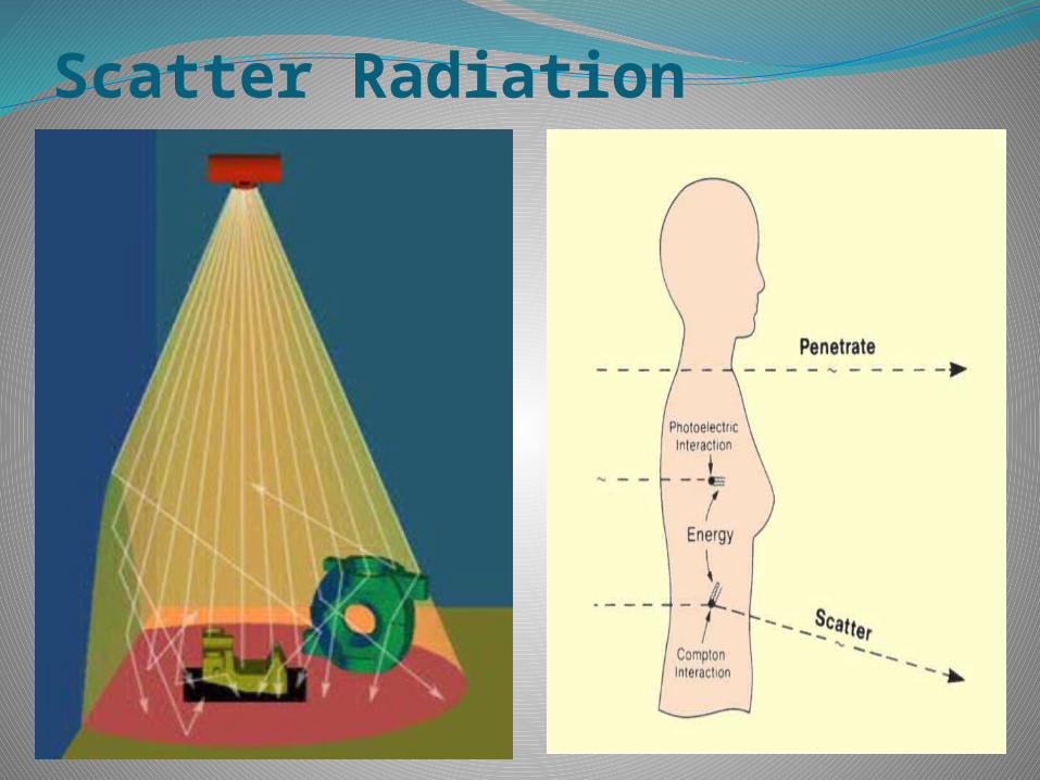
A type of secondary radiation composed of photons of lower energy than the photons that produced them and which travel in a different direction.
The term scatter radiation is synonymous with secondary radiation in the context of x-rays
Scatter radiation & Contrast - overview
Radiographic images are maps of radiation attenuation. Bones attenuate the most, air in lungs the least.
Good radiograph : maximum contrast difference between different tissues.
X-Ray beam enters body.
Large number of interactions producing scatter radiation.
Image contrast reduced depending on scatter radiation content reaching film.
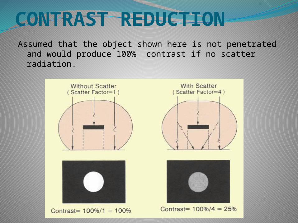
CONTRAST REDUCTIONAssumed that the object shown here is not penetrated and
would produce 100% contrast if no scatter radiation.
Sources of scattered radiation
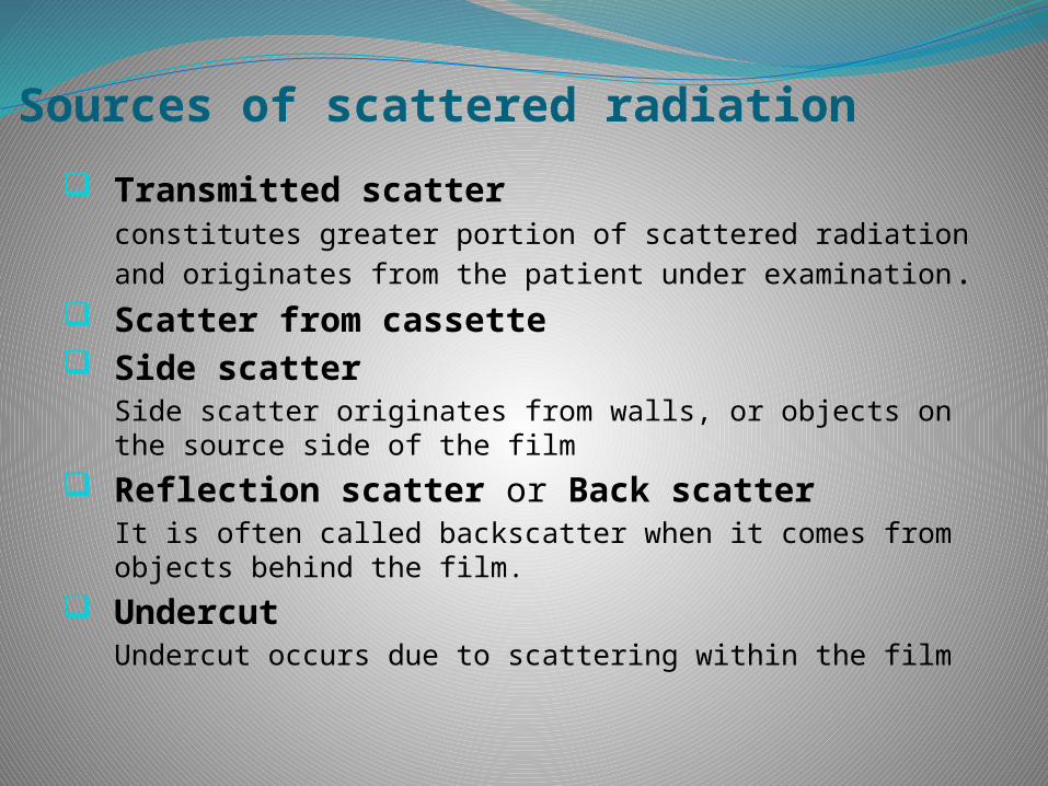
Transmitted scatter constitutes greater portion of scattered radiation and originates from the patient under examination.
Scatter from cassette Side scatter
Side scatter originates from walls, or objects on the source side of the film
Reflection scatter or Back scatter It is often called backscatter when it comes from objects behind the film.
Undercut Undercut occurs due to scattering within the film
Factors affecting scatter radiation
Field size
Kilo voltage (kVp)
Anatomical volume (Part thickness)
Factors affecting scatter radiationScatter radiation is maximum with high kvp
technique, large field , and thick parts---- Unfortunately, this is what we usually deal
with in diagnostic radiology.
The only variable we can control is kvp , but we have less control .
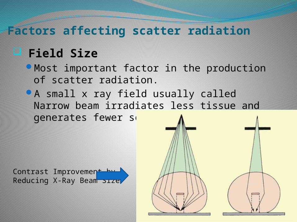
Factors affecting scatter radiation
Field SizeMost important factor in the production of
scatter radiation.A small x ray field usually called Narrow beam
irradiates less tissue and generates fewer scattered photons.
Contrast Improvement by Reducing X-Ray Beam Size
Factors affecting scatter radiationKvp
The effect of kvp on the production of scatter radiation is probably not as important as part thickness , and as field size.
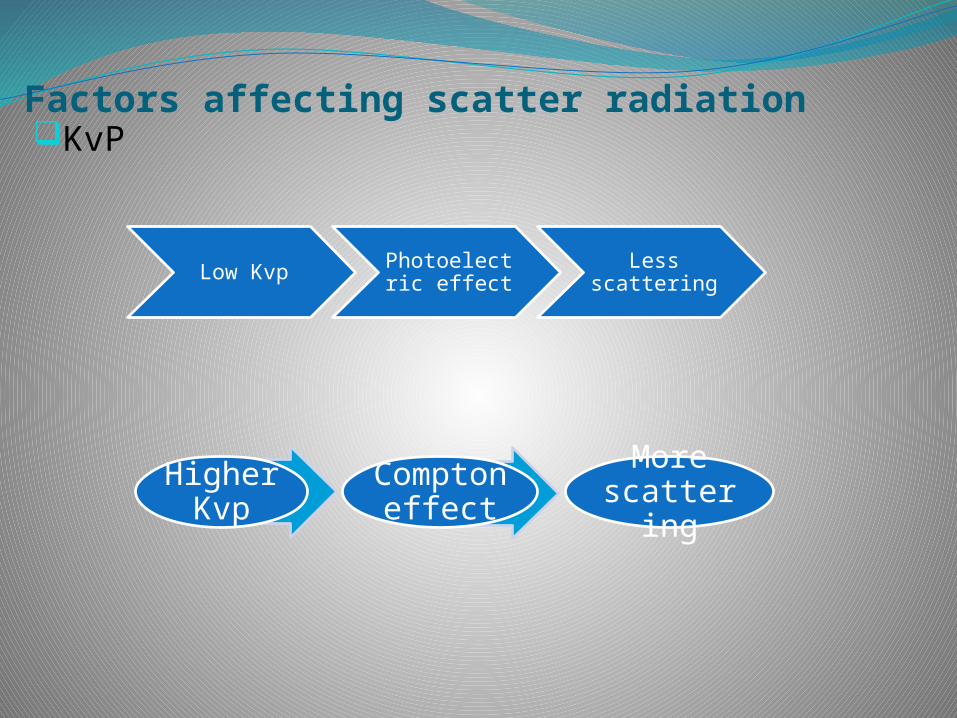
Factors affecting scatter radiationKvP
Low Kvp Photoelectric effect
Less scattering
Higher Kvp
Compton effect
More scattering
Factors affecting scatter radiation
Part thickness
Scatter radiation is directly proportional to the part thickness.
The operator has no control over this parameter.
Effects of scatter radiation
Reduction of contrast: Scattered photons Carry no useful information Contribute to film blackness(film fog)
Increased patient dose
Increased risk to personnel
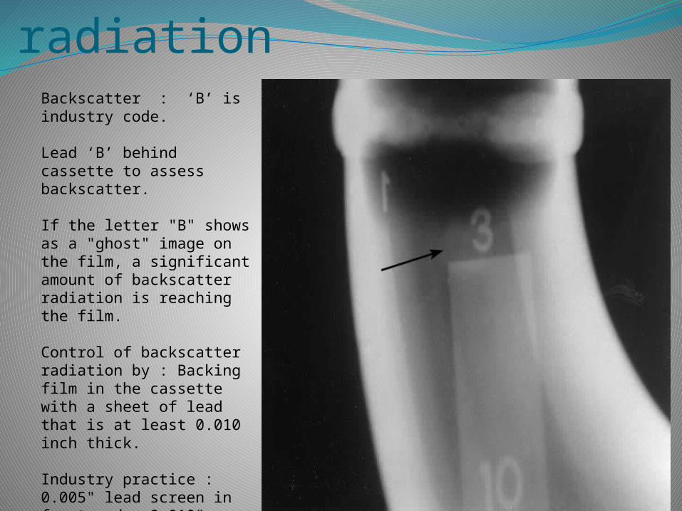
Control of scatter radiationBackscatter : ‘B’ is industry code.
Lead ‘B’ behind cassette to assess backscatter.
If the letter "B" shows as a "ghost" image on the film, a significant amount of backscatter radiation is reaching the film.
Control of backscatter radiation by : Backing film in the cassette with a sheet of lead that is at least 0.010 inch thick.
Industry practice : 0.005" lead screen in front and a 0.010" screen behind the film.
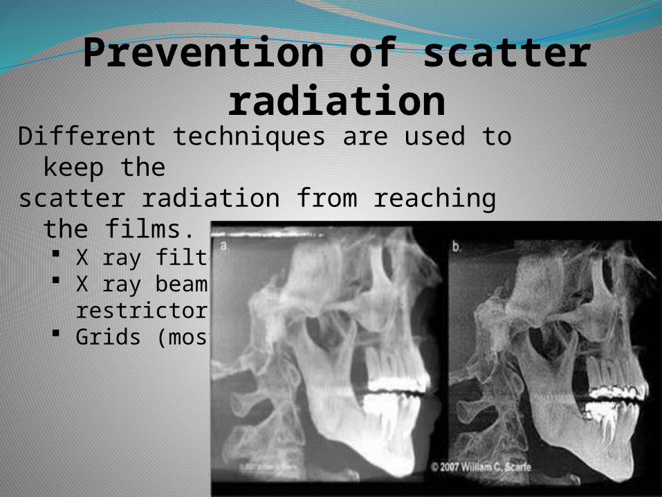
Different techniques are used to keep thescatter radiation from reaching the films.
X ray filters X ray beam
restrictors Grids (most important)
Prevention of scatter radiation
SUMMARYOnly two interactions are important in diagnostic radiology, the Photoelectric
effect & Compton scattering.Ø The Photoelectric effect
is the predominant interaction with low energy radiation & high atomic no. absorbers.
It generates no significant scatter radiation & produces high contrast in the x-ray image.
But, unfortunately it exposes the patient to a great deal of radiation.Ø Compton scattering is the most common interaction at higher diagnostic
energies. responsible for almost all scatter radiation.
radiographic image contrast is less compared to
photoelectric effect.Ø Coherent scattering is numerically unimportant.Ø Pair production & Photodisintegration occur at energies above
the useful energy range.
SUMMARY
Scatter Radiationsecondary radiation composed of photons of lower
energy than the photons that produced them and which travel in a different direction.
Factors affecting it : Field size Kilo voltage (kVp) Anatomical volume (Part thickness)
No useful information, causes film fog and increases patient exposure.

Thank you Have A nice day
T H A N K Y O U