Pharmacokinetic study of the structural components of
40
DMD: #50005 Pharmacokinetic study of the structural components of adenosine diphosphate-encapsulated liposomes coated with fibrinogen γ-chain dodecapeptide as a synthetic platelet substitute Kazuaki Taguchi, Hayato Ujihira, Shigeru Ogaki, Hiroshi Watanabe, Atsushi Fujiyama, Mami Doi, Yosuke Okamura, Shinji Takeoka, Yasuo Ikeda, Makoto Handa, Masaki Otagiri, Toru Maruyama Department of Biopharmaceutics, Graduate School of Pharmaceutical Sciences (K.T., H.U., S.O., H.W., M.O., T.M.), Center for Clinical Pharmaceutical Sciences (H.W., T.M.), Kumamoto University, Department of Life Science and Medical Bioscience, Graduate School of Advanced Science and Engineering (A.F., M.D., S.T., Y.I), Waseda University, Institute of Innovative Science and Technology (Y.O.), Tokai University, Department of Transfusion Medicine & Cell Therapy (M.H.), Keio University, Faculty of Pharmaceutical Sciences (M.O.), DDS Research Institute (M.O.), Sojo University DMD Fast Forward. Published on June 4, 2013 as doi:10.1124/dmd.112.050005 Copyright 2013 by the American Society for Pharmacology and Experimental Therapeutics. This article has not been copyedited and formatted. The final version may differ from this version. DMD Fast Forward. Published on June 4, 2013 as DOI: 10.1124/dmd.112.050005 at ASPET Journals on April 9, 2019 dmd.aspetjournals.org Downloaded from
Transcript of Pharmacokinetic study of the structural components of
DMD: #50005
1
Pharmacokinetic study of the structural components of adenosine
diphosphate-encapsulated liposomes coated with fibrinogen γ-chain dodecapeptide
as a synthetic platelet substitute
Kazuaki Taguchi, Hayato Ujihira, Shigeru Ogaki, Hiroshi Watanabe, Atsushi Fujiyama,
Mami Doi, Yosuke Okamura, Shinji Takeoka, Yasuo Ikeda, Makoto Handa, Masaki
Otagiri, Toru Maruyama
Department of Biopharmaceutics, Graduate School of Pharmaceutical Sciences (K.T.,
H.U., S.O., H.W., M.O., T.M.), Center for Clinical Pharmaceutical Sciences (H.W.,
T.M.), Kumamoto University,
Department of Life Science and Medical Bioscience, Graduate School of Advanced
Science and Engineering (A.F., M.D., S.T., Y.I), Waseda University, Institute of
Innovative Science and Technology (Y.O.), Tokai University,
Department of Transfusion Medicine & Cell Therapy (M.H.), Keio University,
Faculty of Pharmaceutical Sciences (M.O.), DDS Research Institute (M.O.), Sojo
University
DMD Fast Forward. Published on June 4, 2013 as doi:10.1124/dmd.112.050005
Copyright 2013 by the American Society for Pharmacology and Experimental Therapeutics.
This article has not been copyedited and formatted. The final version may differ from this version.DMD Fast Forward. Published on June 4, 2013 as DOI: 10.1124/dmd.112.050005
at ASPE
T Journals on A
pril 9, 2019dm
d.aspetjournals.orgD
ownloaded from
DMD: #50005
2
Running title: Disposition of H12-(ADP)-liposomes
Address for Correspondence:
Toru Maruyama, Ph.D
Department of Biopharmaceutics, Graduate School of Pharmaceutical Sciences,
Kumamoto University,
5-1 Oe-honmachi, Kumamoto 862-0973, Japan;
Tel: +81–96–361–4150
Fax: +81–96–362–7690
E-mail: [email protected]
The number of text pages: 35
The number of tables: 2
The number of figures: 5
The number of references: 37
The number of words in the Abstract: 224
The number of words in the Introduction: 731
The number of words in the Discussion: 1498
Abbreviation: RGD, arginine-glycine-aspartic acid; ADP, adenosine diphosphate; H12,
HHLGGAKQAGDV; H12-(ADP)-liposome, ADP-encapsulated liposomes modified
with a dodecapeptide; GP, glycoprotein; PEG, polyethyleneglycol; DPPC,
1,2-dipalmitoyl-sn-glycero-3-phosphatidylcholine; PEG-DSPE,
2-distearoyl-sn-glycero-3-phosphatidylethanolamine-N-[monomethoxypoly(ethylenegly
This article has not been copyedited and formatted. The final version may differ from this version.DMD Fast Forward. Published on June 4, 2013 as DOI: 10.1124/dmd.112.050005
at ASPE
T Journals on A
pril 9, 2019dm
d.aspetjournals.orgD
ownloaded from
DMD: #50005
3
col)]; DHSG, 1,5-Dihexadecyl-N-succinyl-L-glutamate; HPLC, high performance
liquid chromatography; ID, injected dose; HbV, hemoglobin-vesicles; MPS,
mononuclear phagocyte system.
This article has not been copyedited and formatted. The final version may differ from this version.DMD Fast Forward. Published on June 4, 2013 as DOI: 10.1124/dmd.112.050005
at ASPE
T Journals on A
pril 9, 2019dm
d.aspetjournals.orgD
ownloaded from
DMD: #50005
4
Abstract
Fibrinogen γ-chain (dodecapeptide HHLGGAKQAGDV, H12)-coated,
adenosine-diphosphate (ADP)-encapsulated liposomes (H12-(ADP)-liposomes) were
developed as a synthetic platelet alternative that specifically accumulates at bleeding
sites as the result of interactions with activated platelets via GPIIb/IIIa and augments
platelet aggregation by releasing ADP. The aim of this study is to characterize the
pharmacokinetic properties of H12-(ADP)-liposomes and structural components in rats,
and to predict the blood retention of H12-(ADP)-liposomes in humans. With use of
H12-(ADP)-liposomes in which the encapsulated ADP and liposomal membrane
cholesterol were radiolabled with 14C and 3H, respectively, it was found that the time
courses for the plasma concentration curves of 14C and 3H radioactivity showed that the
H12-(ADP)-liposomes remained intact in the blood circulation for up to 24 h after
injection, and were mainly distributed to the liver and spleen. However, the 14C and 3H
radioactivity of H12-(ADP)-liposomes disappeared from organs within 7 day after
injection. The encapsulated ADP was metabolized to allantoin, which is the final
metabolite of ADP in rodents, and was mainly eliminated in the urine, while the
cholesterol were mainly eliminated in feces. In addition, the half-life of the
H12-(ADP)-liposomes in humans was predicted to be approximately 96 hrs from
pharmacokinetic data obtained for mice, rats and rabbits using an allometric equation.
These results suggest that H12-(ADP)-liposome has potential with proper
pharmacokinetic and acceptable biodegradable properties as synthetic platelet
substitute.
This article has not been copyedited and formatted. The final version may differ from this version.DMD Fast Forward. Published on June 4, 2013 as DOI: 10.1124/dmd.112.050005
at ASPE
T Journals on A
pril 9, 2019dm
d.aspetjournals.orgD
ownloaded from
DMD: #50005
5
Introduction
As the numbers of patients with hematologic malignancies and solid tumors
increase, platelet transfusion represents one of the most essential prophylactic or
therapeutic treatments, because these disorders induce severe thrombocytopenia caused
by the intensive chemotherapy, surgical procedures and radiotherapy. However, platelet
transfusion can introduce a variety of complications such as bacterial infection, allergic
reaction and acute lung injury. In addition, donated platelet for blood transfusions can
only be stored for a period of 4 days in Japan and 5-7 days in the USA and Europe. This
has become a serious concern in our aging society and a stable supply in an emergency
situation such as disasters and pandemics needs to be on hand. To solve these problems,
various platelet substitutes, which consist of materials derived from blood components,
have been developed (Blajchman, 2003), such as solubilized platelet membrane protein
conjugated liposomes (Plateletsome) (Rybak and Renzulli, 1993), infusible platelet
membranes (IPM) (Graham et al., 2001), fibrinogen-coated albumin microcapsules
(Synthocyte) (Levi et al., 1999), red blood cells with bound fibrinogen (Agam and
Livne, 1992), liposomes bearing fibrinogen (Casals et al., 2003),
arginine-glycine-aspartic acid (RGD) peptidebound red blood cells
(Thromboerythrocyte) (Coller et al., 1992) and fibrinogen-conjugated albumin polymers
(Takeoka et al., 2001). However, these platelet substitutes have not yet been approved
for clinical use.
Adenosine diphosphate (ADP)-encapsulated liposomes modified with a
dodecapeptide (HHLGGAKQAGDV, H12) (H12-(ADP)-liposome) was developed as a
new type of synthetic platelet alternative. The glycoprotein (GP) IIb/IIIa, which is
present on the platelet membranes, is converted from an inactive to an active form when
This article has not been copyedited and formatted. The final version may differ from this version.DMD Fast Forward. Published on June 4, 2013 as DOI: 10.1124/dmd.112.050005
at ASPE
T Journals on A
pril 9, 2019dm
d.aspetjournals.orgD
ownloaded from
DMD: #50005
6
platelets adhere to collagen that is exposed on sites of vascular injury (Takagi et al.,
2002; Xiao et al., 2004), and platelet aggregation is mediated by fibrinogen by bridging
adjacent platelets through GPIIb/IIIa in an activation-dependent manner in the
circulation. Among several GPIIb/IIIa recognized sequence sites in fibrinogen such as
the RGD-based sequences (95RGDF98 and 572RGDS575 in the Aα chains) and H12
(400HHLGGAKQAGDV411) in the carboxy-terminus of the γ-chain (Kloczewiak et al.,
1982; Kloczewiak et al., 1984; Hawiger et al., 1989), H12 is a specific binding site of
the ligand for activated GPIIb/IIIa (Lam et al., 1987; Andrieux et al., 1989), whereas
RGD related peptides are non-specific with respect to a wide variety of integrins from
various cell types (Phillips et al., 1991). In addition, when ADP is released from
activated platelets, it functions as potent platelet agonist. Thus, these modifications to
H12-(ADP)-liposomes enable them to specifically interact with activated platelets,
resulting in platelet aggregation. In fact, H12-liposomes with polyethyleneglycol
(PEG)-surface modification specifically accumulate at the site of an injury in vivo and
were determined to shorten bleeding time in a dose-dependent manner in a
thrombocytopenic rat and a rabbit model (Okamura et al., 2005; Okamura et al., 2009;
Okamura et al., 2010a; Okamura et al., 2010b; Nishikawa et al., 2012). Therefore, these
findings prompted us to conclude that H12-(ADP)-liposomes have considerable
potential for use as an alternative for actual platelets in clinical settings.
Before new drugs are approved for clinical use, they are required to undergo a
wide variety of evaluations, including physicochemical tests, pre-clinical studies and
clinical trials. As described above, pre-clinical studies of H12-(ADP)-liposomes have
resulted in pharmacological evidence to indicate that they can be used as a platelet
substitute (Okamura et al., 2005; Okamura et al., 2009; Okamura et al., 2010a; Okamura
This article has not been copyedited and formatted. The final version may differ from this version.DMD Fast Forward. Published on June 4, 2013 as DOI: 10.1124/dmd.112.050005
at ASPE
T Journals on A
pril 9, 2019dm
d.aspetjournals.orgD
ownloaded from
DMD: #50005
7
et al., 2010b; Nishikawa et al., 2012). However, information concerning
pharmacokinetic properties is lacking, especially the disposition and retention of each
component in tissues after injection. Our strategy for the development of
H12-(ADP)-liposome is based on the fact that, not only better pharmacological effects,
but also acceptable biodegradable properties (no accumulation or retention) need to be
documented. In addition, pre-clinical pharmacokinetic studies in various mammalian
species are essential, as the results of such studies can be extrapolated to humans,
allowing appropriate dosing regimens to be estimated in the case of humans.
In the present study, we report on an evaluation of the pharmacokinetic
properties of the H12-(ADP)-liposomes and components thereof, from the standpoint of
stability in the blood circulation and the metabolism and excretion of each component.
For this purpose, we prepared H12-(ADP)-liposomes that were 14C, 3H double
radiolabeled, in which the encapsulated ADP and membrane component (cholesterol)
were labeled with 14C and 3H, respectively. Furthermore, we predicted some important
pharmacokinetic parameters, especially retention in the blood circulation, in humans,
based on data obtained in pharmacokinetic studies in mice, rats and rabbits.
This article has not been copyedited and formatted. The final version may differ from this version.DMD Fast Forward. Published on June 4, 2013 as DOI: 10.1124/dmd.112.050005
at ASPE
T Journals on A
pril 9, 2019dm
d.aspetjournals.orgD
ownloaded from
DMD: #50005
8
Materials and Methods
Reagents
Cholesterol and 1,2-dipalmitoyl-sn-glycero-3-phosphatidylcholine (DPPC)
were purchased from Nippon Fine Chemical (Osaka, Japan), and
2-distearoyl-sn-glycero-3-phosphatidylethanolamine-N-[monomethoxypoly(ethylenegly
col)] (PEG-DSPE, 5.1 kDa) was from NOF (Tokyo, Japan).
1,5-Dihexadecyl-N-succinyl-L-glutamate (DHSG) and H12-PEG-Glu2C18, in which
the fibrinogen γ-chain dodecapeptide (C-HHLGGAKQAGDV, Cys-H12) was
conjugated to the end of the PEG-lipids, were synthesized as previous reported
(Okamura et al., 2005). Allantoin, uric acid, hypoxantine, xanthine and ADP were
obtained from Sigma-Aldrich (St Louis, MO, USA).
Preparation of 14C, 3H double labled H12-(ADP)-liposomes
Firstly, 14C labeled H12-(ADP)-liposomes were prepared under sterile
conditions as previously reported, with minor modifications (Okamura et al., 2009). In
brief, DPPC (1000 mg, 1.36 mmol), cholesterol (527 mg, 1.36 mmol), DHSG (189 mg,
272 μmol), PEG-DSPE (52 mg, 9.0 μmol) and H12-PEG-Glu2C18 (47 mg, 9.0 μmol)
were dissolved in t-butyl alcohol and then freeze-dried. The resulting mixed lipids were
hydrated with phosphate-buffered saline (pH 7.4) containing ADP (1 mM) and
[8-14C]ADP (1.85 MBq; Moravec Biochemicas, Inc., USA), and extruded through
membrane filters (pore size, 0.22 μm; Durapore®; Millipore, Tokyo, Japan). Liposomes
were washed with phosphate-buffered saline by centrifugation (100000 g, 30 min, 4°C),
and the remaining ADP was eliminated by sephadexG25. The diameter and
Zeta-potential of the 14C labeled H12-(ADP)-liposomes used in this study are regulated
This article has not been copyedited and formatted. The final version may differ from this version.DMD Fast Forward. Published on June 4, 2013 as DOI: 10.1124/dmd.112.050005
at ASPE
T Journals on A
pril 9, 2019dm
d.aspetjournals.orgD
ownloaded from
DMD: #50005
9
at 250 ± 50 nm and -10 ± 0.9 mV, respectively. The 5-10% of added ADP was
encapsulated in the inner space of the vesicle.
The 3H labeling of 14C labeled H12-(ADP)-liposomes, to prepare 14C and 3H
double labeled H12-(ADP)-liposomes was carried out according to a previous report
(Taguchi et al., 2009). The 14C labeled H12-(ADP)-liposomes (1 mL) was mixed with
[1,2-3H(N)]-cholesterol solution (10 μL), (PerkinElmer, Yokohama, Japan) and
incubated for 12 hrs at room temperature. 14C, 3H labeled H12-(ADP)-liposomes were
filtered through a sterile filter to remove aggregates (pore size, 450 nm). Before being
used in pharmacokinetic experiments, all of the samples were mixed with unlabeled
H12-(ADP)-liposomes. To employ the same procedure using H12-(ADP)-liposomes and
[1,2-3H(N)]-cholesterol, 3H labeled H12-(ADP)-liposomes, which did not contain
[8-14C]ADP, were prepared for the pharmacokinetic studies in mice and rabbits.
Animals
All animal experiments were undertaken in accordance with the guideline
principle and procedure of Kumamoto University for the care and use of laboratory
animals. Experiments were carried out with male ddY mice (28-30 g body weight;
Japan SLC, Inc. Shizuoka Japan), male Sprague-Dawley (SD) rats (180-210 g body
weight; Kyudou Co. Kumamoto, Japan) and male New Zealand White (NZW) rabbits
(2.0-2.2 kg body weight; Biotek Co. Saga, Japan). All animals were maintained under
conventional housing conditions, with food and water ad libitum in a
temperature-controlled room with a 12-hrs dark/light cycle.
Pharmacokinetic studies
This article has not been copyedited and formatted. The final version may differ from this version.DMD Fast Forward. Published on June 4, 2013 as DOI: 10.1124/dmd.112.050005
at ASPE
T Journals on A
pril 9, 2019dm
d.aspetjournals.orgD
ownloaded from
DMD: #50005
10
Administration and collecting blood and organs in rats
Twenty-four SD rats were anesthetized with diethyl ether and received a single
injection of 14C, 3H labeled H12-(ADP)-liposomes (10 mg lipids/kg (n=16), 20 mg
lipids/kg (n=4) and 40 mg lipids/kg (n=4)). In all rat groups, four rats were selected to
undergo the plasma concentration test. Under ether anesthesia, approximately 200 μL
blood samples in all administration groups were collected from tail vein at multiple time
points after the injection of the 14C, 3H labeled H12-(ADP)-liposomes (3, 10, 30 min, 1,
2, 3, 6, 12, 24, 48 and 168 hrs) and the plasma was separated by centrifugation (3000 g,
5 min). After collecting the last blood sample (168 hrs), the rats were sacrificed for
excision of organs (kidney, liver, spleen, lung and heart). Urine and feces were collected
at fixed intervals in a metabolic cage. In addition, the four rats were sacrificed and
organs were collected at 2, 6, 24 hrs after an injection of 14C, 3H labeled
H12-(ADP)-liposomes at a dose of 10 mg lipids/kg.
Administration and collection of blood and organs in mice and rabbits
Twenty-eight ddY mice received a single injection of 3H labeled
H12-(ADP)-liposomes (10 mg lipids/kg) in the tail vein under ether anesthesia. At each
time after the injection of 3H labeled H12-(ADP)-liposomes (3, 30 min, 1, 3, 6, 12, 24
hrs), four mice were anesthetized with ether and blood was collected from the inferior
vena cava, and plasma was obtained by centrifugation (3000 g, 5 min).
Four NZW rabbits received a single injection of 3H labeled
H12-(ADP)-liposomes at a dose of 10 mg lipids/kg. The blood was collected from the
auricular veins at each time after injection (3, 10, 30 min, 1, 2, 12, 24, 36, 48, 72 hrs),
and plasma was obtained by centrifugation (3000 g, 5 min).
This article has not been copyedited and formatted. The final version may differ from this version.DMD Fast Forward. Published on June 4, 2013 as DOI: 10.1124/dmd.112.050005
at ASPE
T Journals on A
pril 9, 2019dm
d.aspetjournals.orgD
ownloaded from
DMD: #50005
11
Measurement of 14C and 3H radioactivity
Plasma samples were solubilized in a mixture of Soluene-350 (Perkin Elmer,
Yokohama, Japan) and isopropyl alcohol (at a ratio of 1/1) for 24 hrs at 50°C. The organ
samples were rinsed with saline, minced, and solubilized in Soluene-350 for 24 hrs at
50°C. Urine and feces were also weighed and solubilized in Soluene-350. All samples
were decolorized by treatment with a hydrogen peroxide solution after treatment of
Soluene-350 or isopropyl alcohol. The 14C, 3H radioactivity was determined by liquid
scintillation counting (LSC-5121, Aloka, Tokyo, Japan) with Hionic Fluor (Perkin
Elmer, Yokohama, Japan).
Analysis of metabolites of encapsulated ADP
ADP metabolites in urine were determined by high performance liquid
chromatography (HPLC), as described previously (George et al., 2006). A part of the
urine obtained in the pharmacokinetic study in rats was used for this analysis, and
aliquots of urine samples (2.5 mL) were mixed with 200 μL of 10% sulphuric acid. Just
before the analysis, the urine samples were centrifuged and filtered through a
Dismic-25cs (ADVANTEC, Tokyo, Japan, 0.2 、μm pore size) and diluted ten-fold with
water after adjusting the pH to 7 with 0.01N sodium hydroxide and 0.01N sulphuric
acid. A standard solution containing ADP, allantoin, uric acid, hypoxantine and
xanthine was prepared as reported in a previous study (George et al., 2006). The HPLC
system consisted of a Waters 2695 pump (Waters, Massachusetts, USA), a Waters 2487
detector (Waters, Massachusetts, USA) operated at 220 nm. LC analyses were achieved
with a 250 × 4 mm, 5 μm LiChrospher® 100 RP-18 endcapped column (LiChroCART®
This article has not been copyedited and formatted. The final version may differ from this version.DMD Fast Forward. Published on June 4, 2013 as DOI: 10.1124/dmd.112.050005
at ASPE
T Journals on A
pril 9, 2019dm
d.aspetjournals.orgD
ownloaded from
DMD: #50005
12
250-4, Merck, Darmstadt, Germany). Furthermore, each ADP metabolite separated by
HPLC was collected by a fraction collector (CHF121SA, ADVANTEC, Tokyo, Japan)
and 14C radioactivity was determined by liquid scintillation counting with Hionic Fluor.
Interspecies scaling of pharmacokinetic parameters
Allometric relationships between various pharmacokinetic parameters (P) and
body weight (W) were plotted on a log-log scale. Linear regression of the logarithmic
values was calculated using the least-squares method using Eq. (1) (Boxenbaum, 1984).
P = α ・ Wβ (1)
P is the parameter of interest (distribution volume (Vdss) or clearance (CL)), W is the
body weight (kg), and α and β are the coefficient and exponent of the allometric
equation, respectively. The average body weights of 0.034 kg (mouse), 0.242 kg (rat),
2.08 kg (rabbit) and 70 kg (human) were used for prediction of Vdss and CL for human.
After predicting of Vdss and CL for humans (70 kg) using Eq. (1), the half-life for
human was estimated.
Data Analysis
A non-compartment model was used for the pharmacokinetic analysis. Each
parameter, half-life (t1/2, hr), mean residence time (MRT, hr), area under the
concentration-time curve (AUC, hr・% of dose/mL), clearance (CL, mL/hr), distribution
volume (Vdss, mL), was calculated using the moment analysis program available on
Microsoft Excel. (Yamakawa et al., 2013) Data are shown as means ± SD for the
indicated number of animals.
This article has not been copyedited and formatted. The final version may differ from this version.DMD Fast Forward. Published on June 4, 2013 as DOI: 10.1124/dmd.112.050005
at ASPE
T Journals on A
pril 9, 2019dm
d.aspetjournals.orgD
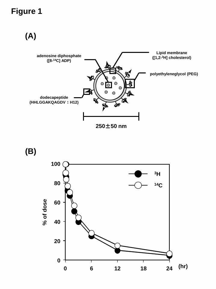
ownloaded from
DMD: #50005
13
Results
Pharmacokinetics of H12-(ADP)-liposome components in rats
In order to investigate the pharmacokinetics of each component of the
H12-(ADP)-liposomes, 14C, 3H labeled H12-(ADP)-liposomes, in which the
encapsulated ADP was labeled with 14C and the membrane component (cholesterol) was
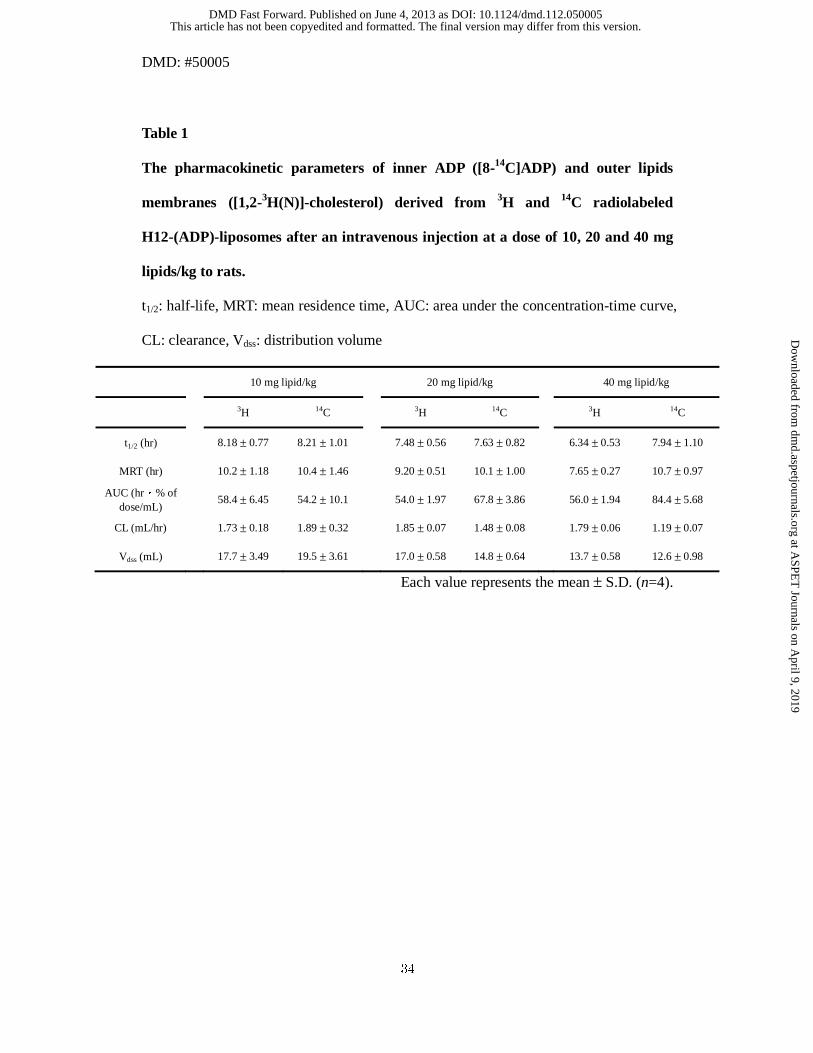
labeled with 3H, were prepared (Figure 1A). As shown in Fig. 1B and Table 1, the
plasma concentration curves and pharmacokinetic parameters for 14C radioactivity and
3H radioactivity were similar. These data indicate that the structure of the
H12-(ADP)-liposomes remained intact in the blood circulation for periods of up to 24
hrs after injection in rats.
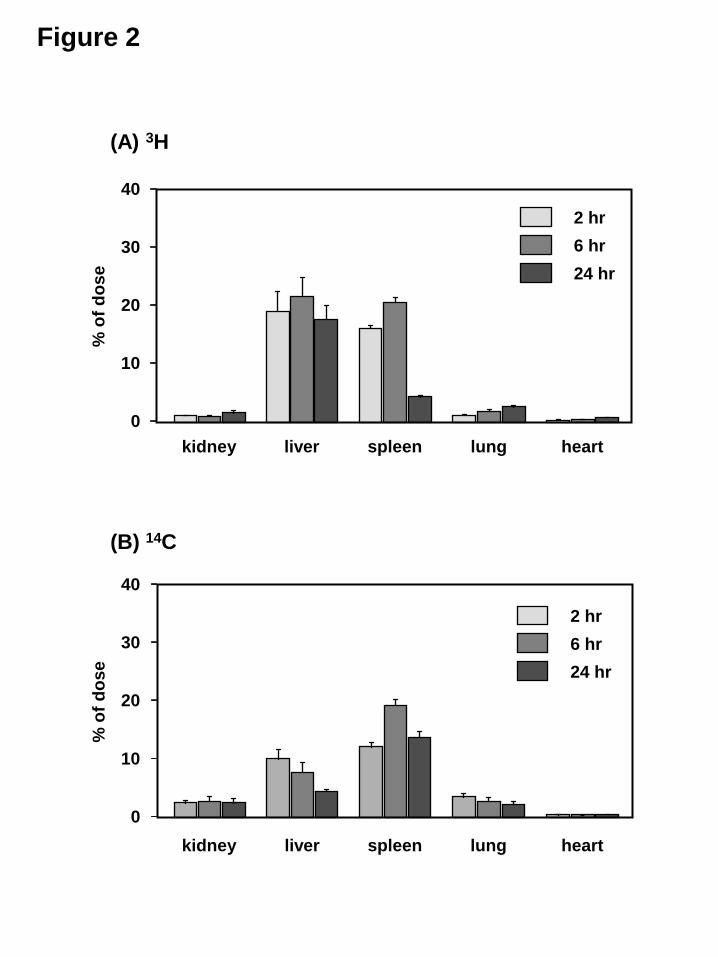
Moreover, we evaluated the tissue distribution of both the encapsulated ADP
and membrane component (cholesterol) of the H12-(ADP)-liposomes. Figure 2 shows
the tissue distribution in organs at 2, 6 and 24 hrs after the administration of 14C, 3H
labeled H12-(ADP)-liposomes at a dose of 10 mg lipids/kg to rats. Among these organs,
the majority of both the 14C and 3H radioactivity of the H12-(ADP)-liposomes were
distributed in the liver and spleen. However, both the 14C and 3H radioactivity of the
H12-(ADP)-liposomes were eliminated from each organ, and the activity essentially
disappeared within 7 days after injection (data not shown). These data indicate that the
H12-(ADP)-liposomes are mainly distributed to the liver and spleen, but the retention in
these organs is negligible.
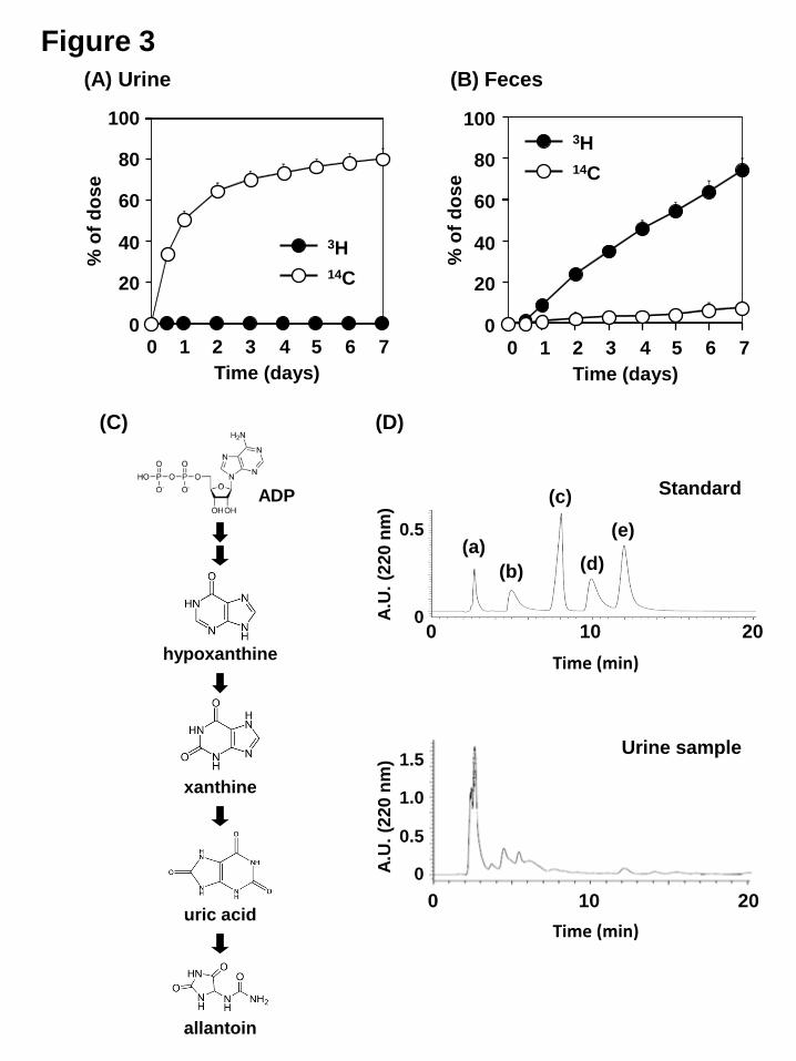
In order to identify the excretion pathway of the H12-(ADP)-liposomes, the
levels of 14C and 3H in urine and feces were measured (Fig. 3A and B). The 14C was
excreted mainly in the urine (80.4±4.9 % of the injected dose (ID) at 7 days after
injection), but was low in feces (7.6±2.7 % of ID at 7 day after injection). On the other
This article has not been copyedited and formatted. The final version may differ from this version.DMD Fast Forward. Published on June 4, 2013 as DOI: 10.1124/dmd.112.050005
at ASPE
T Journals on A
pril 9, 2019dm
d.aspetjournals.orgD
ownloaded from
DMD: #50005
14
hand, the majority of the 3H was excreted in the feces (74.2±5.7% % of ID at 7 days
after injection), and excretion into the urine was essentially nil. In addition, as shown in
Figure 3C, it is well known that, in rodents, endogenous ADP is ultimately metabolized
to allantoin and excreted. Thus, we qualitatively determined the fate of the encapsulated
ADP of the H12-(ADP)-liposomes using an HPLC method. Figure 3D shows the
separated peaks for ADP and its metabolites in the standard solution and in a urine
sample 6 hours after the administration of the H12-(ADP)-liposomes to a rat.
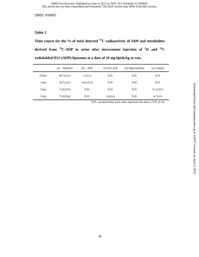
Furthermore, to exclude the effect of endogenous ADP and its metabolites, we
measured the 14C radioactivity of each peak that had been separated by HPLC. As a
result, almost all the 14C radioactivity was detected in the peak corresponding to
allantoin, which is the final metabolite of ADP in rodents, in the urine sample (Table 2).
These results indicate that more than 75% of each structural component of the
H12-(ADP)-liposome is excreted from the body within 7 days after injection, and the
encapsulated ADP and membrane component (cholesterol) derived from
H12-(ADP)-liposomes were metabolized to final metabolites and excreted into the urine
and feces, respectively.
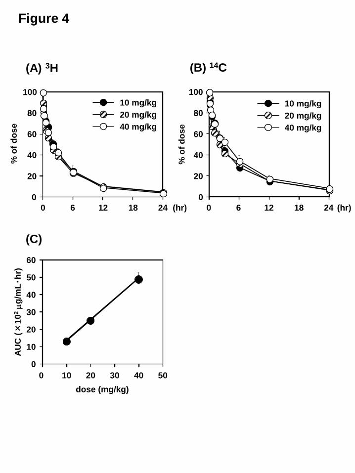
Dose-dependency of H12-(ADP)-liposomes pharmacokinetics.
Figure 4 shows the time courses for the plasma concentration for the 14C, 3H
labeled H12-(ADP)-liposomes administered to rats at doses of 10, 20 and 40 mg
lipids/kg. No significant difference was found in the plasma concentration curve or
pharmacokinetic parameters among all groups (Figure 4A and B). In fact, a linear
relationship between the administration dose and the area under the concentration-time
curve (AUC) was found, the values for which were calculated based on the lipids
This article has not been copyedited and formatted. The final version may differ from this version.DMD Fast Forward. Published on June 4, 2013 as DOI: 10.1124/dmd.112.050005
at ASPE
T Journals on A
pril 9, 2019dm
d.aspetjournals.orgD
ownloaded from
DMD: #50005
15
concentration (Figure 4C). These data indicate that the disposition of the
H12-(ADP)-liposomes is linear for a dose of 40 mg lipids/kg.
Moreover, the tissue distribution of both the encapsulated ADP and the
membrane lipids component (cholesterol) of the 14C, 3H labeled H12-(ADP)-liposomes
was evaluated at 7 days after the injection of H12-(ADP)-liposomes at a dose of 10, 20,
40 mg lipids/kg. The level of 14C and 3H radioactivity was nearly undetectable in the
observed organs (kidney, liver, spleen, lung and heart) (data not shown). In addition, the
radioactive 14C was excreted mainly in the urine (80.4±4.9 %, 52.1±3.6 %, 58.4±7.1 %
of ID at 7 days after the injection at a dose of 10, 20, 40 mg lipids/kg, respectively), but
was low in feces (7.6±2.7 %, 6.5±2.9 %, 2.5±1.9 % of ID at 7 days after the injection at
a dose of 10, 20, 40 mg lipids/kg, respectively). On the other hand, the majority of the
radioactive 3H was excreted in the feces (74.2±5.7%, 98.9±14.9 %, 70.6±6.2 % of ID at
7 days after the injection at a dose of 10, 20, 40 mg lipids/kg, respectively), and small
portion of the 3H radioactivity was excreted into the urine. These data indicate that more
than 75% of H12-(ADP)-liposomes are eliminated within 7 days after injection and
retention in the body can be limited to detect at a dose of up to 40 mg lipids/kg.
Pharmacokinetics of the H12-(ADP)-liposomes in mice and rabbits
To calculate the pharmacokinetic parameters of the H12-(ADP)-liposomes in
mice and rabbits, the 3H labeled H12-(ADP)-liposomes were administered to mice and
rabbits at a dose of 10 mg lipids/kg. According to the pharmacokinetic parameters
calculated from the plasma concentration curve, the CL and Vdss of the 3H labeled
H12-(ADP)-liposomes in mice were 0.54±0.12 mL/hr and 3.81±0.35 mL, respectively,
while the values in the case of rabbits were 23.5±2.8 mL/hr and 827±163 mL,
This article has not been copyedited and formatted. The final version may differ from this version.DMD Fast Forward. Published on June 4, 2013 as DOI: 10.1124/dmd.112.050005
at ASPE
T Journals on A
pril 9, 2019dm
d.aspetjournals.orgD
ownloaded from
DMD: #50005
16
respectively (Supplemental Table 1).
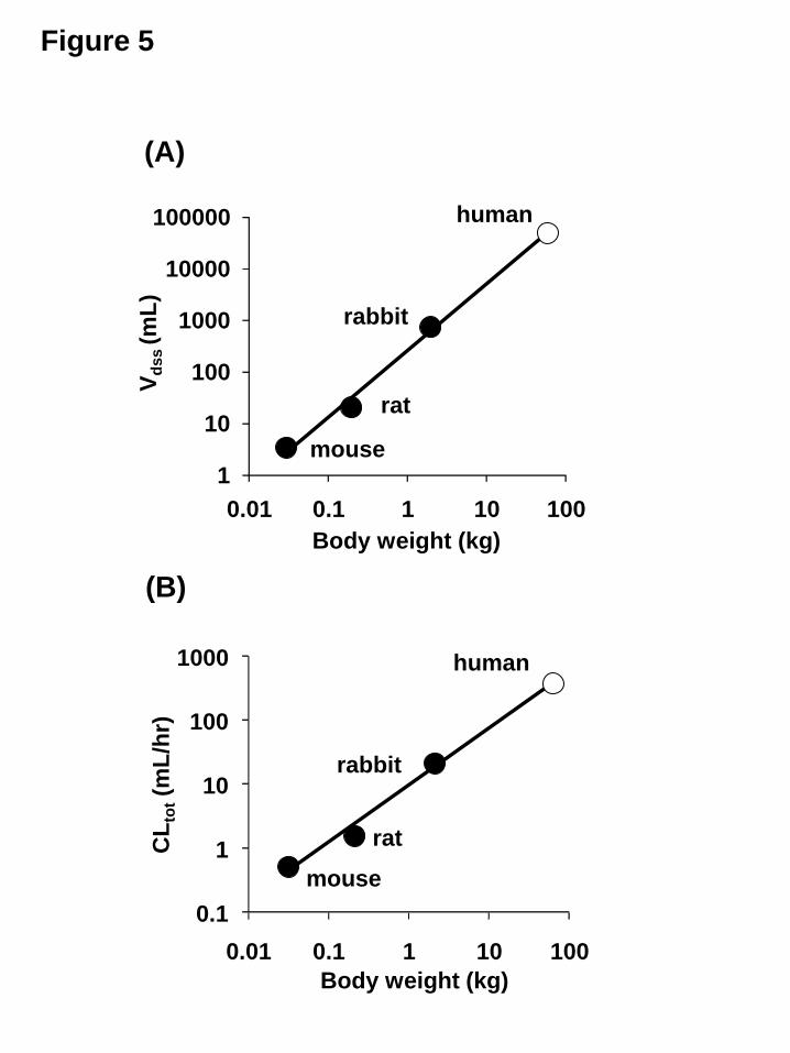
Prediction of pharmacokinetics of the H12-(ADP)-liposomes in human.
To predict the pharmacokinetics in humans, we examined the allometric
relationship between Vdss and body weight (Fig. 5A) and CL and body weight (Fig. 5B)
in mice, rats, rabbits using the results summarized in Table 1 and supplemental Table 1.
As shown in Figure 5A and B, a good correlation in both relationships was observed.
Furthermore, we calculated the half-life, based on extrapolation, of the
H12-(ADP)-liposomes that were administered at a dose of 10 mg lipids/kg in humans to
be approximately 96 hrs.
This article has not been copyedited and formatted. The final version may differ from this version.DMD Fast Forward. Published on June 4, 2013 as DOI: 10.1124/dmd.112.050005
at ASPE
T Journals on A
pril 9, 2019dm
d.aspetjournals.orgD
ownloaded from
DMD: #50005
17
Discussion
In the present study, the pharmacokinetic properties of H12-(ADP)-liposomes
and structural components thereof, including the encapsulated ADP and membrane
components (cholesterol) were characterized. The findings confirmed that the product
has proper pharmacological functions and acceptable biodegradable properties (little
retention). This leads to the conclusion that the H12-(ADP)-liposomes have the
potential for use as a synthetic platelet substitute from the viewpoint of the
pharmacokinetic properties in rodents.
We encapsulated ADP into H12 coated liposomes to strengthen the hemostatic
ability of the H12 coated liposome as a platelet substitute, because this physiologically
relevant platelet agonist is stored in dense granules and released upon cellular activation,
then functions to reinforce or maintain platelet aggregation through corresponding
platelet nucleotide receptors P2Y1 and P2Y12. Thus, the stable encapsulation of ADP
in liposomes permits them to function at sites of vascular injuries. The findings herein
clearly show that, for up to 24 h after injection in rats, the plasma concentration curves
for 14C, 3H radiolabeled H12-(ADP)-liposome exhibited similar behaviors (Figure 1),
indicating that the H12-(ADP)-liposomes circulate in the bloodstream without any
leakage of ADP. In addition, we also realized that the non-liposomal ADP was
immediately eliminated from blood (data not shown), because ADP released into blood
was metabolized by leukocytes, erythrocytes and endothelial cells (Marcus et al., 2003;
Heptinstall et al., 2005). This means that ADP encapsulated in the vesicle has
advantages that is not only specific delivery ADP to injury site but also improvement of
the blood retention of ADP. Previous in vivo hemostatic studies of
H12-(ADP)-liposomes using a rat model with busulphan-induced thrombocytopenia
This article has not been copyedited and formatted. The final version may differ from this version.DMD Fast Forward. Published on June 4, 2013 as DOI: 10.1124/dmd.112.050005
at ASPE
T Journals on A
pril 9, 2019dm
d.aspetjournals.orgD
ownloaded from
DMD: #50005
18
(platelet counts; 1.9 ± 0.2×10⁵ μL-1) clearly showed that the tail vein bleeding times of
thrombocytopenic rats after an infusion of H12-(ADP)-liposomes (10 mg lipids/kg)
were significantly reduced compared to that of controls (H12-liposome (10 mg
lipids/kg) and (ADP)-liposome (10 mg lipids/kg)) (Okamura et al., 2009). Furthermore,
the specific accumulation of H12-(iopamidol)-liposomes at the injury site at the rat tail
vein and jugular vein were identified using an explore Locus CT system (Okamura et al.,
2009; Okamura et al., 2010a). These results indicate that the H12-(ADP)-liposomes
circulate in the bloodstream in a stable form until reaching the site of a vascular injury,
and successfully augments hematostatic effects.
Retention in the blood is also an important factor in the evaluation of the
hematostatic effects of H12-(ADP)-liposomes, because if the systemic half-life of the
H12-(ADP)-liposome is too short, it cannot effectively function as a platelet substitute.
From the viewpoint of future clinical applications, an allometric prediction of human
pharmacokinetics based on data obtained from animal studies—so called, “animal
scale-up”—is important for the determination of optimal doses and intervals (Izumi et
al., 1996). In fact, we successfully predicted the blood retention properties of
hemoglobin-vesicles (HbV), the liposomal characteristics of which have similar
characteristics in terms of liposomal structure to H12-(ADP)-liposomes. This was
accomplished using an allometric equation that is generally applied in animal scale-up
studies to extrapolate the half-life of pharmaceuticals in humans. In the present study,
we showed that the predicted half-life of H12-(ADP)-liposomes in humans would be
approximately 96 hrs (Figure 5) using the above approach. The results obtained for a
single-dose pharmacokinetic study of recombinant factor VIIa (rFVIIa), which is widely
used as a hemostatic agent in clinical settings, showed that its half-life was 2-3 hrs in
This article has not been copyedited and formatted. The final version may differ from this version.DMD Fast Forward. Published on June 4, 2013 as DOI: 10.1124/dmd.112.050005
at ASPE
T Journals on A
pril 9, 2019dm
d.aspetjournals.orgD
ownloaded from
DMD: #50005
19
patients with hemophilia (Lindley et al., 1994). These results indicate that
H12-(ADP)-liposomes would be expected to adequately function as a hemostatic agent
in the treatment of massive bleeding in humans.
Since H12-(ADP)-liposomes were developed as synthetic platelet substitute, it
is necessary to characterize the biodegradable properties of these particles, such as the
determination of their metabolism and excretion pathways. Liposomes are generally
captured and degraded by mononuclear phagocyte system (MPS) in the liver and spleen,
such as by Kupffer cells and splenic macrophages (Kiwada et al., 1998). As expected,
more than 10% of initial dose of the H12-(ADP)-liposomes were distributed to the liver
and spleen (Figure 2), which is in good agreement with a previous in vivo study using
HbV (Sakai et al., 2001; Sakai et al., 2004). In addition, an in vitro finding also reported
that the specific uptake and degradation of HbV were observed only in macrophage
cells but not in parenchymal and endothelial cells in the liver (Taguchi et al., 2009).
Furthermore, linear pharmacokinetics were found for the H12-(ADP)-liposomes within
the dose of 40 mg lipids/kg (Figure 4). These results strongly suggest that the majority
of the H12-(ADP)-liposomes are also scavenged and degraded by the MPS, such as by
Kupffer cells or splenic macrophages, and that this process was not saturated at a doses
of 40 mg lipids/kg. However, it was observed the different amount of 3H and 14C
distribution in liver and spleen (Figure 2). This was similar to our previous finding
using HbV that inner hemoglobin was rapidly eliminated from organs to urine and outer
lipid component (cholesterol) was delayed to eliminate from organs to feces (Taguchi et
al., 2009).Therefore, the different elimination pathway would be related to the retention
in liver and spleen. Further study will be needed in this point.
The findings herein also showed that most of the ADP in
This article has not been copyedited and formatted. The final version may differ from this version.DMD Fast Forward. Published on June 4, 2013 as DOI: 10.1124/dmd.112.050005
at ASPE
T Journals on A
pril 9, 2019dm
d.aspetjournals.orgD
ownloaded from
DMD: #50005
20
H12-(ADP)-liposomes was mainly metabolized to allantoin and excreted into the urine
within 7 days after the injection of the 14C, 3H labeled H12-(ADP)-liposomes (Figure 3).
It is well known that uric acid is the final metabolite of purines, such as adenosine 3’,
5’-phosphate, in mammals. On the other hand, the principal metabolite of exogenous
cyclic nucleotides in the rat is allantoin, and not uric acid (Coulson, 1976). Furthermore,
another study showed that, in rats, hepatic uricase converts most of uric acid into
allantoin, a form that allows it to be excreted in the urine more readily (Friedman and
Byers, 1947). Taken together these findings indicate that the ADP encapsulated by
H12-(ADP)-liposome was completely metabolized and excreted into the urine even
though ADP was encapsulated within liposome. However, 14C radioactivity were not
completely recovered until 7 days after 14C labeled H12-(ADP)-liposome administration.
Although we could not explain the reason why the recovery of 14C radioactivity was
less than 100% at higher doses, it was suggested that a part of encapsulated ADP was
used in the body as endogenous ADP.
The [3H]cholesterol in H12-(ADP)-liposomes was mainly excreted into feces
within 7 days after the injection of 3H labeled H12-(ADP)-liposomes. This result is in
good agreement with the disposition of HbV, using HbV labeled with [3H] cholesterol
after an injection of HbV, which revealed that the majority of outer lipids component
(cholesterol) was excreted via feces within 7 days (Taguchi et al., 2009). Kuipers et al.
previously reported that cholesterol in vesicles reappear in the blood mainly as
lipoprotein-cholesterol complexes after entrapment in Kupffer cells and should then be
excreted in the bile after entrapment of the lipoprotein cholesterol complex by
hepatocytes (Kuipers et al., 1986). Therefore, a knowledge of whether the behavior of
cholesterol as the lipids components of H12-(ADP)-liposome is the same as that of
This article has not been copyedited and formatted. The final version may differ from this version.DMD Fast Forward. Published on June 4, 2013 as DOI: 10.1124/dmd.112.050005
at ASPE
T Journals on A
pril 9, 2019dm
d.aspetjournals.orgD
ownloaded from
DMD: #50005
21
endogenous cholesterol after the metabolization of H12-(ADP)-liposome in the MPS
would be highly desirable. On the other hand, we did not directly examine the
disposition of the DPPC, DHSG, PEG-DSPE, and H12-PEG-Glu2C18 in
H12-(ADP)-liposomes. Previous reports have shown that the phospholipids in liposome
are metabolized in the MPS and reused as cell membranes or are excreted into the bile
(Dijkstra et al., 1985; Verkade et al., 1991). Therefore, it is also possible that
phospholipids in H12-(ADP)-liposome are also metabolized and excreted in the same
manner as the other liposome components, as mentioned above.
From the standpoint of biodegradable properties, it is also important to realize
the possibility that H12-(ADP)-liposomes and components might accumulate in tissues,
because it is well known that cholesterol is a risk factor for several diseases, including
arteriosclerosis and hyperlipidemia. The findings reported herein indicate that both
H12-(ADP)-liposomes and components derived from them disappeared from the
bloodstream and organs within the 7 days after the injection of the
H12-(ADP)-liposomes, indicating that H12-(ADP)-liposomes and components derived
from them possess low accumulative properties. Therefore, H12-(ADP)-liposomes
contain the appropriate components and have the potential for use as a synthetic platelet
substitute, because they possess acceptable biodegradable properties.
Based on the present findings, we provide the first demonstration to show that
the disposition of H12-(ADP)-liposomes and components derived from them, occurs as
follows. After being systemically administrated, the H12-(ADP)-liposomes are stable
and circulate in an intact form in the circulation. As a result, some of the
H12-(ADP)-liposomes would be specifically recruited at an injury site and would exert
a pharmacological action, while the rest mainly are distributed to the liver and spleen,
This article has not been copyedited and formatted. The final version may differ from this version.DMD Fast Forward. Published on June 4, 2013 as DOI: 10.1124/dmd.112.050005
at ASPE
T Journals on A
pril 9, 2019dm
d.aspetjournals.orgD
ownloaded from
DMD: #50005
22
where they are degraded by the MPS. Finally, the encapsulated ADP and membrane
components are eliminated mainly to the urine and feces, respectively, as final
metabolites. In addition, our pharmacokinetic study, using different animal species,
enabled us to predict that the half-life of H12-(ADP)-liposomes in humans is 96 hours.
The above findings provide usable information for the development of the
H12-(ADP)-liposomes for use as a platelet substitute.
This article has not been copyedited and formatted. The final version may differ from this version.DMD Fast Forward. Published on June 4, 2013 as DOI: 10.1124/dmd.112.050005
at ASPE
T Journals on A
pril 9, 2019dm
d.aspetjournals.orgD
ownloaded from
DMD: #50005
23
Acknowledgments
We would like to thank S. Katsuno and M. Arai at Waseda University for
preparation of liposome samples.
This article has not been copyedited and formatted. The final version may differ from this version.DMD Fast Forward. Published on June 4, 2013 as DOI: 10.1124/dmd.112.050005
at ASPE
T Journals on A
pril 9, 2019dm
d.aspetjournals.orgD
ownloaded from
DMD: #50005
24
Authorship Contributions
Participated in research design: Taguchi, Otagiri and Maruyama
Conducted experiments: Taguchi, Ujihira, Ogaki, Fujiyama, and Doi
Contributed new reagents or analytic tools: Ikeda and Handa
Performed data analysis: Taguchi, Ujihira and Watanabe
Wrote or contributed to the writing of the manuscript: Taguchi, Okamura, Takeoka,
Handa, Otagiri, and Maruyama
This article has not been copyedited and formatted. The final version may differ from this version.DMD Fast Forward. Published on June 4, 2013 as DOI: 10.1124/dmd.112.050005
at ASPE
T Journals on A
pril 9, 2019dm
d.aspetjournals.orgD
ownloaded from
DMD: #50005
25
References
Agam G and Livne AA (1992) Erythrocytes with covalently bound fibrinogen as a
cellular replacement for the treatment of thrombocytopenia. Eur J Clin Invest
22:105-112.
Andrieux A, Hudry-Clergeon G, Ryckewaert JJ, Chapel A, Ginsberg MH, Plow EF and
Marguerie G (1989) Amino acid sequences in fibrinogen mediating its
interaction with its platelet receptor, GPIIbIIIa. J Biol Chem 264:9258-9265.
Blajchman MA (2003) Substitutes and alternatives to platelet transfusions in
thrombocytopenic patients. J Thromb Haemost 1:1637-1641.
Boxenbaum H (1984) Interspecies pharmacokinetic scaling and the
evolutionary-comparative paradigm. Drug Metab Rev 15:1071-1121.
Casals E, Verdaguer A, Tonda R, Galan A, Escolar G and Estelrich J (2003) Atomic
force microscopy of liposomes bearing fibrinogen. Bioconjug Chem 14:593-600.
Coller BS, Springer KT, Beer JH, Mohandas N, Scudder LE, Norton KJ and West SM
(1992) Thromboerythrocytes. In vitro studies of a potential autologous,
semi-artificial alternative to platelet transfusions. J Clin Invest 89:546-555.
Coulson R (1976) Metabolism and excretion of exogenous adenosine
3':5'-monophosphate and guanosine 3':5'-monophosphate. Studies in the isolated
perfused rat kidney and in the intact rat. J Biol Chem 251:4958-4967.
Dijkstra J, van Galen M, Regts D and Scherphof G (1985) Uptake and processing of
liposomal phospholipids by Kupffer cells in vitro. Eur J Biochem 148:391-397.
Friedman M and Byers SO (1947) Clearance of allantoin in the rat and dog as a measure
of glomerular filtration rates. Am J Physiol 151:192-197.
George SK, Dipu MT, Mehra UR, Singh P, Verma AK and Ramgaokar JS (2006)
This article has not been copyedited and formatted. The final version may differ from this version.DMD Fast Forward. Published on June 4, 2013 as DOI: 10.1124/dmd.112.050005
at ASPE
T Journals on A
pril 9, 2019dm
d.aspetjournals.orgD
ownloaded from
DMD: #50005
26
Improved HPLC method for the simultaneous determination of allantoin, uric
acid and creatinine in cattle urine. J Chromatogr B Analyt Technol Biomed Life
Sci 832:134-137.
Graham SS, Gonchoroff NJ and Miller JL (2001) Infusible platelet membranes retain
partial functionality of the platelet GPIb/IX/V receptor complex. Am J Clin
Pathol 115:144-147.
Hawiger J, Kloczewiak M, Bednarek MA and Timmons S (1989) Platelet receptor
recognition domains on the alpha chain of human fibrinogen: structure-function
analysis. Biochemistry 28:2909-2914.
Heptinstall S, Johnson A, Glenn JR and White AE (2005) Adenine nucleotide
metabolism in human blood--important roles for leukocytes and erythrocytes. J
Thromb Haemost 3:2331-2339.
Izumi T, Enomoto S, Hosiyama K, Sasahara K, Shibukawa A, Nakagawa T and
Sugiyama Y (1996) Prediction of the human pharmacokinetics of troglitazone, a
new and extensively metabolized antidiabetic agent, after oral administration,
with an animal scale-up approach. J Pharmacol Exp Ther 277:1630-1641.
Kiwada H, Matsuo H and Harashima H (1998) Identification of proteins mediating
clearance of liposomes using a liver perfusion system. Adv Drug Deliv Rev
32:61-79.
Kloczewiak M, Timmons S and Hawiger J (1982) Localization of a site interacting with
human platelet receptor on carboxy-terminal segment of human fibrinogen
gamma chain. Biochem Biophys Res Commun 107:181-187.
Kloczewiak M, Timmons S, Lukas TJ and Hawiger J (1984) Platelet receptor
recognition site on human fibrinogen. Synthesis and structure-function
This article has not been copyedited and formatted. The final version may differ from this version.DMD Fast Forward. Published on June 4, 2013 as DOI: 10.1124/dmd.112.050005
at ASPE
T Journals on A
pril 9, 2019dm
d.aspetjournals.orgD
ownloaded from
DMD: #50005
27
relationship of peptides corresponding to the carboxy-terminal segment of the
gamma chain. Biochemistry 23:1767-1774.
Kuipers F, Spanjer HH, Havinga R, Scherphof GL and Vonk RJ (1986) Lipoproteins and
liposomes as in vivo cholesterol vehicles in the rat: preferential use of
cholesterol carried by small unilamellar liposomes for the formation of
muricholic acids. Biochim Biophys Acta 876:559-566.
Lam SC, Plow EF, Smith MA, Andrieux A, Ryckwaert JJ, Marguerie G and Ginsberg
MH (1987) Evidence that arginyl-glycyl-aspartate peptides and fibrinogen
gamma chain peptides share a common binding site on platelets. J Biol Chem
262:947-950.
Levi M, Friederich PW, Middleton S, de Groot PG, Wu YP, Harris R, Biemond BJ,
Heijnen HF, Levin J and ten Cate JW (1999) Fibrinogen-coated albumin
microcapsules reduce bleeding in severely thrombocytopenic rabbits. Nat Med
5:107-111.
Lindley CM, Sawyer WT, Macik BG, Lusher J, Harrison JF, Baird-Cox K, Birch K,
Glazer S and Roberts HR (1994) Pharmacokinetics and pharmacodynamics of
recombinant factor VIIa. Clin Pharmacol Ther 55:638-648.
Marcus AJ, Broekman MJ, Drosopoulos JH, Islam N, Pinsky DJ, Sesti C and Levi R
(2003) Metabolic control of excessive extracellular nucleotide accumulation by
CD39/ecto-nucleotidase-1: implications for ischemic vascular diseases. J
Pharmacol Exp Ther 305:9-16.
Nishikawa K, Hagisawa K, Kinoshita M, Shono S, Katsuno S, Doi M, Yanagawa R,
Suzuki H, Iwaya K, Saitoh D, Sakamoto T, Seki S, Takeoka S and Handa M
(2012) Fibrinogen gamma-chain peptide-coated, ADP-encapsulated liposomes
This article has not been copyedited and formatted. The final version may differ from this version.DMD Fast Forward. Published on June 4, 2013 as DOI: 10.1124/dmd.112.050005
at ASPE
T Journals on A
pril 9, 2019dm
d.aspetjournals.orgD
ownloaded from
DMD: #50005
28
rescue thrombocytopenic rabbits from non-compressible liver hemorrhage. J
Thromb Haemost 10:2137-2148.
Okamura Y, Eto K, Maruyama H, Handa M, Ikeda Y and Takeoka S (2010a)
Visualization of liposomes carrying fibrinogen gamma-chain dodecapeptide
accumulated to sites of vascular injury using computed tomography.
Nanomedicine 6:391-396.
Okamura Y, Katsuno S, Suzuki H, Maruyama H, Handa M, Ikeda Y and Takeoka S
(2010b) Release abilities of adenosine diphosphate from phospholipid vesicles
with different membrane properties and their hemostatic effects as a platelet
substitute. J Control Release 148:373-379.
Okamura Y, Maekawa I, Teramura Y, Maruyama H, Handa M, Ikeda Y and Takeoka S
(2005) Hemostatic effects of phospholipid vesicles carrying fibrinogen gamma
chain dodecapeptide in vitro and in vivo. Bioconjug Chem 16:1589-1596.
Okamura Y, Takeoka S, Eto K, Maekawa I, Fujie T, Maruyama H, Ikeda Y and Handa
M (2009) Development of fibrinogen gamma-chain peptide-coated, adenosine
diphosphate-encapsulated liposomes as a synthetic platelet substitute. J Thromb
Haemost 7:470-477.
Phillips DR, Charo IF and Scarborough RM (1991) GPIIb-IIIa: the responsive integrin.
Cell 65:359-362.
Rybak ME and Renzulli LA (1993) A liposome based platelet substitute, the
plateletsome, with hemostatic efficacy. Biomater Artif Cells Immobilization
Biotechnol 21:101-118.
Sakai H, Horinouchi H, Tomiyama K, Ikeda E, Takeoka S, Kobayashi K and Tsuchida E
(2001) Hemoglobin-vesicles as oxygen carriers: influence on phagocytic activity
This article has not been copyedited and formatted. The final version may differ from this version.DMD Fast Forward. Published on June 4, 2013 as DOI: 10.1124/dmd.112.050005
at ASPE
T Journals on A
pril 9, 2019dm
d.aspetjournals.orgD
ownloaded from
DMD: #50005
29
and histopathological changes in reticuloendothelial system. Am J Pathol
159:1079-1088.
Sakai H, Masada Y, Horinouchi H, Ikeda E, Sou K, Takeoka S, Suematsu M, Takaori M,
Kobayashi K and Tsuchida E (2004) Physiological capacity of the
reticuloendothelial system for the degradation of hemoglobin vesicles (artificial
oxygen carriers) after massive intravenous doses by daily repeated infusions for
14 days. J Pharmacol Exp Ther 311:874-884.
Taguchi K, Urata Y, Anraku M, Maruyama T, Watanabe H, Sakai H, Horinouchi H,
Kobayashi K, Tsuchida E, Kai T and Otagiri M (2009) Pharmacokinetic study of
enclosed hemoglobin and outer lipid component after the administration of
hemoglobin vesicles as an artificial oxygen carrier. Drug Metab Dispos
37:1456-1463.
Takagi J, Petre BM, Walz T and Springer TA (2002) Global conformational
rearrangements in integrin extracellular domains in outside-in and inside-out
signaling. Cell 110:599-511.
Takeoka S, Teramura Y, Okamura Y, Handa M, Ikeda Y and Tsuchida E (2001)
Fibrinogen-conjugated albumin polymers and their interaction with platelets
under flow conditions. Biomacromolecules 2:1192-1197.
Verkade HJ, Derksen JT, Gerding A, Scherphof GL, Vonk RJ and Kuipers F (1991)
Differential hepatic processing and biliary secretion of head-group and acyl
chains of liposomal phosphatidylcholines. Biochem J 275 ( Pt 1):139-144.
Xiao T, Takagi J, Coller BS, Wang JH and Springer TA (2004) Structural basis for
allostery in integrins and binding to fibrinogen-mimetic therapeutics. Nature
432:59-67.
This article has not been copyedited and formatted. The final version may differ from this version.DMD Fast Forward. Published on June 4, 2013 as DOI: 10.1124/dmd.112.050005
at ASPE
T Journals on A
pril 9, 2019dm
d.aspetjournals.orgD
ownloaded from
DMD: #50005
30
Yamakawa N, Suemasu S, Watanabe H, Tahara K, Tanaka KI, Okamoto Y, Ohtsuka M,
Maruyama T and Mizushima T (2013) Comparison of pharmacokinetics
between loxoprofen and its derivative with lower ulcerogenic activity,
fluoro-loxoprofen. Drug Metab Pharmacokinet.
This article has not been copyedited and formatted. The final version may differ from this version.DMD Fast Forward. Published on June 4, 2013 as DOI: 10.1124/dmd.112.050005
at ASPE
T Journals on A
pril 9, 2019dm
d.aspetjournals.orgD
ownloaded from
DMD: #50005
31
Footnotes
This work was supported in part by Health and Labor Sciences Research
Grants (Research on Public Essential Drugs and Medical Devices, S.T., Y.I., M.H., and
T.M.) from the Ministry of Health, Labour and Welfare, Japan.
This article has not been copyedited and formatted. The final version may differ from this version.DMD Fast Forward. Published on June 4, 2013 as DOI: 10.1124/dmd.112.050005
at ASPE
T Journals on A
pril 9, 2019dm
d.aspetjournals.orgD
ownloaded from
DMD: #50005
32
Figure legends
Figure 1
(A) Structure and regiospecifically-3H, 14C radiolabled of H12-(ADP)-liposome. (B)
Time course for the plasma concentration of 3H and 14C radiolabled
H12-(ADP)-liposome after intravenous injection at a dose of 10 mg lipids/kg to rats.
Each point represents the mean ± S.D. (n=4).
Figure 2
The tissue distribution of (A) 3H and (B) 14C radioactivity at 2, 6, 24 hours after an
intravenous injection of 3H and 14C radiolabled H12-(ADP)-liposome at a dose of
10 mg lipids/kg to rats. Each point represents the mean ± S.D. (n=4).
Figure 3
Time course for radioactivity in urine (A) and feces (B) after the administration of
3H and 14C radiolabled H12-(ADP)-liposome to rats. Each point represents the mean
± S.D. (n=4). (C) Scheme of metabolism pathway from ADP to allantoin in rodents.
(D) Chromatogram of standard mixture and urine sample analyzed by HPLC. The
standard peaks are (a) allantoin, (b) ADP, (c) uric acid, (d) hypoxanthine and (e)
xanthine. The urine sample was collected 6 hour after intravenous injection of 3H and
14C radiolabled H12-(ADP)-liposome at a dose of 10 mg lipids/kg to rats.
Figure 4
Dose-dependent plasma concentration curve of (A) 3H and (B) 14C radiolabeled
H12-(ADP)-liposome after intravenous injection at a dose of 10, 20 and 40 mg
This article has not been copyedited and formatted. The final version may differ from this version.DMD Fast Forward. Published on June 4, 2013 as DOI: 10.1124/dmd.112.050005
at ASPE
T Journals on A
pril 9, 2019dm
d.aspetjournals.orgD
ownloaded from
DMD: #50005
33
lipids/kg to rats. Each point represents the mean ± S.D. (n=4). (C) Relationship
between the dose of H12-(ADP)-liposome and the area under the blood
concentration-time curve. The linear regression of logarithmic values was calculated
using the least-squares method (y=98.33x+124.98, r2=1)
Figure 5
Allometric relationships between body weight and distribution volume (Vdss) (A)
and body weight and clearance (CL) (B). The linear regression of the logarithmic
values was calculated using the least-squares method (A, y=257.71x1.2947, r2=0.965; B,
y=10.246x0.8928, r2=0.97). The extrapolated human values based on a body weight of 70
kg (open circle) and the values from individual animals (grey circle) are also shown.
This article has not been copyedited and formatted. The final version may differ from this version.DMD Fast Forward. Published on June 4, 2013 as DOI: 10.1124/dmd.112.050005
at ASPE
T Journals on A
pril 9, 2019dm
d.aspetjournals.orgD
ownloaded from
DMD: #50005
34
Table 1
The pharmacokinetic parameters of inner ADP ([8-14C]ADP) and outer lipids
membranes ([1,2-3H(N)]-cholesterol) derived from 3H and 14C radiolabeled
H12-(ADP)-liposomes after an intravenous injection at a dose of 10, 20 and 40 mg
lipids/kg to rats.
t1/2: half-life, MRT: mean residence time, AUC: area under the concentration-time curve,
CL: clearance, Vdss: distribution volume
10 mg lipid/kg 20 mg lipid/kg 40 mg lipid/kg
3H 14C
3H 14C
3H 14C
t1/2 (hr)
8.18 ± 0.77 8.21 ± 1.01
7.48 ± 0.56 7.63 ± 0.82
6.34 ± 0.53 7.94 ± 1.10
MRT (hr)
10.2 ± 1.18 10.4 ± 1.46
9.20 ± 0.51 10.1 ± 1.00
7.65 ± 0.27 10.7 ± 0.97
AUC (hr・% of dose/mL)
58.4 ± 6.45 54.2 ± 10.1
54.0 ± 1.97 67.8 ± 3.86
56.0 ± 1.94 84.4 ± 5.68
CL (mL/hr)
1.73 ± 0.18 1.89 ± 0.32
1.85 ± 0.07 1.48 ± 0.08
1.79 ± 0.06 1.19 ± 0.07
Vdss (mL)
17.7 ± 3.49 19.5 ± 3.61
17.0 ± 0.58 14.8 ± 0.64
13.7 ± 0.58 12.6 ± 0.98
Each value represents the mean ± S.D. (n=4).
This article has not been copyedited and formatted. The final version may differ from this version.DMD Fast Forward. Published on June 4, 2013 as DOI: 10.1124/dmd.112.050005
at ASPE
T Journals on A
pril 9, 2019dm
d.aspetjournals.orgD
ownloaded from
DMD: #50005
35
Table 2
Time course for the % of total detected 14C radioactivity of ADP and metabolites
derived from 14C-ADP in urine after intravenous injection of 3H and 14C
radiolabled H12-(ADP)-liposome at a dose of 10 mg lipids/kg to rats.
(a) Allantoin (b) ADP (c) Uric acid (d) Hypoxanthine (e) Xantine
6 hour 89.7±12.2 1.3±1.2 N.D. N.D. N.D.
1 day 78.7±13.3 16.0±15.8 N.D. N.D. N.D.
3 day 71.8±15.9 N.D. N.D. N.D. 11.2±10.5
5 day 75.0±20.8 N.D. 3.9±3.4 N.D. 6.7±5.9
N.D.; not determine, Each value represents the mean ± S.D. (n=4).
This article has not been copyedited and formatted. The final version may differ from this version.DMD Fast Forward. Published on June 4, 2013 as DOI: 10.1124/dmd.112.050005
at ASPE
T Journals on A
pril 9, 2019dm
d.aspetjournals.orgD
ownloaded from
% o
f d
os
e
0
20
40
60
80
100
0 6 12 18 24 (hr)
3H
14C
Figure 1
polyethyleneglycol (PEG)
dodecapeptide
(HHLGGAKQAGDV : H12)
adenosine diphosphate
([8-14C] ADP)
250±50 nm
Lipid membrane
([1,2-3H] cholesterol)
(A)
(B)
This article has not been copyedited and formatted. The final version may differ from this version.DMD Fast Forward. Published on June 4, 2013 as DOI: 10.1124/dmd.112.050005
at ASPE
T Journals on A
pril 9, 2019dm
d.aspetjournals.orgD
ownloaded from
0
10
20
30
40
% o
f d
ose
(B) 14C
0
10
20
30
40
kidney liver spleen lung heart
% o
f d
ose
(A) 3H
kidney liver spleen lung heart
2 hr
6 hr
24 hr
2 hr
6 hr
24 hr
Figure 2
This article has not been copyedited and formatted. The final version may differ from this version.DMD Fast Forward. Published on June 4, 2013 as DOI: 10.1124/dmd.112.050005
at ASPE
T Journals on A
pril 9, 2019dm
d.aspetjournals.orgD
ownloaded from
0 10 20
0
0.5
1.5
1.0
A.U
. (2
20
nm
)
0 10 20 0
0.5
(A) Urine
0 1 2 3 4 5 6 7
Time (days)
% o
f d
ose
0
20
40
60
80
100 3H
14C
0 1 2 3 4 5 6 7
Time (days)
% o
f d
ose
0
20
40
60
80
100
3H
14C
(B) Feces
(C)
ADP
hypoxanthine
xanthine
uric acid
allantoin
A.U
. (2
20
nm
)
Time (min)
Time (min)
(a)
(b)
(c)
(d)
(e)
(D)
Standard
Urine sample
Figure 3
This article has not been copyedited and formatted. The final version may differ from this version.DMD Fast Forward. Published on June 4, 2013 as DOI: 10.1124/dmd.112.050005
at ASPE
T Journals on A
pril 9, 2019dm
d.aspetjournals.orgD
ownloaded from
% o
f d
ose
0
20
40
60
80
100
0 6 12 18 24
% o
f d
ose
0
20
40
60
80
100
0 6 12 18 24
(A) 3H (B) 14C
0
10
20
30
40
50
60
0 10 20 30 40 50
dose (mg/kg)
AU
C (×
10
2 m
g/m
L・h
r)
10 mg/kg
20 mg/kg
40 mg/kg
(hr)
10 mg/kg
20 mg/kg
40 mg/kg
(hr)
(C)
Figure 4
This article has not been copyedited and formatted. The final version may differ from this version.DMD Fast Forward. Published on June 4, 2013 as DOI: 10.1124/dmd.112.050005
at ASPE
T Journals on A
pril 9, 2019dm
d.aspetjournals.orgD
ownloaded from
1
10
100
1000
10000
100000
0.01 0.1 1 10 100
Vd
ss (m
L)
Body weight (kg)
rat
mouse
rabbit
human
0.1
1
10
100
1000
0.01 0.1 1 10 100
Body weight (kg)
CL
tot (m
L/h
r)
rat
mouse
rabbit
human
Figure 5
(A)
(B)
This article has not been copyedited and formatted. The final version may differ from this version.DMD Fast Forward. Published on June 4, 2013 as DOI: 10.1124/dmd.112.050005
at ASPE
T Journals on A
pril 9, 2019dm
d.aspetjournals.orgD
ownloaded from


![High optical and structural quality of GaN epilayers grown ...projects.itn.pt/marco_fct/[4]High optical and structural quality of GaN... · High optical and structural quality of](https://static.fdocument.org/doc/165x107/5e880c2016bca472f2564feb/high-optical-and-structural-quality-of-gan-epilayers-grown-4high-optical-and.jpg)