Partners In Care...Partners In Care ˜ Summer 2016 olume 1:2 (W/B) Services available at our Waltham...
12
Veterinary Referral News from Angell Animal Medical Center Summer 2016 π Volume 10:2 π angell.org π facebook.com/AngellReferringVeterinarians Partners In Care Alfaxalone: The Newest Anesthetic Induction Agent π Stephanie Krein, DVM, DACVAA angell.org/anesthesia [email protected] 617-541-5048 S ince the introduction of anesthesia hundreds of years ago, the agents used to provide sedation, induction, and maintenance of anesthesia have become profoundly safer. Although the drugs we use today are much safer, there still exists no perfect anesthetic agent. Since propofol came onto the market, it has been the most widely used and arguably one of the safest anesthetic induction agents used in both human and veterinary anesthesia. Alfaxalone, brand name Alfaxan, is the newest anesthetic induction agent to enter the United States market after approval by the FDA in 2014. Although it is new in this country, it has been available and used widely throughout the world since 2001. Alfaxalone is a neurosteroid that enhances the actions of GABAA, the most important inhibitory neurotransmitter in the body, to produce muscle relaxation and anesthesia. Alfaxalone comes as a clear, colorless, non-irritating, aqueous formulation and is marketed for intravenous use only, although it can be used off label and administered intramuscularly (IM) as part of a sedation protocol. Alfaxalone is a class IV controlled substance and needs to be kept in a lock box, and a log sheet must be created to record the amounts used each time. ANESTHESIA V eterinarians commonly auscult heart murmurs during routine exams for pets presenting for non-cardiac reasons (for example, annual vaccine appointments. Although it would be wonderful if every such pet could have a full cardiac workup, not every client may have the resources or inclination to follow up with an echocardiogram or visit to a cardiologist, requiring general practitioners to be able to provide information to enable their clients to make the best diagnostic decisions and plans for their pets. In 2015, a group of board- certified cardiologists brought together as the “Working Group of the ACVIM Specialty of Cardiology on Incidentally Detected Heart Murmurs” published a reference article in JAVMA providing current information by species and age group to help veterinarians in this endeavor. 1 This summary will highlight some of the key points made by the authors, although it is worth noting that the full article provides even more information that is well worth reading. To Echo or Not to Echo? Incidentally Detected Heart Murmurs in Dogs and Cats π Rebecca Malakoff, DVM, DACVIM (Cardiology) MSPCA-Angell West angell.org/cardiology [email protected] 617-541-5038 CARDIOLOGY 1. TO ECHO OR NOT TO ECHO? INCIDENTALLY DETECTED HEART MURMURS IN DOGS AND CATS CANINE GI NEOPLASIA ALFAXALONE: THE NEWEST ANESTHETIC INDUCTION AGENT A PRIMER ON OSTEOARTHRITIS: MYTHS AND TRUTHS PROXIMAL ABDUCTING ULNA OSTEOTOMY PAGE 1 PAGE 1 PAGE 4 PAGE 6 PAGE 8 SEE POSTCARD INSIDE For Fall CE details and Referral Contact information (CONTINUED ON NEXT PAGE) (CONTINUED ON PAGE 3)
Transcript of Partners In Care...Partners In Care ˜ Summer 2016 olume 1:2 (W/B) Services available at our Waltham...
Veterinary Referral News from Angell Animal Medical Center
Summer 2016 π Volume 10:2 π angell.org π facebook.com/AngellReferringVeterinarians
Partners In Care
Alfaxalone: The Newest Anesthetic Induction Agent
π Stephanie Krein, DVM, DACVAA
angell.org/[email protected]
Since the introduction of anesthesia hundreds of years ago, the agents used to provide sedation, induction, and maintenance of anesthesia have become profoundly safer. Although the drugs we use today are much safer, there still exists no perfect anesthetic
agent. Since propofol came onto the market, it has been the most widely used and arguably one of the safest anesthetic induction agents used in both human and veterinary anesthesia. Alfaxalone, brand name Alfaxan, is the newest anesthetic induction agent to enter the United States market after approval by the FDA in 2014. Although it is new in this country, it has been available and used widely throughout the world since 2001. Alfaxalone is a neurosteroid that enhances the actions of GABAA, the most important inhibitory neurotransmitter in the body, to produce muscle relaxation and anesthesia. Alfaxalone comes as a clear, colorless, non-irritating, aqueous formulation and is marketed for intravenous use only, although it can be used off label and administered intramuscularly (IM) as part of a sedation protocol. Alfaxalone is a class IV controlled substance and needs to be kept in a lock box, and a log sheet must be created to record the amounts used each time.
ANESTHESIA
V eterinarians commonly auscult heart murmurs during routine exams for pets presenting for non-cardiac reasons (for example, annual vaccine appointments. Although it would be wonderful if every such pet could
have a full cardiac workup, not every client may have the resources or inclination to follow up with an echocardiogram or visit to a cardiologist, requiring general practitioners to be able to provide information to enable their clients to make the best diagnostic decisions and plans for their pets. In 2015, a group of board-certified cardiologists brought together as the “Working Group of the ACVIM Specialty of Cardiology on Incidentally Detected Heart Murmurs” published a reference article in JAVMA providing current information by species and age group to help veterinarians in this endeavor.1 This summary will highlight some of the key points made by the authors, although it is worth noting that the full article provides even more information that is well worth reading.
To Echo or Not to Echo? Incidentally Detected Heart Murmurs in Dogs and Cats
π Rebecca Malakoff, DVM, DACVIM (Cardiology)
MSPCA-Angell Westangell.org/[email protected]
CARDIOLOGY
1.
TO ECHO OR NOT TO ECHO? INCIDENTALLY
DETECTED HEART MURMURS IN DOGS
AND CATS
CANINE GI NEOPLASIAALFAXALONE: THE
NEWEST ANESTHETIC INDUCTION AGENT
A PRIMER ON OSTEOARTHRITIS:
MYTHS AND TRUTHS
PROXIMAL ABDUCTING ULNA
OSTEOTOMY
PAGE 1 PAGE 1 PAGE 4 PAGE 6 PAGE 8
SEE POSTCARD INSIDE
For Fall CE details and
Referral Contact information
(CONTINUED ON NEXT PAGE) (CONTINUED ON PAGE 3)
ANESTHESIA
Continued from page 1
CARDIOVASCULAR EFFECTS OF ALFAXALONECats: There are several different studies evaluating the effects of alfaxalone on heart rate, systemic vascular resistance, contractility, and cardiac output, and these studies do not all agree in their findings. The overall picture of alfaxalone in cats is that it may decrease, although usually not to a clinically significant value, heart rate, decrease systemic vascular resistance, and mildly decrease contractility and cardiac output.1,2 When compared to propofol, it is thought that alfaxalone is similar in its effects, although it may maintain systemic vascular resistance better than propofol, thereby leading to higher blood pressures.
Dogs: Alfaxalone has been shown to cause an increase in heart rate and a decrease in systemic vascular resistance (although mild and still within normal physiologic range) after induction.3 It is thought to be a useful induction agent in both healthy and hemodynamically unstable dogs.4 In dogs with advanced heart failure, etomidate still remains the drug with minimal effects on the cardiovascular system.3
RESPIRATORY EFFECTS OF ALFAXALONECats: A recent study has shown that alfaxalone maintains respiratory rate and end tidal CO2 at normal levels as compared to the decrease seen with propofol.5
Dogs: Respiratory rate has been shown to be decreased from baseline after induction with alfaxalone, but apnea is less commonly noted than with propofol.3
INDUCTION AND RECOVERY QUALITY WITH ALFAXALONECats: Induction and recovery from alfaxalone anesthesia has been shown to be satisfactory in
cats.1,6,7 Although overall satisfactory recoveries are noted, things such as tremors, twitching, paddling, and face rubbing can occur. Longer recovery times are noted with alfaxalone than with propofol (may not be true for constant rate infusions). Alfaxalone recoveries are more satisfactory than those seen with ketamine and midazolam/diazepam.
Dogs: Induction has been shown to be smooth in dogs, although events such as myoclonus and tremors can be seen, especially in inappropriately premedicated dogs. Recovery has also been proven to be overall satisfactory with the use of alfaxalone, although events such as head shaking, excitation, and sensitivity to noise have been seen.8
EFFECTS ON OTHER BODY SYSTEMSIntracranial pressure: Although not thoroughly studied at this point, it is thought that alfaxalone is safe for use in patients with intracranial disease.1,9
Intraocular pressure and tear production: Alfaxalone has been shown to increase intraocular pressure to the same degree as propofol (clinical significance unknown) and to decrease tear production to a greater degree than propofol.10,11
Hepatic and renal: Alfaxalone does not appear to have any unwanted effects on either the hepatic or the renal system.1
CLINICAL USE OF ALFAXALONEDogs: Alfaxalone use in dogs is associated with a smooth induction of anesthesia, minimal cardiorespiratory depression (although a decrease in systemic vascular resistance and blood pressure can be seen), and a satisfactory recovery. In dogs in which an increase in heart rate is desired, such as those with degenerative valvular disease, alfaxalone may be a more appropriate agent than propofol. Although solely labeled for intravenous administration, alfaxalone can be given IM as part of the premedication in difficult-to-handle dogs. Alfaxalone does not provide analgesia, and adequate premedication and pain control must be used in order to provide smooth inductions and recoveries with this drug. In addition to its use both IV and IM, the author of this article has used intranasal administration to deliver alfaxalone in a Doberman with severe Von Willebrand’s disease. This technique has not been described in the literature. Alfaxalone can be used in both healthy dogs and in those less stable to provide sufficient induction into anesthesia.4 Alfaxalone can also be used as a constant rate infusion to maintain anesthesia throughout various procedures.
Cats: Alfaxalone use in cats is associated with a smooth induction of anesthesia, minimal cardiorespiratory depression, and a satisfactory recovery, although events such as tremors and twitching can be noted. Since cats can be difficult to handle, often requiring intramuscular sedation, alfaxalone could fill a much-needed void in providing an option that has overall minimal cardiovascular effects. Cardiovascular disease often goes undiagnosed in cats, especially those that are difficult to handle, and the drugs used to sedate cats IM currently have many unwanted cardiovascular side effects. The use of an opioid, a sedative (midazolam or dexmedetomidine), and alfaxalone frequently provides a rapid and satisfactory sedation of these cats, as well as a smooth recovery. It is my clinical opinion that alfaxalone causes less hypotension after induction than does propofol in cats. Alfaxalone can also be used as a constant rate infusion for maintenance of anesthesia in cats. For these reasons, alfaxalone may indeed become widely used in feline anesthesia.
Other species: Although beyond the scope of this article, alfaxalone has been used and studied in many other species, including alpacas, ruminants, rabbits, ferrets, fish, and reptiles.
For more information about Angell’s Anesthesia Service, please call 617-541-5048 or email [email protected].
REFERENCES
1 Warne LN, Beths T, Whittem T, Carter JE, Bauquier SH. A review of the pharmacology and clinical application of alfaxalone in cats. Vet J. 2015 Feb;203(2):141–8.
2 Ribas T, Bublot I, Junot S, Beaufrère H, Rannou B, Gagnière P, et al. Effects of intramuscular sedation with alfaxalone and butorphanol on echocardiographic measurements in healthy cats. J Feline Med Surg. SAGE Publications; 2015 Jun;17(6):530–6.
3 Rodríguez JM, Muñoz-Rascón P, Navarrete-Calvo R, Gómez-Villamandos RJ, Domínguez Pérez JM, Fernández Sarmiento JA, et al. Comparison of the cardiopulmonary parameters after induction of anaesthesia with alphaxalone or etomidate in dogs. Veterinary Anaesthesia and Analgesia. Blackwell Publishing Ltd; 2012 Jul;39(4):357–65.
4 Psatha E, Alibhai HI, Jimenez Lozano A, Armitage Chan E, Brodbelt DC. Clinical efficacy and cardiorespiratory effects of alfaxalone, or diazepam/fentanyl for induction of anaesthesia in dogs that are a poor anaesthetic risk. Veterinary Anaesthesia and Analgesia. Blackwell Publishing Ltd; 2011 Jan 1;38(1):24–36.
(CONTINUED ON PAGE 10)
AN
EST
HE
SIA
4 Alfaxalone, brand name Alfaxan, is the newest anesthetic induction agent to enter the United States market after approval by the FDA in 2014.
FIGURE 1
Partners In Care π Summer 2016 π Volume 10:22.
The group described characteristics that aid in determining whether a murmur is pathological (resulting from a cardiovascular lesion) as opposed to nonpathological (associated with a structurally normal heart). A nonpathological murmur may be termed functional if there is a physiological cause, such as anemia, or innocent if no obvious physiological cause is identified. The authors modified a “6-S” rubric used in human cardiology to indicate features more likely to be associated with a nonpathological murmur: murmurs that are:
1. Soft (generally grade 1 or 2/6)
2. Systolic
3. Small (localized to left heart base or to one location with no radiation)
4. Single (no other abnormal heart sounds, such as clicks, gallops, or arrhythmias)
5. Short (predominantly heard in early or mid systole) and
6. Sensitive (absent or much softer at rest than with excitement or exercise)
It is important to note that this rubric may be more useful in dogs, as with cats, it is often not possible to classify systolic murmur grades 1 to 3/6 as pathological or nonpathological.
Nonpathological murmurs are commonly ausculted in puppies, but pathological murmurs are heard as well, typically resulting from congenital heart disease. The most common congenital heart defects diagnosed in dogs in the United States are pulmonic stenosis, subaortic stenosis, patent ductus arteriosus, and ventricular septal defect, with some breed predispositions to particular disorders. Further investigation of an incidentally ausculted murmur in a young dog is warranted if the murmur is continuous, diastolic, prolonged such that it obscures the second or both heart sounds, or accompanied by transient abnormal heart sounds (such as a split-second heart sound). Murmurs ausculted in locations other than the left heart base (such as radiating to the carotid region, over the right hemithorax, or best heard at the left apical region over the mitral valve) or that are loud (grade 3/6 or louder), with PMI over the left heart base, warrant further testing.
Young cats (<6 months) may have nonpathological murmurs or pathological murmurs, and as previously emphasized, the specific characteristics that separate these two categories fail to reliably do so in most cats with grade 1 to 3/6 systolic murmurs. If such a murmur is ausculted, the author suggests three different possible approaches: further
cardiovascular testing (echocardiography), second opinion auscultation by a cardiologist, or reexamination and reauscultation after a period of two to four weeks. Any murmurs louder than a grade 3/6 or continuous in duration warrant further investigation. Murmur intensity and location is less helpful for predicting specific diagnoses and prognoses for cats than dogs. For example, cats with a small, clinically insignificant ventricular septal defect may have a loud (grade 5/6) systolic murmur.
For both puppies and kittens with heart murmurs warranting further investigation, 2-D and Doppler echocardiography by a cardiologist are recommended as providing the best diagnostic and prognostic information. ECG and thoracic radiographs may provide useful ancillary information but cannot provide a definitive diagnosis of the cause of the murmur. Assessment of cardiac size on thoracic radiographs in cats can have limited accuracy, especially as concentric ventricular hypertrophy is not radiographically apparent.
For adult and geriatric dogs with an incidental heart murmur, consider signalment aids in determining the most likely etiology of pathological murmurs. The majority of small-breed (<20 kg) dogs with a systolic murmur over the left apex have degenerative mitral valve disease. Although echocardiography provides more precise and accurate information regarding the cause of the murmur and is ideal in these cases, thoracic radiographs are often performed first because of lower cost and
greater availability. If the thoracic radiographic findings are normal, clinically important heart disease is uncommon (absence of cardiomegaly may suggest a nonpathological murmur or mild/early degenerative mitral valve disease). If there is cardiomegaly or other cardiovascular abnormalities, echocardiography is more strongly recommended. For large-breed dogs (>20 kg), fewer conclusions may be reached confidently based solely on physical exam findings. Large-breed dogs with a left apical systolic murmur may have dilated cardiomyopathy or degenerative mitral valve disease, and degenerative valve disease can progress more rapidly in dogs of this size compared to small-breed dogs. Therefore, for this size group of dogs, echocardiography should be recommended as the initial diagnostic test of choice. Other findings to prompt recommendation for echocardiography in adult and geriatric dogs share some similarities as with juveniles: diastolic or continuous murmurs, murmur location other than left apical, murmur accompanied by arrhythmia, or murmur that is recent in onset and coexists with vague systemic signs (to rule out possible infective endocarditis).
Systolic heart murmurs are fairly common in healthy adult and geriatric cats. The most common form of heart disease causing a pathological murmur in cats is hypertrophic cardiomyopathy, and the most common cause of a nonpathological murmur is dynamic right ventricular outflow tract obstruction. Because
(CONTINUED ON PAGE 10)
CA
RD
IOLO
GY
CARDIOLOGY
Continued from page 1
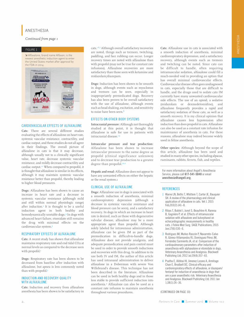
4 Dr. Malakoff performs an echocardiogram on a feline patient
FIGURE 1
Partners In Care π Summer 2016 π Volume 10:2 3.
PA
IN M
EDIC
INEPAIN MEDICINE
Osteoarthritis (OA – here used synonymously with degenerative joint disease (DJD)) is probably the most common chronic disease
that veterinarians see, yet we don’t think too much about the details of pathophysiology, diagnosis, and progression. In Angell Animal Medical Center’s Pain Medicine Service, osteoarthritis is present in nearly every patient evaluated. Oftentimes, clients have a limited understanding of this disease and their view is usually directly extrapolated from their own experiences. Below are some of the myths clients may have about clinical features of osteoarthritis (OA or DJD) and some key facts about this disease that may be useful to review.
MYTH: OSTEOARTHRITIS IS A DISEASE OF AGINGSadly, some of our patients will have significant OA even before they reach skeletal maturity. OA can be caused by normal forces on abnormal joints or by abnormal forces on normal joints (i.e., obesity). The most common examples of OA in young patients are due to congenital joint asymmetry, as in coxofemoral or elbow dysplasia. For these patients, clients may recognize signs of lameness, exercise intolerance, or even problems with social interactions in puppies before they are six months of age. Clients are usually quite surprised to learn that their puppies already have OA and not an acute injury. Of course, OA is very prevalent in aged dogs, cats, and people. Estimates are that OA is present in over 80 percent of people over 55 years of age and more than one in five adult dogs.1
MYTH: OSTEOARTHRITIS IS A DISEASE OF CARTILAGEOf course, cartilage is affected by OA, but OA is a disease that is characterized by abnormalities in all tissues of a moveable joint. In some species, namely humans, cartilage loss is a hallmark of the disease. This is less true in dogs and cats. One definition of OA is “the clinical and pathological outcome of a range of disorders that result in structural and functional failure of the synovial joints with meniscal degeneration, subchondral bone alterations, bone and cartilage overgrowth (osteophytes),
loss of articular cartilage, and a synovial inflammatory response.”2 OA is a reflection of an unbalancing of the dynamic equilibrium between breakdown and repair of joint tissues. In many patients, the body’s overexuberant attempts to repair the damage to the joint tissues instigates pain and disability.
MYTH: RADIOGRAPHIC OSTEOARTHRITIS IS CORRELATED WITH PAIN AND DISABILITY Many people have radiographic evidence of OA (i.e., structural damage) without reporting any pain or decreased mobility. As best as we can infer without self-reporting, this is also true of dogs and cats. Conversely, patients of any species can have OA pain without significant structural damage visible on radiographs. This is particularly true of cats, which have less of a tendency to form osteophytes or develop sclerosis of subchondral bone than other species.3 This may be one reason why feline OA appears to be seriously underdiagnosed. Pain and disability from OA are not directly linked
to radiographic severity because pain generation is due to an intricate, dynamic relationship of multiple factors. Fox describes the origin of OA pain as a complex interaction of structural change, biochemical alteration, pain processing in the nervous system, and individual pain cognition.1
MYTH: OSTEOARTHRITIS IS A NONINFLAMMATORY CONDITION, IN CONTRAST TO RHEUMATOID AND EROSIVE-TYPE ARTHRITISFor many years, osteoarthritis was termed noninflammatory. In fact, osteoarthritis pathogenesis does involve a nonpurulent inflammatory degenerative process that varies in intensity at different phases of the disease. Primary immune mediated arthritis, like IMPA or rheumatoid arthritis, is a systemic disease, while OA is not. Inflammation does play a large role in the pathogenesis of pain (both peripheral and central) but also has important consequences for the permanent damage to affected tissues via chemical mediators. The disease course naturally waxes and wanes, leading to the idea that levels of inflammation may relate to variable clinical signs of pain over time.
MYTH: WEIGHT REDUCTION REDUCES JOINT PAIN BECAUSE OF DECREASED FORCE ON THE JOINTExcessive weight is a common and significant factor leading to degeneration of a joint and pain, but its effect is far from a simple loading of force. There is a complex interaction of weight and joint pain that clients rarely understand. Education about pathogenesis and the direct pain-relieving benefits of weight loss can be a powerful tool in weight-reduction compliance. Excess weight may initiate joint degeneration, leading to a vicious cycle of decreased exercise due to pain, muscle atrophy, and further weight gain. Clients may not understand that fat is biologically active and directly contributes to an inf lammatory pain state via the production of pro-inf lammatory cytokines. Weight reduction is directly pain relieving for patients experiencing chronic pain. Once pain is reduced and activity can be increased, further pain reduction occurs because of increases in endogenous analgesic mechanisms
A Primer on Osteoarthritis: Myths and Truths π Lisa Moses, VMD, DACVIM, CVMA
angell.org/[email protected]
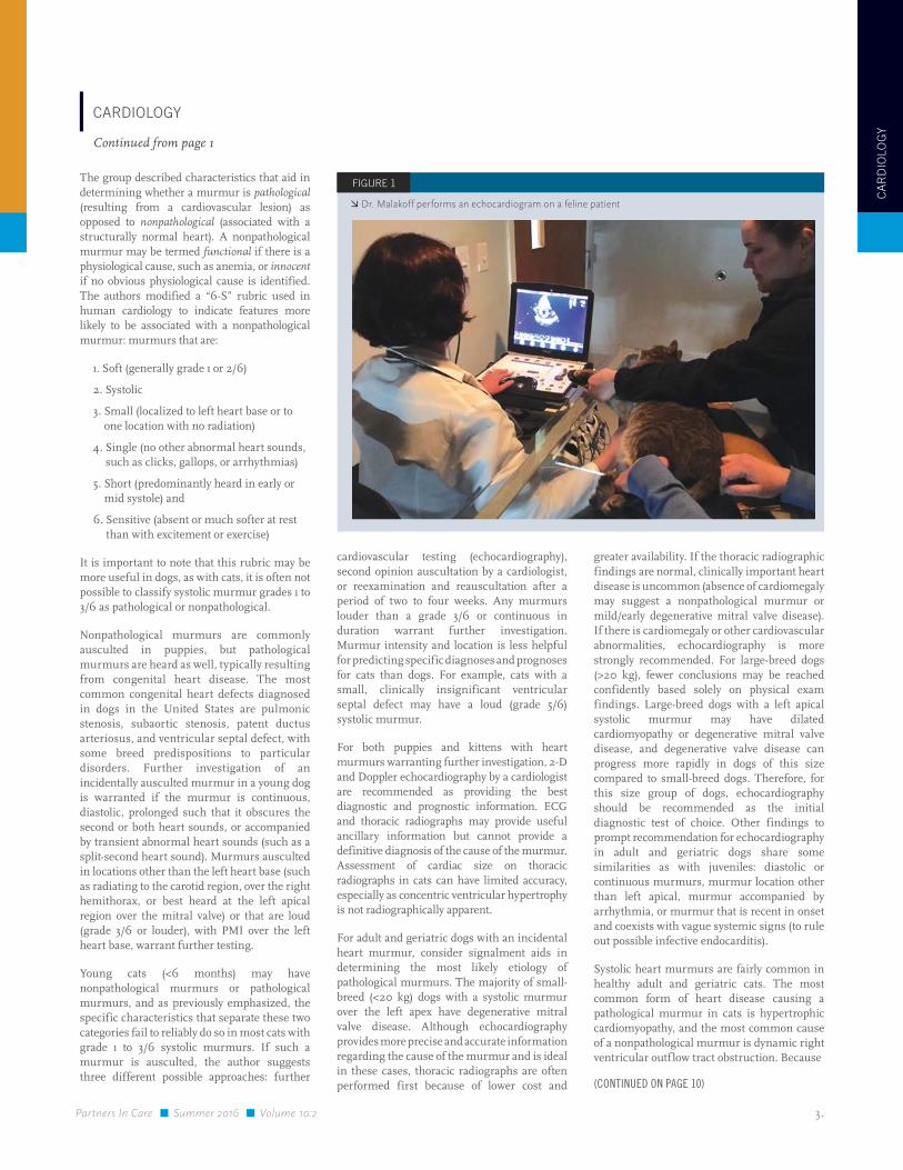
FIGURES 1 & 2
Partners In Care π Summer 2016 π Volume 10:24.
PAIN MEDICINE
associated with activity and muscle mass. When significant weight reductions occur in OA patients, there is also an eventual reduction in hyperalgesia and altered pain thresholds. In the Pain Medicine Service at Angell, weight reduction is the most effective method used to reduce pharmacological dependence on analgesics.
MYTH: DISEASE-MODIFYING OSTEOARTHRITIS DRUGS (DMOADS) REDUCE OA SEVERITY AND RESULTING PAINLike the rest of the story about OA, the truth about DMOADs is complex. When the pathogenesis of OA was being uncovered years ago, cartilage degeneration was thought to be the dominant process in pain generation and disability. Pharmacological substances that impact cartilage degeneration were originally called chondroprotective agents. This label was changed to DMOADs because they were thought to impact the course of the degeneration and to have clinical benefit. Since that terminology change, the FDA and its European counterpart have defined DMOADs as agents that modify OA structure by slowing joint-space narrowing (which is much more common in people) and provide pain and disability symptom reduction. Currently, no DMOAD exists that meets this criterion. Some of these agents do slow structural progression but do not improve function or pain in patients. Some of these agents improve pain in certain patients but do not slow progression of structural changes. Because pain and dysfunction are not directly or solely related to structural progression, delaying structural changes doesn’t always show clinical benefit. The bottom line is that some of these agents are beneficial for individual patients, but there is no evidence suggesting long-term benefit or prophylaxis from DMOADs in most patients. For most patients, these agents do not actually modify the disease course at all. For patients in the Pain Medicine Service, we advise a one- to two-month trial of so-called DMOADs and then a reevaluation of their benefits. It has been suggested that some joint supplements may have anti-inflammatory effects in particular individuals using an unknown mechanism. Our clinical experience using these agents would support that idea. If clear reduction in disability or
pain isn’t seen, then clients are likely wasting their money and may be doing harm if patients have GI or other adverse effects from these agents.
For more information about Angell’s Pain Medicine Service, please call 617-541-5140 or email [email protected].
REFERENCES AND SUGGESTED READINGS
1 Fox, SM. Chapter 2: Pathophysiology of Osteoarthritic Pain in Chronic Pain in Small Animal Medicine. Manson Publishing, London, 2010.
2 Hunter DJ and Hellio Le Graverand-Gastineau M. How Close Are We to Having Structure-Modifying Drugs Available? Med Clin N Am 93: 223-234, 2009.
3 Lascelles, BDX. Feline Degenerative Joint Disease. Veterinary Surgery 39; 2-13, 2010
Sofat N, Ejindu V, Kiely, P. What Makes Osteoarthritis Painful? The evidence for local and central pain processing. Rheumatology 50: 2157-2165, 2011.
Vandeweerd JM, et al. Systematic Review of Efficacy of Nutraceuticals to Alleviate Clinical Signs of Osteoarthritis. J Vet Intern Med 26: 448-456, 2012.
ANGELL AT NASHOBA: LOW COST CARE FOR QUALIFIED LOW INCOME CLIENTS
Angell Animal Medical Center and Nashoba Valley Technical High School have partnered to create Angell at Nashoba, a one-doctor clinic dedicated to providing quality veterinary care to low income pet owners and training more technicians within Northern Massachusetts. For the pets of owners that financially qualify, Angell at Nashoba provides discounted:
Spay/neuter services
Vaccinations
Basic veterinary care
Open weekdays from 7:45am-4:00pm throughout the year, the clinic does not provide overnight care, specialty service care, nor 24/7 emergency service as Angell’s Boston and Waltham facilities do, but will refer cases as appropriate to surrounding specialty veterinary referral hospitals.
To contact the Angell at Nashoba clinic, please call 978-577-5992. The clinic is located at: 100 Littleton Road, Westford, Massachusetts.
THE STAFF AT ANGELL AT NASHOBA
LAURENCE SAWYER, DVMDr. Sawyer has been in small animal private practice since 2001. She has also volunteered regularly at Tufts at Tech Community Veterinary Clinic since its inception in 2012. She enjoys clinical medicine and surgery, teaching, and
community service and is excited to be part of the Angell team that has established a community veterinary clinic at Nashoba Valley.
LISA QUINONES, CVTLisa has been working as a veterinary technician since 2007, starting her career in a fast-paced emergency and critical care practice. In 2010, she moved to Boston and joined MSPCA-Angell’s surgery team, working as an anesthesia tech
and becoming the department’s supervisor in 2013. Lisa enjoys volunteer work, painting, yoga, gardening, reading, and being outdoors with her two dogs.
Additionally, Department of Elementary and Secondary Education (DESE) licensed high school animal science teachers will be part of the program. Their main focus will be the education of the high school students in the necessary theoretical and clinical elements for acquiring a veterinary assisting license. Students and staff will also work alongside the staff at Angell at Nashoba.
For more information, please visit: www.angell.org/nashoba. Follow the clinic on Facebook: www.facebook.com/AngellAtNashoba.
Partners In Care π Summer 2016 π Volume 10:2 5.

PATHOLOGY
PAT
HO
LOG
Y
Canine GI Neoplasia π Pamela Mouser, DVM, MS, DACVP Anatomic Pathologist, Angell Animal Medical Center
angell.org/[email protected]
T he most enjoyable cases from a diagnostic pathologist’s perspective are those that have beautiful, picturesque lesions. I am often partial
toward nice gross lesions, and gastrointestinal neoplasia can certainly provide some great photo opportunities. In this article, I will summarize just over six years of surgical biopsy data for canine gastrointestinal neoplasia (mid-2009 through late 2015), including samples taken from the stomach to the rectum; illustrate the gross appearance of common neoplastic lesions; and compare findings to published data.
ANGELL DATA SUMMARYOf the 108 canine gastrointestinal (GI) neoplasms in our database from the past six years, the most common diagnoses in descending order were adenoma or adenomatous hyperplasia (32, 26 of which were rectal papillary adenoma submissions), adenocarcinoma (21), and lymphoma (13). If all sarcomas were considered together as a group, they would weigh in at a total of 24 cases.
Angell submissions were further tabulated by section of the GI tract affected, and it is interesting to note that some tumor types predominated in certain areas (see Table 1). For example, eight of the 13 gastric neoplasms were epithelial proliferations ranging from adenomatous hyperplasia to gastric adenocarcinoma. Interestingly, there were no lymphoma diagnoses in the stomach, although this may represent case bias since a cytological diagnosis of lymphoma may preclude surgical biopsy. One-third of the neoplastic processes in the small intestine were diagnosed as carcinoma and another third as lymphoma. This is in contrast to the ileocecocolic junction, cecum, and colon, where adenocarcinoma and lymphoma were only diagnosed once each out of 17 neoplastic submissions. Gastrointestinal stromal tumors (GISTs) appeared to favor the cecum, although some of the unclassified sarcomas occurring at other sites may have been recategorized as GISTs if immunohistochemical phenotyping had been performed. Over half of the rectal masses were papillary adenomas, and three cases classified as carcinoma were noted
to be arising from rectal papillary adenomas. Rectal plasma cell tumors (plasmacytomas) were the next most-common rectal tumor type and tended to have a high recurrence rate (five of seven tumors).
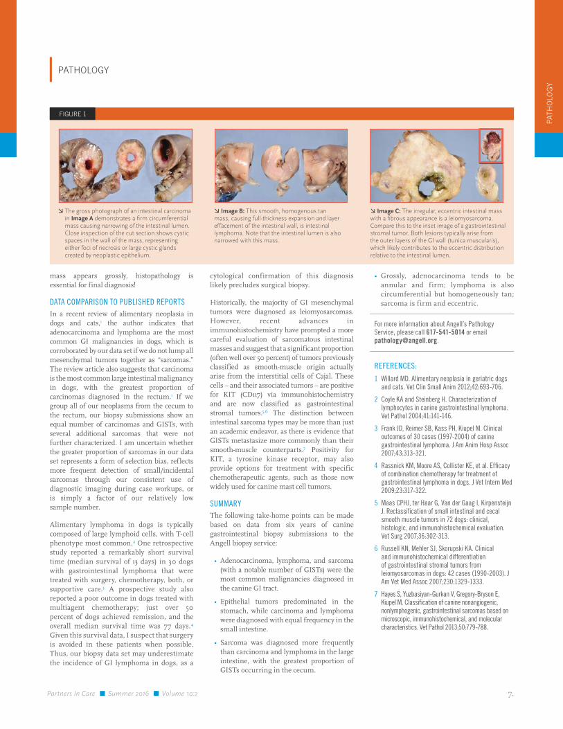
GROSS LESION APPEARANCEThe three major types of gastrointestinal malignancy are illustrated in Figure 1, using small intestinal lesions as examples. Adenocarcinomas arise from the inner aspect of the GI wall (tunica mucosa), are often circumferential in the small and large intestine, and induce a desmoplastic response. These tumors are therefore firm, frequently cause stricture of the intestinal lumen, and may cause annular constriction of the wall referred to as a “napkin-ring” lesion. Lymphoma is also often circumferential, but tends to be more expansile than carcinoma, causing effacement of all layers with a homogenous tan color. Intestinal sarcomas range from pale tan to almost white, are firm to fibrous, and have a more eccentric distribution compared to the other two lesions. Of course, regardless of how characteristic a
4 Table 1: Canine gastrointestinal neoplasia diagnosed via biopsy between mid-2009 and late 2015, separated by submission site.
SMALL ILEOCECOCOLIC STOMACH INTESTINE JUNCTION CECUM COLON RECTUM TOTAL
Adenocarcinoma 3 11 1 6* 21
Lymphoma 11 1 1 13
Sarcoma† 1 4 1 2 1 1 10
GIST 1 2 6 1 10
Leiomyosarcoma 2 1 1 4
Adenoma/polyp 5 1 26 32
Plasmacelltumor 1 7 8
Leiomyoma 2 1 2 5
Mastcelltumor 1 1 1 3
Neuroendocrinecarcinoma 1(insulinoma) 1 2
TOTAL 13 33 1 10 6 45 108
* Three cases of rectal carcinoma were in situ carcinomas arising within rectal papillary adenomas.† Masses classified as sarcomas included lesions without IHC for further distinction, KIT-negative sarcomas that did not have morphology typical of leiomyosarcoma, and
one extraskeletal osteosarcoma invading the small intestine.
6. Partners In Care π Summer 2016 π Volume 10:2
PAT
HO
LOG
Y
PATHOLOGY
mass appears grossly, histopathology is essential for final diagnosis!
DATA COMPARISON TO PUBLISHED REPORTSIn a recent review of alimentary neoplasia in dogs and cats,1 the author indicates that adenocarcinoma and lymphoma are the most common GI malignancies in dogs, which is corroborated by our data set if we do not lump all mesenchymal tumors together as “sarcomas.” The review article also suggests that carcinoma is the most common large intestinal malignancy in dogs, with the greatest proportion of carcinomas diagnosed in the rectum.1 If we group all of our neoplasms from the cecum to the rectum, our biopsy submissions show an equal number of carcinomas and GISTs, with several additional sarcomas that were not further characterized. I am uncertain whether the greater proportion of sarcomas in our data set represents a form of selection bias, reflects more frequent detection of small/incidental sarcomas through our consistent use of diagnostic imaging during case workups, or is simply a factor of our relatively low sample number.
Alimentary lymphoma in dogs is typically composed of large lymphoid cells, with T-cell phenotype most common.2 One retrospective study reported a remarkably short survival time (median survival of 13 days) in 30 dogs with gastrointestinal lymphoma that were treated with surgery, chemotherapy, both, or supportive care.3 A prospective study also reported a poor outcome in dogs treated with multiagent chemotherapy; just over 50 percent of dogs achieved remission, and the overall median survival time was 77 days.4 Given this survival data, I suspect that surgery is avoided in these patients when possible. Thus, our biopsy data set may underestimate the incidence of GI lymphoma in dogs, as a
cytological confirmation of this diagnosis likely precludes surgical biopsy.
Historically, the majority of GI mesenchymal tumors were diagnosed as leiomyosarcomas. However, recent advances in immunohistochemistry have prompted a more careful evaluation of sarcomatous intestinal masses and suggest that a significant proportion (often well over 50 percent) of tumors previously classified as smooth-muscle origin actually arise from the interstitial cells of Cajal. These cells – and their associated tumors – are positive for KIT (CD117) via immunohistochemistry and are now classified as gastrointestinal stromal tumors.5,6 The distinction between intestinal sarcoma types may be more than just an academic endeavor, as there is evidence that GISTs metastasize more commonly than their smooth-muscle counterparts.7 Positivity for KIT, a tyrosine kinase receptor, may also provide options for treatment with specific chemotherapeutic agents, such as those now widely used for canine mast cell tumors.
SUMMARYThe following take-home points can be made based on data from six years of canine gastrointestinal biopsy submissions to the Angell biopsy service:
• Adenocarcinoma, lymphoma, and sarcoma (with a notable number of GISTs) were the most common malignancies diagnosed in the canine GI tract.
• Epithelial tumors predominated in the stomach, while carcinoma and lymphoma were diagnosed with equal frequency in the small intestine.
• Sarcoma was diagnosed more frequently than carcinoma and lymphoma in the large intestine, with the greatest proportion of GISTs occurring in the cecum.
• Grossly, adenocarcinoma tends to be annular and firm; lymphoma is also circumferential but homogeneously tan; sarcoma is firm and eccentric.
For more information about Angell’s Pathology Service, please call 617-541-5014 or email [email protected].
REFERENCES:
1 Willard MD. Alimentary neoplasia in geriatric dogs and cats. Vet Clin Small Anim 2012;42:693-706.
2 Coyle KA and Steinberg H. Characterization of lymphocytes in canine gastrointestinal lymphoma. Vet Pathol 2004;41:141-146.
3 Frank JD, Reimer SB, Kass PH, Kiupel M. Clinical outcomes of 30 cases (1997-2004) of canine gastrointestinal lymphoma. J Am Anim Hosp Assoc 2007;43:313-321.
4 Rassnick KM, Moore AS, Collister KE, et al. Efficacy of combination chemotherapy for treatment of gastrointestinal lymphoma in dogs. J Vet Intern Med 2009;23:317-322.
5 Maas CPHJ, ter Haar G, Van der Gaag I, Kirpensteijn J. Reclassification of small intestinal and cecal smooth muscle tumors in 72 dogs: clinical, histologic, and immunohistochemical evaluation. Vet Surg 2007;36:302-313.
6 Russell KN, Mehler SJ, Skorupski KA. Clinical and immunohistochemical differentiation of gastrointestinal stromal tumors from leiomyosarcomas in dogs: 42 cases (1990-2003). J Am Vet Med Assoc 2007;230:1329-1333.
7 Hayes S, Yuzbasiyan-Gurkan V, Gregory-Bryson E, Kiupel M. Classification of canine nonangiogenic, nonlymphogenic, gastrointestinal sarcomas based on microscopic, immunohistochemical, and molecular characteristics. Vet Pathol 2013;50:779-788.
FIGURE 1
4 The gross photograph of an intestinal carcinoma in ImageA demonstrates a firm circumferential mass causing narrowing of the intestinal lumen. Close inspection of the cut section shows cystic spaces in the wall of the mass, representing either foci of necrosis or large cystic glands created by neoplastic epithelium.
4 ImageB: This smooth, homogenous tan mass, causing full-thickness expansion and layer effacement of the intestinal wall, is intestinal lymphoma. Note that the intestinal lumen is also narrowed with this mass.
4 ImageC: The irregular, eccentric intestinal mass with a fibrous appearance is a leiomyosarcoma. Compare this to the inset image of a gastrointestinal stromal tumor. Both lesions typically arise from the outer layers of the GI wall (tunica muscularis), which likely contributes to the eccentric distribution relative to the intestinal lumen.
Partners In Care π Summer 2016 π Volume 10:2 7.
Perhaps it reflects breed popularity (golden retrievers, Labradors, Bernese mountain dogs, rottweilers), but at Angell, the dominant subtypes of
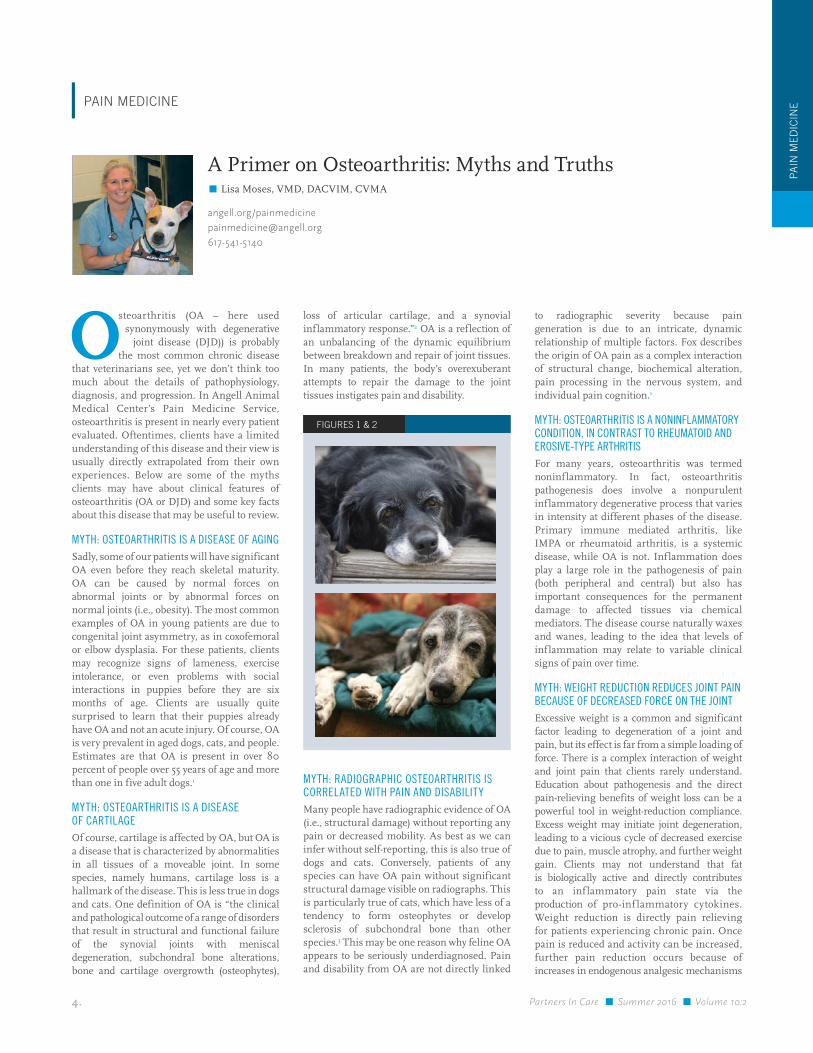
canine elbow dysplasia are fragmented medial coronoid process (FMCP) and osteochondritis dissecans (OCD). FMCP is a separation of the medial aspect of the coronoid process from the ulna, whereas OCD is caused by failure of normal endochondral ossification on the medial aspect of the humerus. The long-term effect of these developmental imperfections is degenerative joint disease (DJD), leading to wear and erosion of cartilage on the medial aspect of the elbow joint. When visualized arthroscopically (Figure 1), the delineation between destroyed cartilage medially and healthy white pristine cartilage in the lateral compartment is striking.
Biomechanical studies have suggested that dogs load about 65 percent on the medial aspect of the elbow joint and 35 percent on the lateral aspect. What if it were possible to make a subtle change in limb alignment that would unload the medial compartment, make better use of that healthy lateral cartilage, and in doing so,
reduce pain and improve elbow function? This is, in broad terms, the general premise behind PAUL, the proximal abducting ulna osteotomy (Kyon Veterinary Surgical Products).
Pendo, a five-year-old neutered male golden retriever had been medically managed for DJD secondary to left elbow dysplasia for many years. More recently, his function had been declining despite excellent weight management, exercise limitations, joint supplements, and meloxicam. Pendo appeared to be a good candidate for a PAUL procedure. Following radiographic preoperative planning, the affected elbow was scoped to ensure cartilage damage was limited to the medial compartment and, in Pendo’s case, to remove a loose coronoid fragment. The caudal approach to the proximal ulna is simple, with minimal soft tissue dissection, allowing an ulna osteotomy with an oscillating saw to create a small caudal step of two to three millimeters in the proximal ulna, lowering the coronoid. This is secured using a titanium six-hole, PAUL plate (Kyon) applied to the lateral aspect of the ulna, spanning the osteotomy gap (Figure 2 and Figure 3).
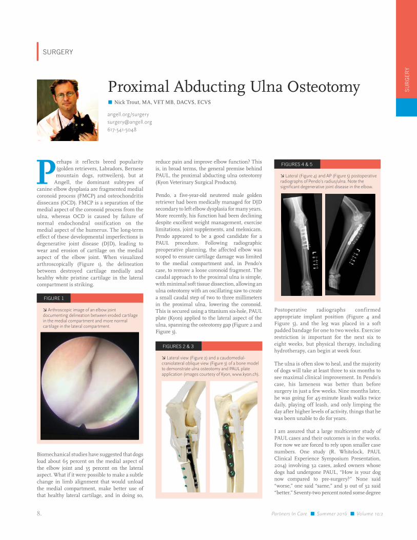
Postoperative radiographs confirmed appropriate implant position (Figure 4 and Figure 5), and the leg was placed in a soft padded bandage for one to two weeks. Exercise restriction is important for the next six to eight weeks, but physical therapy, including hydrotherapy, can begin at week four.
The ulna is often slow to heal, and the majority of dogs will take at least three to six months to see maximal clinical improvement. In Pendo’s case, his lameness was better than before surgery in just a few weeks. Nine months later, he was going for 45-minute leash walks twice daily, playing off leash, and only limping the day after higher levels of activity, things that he was been unable to do for years.
I am assured that a large multicenter study of PAUL cases and their outcomes is in the works. For now we are forced to rely upon smaller case numbers. One study (R. Whitelock, PAUL Clinical Experience Symposium Presentation, 2014) involving 32 cases, asked owners whose dogs had undergone PAUL, “How is your dog now compared to pre-surgery?” None said “worse,” one said “same,” and 31 out of 32 said “better.” Seventy-two percent noted some degree
Proximal Abducting Ulna Osteotomy π Nick Trout, MA, VET MB, DACVS, ECVS
angell.org/surgery [email protected] 617-541-5048
SURGERY
SUR
GER
Y
4 Arthroscopic image of an elbow joint documenting delineation between eroded cartilage in the medial compartment and more normal cartilage in the lateral compartment.
FIGURE 1
4 Lateral view (Figure 2) and a caudomedial-craniolateral oblique view (Figure 3) of a bone model to demonstrate ulna osteotomy and PAUL plate application (images courtesy of Kyon, www.kyon.ch).
FIGURES 2 & 3
4 Lateral (Figure 4) and AP (Figure 5) postoperative radiographs of Pendo’s radius/ulna. Note the significant degenerative joint disease in the elbow.
FIGURES 4 & 5
8. Partners In Care π Summer 2016 π Volume 10:2

ANGELL WELCOMES NEW STAFF DENTIST AND RADIOLOGIST
Angell Animal Medical Center is extremely pleased to announce that Jessica Riehl, DVM, DAVDC has joined our Dentistry Service, and Ruth Van Hatten, DVM, DACVR has joined our Diagnostic Imaging Service in Boston.
JESSICA RIEHL, DVM, DAVDCDr. Riehl joined Angell in 2016 after practicing at a veterinary specialty hospital in Seattle, Washington. She grew up in Connecticut and obtained her Bachelor’s Degree in Zoology at Connecticut College. Dr. Riehl graduated from University of Wisconsin-Madison School of Veterinary Medicine in 2010. She then went on to complete a small animal rotating internship in West Islip, NY followed by a residency in
Dentistry and Oral Surgery at the University of Wisconsin-Madison. Dr. Riehl became a Diplomate of the American Veterinary Dental College in 2014 and currently serves on the Credentials Committee for the AVDC. She is excited to return to New England. Outside of work Dr. Riehl enjoys cooking, outdoors, and spending time with her husband, son, and pets (Fern and Kota).
EDUCATION π University of Wisconsin-Madison School of Veterinary Medicine, DVM, 2010
π Connecticut College, BA, 2004
SPECIALTY TRAINING π Veterinary Medical Center of Long Island, Small Animal Internship 2010-2011
π University of Wisconsin-Madison School of Veterinary Medicine, Residency in Dentistry and Oral Surgery, 2011-2014
CERTIFICATION π Diplomate, American Veterinary Dental College, 2014
RUTH VAN HATTEN, DVM, DACVRDr. Ruth Van Hatten attended Boston University, where she received her Bachelor’s degree in Biology with a minor in Anthropology. She earned her DVM degree at Tufts University in 2010. Upon graduation, she completed a small animal rotating internship at Angell Animal Medical Center in Boston and a residency in diagnostic imaging at Cornell University. After
finishing her residency, she stayed on as faculty in the diagnostic imaging service at Cornell University Hospital for Animals. Dr. Van Hatten has a special interest in abdominal imaging, specifically ultrasound and CT. Dr. Van Hatten grew up in upstate NY, but considers Boston her home. In her free time, she enjoys watching Boston sports, traveling, and hiking, with some shopping mixed in!
EDUCATION π Tufts University School of Veterinary Medicine, DVM, 2010
π Boston University, BA, 2001
SPECIALTY TRAINING π Cornell University, Residency in Diagnostic Imaging, 2011-2014
π Angell Animal Medical Center, Small Animal Rotating Internship, 2010-2011
CERTIFICATION π Diplomate, American College of Veterinary Radiology, 2014
of lameness during certain activities, but 97 percent reported meaningful improvement.
Pendo may be my poster dog for PAUL, and I am always wary of anecdotes as proof of success; however, PAUL does appear to have a place as a viable treatment alternative in carefully selected cases where clinicians and owners are frustrated by the challenges of medical management for chronic elbow dysplasia.
For more information about Angell’s Surgery Service, please call 617-541-5048 or email [email protected].
Partners In Care π Summer 2016 π Volume 10:2 9.
ANESTHESIA
Continued from page 2
5 Campagna I, Schwarz A, Keller S, Bettschart-Wolfensberger R, Mosing M. Comparison of the effects of propofol or alfaxalone for anaesthesia induction and maintenance on respiration in cats. Veterinary Anaesthesia and Analgesia. 2015 Sep;42(5):484–92.
6 Mathis A, Pinelas R, Brodbelt DC, Alibhai HI. Comparison of quality of recovery from anaesthesia in cats induced with propofol or alfaxalone. Veterinary Anaesthesia and Analgesia. Blackwell Publishing Ltd; 2012 May 1;39(3):282–90.
7 Gieseg M, Hon H, Bridges J, Walsh V. A comparison of anaesthetic recoveries in cats following induction with either alfaxalone or ketamine and diazepam. N Z Vet J. 2014 May;62(3):103–9.
8 Maney JK, Shepard MK, Braun C, Cremer J, Hofmeister EH. A comparison of cardiopulmonary and anesthetic effects of an induction dose of alfaxalone or propofol in dogs. Veterinary Anaesthesia and Analgesia. 2013 May;40(3):237–44.
9 Warne LN, Beths T, Fogal S, Bauquier SH. The use of alfaxalone and remifentanil total intravenous anesthesia in a dog undergoing a craniectomy for tumor resection. Can Vet J. 2014 Nov;55(11):1083–8.
10 Hasiuk MMM, Forde N, Cooke A, Ramey K, Pang DSJ. A comparison of alfaxalone and propofol on intraocular pressure in healthy dogs. Vet Ophthalmol. 2014 Nov;17(6):411–6.
11 Costa D, Leiva M, Moll X, Aguilar A, Peña T, Andaluz A. Alfaxalone versus propofol in dogs: a randomised trial to assess effects on peri-induction tear production, intraocular pressure and globe position. Vet Rec. BMJ Publishing Group Limited; 2015 Jan 17;176(3):73–3.
CARDIOLOGY
Continued from page 3
there is so much overlap in how these murmurs sound, it is generally impossible to differentiate pathological and nonpathological murmurs in cats based on auscultation alone. Additional findings that would strengthen the recommendation for echocardiography in an adult cat include presence of a gallop sound, arrhythmia, a murmur that is diastolic or continuous, a murmur that is loud (grade 3/6 or louder), or increased strength of the apex beat. Thoracic radiography is less useful for determining whether significant cardiac disease is present, as concentric hypertrophy (such as with hypertrophic cardiomyopathy) may not result in an apparently enlarged cardiac silhouette. Certainly, if cardiomegaly is apparent radiographically, this strengthens the recommendation for echocardiography, as would the finding of an abnormal NT-proBNP level.
Finally, several considerations beyond physical exam findings or other test results can help in determining whether a juvenile or adult veterinary patient should have an echocardiogram. For example, client concern, anxiety, or desire to be as informed as possible about the cause of the murmur can influence whether an echocardiogram is pursued. The veterinarian should also consider whether the patient requires general anesthesia in the near future or any treatments with relative cardiac contraindications. If the dog or cat is to be used for breeding, echocardiography to try to obtain a definitive diagnosis of the cause of the
murmur becomes of more paramount importance as well.
For more information, about Angell’s Cardiology Service, please call 617-541-5038 or email [email protected]. Dr. Malakoff works full time at our Waltham location.
REFERENCES
1 Cote E, Edwards NJ, Ettinger SJ et al. Management of incidentally detected heart murmurs in dogs and cats. J Am Vet Med Assoc 2015;246(10): 1076-1088.
YAO YAO, VMDDr. Yao received her bachelor’s degree in molecular biology with a minor in art history from Bryn Mawr and Haverford College prior to attending University of Pennsylvania, where she obtained her Doctor of Veterinary Medicine degree. After graduation, Dr. Yao completed one year internship in small animal medicine and surgery at Red Bank Veterinary Hospital in Tinton Falls, NJ. Before becoming a full time veterinarian, Dr. Yao dedicated a lot of time in academic research projects including ones involving human esophageal and pancreatic cancer. In her free time Dr. Yao enjoys traveling, cooking, painting, spending time with family, friends and her lazy grey tabby Logan.
EDUCATION π Doctor of Veterinary Medicine, University of Pennsylvania, 2012
π Bachelor of Science, Bryn Mawr and Haverford College, 2008
SPECIALTY TRAINING π Internship in Small Animal Medicine and Surgery, Red Bank Veterinary Hospital, 2012-2013
MSPCA-Angell West is extremely pleased to announce that Yao Yao, VMD, has joined the Emergency and Critical Care Service in Waltham.
10. Partners In Care π Summer 2016 π Volume 10:2
(W/B) Services available at our Waltham and Boston locations.* Boston-based radiologists and pathologists serve both Boston & Waltham locations.
STA
FF D
OC
TOR
S
π We encourage you to contact Angell’s specialists with questions.
Main Phone: 617-522-7282 (Boston), 781-902-8400 (Waltham), 978-577-5992 (Angell at Nashoba) Veterinary Referrals: 617-522-5011
CHIEF OF STAFF
Ann Marie Greenleaf, DVM, DACVECC
24/7 EMERGENCY & CRITICAL CARE (W/B)
Lauren Baker, DVM
Kiko Bracker, DVM, DACVECC
Emily Finn, DVM
Jessica Hamilton, DVM
Roxanna Khorzad, DVM
Amanda Lohin, DVM
Courtney Peck, DVM
Virginia Sinnott, DVM, DACVECC
Catherine Sumner, DVM, DACVECC
Megan Whelan, DVM, DACVECC
Yao Yao, VMD
ANESTHESIOLOGY
Stephanie Krein, DVM, DACVAA
AVIAN & EXOTIC MEDICINE (W/B)
Brendan Noonan, DVM, DABVP
(Avian Practice)
Elisabeth Simone-Freilicher, DVM, DABVP
(Avian Practice)
BEHAVIOR (W/B)
Terri Bright, Ph.D., BCBA-D
Taylor Kirby-Madden, DVM
CARDIOLOGY (W/B)Ashley Jones, DVM, DACVIM (Cardiology)[email protected]
Nancy Laste, DVM, DACVIM (Cardiology) [email protected]
Rebecca Malakoff, DVM, DACVIM (Cardiology) [email protected]
Rebecca Quinn, DVM, DACVIM (Cardiology and Internal Medicine) [email protected]
DENTISTRYErin Abrahams, DVM [email protected]
William Rosenblad, DVM [email protected]
Jessica Riehl, DVM, DAVDC [email protected]
DERMATOLOGYKlaus Loft, DVM [email protected]
DIAGNOSTIC IMAGING*Rebecca Manley, DVM, DACVR [email protected]
Steven Tsai, DVM, DACVR [email protected]
Ruth Van Hatten, DVM, DACVR [email protected]
INTERNAL MEDICINE (W/B)Daniela Ackley, DVM, DACVIM [email protected]
Doug Brum, DVM [email protected]
Maureen Carroll, DVM, DACVIM [email protected]
Erika de Papp, DVM, DACVIM [email protected]
Jean Marie Duddy, DVM [email protected]
Kirstin Johnson, DVM, DACVIM [email protected]
Shawn Kearns, DVM, DACVIM [email protected]
Susan O’Bell, DVM, MPH, DACVIM [email protected]
Cynthia Talbot, DVM [email protected]
NEUROLOGY (W/B)Rob Daniel, DVM, DACVIM (Neurology) [email protected]
Allen Sisson, DVM, MS, DACVIM (Neurology) [email protected]
NUTRITIONDana Hutchinson, DVM, DACVN [email protected]
ONCOLOGYLyndsay Kubicek, DVM, DACVR (Radiation Oncology) [email protected]
J. Lee Talbott, DVM (Practice Limited to Medical Oncology) [email protected]
OPHTHALMOLOGYDaniel Biros, DVM, DACVO [email protected]
Martin Coster, DVM, MS, DACVO [email protected]
PAIN MEDICINELisa Moses, VMD, DACVIM, CVMA [email protected]
PATHOLOGY (CLINICAL & ANATOMIC)*Patty Ewing, DVM, MS, DACVP [email protected]
Pamela Mouser, DVM, MS, DACVP [email protected]
SURGERY (W/B)Sue Casale, DVM, DACVS [email protected]
Michele Kudisch, DVM, DACVS [email protected]
Michael Pavletic, DVM, DACVS [email protected]
Meghan Sullivan, DVM, DACVS [email protected]
Nicholas Trout, MA, VET MB, MRCVS, DACVS, DECVS [email protected]
ANGELL AT NASHOBALaurence Sawyer, DVM [email protected]
STAFF DOCTORS
Partners In Care π Summer 2016 π Volume 10:2 11.
Nonprofit Org.
US Postage
PAID
Permit No. 1141
Boston, MA
350 South Huntington Avenue
Boston, MA 02130
617-522-5011
angell.org
293 Second Avenue
Waltham, MA 02451
781-902-8400
angell.org/waltham
MSP
CA
-AN
GEL
LM
SPC
A-A
NG
ELL
WES
T
We mail one complimentary copy of our newsletter to each of our referring partners. Please circulate this copy within your practice.
Summer 2016 π Volume 10:2 π angell.org π facebook.com/AngellReferringVeterinarians
π angell.org/directions (free parking) π angell.org/hours π angell.org/ce
4 ANGELL AT NASHOBA NOW ACCEPTING APPOINTMENTS
Angell Animal Medical Center and Nashoba Valley Technical High School have partnered to create Angell at Nashoba, a veterinary clinic for low income pet owners that also serves as a rigorous academic and experiential training program for students enrolled at Nashoba Valley Technical High School. Opened on February 3, 2016, the clinic provides discounted:
Spay/neuter services
Vaccinations
Basic veterinary care
Please consider adding Angell’s main numbers to your after-hours phone message.
Financial Qualifications for Clients
To qualify for Angell at Nashoba services, clients must present a photo ID and one of the following:
The person whose name is on the card or documents must be present (i.e., they can’t send a relative or friend). The only exception is a spouse with the same last name and address.
Women, Infants, and Children (WIC) program card
Supplemental Nutrition Assistance Program (SNAP) card (formerly known as Food Stamps/EBT card)
Spay and Neuter Assistance Program certificate
Letter/lease from the owner’s local housing authority showing that the owner is a participant in public housing
To reach the clinic, please call 978-577-5992.
The clinic is located at: 100 Littleton Road, Westford, Massachusetts.
For more information, visit www.angell.org/nashoba.
4 Fourteen-year-old Rocky, a Jack Russell Terrier, was Angell at Nashoba’s first patient.