![Journal of Molecular and Cellular Cardiology€¦ · Cardif, and VISA) adapter protein and promote MAVS oligomerization [42,44,78,101,102]. MAVS localizes primarily to the outer mitochon-drial](https://static.fdocument.org/doc/165x107/5ff8125ed446ec04280eefb4/journal-of-molecular-and-cellular-cardiology-cardif-and-visa-adapter-protein-and.jpg)
Page 1 of 41 Diabetes · 14.1%, then decreases to 7.5% (grey bars). Interestingly, cyclin D3...
41
1 DB-12-0778 Revision 2 Cytoplasmic-Nuclear Trafficking of G1/S Cell Cycle Molecules and Adult Human Beta Cell Replication: A Revised Model of Human Beta Cell G1/S Control Running Title: Nuclear Trafficking and β-Cell Replication Nathalie M. Fiaschi-Taesch PhD 1 , Jeffrey W. Kleinberger BS 1 , Fatimah Salim BS 1 , Ronnie Troxell BS 1 , Rachel Wills BS 1 , Mansoor Tanwir MD 1 , Gabriella Casinelli BD 1 , Amy E. Cox MD 1 , Karen K. Takane PhD 1 , Harish Srinivas PhD 1 , Donald K. Scott PhD 1,2 , and Andrew F. Stewart MD 1,2 From the 1 Division of Endocrinology, The University of Pittsburgh School of Medicine, Pittsburgh, PA, 15217. 2 Current address: Diabetes Obesity and Metabolism Institute, Mount Sinai School of Medicine, NY, NY, 10029 Address Correspondence to: Nathalie M. Fiaschi-Taesch PhD Division of Endocrinology and Metabolism BST E-1140 University of Pittsburgh School of Medicine 200 Lothrop St., Pittsburgh PA 15260 Phone: 412-648-9316 Fax: 412-648-3290 e-mail: [email protected] Abstract: 200 words Main Text: 4394 words References: 39 Figures: 8 Tables 0 Page 1 of 41 Diabetes Diabetes Publish Ahead of Print, published online March 14, 2013
Transcript of Page 1 of 41 Diabetes · 14.1%, then decreases to 7.5% (grey bars). Interestingly, cyclin D3...
1
DB-12-0778 Revision 2
Cytoplasmic-Nuclear Trafficking of G1/S Cell Cycle Molecules and Adult Human Beta Cell
Replication: A Revised Model of Human Beta Cell G1/S Control
Running Title: Nuclear Trafficking and β-Cell Replication
Nathalie M. Fiaschi-Taesch PhD1, Jeffrey W. Kleinberger BS1, Fatimah Salim BS1, Ronnie
Troxell BS1, Rachel Wills BS1, Mansoor Tanwir MD1, Gabriella Casinelli BD1, Amy E. Cox MD1,
Karen K. Takane PhD1, Harish Srinivas PhD1, Donald K. Scott PhD1,2,
and Andrew F. Stewart MD1,2
From the 1Division of Endocrinology, The University of Pittsburgh School of Medicine,
Pittsburgh, PA, 15217. 2Current address: Diabetes Obesity and Metabolism Institute, Mount
Sinai School of Medicine, NY, NY, 10029
Address Correspondence to:
Nathalie M. Fiaschi-Taesch PhD Division of Endocrinology and Metabolism BST E-1140 University of Pittsburgh School of Medicine 200 Lothrop St., Pittsburgh PA 15260 Phone: 412-648-9316 Fax: 412-648-3290 e-mail: [email protected] Abstract: 200 words Main Text: 4394 words References: 39 Figures: 8 Tables 0
Page 1 of 41 Diabetes
Diabetes Publish Ahead of Print, published online March 14, 2013
2
Abstract
Harnessing control of human beta cell proliferation has proven frustratingly difficult.
Most G1/S control molecules, generally presumed to be nuclear proteins in the human beta cell,
are in fact constrained to the cytoplasm. Here, we asked whether G1/S molecules might traffic
into and out of the cytoplasmic compartment in association with activation of cell cycle
progression. Cdk6 and cyclin D3 were used to drive human beta cell proliferation, and promptly
translocated into the nucleus in association with proliferation. In contrast, the cell cycle
inhibitors p15, p18 and p19 did not alter their location, remaining cytoplasmic. Conversely, p16,
p21, p27 all increased their nuclear frequency. In contrast once again, p57 decreased its
nuclear frequency. While proliferating beta cells contained nuclear cyclin D3 and cdk6,
proliferation generally did not occur in beta cells that contained nuclear cell cycle inhibitors,
except p21. Dynamic cytoplasmic-nuclear trafficking of cdk6 was confirmed using GFP-tagged
cdk6 and live-cell imaging. Thus, we provide novel working models describing the control of cell
cycle progression in the human beta cell. In addition to known obstacles to beta cell
proliferation, cytoplasmic-to-nuclear trafficking of G1/S molecules may represent both an
obstacle, as well as a therapeutic opportunity, for human beta cell expansion.
Key words: proliferation, adenovirus, trafficking, human beta cell, cdk, cyclin
Page 2 of 41Diabetes
3
Introduction
In an accompanying report (1), we have developed a novel “human beta cell G1/S
molecule atlas”, which reveals that essentially all of the G1/S molecules are present not only in
the human islet, but actually are present in the human beta cell. Surprisingly, although the G1/S
molecules are widely considered to be nuclear proteins, we encountered them principally in the
cytoplasm, where they presumably would be unable to direct cell cycle progression. Indeed, the
only G1/S molecules encountered in the nucleus of the human beta cell were cell cycle
inhibitors, pRb, p57, and, variably, p21. In contrast, all of the cell cycle-activating cyclins and
cdks were restricted to the cytoplasm. These studies were performed in quiescent human beta
cells, and shed no light on the functional activities of G1/S molecules during cell cycle
progression.
In this report, we explored whether G1/S molecules might be able to be induced to
shuttle from the cytoplasm to the nuclear compartment in association with activation of cell cycle
progression. We find that several cell cycle inhibitors as well as activators do indeed actively
traffic from the cytoplasm to the nucleus in association with activation of proliferation. These
results lead to a substantially altered model of G1/S trafficking and its control in the human beta
cell.
Page 3 of 41 Diabetes
4
Methods
Human cadaveric and rat islets. One hundred and sixty-four different cadaveric islet
preparations were used for these studies. The demographics and sources of the islets are
described in the accompanying report (1). Dispersal of the human islets was performed as
described in detail previously (1-5). Rat islets were isolated from 2-3 month old Sprague
Dawley rats, dispersed and cultured as detailed previously (5,6). Rat studies were approved in
advance by the University of Pittsburgh Institutional Animal Care and Use Committee.
Adenovirus production and transduction. Adenovirus preparation is described previously (1).
The efficiency of adenoviral transduction, assessed using ß-galactosidase and insulin co-
staining of human islets transduced with Ad.lacZ, was (mean±SEM) 65.1±3.0%, 67.9±2.5%,
and 75.7±2.8%, at 24, 48 and 72 hours following transduction. In addition, to prepare a GFP-
tagged cdk6 adenovirus, human cdk6 cDNA was subcloned into pcDNA3.1/CT-GFP plasmid
(Invitrogen, Carlsbad, CA) using a GFP fusion TOPO®TA expression kit (Invitrogen), which
places the GFP at the C-terminus of cdk6. This was subcloned into the adenovirus shuttle
vector, pACCMV, and adenovirus prepared as described (1-7).
Immunocytochemistry. Islets were dispersed to single cells, fixed and labeled as described (1-
7). For studies under proliferating conditions, dispersed islets were transduced with either
Ad.LacZ or Ad.cdk6+Ad.cyclinD3 (100 moi) for two hours, cultured for 24, 48 and 72 hours as
described in the Figures, and immunolabeled using antisera as described in Supplemental
Table 1 of the accompanying manuscript (1). Labeled cells were visualized using laser
confocal microscopy. Each experiment shown is representative of 3-6 human islet
preparations.
Immunoblotting. Immunoblotting was performed as described (1-7). Antibodies used to detect
the G1/S molecules are described in detail in Supplemental Table 1 to reference 1. Each
experiment shown is representative of 3-6 human islet preparations.
Page 4 of 41Diabetes
5
Live Cell Imaging. Rat Insulinoma cells (Ins1 832/13) were washed in PBS twice and
trypsinized for 5 min. Complete medium {RPMI medium (Gibco, Grand Isle, NY) containing 5.5
mM glucose, 1% penicillin and streptomycin, 10% fetal bovine serum, 10 mmol/l HEPES, 2
mmol/l glutamine, 1 mmol/l sodium pyruvate, and 50 µmol/l β-mercaptoethanol} was added and
a suspension of 200,000 cells was plated on a glass-bottomed microwell dish (MatTek,
Ashland, MA). The cell suspension was transduced with 100 moi of Ad.cdk6-GFP for two hours.
Human islets (200 IEQ) were dispersed as described above and plated on glass-bottomed
microwell dish (MatTek, Ashland, MA) and were transduced for two hours with 100 moi
Ad.cdk6-GFP. The transduction was stopped by adding 1 ml of complete medium to the Ins1
cells or the dispersed human islets. Transduced Ins1 cells or dispersed human islets were
imaged 24h after infection using a Nikon A1 Confocal Live Cell System and the NLS-Element
software (Nikon, Melville, NY).
Statistics. Statistical analysis for Figs 4&5 was performed using one-way ANOVA with
Bonferroni’s post-hoc correction. Student’s paired, two-tailed T-test was employed in Figs
2A&C. All values are expressed as means ± SEM. “p” values < 0.05 were considered
significant.
Page 5 of 41 Diabetes
6
Results
Induction of Proliferation Leads to Nuclear Translocation of Some, But Not All, G1/S
Molecules. In the absence of mitogenic growth factors or nutrients that are able to induce
robust human beta cell proliferation, we have employed adenoviral expression of D-cyclins,
cyclin E, and/or cdks 2,4 and 6 to drive human beta cell proliferation (2-4,7). For the current
studies, we selected the combination of Ad.cdk6 + Ad.cyclin D3 because this combination
yielded the highest rates of BrdU and Ki-67 incorporation (3). We asked whether proliferation
associated with adenoviral expression of cyclins and cdks might influence the subcellular
localization of G1/S components.
As previously reported (3,4), the expression of Ad.cdk6 and Ad.cyclin D3 led to beta cell
proliferation, and also the nuclear appearance of these two molecules (Fig 1A). Specifically,
following transduction with Ad.cdk6/cyclinD3, Ki67 labeling increased from 0.1% to
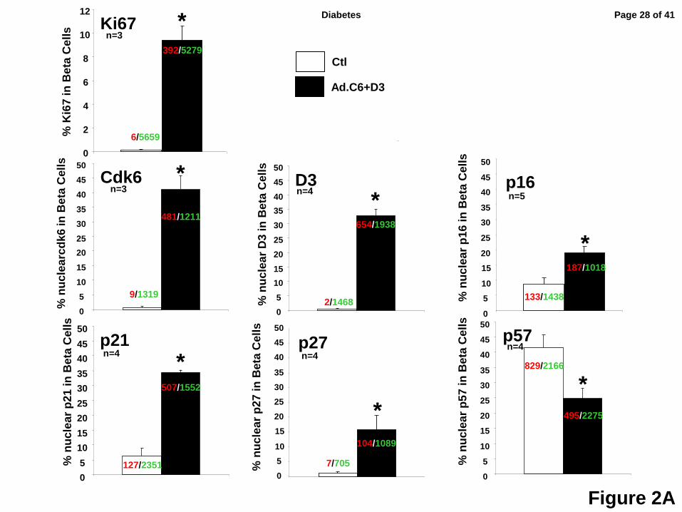
approximately 9.4% of beta cells; the percent of beta cells that contained nuclear cdk6
increased from 0.8% to 46.1%; and, the percent of beta cells that contained nuclear cyclin D3
increased from 0.3% to 29.2% (Fig 2A).
Exploring whether other G1/S molecules altered subcellular location in response to
induction of proliferation, three INK4 family members were found not to be altered: p15, p18 and
p19 all remained cytoplasmic (Fig 1B). On the other hand, p16, p21 and p27 all shifted into the
nuclear compartment (Figs 1C, 2A). In quantitative terms, p16, which was nuclear in only 8.4%
of beta cells under basal conditions, became nuclear in 16.1%. Similarly, p21, which was
nuclear in 7.9% of quiescent beta cells, became nuclear in 35.6%. p27 which had been nuclear
in 1.9% of quiescent beta cells became prominently nuclear in 11.2%. Surprisingly, p57 which
had been so predominantly nuclear in quiescent cells (42.1%) became less prominent in the
nucleus (23.8%) with induction of proliferation (Figs 1C, 2A).
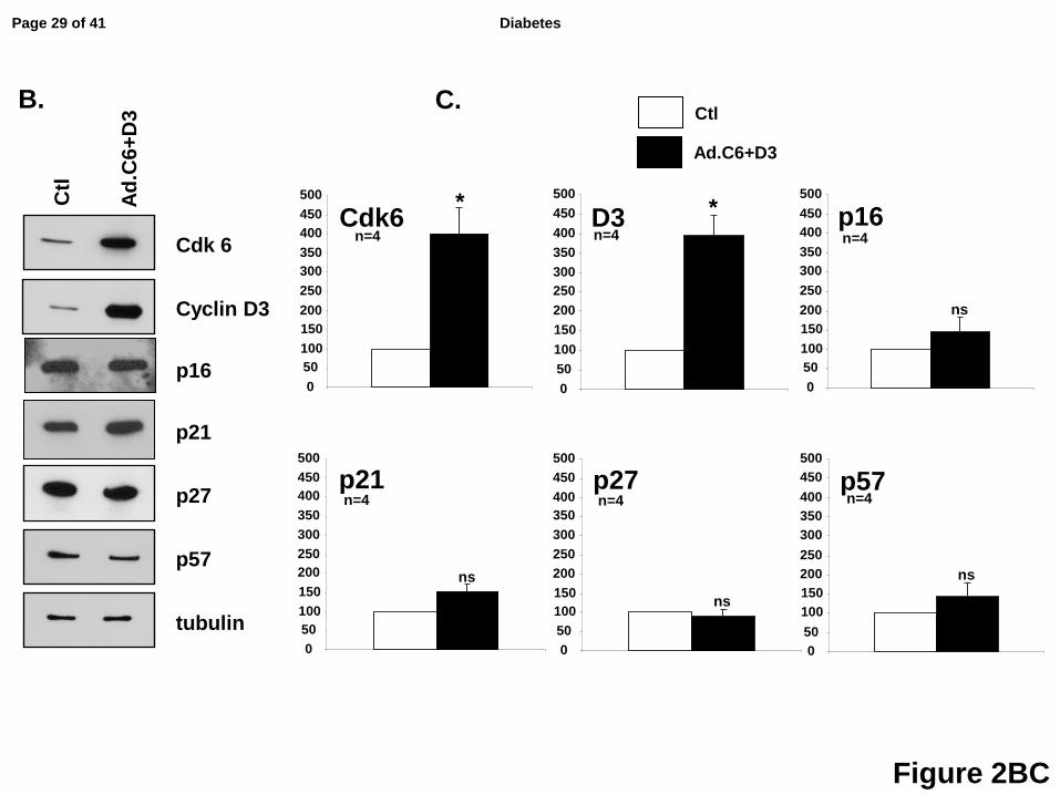
To determine whether the changes in nuclear abundance of p16, p21 and p27, and the
decrease in p57, were a result of changes in the total abundance of these molecules, we
Page 6 of 41Diabetes
7
examined their relative abundance with or without expression of cdk6 and cyclin D3. As shown
in Figs 2BC, although these molecules shifted location with proliferation, there were no changes
in their absolute abundance. Collectively, these results suggest that p16, p21, p27 and p57 all
markedly alter their subcellular localization, but not their overall abundance, in response to
induction of proliferation, with p16, p21 and p27 all displaying a pronounced increase in their
nuclear frequency, while nuclear p57 declined in nuclear frequency.
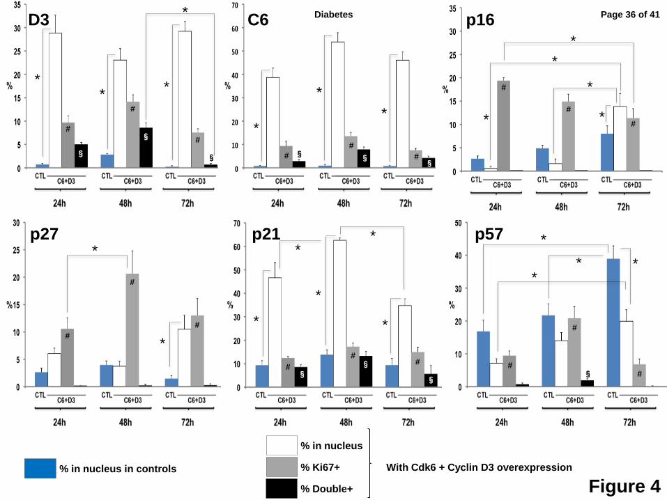
Human Beta Cell Proliferation Broadly Correlates With Nuclear Cdks and Cyclins, and
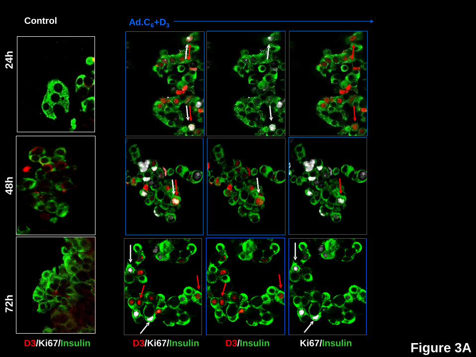
Inversely With Nuclear Cell Cycle Inhibitors. We next studied G1/S molecule nuclear
presence vs. proliferation (Ki67), as well as the 72-hour time courses of translocation of cdk6,
cyclin D3, p16, p21, p27 and p57. Ki67 was selected rather than BrdU since it reflects cell cycle
progression at the time of labeling, whereas BrdU incorporation reflects both active and past
proliferation. Examples of these studies are shown in Fig 3, and their quantification in Fig 4. In
Fig 4, the blue bars represent non-transduced control beta cells at 24, 48 and 72 hours
following dispersal and plating. The subsequent three bars reveal events in beta cells
transduced with both Ad.cdk6 plus Ad.cyclin D3 at these same time points: white indicates the
percent of beta cell nuclei that contain the G1/S molecule in question; grey, the percent of beta
cells containing Ki67 in their nuclei; and, black, the percent of beta cells that are doubly positive
for both Ki67 plus nuclear presence of the G1/S molecule in question.
Cyclin D3 (Figs 3A, 4) in control (blue bar) beta cells, appears in the nucleus of only
0.7% of beta cells at 24 hours, increases to 2.8% at 48 hours and declines to 0.3% at 72 hours.
With Ad.cdk6+cyclin D3 transduction, 25-30% of beta cells display nuclear cyclin D3 at all three
time points (white bars), and the percent of Ki67+ beta cells initially increases from 9.7% to
14.1%, then decreases to 7.5% (grey bars). Interestingly, cyclin D3 co-localizes in the nuclei
with Ki67 in 5.0%, 8.6% and 0.7% of beta cells at 24, 48 and 72 hours (black bars), suggesting
that it may enter the nucleus early, partner with cdk6, activate proliferation, and then be
Page 7 of 41 Diabetes
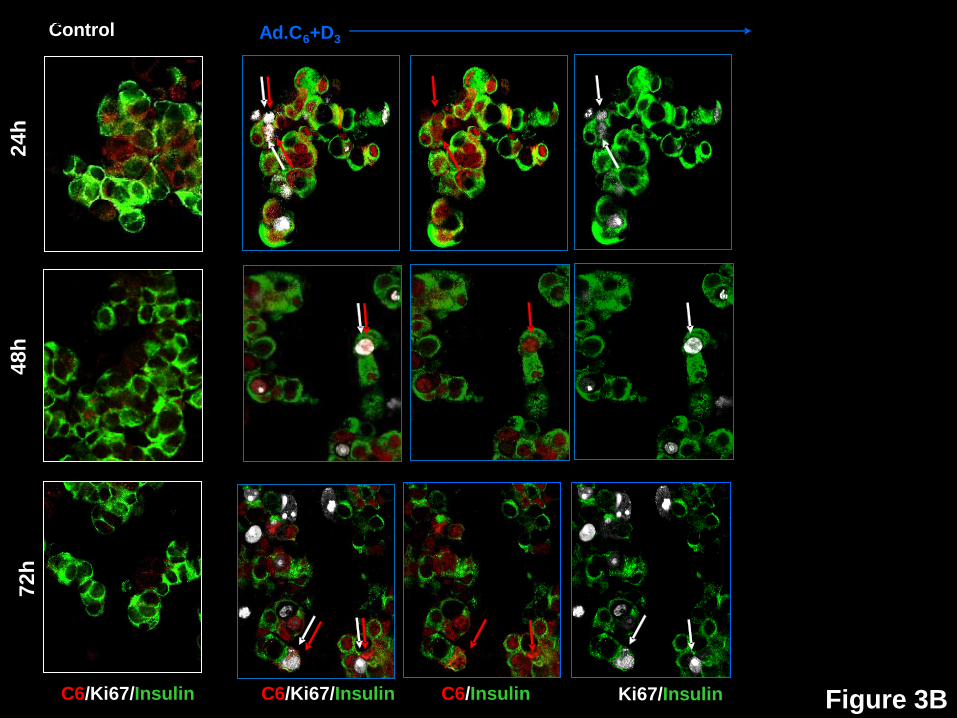
8
exported, degraded, or both, by 72 hours. Cdk6 reveals a similar bi-phasic pattern, suggesting
that it enters the nucleus transiently, then declines through one of the above mechanisms (Fig
3B,4).
For p16 (Figs 3C,4), in control, non-transduced beta cells (blue bars), its nuclear
abundance of increases with time, rising from 2.7% to 8.0% from 24-72 hours. With activation
of proliferation, nuclear p16 increases to levels above baseline and above controls by 72 hours.
Significantly, p16 is essentially never in the nucleus of a beta cell that is also Ki67+ (black bars)
at any of the three time points, suggesting that nuclear p16 is incompatible with proliferation.
p27 (Figs 3D,4) reveals a pattern that is generally similar to p16, with the principal
observations being that it is only occasionally (1.5-4.0%) nuclear in control beta cells, markedly
increases with induction of proliferation at 72 hours (10.5%), and is essentially never in the
nucleus of beta cells that are proliferating (black bars), suggesting that its nuclear presence may
also be incompatible with proliferation.
p21 (Figs 3E,4) reveals a different pattern. It does not change substantially in control
beta cells (blue bars) over time, but its nuclear abundance (white bars) increases dramatically in
transduced beta cells at every time point, ranging from 9.4-13.9% in controls, to 34.8-62.5%.
Additionally, p21 is the only cell cycle inhibitor that coincides with Ki67 in the nucleus of beta
cells, and it may do this in a time-dependent manner, rising from 8.6 to 13.3% of beta cells, then
declining to 5.7% at 24, 48 and 72 hours, respectively. These observations are compatible with
the assembly/chaperon and nuclear translocation functions of p21 in other cell types, discussed
below.
p57 (Figs 3F,4) reveals still a different pattern. It is the only INK4 or KIP/CIP molecule
that is present in the nuclei of large numbers of control beta cells and increases with time in
culture (blue bars). Also, although its nuclear abundance increases with induction of
proliferation at every time point (white bars), it is always lower in transduced beta cells than in
Page 8 of 41Diabetes
9
control beta cells. Like p16 and p27, it essentially never is present in the nuclei of proliferating
beta cells (black bars).
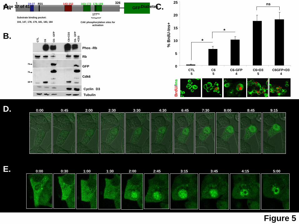
Confirmation of G1/S Molecule Cytoplasmic-to-Nuclear Trafficking with Live-Cell
Imaging. The preceding results suggest a model in which G1/S molecules may be able to
traffic into (cdk6, cyclin D3, p16, p21, p27) or out of (cyclin D3, p57) the nucleus of beta cells in
a manner that is associated with proliferation. To directly confirm whether G1/S molecule
trafficking can occur, an adenovirus in which human cdk6 was coupled to green fluorescent
protein (GFP) (Fig 5A) was prepared, so that trafficking could be directly observed in real-time,
if it occurred. Ad.cdk6-GFP was selected because cdk6 can co-localize with Ki67 in human
beta cells (Figs 3B,4), and because Ad.cdk6 is capable of driving human beta cell proliferation
in vitro and in vivo and enhances human β-cell engraftment in vivo (1-3). To confirm that the
Ad.cdk6-GFP construct was fully biologically active, it was compared to an adenovirus
expressing “wild-type”, untagged human cdk6 (Ad.cdk6), examining their relative levels of
expression and their abilities, with or without Ad.cyclinD3, to induce pRb phosphorylation and
human beta cell proliferation. These studies confirmed the efficiency of expression, bioactivity
and potency {induction of pRb phosphorylation (Fig 5B) and beta cell proliferation (Fig 7C)} of
the Ad.cdk6-GFP as compared to “wild-type” cdk6.
The rat insulinoma line, Ins1, and dispersed human islet cells, were transduced with the
Ad.cdk6-GFP construct, and live-cell imaging was performed, beginning 24 hours after
transduction. As can be seen in the live-cell time-lapse images in Figs 5D,E, also seen in video
format (see Supplemental Movies 1 & 2), cdk6-GFP can be observed clearly in the
cytoplasmic compartment initially, and then shifts into the nuclear compartment with time. That
this is not simply the natural progression of cytoplasmic translation of what will ultimately be a
protein which is then targeted to the nuclear compartment is evident from the observations in
Figs 1-4 and references 1,3,4 that cdk6 is present in the cytoplasm, but does not appear in the
Page 9 of 41 Diabetes
10
nuclear compartment in quiescent cells. Thus, these studies document in dynamic terms that
cdk6 is a principally cytoplasmic protein in quiescent, non-proliferating human beta cells, but is
able to shuttle to the nucleus in association with proliferation.
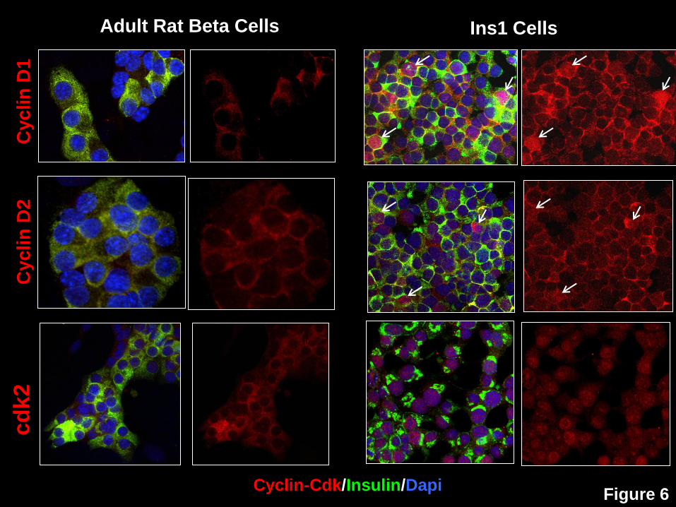
The Quiescent-to-Proliferative G1/S Molecule Shift Applies to Rat Beta Cells. To
determine whether the cytoplasmic G1/S molecule paradigm is restricted to human beta cells, or
might be a feature of rodent beta cells as well, we examined cyclin D1, D2 and cdk2 in rat islets
(Fig 6) and compared their localization to that observed in the rat insulinoma cell line, Ins1
832/13. We selected cyclin D1 and cdk2 because they are present in the cytoplasm of human
islets, and cyclin D2 because it is the most important D-cyclin in rat islets, therefore comparable
to cyclin D3 in human islets. As can be seen in the figure, these three molecules are most
intensely apparent in the cytoplasmic compartment in rat beta cells. On the other hand, using
the same antisera, the same fixation methods and the same species, these three molecules are
readily apparent in the nuclei of Ins1 cells.
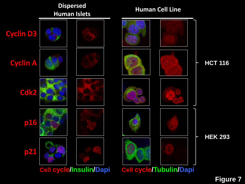
Comparison of Cytoplasmic Human Beta Cell G1/S Molecules in Transformed Human Cell
Lines. To determine whether the molecules that appeared cytoplasmic in non-proliferating
human beta cells could be observed in the nucleus of transformed, proliferating human cell lines
using the same antisera and same fixation methods in a human homologous system, we re-
examined cyclin D3, Cyclin A, cdk2, p16 and p21 in human beta cells, and compared them to
the human colonic cancer cell line, HCT116, and to HEK293 human embryonal kidney cells. As
can be seen in Fig 7, in human beta cells, each of the molecules showed the same principally
cytoplasmic pattern described above. In contrast, in HCT116 and HEK293 cells, cyclin D3,
cyclin A, cdk2, and p16 could be readily observed in the nuclei of many cells. p21 which is
nuclear and/or cytoplasmic in human beta cells, was also observed in both nuclei and cytoplasm
in HEK293 cells.
Page 10 of 41Diabetes
11
Discussion
Most prior models of human beta cell replication assumed that all G1/S molecules are
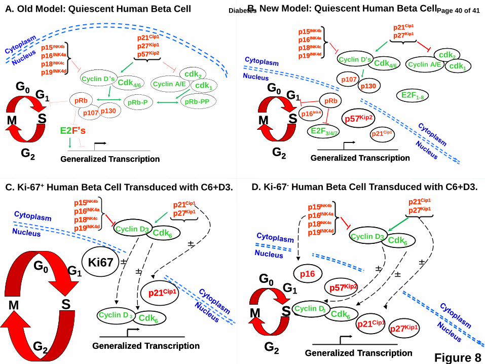
present in the nuclear compartment, as depicted in Fig 8A, awaiting activation (1-21). Instead,
in quiescent adult human beta cells, most are found in the cytoplasm, as shown in the model in
Fig 8B (1). Indeed, the only three abundant nuclear molecules in quiescent human beta cells
are cell cycle inhibitors: pRb, p57 and in some cases p21 (1), all of which would be anticipated
to discourage cell cycle progression. Further, the quiescent human beta cell model depicted in
Fig 8B is not static, but one that is active, motile, fluid, and characterized by shuttling of G1/S
molecules to and from the cytoplasm into the nuclear compartment in association with beta cell
proliferation, as shown in the models in Figs 8C and D. These models demonstrate that
several G1/S molecules can be induced to traffic into the nucleus in association with induction of
proliferation. Further, the observations suggest that this trafficking may be selective, with some
G1/S molecules engaging in nuclear trafficking (eg., cdks, cyclins, p16, p21, p27, p57), while
others may not (p15, p18, p19), and also suggest a directionality to this trafficking, with some
molecules entering the nucleus (cyclins, cdks, p16, p21, p27), while others exit, degrade or
otherwise disappear (cdk6, cyclin D3, p57).
The evidence for G1/S molecule cytoplasmic-nuclear trafficking seems clear.
Specifically, cdk6 and cyclin D3 clearly become more abundant in the nucleus, as we have
shown previously for cyclins D1 and D2, when overexpressed (1-4). In parallel, the cell cycle
inhibitors p16, p21 and p27 also become more abundant in the nuclear compartment, without an
overall increase in the amounts of these molecules. Conversely, the nuclear abundance of p57
declines, without an overall change in the amount of p57. In contrast yet again, p15, p18 and
p19 reveal a third pattern: no shift in intracellular location in response to proliferation. The most
unequivocal evidence for trafficking was observed for cdk6-GFP, which is absent from the
nuclear compartment under basal conditions, but can be observed trafficking into the nucleus
using live-cell, GFP imaging approaches in Ins1 and human islet cells.
Page 11 of 41 Diabetes
12
Superficially, the mechanism of proliferation seems obvious: cdk6 and cyclin D3 enter
the nucleus, phosphorylate resident pRb, and cell cycle progresses. On reflection, however, the
models raise fundamental questions about the control of trafficking of cell cycle molecules. In a
widely held model (11,12,22-27), cdk6, cdk4 and the D-cyclins lack a nuclear localization signal
(NLS), and require assembly in the cytosol by p21 and p27 (and perhaps p57) following their
translation. Paradoxically, p21 and p27 serve not only as chaperones, but also as members of
cyclin-cdk-inhibitor complexes. These complexes are guided into the nuclear compartment by
the NLS that is present in both p21 and p27. This NLS is in proximity to phosphorylation sites,
suggesting that kinases may phosphorylate p21 and/or p27 and activate the nuclear
translocation of these molecules and their cdk-cyclin cargo. In this model, the abundance of
p21 and p27 “titrates” proliferation, such that complete lack of p21 and p27 causes cell cycle
arrest because cyclins/cdks cannot enter the nucleus, and conversely, marked upregulation of
p21 and p27 cause cell cycle arrest because of their ability to block cyclin A/E/cdk1/cdk2
activity. Proliferation only occurs when p21 and p27 are present in the correctly titrated
concentration. Of course, most of these data are derived from rapidly proliferating genetically
modified mouse embryonic fibroblasts and cancer cell lines, where all of these cyclins and cdks
as well as CIP/KIPs are nuclear (22-27). And these events may be cell-type specific.
With this background, the data summarized in Figs 3 and 4 may indicate that since
among the cell cycle inhibitors studied, p21 is the one most commonly present in the nucleus of
Ki67+ beta cells, it may serve the chaperone/nuclear translocator function in the human beta
cell. Conversely, since p27 and p57, like p16, essentially never appear in the nucleus of Ki67+
beta cells, they may serve a conventional cell cycle inhibitor function. A similar trafficking-
mediated proliferation-inhibitory function has also been suggested for p21 (28). Further, the
observations that p16, p21, p27 and p57 all increase in nuclear frequency or abundance with
the induction of proliferation {(white bars), although for p57, always lower than in control cells
(blue bars)}, may reflect chaperone/nuclear translocator functions, eg, for p21, or an overall
Page 12 of 41Diabetes
13
reactive increase in inhibitory cell cycle “tone” in cells driven to proliferate. This model raises
the question, “What is restraining the cdks and cyclins, the CIP/KIP family and the INK4 family
in the cytoplasmic compartment of quiescent adult human beta cells, preventing them from
entering the nucleus?” Perhaps they are assembled in the cytoplasm by the CIP/KIP
molecules, but for some reason (e.g., lack of peri-NLS phosphorylation?), these complexes are
constrained to the cytoplasm, and cannot transport the cdk/cyclins into the nucleus. Or perhaps
the INK4s or other, as yet unidentified, cytoplasmic molecules anchor the cdk/cyclins in the
cytoplasm. Unraveling these issues will require silencing the CIP/KIPs and INK4s to see which,
if any, influences trafficking of cdks/cyclins or proliferation. It will also require
immunoprecipitation and proteomic studies to identify additional molecules that are bound to the
cdk/cyclins. These studies will be particularly challenging given the complexity of human islet
cellular composition (2,29,30), the relative paucity of beta cells within intact islets, and the lack
of availability of large numbers of pure populations of human beta cells for study.
As good working models should, these models raise many additional questions. For
example, we have not studied the trafficking of all potential G1/S molecules. Do E2Fs and/or
late G1/S molecules (cyclins A/E, cdks1/2) and/or p107/p130 also traffic to the nucleus when
proliferation is activated? Does pRb exit the nucleus? If so, what orchestrates all of this? Or, if
the E2Fs remain in the cytoplasm, how can cell cycle become active? Or, as suggested by
some (31,32), are E2Fs dispensable for proliferation? As another example, Figs 3,4 suggest
that cdk/cyclin D complexes activate proliferation in certain cells that lack cell cycle inhibitors in
the nuclear compartment, but what decides whether a given beta cell does or does not contain
nuclear cell cycle inhibitors? And if cdk6 and cyclin D3 are present in the nucleus of 30-40% of
transduced cells (Figs 2-4), why are all of these cells not proliferating? Of course, we could not
study the subcellular localization and partnering of every G1/S molecule in a single beta cell, so
that knowledge of the precise molecular complexes and the stoichiometry of their components
within the cytoplasm or nucleus of a given beta cell remains incomplete. Finally, these studies
Page 13 of 41 Diabetes
14
were performed using adult human beta cells, which are recalcitrant to proliferation. Since
replication is believed to occur at greater rates in fetal and neonatal human beta cells
(17,20,33), it will be informative to develop a model(s) such as that shown in Fig. 8 using these
types of islets.
The observation that G1/S molecules that are cytoplasmic in primary rat beta cells can
be nuclear in proliferating rat Ins1 insulinoma cells (Fig 6) is consonant with the concept that the
cytoplasmic-to-nuclear translocation occurs in association with activation of proliferation.
Coupled with data from intact human pancreas in the accompanying manuscript (1), they further
suggests that the subcellular localization to the cytoplasm in human beta cells is not an artifact
of fixation, antiserum choice, brain death, or islet isolation, since all of these variables can be
controlled for in the rodent models. Indeed, the “cytoplasmic G1/S quiescence model” may
apply more broadly beyond the human or rodent beta cell, extending to quiescent, differentiated
mammalian cells in general. Thus, in addition to the Discussion in the accompanying
manuscript documenting cytoplasmic localization of G1/S molecules in differentiated, quiescent
non-beta cells, the concept is further supported by Fig 7, contrasting quiescent human beta
cells to proliferating human HEK293 and HCT116 cancer cells, immunolabeled under identical
conditions.
Nonetheless, this work has important limitations. First, we have used cdk6 and cyclin
D3 to force proliferation, an artificial approach. While it would have been more “physiological” to
have employed growth factors, or nutrients to induce proliferation, no growth factors or nutrients
that can induce robust proliferation in adult human beta cells have been identified, so the
cdk/cyclin approach represents the only currently feasible approach. In addition, while it seems
reasonable to assume that G1/S molecules traffic into and out of the nucleus, most of the data
provided are “descriptive” and not “functional” or “mechanistic”, with the exception of the live-cell
imaging for cdk6. Along these lines, while several G1/S molecules appear in the nucleus, and
are less frequent at later time points, we have not documented that this nuclear diminution is a
Page 14 of 41Diabetes
15
result of nuclear exit, intranuclear degradation or another process, all of which are known to
occur in other systems (26,27,35-39). Also, it also should be emphasized that the majority of
studies were performed in isolated, dispersed, cultured, cadaveric beta cells, all conditions that
might alter the normal distribution, function and immunodetection of cell cycle molecules. This
was unavoidable because the usual harsh fixation methods used for intact pancreas pathology
specimens, eg., long term 10% formalin, prevented immunolabeling for most of the G1/S
molecules studied. However, we were able to confirm the subcellular location in intact human
pancreas of pRb p18, p57, p107 and p16, i.e., in five of five cases in which immunolabeling of
intact pancreas was possible (1). Further, the data are complimented by similar data in non-
proliferating rodent beta cells (Fig 7, references 37,38), so these observations seem firm. Also,
while we use the broad term “G1/S” throughout, and although many of these same molecules
participate in the G0/G1 transition, whether there are additional molecules that may “gate” the
G0/G1 interface in beta cells is unknown. Importantly, we have also used terms such as
“proliferation” and “replication” advisedly, employing a surrogate for beta cell proliferation, Ki67,
rather than authentic expansion of beta cell numbers. Unfortunately, this is unavoidable,
because no laboratory has developed techniques that provide unequivocal evidence of
productive human beta cell expansion in vitro or in vivo (14). In a related manner, we variously
use the terms “quiescent”, “senescent” and “irreversible” interchangeably in the context of non-
proliferating human beta cells. While some would argue that these are different states,
distinguishing among them in cadaveric human beta cells is difficult or impossible. Another
important consideration is that while we have demonstrated an association between nuclear-
cytoplasmic trafficking and cell cycle activation, we have not proven in formal terms that this is
required for cell cycle entry in the human beta cell. It also is important to emphasize that while it
is likely that G1/S molecule trafficking is one important regulatory process involved in beta cell
cycle entry, it is only one of many important mechanisms, with other examples being
phosphorylation and dephosphorylation of key substrates, transcriptional and posttranscriptional
Page 15 of 41 Diabetes
16
induction and/or stabilization/destabilization of cyclins/cdks, or comparable reductions in cell
cycle inhibitors, to name a few. Finally, while we believe we have controlled as well as
reasonably possible for immunocyto/histochemical variation and error, antibody specificity can
be troublesome. Thus, it must be emphasized that the models in Fig 8 are just that: working
models that can be used for confirmation, modification, correction, extension, and hypothesis
generation: they will likely evolve further with time.
In summary, these studies provide three new models of “human beta cell G1/S cell cycle
control”, one in quiescence (Fig 8B), one for proliferating beta cells induced by cdk6/cyclin D3
(Fig 8C), and one for beta cells that fail to proliferate despite cdk6/cyclin D3 expression (Fig
8D). The models should be useful for hypothesis generation, and provide new questions and
potential therapeutic approaches to human beta cell expansion, such as high-throughput drug
screens examining nuclear translocation, or kinome profiling to identify kinases that
phosphorylate key cell cycle inhibitors, and induce their translocation.
Page 16 of 41Diabetes
17
Author Contributions
N.F.T. and A.F.S. are guarantors and take full responsibility for the manuscript and its originality. N.F.T. researched data, contributed to the discussion, and wrote the manuscript. J.W.K., F.G.S., R.T., H.S., K.K.T, R.W, G.C., M.T. and A.E.C researched data. D.K.S. contributed to the discussion. A.F.S. contributed to the discussion and also wrote the manuscript. No potential conflicts of interest relevant to this article were reported.
Acknowledgements
The authors wish to thank Adolfo Garcia-Ocaña PhD, Rupangi C. Vasavada PhD, and Laura C. Alonso MD at the University Of Pittsburgh School Of Medicine for many helpful discussions during the preparation of these studies. This work was supported by the NIDDK and the Beta Cell Biology Consortium through NIH Grants U-01 DK 089538, R-01 DK55023, R56 DK065149 and T32-07052, by JDRF Grants 1-2008-39 and 34-2008-630, by ADA Grant 7-12-BS-046 and by a University of Pittsburgh Junior Faculty Award to NFT. We also thank the Kroh and Wagner Family Foundations for their support of this work. Human islets were generously supplied by the NIDDK- and JDRF-supported Integrated Islet Distribution Program (IIDP), and by Tatsuya Kin MD at the University of Alberta.
Page 17 of 41 Diabetes
18
References
1. Fiaschi-Taesch NM, Kleinberger JW, Salim F, Troxell R, Cox AE, Takane KK, Scott DK,
Stewart AF. A Human Pancreatic Beta Cell G1/S Molecule Cell Cycle Atlas. Diabetes (accompanying manuscript).
2. Cozar-Castellano I, Takane KK, Bottino R, Balamurugan AN, Stewart AF. Induction of Beta Cell Proliferation and Retinoblastoma Protein Phosphorylation in Rat and Human Islets Using Adenoviral Delivery of Cyclin-Dependent Kinase-4 and Cyclin D1. Diabetes 53:149-59, 2004.
3. Fiaschi-Taesch NM, Bigatel TA, Sicari BM, Takane KK, Velazquez-Garcia S, Harb G, Karen Selk K, Cozar-Castellano I, Stewart AF. A survey of the human pancreatic beta cell G1/S proteome reveals a potential therapeutic role for cdk-6 and cyclin D1 in enhancing human beta cell replication and function in vivo. Diabetes 58:882-93, 2009.
4. Fiaschi-Taesch NM, Salim F, Kleinberger J, Troxell R, Cozar-Castellano I, Selk K, Cherok E, Takane KK, Stewart AF. Induction of human beta cell proliferation and engraftment using a single G1/S regulatory molecule, cdk6. Diabetes 59:1926-36, 2010.
5. Karslioglu E, Kleinberger J, Salim F, Cox A, Takane KK, Donald K. Scott DK, Stewart AF. cMyc is the principal upstream driver of beta cell proliferation in rat insulinoma cell lines and Is an effective mediator of human beta cell replication. Mol Endocrinology 25:1760-72, 2011.
6. Cozar-Castellano I, Harb G, Selk K, Takane KK, Vasavada RC, Sicari B, Law B, Zhang P, Scott DK, Fiaschi-Taesch N, Stewart AF. Lessons from the First Comprehensive Molecular Characterization of Cell Cycle Control in Rodent Insulinoma Cell Lines. Diabetes 57:3056-68, 2008.
7. Kondegowda N G, Joshi-Gokhale S, Harb G, Williams K, Zhang XY, Takane KK, Zhang P, Scott DK, Stewart AF, Garcia-Ocaña A, Vasavada RC. Parathyroid hormone-related protein enhances human beta cell proliferation and function with simultaneous induction of cyclin-dependent kinase 2 and cyclin E expression. Diabetes 59:3131-38, 2010.
8. Lavine JA, Raess PW, Davis DB, Rabaglia ME, Presley BK, Keller MP, Beinfeld MC, Kopin AS, Newgard CN, Attie AD. Overexpression of pre-pro-cholecystokinin stimulates beta cell proliferation in mouse and human islets with retention of islet function. Mol Endocrinol 22:2716-28, 2008.
9. Davis DB, Lavine JA, Suhonen JI, Krautkramer KA, Rabaglia ME, Sperger JM, Fernandez LA, Yandell BS, Keler MP, Wang IM, Schadt EE, Attie AD. FoxM1 is up-regulated in obesity and stimunates beta-cell proliferation. Mol Endocrinol 24:1822-34, 2010.
10. Fatrai S, Elghazi L, Balcazar N, Cras-Meneur C, Krits I, Kiyokawa H, and Bernal-Mizrachi E. Akt Induces β-cell proliferation by regulating cyclin D1, Cyclin D2, and p21 levels and cyclin-dependent kinase-4 activity. Diabetes 55:318-325, 2006.
11. Cozar-Castellano I, Fiaschi-Taesch N, Bigatel TA, Takane KK, Garcia-Ocana A, Vasavada RC, Stewart AF. Molecular Control of Cell Cycle Progression in the Pancreatic Beta Cell. Endocrine Reviews 27:356-370, 2006.
12. Heit JJ, Karnik SK, Kim SK. Intrinsic regulators of pancreatic beta-cell proliferation. Annu Rev Cell Dev Biol 22:311-38, 2006.
13. Cozar-Castellano I, Weinstock M, Haught M, Velázquez-Garcia S, Sipula D, Stewart AF. Evaluation of beta cell replication in mice transgenic for hepatocyte growth factor, placental lactogen or both: comprehensive characterization of the G1/S regulatory proteins reveals unique involvement of p21cip. Diabetes 55:70-77, 2006.
14. Rieck S, Li Z, Sandhu AK, Liu C, Naji Al, Takane KK, Fiaschi-Taesch NM, Stewart AF, Kushner JA, Kaestner KH. Overexpression of hepatocyte nuclear factor 4α initiates cell cycle entry, but it not sufficient to promote ß-cell expansion in human islets. Mol Endocrinol 26:1590-1602, 2012.
Page 18 of 41Diabetes
19
15. Kulkarni RN, Bernal-Mizrachi E, Garcia-Ocaña A, Stewart AF. Human ß-cell proliferation and intracellular signaling: driving in the dark without a roadmap. Diabetes 61:2205-2213, 2012.
16. Zhong L, Georgia S, Tschen S, Nakayama K, Nakayama K, Bhushan A. Essential role of skp-2-mediated p27 degradation in growth and adaptive expansion of pancreatic beta cells. J Clin Invest. 117:2869-76, 2007.
17. Chen H, Gu X, Liu Y, Wang J, Wirt SE, Bottino R, Schorle H, Sage J, Kim SK. PDGF
signaling controls age-dependent proliferation in pancreatic β-cells. Nature 478: 349-55, 2011.
18. Beattie GM, Itkin-Ansari P, Cirulli V, Leibowitz G, Lopez AD, Bossie S, Mally MI, Levine F, Hayek A. Sustained proliferation of pdx-1+ cells derived from human islets. Diabetes 48:1013-19, 1999.
19. Krishnamurthy J, Ramsey MR, Ligon KL, Torrice C, Koh A, Bonner-Weir S, Sharpless NE. p16ink4a induces an age-dependent decline in islet regenerative potential. Nature 443:453-7, 2006.
20. Kohler CU, Olewinski M, Tannapfel A, Schmidt WE, Fritsch H, Meier JJ. Cell cycle control of beta cell replication in the prenatal and postnatal human pancreas. Am J Physiol Endocrinol Metab 300:E221-230, 2011.
21. Porat S, Weinberg-Corem N, Tornovsky-Babaey S, Schyr-Ben-Haroush R, Hija A, Stolovich-Rain M, Dadon D, Granot Z, Ben-Hur V, White P, Girard CA, Karni R, Kaestner KH, Ashcroft FM, Magnuson MA, Saada A, Grimsby J, Glaser B, Dor Y. Control of pancreatic beta cell regeneration by glucose metabolism. Cell Metab 13:440-449, 2011.
22. Sherr CJ Roberts JM. Living with or without cyclins and cyclin-dependent kinases. Genes Dev 18:2699-2711, 2004.
23. Sherr CJ, Roberts JM. CDK inhibitors: positive and negative regulators of G1-phase progression. Genes & Dev 13:1501-12, 1999.
24. Malumbres M, Barbacid M. Mammalian cyclin-dependent kinases. Trends in Biological Sciences. 30:630-641, 2005.
25. Cobrinik D. Pocket Proteins and cell cycle control. Oncogene 24:2796-2809, 2005. 26. Malumbres, M. Physiological relevance of cell cycle kinases. Physiol Rev. 91:973-1007,
2011. 27. Satyanarayana A, Kaldis P. Mammalian cell cycle regulation: several cdks, numerous
cyclins and diverse compensatory mechanisms. Oncogene 28:2925-39, 2009. 28. Ranta F, Leveringhaus J, Theilig D, Schulz-Raffelt G, Hennige AM, Hildebrand DG, Handrick
R, Jendrossek V, Bosch F, Schulze-Osthoff K, Haring H-U, Ullrich S. Protein kinase C delta (PKC∂) affects proliferation of insulin-secreting cells by promoting nuclear extrusion of the cell cycle inhibitor p21cip/waf1. PLoS One 12:e28828, 2011.
29. Cabrera O, Berman D, Kenyon NS, Ricordi C, Berggren P-O, Caicedo A. The unique architecture of the human pancreatic islet has implications for islet cell function. Proc Nat Acad Sci USA 103:2334-39, 2006.
30. Brissova M, Fowler MJ, Nicholson WE, Cho A, Hirshberg B, Harlan DM, Powers AC. Assessment of human pancreatic islet architecture and composition by laser scanning confocal microscopy. J Histochem Cytochem. 53:1087-97, 2005.
31. Chong JL, Wenzel PL, Saenz-Robles MT, Nair V, Ferrey A, Hagan A, Gomez YM, Sharma N, Chen HZ, Ouseph M, Wang SH, Trikha P, Culp B, Mezache L, Winton DJ, Sansom OJ, Chen D, Bremner R, Cantalupo PG, Robinson ML, Pipas JM, Leone G. E2f1-3 switch from activators in progenitor cells to repressors on differentiated cells. Nature 462:930-4, 2009.
32. Chen D, Pascal M, Wenzel P, Knopfler PS, Leone G, Bremner R. Division and apoptosis-deficient retinal progenitors. Nature 462:925-29, 2009.
33. Meier JJ, Butler AE, Saisho Y, Monchamp T, Galasso R, Bhushan A, Rizza RA, Butler PC. Beta cell replication is the primary mechanism subserving the postnatal expansion of beta cell mass in humans. Diabetes 57:1584-94, 2008.
Page 19 of 41 Diabetes
20
34. Kassem SA, Ariel I, Thornton PS, Scheimberg I, Glaser B. Beta cell proliferation and apoptosis in the developing normal human pancreas and in hyperinsulinism of infancy. Diabetes 49:1325-1333, 2000.
35. Ivanova IA, Vespa A, Dagnino L. A novel mechanism of E2F1 regulation via nucleocytoplasmic shuttling. Cell Cycle 6:2186-95, 2007.
36. Veiga-Fernanandes H, Rocha B. High expression of active cdk6 in the cytoplasm of CD8 memory cells favors rapid division. Nature Immunol 5:31-37, 2004.
37. He LM, Sartori DJ, Teta M, Opare-Addo, Rankin M, Long SY, Diehl JA, Kushner JA. Cyclin D2 protein stability is regulated in pancreatic beta cells. Mol Endocrinol. 23:1865-75, 2009.
38. Alonso LC, Yokoe T, Zhang P, Scott DK, Kim SK, O’Donnell CP, Garcia-Ocaña A. Glucose infusion in mice: a new model to induce beta-cell replication. Diabetes 56:1792-801, 2007.
39. Van Dross R, Yao S, Asad S, Westlake G, Mays DJ, Barquero L, Duell S, Pietenpol JA, Browning PJ. Constitutively active K-cyclin-cdk6 kinase in Kaposi sarcoma-associated herpesvirus infection. J Nat Cancer Inst 97:656-66, 2005.
Page 20 of 41Diabetes
21
Figure Legends
Figure 1: Nuclear translocation of the cdk6, cyclin D3, p16, p21, p27, but not p15, p18,
p19 or p57, in human beta cells in response to expression of cdk6 and cyclin D3. A.
Dispersed human islets were transduced with control adenovirus (“control”, white frames) or
with Ad.cdk6 and Ad.cyclin D3 (“Ad.C6+D3”, blue frames). Immunolabeling for cdk6 (upper two
panels) and cyclin D3 (lower panels) is shown in red, and insulin in green. Note that the nuclear
frequency of both cdk6 and cyclin D3 increase with their adenoviral expression, confirming a
prior report (4). B. In contrast to cdk6 and cyclin D3, there is no change in the cellular location
of p15, p18 or p19. C. In contrast, in Panel C, p16, p21, p27 and p57 all alter their subcellular
distribution in response to cdk6/cyclin D3. These experiments are representative of 3-5
separate human cadaver islet preparations. In some panels, insulin staining is removed to allow
visualization of the relevant G1/S molecule in the cytoplasm, but in all cases, the cells shown
are also insulin+ as shown for cdk6 and p16.
Figure 2: Quantification of the nuclear translocation of the G1/S molecules and their
absolute expression in human beta cells in response to overexpression of cdk6 and
cyclin D3. Panel A. White bars represent control dispersed human beta cells, and black bars
represent those transduced with Ad.cdk6+cyclin D3. Each panel represents observations for
the G1/S molecule indicated. The numbers shown within or above the bars indicate the number
of insulin+ cells showing nuclear G1/S molecule (red) and the total number of insulin+ (green)
cells counted. Bars indicate mean ± SEM; * = p<0.05 vs control. Quantification of Ki67+ and
insulin+ cells as a function of total insulin+ cells. Panel B. Representative immunoblots of cdk6,
cyclin D3, p16, p21, p27 and p57 from islet preps used for panel C. Panel C. Densitometric
quantification of four immunoblots for cdk6, cyclin D3, p16, p21, p27 and p57. * = p<0.05 vs
control; ns, non-significant vs control. These panels make the point that when p16, p21 and p27
increase in nuclear intensity, there is no overall increase in the amounts of these three
molecules, and the inverse is true for p57, suggesting that the changes in subcellular
localization observed are not simply a reflection of a generalized increase in p16, p21, p27 and
p57, but likely reflect changes in their nuclear trafficking and/or stability.
Figure 3: Time courses of subcellular localization of G1/S molecules and Ki67 in human
beta cells in response to expression of cdk6 and cyclin D3. Dispersed human islets were
transduced with control adenovirus (“control”, white frames on the left) or with Ad.cdk6 and
Page 21 of 41 Diabetes
22
Ad.cyclinD3 (“Ad.C6+D3”, blue frames on the right). Cells were fixed 24, 48 or 72 hr after
transduction, and immunolabeled for the G1/S molecule indicated, cyclin D3, cdk6, p16, p27,
p21 or p57. White arrows indicate examples of proliferative beta cells, as assessed by Ki67.
Red arrows illustrate examples of a nuclear the G1/S molecule in question. Double red and
white arrows indicate co-localization of a nuclear G1/S molecule with Ki67 in insulin+ cells.
Figure 4: Quantification of changes in cyclin D3, cdk6, p16, pp27, p21 and p57 with Ki67
in human beta cells in response to expression of cdk6 and cyclin D3. These data
represent the quantification data in Fig. 3 from different experiments using islets from 4-6
different donors for each cell cycle molecule, at 24, 48 and 72 hours after cdk6/cyclin D3
transduction. The blue bars indicate the % nuclear localization of the six molecules shown in
insulin+ (beta) cells from control, non-transduced, dispersed human islet cells. The white bars
represent the % of insulin+ (beta) cells that contain the indicated G1/S molecule in question in
the nucleus at 24, 48 and 72 hours when cdk6/cyclin D3 are expressed. The grey bars
represent the % of Ki67+/insulin+ cells. The black bars represent insulin+ cells that are doubly
positive for both Ki67 and nuclear presence of the G1/S molecule indicated. Bars indicate mean
± SEM. * = <0.05 for differences shown by the solid lines. # = p<0.05 for Ki67+ beta cells (grey
bars) vs. Ki67+ untransduced beta cells at 24, 48 and 72 hrs. § = p<0.05 vs. untransduced beta
cells. Unless a symbol is shown, the changes are not significant. The range of Ki67 induction
(grey bars) is large and spans approximately 10-20% of cells, in accord with prior studies. This
variability likely reflects differences among human islet preparations, adenoviral transduction
efficiency, and other variables.
Figure 5: Cdk6 cytoplasmic-to-nuclear trafficking with live-cell imaging. A. Schematic
representation of the Ad.cdk6-GFP construct used in the live-cell imaging experiments, with a
GFP tag at the C-terminus. Numbers above indicate amino acid residues. The blue box
designates the ATP-binding domain, the black box the INK4-binding domain, the red box the
catalytic loop, and the two green boxes the activation loops. Cdk6 can be phosphorylated on
Thr177 by cdk-activating kinase (CAK). Key amino acids in the substrate-binding pocket are
indicated. B. Representative immunoblots of human islets transduced with Ad.lacZ (CTL),
Ad.cdk-6-GFP, non-tagged Ad.cdk6, with or without Ad.cyclin D3, showing phosphorylated Rb
(phos-Rb), total Rb (Rb), GFP, cdk6 and cyclin D3, and tubulin as a loading control. Small
numbers on the left indicate molecular weight markers. These studies indicate that the
Ad.cdk6-GFP is biologically active since it can phosphorylate pRb. C. BrdU+ and insulin+ cells
Page 22 of 41Diabetes
23
as a percentage of total insulin+ cells. Representative pictures of BrdU (red) and insulin (green)
in the conditions are shown at the bottom on the panel. Dispersed human islets were
transduced with control Ad.lacZ (CTL) or with adenoviruses encoding cdk6 (C6), cdk6-GFP (C6-
GFP), cdk6 and cyclin D3 (C6+D3) or cdk6-GFP and cyclin D3 (C6-GFP+D3). The numbers
below each bar indicate the numbers of different human islet preparations studied. Bars
indicate mean ± SEM; * = p<0.05; ns = non-significant. These studies confirm that the Ad.cdk6-
GFP is biologically active. D. Photomicrographs derived from live-cell imaging of Ins1 cells
transduced with cdk6-GFP (the full movie can be seen at “Supplemental Movies 1”). E.
Photomicrographs derived from live-cell imaging of dispersed human islet cells transduced with
cdk6-GFP. The numbers above each picture in D and E indicate the time from the beginning of
the movie (the full movie can be seen in “Supplemental Movies 2”).
Figure 6: Comparison of distribution of cyclin D1, cyclin D2 and cdk2 in rat beta cells
and rat Ins1 cells. Left two columns. The subcellular location of cyclin D1, cyclin D2 and
cdk2 in primary cultures of dispersed rat pancreatic beta cells. Right two columns. The
subcellular location of the same three molecules in proliferating Ins1 cells. White arrows show
example of Ins1 cells in which cyclins D1 and 2 appear in the nucleus. In each pair of columns,
the left column displays the merged images of insulin (green), the cyclin/cdk of interest (red) and
the nuclear marker DAPI (blue), while the right column shows the cyclin/cdk separated from
insulin and DAPI images. Each experiment is representative of three different rat islet
preparations or Ins1 experiments. Rat islets were isolated and dispersed as detailed previously
(5,6) and both rat islets and Ins1 cells were cultured for 72 hrs in RPMI 1640 with 10% fetal
bovine serum prior to fixing with 4% paraformaldehyde, as for the human islet samples. The
primary antisera were the same used for human islets in the supplemental Table to the
accompanying manuscript. Note that cyclins D1 and D2 and cdk2 are cytoplasmic, and non-
nuclear in quiescent rat beta cells as they are in human beta cells, but each can be readily
detected in the nucleus of proliferating Ins1 cells. These studies indicate that cyclin D1, cyclin
D2 and cdk2 easily can be observed in the nucleus, when present, using the reagents and
conditions employed, and that their localization is correlated with quiescent vs. proliferative
state.
Figure 7: Comparison of G1/S molecule subcellular localization in human beta cells and
transformed human cells lines. Left two columns. Dispersed human islets immunolabeled
for cyclin D3, cyclin A, cdk2, p16 and p21. Right two columns. Immunolabeling for the same
Page 23 of 41 Diabetes
24
five G1/S molecules in HCT116 human colon cancer cells or HEK293 human embryonic kidney
cells. Culture and fixation conditions were identical (4% paraformaldehyde for 15 min), as were
antisera, as shown in Supplemental Table 1 of the accompanying manuscript. Each image is
representative of three different human islet preparations or cell line experiments. Note that
cyclin D3, cyclin A, cdk2, p16 all appear cytoplasmic in quiescent human beta cells, but each is
observed in the nucleus of HEK293 or HCT116 cells. p21, which may be nuclear in some
human beta cells, can also be nuclear in HEK293 cells. These studies are compatible with the
concepts that these G1/S molecules can readily be observed in the nucleus, when present,
using the reagents and conditions employed, and that their localization generally correlates with
quiescent vs. proliferative state.
Figure 8: Prior and revised models of the G1/S cell cycle transition regulation in human
beta cells. The red arrows indicate inhibition, the green arrows activation, and the dotted black
lines, trafficking. (A) A prior model of G1/S control in human beta cells (2-13) in which most cell
cycle molecules were assumed to be present in the nucleus. (B) A revised model for G1/S
molecule distribution in quiescent human beta cells: most G1/S molecules are cytoplasmic, and
the only other frequently nuclear molecules are the cell cycle inhibitors, pRb, p57, accompanied
in occasional beta cells by p16, and/or in some preparations, p21. (C) G1/S molecules in Ki67+
human beta cells that have recently proliferated, or are proliferating. The larger G1/S circle in
this panel, as compared to the others, indicates that these cells have entered the cell cycle.
Following transduction, cdk6 and cyclin D3 transit to the nuclear compartment in association
with p21. (D) G1/S molecules in human beta cells that remain Ki67- (fail to proliferate) despite
transduction with cdk6 and cyclin D3. Cdk6 and cyclin D3 still enter the nucleus in many beta
cells, but are accompanied variably by p16, p21, p27 and/or p57, all or some of which may
attenuate the induction of proliferation. The ± symbols in C and D indicate that these
movements may or may not occur in a given beta cell following cdk6-cyclin D3 transduction.
Page 24 of 41Diabetes
For Peer Review Only
Control Ad.C6+D3
Figure 1A
C6
D3
C6/I
ns
D3/I
ns
Control Ad.C6+D3
Page 25 of 41 Diabetes
For Peer Review Only
Control Ad.C6+D3
p15
p18
p19
Figure 1B
Page 26 of 41Diabetes
For Peer Review Only
Control Ad.C6+D3
p21
p27
p
57
Control Ad.C6+D3
p16/I
ns
p16
Figure 1C
Page 27 of 41 Diabetes
For Peer Review Only
Ctl
Ad.C6+D3
% K
i67
in
Be
ta C
ell
s Ki67
0
2
4
6
8
10
12
*
6/5659
392/5279
n=3
D3
% n
ucle
ar
D3 in
Beta
Cell
s
0
5
10
15
20
25
30
35
40
45
50
*
2/1468
654/1938
n=4 C6
0
5
10
15
20
25
30
35
40
45
50
% n
ucle
arc
dk6 in
Beta
Cell
s
Cdk6 *
9/1319
481/1211
n=3
p21
% n
ucle
ar
p21
in
Beta
Cell
s
0
5
10
15
20
25
30
35
40
45
50
*
127/2351
507/1552
n=4
p57
% n
ucle
ar
p57
in
Beta
Cell
s
0
5
10
15
20
25
30
35
40
45
50
* 829/2166
495/2275
n=4
% n
uc
lea
r p
16
in
Be
ta C
ell
s
p16 n=5
% n
ucle
ar
p27
in
Beta
Cell
s
p27 n=4
Figure 2A
*
0
5
10
15
20
25
30
35
40
45
50
7/705
104/1089
*
0
5
10
15
20
25
30
35
40
45
50
133/1438
187/1018
Page 28 of 41Diabetes
For Peer Review Only
Cyclin D3
Cdk 6
p21
p27
p57
tubulin
p16
Ctl
Ad.C6+D3
Ctl
Ad
.C6
+D
3
Figure 2BC
B. C.
0
50
100
150
200
250
300
350
400
450
500
0
50
100
150
200
250
300
350
400
450
500
0
50
100
150
200
250
300
350
400
450
500
0
50
100
150
200
250
300
350
400
450
500
0
50
100
150
200
250
300
350
400
450
500
0
50
100
150
200
250
300
350
400
450
500
D3 n=4
Cdk6 n=4
p21 n=4
p57 n=4
p16 n=4
p27 n=4
ns
ns
ns ns
* *
Page 29 of 41 Diabetes
For Peer Review Only
Figure 3A D3 /Ki67/ Insulin D3 /Ki67/ Insulin D3 / Insulin Ki67/ Insulin
Control Ad.C 6 +D 3
24
h
48
h
72
h
Page 30 of 41Diabetes
For Peer Review Only
Figure 3B
Control Ad.C 6 +D 3
C6 /Ki67/ Insulin C6 / Insulin Ki67/ Insulin C6 /Ki67/ Insulin
24
h
48
h
72
h
Page 31 of 41 Diabetes
For Peer Review Only
Figure 3C
24
h
48
h
Control Ad.C 6 +D 3
p16 /Ki67/ Insulin p16 /Ki67/ Insulin p16 / Insulin Ki67/ Insulin
72
h
Page 32 of 41Diabetes
For Peer Review Only
Figure 3D p27 /Ki67/ Insulin p27 /Ki67/ Insulin p27 / Insulin Ki67/ Insulin
24
h
48
h
Control Ad.C 6 +D 3
72
h
Page 33 of 41 Diabetes
For Peer Review Only
Figure 3E p21 /Ki67/ Insulin p21 /Ki67/ Insulin p21 / Insulin Ki67/ Insulin
24
h
48
h
72
h
Control Ad.C 6 +D 3
Page 34 of 41Diabetes
For Peer Review Only
Figure 3F p57 /Ki67/ Insulin p57 /Ki67/ Insulin p57 / Insulin Ki67/ Insulin
24
h
48
h
Control Ad.C 6 +D 3
72
h
Page 35 of 41 Diabetes
For Peer Review Only
% Ki67+ With Cdk6 + Cyclin D3 overexpression
Figure 4
C6 D3
% in nucleus in controls
% Double+
p21 p57 p27
p16
* *
*
*
*
*
* *
*
*
*
*
*
*
*
*
*
* *
*
* * *
% in nucleus
#
#
#
#
#
#
#
#
#
#
#
#
#
# # #
#
#
§
§
§ § § §
§
§
§ §
Page 36 of 41Diabetes
For Peer Review Only
1 32619-27 143-152 163-172 176-189
Substrate binding pocket:
104, 147, 178, 179, 181, 183, 184
R31
177Thr
CAK phosphorylation sites for
activation
GFPA.
B.
C.
D.
E.
0:00 0:45 2:00 2:30 3:30 4:30 6:45 7:30 8:00 8:45 9:15
0:00 0:30 1:00 1:30 2:00 2:45 3:15 3:45 4:15 5:00
Figure 5
Brd
U / I
ns
0
5
10
15
20
25
CTL 5
C6 5
C6 - GFP 4
C6+D3 5
C6GFP+D3 4
*
*
ns
% B
rdU
- In
s+
CT
L
C6
C6
- G
FP
C6+
CD
3
C6
- G
FP
+C
D3
Phos - Rb
Cyclin D3
Tubulin
Rb
GFP 76
Cdk6
76
38
Page 37 of 41 Diabetes
For Peer Review Only
Cyc
lin
D1
C
yc
lin
D2
c
dk
2
Adult Rat Beta Cells Ins1 Cells
Cyclin-Cdk/Insulin/Dapi Figure 6
Page 38 of 41Diabetes
For Peer Review Only
Cdk2
Cyclin D3
Dispersed Human Islets
Human Cell Line
Cyclin A
Figure 7
Cell cycle/Insulin/Dapi Cell cycle/Tubulin/Dapi
p16
p21
HCT 116
HEK 293
Page 39 of 41 Diabetes
For Peer Review Only
A. Old Model: Quiescent Human Beta Cell
C. Ki-67+ Human Beta Cell Transduced with C6+D3.
Figure 8
p15 INK4b
p16 INK4a
p18 INK4c
p19 INK4d
Cdk 4/6
Generalized Transcription
p21 Cip1
p27 Kip1
Cyclin D ’ s
Cdk 6
M S
G 1 G 0 G 0
G 2 G 2
p15 INK4b
p16 INK4a
p18 INK4c
p19 INK4d
Cdk 6
Generalized Transcription
p21 Cip1
p27 Kip1
Cyclin D ’ s Cyclin D 3
Cdk 6 3 Cyclin D 3 M S
G G 0
G 2
p15 INK4b
p16 INK4a
p18 INK4c
p19 INK4d
Cdk 4/6
Generalized Transcription
p21 Cip1
p27 Kip1
Cyclin D ’ s
p27 Kip1 p21 Cip1
p16
M S
1
G 0
G 2
p15 INK4b
p16 INK4a
p18 INK4c
p19 INK4d
Cdk 6
Generalized Transcription
p21 Cip1
p27 Kip1
Cyclin D ’ s Cyclin D 3
p27 Kip1 p21 Cip1
p16
p57 Kip2 p57 Kip2
B. New Model: Quiescent Human Beta Cell
p57 Kip2 p57 Kip2 M S
G 1 G 0
G 2
p15 INK4b
p16 INK4a
p18 INK4c
p19 INK4d
Cdk 4/6
Generalized Transcription
Cyclin A/E
cdk 2
p21 Cip1
p27 Kip1
p107 p130
Cyclin D ’ s cdk 1
pRb
p130
E2F 1 - 8
p21 Cip1 E2F 4/6/7
M S
G G 0
G 2
p15 INK4b
p16 INK4a
p18 INK4c
p19 INK4d
Cdk 4/6
Generalized Transcription
Cyclin A/E
cdk 2 cdk 2
p21 Cip1
p27 Kip1
p107 p130
Cyclin D ’ s Cyclin D ’ s cdk 1
pRb
p130
E2F 1 - 8
p21 Cip1 E2F 3/4/7
D. Ki-67- Human Beta Cell Transduced with C6+D3.
p16 Ink4
p21 Cip1 p21 Cip1
Ki67
S Cdk 6 Cyclin D 3 S Cdk 6 Cyclin D 3 Cyclin D 3
±
± ±
±
M S
G 1
G 0
G 2
p15 INK4b
p16 INK4a
p18 INK4c
p19 INK4d
Cdk 4/6
pRb - P
E2 F ’ s
Generalized Transcription
pRb - PP
Cyclin A/E
cdk 2
p21 Cip1
p27 Kip1
p57 Kip2
p107 p130
Cyclin D ’ s cdk 1
pRb
M S
G G 0
G 2
p15 INK4b
p16 INK4a
p18 INK4c
p19 INK4d
Cdk 4/6
pRb - P
E2 F ’ s
Generalized Transcription
pRb - PP
Cyclin A/E
cdk 2 cdk 2
p21 Cip1
p27 Kip1
p57 Kip2
p107 p130
Cyclin D ’ s Cyclin D ’ s cdk 1
pRb
±
±
Page 40 of 41Diabetes
1
Supplemental Movie Legends
Supplemental Movie 1: Cdk6-GFP live-cell imaging in INS1 cells. Rat insulinoma cell line
(INS1) were plated and transduced with adenovirus encoding human cdk6 fused with green
fluorescent protein (GFP) (Ad.cdk6-GFP) as described in Materials and Methods and were
imaged 24 hr after transduction. See text for details.
Supplemental Movie 2: Cdk6-GFP live-cell imaging in human islet cells. Human islets were
dispersed and transduced with Ad.cdk6-GFP as described in Materials and Methods and were
imaged 24 hr after transduction. See text for details.
Page 41 of 41 Diabetes