
Oxygen Activation by Nonheme Iron(II) Complexes: α-Keto Carboxylate versus Carboxylate
15
Oxygen Activation by Nonheme Iron(II) Complexes: r-Keto Carboxylate versus Carboxylate Mark P. Mehn, ² Kiyoshi Fujisawa,* ,‡ Eric L. Hegg, ²,§ and Lawrence Que, Jr.* ,² Contribution from the Department of Chemistry and Center for Metals in Biocatalysis, 207 Pleasant Street Southeast, UniVersity of Minnesota, Minneapolis, Minnesota 55455, and Department of Chemistry, UniVersity of Tsukuba, Tennodai 1-1-1, Tsukuba 305-8571, Japan Received October 8, 2002; E-mail: [email protected]; [email protected] Abstract: Mononuclear iron(II) R-keto carboxylate and carboxylate compounds of the sterically hindered tridentate face-capping ligand Tp Ph2 (Tp Ph2 ) hydrotris(3,5-diphenylpyrazol-1-yl)borate) were prepared as models for the active sites of nonheme iron oxygenases. The structures of an aliphatic R-keto carboxylate complex, [Fe II (Tp Ph2 )(O2CC(O)CH3)], and the carboxylate complexes [Fe II (Tp Ph2 )(OBz)] and [Fe II (Tp Ph2 )(OAc)(3,5- Ph2pzH)] were determined by single-crystal X-ray diffraction, all of which have five-coordinate iron centers. Both the R-keto carboxylate and the carboxylate compounds react with dioxygen resulting in the hydroxylation of a single ortho phenyl position of the Tp Ph2 ligand. The oxygenation products were characterized spectroscopically, and the structure of the octahedral iron(III) phenolate product [Fe III (Tp Ph2* )(OAc)(3,5- Ph2pzH)] was established by X-ray diffraction. The reaction of the R-keto carboxylate model compounds with oxygen to produce the phenolate product occurs with concomitant oxidative decarboxylation of the R-keto acid. Isotope labeling studies show that 18 O2 ends up in the Tp Ph2* phenolate oxygen and the carboxylate derived from the R-keto acid. The isotope incorporation mirrors the dioxygenase nature of the enzymatic systems. Parallel studies on the carboxylate complexes demonstrate that the oxygen in the hydroxylated ligand is also derived from molecular oxygen. The oxygenation of the benzoylformate complex is demonstrated to be first order in metal complex and dioxygen, with activation parameters ΔH q ) 25 ( 2 kJ mol -1 and ΔS q )-179 ( 6 J mol -1 K -1 . The rate of appearance of the iron(III) phenolate product is sensitive to the nature of the substituent on the benzoylformate ligand, exhibiting a Hammett F value of +1.3 indicative of a nucleophilic mechanism. The proposed reaction mechanism involves dioxygen binding to produce an iron(III) superoxide species, nucleophilic attack of the superoxide at the R-keto functionality, and oxidative decarboxylation of the adduct to afford the oxidizing species that attacks the Tp Ph2 phenyl ring. Interestingly, the R-keto carboxylate complexes react 2 orders of magnitude faster than the carboxylate complexes, thus emphasizing the key role that the R-keto functionality plays in oxygen activation by R-keto acid-dependent iron enzymes. R-Ketoglutarate (R-KG) 1 -dependent dioxygenases constitute a large class of nonheme iron-containing enzymes that are essential for the biosynthesis of a diverse array of compounds, 2 catabolism of selected biomolecules, 3 repair of alkylated DNA 4,5 and RNA, 6 and oxygen sensing in cells. 7-9 For instance, prolyl hydroxylase is responsible for the posttranslational modification of proline to hydroxyproline in collagen biosynthesis 10 and in the cellular response to hypoxia, 7-9 clavaminic acid synthase (CAS) catalyzes three distinct steps in the synthesis of the -lactamase inhibitor clavulanic acid, 11 AlkB demethylates 1-methyladenine and 3-methylcytosine in single stranded DNA and RNA, 4-6 and deacetoxycephalosporin C synthase (DAOCS) catalyzes the ring expansion of the thiazolidine ring of the penicillin N nucleus to afford deacetoxycephalosporin C (Scheme 1). 12,13 Although each enzyme carries out a distinct transforma- tion, the members of this class share a requirement for an R-keto ² University of Minnesota. ‡ University of Tsukuba. § Current address: Department of Chemistry, University of Utah, Salt Lake City, UT 84112. (1) Abbreviations: R-KG, R-ketoglutarate; BF, benzoylformate; CAS, cla- vaminate synthase 2; DAOCS, deacetoxycephalosporin C synthase; 3,5- Ph2pzH, 3,5-diphenylprazole; 6-Me3-TPA, tris(6-methyl-2-pyridylmethyl)- amine; PRV, pyruvate; TfdA, 2,4-dichlrophenoxyacetic acid/R-KG dioxygenase; TauD, taurine/R-KG dioxygenase; TPA, tris(2-pyridylmethyl)- amine; Tp tBu,iPr , hydrotris(3-tert-butyl-5-isopropylpyrazol-1-yl)borate (mono- anion); Tp iPr2 , hydrotris(3,5-diisopropylpyrazol-1-yl)borate (monoanion); Tp Me2 , hydrotris(3,5-dimethylpyrazol-1-yl)borate (monoanion); Tp Ph2 , hy- drotris(3,5-diphenylpyrazol-1-yl)borate (monoanion). (2) Prescott, A. G.; Lloyd, M. D. Nat. Prod. Rep. 2000, 17, 367-383. (3) De Carolis, E.; De Luca, V. Phytochemistry 1994, 36, 1093-1107. (4) Falnes, P. Ø.; Johansen, R. F.; Seeberg, E. Nature 2002, 419, 178-182. (5) Trewick, S. C.; Henshaw, T. F.; Hausinger, R. P.; Lindahl, T.; Sedgwick, B. Nature 2002, 419, 174-178. (6) Aas, P. A.; Otterlei, M.; Falnes, P. Ø.; Vågbø, C. B.; Skorpen, F.; Akbari, M.; Sundheim, O.; Bjørås, M.; Slupphaug, G.; Seeberg, E.; Krokan, H. E. Nature 2003, 421, 859-863. (7) Ivan, M.; Kondo, K.; Yang, H.; Kim, W.; Valiando, J.; Ohh, M.; Salic, A.; Asara, J. M.; Lane, W. S.; Kaelin, W. G., Jr. Science 2001, 292, 464-468. (8) Jaakkola, P.; Mole, D. R.; Tian, Y.-M.; Wilson, M. I.; Gielbert, J.; Gaskell, S. J.; von Kriegsheim, A.; Hebestreit, H. F.; Mukherji, M.; Schofield, C. J.; Maxwell, P. H.; Pugh, C. W.; Ratcliffe, P. J. Science 2001, 292, 468- 472. (9) Lando, D.; Peet, D. J.; Whelan, D. A.; Gorman, J. J.; Whitelaw, M. L. Science 2002, 295, 858-861. (10) Kivirikko, K. I.; Myllyla ¨, R.; Pihlajaniemi, T. FASEB J. 1989, 3, 1609- 1617. (11) Salowe, S. P.; Marsh, E. N.; Townsend, C. A. Biochemistry 1990, 29, 6499- 6508. Published on Web 06/10/2003 7828 9 J. AM. CHEM. SOC. 2003, 125, 7828-7842 10.1021/ja028867f CCC: $25.00 © 2003 American Chemical Society
Transcript of Oxygen Activation by Nonheme Iron(II) Complexes: α-Keto Carboxylate versus Carboxylate
Oxygen Activation by Nonheme Iron(II) Complexes:r-Keto Carboxylate versus Carboxylate
Mark P. Mehn,† Kiyoshi Fujisawa,*,‡ Eric L. Hegg,†,§ and Lawrence Que, Jr.*,†
Contribution from the Department of Chemistry and Center for Metals in Biocatalysis,207 Pleasant Street Southeast, UniVersity of Minnesota, Minneapolis, Minnesota 55455, andDepartment of Chemistry, UniVersity of Tsukuba, Tennodai 1-1-1, Tsukuba 305-8571, Japan
Received October 8, 2002; E-mail: [email protected]; [email protected]
Abstract: Mononuclear iron(II) R-keto carboxylate and carboxylate compounds of the sterically hinderedtridentate face-capping ligand TpPh2 (TpPh2 ) hydrotris(3,5-diphenylpyrazol-1-yl)borate) were prepared asmodels for the active sites of nonheme iron oxygenases. The structures of an aliphatic R-keto carboxylatecomplex, [FeII(TpPh2)(O2CC(O)CH3)], and the carboxylate complexes [FeII(TpPh2)(OBz)] and [FeII(TpPh2)(OAc)(3,5-Ph2pzH)] were determined by single-crystal X-ray diffraction, all of which have five-coordinate iron centers.Both the R-keto carboxylate and the carboxylate compounds react with dioxygen resulting in the hydroxylationof a single ortho phenyl position of the TpPh2 ligand. The oxygenation products were characterizedspectroscopically, and the structure of the octahedral iron(III) phenolate product [FeIII(TpPh2*)(OAc)(3,5-Ph2pzH)] was established by X-ray diffraction. The reaction of the R-keto carboxylate model compoundswith oxygen to produce the phenolate product occurs with concomitant oxidative decarboxylation of theR-keto acid. Isotope labeling studies show that 18O2 ends up in the TpPh2* phenolate oxygen and thecarboxylate derived from the R-keto acid. The isotope incorporation mirrors the dioxygenase nature of theenzymatic systems. Parallel studies on the carboxylate complexes demonstrate that the oxygen in thehydroxylated ligand is also derived from molecular oxygen. The oxygenation of the benzoylformate complexis demonstrated to be first order in metal complex and dioxygen, with activation parameters ∆Hq ) 25 (2 kJ mol-1 and ∆Sq ) -179 ( 6 J mol-1 K-1. The rate of appearance of the iron(III) phenolate product issensitive to the nature of the substituent on the benzoylformate ligand, exhibiting a Hammett F value of+1.3 indicative of a nucleophilic mechanism. The proposed reaction mechanism involves dioxygen bindingto produce an iron(III) superoxide species, nucleophilic attack of the superoxide at the R-keto functionality,and oxidative decarboxylation of the adduct to afford the oxidizing species that attacks the TpPh2 phenylring. Interestingly, the R-keto carboxylate complexes react 2 orders of magnitude faster than the carboxylatecomplexes, thus emphasizing the key role that the R-keto functionality plays in oxygen activation by R-ketoacid-dependent iron enzymes.
R-Ketoglutarate (R-KG)1-dependent dioxygenases constitutea large class of nonheme iron-containing enzymes that areessential for the biosynthesis of a diverse array of compounds,2
catabolism of selected biomolecules,3 repair of alkylated DNA4,5
and RNA,6 and oxygen sensing in cells.7-9 For instance, prolylhydroxylase is responsible for the posttranslational modificationof proline to hydroxyproline in collagen biosynthesis10 and in
the cellular response to hypoxia,7-9 clavaminic acid synthase(CAS) catalyzes three distinct steps in the synthesis of theâ-lactamase inhibitor clavulanic acid,11 AlkB demethylates1-methyladenine and 3-methylcytosine in single stranded DNAand RNA,4-6 and deacetoxycephalosporin C synthase (DAOCS)catalyzes the ring expansion of the thiazolidine ring of thepenicillin N nucleus to afford deacetoxycephalosporin C (Scheme1).12,13Although each enzyme carries out a distinct transforma-tion, the members of this class share a requirement for anR-keto
† University of Minnesota.‡ University of Tsukuba.§ Current address: Department of Chemistry, University of Utah, Salt
Lake City, UT 84112.(1) Abbreviations: R-KG, R-ketoglutarate; BF, benzoylformate; CAS, cla-
vaminate synthase 2; DAOCS, deacetoxycephalosporin C synthase; 3,5-Ph2pzH, 3,5-diphenylprazole; 6-Me3-TPA, tris(6-methyl-2-pyridylmethyl)-amine; PRV, pyruvate; TfdA, 2,4-dichlrophenoxyacetic acid/R-KGdioxygenase; TauD, taurine/R-KG dioxygenase; TPA, tris(2-pyridylmethyl)-amine; TptBu,iPr, hydrotris(3-tert-butyl-5-isopropylpyrazol-1-yl)borate (mono-anion); TpiPr2, hydrotris(3,5-diisopropylpyrazol-1-yl)borate (monoanion);TpMe2, hydrotris(3,5-dimethylpyrazol-1-yl)borate (monoanion); TpPh2, hy-drotris(3,5-diphenylpyrazol-1-yl)borate (monoanion).
(2) Prescott, A. G.; Lloyd, M. D.Nat. Prod. Rep.2000, 17, 367-383.(3) De Carolis, E.; De Luca, V.Phytochemistry1994, 36, 1093-1107.(4) Falnes, P. Ø.; Johansen, R. F.; Seeberg, E.Nature2002, 419, 178-182.(5) Trewick, S. C.; Henshaw, T. F.; Hausinger, R. P.; Lindahl, T.; Sedgwick,
B. Nature2002, 419, 174-178.
(6) Aas, P. A.; Otterlei, M.; Falnes, P. Ø.; Vågbø, C. B.; Skorpen, F.; Akbari,M.; Sundheim, O.; Bjørås, M.; Slupphaug, G.; Seeberg, E.; Krokan, H. E.Nature2003, 421, 859-863.
(7) Ivan, M.; Kondo, K.; Yang, H.; Kim, W.; Valiando, J.; Ohh, M.; Salic, A.;Asara, J. M.; Lane, W. S.; Kaelin, W. G., Jr.Science2001, 292, 464-468.
(8) Jaakkola, P.; Mole, D. R.; Tian, Y.-M.; Wilson, M. I.; Gielbert, J.; Gaskell,S. J.; von Kriegsheim, A.; Hebestreit, H. F.; Mukherji, M.; Schofield, C.J.; Maxwell, P. H.; Pugh, C. W.; Ratcliffe, P. J.Science2001, 292, 468-472.
(9) Lando, D.; Peet, D. J.; Whelan, D. A.; Gorman, J. J.; Whitelaw, M. L.Science2002, 295, 858-861.
(10) Kivirikko, K. I.; Myllyla , R.; Pihlajaniemi, T.FASEB J.1989, 3, 1609-1617.
(11) Salowe, S. P.; Marsh, E. N.; Townsend, C. A.Biochemistry1990, 29, 6499-6508.
Published on Web 06/10/2003
7828 9 J. AM. CHEM. SOC. 2003 , 125, 7828-7842 10.1021/ja028867f CCC: $25.00 © 2003 American Chemical Society
acid, dioxygen, and iron(II). The iron(II) is bound to a common2-His-1-carboxylate facial triad,14 which to date has beenobserved crystallographically for several enzymes in thissubclass, including DAOCS,15 p-hydroxyphenylpyruvate di-oxygenase,16 CAS,17 prolyl hydroxylase,18 anthocyanidin syn-thase,19 and taurine/R-ketoglutarate dioxygenase (TauD).20 Theremaining coordination sites are occupied by water moleculesthat can be displaced by exogenous ligands such as theR-ketoacid, NO, and presumably O2.15,17,19-21
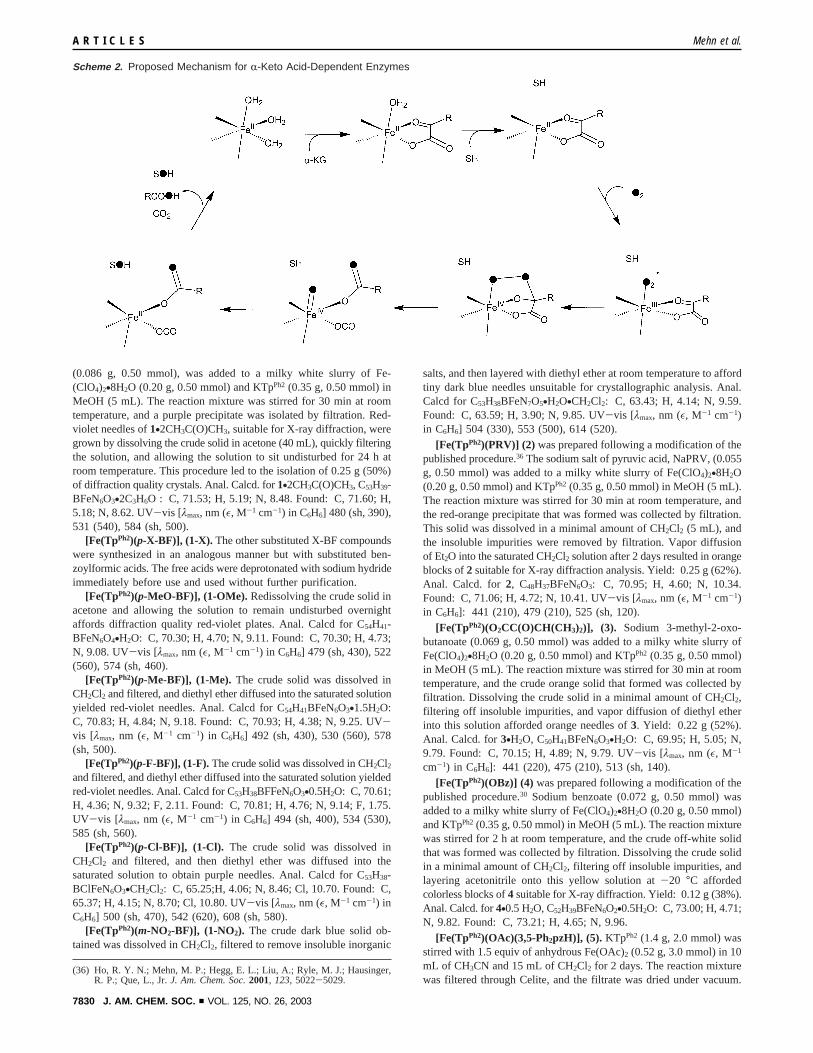
A common mechanism has been proposed for these enzymes(Scheme 2).22-24 In the first step, theR-keto acid chelates tothe iron(II) center, displacing two solvent molecules. Substratebinding in the vicinity of the iron active site then results in thedisplacement of the remaining water ligand, generating acoordinatively unsaturated metal center ready to bind andactivate dioxygen.22-25 O2 binds to the iron(II) center and attacksthe keto carbon of theR-keto acid generating an iron(IV)-peroxointermediate that then undergoes O-O bond cleavage anddecarboxylation to afford an iron(IV)-oxo oxidant. Aftersubstrate oxidation, the product, succinate, and CO2 dissociatefrom the active site to complete the catalytic cycle. Crystalstructures of enzyme-R-KG, enzyme-R-KG-substrate, and enzyme-succinate-CO2 complexes support this scheme.15,17-20,26
While the enzymatic studies provide a firm foundation forthe steps leading to and from substrate oxidation, deeper insightinto the mechanism of the metal-centered oxidation is desirable.Toward this goal, several complexes have been synthesized asmodels that provide a basis for understanding oxygen activationby R-keto acid-dependent enzymes;27-31 yet only for one, [Fe-
(TpPh2)(BF)], has the dioxygenase activity observed inR-ketoacid-dependent enzymes been demonstrated.30 In this case, theoxidative decarboxylation of benzoylformate (BF) results in theincorporation of one atom of dioxygen into the hydroxylatedphenyl ring of the ligand and the other into the benzoatebyproduct of this reaction. To better understand the role of theR-keto carboxylate cofactor in oxygen activation at mononuclearnonheme iron enzymes, we have synthesized an expanded seriesof [FeII(TpPh2)(X)] model complexes withR-keto carboxylatesand corresponding carboxylates and examined the structures andreactivity of these compounds. These studies demonstrate thatthe R-keto acid cofactor serves as a sacrificial two-electronreductant and facilitates production of a reactive oxygen speciesat the iron center that hydroxylates a phenyl ring of the TpPh2
ligand. The corresponding carboxylate model complexes alsoactivate O2, but the reactions proceed much more slowly. Thisdramatic difference emphasizes the importance ofR-keto acidcofactors in promoting the activation of dioxygen at nonhemeiron(II) centers in biology.
Experimental Procedures
General Materials and Syntheses.All reagents and solvents werepurchased from commercial sources and were used without furtherpurification unless otherwise noted. Methanol was distilled from Mg-(OMe)2 before use. Thiophene-free benzene was distilled from sodium/benzophenone ketyl under an argon atmosphere prior to use. Dichlo-romethane, acetonitrile, andn-heptane were carefully purified bydistillation under an argon atmosphere from P2O5, CaH2, and sodium/benzophenone ketyl, respectively.32 Preparation and handling of air-sensitive materials were carried out under inert atmosphere usingstandard Schlenk techniques or a glovebox. Caution: Although noproblems were encountered with these complexes, perchlorate salts arepotentially explosive and should be handled with care.
The ligand K(TpPh2) was prepared as previously reported.33 Ben-zoylformic acid and its sodium salt were purchased from Aldrich.Sodium (p-methoxybenzoyl)formate was prepared by Friedel-Craftsacylation of anisole with ethyl oxalyl chloride followed by hydrolysisof the resultant ester with 4 N NaOH (aq) to yield the sodium salt.34
1H NMR (D2O, 300 MHz)δ (ppm): 7.86 (2H, d, 8 Hz), 7.02 (2H, d,8 Hz), 3.83 (3H, s);13C NMR (D2O, 75.5 MHz)δ (ppm): 196, 173,164, 132, 125, 114, 55. The ethyl esters of (p-methylbenzoyl)formicacid, (p-chlorobenzoyl)formic acid, and (p-fluorobenzoyl)formic acidwere prepared by treating ethylR-oxo-1H-imidazole-1-acetate with theappropriate aryl Grignard reagent.35 The substitutedR-keto acids wereobtained in 75-90% yield by base-catalyzed hydrolysis of the resultingethyl esters with 4 N NaOH (aq) at room temperature for 4 h.27 (m-Nitrobenzoyl)formic acid was prepared by nitration of benzoylformicacid with concentrated HNO3/H2SO4 using literature procedures.27,34
[Fe(TpPh2)(BF)] (1) was prepared following a modification of thepublished procedure.36 The sodium salt of benzoylformic acid, NaBF
(12) Baldwin, J. E.; Abraham, E.Nat. Prod. Rep.1988, 5, 129-145.(13) Baldwin, J. E.; Adlington, R. M.; Crouch, N. P.; Schofield, C. J.; Turner,
N. J.; Aplin, R. T.Tetrahedron1991, 47, 9881-9900.(14) Hegg, E. L.; Que, L., Jr.Eur. J. Biochem.1997, 250, 625-629.(15) Valegard, K.; Terwisscha van Scheltinga, A. C.; Lloyd, M. D.; Hara, T.;
Ramaswamy, S.; Perrakis, A.; Thompson, A.; Lee, H.-J.; Baldwin, J. E.;Schofield, C. J.; Hajdu, J.; Andersson, I.Nature1998, 394, 805-809.
(16) Serre, L.; Sailland, A.; Sy, D.; Boudec, P.; Rolland, A.; Pebay-Peyroula,E.; Cohen-Addad, C.Structure1999, 7, 977-988.
(17) Zhang, Z.; Ren, J.; Stammers, D. K.; Baldwin, J. E.; Harlos, K.; Schofield,C. J.Nat. Struct. Biol.2000, 7, 127-133.
(18) Clifton, I. J.; Hsueh, L.-C.; Baldwin, J. E.; Harlos, K.; Schofield, C. J.Eur. J. Biochem.2001, 268, 6625-6636.
(19) Wilmouth, R. C.; Turnbull, J. J.; Welford, R. W. D.; Clifton, I. J.; Prescott,A. G.; Schofield, C. J.Structure2002, 10, 93-103.
(20) Elkins, J. M.; Ryle, M. J.; Clifton, I. J.; Hotopp, J. C. D.; Lloyd, J. S.;Burzlaff, N. I.; Baldwin, J. E.; Hausinger, R. P.; Roach, P. L.Biochemistry2002, 41, 5185-5192.
(21) Zhang, Z.; Ren, J.-s.; Harlos, K.; McKinnon, C. H.; Clifton, I. J.; Schofield,C. J.FEBS Lett.2002, 517, 7-12.
(22) Hanauske-Abel, H. M.; Gu¨nzler, V. J. Theor. Biol.1982, 94, 421-455.(23) Que, L., Jr.; Ho, R. Y. N.Chem. ReV. 1996, 96, 2607-2624.(24) Solomon, E. I.; Brunold, T. C.; Davis, M. I.; Kemsley, J. N.; Lee, S.-K.;
Lehnert, N.; Neese, F.; Skulan, A. J.; Yang, Y.-S.; Zhou, J.Chem. ReV.2000, 100, 235-349.
(25) Schofield, C. J.; Zhang, Z.Curr. Opin. Struct. Biol.1999, 9, 722-731.(26) Lee, H.-J.; Lloyd, M. D.; Harlos, K.; Clifton, I. J.; Baldwin, J. E.; Schofield,
C. J.J. Mol. Biol. 2001, 308, 937-948.
(27) Chiou, Y.-M.; Que, L., Jr.J. Am. Chem. Soc.1995, 117, 3999-4013.(28) Ha, E. H.; Ho, R. Y. N.; Kisiel, J. F.; Valentine, J. S.Inorg. Chem.1995,
34, 2265-2266.(29) Hikichi, S.; Ogihara, T.; Fujisawa, K.; Kitajima, N.; Akita, M.; Moro-oka,
Y. Inorg. Chem.1997, 36, 4539-4547.(30) Hegg, E. L.; Ho, R. Y. N.; Que, L., Jr.J. Am. Chem. Soc.1999, 121, 1972-
1973.(31) Chiou, Y.-M.; Que, L., Jr.Angew. Chem., Int. Ed. Engl.1994, 33, 1886-
1888.(32) Armarego, W. L. F.; Perrin, D. D.Purification of Laboratory Chemicals,
4th ed.; Butterworth-Heinemann: Oxford, 1997.(33) Kitajima, N.; Fujisawa, K.; Fujimoto, C.; Moro-oka, Y.; Hashimoto, S.;
Kitagawa, T.; Toriumi, K.; Tatsumi, K.; Nakamura, A.J. Am. Chem. Soc.1992, 114, 1277-1291.
(34) Pavia, D. L.; Lampman, G. M.; Kriz, G. S., Jr.Introduction to OrganicLaboratory Techniques, 2nd ed.; Saunders College Publishing: Philadelphia,1982.
(35) Nimitz, J. S.; Mosher, H. S.J. Org. Chem.1981, 46, 211-213.
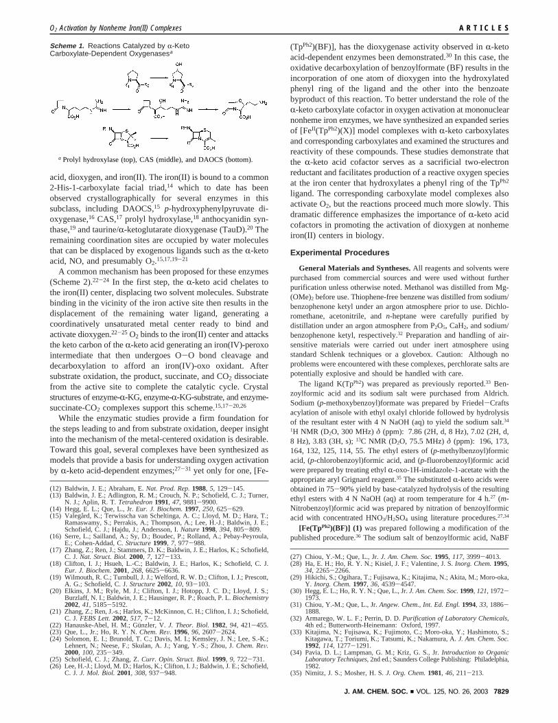
Scheme 1. Reactions Catalyzed by R-KetoCarboxylate-Dependent Oxygenasesa
a Prolyl hydroxylase (top), CAS (middle), and DAOCS (bottom).
O2 Activation by Nonheme Iron(II) Complexes A R T I C L E S
J. AM. CHEM. SOC. 9 VOL. 125, NO. 26, 2003 7829
(0.086 g, 0.50 mmol), was added to a milky white slurry of Fe-(ClO4)2•8H2O (0.20 g, 0.50 mmol) and KTpPh2 (0.35 g, 0.50 mmol) inMeOH (5 mL). The reaction mixture was stirred for 30 min at roomtemperature, and a purple precipitate was isolated by filtration. Red-violet needles of1•2CH3C(O)CH3, suitable for X-ray diffraction, weregrown by dissolving the crude solid in acetone (40 mL), quickly filteringthe solution, and allowing the solution to sit undisturbed for 24 h atroom temperature. This procedure led to the isolation of 0.25 g (50%)of diffraction quality crystals. Anal. Calcd. for1•2CH3C(O)CH3, C53H39-BFeN6O3•2C3H6O : C, 71.53; H, 5.19; N, 8.48. Found: C, 71.60; H,5.18; N, 8.62. UV-vis [λmax, nm (ε, M-1 cm-1) in C6H6] 480 (sh, 390),531 (540), 584 (sh, 500).
[Fe(TpPh2)(p-X-BF)], (1-X). The other substituted X-BF compoundswere synthesized in an analogous manner but with substituted ben-zoylformic acids. The free acids were deprotonated with sodium hydrideimmediately before use and used without further purification.
[Fe(TpPh2)(p-MeO-BF)], (1-OMe). Redissolving the crude solid inacetone and allowing the solution to remain undisturbed overnightaffords diffraction quality red-violet plates. Anal. Calcd for C54H41-BFeN6O4•H2O: C, 70.30; H, 4.70; N, 9.11. Found: C, 70.30; H, 4.73;N, 9.08. UV-vis [λmax, nm (ε, M-1 cm-1) in C6H6] 479 (sh, 430), 522(560), 574 (sh, 460).
[Fe(TpPh2)(p-Me-BF)], (1-Me). The crude solid was dissolved inCH2Cl2 and filtered, and diethyl ether diffused into the saturated solutionyielded red-violet needles. Anal. Calcd for C54H41BFeN6O3•1.5H2O:C, 70.83; H, 4.84; N, 9.18. Found: C, 70.93; H, 4.38; N, 9.25. UV-vis [λmax, nm (ε, M-1 cm-1) in C6H6] 492 (sh, 430), 530 (560), 578(sh, 500).
[Fe(TpPh2)(p-F-BF)], (1-F). The crude solid was dissolved in CH2Cl2and filtered, and diethyl ether diffused into the saturated solution yieldedred-violet needles. Anal. Calcd for C53H38BFFeN6O3•0.5H2O: C, 70.61;H, 4.36; N, 9.32; F, 2.11. Found: C, 70.81; H, 4.76; N, 9.14; F, 1.75.UV-vis [λmax, nm (ε, M-1 cm-1) in C6H6] 494 (sh, 400), 534 (530),585 (sh, 560).
[Fe(TpPh2)(p-Cl-BF)], (1-Cl). The crude solid was dissolved inCH2Cl2 and filtered, and then diethyl ether was diffused into thesaturated solution to obtain purple needles. Anal. Calcd for C53H38-BClFeN6O3•CH2Cl2: C, 65.25;H, 4.06; N, 8.46; Cl, 10.70. Found: C,65.37; H, 4.15; N, 8.70; Cl, 10.80. UV-vis [λmax, nm (ε, M-1 cm-1) inC6H6] 500 (sh, 470), 542 (620), 608 (sh, 580).
[Fe(TpPh2)(m-NO2-BF)], (1-NO2). The crude dark blue solid ob-tained was dissolved in CH2Cl2, filtered to remove insoluble inorganic
salts, and then layered with diethyl ether at room temperature to affordtiny dark blue needles unsuitable for crystallographic analysis. Anal.Calcd for C53H38BFeN7O5•H2O•CH2Cl2: C, 63.43; H, 4.14; N, 9.59.Found: C, 63.59; H, 3.90; N, 9.85. UV-vis [λmax, nm (ε, M-1 cm-1)in C6H6] 504 (330), 553 (500), 614 (520).
[Fe(TpPh2)(PRV)] (2) was prepared following a modification of thepublished procedure.36 The sodium salt of pyruvic acid, NaPRV, (0.055g, 0.50 mmol) was added to a milky white slurry of Fe(ClO4)2•8H2O(0.20 g, 0.50 mmol) and KTpPh2(0.35 g, 0.50 mmol) in MeOH (5 mL).The reaction mixture was stirred for 30 min at room temperature, andthe red-orange precipitate that was formed was collected by filtration.This solid was dissolved in a minimal amount of CH2Cl2 (5 mL), andthe insoluble impurities were removed by filtration. Vapor diffusionof Et2O into the saturated CH2Cl2 solution after 2 days resulted in orangeblocks of2 suitable for X-ray diffraction analysis. Yield: 0.25 g (62%).Anal. Calcd. for2, C48H37BFeN6O3: C, 70.95; H, 4.60; N, 10.34.Found: C, 71.06; H, 4.72; N, 10.41. UV-vis [λmax, nm (ε, M-1 cm-1)in C6H6]: 441 (210), 479 (210), 525 (sh, 120).
[Fe(TpPh2)(O2CC(O)CH(CH3)2)], (3). Sodium 3-methyl-2-oxo-butanoate (0.069 g, 0.50 mmol) was added to a milky white slurry ofFe(ClO4)2•8H2O (0.20 g, 0.50 mmol) and KTpPh2 (0.35 g, 0.50 mmol)in MeOH (5 mL). The reaction mixture was stirred for 30 min at roomtemperature, and the crude orange solid that formed was collected byfiltration. Dissolving the crude solid in a minimal amount of CH2Cl2,filtering off insoluble impurities, and vapor diffusion of diethyl etherinto this solution afforded orange needles of3. Yield: 0.22 g (52%).Anal. Calcd. for3•H2O, C50H41BFeN6O3•H2O: C, 69.95; H, 5.05; N,9.79. Found: C, 70.15; H, 4.89; N, 9.79. UV-vis [λmax, nm (ε, M-1
cm-1) in C6H6]: 441 (220), 475 (210), 513 (sh, 140).
[Fe(TpPh2)(OBz)] (4) was prepared following a modification of thepublished procedure.30 Sodium benzoate (0.072 g, 0.50 mmol) wasadded to a milky white slurry of Fe(ClO4)2•8H2O (0.20 g, 0.50 mmol)and KTpPh2(0.35 g, 0.50 mmol) in MeOH (5 mL). The reaction mixturewas stirred for 2 h atroom temperature, and the crude off-white solidthat was formed was collected by filtration. Dissolving the crude solidin a minimal amount of CH2Cl2, filtering off insoluble impurities, andlayering acetonitrile onto this yellow solution at-20 °C affordedcolorless blocks of4 suitable for X-ray diffraction. Yield: 0.12 g (38%).Anal. Calcd. for4•0.5 H2O, C52H39BFeN6O2•0.5H2O: C, 73.00; H, 4.71;N, 9.82. Found: C, 73.21; H, 4.65; N, 9.96.
[Fe(TpPh2)(OAc)(3,5-Ph2pzH)], (5). KTpPh2 (1.4 g, 2.0 mmol) wasstirred with 1.5 equiv of anhydrous Fe(OAc)2 (0.52 g, 3.0 mmol) in 10mL of CH3CN and 15 mL of CH2Cl2 for 2 days. The reaction mixturewas filtered through Celite, and the filtrate was dried under vacuum.
(36) Ho, R. Y. N.; Mehn, M. P.; Hegg, E. L.; Liu, A.; Ryle, M. J.; Hausinger,R. P.; Que, L., Jr.J. Am. Chem. Soc.2001, 123, 5022-5029.
Scheme 2. Proposed Mechanism for R-Keto Acid-Dependent Enzymes
A R T I C L E S Mehn et al.
7830 J. AM. CHEM. SOC. 9 VOL. 125, NO. 26, 2003
The pale yellow powder was dissolved in CH2Cl2, and the resultingsolution was filtered and evaporated to dryness. The resultant powderwas extracted with CH3CN to yield a white powder. The powder wasrecrystallized from CH2Cl2/CH3CN at-20°C to afford colorless blocks.Yield: 0.82 g (41%). Anal. Calcd. for5, C62H49BFeN8O2: C, 74.11;H, 4.92; N, 11.15. Found: C, 73.85; H, 4.79; N, 11.17.
[Fe(TpPh2*)(OAc)(3,5-Ph2pzH)] (6) was prepared by oxygenationof a dichloromethane solution of the acetate compound5 followed byslow evaporation to obtain dark green X-ray quality crystals. Anal.Calcd. for6, C62H48BFeN8O3: C, 73.20; H, 4.74; N, 10.99. Found: C,72.99; H, 4.84; N, 11.06. UV-vis [λmax, nm (ε, M-1 cm-1) in C6H6]:650 (1400).
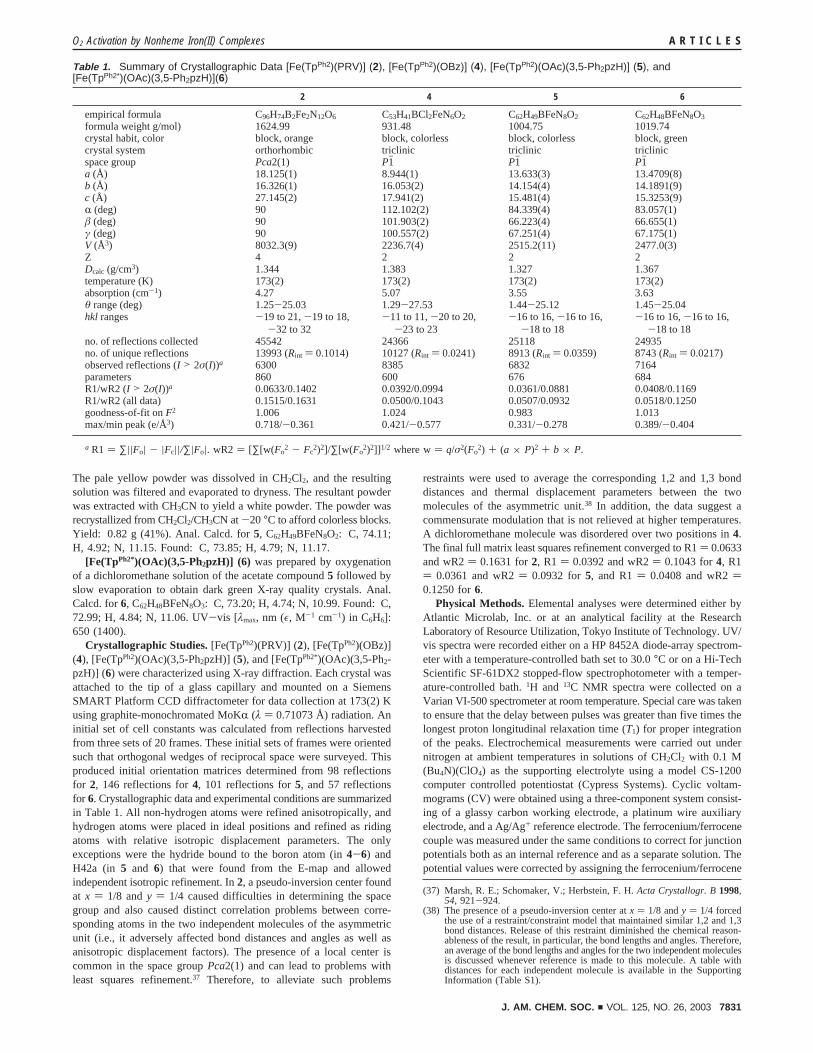
Crystallographic Studies.[Fe(TpPh2)(PRV)] (2), [Fe(TpPh2)(OBz)](4), [Fe(TpPh2)(OAc)(3,5-Ph2pzH)] (5), and [Fe(TpPh2*)(OAc)(3,5-Ph2-pzH)] (6) were characterized using X-ray diffraction. Each crystal wasattached to the tip of a glass capillary and mounted on a SiemensSMART Platform CCD diffractometer for data collection at 173(2) Kusing graphite-monochromated MoKR (λ ) 0.71073 Å) radiation. Aninitial set of cell constants was calculated from reflections harvestedfrom three sets of 20 frames. These initial sets of frames were orientedsuch that orthogonal wedges of reciprocal space were surveyed. Thisproduced initial orientation matrices determined from 98 reflectionsfor 2, 146 reflections for4, 101 reflections for5, and 57 reflectionsfor 6. Crystallographic data and experimental conditions are summarizedin Table 1. All non-hydrogen atoms were refined anisotropically, andhydrogen atoms were placed in ideal positions and refined as ridingatoms with relative isotropic displacement parameters. The onlyexceptions were the hydride bound to the boron atom (in4-6) andH42a (in 5 and 6) that were found from the E-map and allowedindependent isotropic refinement. In2, a pseudo-inversion center foundat x ) 1/8 andy ) 1/4 caused difficulties in determining the spacegroup and also caused distinct correlation problems between corre-sponding atoms in the two independent molecules of the asymmetricunit (i.e., it adversely affected bond distances and angles as well asanisotropic displacement factors). The presence of a local center iscommon in the space groupPca2(1) and can lead to problems withleast squares refinement.37 Therefore, to alleviate such problems
restraints were used to average the corresponding 1,2 and 1,3 bonddistances and thermal displacement parameters between the twomolecules of the asymmetric unit.38 In addition, the data suggest acommensurate modulation that is not relieved at higher temperatures.A dichloromethane molecule was disordered over two positions in4.The final full matrix least squares refinement converged to R1) 0.0633and wR2) 0.1631 for2, R1 ) 0.0392 and wR2) 0.1043 for4, R1) 0.0361 and wR2) 0.0932 for5, and R1) 0.0408 and wR2)0.1250 for6.
Physical Methods.Elemental analyses were determined either byAtlantic Microlab, Inc. or at an analytical facility at the ResearchLaboratory of Resource Utilization, Tokyo Institute of Technology. UV/vis spectra were recorded either on a HP 8452A diode-array spectrom-eter with a temperature-controlled bath set to 30.0°C or on a Hi-TechScientific SF-61DX2 stopped-flow spectrophotometer with a temper-ature-controlled bath.1H and 13C NMR spectra were collected on aVarian VI-500 spectrometer at room temperature. Special care was takento ensure that the delay between pulses was greater than five times thelongest proton longitudinal relaxation time (T1) for proper integrationof the peaks. Electrochemical measurements were carried out undernitrogen at ambient temperatures in solutions of CH2Cl2 with 0.1 M(Bu4N)(ClO4) as the supporting electrolyte using a model CS-1200computer controlled potentiostat (Cypress Systems). Cyclic voltam-mograms (CV) were obtained using a three-component system consist-ing of a glassy carbon working electrode, a platinum wire auxiliaryelectrode, and a Ag/Ag+ reference electrode. The ferrocenium/ferrocenecouple was measured under the same conditions to correct for junctionpotentials both as an internal reference and as a separate solution. Thepotential values were corrected by assigning the ferrocenium/ferrocene
(37) Marsh, R. E.; Schomaker, V.; Herbstein, F. H.Acta Crystallogr. B1998,54, 921-924.
(38) The presence of a pseudo-inversion center atx ) 1/8 andy ) 1/4 forcedthe use of a restraint/constraint model that maintained similar 1,2 and 1,3bond distances. Release of this restraint diminished the chemical reason-ableness of the result, in particular, the bond lengths and angles. Therefore,an average of the bond lengths and angles for the two independent moleculesis discussed whenever reference is made to this molecule. A table withdistances for each independent molecule is available in the SupportingInformation (Table S1).
Table 1. Summary of Crystallographic Data [Fe(TpPh2)(PRV)] (2), [Fe(TpPh2)(OBz)] (4), [Fe(TpPh2)(OAc)(3,5-Ph2pzH)] (5), and[Fe(TpPh2*)(OAc)(3,5-Ph2pzH)](6)
2 4 5 6
empirical formula C96H74B2Fe2N12O6 C53H41BCl2FeN6O2 C62H49BFeN8O2 C62H48BFeN8O3
formula weight g/mol) 1624.99 931.48 1004.75 1019.74crystal habit, color block, orange block, colorless block, colorless block, greencrystal system orthorhombic triclinic triclinic triclinicspace group Pca2(1) P1h P1h P1ha (Å) 18.125(1) 8.944(1) 13.633(3) 13.4709(8)b (Å) 16.326(1) 16.053(2) 14.154(4) 14.1891(9)c (Å) 27.145(2) 17.941(2) 15.481(4) 15.3253(9)R (deg) 90 112.102(2) 84.339(4) 83.057(1)â (deg) 90 101.903(2) 66.223(4) 66.655(1)γ (deg) 90 100.557(2) 67.251(4) 67.175(1)V (Å3) 8032.3(9) 2236.7(4) 2515.2(11) 2477.0(3)Z 4 2 2 2Dcalc (g/cm3) 1.344 1.383 1.327 1.367temperature (K) 173(2) 173(2) 173(2) 173(2)absorption (cm-1) 4.27 5.07 3.55 3.63θ range (deg) 1.25-25.03 1.29-27.53 1.44-25.12 1.45-25.04hkl ranges -19 to 21,-19 to 18,
-32 to 32-11 to 11,-20 to 20,
-23 to 23-16 to 16,-16 to 16,
-18 to 18-16 to 16,-16 to 16,
-18 to 18no. of reflections collected 45542 24366 25118 24935no. of unique reflections 13993 (Rint ) 0.1014) 10127 (Rint ) 0.0241) 8913 (Rint ) 0.0359) 8743 (Rint ) 0.0217)observed reflections (I > 2σ(I))a 6300 8385 6832 7164parameters 860 600 676 684R1/wR2 (I > 2σ(I))a 0.0633/0.1402 0.0392/0.0994 0.0361/0.0881 0.0408/0.1169R1/wR2 (all data) 0.1515/0.1631 0.0500/0.1043 0.0507/0.0932 0.0518/0.1250goodness-of-fit onF2 1.006 1.024 0.983 1.013max/min peak (e/Å3) 0.718/-0.361 0.421/-0.577 0.331/-0.278 0.389/-0.404
a R1 ) ∑||Fo| - |Fc||/∑|Fo|. wR2 ) [∑[w(Fo2 - Fc
2)2]/∑[w(Fo2)2]] 1/2 where w) q/σ2(Fo
2) + (a × P)2 + b × P.
O2 Activation by Nonheme Iron(II) Complexes A R T I C L E S
J. AM. CHEM. SOC. 9 VOL. 125, NO. 26, 2003 7831
couple the value of 0.480 V versus SCE.39 X-band EPR spectra wererecorded using a Bruker E-500 spectrometer with sample temperaturemaintained at 20 K with an Oxford Instruments ESR-10 liquid heliumcryostat. Spectra were collected using a time constant of 82 ms, a sweeptime of 82 ms, a modulation amplitude of 10 G, a modulation frequencyof 100 kHz, and a power of 0.2 mW to avoid saturation. Theelectrospray ionization mass spectral data for the oxidized compoundswere obtained using an LCQ mass spectrometer (ThermoFinnigan) onCH2Cl2 solutions that were directly infused into the spectrometer via asyringe pump.
Resonance Raman spectra were collected on an Acton AM-506spectrometer (1200-groove grating) using a Kaiser Optical holographicsuper-notch filter with a Princeton Instruments liquid N2-cooled (LN-1100PB) CCD detector with 4 cm-1 spectral resolution. The laserexcitation lines were obtained with a Spectra Physics 2030-15 argonion laser and a 375B CW dye (Rhodamine 6G). The Raman frequencieswere referenced to indene, and the entire spectral range of 400-1700cm-1 was obtained by collecting spectra at 2-3 different frequencywindows and splicing the spectra together, making sure the solventpeaks were aligned before splicing. The spectra were obtained with apower of 200 mW at 77 K using a backscattering geometry on samplesfrozen on a gold-plated copper coldfinger in thermal contact with aDewar containing liquid N2. Typical accumulation times were 16-32min per frequency window. Curve fits (Gaussian functions) and baselinecorrections (polynomial fits) were carried out using Grams/32 SpectralNotebase Version 4.04 (Galactic).
Kinetics Measurements.Distilled thiophene-free benzene was storedover 3A molecular sieves under nitrogen and passed through a columnof activated alumina prior to use. Crystalline materials were dissolvedin benzene at room temperature. Temperature dependent changes inthe density of benzene and the solubility of oxygen have been reportedpreviously, and corrections were made for these phenomena.40,41 Thesolubility of O2 in benzene is reported to be 9.15 mM at 30°C, andoxygen saturation is assumed in these experiments.41 However, todetermine the molecularity of the reaction with respect to oxygen, loweroxygen concentrations were required. Stopped-flow methods wereutilized to minimize equilibration of oxygen into the headspace. Takinginto account the temperature dependent changes in the density ofbenzene and the changes in oxygen solubility, as well as the 1:1 mixingratio in the stopped-flow experiments, the maximum accessibleconcentration of O2 at 50 °C was approximately 4.5 mM. Solutionswith lower dioxygen concentrations were prepared using gastightsyringes of O2 and N2 saturated solutions that were added in knownproportion to a third syringe. Since the concentration of dioxygen greatlyexceeded that of the iron(II) precursor, it was assumed to be constantthroughout the reaction. The kinetic curves obtained under pseudo-first-order conditions over five to seven half-lives were fit to eq 1.
Alternatively, for the oxygen dependence, kinetic data from 25% ofthe first half-life were fit to a linear function giving dA/dt as the initialrate of absorbance change. All rates reported are the average of at leastthree different trials.
Results and Discussion
The mononuclear iron(II) complexes1-4 were synthesizedby addition of 1 equiv of the sodium salt of the desiredR-ketoacid or sodium benzoate to equimolar portions of FeII-(ClO4)2•8H2O and KTpPh2 in methanol. The acetate complex5was prepared directly from metathesis of KTpPh2 and iron(II)
acetate and was isolated as the pyrazole adduct. Although otherworkers have obtained related FeII(TpR1,R2) complexes ofcarboxylates without an additional pyrazole ligand,29,42,43 theacetate complex could only be isolated as the pyrazole adduct.Addition of excess 3,5-diphenylpyrazole to the reaction mixtureincreased the yield of5.
Structures of Iron(II) Precursors. There are several previ-ously reported examples of benzoylformate complexes,27,29,30
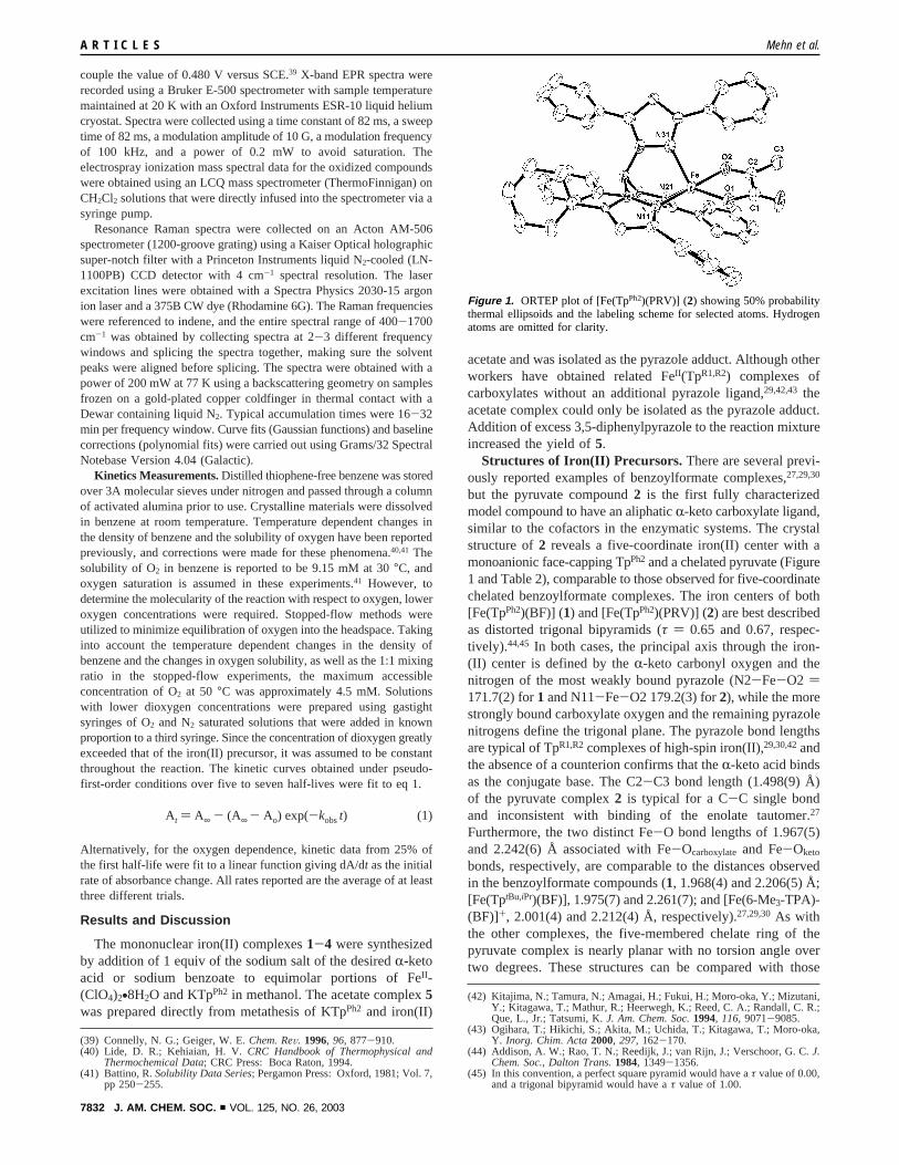
but the pyruvate compound2 is the first fully characterizedmodel compound to have an aliphaticR-keto carboxylate ligand,similar to the cofactors in the enzymatic systems. The crystalstructure of2 reveals a five-coordinate iron(II) center with amonoanionic face-capping TpPh2and a chelated pyruvate (Figure1 and Table 2), comparable to those observed for five-coordinatechelated benzoylformate complexes. The iron centers of both[Fe(TpPh2)(BF)] (1) and [Fe(TpPh2)(PRV)] (2) are best describedas distorted trigonal bipyramids (τ ) 0.65 and 0.67, respec-tively).44,45 In both cases, the principal axis through the iron-(II) center is defined by theR-keto carbonyl oxygen and thenitrogen of the most weakly bound pyrazole (N2-Fe-O2 )171.7(2) for1 and N11-Fe-O2 179.2(3) for2), while the morestrongly bound carboxylate oxygen and the remaining pyrazolenitrogens define the trigonal plane. The pyrazole bond lengthsare typical of TpR1,R2complexes of high-spin iron(II),29,30,42andthe absence of a counterion confirms that theR-keto acid bindsas the conjugate base. The C2-C3 bond length (1.498(9) Å)of the pyruvate complex2 is typical for a C-C single bondand inconsistent with binding of the enolate tautomer.27
Furthermore, the two distinct Fe-O bond lengths of 1.967(5)and 2.242(6) Å associated with Fe-Ocarboxylateand Fe-Oketo
bonds, respectively, are comparable to the distances observedin the benzoylformate compounds (1, 1.968(4) and 2.206(5) Å;[Fe(TptBu,iPr)(BF)], 1.975(7) and 2.261(7); and [Fe(6-Me3-TPA)-(BF)]+, 2.001(4) and 2.212(4) Å, respectively).27,29,30As withthe other complexes, the five-membered chelate ring of thepyruvate complex is nearly planar with no torsion angle overtwo degrees. These structures can be compared with those
(39) Connelly, N. G.; Geiger, W. E.Chem. ReV. 1996, 96, 877-910.(40) Lide, D. R.; Kehiaian, H. V.CRC Handbook of Thermophysical and
Thermochemical Data; CRC Press: Boca Raton, 1994.(41) Battino, R.Solubility Data Series; Pergamon Press: Oxford, 1981; Vol. 7,
pp 250-255.
(42) Kitajima, N.; Tamura, N.; Amagai, H.; Fukui, H.; Moro-oka, Y.; Mizutani,Y.; Kitagawa, T.; Mathur, R.; Heerwegh, K.; Reed, C. A.; Randall, C. R.;Que, L., Jr.; Tatsumi, K.J. Am. Chem. Soc.1994, 116, 9071-9085.
(43) Ogihara, T.; Hikichi, S.; Akita, M.; Uchida, T.; Kitagawa, T.; Moro-oka,Y. Inorg. Chim. Acta2000, 297, 162-170.
(44) Addison, A. W.; Rao, T. N.; Reedijk, J.; van Rijn, J.; Verschoor, G. C.J.Chem. Soc., Dalton Trans.1984, 1349-1356.
(45) In this convention, a perfect square pyramid would have aτ value of 0.00,and a trigonal bipyramid would have aτ value of 1.00.
At ) A∞ - (A∞ - Ao) exp(-kobst) (1)
Figure 1. ORTEP plot of [Fe(TpPh2)(PRV)] (2) showing 50% probabilitythermal ellipsoids and the labeling scheme for selected atoms. Hydrogenatoms are omitted for clarity.
A R T I C L E S Mehn et al.
7832 J. AM. CHEM. SOC. 9 VOL. 125, NO. 26, 2003
recently reported forR-KG-dependent iron(II) enzymes withbound cofactor and substrate.17,19,20 While all three show achelated R-ketoglutarate, the iron center of anthocyanidinsynthase has a water molecule occupying the sixth site to givea distorted octahedral geometry,19 and the other two (DAOCSand TauD) have a distorted square pyramidal geometry (τ )0.2617 and 0.1220) about the iron(II) center.
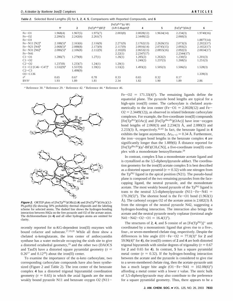
To examine the importance of theR-keto carboxylate, twocorresponding carboxylate compounds have also been synthe-sized (Figure 2 and Table 2). The iron center of the benzoatecomplex 4 has a distorted trigonal bipyramidal coordinationgeometry (τ ) 0.65) in which the axial ligands are the mostweakly bound pyrazole N11 and benzoate oxygen O2 (N11-
Fe-O2 ) 171.33(4)°). The remaining ligands define theequatorial plane. The pyrazole bond lengths are typical for ahigh-spin iron(II) center. The carboxylate is chelated asym-metrically to the iron center (Fe-O1 ) 2.0028(12) and Fe-O2 ) 2.3449(12)), as observed in related bidentate carboxylatecomplexes. For example, the five-coordinate iron(II) compounds[Fe(TpiPr2)(OAc)] and [Fe(TptBu,iPr)(OAc)] have iron-oxygenbond lengths of 2.060(3) and 2.234(3) Å, and 2.080(3) and2.233(3) Å, respectively.42,43 In fact, the benzoate ligand in4exhibits the largest asymmetry,∆rFe-O ) 0.34 Å. Furthermore,the iron-oxygen bond lengths in the benzoate complex4 aresignificantly longer than the 1.889(6) Å distance reported for[Fe(TptBu,iPr)(η1-BF)(CH3CN)], a five-coordinate iron(II) com-plex with a monodentate benzoylformate.29
In contrast, complex5 has a monodentate acetate ligand andis crystallized as the 3,5-diphenylpyrazole adduct. The coordina-tion geometry for the iron(II) acetate complex5 is best describedas a distorted square pyramid (τ ) 0.32) with one nitrogen fromthe TpPh2 ligand in the apical position (N21). The pseudo-basalplane is composed of the two remaining pyrazoles from the face-capping ligand, the neutral pyrazole, and the monodentateacetate. The most weakly bound pyrazole of the TpPh2 ligand istrans to the neutral 3,5-diphenylpyrazole (N11-Fe-N41 )170.20(5)°). The shortest bond is the Fe-O1 bond (1.963(1)Å). The carbonyl oxygen O2 of the acetate anion is 2.682(3) Åfrom the nitrogen of the neutral pyrazole N42, suggesting ahydrogen-bonding interaction. The interaction also brings theacetate and the neutral pyrazole nearly coplanar (torsional angleN41-N42-O2-O1 ) 16.4(1)°).
The structures of2, 4, and5 consist of an [Fe(TpPh2)]+ unitcoordinated by a monoanionic ligand that gives rise to a five-,four-, or seven-membered chelate ring, respectively. Despite thedifferences in bite angle (O1-Fe-O2 ) 77.0(3)° for 2 and59.96(4)° for 4), the iron(II) centers of2 and4 are both distortedtrigonal bipyramids with similar degrees of trigonality (τ ) 0.67for 2 and 0.65 for4). In contrast,5 has a square pyramidalmetal center (τ ) 0.32). If the hydrogen-bonding interactionbetween the acetate and the pyrazole is considered to give riseto a seven-membered chelate ring, then the acetate-pyrazole unithas a much larger bite angle (O1-Fe-N41 ) 102.08(6)°)affording a metal center with a lowerτ value. The steric bulkof 3,5-diphenylpyrazole may also contribute to the preferencefor a square pyramidal geometry. Thus, there appears to be a
Table 2. Selected Bond Lengths (Å) for 1, 2, 4, 5, Comparisons with Reported Compounds, and 6
1a 2 [Fe(TptBu,iPr)(BF)]b[Fe(TptBu,iPr)(η1-BF)(3-iPr-5-tBupzH)]b 4 5 [Fe(TpiPr2)(OAc)]c 6
Fe-O1 1.968(4) 1.967(5) 1.975(7) 2.003(8) 2.0028(12) 1.9634(14) 2.234(3) 1.9740(16)Fe-O2 2.206(5) 2.242(6) 2.261(7) 2.3449(12) 2.060(3)Fe-O3 1.8877(14)Fe-N11 [N2]# 2.188(5)# 2.163(6) 2.116(9) 2.271(9) 2.1762(13) 2.2626(15) 2.073(3) 2.2022(17)Fe-N21 [N4]# 2.068(5)# 2.088(8) 2.173(9) 2.117(9) 2.0916(14) 2.0745(15) 2.093(2) 2.1652(17)Fe-N31 [N6]# 2.086(5)# 2.106(8) 2.112(9) 2.102(8) 2.0653(13) 2.0855(16) 2.092(3) 2.0654(17)Fe-N41 2.22(1) 2.2347(17) 2.2344(17)C1-O1 1.286(7) 1.279(8) 1.27(1) 1.26(1) 1.285(2) 1.263(2) 1.230(5) 1.201(3)C1-O2 1.240(2) 1.237(3) 1.268(5) 1.251(3)C2-O2 1.237(8) 1.233(7) 1.24(1) 1.20(1)C1-C2 [C46-C47]# 1.532(9)# 1.557(9) 1.59(1) 1.53(2) 1.493(2) 1.505(3) 1.506(5) 1.528(3)C2-C3 1.498(9)O3-C136 1.339(3)τd 0.65 0.67 0.78 0.33 0.65 0.32 0.17BVSe 1.93 1.89 1.81 2.34 1.82 1.90 1.89 2.86
a Reference 30.b Reference 29.c Reference 42.d Reference 44.e Reference 46.
Figure 2. ORTEP plots of [Fe(TpPh2)(OBz)] (4) and [Fe(TpPh2)(OAc)(3,5-Ph2pzH)] (5) showing 50% probability thermal ellipsoids and the labelingscheme for selected atoms. The dashed line shows the hydrogen-bondinginteraction between H42a on the free pyrazole and O2 of the acetate anion.The dichloromethane (in4) and all other hydrogen atoms are omitted forclarity.
O2 Activation by Nonheme Iron(II) Complexes A R T I C L E S
J. AM. CHEM. SOC. 9 VOL. 125, NO. 26, 2003 7833
very sensitive interplay between the steric bulk of the mono-anionic ligands and alkyl substitution of the tris(pyrazolyl)borateligand. For example, with the [Fe(TptBu,iPr)]+ fragment, theacetate complex is square pyramidal while the larger benzoyl-formate anion results in a trigonal bipyramidal geometry aboutthe iron center; in fact, the latter converts to a tetrahedralcomplex with a monodentate benzoylformate ligand at hightemperature.29,43 This suggests that the steric bulk of the [Fe-(TpPh2)]+ framework is reduced as compared to [Fe(TptBu,iPr)]+
or [Fe(TpiPr2)]+, consistent with the series for effective stericbulk of substituted tris(pyrazolyl)borate ligands proposed byKitajima and Tolman.47
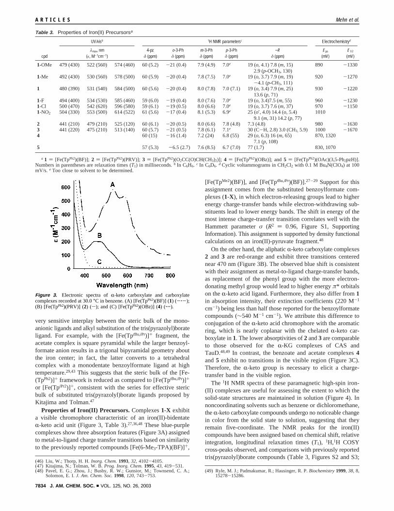
Properties of Iron(II) Precursors. Complexes1-X exhibita visible chromophore characteristic of an iron(II)-bidentateR-keto acid unit (Figure 3, Table 3).27,36,48These blue-purplecomplexes show three absorption features (Figure 3A) assignedto metal-to-ligand charge transfer transitions based on similarityto the previously reported compounds [Fe(6-Me3-TPA)(BF)]+,
[Fe(TpMe2)(BF)], and [Fe(TptBu,iPr)(BF)].27-29 Support for thisassignment comes from the substituted benzoylformate com-plexes (1-X), in which electron-releasing groups lead to higherenergy charge-transfer bands while electron-withdrawing sub-stituents lead to lower energy bands. The shift in energy of themost intense charge-transfer transition correlates well with theHammett parameterσ (R2 ) 0.96, Figure S1, SupportingInformation). This assignment is supported by density functionalcalculations on an iron(II)-pyruvate fragment.48
On the other hand, the aliphaticR-keto carboxylate complexes2 and 3 are red-orange and exhibit three transitions centerednear 470 nm (Figure 3B). The observed blue shift is consistentwith their assignment as metal-to-ligand charge-transfer bands,as replacement of the phenyl group with the more electron-donating methyl group would lead to higher energyπ* orbitalson theR-keto acid ligand. Furthermore, they also differ from1in absorption intensity, their extinction coefficients (220 M-1
cm-1) being less than half those reported for the benzoylformatecompounds (∼540 M-1 cm-1). We attribute this difference toconjugation of theR-keto acid chromophore with the aromaticring, which is nearly coplanar with the chelatedR-keto car-boxylate in1. The lower absorptivities of2 and3 are comparableto those observed for theR-KG complexes of CAS andTauD.48,49 In contrast, the benzoate and acetate complexes4and5 exhibit no transitions in the visible region (Figure 3C).Therefore, theR-keto group is necessary to elicit a charge-transfer band in the visible region.
The 1H NMR spectra of these paramagnetic high-spin iron-(II) complexes are useful for assessing the extent to which thesolid-state structures are maintained in solution (Figure 4). Innoncoordinating solvents such as benzene or dichloromethane,theR-keto carboxylate compounds undergo no noticeable changein color from the solid state to solution, suggesting that theyremain five-coordinate. The NMR peaks for the iron(II)compounds have been assigned based on chemical shift, relativeintegration, longitudinal relaxation times (T1), 1H,1H COSYcross-peaks observed, and comparisons with previously reportedtris(pyrazolyl)borate compounds (Table 3, Figures S2 and S3;
(46) Liu, W.; Thorp, H. H.Inorg. Chem.1993, 32, 4102-4105.(47) Kitajima, N.; Tolman, W. B.Prog. Inorg. Chem.1995, 43, 419-531.(48) Pavel, E. G.; Zhou, J.; Busby, R. W.; Gunsior, M.; Townsend, C. A.;
Solomon, E. I.J. Am. Chem. Soc.1998, 120, 743-753.(49) Ryle, M. J.; Padmakumar, R.; Hausinger, R. P.Biochemistry1999, 38, 8,
15278-15286.
Table 3. Properties of Iron(II) Precursorsa
UV/visb 1H NMR parametersc Electrochemistryd
cpdλmax, nm
(ε, M-1cm-1)4-pz
δ (ppm)o-3-Phδ (ppm)
m-3-Phδ (ppm)
p-3-Phδ (ppm)
−Rδ (ppm)
Epa
(mV)E1/2
(mV)
1-OMe 479 (430) 522 (560) 574 (460) 60 (5.2)-21 (0.4) 7.9 (4.9) 7.0e 19 (o, 4.1) 7.8 (m, 15)2.9 (p-OCH3, 130)
890 -1330
1-Me 492 (430) 530 (560) 578 (500) 60 (5.9) -20 (0.4) 7.8 (7.5) 7.0e 19 (o, 3.7) 7.9 (m, 19)-4.1 (p-CH3, 111)
920 -1270
1 480 (390) 531 (540) 584 (500) 60 (5.6) -20 (0.4) 8.0 (7.8) 7.0 (7.1) 19 (o, 3.4) 7.9 (m, 25)13.6 (p, 71)
930 -1220
1-F 494 (400) 534 (530) 585 (460) 59 (6.0) -19 (0.4) 8.0 (7.6) 7.0e 19 (o, 3.4)7.5 (m, 55) 960 -12301-Cl 500 (470) 542 (620) 596 (580) 59 (6.1) -19 (0.5) 8.0 (6.6) 7.0e 19 (o, 3.7) 7.6 (m, 37) 970 -11501-NO2 504 (330) 553 (500) 614 (522) 61 (5.6) -17 (0.4) 8.1 (5.3) 6.9e 25 (o′, 4.0) 14.4 (o, 5.4)
9.1 (m, 31) 14.2 (p, 77)1010
2 441 (210) 479 (210) 525 (120) 60 (6.1) -20 (0.5) 8.0 (6.6) 7.8 (4.8) 7.3 (4.8) 980 -16303 441 (220) 475 (210) 513 (140) 60 (5.7) -21 (0.5) 7.8 (6.1) 7.1e 30 (C-H, 2.8) 3.0 (CH3, 5.9) 1000 -16704 60 (15) -16 (1.4) 7.2 (24) 6.8 (55) 29 (o, 6.3) 16 (m, 65)
7.1 (p, 108)870, 1320
5 57 (5.3) -6.5 (2.7) 7.6 (8.5) 6.7 (7.0) 77 (1.7) 830, 1070
a 1 ) [Fe(TpPh2)(BF)]; 2 ) [Fe(TpPh2)(PRV)]; 3 ) [Fe(TpPh2)(O2CC(O)CH(CH3)2)]; 4 ) [Fe(TpPh2)(OBz)]; and5 ) [Fe(TpPh2)(OAc)(3,5-Ph2pzH)].Numbers in parentheses are relaxation times (T1) in milliseconds.b In C6H6. c In C6D6. d Cyclic voltammograms in CH2Cl2 with 0.1 M Bu4N(ClO4) at 100mV/s. e Too close to solvent to be determined.
Figure 3. Electronic spectra ofR-keto carboxylate and carboxylatecomplexes recorded at 30.0°C in benzene. (A) [Fe(TpPh2)(BF)] (1) (-‚-);(B) [Fe(TpPh2)(PRV)] (2) (s); and (C) [Fe(TpPh2)(OBz)] (4) (‚‚‚).
A R T I C L E S Mehn et al.
7834 J. AM. CHEM. SOC. 9 VOL. 125, NO. 26, 2003
see Table S2 for full table). In all cases, the tris(pyrazolyl)-borate arms are magnetically equivalent in solution.
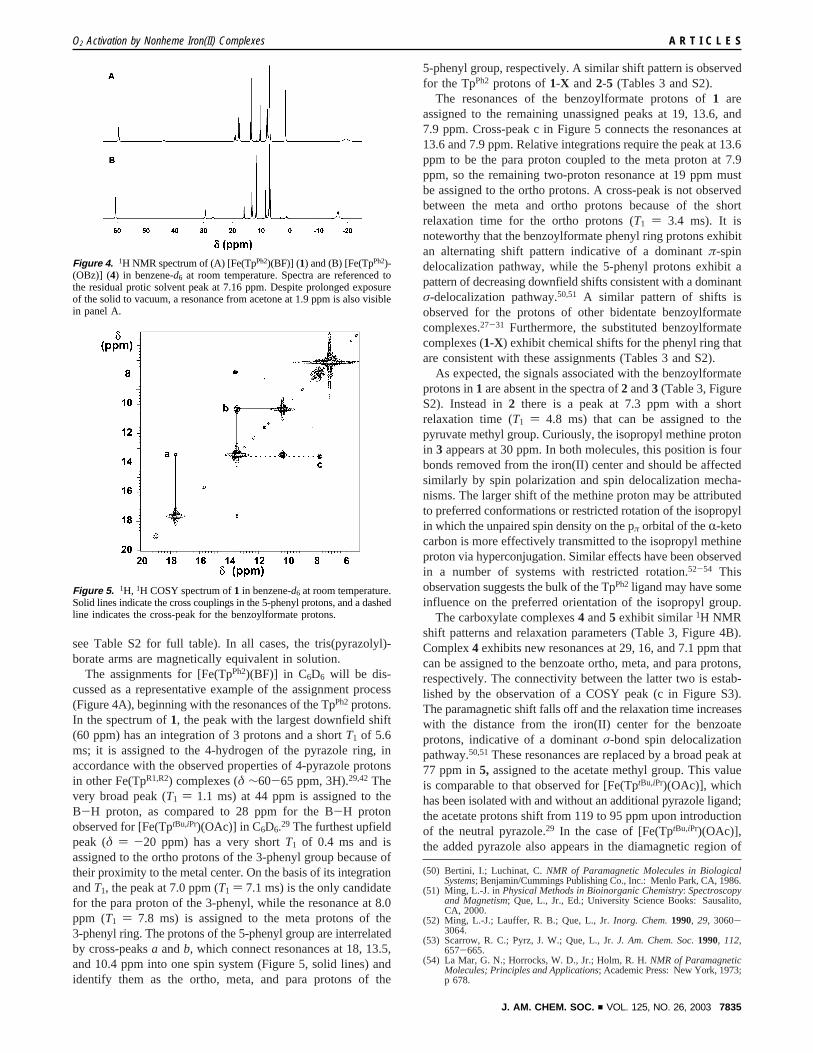
The assignments for [Fe(TpPh2)(BF)] in C6D6 will be dis-cussed as a representative example of the assignment process(Figure 4A), beginning with the resonances of the TpPh2protons.In the spectrum of1, the peak with the largest downfield shift(60 ppm) has an integration of 3 protons and a shortT1 of 5.6ms; it is assigned to the 4-hydrogen of the pyrazole ring, inaccordance with the observed properties of 4-pyrazole protonsin other Fe(TpR1,R2) complexes (δ ∼60-65 ppm, 3H).29,42Thevery broad peak (T1 ) 1.1 ms) at 44 ppm is assigned to theB-H proton, as compared to 28 ppm for the B-H protonobserved for [Fe(TptBu,iPr)(OAc)] in C6D6.29 The furthest upfieldpeak (δ ) -20 ppm) has a very shortT1 of 0.4 ms and isassigned to the ortho protons of the 3-phenyl group because oftheir proximity to the metal center. On the basis of its integrationandT1, the peak at 7.0 ppm (T1 ) 7.1 ms) is the only candidatefor the para proton of the 3-phenyl, while the resonance at 8.0ppm (T1 ) 7.8 ms) is assigned to the meta protons of the3-phenyl ring. The protons of the 5-phenyl group are interrelatedby cross-peaksa andb, which connect resonances at 18, 13.5,and 10.4 ppm into one spin system (Figure 5, solid lines) andidentify them as the ortho, meta, and para protons of the
5-phenyl group, respectively. A similar shift pattern is observedfor the TpPh2 protons of1-X and2-5 (Tables 3 and S2).
The resonances of the benzoylformate protons of1 areassigned to the remaining unassigned peaks at 19, 13.6, and7.9 ppm. Cross-peak c in Figure 5 connects the resonances at13.6 and 7.9 ppm. Relative integrations require the peak at 13.6ppm to be the para proton coupled to the meta proton at 7.9ppm, so the remaining two-proton resonance at 19 ppm mustbe assigned to the ortho protons. A cross-peak is not observedbetween the meta and ortho protons because of the shortrelaxation time for the ortho protons (T1 ) 3.4 ms). It isnoteworthy that the benzoylformate phenyl ring protons exhibitan alternating shift pattern indicative of a dominantπ-spindelocalization pathway, while the 5-phenyl protons exhibit apattern of decreasing downfield shifts consistent with a dominantσ-delocalization pathway.50,51 A similar pattern of shifts isobserved for the protons of other bidentate benzoylformatecomplexes.27-31 Furthermore, the substituted benzoylformatecomplexes (1-X) exhibit chemical shifts for the phenyl ring thatare consistent with these assignments (Tables 3 and S2).
As expected, the signals associated with the benzoylformateprotons in1 are absent in the spectra of2 and3 (Table 3, FigureS2). Instead in2 there is a peak at 7.3 ppm with a shortrelaxation time (T1 ) 4.8 ms) that can be assigned to thepyruvate methyl group. Curiously, the isopropyl methine protonin 3 appears at 30 ppm. In both molecules, this position is fourbonds removed from the iron(II) center and should be affectedsimilarly by spin polarization and spin delocalization mecha-nisms. The larger shift of the methine proton may be attributedto preferred conformations or restricted rotation of the isopropylin which the unpaired spin density on the pπ orbital of theR-ketocarbon is more effectively transmitted to the isopropyl methineproton via hyperconjugation. Similar effects have been observedin a number of systems with restricted rotation.52-54 Thisobservation suggests the bulk of the TpPh2ligand may have someinfluence on the preferred orientation of the isopropyl group.
The carboxylate complexes4 and5 exhibit similar1H NMRshift patterns and relaxation parameters (Table 3, Figure 4B).Complex4 exhibits new resonances at 29, 16, and 7.1 ppm thatcan be assigned to the benzoate ortho, meta, and para protons,respectively. The connectivity between the latter two is estab-lished by the observation of a COSY peak (c in Figure S3).The paramagnetic shift falls off and the relaxation time increaseswith the distance from the iron(II) center for the benzoateprotons, indicative of a dominantσ-bond spin delocalizationpathway.50,51These resonances are replaced by a broad peak at77 ppm in5, assigned to the acetate methyl group. This valueis comparable to that observed for [Fe(TptBu,iPr)(OAc)], whichhas been isolated with and without an additional pyrazole ligand;the acetate protons shift from 119 to 95 ppm upon introductionof the neutral pyrazole.29 In the case of [Fe(TptBu,iPr)(OAc)],the added pyrazole also appears in the diamagnetic region of
(50) Bertini, I.; Luchinat, C.NMR of Paramagnetic Molecules in BiologicalSystems; Benjamin/Cummings Publishing Co., Inc.: Menlo Park, CA, 1986.
(51) Ming, L.-J. inPhysical Methods in Bioinorganic Chemistry: Spectroscopyand Magnetism; Que, L., Jr., Ed.; University Science Books: Sausalito,CA, 2000.
(52) Ming, L.-J.; Lauffer, R. B.; Que, L., Jr.Inorg. Chem.1990, 29, 3060-3064.
(53) Scarrow, R. C.; Pyrz, J. W.; Que, L., Jr.J. Am. Chem. Soc.1990, 112,657-665.
(54) La Mar, G. N.; Horrocks, W. D., Jr.; Holm, R. H.NMR of ParamagneticMolecules; Principles and Applications; Academic Press: New York, 1973;p 678.
Figure 4. 1H NMR spectrum of (A) [Fe(TpPh2)(BF)] (1) and (B) [Fe(TpPh2)-(OBz)] (4) in benzene-d6 at room temperature. Spectra are referenced tothe residual protic solvent peak at 7.16 ppm. Despite prolonged exposureof the solid to vacuum, a resonance from acetone at 1.9 ppm is also visiblein panel A.
Figure 5. 1H, 1H COSY spectrum of1 in benzene-d6 at room temperature.Solid lines indicate the cross couplings in the 5-phenyl protons, and a dashedline indicates the cross-peak for the benzoylformate protons.
O2 Activation by Nonheme Iron(II) Complexes A R T I C L E S
J. AM. CHEM. SOC. 9 VOL. 125, NO. 26, 2003 7835
the spectrum; however, similar peaks have not been observedfor the neutral pyrazole in [Fe(TpPh2)(OAc)(3,5-Ph2pzH)],suggesting that it may be in an intermediate exchange regime.
The electrochemical properties of the iron(II) precursors havebeen examined by cyclic voltammetry of anaerobic dichlo-romethane solutions with tetrabutylammonium perchlorate asthe supporting electrolyte (Table 3). Complex1 shows an anodicone-electron wave at 930 mV versus SCE, corresponding tothe oxidation of iron(II). There is a cathodic wave at 770 mVversus SCE but with a smaller current, suggesting an irreversiblechange after oxidation to iron(III). Substitution of the benzoyl-formate group results in the modulation of this potential from890 to 1010 mV versus SCE. The otherR-keto carboxylatecompounds show similar anodic peaks at 980 mV for2 and1000 mV for 3, while the carboxylate compounds exhibitmultiple irreversible waves (4, Epa ) 870 and 1320 mV,5 830and 1070 mV versus SCE). It is interesting to note that all fivecomplexes have FeIII /FeII potentials near 1 V despite the factthat carboxylates are much stronger bases thanR-keto carboxy-lates (pKa ) 4.19 and 4.7555 for benzoic acid and acetic acid,respectively, vs 1.3956 (or 1.157) and 1.9458 for benzoylformicand pyruvic acid, respectively).
For 1-3, there is an additional quasi-reversible wave thatcan be assigned to the formation of a ketyl anion radical. Theketone/ketyl couple is observed at-1220 mV versus SCE (∆E) 160 mV) for1 and is shifted to-1630 mV versus SCE (∆E) 210 mV) for2, and-1670 mV versus SCE (∆E ) 210 mV)for 3. Substitution of the benzoylformate ring also affects thiscouple, and while1-NO2 exhibits irreversible waves, moreelectron-releasing groups result in cathodic shifts of the ketone/ketyl couple (Figure S4). The cathodic shift of the ketyl couplesuggests theπ*-acceptor orbital of the BF ligand in1 isstabilized relative to those of theR-keto acid ligands in2 and3, probably because of conjugation with the phenyl ring. Thistrend is consistent with the blue shifts in the MLCT transitionsobserved for2 and3. The conjugation may also stabilize thedianionic enolate form of BF and rationalize the lower FeIII /FeII potential of1 relative to2 and3.
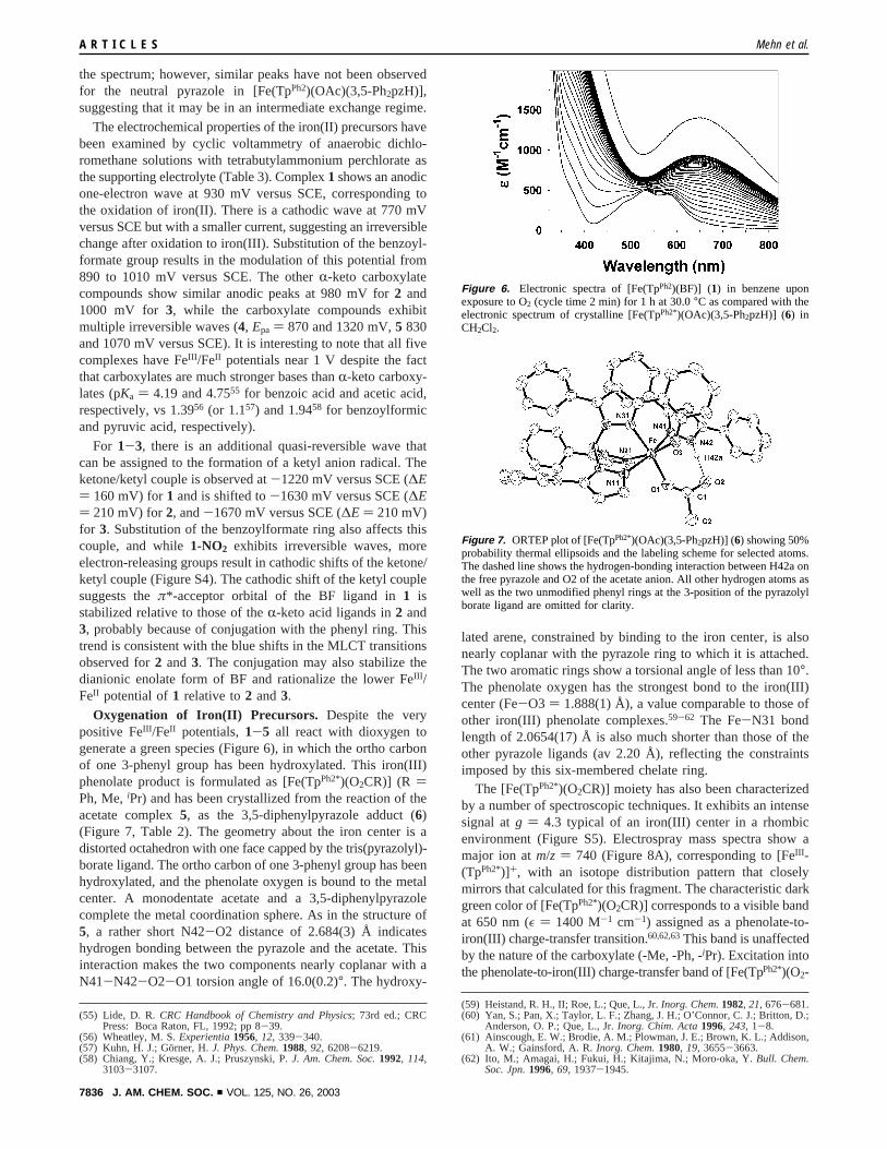
Oxygenation of Iron(II) Precursors. Despite the verypositive FeIII /FeII potentials,1-5 all react with dioxygen togenerate a green species (Figure 6), in which the ortho carbonof one 3-phenyl group has been hydroxylated. This iron(III)phenolate product is formulated as [Fe(TpPh2*)(O2CR)] (R )Ph, Me,iPr) and has been crystallized from the reaction of theacetate complex5, as the 3,5-diphenylpyrazole adduct (6)(Figure 7, Table 2). The geometry about the iron center is adistorted octahedron with one face capped by the tris(pyrazolyl)-borate ligand. The ortho carbon of one 3-phenyl group has beenhydroxylated, and the phenolate oxygen is bound to the metalcenter. A monodentate acetate and a 3,5-diphenylpyrazolecomplete the metal coordination sphere. As in the structure of5, a rather short N42-O2 distance of 2.684(3) Å indicateshydrogen bonding between the pyrazole and the acetate. Thisinteraction makes the two components nearly coplanar with aN41-N42-O2-O1 torsion angle of 16.0(0.2)°. The hydroxy-
lated arene, constrained by binding to the iron center, is alsonearly coplanar with the pyrazole ring to which it is attached.The two aromatic rings show a torsional angle of less than 10°.The phenolate oxygen has the strongest bond to the iron(III)center (Fe-O3 ) 1.888(1) Å), a value comparable to those ofother iron(III) phenolate complexes.59-62 The Fe-N31 bondlength of 2.0654(17) Å is also much shorter than those of theother pyrazole ligands (av 2.20 Å), reflecting the constraintsimposed by this six-membered chelate ring.
The [Fe(TpPh2*)(O2CR)] moiety has also been characterizedby a number of spectroscopic techniques. It exhibits an intensesignal atg ) 4.3 typical of an iron(III) center in a rhombicenvironment (Figure S5). Electrospray mass spectra show amajor ion atm/z ) 740 (Figure 8A), corresponding to [FeIII -(TpPh2*)]+, with an isotope distribution pattern that closelymirrors that calculated for this fragment. The characteristic darkgreen color of [Fe(TpPh2*)(O2CR)] corresponds to a visible bandat 650 nm (ε ) 1400 M-1 cm-1) assigned as a phenolate-to-iron(III) charge-transfer transition.60,62,63This band is unaffectedby the nature of the carboxylate (-Me, -Ph, -iPr). Excitation intothe phenolate-to-iron(III) charge-transfer band of [Fe(TpPh2*)(O2-
(55) Lide, D. R.CRC Handbook of Chemistry and Physics; 73rd ed.; CRCPress: Boca Raton, FL, 1992; pp 8-39.
(56) Wheatley, M. S.Experientia1956, 12, 339-340.(57) Kuhn, H. J.; Go¨rner, H.J. Phys. Chem.1988, 92, 6208-6219.(58) Chiang, Y.; Kresge, A. J.; Pruszynski, P.J. Am. Chem. Soc.1992, 114,
3103-3107.
(59) Heistand, R. H., II; Roe, L.; Que, L., Jr.Inorg. Chem.1982, 21, 676-681.(60) Yan, S.; Pan, X.; Taylor, L. F.; Zhang, J. H.; O’Connor, C. J.; Britton, D.;
Anderson, O. P.; Que, L., Jr.Inorg. Chim. Acta1996, 243, 1-8.(61) Ainscough, E. W.; Brodie, A. M.; Plowman, J. E.; Brown, K. L.; Addison,
A. W.; Gainsford, A. R.Inorg. Chem.1980, 19, 3655-3663.(62) Ito, M.; Amagai, H.; Fukui, H.; Kitajima, N.; Moro-oka, Y.Bull. Chem.
Soc. Jpn.1996, 69, 1937-1945.
Figure 6. Electronic spectra of [Fe(TpPh2)(BF)] (1) in benzene uponexposure to O2 (cycle time 2 min) for 1 h at30.0°C as compared with theelectronic spectrum of crystalline [Fe(TpPh2*)(OAc)(3,5-Ph2pzH)] (6) inCH2Cl2.
Figure 7. ORTEP plot of [Fe(TpPh2*)(OAc)(3,5-Ph2pzH)] (6) showing 50%probability thermal ellipsoids and the labeling scheme for selected atoms.The dashed line shows the hydrogen-bonding interaction between H42a onthe free pyrazole and O2 of the acetate anion. All other hydrogen atoms aswell as the two unmodified phenyl rings at the 3-position of the pyrazolylborate ligand are omitted for clarity.
A R T I C L E S Mehn et al.
7836 J. AM. CHEM. SOC. 9 VOL. 125, NO. 26, 2003
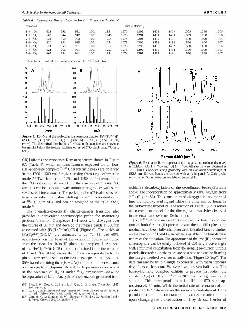
CR)] affords the resonance Raman spectrum shown in Figure9A (Table 4), which contains features expected for an iron-(III)-phenolate complex.63-65 Characteristic peaks are observedin the 1200-1600 cm-1 region arising from ring deformationmodes.64 Two features at 1254 and 1298 cm-1 downshift inthe 18O isotopomer derived from the reaction of1 with 18O2
and thus can be associated with aromatic ring modes with someC-O stretching character. The peak at 621 cm-1 is also sensitiveto isotopic substitution, downshifting 16 cm-1 upon introductionof 18O (Figure 9B), and can be assigned as theν(Fe-OAr)mode.63
The phenolate-to-iron(III) charge-transfer transition alsoprovides a convenient spectroscopic probe for monitoringproduct formation. Complexes1-3 react with dioxygen overthe course of 60 min and develop the intense bands at 650 nmassociated with [Fe(TpPh2*)(O2CR)] (Figure 6). The yields of[Fe(TpPh2*)(O2CR)] are estimated to be 70, 55, and 60%,respectively, on the basis of the extinction coefficient culledfrom the crystalline iron(III) phenolate complex,6. Analysisof the [Fe(TpPh2*)(O2CR)] product obtained from the reactionof 1 and 18O2 (98%) shows that18O is incorporated into thephenolates76% based on the ESI mass spectral analysis and85% based on fitting theν(Fe-OAr) vibration in the resonanceRaman spectrum (Figures 8A and 9). Experiments carried outin the presence of H218O under 16O2 atmosphere show noincorporation of label. Analysis of the benzoate generated from
oxidative decarboxylation of the coordinated benzoylformateshows the incorporation of approximately 80% oxygen from18O2 (Figure S6). Thus, one atom of dioxygen is incorporatedinto the hydroxylated ligand while the other can be found inthe carboxylate byproduct. The reaction of1 with O2 thus servesas an excellent model for the dioxygenase reactivity observedin the enzymatic systems (Scheme 2).
[Fe(TpPh2)(BF)] is an excellent candidate for kinetic examina-tion as both the iron(II) precursor and the iron(III) phenolateproduct have been fully characterized. Detailed kinetic studieson the reaction of1 and O2 in benzene establish the bimolecularnature of the oxidation. The appearance of the iron(III) phenolatechromophore can be easily followed at 650 nm, a wavelengthwith a minimal contribution from the iron(II) precursor. Simplepseudo-first-order kinetic traces are observed and can be fit usingthe integral method over seven half-lives (Figure 10 (top)). Thedata can also be fit to a single exponential with mean standarddeviations of less than 2% over five to seven half-lives. Thebenzoylformate complex exhibits a pseudo-first-order rateconstant (kobs) of 1.0× 10⁻³ s-1 at 30°C in an oxygen-saturatedsolution. This corresponds to a half-life of 670 s or ap-proximately 11 min. While the initial rate of formation of theproduct at 30°C depends on the initial concentration of1, thepseudo-first-order rate constant exhibits no systematic variationupon changing the concentration of1 by almost 1 order of
(63) Pyrz, J. W.; Roe, A. L.; Stern, L. J.; Que, L., Jr.J. Am. Chem. Soc.1985,107, 614-620.
(64) Que, L., Jr. inBiological Applications of Raman Spectroscopy; Spiro, T.G., Ed.; Wiley: New York, 1988; Vol. 3, pp 491-521.
(65) Carrano, C. J.; Carrano, M. W.; Sharma, K.; Backes, G.; Sanders-Loehr,J. Inorg. Chem.1990, 29, 1865-1870.
Table 4. Resonance Raman Data for Iron(III) Phenolate Productsa
compound raman shift (cm-1)
1 + 16O2 621 861 961 1001 1254 1273 1298 1451 1480 1559 1598 16061 + 18O2 605 844 942 1001 1245 1273 1294 1451 1480 1559 1598 16062 + 16O2 621 860 963 1001 1254 1274 1301 1452 1481 1559 1599 16043 + 16O2 622 861 965 1001 1254 1275 1302 1453 1482 1560 1600 16074 + 16O2 621 859 961 1001 1252 1273 1299 1452 1482 1560 1600 16065 + 16O2 621 861 961 1001 1253 1275 1300 1453 1482 1560 1599 16075 + 18O2 605 844 961 1001 1244 1273 1297 1451 1481 1560 1599 1607
a Numbers in bold denote modes sensitive to18O substitution.
Figure 8. ESI-MS of the molecular ion corresponding to [FeIII (TpPh2*)]+.(A) 1 + 16O2 (s) and1 + 18O2 (- -) and (B)5 + 16O2 (s) and5 + 18O2
(- -). The theoretical distributions for these molecular ions are shown asbar graphs below the isotope splitting observed (16O black bars,18O graybars).
Figure 9. Resonance Raman spectra of the oxygenation products dissolvedin CH2Cl2: (A) 1 + 16O2 and (B)1 + 18O2. All spectra were obtained at77 K using a backscattering geometry with an excitation wavelength of632.8 nm. Solvent bands are labeled with an s in panel A. Only peakssensitive to18O substitution are labeled in panel B.
O2 Activation by Nonheme Iron(II) Complexes A R T I C L E S
J. AM. CHEM. SOC. 9 VOL. 125, NO. 26, 2003 7837
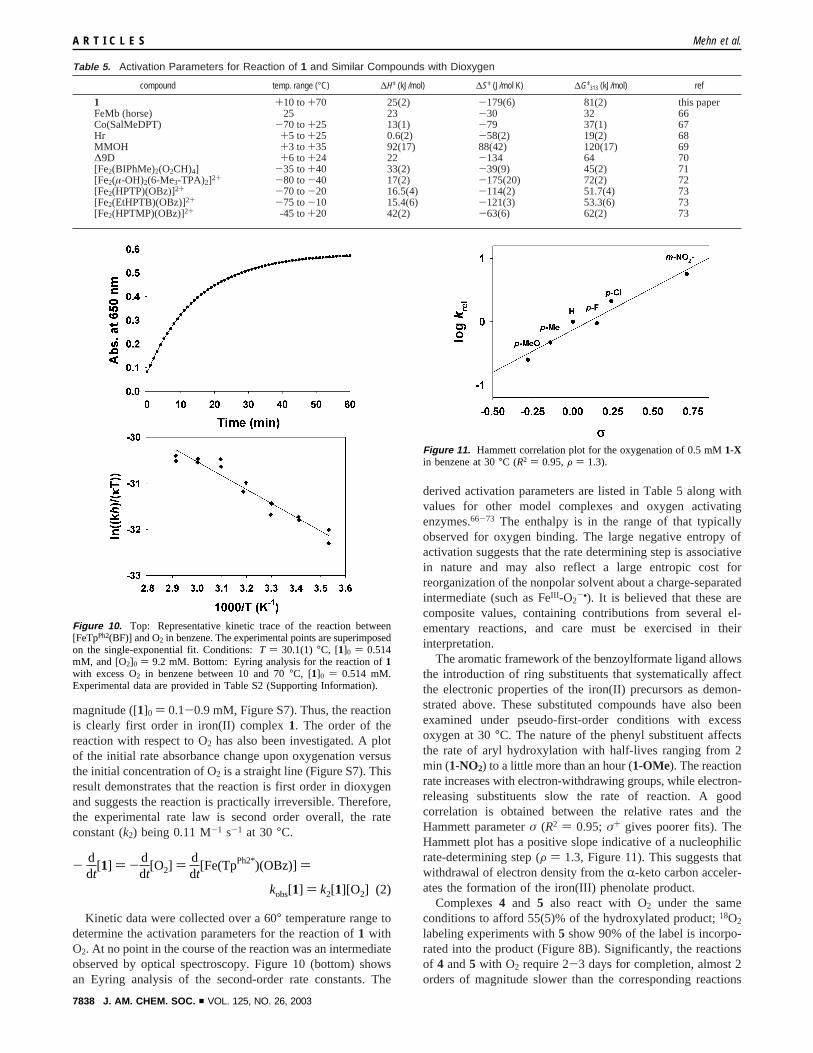
magnitude ([1]0 ) 0.1-0.9 mM, Figure S7). Thus, the reactionis clearly first order in iron(II) complex1. The order of thereaction with respect to O2 has also been investigated. A plotof the initial rate absorbance change upon oxygenation versusthe initial concentration of O2 is a straight line (Figure S7). Thisresult demonstrates that the reaction is first order in dioxygenand suggests the reaction is practically irreversible. Therefore,the experimental rate law is second order overall, the rateconstant (k2) being 0.11 M-1 s-1 at 30°C.
Kinetic data were collected over a 60° temperature range todetermine the activation parameters for the reaction of1 withO2. At no point in the course of the reaction was an intermediateobserved by optical spectroscopy. Figure 10 (bottom) showsan Eyring analysis of the second-order rate constants. The
derived activation parameters are listed in Table 5 along withvalues for other model complexes and oxygen activatingenzymes.66-73 The enthalpy is in the range of that typicallyobserved for oxygen binding. The large negative entropy ofactivation suggests that the rate determining step is associativein nature and may also reflect a large entropic cost forreorganization of the nonpolar solvent about a charge-separatedintermediate (such as FeIII -O2
-•). It is believed that these arecomposite values, containing contributions from several el-ementary reactions, and care must be exercised in theirinterpretation.
The aromatic framework of the benzoylformate ligand allowsthe introduction of ring substituents that systematically affectthe electronic properties of the iron(II) precursors as demon-strated above. These substituted compounds have also beenexamined under pseudo-first-order conditions with excessoxygen at 30°C. The nature of the phenyl substituent affectsthe rate of aryl hydroxylation with half-lives ranging from 2min (1-NO2) to a little more than an hour (1-OMe). The reactionrate increases with electron-withdrawing groups, while electron-releasing substituents slow the rate of reaction. A goodcorrelation is obtained between the relative rates and theHammett parameterσ (R2 ) 0.95; σ+ gives poorer fits). TheHammett plot has a positive slope indicative of a nucleophilicrate-determining step (F ) 1.3, Figure 11). This suggests thatwithdrawal of electron density from theR-keto carbon acceler-ates the formation of the iron(III) phenolate product.
Complexes4 and 5 also react with O2 under the sameconditions to afford 55(5)% of the hydroxylated product;18O2
labeling experiments with5 show 90% of the label is incorpo-rated into the product (Figure 8B). Significantly, the reactionsof 4 and5 with O2 require 2-3 days for completion, almost 2orders of magnitude slower than the corresponding reactions
Table 5. Activation Parameters for Reaction of 1 and Similar Compounds with Dioxygen
compound temp. range (°C) ∆Hq (kJ/mol) ∆Sq (J/mol K) ∆Gq313 (kJ/mol) ref
1 +10 to+70 25(2) -179(6) 81(2) this paperFeMb (horse) 25 23 -30 32 66Co(SalMeDPT) -70 to+25 13(1) -79 37(1) 67Hr +5 to +25 0.6(2) -58(2) 19(2) 68MMOH +3 to +35 92(17) 88(42) 120(17) 69∆9D +6 to +24 22 -134 64 70[Fe2(BIPhMe)2(O2CH)4] -35 to+40 33(2) -39(9) 45(2) 71[Fe2(µ-OH)2(6-Me3-TPA)2]2+ -80 to-40 17(2) -175(20) 72(2) 72[Fe2(HPTP)(OBz)]2+ -70 to-20 16.5(4) -114(2) 51.7(4) 73[Fe2(EtHPTB)(OBz)]2+ -75 to-10 15.4(6) -121(3) 53.3(6) 73[Fe2(HPTMP)(OBz)]2+ -45 to+20 42(2) -63(6) 62(2) 73
Figure 10. Top: Representative kinetic trace of the reaction between[FeTpPh2(BF)] and O2 in benzene. The experimental points are superimposedon the single-exponential fit. Conditions:T ) 30.1(1) °C, [1]0 ) 0.514mM, and [O2]0 ) 9.2 mM. Bottom: Eyring analysis for the reaction of1with excess O2 in benzene between 10 and 70°C, [1]0 ) 0.514 mM.Experimental data are provided in Table S2 (Supporting Information).
Figure 11. Hammett correlation plot for the oxygenation of 0.5 mM1-Xin benzene at 30°C (R2 ) 0.95,F ) 1.3).
- ddt
[1] ) - ddt
[O2] ) ddt
[Fe(TpPh2*)(OBz)] )
kobs[1] ) k2[1][O2] (2)
A R T I C L E S Mehn et al.
7838 J. AM. CHEM. SOC. 9 VOL. 125, NO. 26, 2003
of 1-3 at the same concentrations of iron(II) complex. Interest-ingly, the time course for the formation of6 from 5 is morecomplex than the easily analyzed exponential increase observedfor 1 (Figure 10). The carboxylate compounds exhibit a lagphase that makes further detailed kinetic analysis necessary.Nonetheless, it is clear from the above observations that thepresence of anR-keto acid in the iron coordination spheresignificantly enhances its ability to react with O2.
Mechanistic Implications. Iron(II)-R-keto carboxylate com-plexes have previously been shown to activate dioxygen andmodel the oxidative decarboxylation ofR-keto acids carried outby the enzymatic systems.27,28,31The ancillary ligand in theseiron(II)-R-keto carboxylate complexes exerts a dramatic effecton the reactivity of the complexes toward O2, increasing in theorder 6-Me3-TPA, TPA, TpPh2, and TpMe2. The tetradentate6-Me3-TPA and TPA ligands afford six-coordinate complexesthat react over a period of days,27 while the tridentate TpPh2
and TpMe2 ligands afford five-coordinate complexes that reactwithin an hour.28,30 The difference of nearly 2 orders ofmagnitude in reaction rate strongly suggests that the coordinativeunsaturation of the iron(II) center in the TpR2 complexes andthe consequent availability of an O2 coordination site areimportant in promoting the oxidative decarboxylation of theboundR-keto acid. This mechanistic principle has also beeninvoked in explaining the much higher reactivity of the five-coordinate enzyme-R-KG complex in the presence of theprimary substrate relative to the six-coordinate complex in theabsence of the primary substrate.24,74,75
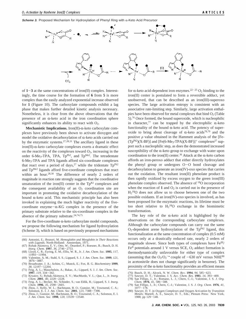
For the five-coordinateR-keto carboxylate model compounds,we propose the following mechanism for ligand hydroxylation(Scheme 3), which is based on previously proposed mechanisms
for R-keto acid-dependent iron enzymes.22-25 O2 binding to theiron(II) center is postulated to form a reversible adduct, yetunobserved, that can be described as an iron(III)-superoxospecies. The large activation entropy is consistent with anassociative rate-limiting step. Similarly, large activation enthal-pies have been observed for metal complexes that bind O2 (Table5).76 Once formed, the bound superoxide, which is nucleophilicin character,77 can be trapped by the electrophilicR-ketofunctionality of the boundR-keto acid. The potency of super-oxide to bring about cleavage ofR-keto acids78,79 and thepositiveF value obtained in the Hammett analysis of the [Fe-(TpPh2)(X-BF)] and [Fe(6-Me3-TPA)(X-BF)]+ complexes27 sup-port such a nucleophilic step, as does the demonstrated increasedsusceptibility of theR-keto group to exchange with water uponcoordination to the iron(II) center.36 Attack at theR-keto carbonaffords an iron-peroxo adduct that either directly hydroxylatesthe phenyl group or undergoes O-O bond cleavage anddecarboxylation to generate an iron(IV)-oxo species that carriesout the oxidation. The resultant iron(II) phenolate product isthen rapidly oxidized by excess oxygen to the green iron(III)phenolate complex observed. The absence of18O incorporationwhen the reaction of1 and O2 is carried out in the presence ofH2
18O does not allow us to choose between one of the twopossible oxidants. If an iron(IV)-oxo species is involved, as hasbeen proposed for the enzymatic reactions, its lifetime must betoo short relative to H218O exchange in the biomimetictransformation.
The key role of theR-keto acid is highlighted by theobservations on the corresponding carboxylate complexes.Although the carboxylate compounds can carry out the sameO2-dependent arene hydroxylation of the TpPh2 ligand, thisfunctionalization at the same concentration of complex (0.5 mM)occurs only at a drastically reduced rate, nearly 2 orders ofmagnitude slower. Since both types of complexes have FeIII /FeII potentials around 1 V versus SCE, O2 adduct formation isthermodynamically unfavorable for either type of complex(assuming that the O2/O2
-• couple of-630 mV versus NHE80
in acetonitrile does not change significantly in benzene). Theproximity of theR-keto functionality provides an efficient means
(66) Antonini, E.; Brunori, M.Hemoglobin and Myoglobin in Their Reactionswith Ligands; North-Holland: Amsterdam, 1971.
(67) Rybak-Akimova, E. V.; Otto, W.; Deardorf, P.; Roesner, R.; Busch, D. H.Inorg. Chem.1997, 36, 2746-2753.
(68) Lloyd, C. R.; Eyring, E. M.; Ellis, W. R., Jr.J. Am. Chem. Soc.1995, 117,11993-11994.
(69) Valentine, A. M.; Stahl, S. S.; Lippard, S. J.J. Am. Chem. Soc.1999, 121,3876-3887.
(70) Broadwater, J. A.; Achim, C.; Munck, E.; Fox, B. G.Biochemistry1999,38, 12197-12204.
(71) Feig, A. L.; Masschelein, A.; Bakac, A.; Lippard, S. J.J. Am. Chem. Soc.1997, 119, 334-342.
(72) Kryatov, S.; Rybak-Akimova, E. V.; MacMurdo, V. L.; Que, L., Jr.Inorg.Chem.2001, 40, 2220-2228.
(73) Feig, A. L.; Becker, M.; Schinder, S.; van Eldik, R.; Lippard, S. J.Inorg.Chem.1996, 35, 2590-2601.
(74) Zhou, J.; Kelly, W. L.; Bachmann, B. O.; Gunsior, M.; Townsend, C. A.;Solomon, E. I.J. Am. Chem. Soc.2001, 123, 7388-7398.
(75) Zhou, J.; Gunsior, M.; Bachmann, B. O.; Townsend, C. A.; Solomon, E. I.J. Am. Chem. Soc.1998, 120, 13539-13540.
(76) Busch, D. H.; Alcock, N. W.Chem. ReV. 1994, 94, 585-623.(77) Sawyer, D. T.; Valentine, J. S.Acc. Chem. Res.1981, 14, 393-400.(78) San Fillipo, J., Jr.; Romano, L. J.; Chern, C.-I.; Valentine, J. S.J. Org.
Chem.1976, 41, 586-588.(79) San Fillipo, J., Jr.; Chern, C.-I.; Valentine, J. S.J. Org. Chem.1976, 41,
1077-178.(80) Sawyer, D. T. inOxygen Complexes and Oxygen ActiVation by Transition
Metals; Martell, A. E., Sawyer, D. T., Eds.; Plenum Press: New York,1988; pp 129-148.
Scheme 3. Proposed Mechanism for Hydroxylation of Phenyl Ring with R-Keto Acid Precursor
O2 Activation by Nonheme Iron(II) Complexes A R T I C L E S
J. AM. CHEM. SOC. 9 VOL. 125, NO. 26, 2003 7839
for trapping the nascent superoxide in the cases of1-3 to drivethe overall reaction to completion. For4 and5, the absence oftheR-keto moiety would require a different superoxide trappingmechanism that may relate to the formation of a (µ-1,2-peroxo)-diiron(III) adduct like that observed for [Fe(TpiPr2)(O2CR)]complexes.42,81 Mechanistic details for the latter reaction havenot been established and will be the focus of a subsequentpublication. Nevertheless, these results emphasize the versatilityof a nonheme iron center to activate dioxygen.
An important feature of theR-keto carboxylate complexesdescribed here is the effective coupling between the oxidativedecarboxylation of theR-keto acid part of the reaction, whichprovides the electrons needed for oxygen activation, and thehydroxylation of the phenyl group. In the best case, about 0.7equiv of phenol is formed perR-keto acid decarboxylated. Thedegree of coupling is much less efficient for other nonhemeiron model systems that mimic enzymatic hydroxylation ofarenes using molecular oxygen and a sacrificial electrondonor.82-87A large excess of reducing agent such as mercap-toethanol, zinc and acetic acid, diphenylhydrazine, hydro-quinone, or ascorbate is typically required, making it difficultto determine the ratio of substrate oxidized to reductant usedin these systems. Invariably, the oxidizing species formed insuch reactions can also oxidize the excess reducing agent insteadof the intended substrate. Enzymes avoid this pitfall bycompartmentalizing the reducing and oxidizing half reactions.The use of theR-keto acid cofactor is a convenient strategy tocontrol the flow of electrons and may explain the many anddiverse types ofR-keto acid-dependent enzymes found inbiology.2,4-9,24 Yet even in the enzymes, there is evidence foruncoupling of oxidative decarboxylation from substrate func-tionalization, especially when substrate analogues are used.88-92
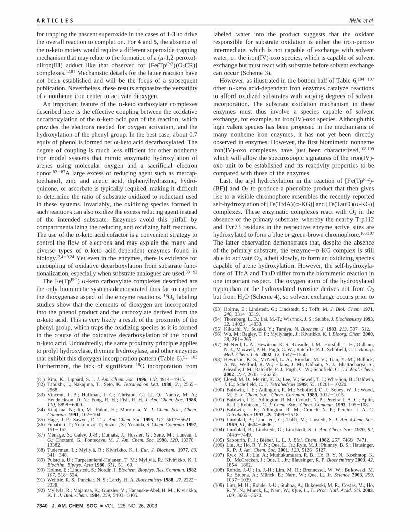
The Fe(TpPh2) R-keto carboxylate complexes described arethe only biomimetic systems demonstrated thus far to capturethe dioxygenase aspect of the enzyme reactions.18O2 labelingstudies show that the elements of dioxygen are incorporatedinto the phenol product and the carboxylate derived from theR-keto acid. This is very likely a result of the proximity of thephenyl group, which traps the oxidizing species as it is formedin the course of the oxidative decarboxylation of the boundR-keto acid. Undoubtedly, the same proximity principle appliesto prolyl hydroxylase, thymine hydroxylase, and other enzymesthat exhibit this dioxygen incorporation pattern (Table 6).93-103
Furthermore, the lack of significant18O incorporation from
labeled water into the product suggests that the oxidantresponsible for substrate oxidation is either the iron-peroxointermediate, which is not capable of exchange with solventwater, or the iron(IV)-oxo species, which is capable of solventexchange but must react with substrate before solvent exchangecan occur (Scheme 3).
However, as illustrated in the bottom half of Table 6,104-107
other R-keto acid-dependent iron enzymes catalyze reactionsto afford oxidized substrates with varying degrees of solventincorporation. The substrate oxidation mechanism in theseenzymes must thus involve a species capable of solventexchange, for example, an iron(IV)-oxo species. Although thishigh valent species has been proposed in the mechanisms ofmany nonheme iron enzymes, it has not yet been directlyobserved in enzymes. However, the first biomimetic nonhemeiron(IV)-oxo complexes have just been characterized,108,109
which will allow the spectroscopic signatures of the iron(IV)-oxo unit to be established and its reactivity properties to becompared with those of the enzymes.
Last, the aryl hydroxylation in the reaction of [Fe(TpPh2)-(BF)] and O2 to produce a phenolate product that then givesrise to a visible chromophore resembles the recently reportedself-hydroxylation of [Fe(TfdA)(R-KG)] and [Fe(TauD)(R-KG)]complexes. These enzymatic complexes react with O2 in theabsence of the primary substrate, whereby the nearby Trp112and Tyr73 residues in the respective enzyme active sites arehydroxylated to form a blue or green-brown chromophore.106,107
The latter observation demonstrates that, despite the absenceof the primary substrate, the enzyme-R-KG complex is stillable to activate O2, albeit slowly, to form an oxidizing speciescapable of arene hydroxylation. However, the self-hydroxyla-tions of TfdA and TauD differ from the biomimetic reaction inone important respect. The oxygen atom of the hydroxylatedtryptophan or the hydroxylated tyrosine derives not from O2
but from H2O (Scheme 4), so solvent exchange occurs prior to
(81) Kim, K.; Lippard, S. J.J. Am. Chem. Soc.1996, 118, 4914-4915.(82) Tabushi, I.; Nakajima, T.; Seto, K.Tetrahedron Lett.1980, 21, 2565-
2568.(83) Vincent, J. B.; Huffman, J. C.; Christou, G.; Li, Q.; Nanny, M. A.;
Hendrickson, D. N.; Fong, R. H.; Fish, R. H.J. Am. Chem. Soc.1988,110, 6898-6900.
(84) Kitajima, N.; Ito, M.; Fukui, H.; Moro-oka, Y.J. Chem. Soc., Chem.Commun.1991, 102-104.
(85) Hage, J. P.; Sawyer, D. T.J. Am. Chem. Soc.1995, 117, 5617-5621.(86) Funabiki, T.; Yokomizo, T.; Suzuki, S.; Yoshida, S.Chem. Commun.1997,
151-152.(87) Menage, S.; Galey, J.-B.; Dumats, J.; Hussler, G.; Seite´, M.; Luneau, I.
G.; Chottard, G.; Fontecave, M.J. Am. Chem. Soc.1998, 120, 13370-13382.
(88) Tuderman, L.; Myllyla¨, R.; Kivirikko, K. I. Eur. J. Biochem.1977, 80,341-348.
(89) Puistola, U.; Turpeenniemi-Hujanen, T. M.; Myllyla¨, R.; Kivirikko, K. I.Biochim. Biphys. Acta1980, 611, 51-60.
(90) Holme, E.; Lindstedt, S.; Nordin, I.Biochem. Biophys. Res. Commun.1982,107, 518-524.
(91) Wehbie, R. S.; Punekar, N. S.; Lardy, H. A.Biochemistry1988, 27, 2222-2228.
(92) Myllyla, R.; Majamaa, K.; Gu¨nzler, V.; Hanauske-Abel, H. M.; Kivirikko,K. I. J. Biol. Chem.1984, 259, 5403-5405.
(93) Holme, E.; Lindstedt, G.; Lindstedt, S.; Tofft, M.J. Biol. Chem.1971,246, 3314-3319.
(94) Thornburg, L. D.; Lai, M.-T.; Wishnok, J. S.; Stubbe, J.Biochemistry1993,32, 14023-14033.
(95) Kikuchi, Y.; Suzuki, Y.; Tamiya, N.Biochem. J.1983, 213, 507-512.(96) Wu, M.; Begley, T. P.; Myllyharju, J.; Kivirikko, K. I.Bioorg. Chem.2000,
28, 261-265.(97) McNeill, L. A.; Hewitson, K. S.; Gleadle, J. M.; Horsfall, L. E.; Oldham,
N. J.; Maxwell, P. H.; Pugh, C. W.; Ratcliffe, P. J.; Schofield, C. J.Bioorg.Med. Chem. Lett.2002, 12, 1547-1550.
(98) Hewitson, K. S.; McNeill, L. A.; Riordan, M. V.; Tian, Y.-M.; Bullock,A. N.; Welford, R. W.; Elkins, J. M.; Oldham, N. J.; Bhattacharya, S.;Gleadle, J. M.; Ratcliffe, P. J.; Pugh, C. W.; Schofield, C. J.J. Biol. Chem.2002, 277, 26351-26355.
(99) Lloyd, M. D.; Merritt, K. D.; Lee, V.; Sewell, T. J.; Wha-Son, B.; Baldwin,J. E.; Schofield, C. J.Tetrahedron1999, 55, 10201-10220.
(100) Baldwin, J. E.; Adlington, R. M.; Schofield, C. J.; Sobey, W. J.; Wood,M. E. J. Chem. Soc., Chem. Commun.1989, 1012-1015.
(101) Baldwin, J. E.; Adlington, R. M.; Crouch, N. P.; Pereira, I. A. C.; Aplin,R. T.; Robinson, C.J. Chem. Soc., Chem. Commun.1993, 105-108.
(102) Baldwin, J. E.; Adlington, R. M.; Crouch, N. P.; Pereira, I. A. C.Tetrahedron1993, 49, 7499-7518.
(103) Lindblad, B.; Lindstedt, G.; Tofft, M.; Linstedt, S.J. Am. Chem. Soc.1969, 91, 4604-4606.
(104) Lindblad, B.; Lindstedt, G.; Lindstedt, S.J. Am. Chem. Soc.1970, 92,7446-7449.
(105) Sabourin, P. J.; Bieber, L. L.J. Biol. Chem.1982, 257, 7468-7471.(106) Liu, A.; Ho, R. Y. N.; Que, L., Jr.; Ryle, M. J.; Phinney, B. S.; Hausinger,
R. P.J. Am. Chem. Soc.2001, 123, 5126-5127.(107) Ryle, M. J.; Liu, A.; Muthukumaran, R. B.; Ho, R. Y. N.; Koehntop, K.
D.; McCracken, J.; Que, L., Jr.; Hausinger, R. P.Biochemistry2003, 42,1854-1862.
(108) Rohde, J.-U.; In, J.-H.; Lim, M. H.; Brennessel, W. W.; Bukowski, M.R.; Stubna, A.; Mu¨nck, E.; Nam, W.; Que, L., Jr.Science2003, 299,1037-1039.
(109) Lim, M. H.; Rohde, J.-U.; Stubna, A.; Bukowski, M. R.; Costas, M.; Ho,R. Y. N.; Munck, E.; Nam, W.; Que, L., Jr.Proc. Natl. Acad. Sci.2003,100, 3665-3670.
A R T I C L E S Mehn et al.
7840 J. AM. CHEM. SOC. 9 VOL. 125, NO. 26, 2003
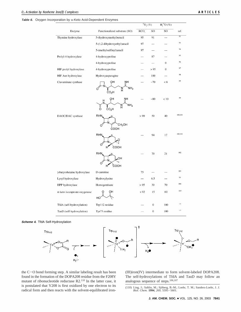
the C-O bond forming step. A similar labeling result has beenfound in the formation of the DOPA208 residue from the F208Ymutant of ribonucleotide reductase R2.110 In the latter case, itis postulated that Y208 is first oxidized by one electron to itsradical form and then reacts with the solvent-equilibrated iron-
(III)iron(IV) intermediate to form solvent-labeled DOPA208.The self-hydroxylations of TfdA and TauD may follow ananalogous sequence of steps.106,107
(110) Ling, J.; Sahlin, M.; Sjo¨berg, B.-M.; Loehr, T. M.; Sanders-Loehr, J.J.Biol. Chem.1994, 269, 5595-5601.
Table 6. Oxygen Incorporation by R-Keto Acid-Dependent Enzymes
Scheme 4. TfdA Self-Hydroxylation
O2 Activation by Nonheme Iron(II) Complexes A R T I C L E S
J. AM. CHEM. SOC. 9 VOL. 125, NO. 26, 2003 7841
The Fe(TpPh2) model system described in this paper thusserves as an excellent functional model for theR-keto acid-dependent iron enzymes, mimicking many of the featuresassociated with the reaction of enzyme-R-KG complexes withO2. Our results particularly emphasize the key role theR-ketoacid plays in oxygen activation by trapping the initial O2 adductand channeling it down the pathway towards O-O bondcleavage. Continued work on this unique family of biomimeticcomplexes should provide further mechanistic insight into thisfascinating and versatile iron(II)R-keto carboxylate center foroxygen activation in biology.
Acknowledgment. This work was supported at the Universityof Minnesota by the National Institutes of Health (Grant GM-33162 to L.Q., Postdoctoral Fellowship GM-18639 to E.L.H.,and Predoctoral Traineeship GM-08700 to M.P.M.) and at theUniversity of Tsukuba by grants from the Japan Society forthe Promotion of Science (Nos. 13555257 and 14350471),Kawaski Steel 21st Century Foundation, and the Asahi GlassFoundation. We thank Dr. Victor G. Young, Jr. and WilliamW. Brennessel of the University of Minnesota X-ray Crystal-
lographic Laboratory for determining the crystal structures of2 and 4-6. K.F. acknowledges Dr. Frank Neese and Prof.Edward I. Solomon (Stanford University) for preliminaryresonance Raman studies of the oxygenated products and Prof.Ken-ichi Okamoto (University of Tsukuba) for his encourage-ment.
Supporting Information Available: Crystallographic data for2 and4-6 (CIF format); structural tables for both independentmolecules in unit cell of2 (Table S1);1H NMR parameters(Table S2); Kinetic Data for Eyring Analysis (Table S3);Correlation of LMCT with Hammettσ (Figure S1);1H NMRspectra of2, 3, and5 (Figure S2); COSY spectrum of4 (FigureS3); CV of1-NO2, 1, and1-OMe and correlation with Hammettσ (Figure S4); EPR spectra of oxygenated products for1 + O2
(Figure S5); Low Resolution MS of PhCO18OMe (Figure S6);and determination of order in1 and O2 (Figure S7) (PDFformat). This material is available free of charge via the Internetat http://pubs.acs.org.
JA028867F
A R T I C L E S Mehn et al.
7842 J. AM. CHEM. SOC. 9 VOL. 125, NO. 26, 2003


![Technische Universität Chemnitz, Center for ...Preparation of aspheric copper nanoparticles Scheme 1: Synthesis of copper nanoparticles by thermolysis of copper(I) carboxylate 1 [7].](https://static.fdocument.org/doc/165x107/60fcc6b8e53c32273d090db6/technische-universitt-chemnitz-center-for-preparation-of-aspheric-copper.jpg)