Overexpression of N141I PS2 increases γ-secretase activity ... · and phosphorus (0.99%) and...
8
249 Lab Anim Res 2016: 32(4), 249-256 https://doi.org/10.5625/lar.2016.32.4.249 ISSN 1738-6055 (Print) ISSN 2233-7660 (Online) Overexpression of N141I PS2 increases γ-secretase activity through up-regulation of Presenilin and Pen-2 in brain mitochondria of NSE/hPS2m transgenic mice Woo Bin Yun 1,# , Jin Ju Park 1,# , Ji Eun Kim 1 , Ji Eun Sung 1 , Hyun Ah Lee 1 , Jae Ho Lee 1 , Chang Jun Bae 2 , Dae Youn Hwang 1, * Department of Biomaterials Science, College of Natural Resources and Life Science/Life and Industry Convergence Research Institute, Pusan National University, Miryang, Korea Biologics Division, Ministry of Food and Drug Administration (MFDS), Cheongju, Korea Alzheimer’s disease (AD) is known to induce alterations of mitochondrial function such as elevation of oxidative stress and activation of apopotosis. The aim of this study was to investigate the effects of human Presenilin 2 mutant (hPS2m) overexpression on the γ-secretase complex in the mitochondrial fraction. To achieve this, alterations of γ-secretase complex expression and activity were detected in the mitochondrial fraction derived from brains of NSE/hPS2m Tg mice and Non-Tg mice. Herein, the following were observed: i) overexpression of the hPS2m gene significantly up-regulated the deposition of Aβ-42 peptides in the hippocampus and cortex of brain, ii) overexpression of hPS2m protein induced alterations of γ-secretase components such as main component protein and activator protein but not stabilization-related proteins, iii) changes in γ-secretase components induced by overexpression of hPS2m protein up-regulated γ-secretase activity in the mitochondrial fraction, and iv) elevation of γ-secretase activity induced production of Aβ-42 peptides in the mitochondrial fraction. Based on these observations, these results indicate that alteration of γ-secretase activity in cells upon overexpression of hPS2m is tightly linked to mitochondrial dysfunction under the specific physiological and pathological conditions of AD. Keywords: Alzheimer’s disease, presenilin 2, γ-secretase complex, Aβ-42 peptides Received 30 November 2016; Revised version received 3 December 2016; Accepted 6 December 2016 γ-Secretase activity, which is responsible for generation of Aβ peptides, is associated with a complex that mediates intramembrane proteolytic cleavage [1]. The catalytic core of this enzyme apparently resides on Presenilin (PS) 1 or 2, but amyloidogenic activity requires three other proteins: nicastrin, anterior pharynx-defective phenotypes (APH-1), and Pen-2 (PS-enhancer) [2]. Furthermore, co- expression of all four members is required for full reconstitution of γ-secretase activity in mammalian cells [3-6]. Activation of γ-secretase cleaves β-APP, Notch, and other type I transmembrane proteins such as receptor tyrosine kinase ErbB4 [7]. In addtion, the activity and components of the γ-secretase complex are found in various cytosolic systems such as the endoplasmic reticulum-Golgi intermediate compartment, Golgi, trans- Golgi network, plamsa membrane, endosomal-lysosomal, and the mitochondria [8]. Mutations detected in the PS1 and PS2 genes are responsible for the majority of autosomal dominant forms of familial Alzheiemr’s disease (FAD) [9]. These proteins undergo physiological endoproteolytic cleavage, yielding N- and C-terminal fragments, under normal These authors contributed equaly this work. *Corresponding author: Dae Youn Hwang, Department of Biomaterials Science, College of Natural Resources and Life Science, Pusan National University, 50 Cheonghak-ri, Samnangjin-eup, Miryang, Gyeongnam 627-706, Korea Tel: +82-55-350-5388; Fax: +82-55-350-5389; E-mail: [email protected] This is an Open Access article distributed under the terms of the Creative Commons Attribution Non-Commercial License (http://creativecommons.org/licenses/ by-nc/3.0) which permits unrestricted non-commercial use, distribution, and reproduction in any medium, provided the original work is properly cited.
Transcript of Overexpression of N141I PS2 increases γ-secretase activity ... · and phosphorus (0.99%) and...
249
Lab Anim Res 2016: 32(4), 249-256
https://doi.org/10.5625/lar.2016.32.4.249
ISSN 1738-6055 (Print)
ISSN 2233-7660 (Online)
Overexpression of N141I PS2 increases γ-secretase activity through up-regulation of Presenilin and Pen-2 in brain mitochondria of
NSE/hPS2m transgenic mice
Woo Bin Yun1,#, Jin Ju Park1,#, Ji Eun Kim1, Ji Eun Sung1, Hyun Ah Lee1,Jae Ho Lee1, Chang Jun Bae2, Dae Youn Hwang1,*
1Department of Biomaterials Science, College of Natural Resources and Life Science/Life andIndustry Convergence Research Institute, Pusan National University, Miryang, Korea
2Biologics Division, Ministry of Food and Drug Administration (MFDS), Cheongju, Korea
Alzheimer’s disease (AD) is known to induce alterations of mitochondrial function such as elevation ofoxidative stress and activation of apopotosis. The aim of this study was to investigate the effects ofhuman Presenilin 2 mutant (hPS2m) overexpression on the γ-secretase complex in the mitochondrialfraction. To achieve this, alterations of γ-secretase complex expression and activity were detected in themitochondrial fraction derived from brains of NSE/hPS2m Tg mice and Non-Tg mice. Herein, thefollowing were observed: i) overexpression of the hPS2m gene significantly up-regulated the deposition ofAβ-42 peptides in the hippocampus and cortex of brain, ii) overexpression of hPS2m protein inducedalterations of γ-secretase components such as main component protein and activator protein but notstabilization-related proteins, iii) changes in γ-secretase components induced by overexpression of hPS2mprotein up-regulated γ-secretase activity in the mitochondrial fraction, and iv) elevation of γ-secretaseactivity induced production of Aβ-42 peptides in the mitochondrial fraction. Based on these observations,these results indicate that alteration of γ-secretase activity in cells upon overexpression of hPS2m is tightlylinked to mitochondrial dysfunction under the specific physiological and pathological conditions of AD.
Keywords: Alzheimer’s disease, presenilin 2, γ-secretase complex, Aβ-42 peptides
Received 30 November 2016; Revised version received 3 December 2016; Accepted 6 December 2016
γ-Secretase activity, which is responsible for generation
of Aβ peptides, is associated with a complex that mediates
intramembrane proteolytic cleavage [1]. The catalytic
core of this enzyme apparently resides on Presenilin (PS)
1 or 2, but amyloidogenic activity requires three other
proteins: nicastrin, anterior pharynx-defective phenotypes
(APH-1), and Pen-2 (PS-enhancer) [2]. Furthermore, co-
expression of all four members is required for full
reconstitution of γ-secretase activity in mammalian cells
[3-6]. Activation of γ-secretase cleaves β-APP, Notch,
and other type I transmembrane proteins such as receptor
tyrosine kinase ErbB4 [7]. In addtion, the activity and
components of the γ-secretase complex are found in
various cytosolic systems such as the endoplasmic
reticulum-Golgi intermediate compartment, Golgi, trans-
Golgi network, plamsa membrane, endosomal-lysosomal,
and the mitochondria [8].
Mutations detected in the PS1 and PS2 genes are
responsible for the majority of autosomal dominant
forms of familial Alzheiemr’s disease (FAD) [9]. These
proteins undergo physiological endoproteolytic cleavage,
yielding N- and C-terminal fragments, under normal
#These authors contributed equaly this work.
*Corresponding author: Dae Youn Hwang, Department of Biomaterials Science, College of Natural Resources and Life Science, PusanNational University, 50 Cheonghak-ri, Samnangjin-eup, Miryang, Gyeongnam 627-706, KoreaTel: +82-55-350-5388; Fax: +82-55-350-5389; E-mail: [email protected]
This is an Open Access article distributed under the terms of the Creative Commons Attribution Non-Commercial License (http://creativecommons.org/licenses/by-nc/3.0) which permits unrestricted non-commercial use, distribution, and reproduction in any medium, provided the original work is properly cited.
250 Woo Bin Yun et al.
Lab Anim Res | December, 2016 | Vol. 32, No. 4
conditions. Accumulation of these proteolytic fragments
is tightly regulated in vivo, suggesting that constitutive
levels of their expression are crucial for cell function
[10,11]. Especially, several studies reported that PS2
leads to enhanced susceptibility to various apoptotic
stimuli. Moreover, FAD-liked mutations of PS2 were
shown to potentiate the pro-apoptotic properties of these
genes in cultured cells and in neurons from transgenic
mice [12-14]. However, there has been no study on the
effects of mutant PS2 on the activity and components of
the γ-secretase complex in mitochondria harvested from
brains of NSE/PS2m Tg mice.
As demonstrated by our data, significant changes were
observed in the activity and component expression of the
γ-secretase complex. These data presented herein provide
strong evidence that overexpression of mPS2 could
contribute to up-regulation of Aβ peptides in mito-
chrondria through activation of γ-secretase. Furthermore,
these changes may impair function of mitochondria in
neuronal cells from subjects with AD.
Materials and Methods
Identification and maintenance of NSE/hPS2m Tg
mice
NSE/hPS2m Tg mice were produced by micro-
injection of the NSE/hPS2m plasmid fragment (Figure
1A), which was kindly provided from Laboratory Animal
Resources Division, National Institute of Food and Drug
Safety Evaluation (Cheongju, Korea) [12]. In order to
identify the NSE/hPS2m Tg mice, the inserted hPS2m
transgene was identified by PCR analysis of genomic
DNA isolated from the tails of 3-week-old founder mice.
The hPS2m genes were synthesized using sense primer
(5'-GAGGA AGAAG TGTGT GATGA G-3) and antisense
primer (5'-CACGA TGACG CTGAT CATGA TG-3),
with complementary hPS2m genes ranging from 817 to
796 nucleotides as the DNA template. After 25 cycles of
amplification, levels of hPS2m products (422-bp) were
quantified using a Kodak Electrophoresis Documentation
and Analysis System 120 on 1% agarose gels.
The protocols for the animal experiment were carefully
reviewed for ethical and scientific care procedures and
approved by the Pusan National University-Institutional
Animal Care and Use Committee (PNU-IACUC; Approval
Number PNU-2012-0050). All mice were provided with
ad libitum access to standard irradiated chow diet
(Samtako Inc., Osan, Korea) consisting of moisture
(12.5%), crude protein (25.43%), crude fat (6.06%),
crude fiber (3.9%), crude ash (5.31%), calcium (1.14%),
and phosphorus (0.99%) and water. During the experiment,
mice were maintained in a specific pathogen-free state
under a strict light cycle (lights on at 08:00 hours and off
at 20:00 hrs) at 23±2oC and 50±10% relative humidity.
The mice were housed in the Pusan National University-
Laboratory Animal Resources Center accredited by the
Korea Ministry of Food and Drug Safety (MFDS) in
accordance with the Laboratory Animal Act (Accredited
Unit Number-000231).
Immunohistochemistry
NSE/hPS2m Tg and age-matched Non-Tg mice were
perfused as described [15]. After perfusion, brain tissue
was fixed in 5% formalin at 4oC for 12 hrs and transferred
successively to 10-20 and 30% sucrose solution. Sections
(10μm) were prepared and pretreated at room temperature
for 30 min with PBS-blocking buffer containing 10%
goat serum (Vector Laboratories Inc. Burlingame, CA,
USA) in PBS for 1 hr. These sections were incubated
with primary rabbit polyclonal anti-Aβ-42 (Chemicon
International, Inc. Billerica, MA, USA) at a dilution of
1:100 in tris-buffered saline (TBS) blocking buffer for
12 hrs. Each complex of antigen-antibody was visualized
with biotinylated secondary antibody (goat anti-rabbit)-
conjugated HRP streptavidin (Zymed, Histostain-Plus
Kit) diluted 1:1,500 in PBS blocking buffer. Aβ peptides
were detected using stable 3,3'-diaminobenzidine (DAB;
Invitrogen, Carlsbad, CA, USA) and observed with
Leica Application Suite (Leica Microsystems).
Purification of cytosol and mitochondrial fractions
from brain
The purification of cytosol and mitochondrial fractions
from the hippocampus and cortex of brain was performed
with a Cytosol/Mitochondria Fraction Kit according to
the manufacturer (Calbiochem Inc., San Diego, CA,
USA). Brain tissues (200 mg) of mice were chopped
with scissors in 1.5 mL of 1× Cytosolic extraction buffer
containing DTT (1 μL) and Protease inhibitor cocktail
(2 μL). Brain mixtures were homogenated with a glass
grinder and incubated on ice for 10 min. The mito-
chondrial fractions were harvested from the homogenized
tissue mixture at 700×g for 10 min at 4oC, after which
the supernatant was transferred into a new tube in order
to use the cytosolic fraction. Harvested pellets were
resuspended with 0.1 mL of mitochondria extraction
Effects of hPS2m overexpression on mitochondria 251
Lab Anim Res | December, 2016 | Vol. 32, No. 4
buffer mix containing DTT (1 μL) and protease inhibitor
cocktail (2 μL), after which the protein concentration
was detected with a BCA kit.
Western blot analysis
The proteins prepared from the hippocampus and
cortex of brain tissues of Non-Tg and NSE/hPS2m Tg
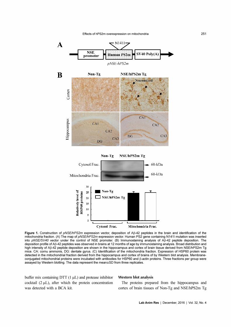
Figure 1. Construction of pNSE/hPS2m expression vector, deposition of Aβ-42 peptides in the brain and identification of themitochondria fraction. (A) The map of pNSE/hPS2m expression vector. Human PS2 gene containing N141I mutation was insertedinto pNSE/SV40 vector under the control of NSE promoter. (B) Immunostaining analysis of Aβ-42 peptide deposition. Thedeposition profile of Aβ-42 peptides was observed in brains at 12 months of age by immunostaining analysis. Broad distribution andhigh intensity of Aβ-42 peptide deposition are shown in the hippocampus and cortex of brain tissue derived from NSE/hPS2m Tgmice. CA: cornu ammonis, DG: dentate gyrus. (C) Identification of the mitochondria fraction. Expression of HSP60 protein wasdetected in the mitochondrial fraction derived from the hippocampus and cortex of brains of by Western blot analysis. Membrane-conjugated mitochondrial proteins were incubated with antibodies for HSP60 and β-actin proteins. Three fractions per group wereassayed by Western blotting. The data represent the mean±SD from three replicates.
252 Woo Bin Yun et al.
Lab Anim Res | December, 2016 | Vol. 32, No. 4
mice were separated by electrophoresis on 4-20% SDS-
PAGE gels for 2 hrs and then transferred to nitro-
cellulose membranes for 2 hrs at 40 V. Each membrane
was incubated separately with primary anti-PS1 FL
(1:1,000, Sigma-Aldrich Co., Missouri, USA), anti-PS1
CTF (1:1,000, Sigma-Aldrich Co.), anti-PS1 NTF
(1:1,000, Sigma-Aldrich Co.), anti-PS2 FL (1:1,000,
Sigma-Aldrich Co.), anti-PS2 CTF (1:1,000, Sigma-
Aldrich Co.), anti-PS2 NTF (1:1,000, Sigma-Aldrich
Co.), anti-NCT (1:1,000, Sigma-Aldrich Co.), anti-APH-
1 (1:1,000, Sigma-Aldrich Co.), anti-Pen-2 (1:2,000,
Calbiochem Inc.), anti-bAPP (1:1,000, Sigma-Aldrich
Co.), anti-C-99 (1:2,000, Calbiochem Inc.), anti-Aβ-42
(1:1,000, Calbiochem Inc.), anti-HSP60 (1:1,500,
Sigma-Aldrich Co.), and anti-β-actin (1:2,000, Sigma-
Aldrich Co.) antibodies overnight at 4oC. The membranes
were washed with washing buffer (137 mM NaCl, 2.7
mM KCl, 10 mM Na2HPO
4, 2 mM KH
2PO
4, and 0.05%
Tween 20) and incubated with horseradish peroxidase-
conjugated goat anti-rabbit IgG (Zymed) at 1:2,000
dilution at room temperature for 2 hrs. Finally, the blots
were developed using Chemiluminescence Reagent Plus
kit (Pfizer Inc., Gladstone, NJ, USA). The signal image
for each protein was acquired using the digital camera
(1.92 MP resolution) of the FluorChem® FC2 Imaging
system (Alpha Innotech Corporation, San Leandro, CA,
USA) and their density was semi-quantified using
AlphaView Program version 3.2.2 (Cell Biosciences,
Inc., Santa Clara, CA).
Assay of γ-secretase activity in mitochondrial fractions
Activity of γ-secretase in the mitochondrial fraction
of brains from Non-Tg and NSE/hPS2m Tg mice was
assayed using the γ-secretase activity assay procedure
and reagents in the γ-secretase Assay kit (R&D System
Inc., Minneapolis, MN, USA). Each mitochondrial fraction
prepared using a cytosol/mitochondria fractionation kit
was diluted with 1× Cell extraction buffer to yield a final
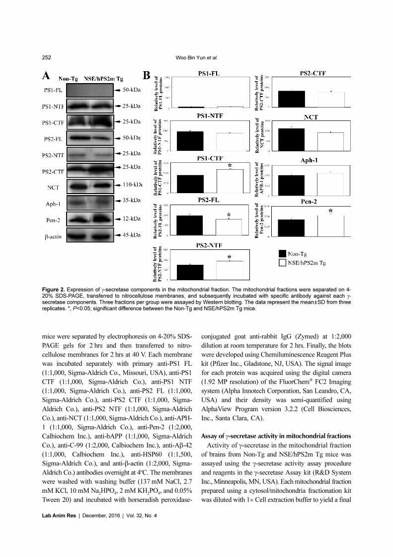
Figure 2. Expression of γ-secretase components in the mitochondrial fraction. The mitochondrial fractions were separated on 4-20% SDS-PAGE, transferred to nitrocellulose membranes, and subsequently incubated with specific antibody against each γ-secretase components. Three fractions per group were assayed by Western blotting. The data represent the mean±SD from threereplicates. *, P<0.05; significant difference between the Non-Tg and NSE/hPS2m Tg mice.
Effects of hPS2m overexpression on mitochondria 253
Lab Anim Res | December, 2016 | Vol. 32, No. 4
protein concentration of roughly 0.5-2.0 mg/mL. The
enzymatic reaction for secretase activity was carried out
using the microplate provided by manufacturer and was
read with a microplate fluorimeter. A total of 50 μL of
2× Reaction Buffer was added into each well containing
about 25-200 μg of total protein (50 μL). Then, 5 μL of
substrate was added to each well, and the plate was
incubated in the dark at 37oC for 1-2 hrs. Finally, the
absorbance of each well was read by a fluorescent
microplate reader using a light filter allowing for
EDANS excitation between wavelengths of 335-355
nm, after which emitted light was collected between
495-510 nm.
Statistical analysis
Statistical analyses were performed using SPSS
software version 10.10 (SPSS, Inc. Chicago, IL, USA).
One-way analysis and post hoc Tukey’s test of variance
was performed to identify significant differences between
Non-Tg and NSE/hPS2m Tg mice. All values are
presented as the mean±standard deviation (SD). A
P<0.05 was considered to indicate a statistically significant
difference.
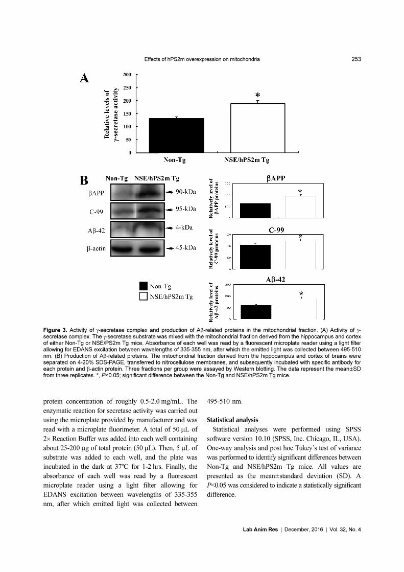
Figure 3. Activity of γ-secretase complex and production of Aβ-related proteins in the mitochondrial fraction. (A) Activity of γ-secretase complex. The γ-secretase substrate was mixed with the mitochondrial fraction derived from the hippocampus and cortexof either Non-Tg or NSE/PS2m Tg mice. Absorbance of each well was read by a fluorescent microplate reader using a light filterallowing for EDANS excitation between wavelengths of 335-355 nm, after which the emitted light was collected between 495-510nm. (B) Production of Aβ-related proteins. The mitochondrial fraction derived from the hippocampus and cortex of brains wereseparated on 4-20% SDS-PAGE, transferred to nitrocellulose membranes, and subsequently incubated with specific antibody foreach protein and β-actin protein. Three fractions per group were assayed by Western blotting. The data represent the mean±SDfrom three replicates. *, P<0.05; significant difference between the Non-Tg and NSE/hPS2m Tg mice.
254 Woo Bin Yun et al.
Lab Anim Res | December, 2016 | Vol. 32, No. 4
Results
Deposition of Aβ-42 peptides in brains of NSE/hPS2m
Tg mice
Firstly, to detect the localization and deposition of Aβ-
42 peptides induced by PS2 expression in brain tissues,
Aβ-42 peptide immunoreactivity was analyzed in the
hippocampus and cerebral cortex of brains using optical
microscopy. These regions were chosen since they are
primary target sites for Aβ peptides deposition and
neuronal loss induced by γ-secretase activity in AD
patients [10,11]. As shown in Figure 1B, immuno-
staining intensity was spread throughout neuronal cells
of the cerebral cortex in brain tissue from NSE/hPS2m
Tg mice. However, the intensity level in Non-Tg
littermates was slightly lower than that in NSE/hPS2m
Tg mice. Many cells in the CA1, CA2, and CA3 regions
were intensively immunostained with a dense line in the
hippocampus of NSE/hPS2m Tg brains, whereas these
regions showed low immunoreactivity in Non-Tg mice.
Therefore, these results suggest that overexpression of
the hPS2m gene could be attributed to the deposition of
Aβ-42 peptides in the hippocampus and cortex of NSE/
hPS2m Tg mice.
Identification of purity for mitochondrial fraction
Cytochrome c, Tim23, and HSP60 proteins are widely
used as mitochondrial markers in various biological
studies, whereas GM130, Syntaxin 13, and N-cadherin
are used as markers of the ER, cis-Golgi, early endosomes,
and plasma membrane [16,17]. To examine the purity of
the mitochondrial fraction, levels of HSP60 protein, as a
mitochondrial marker, were detected in the mitochondrial
fraction derived from brain tissue using Western blot
analysis. HSP60 protein was detected only in the
mitochondrial fraction and not in the cytosolic fraction,
and there was no difference on the expression of HSP60
protein between Non-Tg and NSE/hPS2m Tg mice
(Figure 1C). These results suggest that the methods
applied in this study are very useful tools to harvest the
mitochondrial fraction with high purity from brain
tissue.
Effects of hPS2m overexpression on component levels
of γ-secretase members
To investigate whether or not overexpression of
hPS2m protein is associated with component levels of
the γ-secretase complex in the mitichondria, protein levels
of these components were detected in the mitochondrial
fraction purified from brain tissue. For PS protein, which
is a main catalytic activator of the γ-secretase complex,
full-length PS1 protein was not detected in both mice,
whereas PS2 protein was detected at a high level.
Furthermore, PS2-FL protein was significantly down-
regulated in NSE/hPS2m Tg mice compared to Non-Tg
mice (P<0.032). On the other hand, protein level of PS1
and PS2 c-terminal fragment, a main functional protein,
was significantly higher in brain tissue of NSE/hPS2m
Tg mice compared to Non-Tg mice (P<0.015). Protein
levels of NCT and APH-1, a stabilization protein of γ-
secretase complex, were not significantly different between
Non-Tg and NSE/hPS2m Tg mice, although NCT
protein was slightly down-regulated (P<0.071). However,
protein level of Pen-2, an activator of the γ-secretase
complex, was significantly up-regulated in NSE/hPS2m
Tg mice compared to Non-Tg mice (P<0.043) (Figure
2). These results suggest that over-expression of hPS2m
protein could induce alterations of main component
protein and activator protein of the γ-secretase complex
but not stabilization-related protein in the mitochondria
of NSE/hPS2m Tg mice.
Effects of hPS2m overexpression on γ-secretase
activity
To determine whether or not alteration of γ-secretase
components induced by overexpression of hPS2m protein
can regulate γ-secretase activity in the mitochondria, γ-
secretase activity was measured in the mitochondrial
fraction purified from brain tissues of both mice. As
shown in Figure 3A, γ-secretase activity was significantly
elevated in the mitochondrial fraction of brain tissue
derived from NSE/hPS2m Tg mice compared to Non-Tg
mice (P<0.013). These results suggest that alteration of
γ-secretase components induced by overexpression of
hPS2m protein could up-regulate γ-secretase activity in
the mitochondrial fraction of NSE/hPS2m Tg mice.
Effect of hPS2m overexpression on production of Aβ-
42 peptides
To investigate the effect of γ-secretase changes on
production of Aβ-42 peptides in the mitochondria, the
peptide level of Aβ-42 was analyzed in the mitochondrial
fraction of Non-Tg and NSE/hPS2m Tg mice using a
specific antibody. A high level of Aβ-42 peptides was
successfully detected in the mitochondrial fraction of
NSE/hPS2m Tg mice. Protein levels of bAPP and C-99,
Effects of hPS2m overexpression on mitochondria 255
Lab Anim Res | December, 2016 | Vol. 32, No. 4
which is a C-terminal fragment of APP involving Aβ-42
peptides, were significantly up-regulated in NSE/hPS2m
Tg mice compared to Non-Tg mice (P<0.039) (Figure
3B). Therefore, our results suggest that elevation of γ-
secretase activity induced by hPS2m overexpression
could stimulate production of Aβ-42 peptides in the
mitochondrial fraction.
Discussion
Mitochondria play an important role in the apoptosis
pathway, which is an essential process required for the
development and maintenance of normal tissues [11].
Various apoptotic stimuli originating from the extra-
cellular environment cause increased permeability of the
outer mitochondrial membrane, resulting in release of
mitochondrial apoptogenic proteins such as cytochrome
c and Smac/Diablo into the cytosol [18]. This increased
permeability is negatively and positively regulated by
anti-apoptotic and pro-apoptotic members of the Bcl-2
family, respectively [19,20]. Especially, previous reports
have demonstrated the potential involvement of Aβ
peptides and γ-secretase activity in the induction or
regulation of apoptotic neuronal death [17,21,22].
Therefore, in this study, mitochondria were examined as
a trigger of abnormal onset of neuronal cell death
induced by mPS2 in AD.
Several reports have suggested that all γ-secretase
components are located in the mitochondria. Especially,
the role and importance of γ-secretase activity to
mitochondria were provided from the results that NCT
together with APH-1, Pen-2, and PS1 form a high
molecular weight complex located in mitochondria [17].
In this study, to investigate the effects of mutant PS2
overexpression on the γ-secretasecomplex in mitochondria,
expression levels of γ-secretase components as well as
activity of γ-secretase were measured in the mitochondrial
extract derived from brain tissue of NSE/hPS2m Tg
mice. C-terminal fragments of PS1 and PS2 protein were
significantly up-regulated in the mitochondria of NSE/
hPS2m Tg mice compared to non-transgenic mice,
whereas full-length NTF was unaltered in NSE/hPS2m
Tg mice. Furthermore, the level of Pen-2 protein, as an
activator of γ-secretase, was higher in NSE/hPS2m Tg
mice than in Non-Tg mice. However, there was no
difference in NCT or APH-1 protein level between the
two mice. These results suggest that mutant PS2 could
induce core and activator protein expression of γ-
secretase in mitochondria derived from brain tissue of
NSE/hPS2m Tg mice.
Activity of γ-secretase was detected in mitochondria
using β-APP from BD8 cells or recombinant C100-Flag
as substrates. Formation of AICD was abolished in the
presence of well characterized γ-secretase inhibitors [17].
These studies demonstrated that γ-secretase complexes
located in mitochondria are active and can cleave β-APP.
In this study, the activity level of γ-secretase was detected
in the mitochondrial fraction derived from brain tissue of
NSE/hPS2m Tg mice. mPS2 overexpressed in mitochondria
could induce up-regulation of γ-secretase activity in
brains of NSE/hPS2m Tg mice compared to Non-Tg
mice.
Previous studies have shown that energy depletion and
oxidative stress can induce amyloidogenic changes in
APP processing [23-25]. These results suggested that Aβ
production is potentially linked to mitochondrial dys-
function and oxidative stress [26]. Intracellular Aβ-42
peptides produced by APP processing partially colocalize
with APP and appear predominantly in the golgi complex
and endosomal vesicles. Furthermore, these products
such as Aβ-42 peptides, APP, and C-terminal fragments
have been shown to accumulate in a detergent-insoluble
form in APP-overexpressing cells [27]. Our study showed
that protein levels of βAPP, C-99, and Aβ-42 were
significantly higher than in NSE/hPS2m Tg mice than in
Non-Tg mice. These results suggest that overexpression
of the hPS2m gene may induce accumulation of Aβ-
related byproducts in hPS2m-overexpressing cells derived
from NSE/hPS2m Tg mice similar to APP-overexpressing
cells.
Taken together, these results suggest that overexpression
of mPS2 gene in mitochondria of brain tissue (hippo-
campus and cortex) derived from NSE/hPS2m Tg mice
may induce changes in the component expression and
activity of the γ-secretase complex. A high level of
activity may have up-regulated production of Aβ-related
products in NSE/hPS2m Tg mice. These results suggest
that mutant PS2 plays a critical role in the progression
of AD pathology.
Acknowledgments
This research was supported by a 2-Year Research
Grant of Pusan National University.
Conflict of interests The authors declare that there is
256 Woo Bin Yun et al.
Lab Anim Res | December, 2016 | Vol. 32, No. 4
no financial conflict of interests to publish these results.
References
1. Weihofen A, Martoglio B. Intramembrane-cleaving proteases:controlled liberation of proteins and bioactive peptides. TrendsCell Biol 2003; 13(2): 71-78.
2. De Strooper B. Aph-1, Pen-2, and Nicastrin with Presenilingenerate an active gamma-secretase complex. Neuron 2003;38(1): 9-12.
3. Kimberly WT, LaVoie MJ, Ostaszewski BL, Ye W, Wolfe MS,Selkoe DJ. Gamma-secretase is a membrane protein complexcomprised of presenilin, nicastrin, Aph-1, and Pen-2. Proc NatlAcad Sci U S A 2003; 100(11): 6382-6387.
4. Yu G, Nishimura M, Arawaka S, Levitan D, Zhang L, Tandon A,Song YQ, Rogaeva E, Chen F, Kawarai T, Supala A, Levesque L,Yu H, Yang DS, Holmes E, Milman P, Liang Y, Zhang DM, XuDH, Sato C, Rogaev E, Smith M, Janus C, Zhang Y, Aebersold R,Farrer LS, Sorbi S, Bruni A, Fraser P, St George-Hyslop P.Nicastrin modulates presenilin-mediated notch/glp-1 signaltransduction and betaAPP processing. Nature 2000; 407(6800):48-54.
5. Lee SF, Shah S, Li H, Yu C, Han W, Yu G. Mammalian APH-1interacts with presenilin and nicastrin and is required forintramembrane proteolysis of amyloid-beta precursor protein andNotch. J Biol Chem 2002; 277(47): 45013-45019.
6. Steiner H, Winkler E, Edbauer D, Prokop S, Basset G, YamasakiA, Kostka M, Haass C. PEN-2 is an integral component of thegamma-secretase complex required for coordinated expression ofpresenilin and nicastrin. J Biol Chem 2002; 277(42): 39062-39065.
7. Suh YH, Checler F. Amyloid precursor protein, presenilins, andalpha-synuclein: molecular pathogenesis and pharmacologicalapplications in Alzheimer's disease. Pharmacol Rev 2002; 54(3):469-525.
8. Teng FY, Tang BL. Widespread gamma-secretase activity in thecell, but do we need it at the mitochondria?. Biochem BiophysRes Commun 2005; 328(1): 1-5.
9. St George-Hyslop PH. Molecular genetics of Alzheimer's disease.Biol Psychiatry 2000; 47(3): 183-199.
10. Thinakaran G, Harris CL, Ratovitski T, Davenport F, Slunt HH,Price DL, Borchelt DR, Sisodia SS. Evidence that levels ofpresenilins (PS1 and PS2) are coordinately regulated bycompetition for limiting cellular factors. J Biol Chem 1997;272(45): 28415-28422.
11. Wang HQ, Nakaya Y, Du Z, Yamane T, Shirane M, Kudo T,Takeda M, Takebayashi K, Noda Y, Nakayama KI, Nishimura M.Interaction of presenilins with FKBP38 promotes apoptosis byreducing mitochondrial Bcl-2. Hum Mol Genet 2005; 14(13):1889-1902.
12. Hwang DY, Chae KR, Kang TS, Hwang JH, Lim CH, Kang HK,Goo JS, Lee MR, Lim HJ, Min SH, Cho JY, Hong JT, Song CW,Paik SG, Cho JS, Kim YK. Alterations in behavior, amyloid beta-42, caspase-3, and Cox-2 in mutant PS2 transgenic mouse modelof Alzheimer's disease. FASEB J 2002; 16(8): 805-813.
13. Janicki S, Monteiro MJ. Increased apoptosis arising fromincreased expression of the Alzheimer's disease-associatedpresenilin-2 mutation (N141I). J Cell Biol 1997; 139(2): 485-495.
14. Alves da Costa C, Paitel E, Mattson MP, Amson R, Telerman A,Ancolio K, Checler F. Wild-type and mutated presenilins 2 triggerp53-dependent apoptosis and down-regulate presenilin 1expression in HEK293 human cells and in murine neurons. ProcNatl Acad Sci U S A 2002; 99(6): 4043-4048.
15. van de Craen M, de Jonghe C, van den Brande I, Declercq W, vanGassen G, van Criekinge W, Vanderhoeven I, Fiers W, vanBroeckhoven C, Hendriks L, Vandenabeele P. Identification ofcaspases that cleave presenilin-1 and presenilin-2. Five presenilin-1 (PS1) mutations do not alter the sensitivity of PS1 to caspases.FEBS Lett 1999; 445(1): 149-154.
16. Gandhi S, Muqit MM, Stanyer L, Healy DG, Abou-Sleiman PM,Hargreaves I, Heales S, Ganguly M, Parsons L, Lees AJ,Latchman DS, Holton JL, Wood NW, Revesz T. PINK1 protein innormal human brain and Parkinson's disease. Brain 2006; 129:1720-1731.
17. Hansson CA, Frykman S, Farmery MR, Tjernberg LO, NilsberthC, Pursglove SE, Ito A, Winblad B, Cowburn RF, Thyberg J,Ankarcrona M. Nicastrin, presenilin, APH-1, and PEN-2 formactive gamma-secretase complexes in mitochondria. J Biol Chem2004; 279(49): 51654-51660.
18. Newmeyer DD, Ferguson-Miller S. Mitochondria: releasingpower for life and unleashing the machineries of death. Cell 2003;112(4): 481-490.
19. Tsujimoto Y. Cell death regulation by the Bcl-2 protein family inthe mitochondria. J Cell Physiol 2003; 195(2): 158-167.
20. Green DR, Kroemer G. The pathophysiology of mitochondrial celldeath. Science 2004; 305(5684): 626-629.
21. Yuan J, Yankner BA. Apoptosis in the nervous system. Nature2000; 407(6805): 802-809.
22. Dickson DW. Apoptotic mechanisms in Alzheimer neurofibrillarydegeneration: cause or effect?. J Clin Invest 2004; 114(1): 23-27.
23. Gabuzda D, Busciglio J, Chen LB, Matsudaira P, Yankner BA.Inhibition of energy metabolism alters the processing of amyloidprecursor protein and induces a potentially amyloidogenicderivative. J Biol Chem 1994; 269(18): 13623-13628.
24. Gasparini L, Racchi M, Benussi L, Curti D, Binetti G, BianchettiA, Trabucchi M, Govoni S. Effect of energy shortage andoxidative stress on amyloid precursor protein metabolism in COScells. Neurosci Lett 1997; 231(2): 113-117.
25. Misonou H, Morishima-Kawashima M, Ihara Y. Oxidative stressinduces intracellular accumulation of amyloid beta-protein(Abeta) in human neuroblastoma cells. Biochemistry 2000;39(23): 6951-6959.
26. Busciglio J, Pelsman A, Wong C, Pigino G, Yuan M, Mori H,Yankner BA. Altered metabolism of the amyloid beta precursorprotein is associated with mitochondrial dysfunction in Down'ssyndrome. Neuron 2002; 33(5): 677-688.
27. Yang AJ, Chandswangbhuvana D, Shu T, Henschen A, Glabe CG.Intracellular accumulation of insoluble, newly synthesized abetan-42 in amyloid precursor protein-transfected cells that have beentreated with Abeta1-42. J Biol Chem 1999; 274(29): 20650-20656.

![Crimp Information Sheet - Farnell element14 · 2018. 10. 3. · CCW [mm] Tol CCW [mm] ICH [mm] Tol ICH [mm] ICW [mm] Tol ICW [mm] 10070,50/15366060 2,15 80 1,10 0,05 1,80 0,10 3,50](https://static.fdocument.org/doc/165x107/6119fa6ed77d58264702c930/crimp-information-sheet-farnell-2018-10-3-ccw-mm-tol-ccw-mm-ich-mm.jpg)