Original Article Regulation of WNT/β-catenin signaling by ... · phosphate synthetase 2 (CPSII),...
11
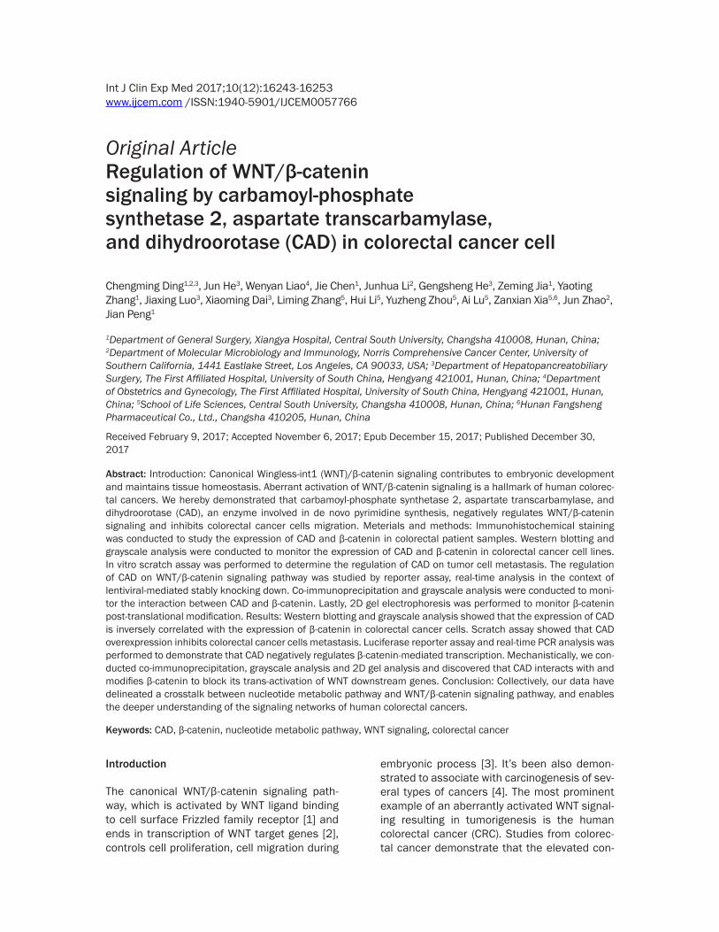
Int J Clin Exp Med 2017;10(12):16243-16253 www.ijcem.com /ISSN:1940-5901/IJCEM0057766 Original Article Regulation of WNT/β-catenin signaling by carbamoyl-phosphate synthetase 2, aspartate transcarbamylase, and dihydroorotase (CAD) in colorectal cancer cell Chengming Ding 1,2,3 , Jun He 3 , Wenyan Liao 4 , Jie Chen 1 , Junhua Li 2 , Gengsheng He 3 , Zeming Jia 1 , Yaoting Zhang 1 , Jiaxing Luo 3 , Xiaoming Dai 3 , Liming Zhang 5 , Hui Li 5 , Yuzheng Zhou 5 , Ai Lu 5 , Zanxian Xia 5,6 , Jun Zhao 2 , Jian Peng 1 1 Department of General Surgery, Xiangya Hospital, Central South University, Changsha 410008, Hunan, China; 2 Department of Molecular Microbiology and Immunology, Norris Comprehensive Cancer Center, University of Southern California, 1441 Eastlake Street, Los Angeles, CA 90033, USA; 3 Department of Hepatopancreatobiliary Surgery, The First Affiliated Hospital, University of South China, Hengyang 421001, Hunan, China; 4 Department of Obstetrics and Gynecology, The First Affiliated Hospital, University of South China, Hengyang 421001, Hunan, China; 5 School of Life Sciences, Central South University, Changsha 410008, Hunan, China; 6 Hunan Fangsheng Pharmaceutical Co., Ltd., Changsha 410205, Hunan, China Received February 9, 2017; Accepted November 6, 2017; Epub December 15, 2017; Published December 30, 2017 Abstract: Introduction: Canonical Wingless-int1 (WNT)/β-catenin signaling contributes to embryonic development and maintains tissue homeostasis. Aberrant activation of WNT/β-catenin signaling is a hallmark of human colorec- tal cancers. We hereby demonstrated that carbamoyl-phosphate synthetase 2, aspartate transcarbamylase, and dihydroorotase (CAD), an enzyme involved in de novo pyrimidine synthesis, negatively regulates WNT/β-catenin signaling and inhibits colorectal cancer cells migration. Meterials and methods: Immunohistochemical staining was conducted to study the expression of CAD and β-catenin in colorectal patient samples. Western blotting and grayscale analysis were conducted to monitor the expression of CAD and β-catenin in colorectal cancer cell lines. In vitro scratch assay was performed to determine the regulation of CAD on tumor cell metastasis. The regulation of CAD on WNT/β-catenin signaling pathway was studied by reporter assay, real-time analysis in the context of lentiviral-mediated stably knocking down. Co-immunoprecipitation and grayscale analysis were conducted to moni- tor the interaction between CAD and β-catenin. Lastly, 2D gel electrophoresis was performed to monitor β-catenin post-translational modification. Results: Western blotting and grayscale analysis showed that the expression of CAD is inversely correlated with the expression of β-catenin in colorectal cancer cells. Scratch assay showed that CAD overexpression inhibits colorectal cancer cells metastasis. Luciferase reporter assay and real-time PCR analysis was performed to demonstrate that CAD negatively regulates β-catenin-mediated transcription. Mechanistically, we con- ducted co-immunoprecipitation, grayscale analysis and 2D gel analysis and discovered that CAD interacts with and modifies β-catenin to block its trans-activation of WNT downstream genes. Conclusion: Collectively, our data have delineated a crosstalk between nucleotide metabolic pathway and WNT/β-catenin signaling pathway, and enables the deeper understanding of the signaling networks of human colorectal cancers. Keywords: CAD, β-catenin, nucleotide metabolic pathway, WNT signaling, colorectal cancer Introduction The canonical WNT/β-catenin signaling path- way, which is activated by WNT ligand binding to cell surface Frizzled family receptor [1] and ends in transcription of WNT target genes [2], controls cell proliferation, cell migration during embryonic process [3]. It’s been also demon- strated to associate with carcinogenesis of sev- eral types of cancers [4]. The most prominent example of an aberrantly activated WNT signal- ing resulting in tumorigenesis is the human colorectal cancer (CRC). Studies from colorec- tal cancer demonstrate that the elevated con-
-
Upload
nguyenhanh -
Category
Documents
-
view
218 -
download
0
Transcript of Original Article Regulation of WNT/β-catenin signaling by ... · phosphate synthetase 2 (CPSII),...
Int J Clin Exp Med 2017;10(12):16243-16253www.ijcem.com /ISSN:1940-5901/IJCEM0057766
Original ArticleRegulation of WNT/β-catenin signaling by carbamoyl-phosphate synthetase 2, aspartate transcarbamylase, and dihydroorotase (CAD) in colorectal cancer cell
Chengming Ding1,2,3, Jun He3, Wenyan Liao4, Jie Chen1, Junhua Li2, Gengsheng He3, Zeming Jia1, Yaoting Zhang1, Jiaxing Luo3, Xiaoming Dai3, Liming Zhang5, Hui Li5, Yuzheng Zhou5, Ai Lu5, Zanxian Xia5,6, Jun Zhao2, Jian Peng1
1Department of General Surgery, Xiangya Hospital, Central South University, Changsha 410008, Hunan, China; 2Department of Molecular Microbiology and Immunology, Norris Comprehensive Cancer Center, University of Southern California, 1441 Eastlake Street, Los Angeles, CA 90033, USA; 3Department of Hepatopancreatobiliary Surgery, The First Affiliated Hospital, University of South China, Hengyang 421001, Hunan, China; 4Department of Obstetrics and Gynecology, The First Affiliated Hospital, University of South China, Hengyang 421001, Hunan, China; 5School of Life Sciences, Central South University, Changsha 410008, Hunan, China; 6Hunan Fangsheng Pharmaceutical Co., Ltd., Changsha 410205, Hunan, China
Received February 9, 2017; Accepted November 6, 2017; Epub December 15, 2017; Published December 30, 2017
Abstract: Introduction: Canonical Wingless-int1 (WNT)/β-catenin signaling contributes to embryonic development and maintains tissue homeostasis. Aberrant activation of WNT/β-catenin signaling is a hallmark of human colorec-tal cancers. We hereby demonstrated that carbamoyl-phosphate synthetase 2, aspartate transcarbamylase, and dihydroorotase (CAD), an enzyme involved in de novo pyrimidine synthesis, negatively regulates WNT/β-catenin signaling and inhibits colorectal cancer cells migration. Meterials and methods: Immunohistochemical staining was conducted to study the expression of CAD and β-catenin in colorectal patient samples. Western blotting and grayscale analysis were conducted to monitor the expression of CAD and β-catenin in colorectal cancer cell lines. In vitro scratch assay was performed to determine the regulation of CAD on tumor cell metastasis. The regulation of CAD on WNT/β-catenin signaling pathway was studied by reporter assay, real-time analysis in the context of lentiviral-mediated stably knocking down. Co-immunoprecipitation and grayscale analysis were conducted to moni-tor the interaction between CAD and β-catenin. Lastly, 2D gel electrophoresis was performed to monitor β-catenin post-translational modification. Results: Western blotting and grayscale analysis showed that the expression of CAD is inversely correlated with the expression of β-catenin in colorectal cancer cells. Scratch assay showed that CAD overexpression inhibits colorectal cancer cells metastasis. Luciferase reporter assay and real-time PCR analysis was performed to demonstrate that CAD negatively regulates β-catenin-mediated transcription. Mechanistically, we con-ducted co-immunoprecipitation, grayscale analysis and 2D gel analysis and discovered that CAD interacts with and modifies β-catenin to block its trans-activation of WNT downstream genes. Conclusion: Collectively, our data have delineated a crosstalk between nucleotide metabolic pathway and WNT/β-catenin signaling pathway, and enables the deeper understanding of the signaling networks of human colorectal cancers.
Keywords: CAD, β-catenin, nucleotide metabolic pathway, WNT signaling, colorectal cancer
Introduction
The canonical WNT/β-catenin signaling path-way, which is activated by WNT ligand binding to cell surface Frizzled family receptor [1] and ends in transcription of WNT target genes [2], controls cell proliferation, cell migration during
embryonic process [3]. It’s been also demon-strated to associate with carcinogenesis of sev-eral types of cancers [4]. The most prominent example of an aberrantly activated WNT signal-ing resulting in tumorigenesis is the human colorectal cancer (CRC). Studies from colorec-tal cancer demonstrate that the elevated con-
CAD inhibits β-catenin
16244 Int J Clin Exp Med 2017;10(12):16243-16253
centration of WNT signaling molecule β-catenin, caused by mutations in adenomatous polypo-sis coli (APC), accounts for the constitutive overexpression of WNT target genes [5-7]. APC is a negative regulator of β-catenin that, togeth-er with Axin, serves as a scaffold to assemble the β-catenin destruction complex, leading to the sequential events of β-catenin phosphory-lation, ubiquitination and proteasomal degra-dation [8]. As a result, in resting cells, the cyto-plasmic pool of β-catenin is maintained at low concentration. Upon stimulation with WNT ligand, phosphorylation of APC and Axin is reversed by protein phosphatase 2A [9], result-ing in disassembly of the destruction complex and stabilization of β-catenin. As a conse-quence, accumulated β-catenin translocates into the nucleus and transactivates WNT target genes [10]. Apart from APC, other mutations in components of the β-catenin destruction com-plex and downstream signaling molecules, which are associated with various cancers, often result in similar aberrant stabilization of β-catenin and inappropriate expression of WNT target genes in the absence of stimulation [11, 12]. Besides APC, kinases belonging to distinct pathways are reported to modify β-catenin, leading to upregulated or downregulated WNT signaling [13]. In this study, we uncover a novel regulation of β-catenin by a nucleotide meta-bolic enzyme, CAD.
CAD is an acronym derived from carbamoyl-phosphate synthetase 2 (CPSII), aspartate transcarbamylase (ATC), and dihydroorotase (DHO). It is a 250-kD protein consisting of three functionally-independent enzymes which cata-lyze the first three steps of de novo pyrimidine synthesis [14, 15]. CPSII, the first and rate limit-ing enzyme of CAD, catalyzes the formation of carbomyl-phosphate from ammonia, bicarbon-ate and ATP [16]. The ammonia group is pro-vided by glutamine via deamidation by GLNase domain (GAT domain) of CPSII. ATC, as the sec-ond step, catalyzes the formation the carbomyl-aspartate based on aspartate and carbomyl phosphate. Finally, DHO is responsible for the formation of the pyrimidine ring. CAD is potently activated during S phase of the cell cycle when DNA undergoes rapid replication [17], which requires sufficient amount of nucleotides as building blocks. The enzymatic activity of CAD undergoes allosteric regulation. The final prod-uct of the de novo synthesis, UTP, has a nega-tive feedback on CAD. On the contrary, Phosphoribosyl 1-pyrophosphate (PRPP), an
intermediate reactant downstream of CAD, pro-motes its enzymatic activity [18].
Several previous findings suggested that CAD is highly expressed and associated with the development/recurrence of numerous tumors [19-22]. Traditional views regarding the role of CAD in tumorigenesis is that it promotes tumor growth by supporting nucleotide biosynthesis. In addition, recent studies reported that, as a nucleotide metabolic enzyme, CAD also regu-lates a number of key signaling molecules in distinctly related oncogenic pathways. For ex- ample, CAD negatively regulates NOD2 activa-tion by its ligand MDP in a GAT-activity depen-dent manner and is a potential therapeutic tar-get for human crohn’s disease (CD) [23]. CAD also interacts with important proteins in NF-κB signaling axis, including NEMO and RelA [24]. Thus, the biological roles of CAD to tumor cell proliferation, cell migration and intracellular signaling is likely beyond biosynthesis,, which remains poorly understood. In this study, we identified the interaction of CAD with β-catenin, which accounts for its negative regulation on WNT/β-catenin signaling pathway and CRC cell migration. This study further expands our understanding on the crosstalk and regulation between metabolic pathways and WNT/β-ca- tenin signaling pathway.
Materials and methods
Cell lines
Human embryonic kidney 293T cells (HEK293T, ATCC, USA) and HCT116 (ATCC, USA) were cul-tured in Dulbecco’s modified Eagle’s medium (DMEM, Corning, USA) supplemented with 10% heat-inactivated fetal bovine serum (FBS; HyClone, USA), penicillin (100 U/mL) and strep-tomycin (100 μg/mL) (Corning, USA). Colorectal cancer cell lines, including Sw480, Sw620, DLD1, Lim2045, RKO, HT29, Colo205, Lovo-p1, DiFi, were gifts from Dr. Chengyu Liang (USC, originally from ATCC, USA). Cells were incubat-ed in a humidified incubator at 37°C and 5% CO2.
Plasmids
Luciferase reporter plasmids for the TOPflash and FOPflash promoter were kindly provided by Dr. Chengyu Liang (USC, originally from Addgene, USA) [25, 26], mammalian expres-sion plasmids for CAD (Thermo Scientific, USA), IKKε (Invitrogen, USA), P65 (Invitrogen, USA) and β-catenin (Addgene, USA) were described
CAD inhibits β-catenin
16245 Int J Clin Exp Med 2017;10(12):16243-16253
previously [27]. The non-silencing (control) shRNA, shRNA against CAD and PFAS were pur-chased from Thermo Scientific, USA.
Antibodies and reagents
Antibodies against GST (Z-5, Santa Cruz Bio- technology, USA), FLAG (M2, Sigma, USA), CAD (A301-374A, Bethyl, USA), β-catenin (H102, Santa Cruz Biotechnology, USA) and β-actin (Ab8226, Abcam, USA) were purchased from the indicated suppliers. Glutathione-conjugated agarose (17075601) and Immobiline Drystrip Gels (17-6001) were purchased from GE health-care, USA. Flag antibody-conjugated agarose (A2220) was purchased from Sigma, USA. 2- iodoacetamide (1632109) was purchased from Bio-rad, USA.
DNA transfection
For plasmid transfection in HEK293T cells, cal-cium phosphate transfection method was applied. HEK293T cells were plated at around 50%-60% confluence. For plasmid transfection in HCT116, Polyethylenimine (PEI, Sigma, USA) method was applied.
Lentivirus-mediated stable cell line construc-tion
Stable cell line was constructed as previously described [28]. Briefly, HEK293T cells were transfected with the packaging plasmids VSV-G and DR8.9 and the lentiviral shRNA plasmids. At 48 h post transfection, supernatant was har-vested and filtered (and concentrated by cen-trifugation if necessary). HEK293T cells or HCT116 cells were infected with the superna-tant in the presence of polybrene (8 μg/ml) with centrifugation at 1800 rpm for 45 minutes. Cells were selected at 48 h post infection and maintained in 10% FBS DMEM supplemented with puromycin (1~2 μg/ml).
Luciferase reporter assay
HEK293T cells, seeded in 24-well plates (~50% cell density), were transfected with TOPflash or FOPflash reporter plasmid cocktail (50 ng of luciferase reporter plasmid and 100 ng of β-galactosidase control vector) and β-catenin by calcium phosphate precipitation. Whole cell lysates were used to determine the activity of
firefly luciferase and β-galactosidase by a microplate reader (FLUOstar Omega, Germany).
Co-immunoprecipitation (Co-IP) and immunob-lotting
For Co-IP using exogenous protein, HEK293T cells were transfected with indicated expres-sion plasmids for 48 h. Whole cell lysates were prepared with NP40 buffer (50 mM Tris-HCl, pH 7.4, 150 mM NaCl, 1% NP-40, 5 mM EDTA) sup-plemented with 20 mM β-glycerophosphate and 1 mM sodium orthovanadate. Whole cell lysates were sonicated, centrifuged and pre-cleared with protein A/G agarose for 1 h. Pre-cleared samples were then incubated with anti-body/glutathione-conjugated agarose for 4 h at 4°C. The agarose beads were washed exten-sively and samples were eluted by boiling at 95°C for 10 min. Precipitated proteins were analyzed by SDS gel electrophoresis and immunoblotting.
All immunoblottings were performed using the indicated primary antibodies (1:1000 dilution) and IRDye800-conjugated secondary antibod-ies (1:10,000 dilution, Licor, USA). Proteins were visualized by Odyssey infrared imaging system (Licor, USA).
Quantitative real-time PCR (qRT-PCR)
Quantitative Real-time PCR was performed as previously described [29]. Cells were infected or treated with viruses or agents for indicated time period. Total RNA was extracted using TRIzol reagent (Invitrogen, USA). Complemen- tary cDNA was synthesized from DNase I-treated total RNA using reverse transcriptase (Invitrogen, USA). cDNA was diluted and qRT-PCR was performed using SYBR Green Master Mix (Applied Biosystems, USA) by real-time PCR instrument (Applied Biosystems, USA). Relative mRNA expression for each target gene was cal-culated by the 2-ΔΔCt method using β-Actin as an internal control. The sequences of qRT-PCR primers are as follows:
Human β-actin Forward 5’-CTGGCACCCAGCACAATG-3’Reverse 5’-GCCGATCCACACGGAGTACT-3’
Human MMP2 Forward 5’-TTGGACTGCCCCAGACAGGT-3’Reverse 5’-CACTTGGGCTTGCGAGGGAA-3’
Human C-Myc Forward 5’-GCCTCAGAGTGCATCGACCC-3’Reverse 5’-GCGGTGTCTCCTCATGGAGC-3’
CAD inhibits β-catenin
16246 Int J Clin Exp Med 2017;10(12):16243-16253
Two-dimensional gel electrophoresis
Cells (1×10⁶) were lysed in 150 µl rehydration buffer (6 M Urea, 2 M Thio-urea, 2% CHAPS, 0.5% IPG Buffer, 0.002% bromophenol blue) by one pulse of sonication and whole cell lysates were centrifuged at 20,000 g for 15 min. Supernatants were loaded to IEF strips for focusing with a program comprising: 20 V, 10 h (rehydration); 100 V, 1 h; 500 V, 1 h; 1000 V, 1 h; 2000 V, 1 h; 4000 V, 1 h; 5000 V, 4 h. After IEF, strips were incubated with SDS equilibra-tion buffer (50 mM Tris-HCl [pH 8.8], 6 M urea, 30% glycerol, 2% SDS, 0.001% Bromophenol Blue) containing 10 mg/ml DTT for 15 min and then SDS equilibration buffer containing 2-iodo-acetamide for 15 min. Strips were washed with SDS-PAGE buffer, resolved by SDS-PAGE, and analyzed by immunoblotting.
In vitro scratch wound assay
HCT116 cells were transfected with a plasmid containing CAD by Polyethylenimine (PEI). 48 hours later, the monolayer of cells was scr- atched with a pipette tip to form a wound. Cells were imaged at time 0 and 12 hours post scratching.
Immunohistochemistry analysis
Colorectal cancer patient tissues were fixed in 10% neutral buffered formalin (Sigma) over-night at room temperature. Tissues were then dehydrated, embedded in paraffin, and cut into 3-μm sections. After antigen retrieval, tissue sections were subject to immunohistochemical staining as indicated below with control anti-bodies or antibodies against CAD and β-catenin. Colorectal cancer patients with informed con-sent donated their colorectal cancer tissues for the study and ethical approval was given by the medical ethics committee of Xiangya Hospital of Central South University with the following reference number: 201410063.
All tumor tissues section staining was per-formed as according to the manufacturer’s pro-tocol. The CAD and β-catenin immunohisto-chemical analysis kits were used (Thermo Scientific, USA). Each tissue section was inde-pendently assessed by two pathologists with-out prior knowledge of patient data. The immu-nohistochemical staining results were assigned mean score, considered both the intensity of
staining and the proportion of cells with an unequivocal positive re-action. Positive reac-tions were defined as those showing brown sig-nals in the cell cytoplasm and cytomembrane. The staining index (values, 0-12) was deter-mined by multiplying the score for staining intensity with the score for proportion of posi-tive cells. The staining intensity was scored as follows: 0 = negative, 1 = weak, 2 = moderate, and 3 = strong. The proportion of positive cells was defined as follows: 0 = if less than 5% cells were stained, 1 = if 5% to 25% cells were stained; 2 = if 26% to 50% cells were stained; 3 = if 51% to 75% cells were stained; and 4 = if more than 75% cells were stained. When the staining was heterogeneous, we scored it as follows: each component was scored indepen-dently and summed for the results. For exam-ple, a specimen containing 75% cells with mod-erate intensity (3×2 = 6) and the other 25% cells with weak intensity (1×1 = 1), so we received a final score 7, which was from (6+1). For statistical analysis, scores of 0 to 7 were considered low expression and scores of 8 to 12 were considered high expression.
Statistical analysis
The data were shown as the mean ± SD, and statistical analysis was performed by unpaired two-tailed Student’s t-test of SPSS. One-way ANOVA was applied in the comparison of multi-ple groups. When homogeneity of variance, the LSD and SNK methods were used, when heteroskedasticity, Tamhane’s T2 or Dunnett’s T3 method was applied. A p-value less than 0.05 was considered statistically significant, and a p-value <0.01 was regarded as strong significant.
Grayscale analysis: Mean grayscale values of each band for western blotting results were obtained by using the image editing program: ImageJ (version 1.48).
Results
CAD was highly expressed and inversely corre-lated with the expression of β-catenin in Colo- rectal Cancer (CRC). When screening for poten-tial oncogenes or tumor suppressors of colorec-tal cancers, we identified CAD as one of the potential candidates. Immunohistochemistry staining demonstrated that the expression of CAD protein in CRC tissues was higher than

CAD inhibits β-catenin
16247 Int J Clin Exp Med 2017;10(12):16243-16253
that in the normal tissues (Figure 1A), which was also supported by a recent study showing CAD’s role in negative regulation of NOD2 sig-naling in colorectal cancer cell lines [23]. We determined the expression of CAD and β-ca- tenin in a series of colorectal cancer cell lines. As shown in Figure 1B and 1C, the expression of CAD might be inversely correlated with the expression of β-catenin in the colorectal can-cer cell lines of DLD1, Lim2405, and RKO. These data prompted us to study the biological relevance of CAD in colorectal tumorigenesis.
CAD inhibited CRC cells proliferation and migration
To investigate the physiological role of CAD in colorectal cancer development, we first sought out to study its role in the proliferation of tumor
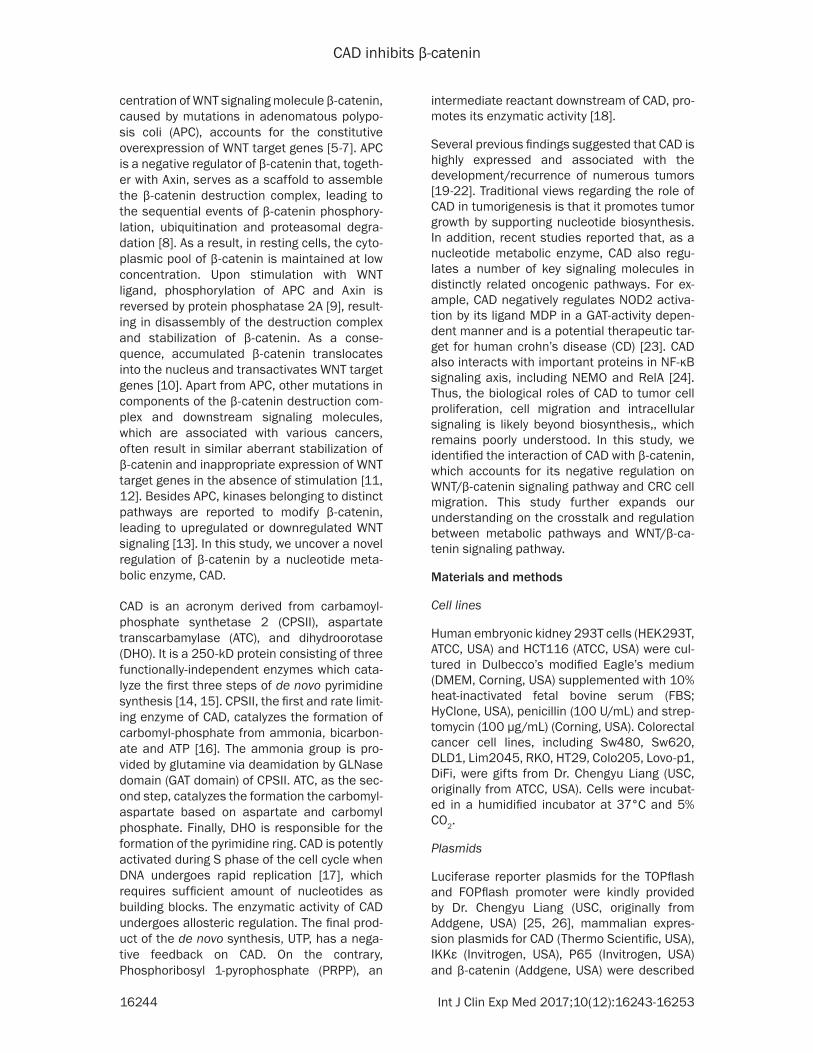
cells. Our results showed that overexpression of CAD could inhibit the proliferation of HCT116 (data not shown). We then went on to test the hypothesis that CAD could play a role in regulat-ing tumor cell metastasis, which is another WNT/β-catenin signaling-manipulated pheno-type. Over-expression of CAD in HCT116 cells indeed inhibited cell migration in the in vitro scratch assay, indicating that it plays an inhibi-tory role in the process of colorectal cancer cell metastasis (Figure 2A, 2B).
CAD negatively regulated β-catenin-mediated WNT signaling transduction
β-catenin plays important role in colorectal cancer metastasis. On one hand, it localizes in adherens junctions which regulate cytoskele-ton organization and cell-cell adhesion. On the
Figure 1. CAD was highly expressed and inversely correlated with the expression of β-catenin in colorectal cancer. (A) Immunohistochemistry staining of CAD and β-catenin in colorectal cancer patient samples. (B) Whole cell lysates were extracted from different colorectal cancer cell lines and analyzed by immunoblotting with indicated antibod-ies. (C) The bands of CAD and β-catenin in (B) were measured by ImageJ Software for grayscale analysis. Relative intensity of the protein band was calculated by the intensity of the target protein divided by the amount of β-actin in the same cell.
CAD inhibits β-catenin
16248 Int J Clin Exp Med 2017;10(12):16243-16253
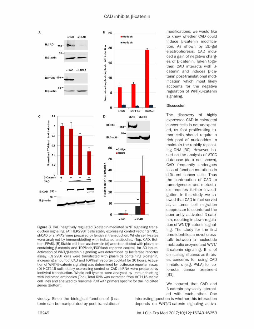
other hand, it enables the expression of WNT target genes necessary for the epithelial-to-mesenchymal transition (EMT). We hypothesize that the inhibition of CAD on colorectal cancer cells proliferation and migration might be dependent on the WNT/β-catenin signaling. To test this hypothesize, we established 293T sta-bly cell lines expressing vector control shNC or shRNA against CAD (shCAD) and PFAS (shP-FAS), a closely related GAT family enzyme in- volved in de novo purine synthesis (Figure 3A). When compared to control cells and PFAS knock down cells, knocking down of CAD signifi-cantly promoted the transcription of luciferase from TOPflash induced by β-catenin overex-pression (Figure 3B). Consistently, overexpres-sion of CAD inhibited the transcription of lucif-erase from TOPflash (Figure 3C). In addition, we examined the expression of β-catenin target genes in colorectal cancer cell line HCT116 by real-time PCR. As depicted in Figure 3D, the depletion of endogenous CAD significantly ele-
vated the expression levels of WNT/β-catenin downstream genes, including c-Myc and MMP2. Altogether, these data demonstrated that CAD negatively regulates β-catenin-mediated WNT signaling transduction.
CAD interacted with β-catenin and induced β-catenin post-translational modification
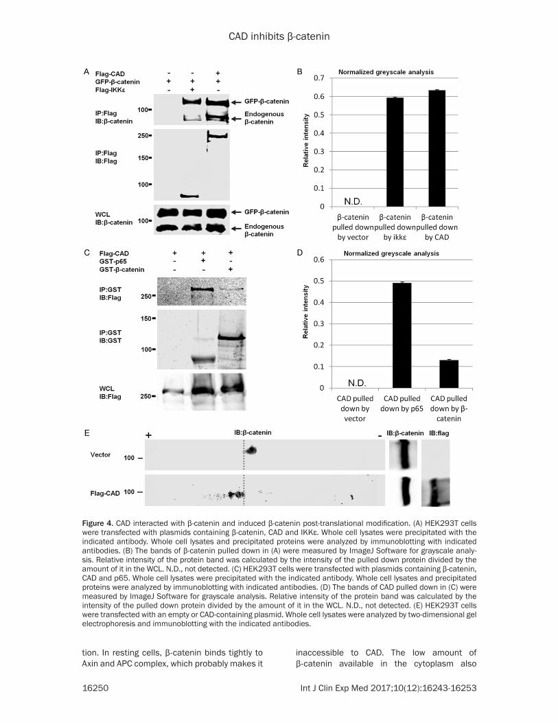
As β-catenin is the key signaling molecule in WNT/β-catenin signaling, to investigate wheth-er CAD inhibits WNT/β-catenin signaling throu- gh regulating β-catenin, we sought out to study the interaction between CAD and β-catenin. As shown by co-immunuoprecipitation assay, CAD readily pulled down β-catenin in 293T cells (Figure 4A and 4B). When we performed the reversed the immunoprecipitation, we could also detect the presence of CAD in β-catenin precipitate (Figure 4C and 4D). Notably, we used IKKε and P65 as positive controls, respec-tively, as these interactions were reported pre-
Figure 2. Overexpression of CAD in-hibited the CRC cell migration ability. A. HCT116 cells were transfected with control vector or a plasmid expressing CAD. Whole cell lysates were analyzed by immunoblotting with indicated an-tibodies. B. HCT116 cells transfected with control vector or plasmid express-ing CAD were scratched and imaged at 0 and 12 hours. Dashed lines indi-cate the boundary of the edges of the wound at 0 and 12 hours.
CAD inhibits β-catenin
16249 Int J Clin Exp Med 2017;10(12):16243-16253
viously. Since the biological function of β-ca- tenin can be manipulated by post-translational
interesting question is whether this interaction depends on WNT/β-catenin signaling activa-
Figure 3. CAD negatively regulated β-catenin-mediated WNT signaling trans-duction signaling. (A) HEK293T cells stably expressing control vector (shNC), shCAD or shPFAS were prepared by lentiviral transduction. Whole cell lysates were analyzed by immunoblotting with indicated antibodies. (Top: CAD, Bot-tom: PFAS). (B) Stable cell lines as shown in (A) were transfected with plasmids containing β-catenin and TOPflash/FOPflash reporter cocktail for 30 hours. Activation of WNT/β-catenin signaling was determined by luciferase reporter assay. (C) 293T cells were transfected with plasmids containing β-catenin, increasing amount of CAD and TOPflash reporter cocktail for 30 hours. Activa-tion of WNT/β-catenin signaling was determined by luciferase reporter assay. (D) HCT116 cells stably expressing control or CAD shRNA were prepared by lentiviral transduction. Whole cell lysates were analyzed by immunoblotting with indicated antibodies (Top). Total RNA was extracted from HCT116 stable cell lines and analyzed by real-time PCR with primers specific for the indicated genes (Bottom).
modifications, we would like to know whether CAD could induce β-catenin modifica-tion. As shown by 2D-gel electrophoresis, CAD indu- ced a gain of negative charg-es of β-catenin. Taken toge- ther, CAD interacts with β- catenin and induces β-ca- tenin post-translational mod-ification which most likely accounts for the negative regulation of WNT/β-catenin signaling.
Discussion
The discovery of highly expressed CAD in colorectal cancer cells is not unexpect-ed, as fast proliferating tu- mor cells should require a rich pool of nucleotides to maintain the rapidly replicat-ing DNA [30]. However, ba- sed on the analysis of ATCC database (data not shown), CAD frequently undergoes loss-of-function mutations in different cancer cells. Thus the contribution of CAD to tumorigenesis and metasta-sis requires further investi-gation. In this study, we sh- owed that CAD in fact served as a tumor cell migration suppressor to counteract the aberrantly activated β-cate- nin, resulting in down-regula-tion of WNT/β-catenin signal-ing. The study for the first time identifies a novel cross-talk between a nucleotide metabolic enzyme and WNT/β-catenin signaling. It is of clinical significance as it rais-es concerns for using CAD inhibitors (e.g. PALA) for co- lorectal cancer treatment [31].
We showed that CAD and β-catenin physically interact-ed with each other. One
CAD inhibits β-catenin
16250 Int J Clin Exp Med 2017;10(12):16243-16253
tion. In resting cells, β-catenin binds tightly to Axin and APC complex, which probably makes it
inaccessible to CAD. The low amount of β-catenin available in the cytoplasm also
Figure 4. CAD interacted with β-catenin and induced β-catenin post-translational modification. (A) HEK293T cells were transfected with plasmids containing β-catenin, CAD and IKKε. Whole cell lysates were precipitated with the indicated antibody. Whole cell lysates and precipitated proteins were analyzed by immunoblotting with indicated antibodies. (B) The bands of β-catenin pulled down in (A) were measured by ImageJ Software for grayscale analy-sis. Relative intensity of the protein band was calculated by the intensity of the pulled down protein divided by the amount of it in the WCL. N.D., not detected. (C) HEK293T cells were transfected with plasmids containing β-catenin, CAD and p65. Whole cell lysates were precipitated with the indicated antibody. Whole cell lysates and precipitated proteins were analyzed by immunoblotting with indicated antibodies. (D) The bands of CAD pulled down in (C) were measured by ImageJ Software for grayscale analysis. Relative intensity of the protein band was calculated by the intensity of the pulled down protein divided by the amount of it in the WCL. N.D., not detected. (E) HEK293T cells were transfected with an empty or CAD-containing plasmid. Whole cell lysates were analyzed by two-dimensional gel electrophoresis and immunoblotting with the indicated antibodies.
CAD inhibits β-catenin
16251 Int J Clin Exp Med 2017;10(12):16243-16253
makes it difficult to be targeted by CAD. In our study, we overexpressed β-catenin, which mim-ics the activation of WNT signaling. Thus, the coIP data indicated that CAD might physically interact with β-catenin upon its release from the destruction complex. Additionally, the bio-logical consequence of this interaction requires further investigation. One possibility is that CAD traps β-catenin inside the cytoplasm to prevent its transactivation of WNT target genes. Although CAD localizes both in the cytoplasm and nucleus, the majority fraction of it resides in the cytoplasm [32]. Additionally, there could also be a shuttling mechanism which exports nuclear β-catenin to the cytoplasm by CAD.
Last but not the least, we performed 2D gel electrophoresis and observed the charge change of β-catenin induced by CAD. It would be very important to figure out the modification causing this charges change. Phosphorylation is the most common protein post-translational modification resulting in net gain of negative charges. Moreover, β-catenin undergoes a number of phosphorylations by distinct kinas-es, with most of the phosphorylation events ending in inhibition of its signaling [33]. Since CAD contains no kinase domain, if it were to induce β-catenin phosphorylation, it would have to bring another yet unidentified kinase. An alternative explanation would be a novel modification on β-catenin brought by CAD itself. Further investigation will be necessary to unveil the mechanism of the modification.
To summarize, our study identified an unex-pected metabolic enzyme regulating colore- ctal cancers cell migration. We demonstrated that WNT/β-catenin can be manipulated by a metabolic enzyme via post-translational mo- dification.
Acknowledgements
This work was supported by Hunan Provincial Natural Science Fund in China (2017JJ2334) (to J.P.), the Joint Funds of the National Natural Science Foundation of China (U1603126) (to Z.X.) and the National Key Research and Development Program of China (2016YFC12- 00200 and 2016YFD0500300) (to Z.X.). The authors are grateful to Professor Pinghui Feng (University of Southern California), Chengyu Liang (University of Southern California) and other study participants.
Disclosure of conflict of interest
None.
Address correspondence to: Jian Peng, Department of General Surgery, Xiangya Hospital, Central South University, Changsha 410008, Hunan, China. Tel: +86-13607441906; E-mail: [email protected]; Jun Zhao, Department of Molecular Micro- biology and Immunology, Norris Comprehensive Cancer Center, University of Southern California, 1441 Eastlake Street, Los Angeles, CA 90033, USA.Tel: 13238650657; E-mail: [email protected]; Zanxian Xia, School of Life Sciences, Central South University, Changsha, Hunan 410008, China. Tel: +86-18673- 175161; E-mail: [email protected]
References
[1] Phesse T, Flanagan D, Vincan E. Frizzled7: a promising Achilles’ heel for targeting the Wnt receptor complex to treat cancer. Cancers 2016; 8: 1-33.
[2] Cisternas P, Salazar P, Silva-Álvarez C, Barros LF, Inestrosa NC. Activation of Wnt signaling in cortical neurons enhances glucose utilization through hlycolysis. J Biol Chem 2016; 291: 25950-25964.
[3] Jiang Y, Sheng H, Meng L, Yue H, Li B, Zhang A, Dong Y, Liu Y. RBM5 inhibits tumorigenesis of gliomas through inhibition of Wnt/β-catenin signaling and induction of apoptosis.World J Surg Oncol 2017; 15: 9.
[4] Razzaque MS, Atfi A. TGIF function in oncogen-ic Wnt signaling. Biochim Biophys Acta 2016; 1865: 101-104.
[5] Yedid N, Kalma Y, Malcov M, Amit A, Kariv R, Caspi M, Rosin-Arbesfeld R, Ben-Yosef D. The effect of a germline mutation in the APC gene on β-catenin in human embryonic stem cells. BMC Cancer 2016; 16: 952.
[6] Kahouli I, Malhotra M, Westfall S, Alaoui-Jama-li MA, Prakash S. Design and validation of an orally administrated active L. fermentum-L. acidophilus probiotic formulation using col- orectal cancer Apc Min/+ mouse model. Appl Microbiol Biotechnol 2017; 101: 1999-2019.
[7] Cho YH, Cha PH, Kaduwal S, Park JC, Lee SK, Yoon JS, Shin W, Kim H, Ro EJ, Koo KH, Park KS3, Han G, Choi KY. KY1022, a small mole-cule destabilizing Ras via targeting the Wnt/β-catenin pathway, inhibits development of met-astatic colorectal cancer. Oncotarget 2016; 7: 81727-81740.
[8] Schweigert A, Fischer C, Mayr D, von Schwein-itz D, Kappler R, Hubertus J. Activation of the Wnt/β-catenin pathway is common in wilms
CAD inhibits β-catenin
16252 Int J Clin Exp Med 2017;10(12):16243-16253
tumor, but rarely through β-catenin mutation and APC promoter methylation. Pediatr Surg Int 2016: 32: 1141-1146.
[9] Hong CS, Ho W, Zhang C, Yang C, Elder JB, Zhuang Z. LB100, a small molecule inhibitor of PP2A with potent chemo- and radio-sensitizing potential. Cancer Biology & Therapy 2015; 16: 821-833.
[10] Bonfim DC, Dias RB, Fortuna-Costa A, Chicay-bam L, Lopes DV, Dutra HS, Borojevic R, Bon-amino M, Mermelstein C, Rossi MI. PS1/γ-Secretase-mediated cadherin cleavage induc-es β-catenin nuclear translocation and osteo-genic differentiation of human bone marrow stromal cells. Stem Cells Int 2016; 2016: 3865315.
[11] Satoh S, Daigo Y, Furukawa Y, Kato T, Miwa N, Nishiwaki T, Kawasoe T, Ishiguro H, Fujita M, Tokino T, Sasaki Y, Imaoka S, Murata M, Shi-mano T, Yamaoka Y, Nakamura Y. AXIN1 muta-tions in hepatocellular carcinomas, and growth suppression in cancer cells by virus-mediated transfer of AXIN1. Nat Genet 2000; 24: 245-250.
[12] Shimizu Y, Ikeda S, Fujimori M, Kodama S, Na-kahara M, Okajima M, Asahara T. Frequent al-terations in the Wnt signaling pathway in colorectal cancer with microsatellite instability. Genes Chromosomes Cancer 2002; 33: 73-81.
[13] Tracy M. Covey, Kornelia Edes, Gary S. Coombs, David M. Virshup, Frank A. Fitzpatrick. Alkyla-tion of the tumor suppressor PTEN activates Akt and β-catenin signaling: a mechanism link-ing inflammation and oxidative stress with can-cer. PLoS One 2010; 5: e13545.
[14] Rabinovich S, Adler L, Yizhak K, Sarver A, Sil-berman A, Agron S, Stettner N, Sun Q, Brandis A, Helbling D, Korman S, Itzkovitz S, Dimmock D, Ulitsky I, Nagamani SC, Ruppin E, Erez A. Diversion of aspartate in ASS1-deficient tu-mours fosters de novo pyrimidine synthesis. Nature 2015; 527: 379-383.
[15] Ruiz-Ramos A, Lallous N, Grande-García A, Ramón-Maiques S. Expression, purification, crystallization and preliminary X-ray diffraction analysis of the aspartate transcarbamoylase domain of human CAD. Acta Crystallogr Sect F Struct Biol Cryst Commun 2013; 69: 1425-1430.
[16] Guy HI, Evans DR. Cloning, expression, and functional interactions of the amidotransfer-ase domain of mammalian CAD carbamyl phosphate synthetase. J Biol Chem 1994; 269: 7702-7708.
[17] Sigoillot FD, Berkowski JA, Sigoillot SM, Kotsis DH, Guy HI. Cell cycle-dependent regulation of pyrimidine biosynthesis. J Biol Chem 2003; 278: 3403-3409.
[18] Nakashima A, Kawanishi I, Eguchi S, Yu EH, Eguchi S, Oshiro N, Yoshino K, Kikkawa U, Yo-nezawa K. Association of CAD, a multifunction-al protein involved in pyrimidine synthesis, with mLST8, a component of the mTOR complexes. J Biomed Sci 2013; 20: 24.
[19] Reardon MA, Dixon JE, Weber G. Increased messenger RNA concentration for carbamoyl-phosphate synthase II in hepatoma 3924A. Biochem Biophys Res Commun 1987; 147: 494-500.
[20] Sigoillot FD, Sigoillot SM, Guy HI. Breakdown of the regulatory control of pyrimidine biosynthe-sis in human breast cancer cells. Int J Cancer 2004; 109: 491-498.
[21] Morin A, Fritsch L, Mathieu JR, Gilbert C, Guar-mit B, Firlej V, Gallou-Kabani C, Vieillefond A, Delongchamps NB, Cabon F. Identification of CAD as an androgen receptor interactant and an early marker of prostate tumor recurrence. FASEB J 2012; 26: 460-467.
[22] Otto E, McCord S, Tlsty TD. Increased incidence of CAD gene amplification in tumorigenic rat lines as an indicator of genomic instability of neoplastic cells. J Biol Chem 1989; 264: 3390-3396.
[23] Richmond AL, Kabi A, Homer CR, Marina-Gar-cia N, Nickerson KP, Nesvizhskii AI, Sreekumar A, Chinnaiyan AM, Nunez G, McDonald C. The nucleotide synthesis enzyme CAD inhibits NOD2 antibacterial function in human intesti-nal epithelial cells. Gastroenterology 2012; 142: 1483-1492.
[24] Bouwmeester T, Bauch A, Ruffner H, Angrand PO, Bergamini G, Croughton K, Cruciat C, Eber-hard D, Gagneur J, Ghidelli S, Hopf C, Huhse B, Mangano R, Michon AM, Schirle M, Schlegl J, Schwab M, Stein MA, Bauer A, Casari G, Drew-es G, Gavin AC, Jackson DB, Joberty G, Neu-bauer G, Rick J, Kuster B, Superti-Furga G. A physical and functional map of the human TNF-alpha, NF-kappa, B signal transduction pathway. Nat Cell Biol 2004; 6: 97-105.
[25] Ishitani T, Ninomiya-Tsuji J, Nagai S, Nishita M, Meneghini M, Barker N, Waterman M, Bower-man B, Clevers H, Shibuya H, Matsumoto K. The TAK1-NLK-MAPK-related pathway antago-nizes signalling between beta-catenin and transcription factor TCF. Nature 1999; 399: 798-802.
[26] Korinek V, Barker N, Willert K, Molenaar M, Roose J, Wagenaar G, Markman M, Lamers W, Destree O, Clevers H. Two members of the Tcf family implicated in Wnt/beta-catenin signal-ing during embryogenesis in the mouse. Mol Cell Biol 1998; 18: 1248-1256.
[27] He Z, Zhao J, Zhang J, Jung JU, Feng P. NF-kap-paB activation coordinated by IKKbeta and IK-Kepsilon enables latent infection of Kaposi’s
CAD inhibits β-catenin
16253 Int J Clin Exp Med 2017;10(12):16243-16253
sarcoma-associated herpesvirus. J Virol 2014; 88: 444-455.
[28] Zhao J, Zeng Y, Xu S, Chen J, Shen G, Yu C, Knipe D, Yuan W, Peng J, Xu W, Zhang C, Xia Z, Feng P. A viral deamidase targets the helicase domain of RIG-I to block RNA-induced activa-tion. Cell Host Microbe 2016; 20: 770-784.
[29] He S, Zhao J, Song S, He X, Minassian A, Zhou Y, Zhang J, Brulois K, Wang Y, Cabo J, Zandi E, Liang C, Jung JU, Zhang X, Feng P. Viral pseudo-enzymes activate RIG-I via deamidation to evade cytokine production. Mol Cell 2015; 58: 134-146.
[30] Liao WS, Heller R, Green P, Stark GR. Regula-tion of carbamoyl phosphate synthetase-as-partate transcarbamoylase-dihydroorotase ge- ne expression in growing and arrested cells. J Biol Chem 1986; 261: 15577-15581.
[31] Johnson RK, Inouye T, Goldin A, Stark GR. Anti-tumor activity of N-(phosphonacetyl)-L-aspartic acid, a transition-state inhibitor of aspartate transcarbamylase. Cancer Res 1976; 36: 2720-2725.
[32] Sigoillot FD, Kotsis DH, Serre V, Sigoillot SM, Evans DR, Guy HI. Nuclear localization and mitogen-activated protein kinase phosphoryla-tion of the multifunctional protein CAD. J Biol Chem 2005; 280: 25611-25620.
[33] Daugherty RL, Gottardi CJ. Phospho-regulation of Beta-catenin adhesion and signaling func-tions. Physiology (Bethesda) 2007; 22: 303-309.