Onsite Program Guide & Exhibit or Guide Exhibitor ......Please join us in welcoming back these...
222
Onsite Program Guide & Exhibitor Information www.microscopy.org/MandM/2019 Exhibitor Guide INCLUDED!
Transcript of Onsite Program Guide & Exhibit or Guide Exhibitor ......Please join us in welcoming back these...
Onsite Program Guide & Exhibitor Information
www.microscopy.org/MandM/2019
Exhibitor Guide
INCLUDED!
0.20.1 0.3 0.4Energy loss (eV)
IR
Nion Iris (2019)
Original guanineresult (2016)
Intensity
0-20 20
4.2 meV
ΔE (meV)
Intensity
e-
X-ray energy (keV)
CPSpe
reV
1
2
4
2 3
(1.2 Å)-1
FFT
2 nm
MAADF, 30kV
Fourier-filtered
FFT
(1.07
Å)-1
0.5 nm
12C13CAlanine
Energy loss (meV)100 140 180 220
Intensity
(a.u.)
"ADF"
ZLP:-10→10 meV
LA-TA+LO-TO:50→200 meV
2 Å
Energy
loss
ω(m
eV)
200
150
100
50
1
Γ Μ Γ' Μ'
2 3 4 50q (Å-1)
Probe
Energygain
Transferred energy (meV)
Intensity
(a.u.)
Energyloss
-60 0 60 120
300 K600 K800 K
Ronchigram int. (a.u.)0 16000
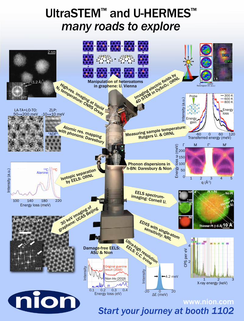
30keV
imaging of
graphe
ne:UCA
S Beijin
g
High-res. imaging at liquid
N2 temperature: CNRS Orsay
Phonon dispersions inh-BN: Daresbury & Nion
Manipulation of heteroatomsin graphene: U. Vienna
EDXS with single-atom
sensitivity: NRL
Damage-free EELS:ASU & Nion
EELS spectrum-imaging: Cornell U.
Ultra-high resolution
EELS: U.C. Irvine
Measuring sample temp
erature:
Rutgers U. &ORNL
Imaging elect
ricfields b
y
4DSTE
M in DyScO3:ORNL
Isotopic sep
aration
by EELS: ORN
L
Atomic res. mappingwith phonons: Daresbury
Start your journey at booth 1102www.nion.com
UltraSTEM™ and U-HERMES™many roads to explore
MICROSCOPY & MICROANALYSIS 2019 MEETING | August 4-8 | Portland, OR 3
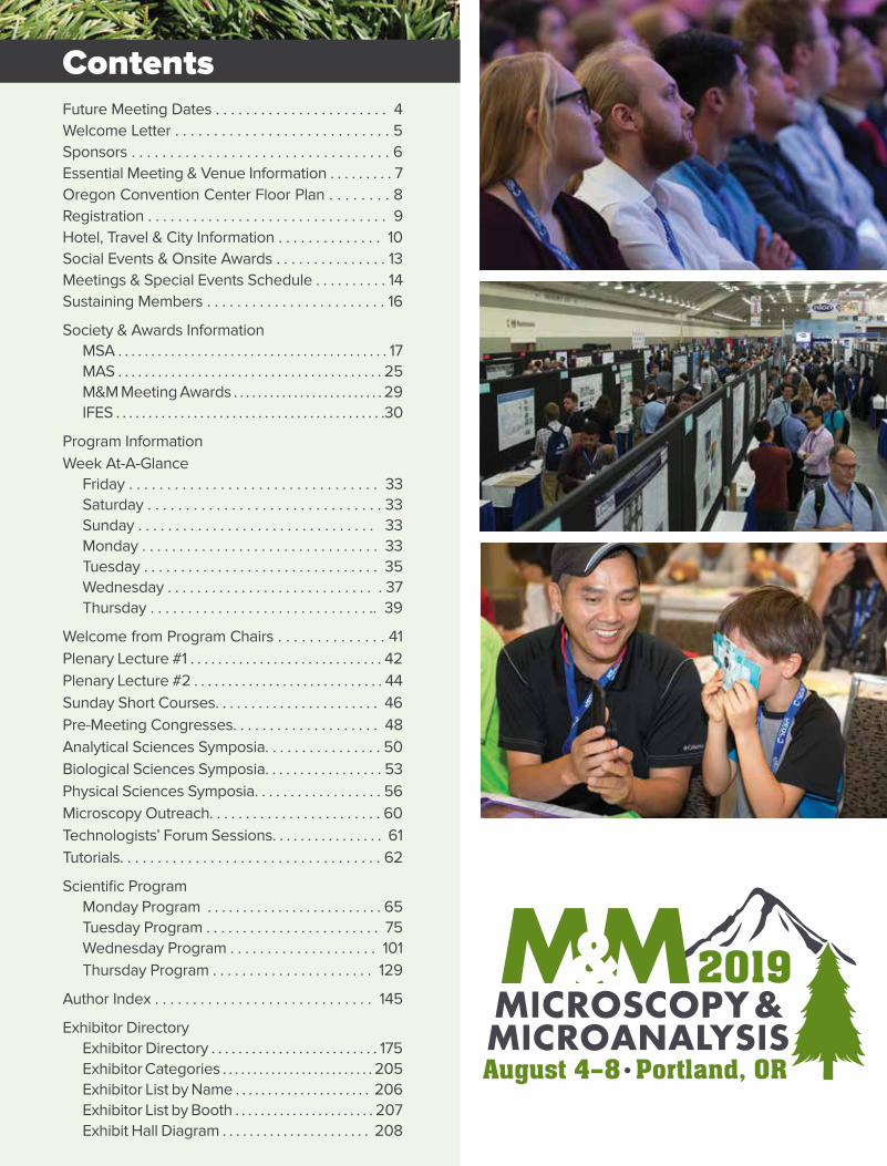
Future Meeting Dates . . . . . . . . . . . . . . . . . . . . . . . 4Welcome Letter . . . . . . . . . . . . . . . . . . . . . . . . . . . . 5Sponsors . . . . . . . . . . . . . . . . . . . . . . . . . . . . . . . . . . 6Essential Meeting & Venue Information . . . . . . . . . 7Oregon Convention Center Floor Plan . . . . . . . . 8Registration . . . . . . . . . . . . . . . . . . . . . . . . . . . . . . . . 9Hotel, Travel & City Information . . . . . . . . . . . . . . 10Social Events & Onsite Awards . . . . . . . . . . . . . . . 13Meetings & Special Events Schedule . . . . . . . . . . 14Sustaining Members . . . . . . . . . . . . . . . . . . . . . . . . 16
Society & Awards InformationMSA . . . . . . . . . . . . . . . . . . . . . . . . . . . . . . . . . . . . . . . . . 17MAS . . . . . . . . . . . . . . . . . . . . . . . . . . . . . . . . . . . . . . . . 25M&M Meeting Awards . . . . . . . . . . . . . . . . . . . . . . . . . 29IFES . . . . . . . . . . . . . . . . . . . . . . . . . . . . . . . . . . . . . . . . . .30
Program InformationWeek At-A-Glance
Friday . . . . . . . . . . . . . . . . . . . . . . . . . . . . . . . . . 33Saturday . . . . . . . . . . . . . . . . . . . . . . . . . . . . . . . 33Sunday . . . . . . . . . . . . . . . . . . . . . . . . . . . . . . . . 33Monday . . . . . . . . . . . . . . . . . . . . . . . . . . . . . . . . 33Tuesday . . . . . . . . . . . . . . . . . . . . . . . . . . . . . . . . 35Wednesday . . . . . . . . . . . . . . . . . . . . . . . . . . . . . 37Thursday . . . . . . . . . . . . . . . . . . . . . . . . . . . . . .. 39
Welcome from Program Chairs . . . . . . . . . . . . . . 41Plenary Lecture #1 . . . . . . . . . . . . . . . . . . . . . . . . . . . . 42Plenary Lecture #2 . . . . . . . . . . . . . . . . . . . . . . . . . . . . 44Sunday Short Courses. . . . . . . . . . . . . . . . . . . . . . . 46Pre-Meeting Congresses. . . . . . . . . . . . . . . . . . . . 48Analytical Sciences Symposia. . . . . . . . . . . . . . . . 50Biological Sciences Symposia. . . . . . . . . . . . . . . . . 53Physical Sciences Symposia. . . . . . . . . . . . . . . . . . 56Microscopy Outreach. . . . . . . . . . . . . . . . . . . . . . . . 60Technologists’ Forum Sessions. . . . . . . . . . . . . . . . 61Tutorials. . . . . . . . . . . . . . . . . . . . . . . . . . . . . . . . . . . 62
Scientific ProgramMonday Program . . . . . . . . . . . . . . . . . . . . . . . . . 65Tuesday Program . . . . . . . . . . . . . . . . . . . . . . . . 75Wednesday Program . . . . . . . . . . . . . . . . . . . . 101Thursday Program . . . . . . . . . . . . . . . . . . . . . . 129
Author Index . . . . . . . . . . . . . . . . . . . . . . . . . . . . . 145
Exhibitor DirectoryExhibitor Directory . . . . . . . . . . . . . . . . . . . . . . . . . 175Exhibitor Categories . . . . . . . . . . . . . . . . . . . . . . . . . 205Exhibitor List by Name . . . . . . . . . . . . . . . . . . . . . 206Exhibitor List by Booth . . . . . . . . . . . . . . . . . . . . . . 207Exhibit Hall Diagram . . . . . . . . . . . . . . . . . . . . . . 208
Contents
www.microscopy.org/MandM/2019 4
With the M&M 2019 mobile app, you can:• Receive Up-to-the-
Minute Meeting &Presenter Info
• Multi-Device Sync• Receive Alerts• See Exhibitors• Make Your Schedule• View Maps & Floor Plans• Connect with
Colleagues & Friends• Join in on Social Media
with #MM2019Portland• And much, much more!
Downloading the App is Easy!SEARCH: The App Store or Google Play for “M&M Annual Meeting”
SCAN:
For your Desktop, and all other web-enabled devices, point your browser to:
http://m.core-apps.com/msa_2019
Navigate the meeting like a pro with the M&M 2019 mobile app, powered by core-apps.com.
Blackberry users will be pointed to the Google Play store to download the mobile app. Windows devices will be able to see the web version, but it is not mobile-enabled.
This Web version will allow you to search/view all meeting contents, as well as set your personal schedule and Favorites. Simply login (your personal/login information is not shared anywhere), and then your choices are synced to all your logged-in devices!
(You can elect to make your profile public within the app so others can find you, but you can determine what others see.)
Should you have any questions, please contact [email protected].
Future Meeting Dates
August 1-5, 2021PITTSBURGH, PA
July 31-August 4, 2022PORTLAND, OR
July 23-27, 2023MINNEAPOLIS, MN
July 28-August 1, 2024CLEVELAND, OH
August 2-6 Milwaukee, WI
Download the 2019 Mobile App!
MICROSCOPY & MICROANALYSIS 2019 MEETING | August 4-8 | Portland, OR 5
Welcome from the Society Presidents
On behalf of the Microscopy Society of America, the Microanalysis Society, and the International Field Emission Society, we welcome you to Microscopy & Microanalysis 2019 at the Oregon Convention Center in Portland, Oregon. It’s an excellent venue with wonderful restaurants, lots of activities for the family, and a comfortable climate.
The Program Committee, led by Alice Dohnalkova, Huolin Xin, Assel Aitkaliyeva, and Baptiste Gault, has developed an exciting group of symposia, spanning advances in instrumentation and techniques development, as well as applications in the analytical, biological, and physical sciences. A record number of paper submissions this year is sure to guarantee an exciting and robust program of cutting-edge research!
The main meeting starts with the Opening Welcome Reception on Sunday evening. The Sunday reception is a great place for all attendees to meet new colleagues and reconnect with old friends. On Monday morning, the Plenary Session kicks off the scientific program with two exciting plenary lectures from 2017 Nobel Prize in Chemistry co-winners, Professors Joachim Frank and Richard Henderson, the presentations of the M&M meeting awards, and awards from the sponsoring societies. Please join us in welcoming back these long-time MSA members and frequent M&M attendees as they discuss their groundbreaking work in cryo-electron microscopy.
In addition to the strong scientific program, what sets the M&M meeting apart is the Exhibit Hall, the world’s largest annual microscopy exhibition, which showcases the latest in microscopy instrumentation and accessories. Don’t miss the highly popular vendor tutorials, held Monday through Wednesday after hours in the Exhibit Hall. Other educational opportunities throughout the week include focused biological and physical science tutorials, educational outreach programs, and our Technologists’ Forum special and roundtable sessions.
In short, M&M 2019 is an outstanding opportunity to stay abreast of the latest technologies, hear about new developments in applications across all areas of microscopy and microanalysis, and most importantly network with colleagues.
Welcome to Portland!
Paul Kotula
Sandia National Laboratories
President, Microscopy Society of America
Rhonda Stroud
U.S. Naval Research Laboratory
President, Microanalysis Society
David Larson
AMETEK, Inc.
President, International Field Emission Society
www.microscopy.org/MandM/2019 6
Sponsor List as of 7/1/19
Sponsors
ProtochipsQuantifiably Better™NSF SCIENCE AND TECHNOLOGY CENTER
Microscopy Products for Science and Industry
Research Inc.
HREM
MICROSCOPY & MICROANALYSIS 2019 MEETING | August 4-8 | Portland, OR 7
AccessibilityIf you require special accommodation in order to participate fully in the meeting, please ask to speak with the meeting manager, or email [email protected]. Requests made onsite will be accommodated as much as possible.
AwardsMajor Society Awards for MSA, MAS, and IFES, along with M&M student awards, will be presented at the Plenary Session immediately following the first Plenary Talk (Monday morning). For detailed listings of all awards, criteria, and award winners, please visit http://microscopy.org/MandM/2019/.
Cancellation and Refund PolicyRefund requests received prior to July 19, 2019 will be honored less a $65 administrative fee. No refunds will be issued for cancellations (for any reason) received on or after July 19, 2019, and no refunds will be issued on-site in Portland. E-mail: [email protected] or fax (703) 964-1246.
Food for PurchaseInexpensive, portable breakfast and snack items are available for purchase in the convention center on the exhibit/registration level (7:30 am–10:30 am). Lunch concessions are available for purchase inside the exhibit hall during lunch hours (11:00 am–2:00 pm).
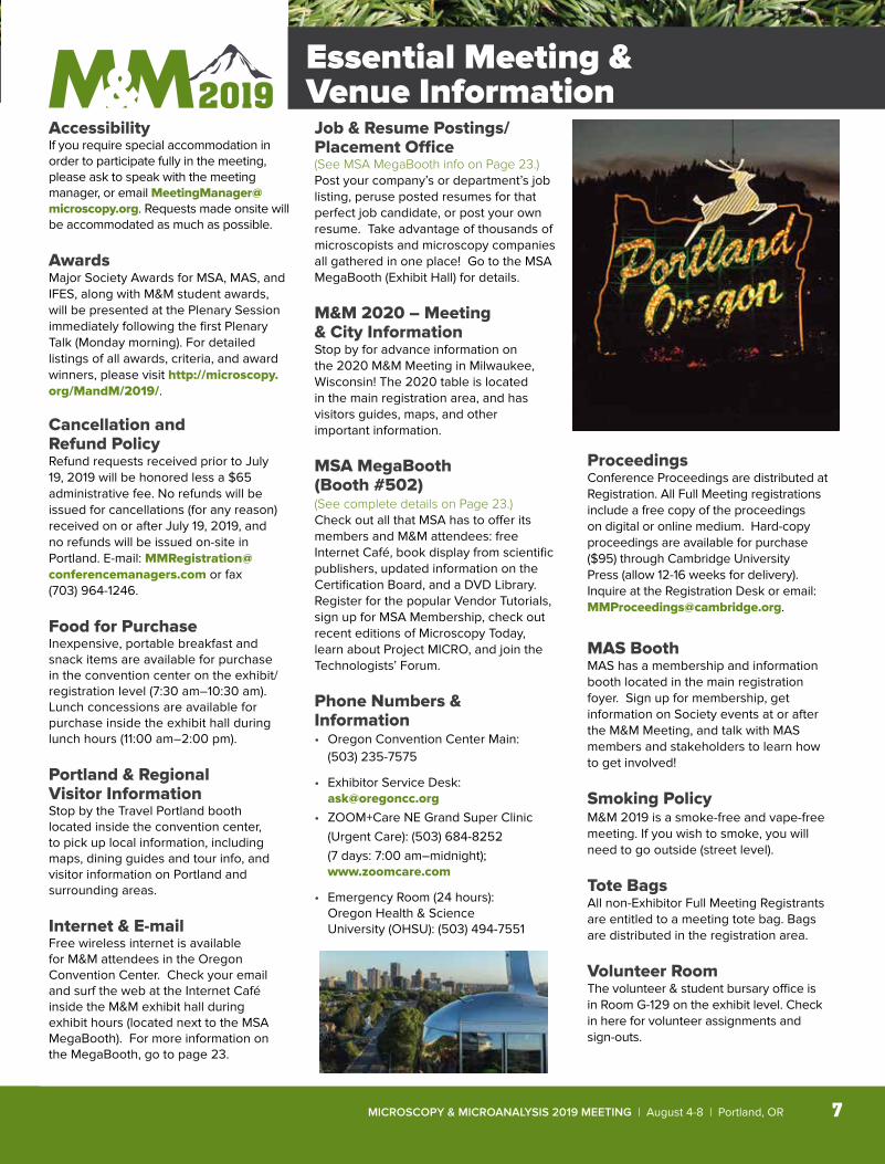
Portland & Regional Visitor InformationStop by the Travel Portland booth located inside the convention center, to pick up local information, including maps, dining guides and tour info, and visitor information on Portland and surrounding areas.
Internet & E-mailFree wireless internet is available for M&M attendees in the Oregon Convention Center. Check your email and surf the web at the Internet Café inside the M&M exhibit hall during exhibit hours (located next to the MSA MegaBooth). For more information on the MegaBooth, go to page 23.
Job & Resume Postings/Placement Office (See MSA MegaBooth info on Page 23.)Post your company’s or department’s job listing, peruse posted resumes for that perfect job candidate, or post your own resume. Take advantage of thousands of microscopists and microscopy companies all gathered in one place! Go to the MSA MegaBooth (Exhibit Hall) for details.
M&M 2020 – Meeting & City InformationStop by for advance information on the 2020 M&M Meeting in Milwaukee, Wisconsin! The 2020 table is located in the main registration area, and has visitors guides, maps, and other important information.
MSA MegaBooth (Booth #502)(See complete details on Page 23.) Check out all that MSA has to offer its members and M&M attendees: free Internet Café, book display from scientific publishers, updated information on the Certification Board, and a DVD Library. Register for the popular Vendor Tutorials, sign up for MSA Membership, check out recent editions of Microscopy Today, learn about Project MICRO, and join the Technologists’ Forum.
Phone Numbers & Information• Oregon Convention Center Main:
(503) 235-7575
• Exhibitor Service Desk: [email protected]
• ZOOM+Care NE Grand Super Clinic (Urgent Care): (503) 684-8252
(7 days: 7:00 am–midnight); www.zoomcare.com
• Emergency Room (24 hours): Oregon Health & Science
University (OHSU): (503) 494-7551
ProceedingsConference Proceedings are distributed at Registration. All Full Meeting registrations include a free copy of the proceedings on digital or online medium. Hard-copy proceedings are available for purchase ($95) through Cambridge University Press (allow 12-16 weeks for delivery). Inquire at the Registration Desk or email: [email protected].
MAS BoothMAS has a membership and information booth located in the main registration foyer. Sign up for membership, get information on Society events at or after the M&M Meeting, and talk with MAS members and stakeholders to learn how to get involved!
Smoking PolicyM&M 2019 is a smoke-free and vape-free meeting. If you wish to smoke, you will need to go outside (street level).
Tote BagsAll non-Exhibitor Full Meeting Registrants are entitled to a meeting tote bag. Bags are distributed in the registration area.
Volunteer RoomThe volunteer & student bursary office is in Room G-129 on the exhibit level. Check in here for volunteer assignments and sign-outs.
Essential Meeting & Venue Information
www.microscopy.org/MandM/2019 8
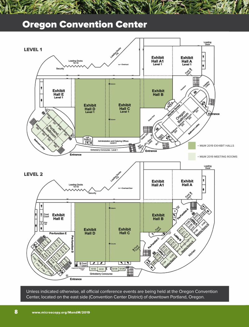
Unless indicated otherwise, all official conference events are being held at the Oregon Convention Center, located on the east side (Convention Center District) of downtown Portland, Oregon.
G132 G129G130G131
LEVEL 1
LEVEL 2
= M&M 2019 EXHIBIT HALLS
= M&M 2019 MEETING ROOMS
Oregon Convention Center

MICROSCOPY & MICROANALYSIS 2019 MEETING | August 4-8 | Portland, OR 9
= M&M 2019 MEETING ROOMS
Onsite Registration Desk Oregon Convention Center
Pick up your badge and materials at the Registration desk according to the schedule below. The Sunday Welcome Reception starts at 6:30 PM in the Portland Ballroom (upper level of the Oregon Convention Center).
Registration Hours: Friday, August 2* 8:00 am – 1:00 pm Friday, August 2 1:00 pm – 6:00 pm Saturday, August 3 8:00 am – 6:00 pm Sunday, August 4 7:00 am – 7:30 pm Monday, August 5 7:00 am – 6:00 pm Tuesday, August 6 7:30 am – 5:00 pm Wednesday, August 7 7:30 am – 5:00 pm Thursday, August 8 7:30 am – 3:00 pm*Exhibitors Only
Commercial Exhibition Hours:Monday, August 5 12:00 pm – 5:30 pmTuesday, August 6 10:00 am – 5:30 pmWednesday, August 7 10:00 am – 5:30 pmThursday, August 8 10:00 am – 2:00 pm
Exhibitor Move-In:Thursday, August 1* 8:00 am – 4:00 pm Friday, August 2 8:00 am – 4:30 pmSaturday, August 3 8:00 am – 4:30 pmSunday, August 4 8:00 am – 4:30 pm*Targeted Island Booths Only
Exhibitor Move-Out:Thursday, August 8 2:00 pm – 7:00 pmFriday, August 9 8:00 am – 5:00 pm
Registration Information
www.microscopy.org/MandM/2019 10
WILLAM
ETTE
RIVER
STREETCAR
MAX
LIG
HT
RAIL
MAX
LIG
HT
RAIL
egdirBleetS
egdirBnosirr
oM
egdirBenrhtwaH
egdirBdnalsIssoR5
405
405
5
5
84
405
STREETCAR
To Airport
Burnside Bridge
egdirBmauqraM
egdirByawdaorB
LIARTHGILXAM
Tilikum Bridge
OregonConvention
Center
NE Broadway
NE Weidler
NE Holladay
SE 12
thSE
11th
East Burnside
SE Morrison
SE Belmont
SE Madison
SE Hawthorne
Mar
tin
Luth
er K
ing
Jr. B
lvd.
Gra
nd A
venu
e.
SE W
ater
SW WashingtonSW Morrison
SE Division
SE Powell Blvd.
. evAydoo
M
N Interstate Ave
NW Northrup
NW Lovejoy
NW
11th
NW
10th
West Burnside
NW Everett
SW11t
hSW
10th
SWBr
oadw
ay
Nai
toPa
rkw
ay
SW MainSW Madison
yawkraPreviRSW Lincoln
SW Yamhill
SWSi
xth
SWFi
fth
NW Glisan
o
8
HOTELS
123456789
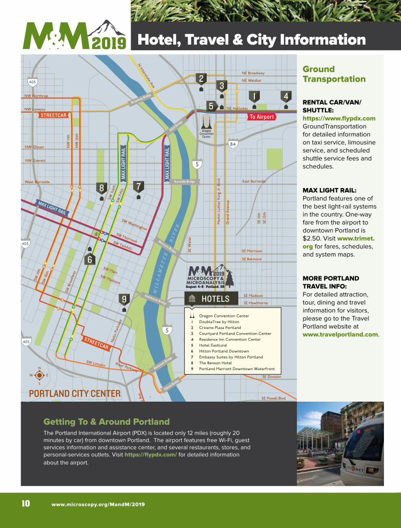
Oregon Convention CenterDoubleTree by HiltonCrowne Plaza PortlandCourtyard Portland Convention CenterResidence Inn Convention CenterHotel EastlundHilton Portland DowntownEmbassy Suites by Hilton PortlandThe Benson HotelPortland Marriott Downtown Waterfront
1 4
7
6
5
23
9
PORTLAND CITY CENTER
Ground Transportation
RENTAL CAR/VAN/SHUTTLE: https://www.flypdx.comGroundTransportation for detailed information on taxi service, limousine service, and scheduled shuttle service fees and schedules.
MAX LIGHT RAIL: Portland features one of the best light-rail systems in the country. One-way fare from the airport to downtown Portland is $2.50. Visit www.trimet.org for fares, schedules, and system maps.
MORE PORTLAND TRAVEL INFO: For detailed attraction, tour, dining and travel information for visitors, please go to the Travel Portland website at www.travelportland.com.
.
Getting To & Around PortlandThe Portland International Airport (PDX) is located only 12 miles (roughly 20 minutes by car) from downtown Portland. The airport features free Wi-Fi, guest services information and assistance center, and several restaurants, stores, and personal-services outlets. Visit https://flypdx.com/ for detailed information about the airport.
Hotel, Travel & City Information
MICROSCOPY & MICROANALYSIS 2019 MEETING | August 4-8 | Portland, OR 11
Hotel, Travel & City Information
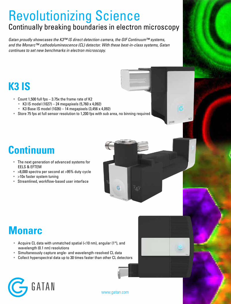
Monarc
• Acquire CL data with unmatched spatial (<10 nm), angular (1°), and wavelength (0.1 nm) resolutions
• Simultaneously capture angle- and wavelength-resolved CL data• Collect hyperspectral data up to 30 times faster than other CL detectors
Gatan proudly showcases the K3™ IS direct detection camera, the GIF Continuum™ systems, and the Monarc™ cathodoluminescence (CL) detector. With these best-in-class systems, Gatan continues to set new benchmarks in electron microscopy.
Revolutionizing Science Continually breaking boundaries in electron microscopy
K3 IS• Count 1,500 full fps – 3.75x the frame rate of K2
• K3 IS model (1027) – 24 megapixels (5,760 x 4,092)• K3 Base IS model (1026) – 14 megapixels (3,456 x 4,092)
• Store 75 fps at full sensor resolution to 1,200 fps with sub area, no binning required
Continuum
• The next generation of advanced systems for EELS & EFTEM
• >8,000 spectra per second at >95% duty cycle• >10x faster system tuning• Streamlined, workflow-based user interface
www.gatan.com
www.microscopy.org/MandM/2019 12
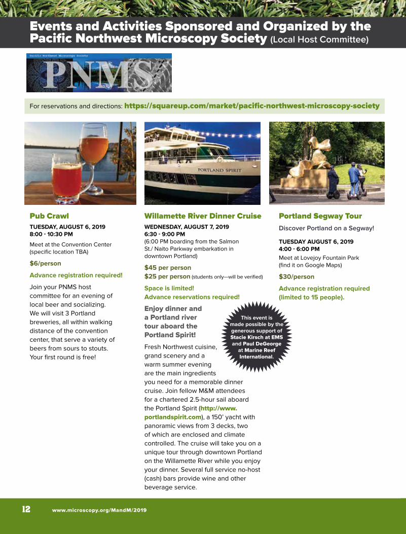
For reservations and directions: https://squareup.com/market/pacific-northwest-microscopy-society
Pub CrawlTUESDAY, AUGUST 6, 20198:00 - 10:30 PM
Meet at the Convention Center (specific location TBA)
$6/person
Advance registration required!
Join your PNMS host committee for an evening of local beer and socializing. We will visit 3 Portland breweries, all within walking distance of the convention center, that serve a variety of beers from sours to stouts. Your first round is free!
Willamette River Dinner CruiseWEDNESDAY, AUGUST 7, 20196:30 - 9:00 PM (6:00 PM boarding from the Salmon St./ Naito Parkway embarkation in downtown Portland)
$45 per person$25 per person (students only—will be verified)
Space is limited! Advance reservations required!
Enjoy dinner and a Portland river tour aboard the Portland Spirit!
Fresh Northwest cuisine, grand scenery and a warm summer evening are the main ingredients you need for a memorable dinner cruise. Join fellow M&M attendees for a chartered 2.5-hour sail aboard the Portland Spirit (http://www.portlandspirit.com), a 150’ yacht with panoramic views from 3 decks, two of which are enclosed and climate controlled. The cruise will take you on a unique tour through downtown Portland on the Willamette River while you enjoy your dinner. Several full service no-host (cash) bars provide wine and other beverage service.
Portland Segway TourDiscover Portland on a Segway!
TUESDAY AUGUST 6, 20194:00 - 6:00 PMMeet at Lovejoy Fountain Park (find it on Google Maps)
$30/person
Advance registration required (limited to 15 people).
This event is made possible by the generous support of Stacie Kirsch at EMS and Paul DeGeorge
at Marine Reef International.
Events and Activities Sponsored and Organized by the Pacific Northwest Microscopy Society (Local Host Committee)
MICROSCOPY & MICROANALYSIS 2019 MEETING | August 4-8 | Portland, OR 13
M&M 2019 Sunday Evening Welcome ReceptionOregon Convention Center – Portland Ballroom (Upper Level)
SUNDAY, AUGUST 4, 2019 6:30 PM - 9:00 PM
One ticket is included with most registrations (see Registration Page for details). Additional tickets: $50 each for adults; $25 each for children 12 and under.
PLEASE NOTE: Onsite availability of tickets is not guaranteed.
This year’s welcome event at the Oregon Convention Center will be a fun and informal get-together. Enjoy a delicious Northwest-inspired supper buffet and local brews; and catch up with friends and colleagues. After the reception, grab some old and new friends and head out to one of Portland’s numerous pubs, microbreweries, or wine bars to continue the fun!
MAS Business Meeting and Social Event – for MAS Members Only!
WEDNESDAY, AUGUST 7, 20195:30 PM - 7:30 PM
Stop by the MAS booth in the lobby to check your membership status and pick up your ticket to the MAS Business Meeting and Social Event, starting on Wednesday, August 7, at 5:30 PM. Meeting and social event will be held in an offsite venue, away from the convention center.
Student Poster Awards (Immediately following daily Poster Presentations & Happy Hours!)
Poster presentations are an excellent format for all participants to engage in intensive discussion with other researchers in the field. MSA provides cash awards to the most outstanding student posters (first author) each day (up to two in each of three categories). Student poster awards will be presented immediately following each day’s poster session, in the Exhibit Hall.
Microscopy Today Innovation AwardsWEDNESDAY, AUGUST 7, 20194:00 PM - Poster Awards Stage, Exhibit Hall
For reservations and directions: https://squareup.com/market/pacific-northwest-microscopy-society
Social EventsEvents and Activities Sponsored and Organized by the Pacific Northwest Microscopy Society (Local Host Committee)
www.microscopy.org/MandM/2019 14
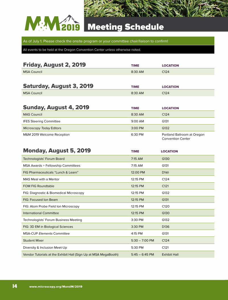
Meeting ScheduleAs of July 1. Please check the onsite program or your committee chair/liaison to confirm!
All events to be held at the Oregon Convention Center unless otherwise noted.
Friday, August 2, 2019 TIME LOCATION
MSA Council 8:30 AM C124
Saturday, August 3, 2019 TIME LOCATION
MSA Council 8:30 AM C124
Sunday, August 4, 2019 TIME LOCATION
MAS Council 8:30 AM C124
IFES Steering Committee 9:00 AM G131
Microscopy Today Editors 3:00 PM G132
M&M 2019 Welcome Reception 6:30 PM Portland Ballroom at Oregon Convention Center
Monday, August 5, 2019 TIME LOCATION
Technologists’ Forum Board 7:15 AM G130
MSA Awards + Fellowship Committees 7:15 AM G131
FIG Pharmaceuticals “Lunch & Learn” 12:00 PM D140
MAS Meal with a Mentor 12:15 PM C124
FOM FIG Roundtable 12:15 PM C121
FIG: Diagnostic & Biomedical Microscopy 12:15 PM G132
FIG: Focused Ion Beam 12:15 PM G131
FIG: Atom Probe Field Ion Microscopy 12:15 PM C120
International Committee 12:15 PM G130
Technologists’ Forum Business Meeting 3:30 PM G132
FIG: 3D EM in Biological Sciences 3:30 PM D136
MSA-CUP Elements Committee 4:15 PM G131
Student Mixer 5:30 – 7:00 PM C124
Diversity & Inclusion Meet-Up 5:30 PM C121
Vendor Tutorials at the Exhibit Hall (Sign Up at MSA MegaBooth) 5:45 – 6:45 PM Exhibit Hall
MICROSCOPY & MICROANALYSIS 2019 MEETING | August 4-8 | Portland, OR 15
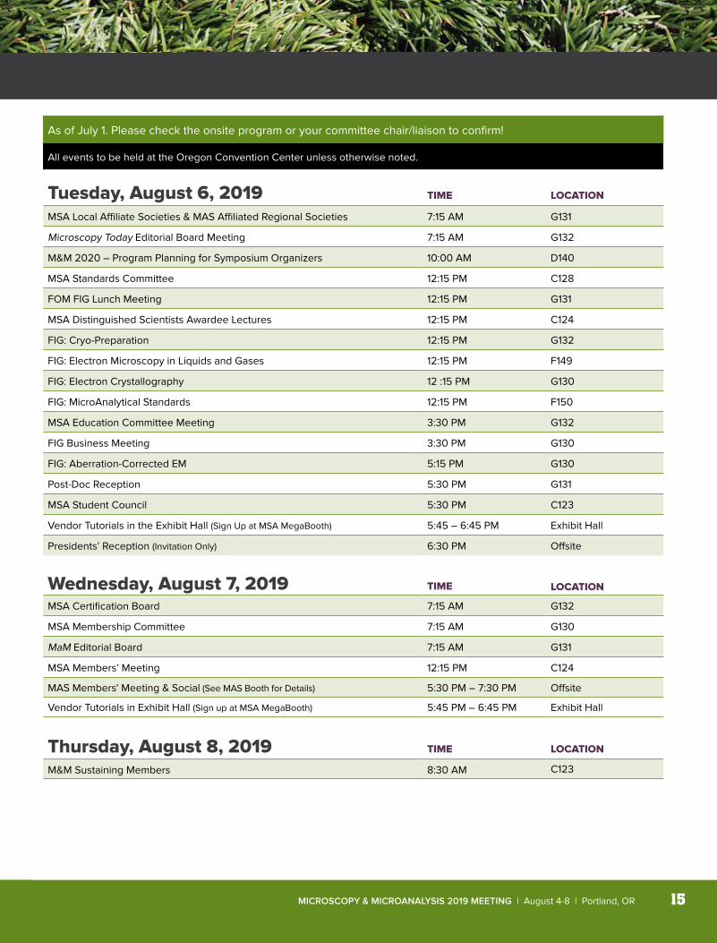
Meeting ScheduleAs of July 1. Please check the onsite program or your committee chair/liaison to confirm!
All events to be held at the Oregon Convention Center unless otherwise noted.
Tuesday, August 6, 2019 TIME LOCATION
MSA Local Affiliate Societies & MAS Affiliated Regional Societies 7:15 AM G131
Microscopy Today Editorial Board Meeting 7:15 AM G132
M&M 2020 – Program Planning for Symposium Organizers 10:00 AM D140
MSA Standards Committee 12:15 PM C128
FOM FIG Lunch Meeting 12:15 PM G131
MSA Distinguished Scientists Awardee Lectures 12:15 PM C124
FIG: Cryo-Preparation 12:15 PM G132
FIG: Electron Microscopy in Liquids and Gases 12:15 PM F149
FIG: Electron Crystallography 12 :15 PM G130
FIG: MicroAnalytical Standards 12:15 PM F150
MSA Education Committee Meeting 3:30 PM G132
FIG Business Meeting 3:30 PM G130
FIG: Aberration-Corrected EM 5:15 PM G130
Post-Doc Reception 5:30 PM G131
MSA Student Council 5:30 PM C123
Vendor Tutorials in the Exhibit Hall (Sign Up at MSA MegaBooth) 5:45 – 6:45 PM Exhibit Hall
Presidents’ Reception (Invitation Only) 6:30 PM Offsite
Wednesday, August 7, 2019 TIME LOCATION
MSA Certification Board 7:15 AM G132
MSA Membership Committee 7:15 AM G130
MaM Editorial Board 7:15 AM G131
MSA Members’ Meeting 12:15 PM C124
MAS Members' Meeting & Social (See MAS Booth for Details) 5:30 PM – 7:30 PM Offsite
Vendor Tutorials in Exhibit Hall (Sign up at MSA MegaBooth) 5:45 PM – 6:45 PM Exhibit Hall
Thursday, August 8, 2019 TIME LOCATION
M&M Sustaining Members 8:30 AM C123
www.microscopy.org/MandM/2019 16
2019
Advanced Microscopy Techniques
Angstrom Scientific, Inc.
Applied Physics Technologies, Inc.
Birla Carbon Company
Bruker Nano Analytics
Carl Zeiss Microscopy, LLC
Carnegie Mellon University
Denton Vacuum, LLC
Dectris, Ltd.
Diatome U.S.
Direct Electron, LP
Duniway Stockroom Corp.
E.A. Fischione Instruments, Inc.
EDAX, Inc.
Electron Microscopy Sciences
EMSIS GmbH
EXpressLO, LLC
Gatan, Inc.
Geller Microanalytical Laboratory, Inc.
High-Field Consultants, Inc.
Hitachi High Technologies America
HREM Research, Inc.
Hummingbird Precision Machine Co.
ibss Group, Inc.
Integrated Dynamics Engineering, Inc.
International Centre for Diffraction Data
IXRF Systems, Inc.
JEOL USA, Inc.
Lehigh Microscopy School
Leica Microsystems, Inc.
Mager Scientific, Inc.
Micron, Inc.
NanoSpective
Nion Co.
Oxford Instruments
PIE Scientific, LLC
PNDetector
Probe Software
Quantum Design, Inc.
Raith America, Inc.
RaySpec, Ltd.
Scientific Instrumentation Services, Inc.
RMC Boeckeler
SEMTech Solutions, Inc.
SEMTEC Laboratories, Inc.
SPI Supplies/ Structure Probe, Inc.
Ted Pella, Inc.
Tescan USA, Inc.
Thermo Fisher Scientific
Tousimis Research Corporation
TSS Microscopy, LLC
XEI Scientific, Inc.
to our sustaining membersThank you
Societies &
Aw
ards
ultra 45° • cryo • histo • ultra 35° histo jumbo • STATIC LINE II • cryo immunoultra sonic • ultra AFM & cryo AFMtrimtool 20, 45, and 90
DiATOME U.S.P.O. Box 550 • 1560 Industry Rd. • Hatfield, PA 19440
Tel: (215) 412-8390 • Fax: (215) 412-8450email: [email protected] or [email protected]
www.emsdiasum.com
Visit us at Booth 938Diamond Knives
at the forefront of innovation
NEW! Ultra ATS The Ultra ATS Diamond knife isperfect for placing sections on Si wafers to view underthe SEM. The knife comes in 3.0mm size with 35° angle.
DiATOME_MandM Ad_2019_Layout 1 6/20/19 6:07 PM Page 1

Soc
ieti
es &
Aw
ards
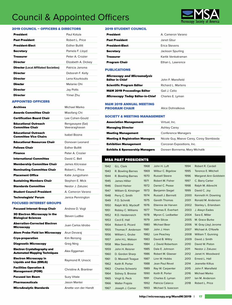
2019 COUNCIL – OFFICERS & DIRECTORS
President Paul Kotula
Past President Robert L. Price
President-Elect Esther Bullitt
Secretary Pamela F. Lloyd
Treasurer Peter A. Crozier
Director Elizabeth A. Dickey
Director (Local Affiliated Societies) Patricia Jansma
Director Deborah F. Kelly
Director Lena Kourkoutis
Director Melanie Ohi
Director Jay Potts
Director Yimei Zhu APPOINTED OFFICERS
Archives Michael Marko
Awards Committee Chair Miaofang Chi
Certification Board Chair Lee Cohen-Gould
Educational Outreach Committee Chair
Rengasayee (Sai) Veeraraghavan
Educational Outreach Committee Vice Chairs Isabel Boona
Educational Resources Chair Donovan Leonard
Fellows Chair Esther Bullitt
Finance Peter A. Crozier
International Committee David C. Bell
Membership Committee Chair James Kilcrease
Nominating Committee Chair Robert L. Price
Placement Office Katie Jungjohann
Sustaining Members Chair Stephen E. Mick
Standards Committee Nestor J. Zaluzec
Student Council President A. Cameron Varano
Technologists’ Forum Janice Pennington
FOCUSED INTEREST GROUPS
Focused Interest Group Chair Andrew D. Vogt
3D Electron Microscopy in the Biological Sciences Steven Ludtke
Aberration-Corrected Electron Microscopy Juan Carlos Idrobo
Atom Probe Field Ion Microscopy Arun Devaraj
Cryo-preparation Kim Rensing
Diagnostic Microscopy Greg Ning
Electron Crystallography and Automated Mapping Techniques Alex Eggeman
Electron Microscopy in Liquids and Gas (EMLG) Raymond R. Unocic
Facilities Operation & Management (FOM) Christine A. Brantner
Focused Ion Beam Suzy Vitale
Pharmaceuticals Jason Mantei
MicroAnalytic Standards Anette von der Handt
http://microscopy.org/MandM/2019 | 17
Council & Appointed Officers2019 STUDENT COUNCIL
President A. Cameron Varano
Past President Janet Gbur
President-Elect Erica Stevens
Secretary Jackson Spurling
Treasurer Kartik Venkatraman
Program Chair Ethan L. Lawrence
PUBLICATIONS
Microscopy and Microanalysis Editor in Chief John F. Mansfield
Scientific Program Editor Richard L. Martens
M&M 2019 Proceedings Editor Gail J. Celio
Microscopy Today Editor-in-Chief Charles E. Lyman
M&M 2019 ANNUAL MEETING PROGRAM CHAIR
SOCIETY & MEETING MANAGEMENT
Association Management Virtual, Inc.
Managing Director Ashley Carey
Meeting Management Conference Managers
Meeting & Registration Managers Nicole Guy, Maeve Carey, Corey Siembieda
Exhibition Management Corcoran Expositions, Inc.
Exhibits & Sponsorship Managers Doreen Bonnema, Mary Michalik
1994 Robert R. Cardell
1995 Terence E. Mitchell
1996 Margaret Ann Goldstein
1997 C. Barry Carter
1998 Ralph M. Albrecht
1999 David C. Joy
2000 Kenneth H. Downing
2001 Ronald M. Anderson
2002 Stanley L. Erlandsen
2003 J. Alwyn Eades
2004 Sara E. Miller
2005 M. Grace Burke
2006 W. Gray (Jay) Jerome
2007 Michael A. O’Keefe
2008 William T. Gunning
2009 David J. Smith
2010 David W. Piston
2011 Nestor J. Zaluzec
2012 Janet H. Woodward
2013 Ernest L. Hall
2014 Jeanette Killius
2015 John F. Mansfield
2016 Michael Marko
2017 Ian M. Anderson
2018 Robert L. Price
Alice Dohnalkova
1968 John H. Luft
1969 Wilbur C. Bigelow
1970 Russell Steere
1971 Robert M. Fisher
1972 Daniel C. Pease
1973 Benjamin Siegel
1974 Russell J. Barrnett
1975 Gareth Thomas
1976 Etienne de Harven
1977 Thomas E. Everhart
1978 Myron C. Ledbetter
1979 John Silcox
1980 Michael Beer
1981 John J. Hren
1982 Lee Peachey
1983 David B. Wittry
1984 J. David Robertson
1985 Dale E. Johnson
1986 Robert M. Glaeser
1987 Linn W. Hobbs
1988 Jean Paul Revel
1989 Ray W. Carpenter
1990 Keith R. Porter
1991 Charles E. Lyman
1992 Patricia Calarco
1993 Michael S. Isaacson
MSA PAST PRESIDENTS
1942 G.L. Clark
1943 R. Bowling Barnes
1944 R. Bowling Barnes
1945 James Hillier
1946 David Harker
1947 William G. Kinsinger
1948 Perry C. Smith
1949 F.O. Schmitt
1950 Ralph W.G. Wyckoff
1951 Robley C. Williams
1952 R.D. Heidenreich
1953 Cecil E. Hall
1954 Robert G. Picard
1955 Thomas F. Anderson
1956 William L. Grube
1957 John H.L. Watson
1958 Max Swerdlow
1959 John H. Reisner
1960 D. Gordon Sharp
1961 D. Maxwell Teague
1962 Keith R. Porter
1963 Charles Schwartz
1964 Sidney S. Breese
1965 Virgil G. Peck
1966 Walter Frajola
1967 Joseph J. Comer
Modi�ed A
Thank you
1942-201775 ye
ars
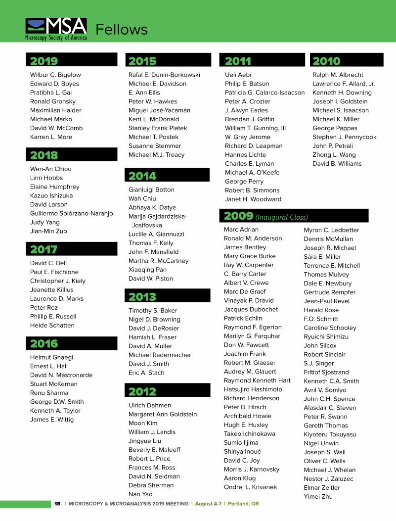
Fellows
2015 Rafal E. Dunin-BorkowskiMichael E. DavidsonE. Ann EllisPeter W. HawkesMiguel José-YacamánKent L. McDonaldStanley Frank PlatekMichael T. PostekSusanne StemmerMichael M.J. Treacy
2014Gianluigi BottonWah ChiuAbhaya K. DatyeMarija Gajdardziska- JosifovskaLucille A. GiannuzziThomas F. KellyJohn F. MansfieldMartha R. McCartneyXiaoqing PanDavid W. Piston
2013Timothy S. BakerNigel D. BrowningDavid J. DeRosierHamish L. FraserDavid A. MullerMichael RadermacherDavid J. SmithEric A. Stach
2012Ulrich DahmenMargaret Ann GoldsteinMoon KimWilliam J. LandisJingyue LiuBeverly E. MaleeffRobert L. PriceFrances M. RossDavid N. SeidmanDebra ShermanNan Yao
2019Wilbur C. BigelowEdward D. BoyesPratibha L. GaiRonald GronskyMaximilian HaiderMichael MarkoDavid W. McCombKarren L. More
2018Wen-An ChiouLinn HobbsElaine HumphreyKazuo IshizukaDavid LarsonGuillermo Solórzano-NaranjoJudy YangJian-Min Zuo
2017David C. BellPaul E. FischioneChristopher J. KielyJeanette KilliusLaurence D. MarksPeter RezPhillip E. RussellHeide Schatten
2016Helmut GnaegiErnest L. HallDavid N. MastronardeStuart McKernanRenu Sharma George D.W. Smith Kenneth A. Taylor James E. Wittig
2011Ueli AebiPhilip E. BatsonPatricia G. Calarco-IsaacsonPeter A. CrozierJ. Alwyn EadesBrendan J. GriffinWilliam T. Gunning, IIIW. Gray JeromeRichard D. LeapmanHannes LichteCharles E. LymanMichael A. O’KeefeGeorge PerryRobert B. SimmonsJanet H. Woodward
2009 (Inaugural Class)Marc AdrianRonald M. AndersonJames BentleyMary Grace BurkeRay W. CarpenterC. Barry CarterAlbert V. CreweMarc De GraefVinayak P. DravidJacques DubochetPatrick EchlinRaymond F. EgertonMarilyn G. FarquharDon W. FawcettJoachim FrankRobert M. GlaeserAudrey M. GlauertRaymond Kenneth HartHatsujiro HashimotoRichard HendersonPeter B. HirschArchibald HowieHugh E. HuxleyTakeo IchinokawaSumio IijimaShinya Inoué David C. JoyMorris J. KarnovskyAaron KlugOndrej L. Krivanek
Myron C. LedbetterDennis McMullanJoseph R. MichaelSara E. MillerTerrence E. MitchellThomas MulveyDale E. NewburyGertrude RempferJean-Paul RevelHarald RoseF.O. SchmittCaroline SchooleyRyuichi ShimizuJohn SilcoxRobert SinclairS.J. SingerFritiof SjostrandKenneth C.A. SmithAvril V. SomlyoJohn C.H. SpenceAlasdair C. StevenPeter R. SwannGareth ThomasKiyoteru TokuyasuNigel UnwinJoseph S. WallOliver C. WellsMichael J. WhelanNestor J. ZaluzecElmar ZeitlerYimei Zhu
2010Ralph M. AlbrechtLawrence F. Allard, Jr.Kenneth H. DowningJoseph I. GoldsteinMichael S. IsaacsonMichael K. MillerGeorge PappasStephen J. PennycookJohn P. PetraliZhong L. WangDavid B. Williams
| MICROSCOPY & MICROANALYSIS 2019 MEETING | August 4-7 | Portland, OR18
Distinguished Scientist Awards1942-201775 ye
ars
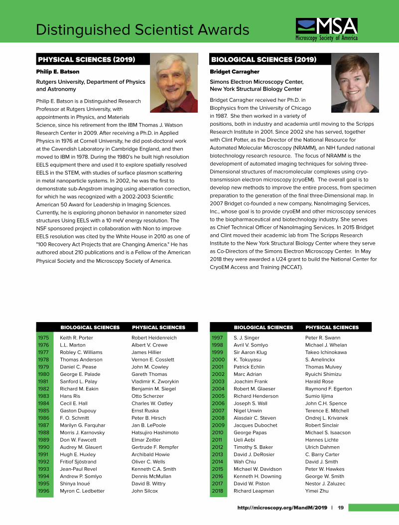
PHYSICAL SCIENCES (2019)
Philip E. Batson
Rutgers University, Department of Physics and Astronomy
Philip E. Batson is a Distinguished Research Professor at Rutgers University, with appointments in Physics, and Materials Science, since his retirement from the IBM Thomas J. Watson Research Center in 2009. After receiving a Ph.D. in Applied Physics in 1976 at Cornell University, he did post-doctoral work at the Cavendish Laboratory in Cambridge England, and then moved to IBM in 1978. During the 1980’s he built high resolution EELS equipment there and used it to explore spatially resolved EELS in the STEM, with studies of surface plasmon scattering in metal nanoparticle systems. In 2002, he was the first to demonstrate sub-Angstrom imaging using aberration correction, for which he was recognized with a 2002-2003 Scientific American 50 Award for Leadership in Imaging Sciences. Currently, he is exploring phonon behavior in nanometer sized structures Using EELS with a 10 meV energy resolution. The NSF sponsored project in collaboration with Nion to improve EELS resolution was cited by the White House in 2010 as one of "100 Recovery Act Projects that are Changing America." He has authored about 210 publications and is a Fellow of the American
Physical Society and the Microscopy Society of America.
BIOLOGICAL SCIENCES (2019)
Bridget Carragher
Simons Electron Microscopy Center, New York Structural Biology Center
Bridget Carragher received her Ph.D. in Biophysics from the University of Chicago in 1987. She then worked in a variety of positions, both in industry and academia until moving to the Scripps Research Institute in 2001. Since 2002 she has served, together with Clint Potter, as the Director of the National Resource for Automated Molecular Microscopy (NRAMM), an NIH funded national biotechnology research resource. The focus of NRAMM is the development of automated imaging techniques for solving three-Dimensional structures of macromolecular complexes using cryo-transmission electron microscopy (cryoEM). The overall goal is to develop new methods to improve the entire process, from specimen preparation to the generation of the final three-Dimensional map. In 2007 Bridget co-founded a new company, NanoImaging Services, Inc., whose goal is to provide cryoEM and other microscopy services to the biopharmaceutical and biotechnology industry. She serves as Chief Technical Officer of NanoImaging Services. In 2015 Bridget and Clint moved their academic lab from The Scripps Research Institute to the New York Structural Biology Center where they serve as Co-Directors of the Simons Electron Microscopy Center. In May 2018 they were awarded a U24 grant to build the National Center for CryoEM Access and Training (NCCAT).
1997 S. J. Singer Peter R. Swann1998 Avril V. Somlyo Michael J. Whelan1999 Sir Aaron Klug Takeo Ichinokawa2000 K. Tokuyasu S. Amelinckx2001 Patrick Echlin Thomas Mulvey2002 Marc Adrian Ryuichi Shimizu2003 Joachim Frank Harald Rose2004 Robert M. Glaeser Raymond F. Egerton2005 Richard Henderson Sumio Iijima2006 Joseph S. Wall John C.H. Spence2007 Nigel Unwin Terence E. Mitchell2008 Alasdair C. Steven Ondrej L. Krivanek2009 Jacques Dubochet Robert Sinclair2010 George Papas Michael S. Isaacson2011 Ueli Aebi Hannes Lichte2012 Timothy S. Baker Ulrich Dahmen2013 David J. DeRosier C. Barry Carter2014 Wah Chiu David J. Smith2015 Michael W. Davidson Peter W. Hawkes2016 Kenneth H. Downing George W. Smith 2017 David W. Piston Nestor J. Zaluzec2018 Richard Leapman Yimei Zhu
BIOLOGICAL SCIENCES PHYSICAL SCIENCES BIOLOGICAL SCIENCES PHYSICAL SCIENCES
1975 Keith R. Porter Robert Heidenreich1976 L.L. Marton Albert V. Crewe1977 Robley C. Williams James Hillier1978 Thomas Anderson Vernon E. Cosslett1979 Daniel C. Pease John M. Cowley1980 George E. Palade Gareth Thomas1981 Sanford L. Palay Vladimir K. Zworykin1982 Richard M. Eakin Benjamin M. Siegel1983 Hans Ris Otto Scherzer1984 Cecil E. Hall Charles W. Oatley1985 Gaston Dupouy Ernst Ruska1986 F. O. Schmitt Peter B. Hirsch1987 Marilyn G. Farquhar Jan B. LePoole1988 Morris J. Karnovsky Hatsujiro Hashimoto1989 Don W. Fawcett Elmar Zeitler1990 Audrey M. Glauert Gertrude F. Rempfer1991 Hugh E. Huxley Archibald Howie1992 Fritiof Sjöstrand Oliver C. Wells1993 Jean-Paul Revel Kenneth C.A. Smith1994 Andrew P. Somlyo Dennis McMullan1995 Shinya Inoué David B. Wittry1996 Myron C. Ledbetter John Silcox
http://microscopy.org/MandM/2019 | 19
1942-201775 ye
ars
Major Society AwardsBURTON MEDAL AWARD (2019)
Hari ShroffNational Institute of Health
Dr. Hari Shroff received a B.S.E. in bioengineering from the University of Washington in 2001, and under the supervision of Dr. Jan Liphardt, completed his Ph.D. in biophysics at the University of California at Berkeley in 2006 . He spent the next three years performing postdoctoral research under the mentorship of Eric Betzig at the Howard Hughes Medical Institute's Janelia Farm Research Campus where his research focused on development of photactivated localization microscopy (PALM), an optical super-resolution technique. Dr. Shroff is now chief of NIBIB's Section on High Resolution Optical Imaging laboratory, where he and his staff are developing new imaging tools for application in biological research. Current research areas include further development of super-resolution microscopy, light-sheet microscopy, inverse imaging problems, deep learning for microscopy, and the study of neurodevelopment in C. elegans.
YEAR RECIPIENT
1975 James Lake1976 Michael S. Isaacson1977 Robert Sinclair1978 David C. Joy1979 Norton B. Gilula1980 John C.H. Spence1981 Barbara J. Panessa-Warren1982 Nestor J. Zaluzec1983 Ronald Gronsky1984 David B. Williams1985 Richard D. Leapman1986 J. Murray Gibson1987 Ron A.Milligan1988 A.D. Romig, Jr.1989 Laurence D. Marks1990 W. Mason Skiff1991 Joseph R. Michael1992 Kannan M. Krishnan1993 Joseph A.N. Zasadzinski1994 Jan M. Chabala1995 Joanna L. Batstone1996 Vinayak P. Dravid
1997 P.M. Ajayan1998 Ian M. Anderson1999 Zhong Lin Wang2000 Eva Nogales2001 Jian Min Zuo2002 Nigel D. Browning2003 Frances M. Ross2004 Z. Hong Zhou2005 David J. Larson2006 David A. Muller2007 Peter D. Nellist2008 Steven J. Ludtke2009 Eric A. Stach2010 Sergei V. Kalinin2011 Radostin Danev2012 David S. Ginger2013 John L. Rubinstein2014 Maria Varela2015 Andrew M. Minor2016 Miaofang Chi2017 Christopher J. Russo2018 Lena F. Kourkoutis
MORTON D. MASER DISTINGUISHED SERVICE AWARD (2019)
No 2019 awardee.
YEAR RECIPIENT
1992 Ronald M. AndersonG. W. BaileyFrances L. BallM. Blair BowersDeborah L. ClaytonJoseph HarbKenneth R. LawlessMorton D. MaserCaroline SchooleyJohn H.L. Watson
1993 E. Laurence Thurston1994 Richard F.E. Crang1995 Raymond K. Hart1996 José A. Mascorro1997 William T. Gunning III1998 Nestor J. Zaluzec1999 Charles E. Lyman2000 Barbara A. Reine
Hildegard H. Crowley
2002 Beverly E. Maleeff2003 M. Grace Burke2004 Ralph M. Albrecht2005 W. Gray (Jay) Jerome2006 Jeanette Killius2007 Robert L. Price2008 Stuart McKernan2010 Pamela F. Lloyd2011 Janet H. Woodward2012 Gina E. Sosinsky2013 Caroline A. Miller2014 Michael Marko2015 JoAn Hudson2016 Amanda Lawrence2017 David W. Tomlin2018 Donovan N. Leonard
| MICROSCOPY & MICROANALYSIS 2019 MEETING | August 4-7 | Portland, OR20
Major Society Awards1942-201775 ye
ars
YEAR RECIPIENT
2012 Gabriel C. Lander2013 Peng Ge2014 Ricardo C. Guerrero-Ferreira2015 Alexey Amunts2016 Dmitry Lyumkis2017 Rengasayee Veeraraghavan2018 Not awarded
YEAR RECIPIENT
2012 Wu Zhou2013 Lena Fitting-Kourkoutis2014 Jinwoo Hwang2015 Meng Gu2016 Ryo Ishikawa2017 Pinshane Y. Huang 2018 Timothy Pennycook
ALBERT CREWE AWARD (2019)
B. Layla MehdiUniversity of Liverpool
Dr B. Layla Mehdi is currently an Assistant Professor and Associate Director of the Imaging Centre at the University of Liverpool (ICaL), UK. She received her Master’s in Analytical Chemistry from the University of Warsaw, Poland and her Ph. D. in Chemistry from Miami University, USA working in the area of electrochemical detectors coupled with gas chromatography for cancer therapy. Following her Ph.D., in 2013 she joined the Physical Sciences Directorate at the Pacific Northwest National Laboratory (PNNL) as a postdoctoral research associate and in 2016 was promoted to Staff Scientist. Her work at PNNL involved the development of an In Situ stage to study dynamic processes in next generation batteries with applications to Li-Ion and beyond Li chemistries being supported as part of the Joint Centre for Energy Storage Research (JCESR) funded by the US Department of Energy. She has received numerous international awards for this work, including the 2015 MRS postdoctoral award, the 2015 Microscopy Society of America postdoctoral award and the 2014 Microscopy & Microanalysis Presidential award. In 2016 she also received JSPS Postdoctoral Fellowship to perform Research at Nagoya University, Japan in collaboration with TOYOTA Japan. She has over 20 publications in the development and application of low-dose methods to the operando and high resolution study of beam sensitive materials and processes. She has organized 4 international In Situ liquid TEM workshops, an international In Situ TEM symposium, has given over 25 invited talks at international meetings and institutions, and is the Associate Editor covering In Situ TEM for the SpringerNature journal, Advanced Structural and Chemical Imaging. Currently, her research group focuses on developing advanced new microscopy methods to generate an in depth understanding of reaction kinetics at solid/liquid and solid/gas interfaces in batteries, electrocatalysts and pharmaceuticals.
GEORGE PALADE AWARD (2019)
http://microscopy.org/MandM/2019 | 21
Alex NobleNew York Structural Biology Center
Alex Noble earned his BS in Physics and BA in Applied Mathematics from UC San Diego and his MS and PhD in Physics at Florida State University. He is currently an NIH Kirschstein Postdoctoral fellow in the laboratory of Bridget Carragher and Clint Potter at the Simons Electron Microscopy Center (SEMC) in the New York Structural Biology Center. He is broadly focused on developing, distributing, and applying methods that further the progress of the cryo-electron microscopy (cryoEM) field and individual cryoEM projects, along with applying those methods himself to specific biological systems. Each focus is driven by a motivation to make positive biomedical and thus humanitarian impacts. As a graduate student, he investigated a model of a coat protein complex II cage by combining cryoEM, hydrogen-deuterium exchange mass spectrometry, and molecular modelling. Beginning as a graduate student and continuing into his postdoctoral training, he develops and maintains software, Appion-Protomo, that enables researchers of all experience levels to process cryo-electron tomography (cryoET) images without the need for additional sample or imaging optimization. As a postdoctoral fellow in a highly-collaborative environment, he has illuminated and investigated a long-standing and widespread problem with single particle cryoEM sample preparation that has numerous broad implications – protein adsorption to the air-water interface. Through collaborative efforts internal and external to SEMC, he investigated solutions to this problem in the form of cryoEM grid freezing techniques (the Spotiton grid preparation robot) and sample detergent optimization. His postdoctoral fellowship, spurred by a fruitful cryoET collaboration, seeks to understand the structural behavior of type II cadherins on membranes, which are a set of cell adhesion molecules with several pathological implications if malformed. His current investigations and efforts also include several projects designed to obtain three-Dimensional cryoET structures of cellular interiors Using cryo-focused ion beam preparation, developing and overseeing deep learning applications in cryoEM/ET, assisting cryoET users at SEMC, and leading cryoET training workshops.
HILDEGARD H. CROWLEY OUTSTANDING TECHNOLOGIST AWARD FOR BIOLOGICAL SCIENCES (2019)
Matthew S. JoensWashington University School of Medicine
Matthew is an analytical chemist by undergraduate training and has over 10 years of electron microscopy experience. His introduction into microscopy included internships at the University of California – San Diego (UCSD) with Dr. Timothy Baker, one of the founding fathers of cryo-electron microscopy, and with the National Center for Microscopy Imaging Research (NCMIR), a NIH funded lab headed by Dr. Mark Ellisman. Following these internships, he moved to the Salk Institute for Biological Studies where he worked with Dr. James Fitzpatrick to establish and grow the electron microscopy division of the Waitt Advanced Biophotonics Center. Matthew was later recruited by Dr. Fitzpatrick to help build the Center for Cellular Imaging at the Washington University School of Medicine in St. Louis. His background includes extensive experience with cryo sample preparation, immunolabeling, 3D electron microscopy, and selective staining chemistries. His main interests are in correlative sample preparation and imaging techniques, spanning light, X-Ray, ion, and electron microscopies.
1942-201775 ye
ars
| MICROSCOPY & MICROANALYSIS 2019 MEETING | August 4-7 | Portland, OR22
Major Society Awards
CHUCK FIORI OUTSTANDING TECHNOLOGIST AWARD FOR PHYSICAL SCIENCES (2019)
Dmitri ZakharovBrookhaven National Laboratory
Dmitri N. Zakharov received his M.S. degree in Solid State Physics from Moscow State Engineering Physical Institute, Department of Theoretical and Experimental Physics in 1995, his Ph.D. in Solid State Physics from the Institute of Crystallography of the Russian Academy of Sciences in 2001, and postdoctoral trainings at Max Planck Institute of Microstructure Physics and Lawrence Berkeley National Laboratory. After spending 6 years as a Staff Scientist at Birck Nanotechnology Center at Purdue University, Dr. Zakharov joined the Center for Functional Nanomaterials at Brookhaven National Laboratory in 2012 in a Staff Scientist role. Dr. Zakharov’s research interests include environmental scanning/transmission electron microscopy (ETEM) technique; automated low dose image acquisition of electron beam sensitive materials; Machine Learning algorithms and Big Data analysis for Real-Time image streams processing to extract quantitative information; operando experimental setup utilizing ETEM and the National Synchrotron Light Source II as two complimentary techniques; carbon nanotube nucleation, growth, termination and chirality control; 2D and III-Nitride materials and devices. Dr. Zakharov has over 150 journal and conference publications, which have been cited more then 3,200 times (h-index 33), and he has presented over 30 invited talks in the US and abroad. Dmitri was recipient of 2014 Spotlight Award by Brookhaven National Laboratory and Mikhail Teplov Scholarship in 2000. He also served as Graduate Faculty at the School of Materials Engineering at Purdue University.
YEAR RECIPIENT
1993 not awarded1994 Bernard J. Kestel1995 not awarded1996 David W. Ackland1997 Stanley J. Klepeis1998 Charles J. Echer1999 John C. Wheatley2000 not awarded2001 Conrad G. Bremer2002 not awarded2003 Edward A. Ryan2004 Mark C. Reuter2005 Chris Nelson2008 not awarded
YEAR RECIPIENT
1993 Ben O. Spurlock1994 not awarded1995 Kai Chien1996 not awarded1997 John P. Benedict1998 Hilton H. Mollenhauer1999 John M. Basgen2000 Nancy Crise Smith2001 not awarded2002 José A. Mascorro2003 not awarded2004 not awarded2005 John J. Bozzola2008 Thomas Deerinck2009 Mary Morphew2010 E. Ann Ellis2011 Robert Grassucci2012 Kunio Nagashima2013 Robyn Roth2014 Hong Yi2015 Norman Olson2016 Frank Macaluso2017 Patricia S. Connelly2018 Anchi Cheng
http://microscopy.org/MandM/2014 for program details [23]
The MSA MEGABOOTH showcases all that MSA
a member, stop by to catch up on all the new society developments. Member information available at Regular, Sustaining (corporate), and Student levels.
Sign up for VENDOR TUTORIALS here! These popular sessions are presented on Monday, Tuesday, and Wednesday evenings after the exhibit hall has closed for the day. Don’t miss out – advance registration is required!
The TECHNOLOGISTS’ FORUM (TF): Attention
grow and develop your skills, your professional career, and your network by joining the Forum!
The PLACEMENT OFFICE is MSA’s job-listing service. Post a job, peruse job listings, post a
for your job opening. All for FREE during the meeting!
MSA MegaBooth in the Exhibit Hall Open during all
exhibit hall hours
The INTERNET CAFÉ and PHONE CHARGING STATION are open to all meeting attendees during all exhibit hall hours. Bring Your Own Device! Lots of places to sit and rest your feet for a few minutes while you charge your mobile phone, check your email, put the �nishing touches on your talk, or collaborate with colleagues.
For more information, visit http://microscopy.org
Check out the BOOK DISPLAY – publisher-donated books, divided into biological/physical topics. Several new titles added every year! Come and browse the newest titles.
CERTIFICATION BOARD – Find out about MSA’s certi�cation program for Electron Microscopy Technologists and how being certi�ed can help you in your next job search!
MICROSCOPY TODAY and MICROSCOPY and MICROANALYSIS are the society’s two publications – one a magazine format, the other a peer-reviewed scienti�c journal. Information for authors and advertisers is available here.
EDUCATIONAL OUTREACH – Includes MSA’s educational outreach program. Browse the materials and �nd out how to start an outreach program in your local area. Get details on the special programming at the M&M meeting for educators and kids of all ages.
Visit the updated Project MICRO display to learn about this organization's education and outreach goals.
2009 Lynne Gignac2010 not awarded2011 not awarded2012 not awarded2013 K. Shawn Reeves2014 Eddy Garcia-Meitin2015 Masahiro Kawasaki2016 not awarded2017 Richard L. Martens2018 Chengyu Song
edax.com
Elite technology for elite results
What’s New at M&M 2019,
EDAX Booth #428?
• Latest developments in detector technology for EBSD analysis
• The fastest EBSD camera in the world powered by a CMOS sensor ★ Velocity™ Plus up to 3,000 indexed points per second ★ Velocity™ Super up to 4,500 indexed points per second • Dictionary indexing/Dynamic pattern simulation module for OIM Analysis™ • Preview APEX™ 2.0 software for integrated EDS-EBSD analysis
• Elite T EDS System for TEM now with 70 mm2 and 160 mm2 sensors • Coming soon....patented CdTe module for high-energy X-ray analysis for TEM
For more information about what’s new at EDAX, please visit edax.com/MM2019 or contact us at [email protected]
M&M_showguide_ad_8.5x11_2019.qxp_Layout 1 5/10/19 10:05 AM Page 1
http://microscopy.org/MandM/2019 | 25
Microanalysis Society Officers
PAST PRESIDENTS
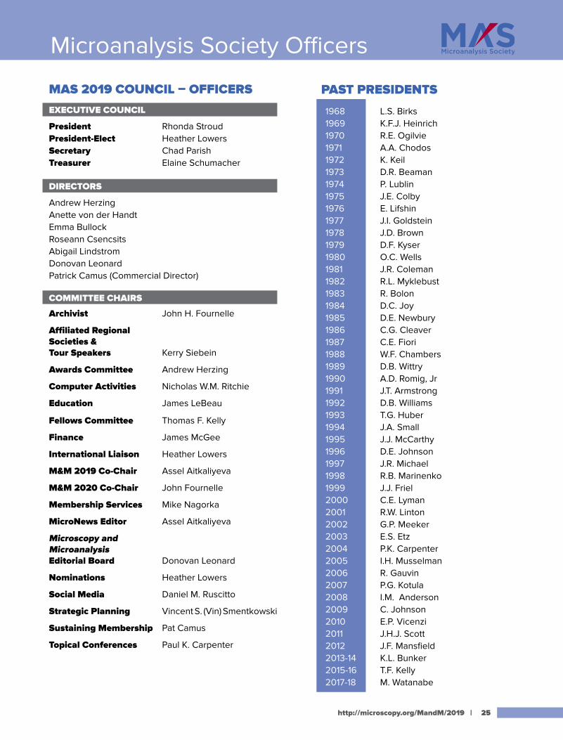
1968 L.S. Birks1969 K.F.J. Heinrich1970 R.E. Ogilvie1971 A.A. Chodos1972 K. Keil1973 D.R. Beaman1974 P. Lublin1975 J.E. Colby1976 E. Lifshin1977 J.I. Goldstein1978 J.D. Brown1979 D.F. Kyser1980 O.C. Wells1981 J.R. Coleman1982 R.L. Myklebust1983 R. Bolon1984 D.C. Joy1985 D.E. Newbury1986 C.G. Cleaver1987 C.E. Fiori1988 W.F. Chambers1989 D.B. Wittry1990 A.D. Romig, Jr1991 J.T. Armstrong1992 D.B. Williams1993 T.G. Huber1994 J.A. Small1995 J.J. McCarthy1996 D.E. Johnson1997 J.R. Michael1998 R.B. Marinenko1999 J.J. Friel2000 C.E. Lyman2001 R.W. Linton2002 G.P. Meeker2003 E.S. Etz2004 P.K. Carpenter2005 I.H. Musselman2006 R. Gauvin2007 P.G. Kotula2008 I.M. Anderson2009 C. Johnson2010 E.P. Vicenzi2011 J.H.J. Scott2012 J.F. Mansfield2013-14 K.L. Bunker2015-16 T.F. Kelly2017-18 M. Watanabe
MAS 2019 COUNCIL – OFFICERS EXECUTIVE COUNCIL
President Rhonda StroudPresident-Elect Heather LowersSecretary Chad ParishTreasurer Elaine Schumacher
DIRECTORS
Andrew HerzingAnette von der HandtEmma BullockRoseann CsencsitsAbigail LindstromDonovan LeonardPatrick Camus (Commercial Director)
COMMITTEE CHAIRS
Archivist John H. Fournelle
Affiliated Regional Societies & Tour Speakers Kerry Siebein
Awards Committee Andrew Herzing
Computer Activities Nicholas W.M. Ritchie
Education James LeBeau
Fellows Committee Thomas F. Kelly
Finance James McGee
International Liaison Heather Lowers
M&M 2019 Co-Chair Assel Aitkaliyeva
M&M 2020 Co-Chair John Fournelle
Membership Services Mike Nagorka
MicroNews Editor Assel Aitkaliyeva
Microscopy and Microanalysis Editorial Board Donovan Leonard
Nominations Heather Lowers
Social Media Daniel M. Ruscitto
Strategic Planning Vincent S. (Vin) Smentkowski
Sustaining Membership Pat Camus
Topical Conferences Paul K. Carpenter
Major Society Awards
PREVIOUS AWARDEES
2007 D.B. Williams
2008 J. I. Goldstein
2009 D.E. Newbury
2010 D.C. Joy
2011 J.R. Michael
2012 J. Bentley
2013 E. Lifshin
2014 O. L. Krivanek
2015 P. J. Statham
2016 D. Muller
2017 T. F. Kelly
2018 R.D. Leapman
PREVIOUS AWARDEES
1986 P.J. Statham 1987 J.T. Armstrong1988 D.B. Williams1989 R.D. Leapman1990 R.W. Linton1991 A.D. Romig, Jr.1992 S.J. Pennycook1993 P.E. Russell1994 J.R. Michael1995 E.N. Lewis1997 R. Gauvin1998 V.P. Dravid1999 J. Bruley2000 H. Ade2001 C. Jacobsen
2002 D.A. Wollman2005 M. Watanabe2006 M. Toth2007 G. Kothleitner2008 P.G. Kotula2009 D. Drouin2010 H. Demers2011 L.N. Brewer2012 E.A. Marquis2013 J.M. LeBeau2014 B.P. Gorman2015 P. Pinard2016 J. Allaz2017 A. Herzing
2018 Y.N. Picard
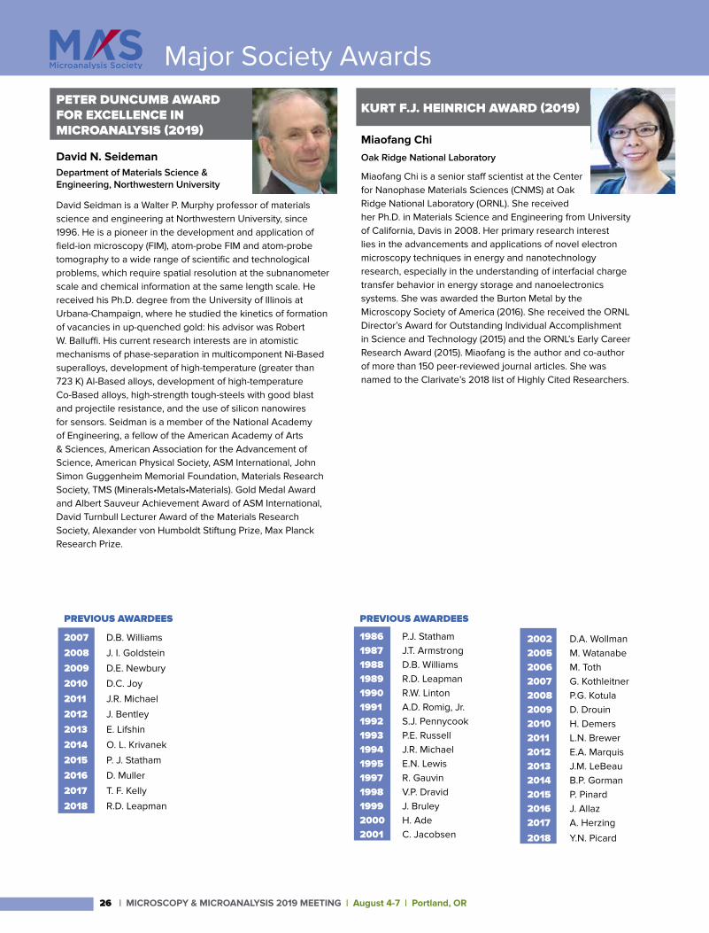
PETER DUNCUMB AWARD FOR EXCELLENCE IN MICROANALYSIS (2019)
David N. SeidemanDepartment of Materials Science & Engineering, Northwestern University
David Seidman is a Walter P. Murphy professor of materials science and engineering at Northwestern University, since 1996. He is a pioneer in the development and application of field-ion microscopy (FIM), atom-probe FIM and atom-probe tomography to a wide range of scientific and technological problems, which require spatial resolution at the subnanometer scale and chemical information at the same length scale. He received his Ph.D. degree from the University of Illinois at Urbana-Champaign, where he studied the kinetics of formation of vacancies in up-quenched gold: his advisor was Robert W. Balluffi. His current research interests are in atomistic mechanisms of phase-separation in multicomponent Ni-Based superalloys, development of high-temperature (greater than 723 K) Al-Based alloys, development of high-temperature Co-Based alloys, high-strength tough-steels with good blast and projectile resistance, and the use of silicon nanowires for sensors. Seidman is a member of the National Academy of Engineering, a fellow of the American Academy of Arts & Sciences, American Association for the Advancement of Science, American Physical Society, ASM International, John Simon Guggenheim Memorial Foundation, Materials Research Society, TMS (Minerals•Metals•Materials). Gold Medal Award and Albert Sauveur Achievement Award of ASM International, David Turnbull Lecturer Award of the Materials Research Society, Alexander von Humboldt Stiftung Prize, Max Planck Research Prize.
KURT F.J. HEINRICH AWARD (2019)
Miaofang ChiOak Ridge National Laboratory
Miaofang Chi is a senior staff scientist at the Center for Nanophase Materials Sciences (CNMS) at Oak Ridge National Laboratory (ORNL). She received her Ph.D. in Materials Science and Engineering from University of California, Davis in 2008. Her primary research interest lies in the advancements and applications of novel electron microscopy techniques in energy and nanotechnology research, especially in the understanding of interfacial charge transfer behavior in energy storage and nanoelectronics systems. She was awarded the Burton Metal by the Microscopy Society of America (2016). She received the ORNL Director’s Award for Outstanding Individual Accomplishment in Science and Technology (2015) and the ORNL’s Early Career Research Award (2015). Miaofang is the author and co-author of more than 150 peer-reviewed journal articles. She was named to the Clarivate’s 2018 list of Highly Cited Researchers.
| MICROSCOPY & MICROANALYSIS 2019 MEETING | August 4-7 | Portland, OR26
http://microscopy.org/MandM/2019 | 27
Major Society Awards
PREVIOUS AWARDEES
1977 P. Lublin1978 D.R. Beaman1979 M.A. Giles1980 A.A. Chodos1981 R.L. Myklebust1982 J. Doyle 1983 D.E. Newbury1984 J.I. Goldstein1985 M.C. Finn1986 V. Shull1987 D.C. Joy1988 C.G. Cleaver1989 W.F. Chambers1990 C.E. Fiori1991 T.G. Huber1992 E.S. Etz1993 H.A. Freeman1994 J.L. Worrall1995 R.W. Linton1996 P. F. Hlava1997 J.A. Small
1998 J.J. McCarthy1999 T.G. Huber2000 R.B. Marinenko2001 C.E. Lyman2002 J.F. Mansfield2003 I.H. Musselman2004 J.R. Michael2005 G.P. Meeker2006 H.A. Freeman2007 P.K. Carpenter2008 L.M. Ross2009 V. Woodward2010 S.A. Wight2011 D.T. Kremser2012 C. Johnson2013 J.J. McGee2014 I.M. Anderson2015 S. McKernan2016 H. Lowers2017 D. Kremser2018 V. Robertson
PREVIOUS AWARDEES
1977 R. Castaing1978 K.F.J. Heinrich1979 P. Duncumb1980 D.B. Wittry1981 S.J.B. Reed1982 R. Shimizu1983 J. Philibert1984 L.S. Birks1985 E. Lifshin1986 R.L. Myklebust1987 O.C. Wells1988 J.D. Brown1989 J. Hillier1990 T.E. Everhart1997 D.B. Williams1998 F.H. Schamber1999 R.A. Sareen2000 R.F. Egerton2001 P.E. Batson
2002 K. Keil2003 P.E. Russell2004 J.T. Armstrong2005 G. Slodzian2006 B.J. Griffin2007 R.D. Leapman2008 T. F. Kelly2009 J.R. Michael2010 J.J. Donovan2011 P.J. Statham2012 N.J. Zaluzec2013 P. Echlin2014 H.L. Fraser2015 M.R. Keenan2016 M. Jercinovic2017 M.K. Miller2018 M.G. Burke
2002 D.A. Wollman2005 M. Watanabe2006 M. Toth2007 G. Kothleitner2008 P.G. Kotula2009 D. Drouin2010 H. Demers2011 L.N. Brewer2012 E.A. Marquis2013 J.M. LeBeau2014 B.P. Gorman2015 P. Pinard2016 J. Allaz2017 A. Herzing
2018 Y.N. Picard
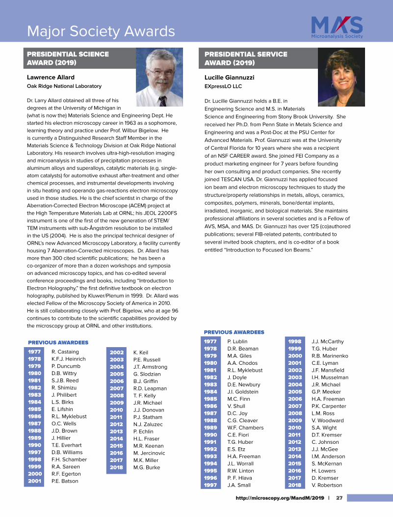
PRESIDENTIAL SCIENCE AWARD (2019)
Lawrence AllardOak Ridge National Laboratory
Dr. Larry Allard obtained all three of his degrees at the University of Michigan in (what is now the) Materials Science and Engineering Dept. He started his electron microscopy career in 1963 as a sophomore, learning theory and practice under Prof. Wilbur Bigelow. He is currently a Distinguished Research Staff Member in the Materials Science & Technology Division at Oak Ridge National Laboratory. His research involves ultra-high-resolution imaging and microanalysis in studies of precipitation processes in aluminum alloys and superalloys, catalytic materials (e.g. single-atom catalysts) for automotive exhaust after-treatment and other chemical processes, and instrumental developments involving in situ heating and operando gas-reactions electron microscopy used in those studies. He is the chief scientist in charge of the Aberration-Corrected Electron Microscope (ACEM) project at the High Temperature Materials Lab at ORNL; his JEOL 2200FS instrument is one of the first of the new generation of STEM/TEM instruments with sub-Ångström resolution to be installed in the US (2004). He is also the principal technical designer of ORNL’s new Advanced Microscopy Laboratory, a facility currently housing 7 Aberration-Corrected microscopes. Dr. Allard has more than 300 cited scientific publications; he has been a co-organizer of more than a dozen workshops and symposia on advanced microscopy topics, and has co-edited several conference proceedings and books, including “Introduction to Electron Holography,” the first definitive textbook on electron holography, published by Kluwer/Plenum in 1999. Dr. Allard was elected Fellow of the Microscopy Society of America in 2010. He is still collaborating closely with Prof. Bigelow, who at age 96 continues to contribute to the scientific capabilities provided by the microscopy group at ORNL and other institutions.
PRESIDENTIAL SERVICE AWARD (2019)
Lucille GiannuzziEXpressLO LLC
Dr. Lucille Giannuzzi holds a B.E. in Engineering Science and M.S. in Materials Science and Engineering from Stony Brook University. She received her Ph.D. from Penn State in Metals Science and Engineering and was a Post-Doc at the PSU Center for Advanced Materials. Prof. Giannuzzi was at the University of Central Florida for 10 years where she was a recipient of an NSF CAREER award. She joined FEI Company as a product marketing engineer for 7 years before founding her own consulting and product companies. She recently joined TESCAN USA. Dr. Giannuzzi has applied focused ion beam and electron microscopy techniques to study the structure/property relationships in metals, alloys, ceramics, composites, polymers, minerals, bone/dental implants, irradiated, inorganic, and biological materials. She maintains professional affiliations in several societies and is a Fellow of AVS, MSA, and MAS. Dr. Giannuzzi has over 125 (co)authored publications; several FIB-related patents, contributed to several invited book chapters, and is co-editor of a book entitled “Introduction to Focused Ion Beams.”
Outstanding Paper Awards for 2018
MAS OUTSTANDING PAPER AWARDS (2019)
These awards are presented annually to the authors of outstanding papers from the previous annual meeting in each of four categories.
RAYMOND CASTAING – BEST STUDENT PAPER AWARD:
Analysis of Redox Changes in Silicate Glasses Using EPMA and Raman Spectroscopy
Ery Hughes, University of Bristol, United Kingdom
V.G. MACRES – BEST INSTRUMENTATION/SOFTWARE PAPER AWARD:
The MTF and DQE of Annular Dark Field STEM: Implications for Low-dose Imaging and Compressed Sensing
Lewys Jones, Trinity College Dublin, Ireland
L.S. BIRKS – BEST CONTRIBUTED PAPER AWARD:
Low Energy STEM-EELS Characterization of Primitive Organic Matter and Silicates in the Meteorite LAP 02342Bradley De Gregorio, U.S. Naval Research Laboratory
V.E. COSSLETT – BEST INVITED PAPER AWARD:
Novel EELS Experiments in the Newly Opened Monochromatic RegimeJordan Hachtel, Oak Ridge National Laboratory
| MICROSCOPY & MICROANALYSIS 2019 MEETING | August 4-7 | Portland, OR28
Ian Anderson Phil Batson Paul Carpenter Bill Chambers John Donovan Vinayak Dravid Ray Egerton John Fournelle Hamish Fraser Raynald Gauvin Paul Hlava Thomas HuberMichael Jercinovic
Cathy Johnson Thomas Kelly Paul Kotula Charles Lyman John Mansfield Joseph Michael Inga Musselman Nicholas Ritchie John Henry Scott John Small Ed Vicenzi Masashi Watanabe Valerie Woodward
2019 MAS FELLOWS (INAUGURAL CLASS):
MICROSCOPY & MICROANALYSIS 2019 MEETING | August 4-8 | Portland, OR 29
Meeting Awards
M&M STUDENT SCHOLAR AWARDS—SPONSORED BY MSA
Fatemeh Abbasi Yeganeh, Florida State University
Yanyan Zhao, Stanford University (Miller Award)
Paul Cueva, Cornell University
Ha Dang, University of Washington
Julia Doh, Oregon Health & Science University
Amanda Erwin, University of Michigan
Alice Greenberg, University of Oregon
Catherine Groschner, University of California-Berkeley
Shen Han, Max Planck Institute for Polymer Research,
Germany Daniel Kelly, Manchester University, United Kingdom
Abinash Kumar, North Carolina State University
Ethan Lawrence, Arizona State University
Brandon McKeon, Arizona State University
Arthur Moya, University of Oxford, United Kingdom
Akshay Murthy, Northwestern University
Colum O'Leary, University of Oxford, United Kingdom
Will Parker, University of Oregon
Timothy Pegg, Miami University
Graham Rykiel, Oregon Health & Science University
Jonathan Schwartz, University of Michigan
Alexandra Sheader, University of Oxford, United Kingdom
Michelle Smeaton, Cornell University
Louisa Mezache, The Ohio State University
Janis Wirth, Friedrich-Alexander University, Germany
Yao Long Xing, Sung Kyun Kwan University, Korea
Reed Yalisove, University of Michigan
Hwanhui Yun, University of Minnesota
Ruopeng Zhang, University of California-Berkeley
Anika Burrell, University of Washington
M&M STUDENT SCHOLAR AWARDS - SPONSORED BY MAS
Kousuke Ooe, University of Tokyo, Japan
Kevin Schweinar, Max Planck Institute for Iron Research, Germany
Berit Goodge, Cornell University
Brian Zutter, University of California-Los Angeles
Charles Fletcher, The University of Oxford, United Kingdom
Meredith Sharps, University of Oregon
Heena Inani, University of Vienna, Austria
Yichao Zhang, University of Minnesota
Parivash Moradifar, Pennsylvania State University
Yitian Zeng, Stanford University
Komal Syed, University of Califormia-Irvine
M&M POSTDOCTORAL SCHOLAR AWARDS
Benjamin Apeleo Zubiri, Friedrich-Alexander University, Germany (APKARIAN AWARD–BIOLOGICAL SCIENCES)
Axel Brilot, University of California-San Francisco (APKARIAN AWARD–PHYSICAL SCIENCES)
Hamish Brown, Lawrence Berkeley Laboratory
Michael Buch, National Institutes of Health
Johannes Elferich, Oregon Health & Science University
Wenpei Gao, University of California-Irvine
Vivian Merk , Northwestern University
Aubrey Penn, North Carolina State University (ERIC SAMUELS SCHOLARSHIP)
Paul Smeets, Northwestern University
Wei-Chang Yang, National Institute of Standards and Technology
Andrew Yankovich, Chalmers University of Technology, Sweden
M&M PROFESSIONAL TECHNICAL STAFF AWARDS Leslie Cummins, Albert Einstein College of Medicine
Pauline Mochama, University of Minnesota
Sara Dickens, Sandia National Laboratories
Ann Johnson, The Dow Chemical Company
Society Information
30 | MICROSCOPY & MICROANALYSIS 2019 MEETING | August 4-7 | Portland, OR
Current IFES Steering Committee
David J. Larson PresidentJulie Cairney Vice-PresidentRoss Marceau SecretaryPaul Bagot TreasurerMattias ThuvanderArun DeverajGang ShaDavid SaxeyBaptiste Gault
IFES Past Presidents
2014 -present D.J. Larson
2008 - 2014 N. Kruse
2006 – 2008 T.F. Kelly
2002 – 2006 R.G. Forbes
2000 – 2001 D.N Seidman
1996 – 2000 R.G. Forbes
1993 – 1996 M.K. Miller
1990 – 1993 G.D.W. Smith
1987 – 1990 J.H. Block
IFES Fellows
Hans-Olof AndrénDidier BlavetteAlfred CerezoPaul CutlerFrédéric DanoixRichard ForbesGeorgiy FurseyRobert GomerKazuhiro HonoGary KelloggThomas KellyHans Juergen KreuzerNorbert KruseAllan Melmed
E.W. Müller Young Scientist Award(1978) A.R. Waugh
(1979) H.-W. Fink
(1980) Y. Kuk
(1981) S.J. Banard
(1982) J.M. Derochette
(1983) D.R. Kingham
(1984) M.G. Hetherington
(1985) M. Ahmad
(1986) L. Karlsson
(1987) P.P. Camus
(1988) A. Cerezo
(1989) J. Dirks
(1990) J.E. Brown
(1991) F. Danoix
(1992) H. Schmid
(1993) M.C. Reckzügel
(1994) R.C. Thomson
(1995) C. Voss
(1996) L. Li
(1997) C. Schmuck-Pareige
(1998) K. Nagaoka
(2001) Ch. Lang
(2002) E. A. Marquis
(2004) B. Cho
(2006) W.M. Tsang
(2008) M. Moors
(2010) P. Stender
(2012) M. Roussel
(2014) C. Oberdorfer
(2016) M. Dagan
(2018) S. Lambeets
The International Field Emission Society (IFES) is centred around the physics and application of high-field nanoscience, and in particular its application to Nano-Scale materials characterisation by atom probe microscopy. A major focus of the society is the promotion and development of atom probe microscopy methods and research.
Michael MillerMarwan MousaOsamu NishikawaJohn PanitzSimon RingerGuido SchmitzDavid SeidmanGeorge SmithKrystyna StillerLyn SwansonTien Tzou TsongNelia Wanderka
BE PREPARED at the Oregon Convention Center!In case of fire, medical emergency, or another emergency situation, Do Not Call 911. Call Building Security at (503) 731-7849.• Tell them the type of emergency (fire, medical) and the location and level.• Remain calm and follow directions.• Use (503) 731-7849 to report any other Security concerns.
Earthquakes• Portland is in an earthquake-prone area. If you experience an earthquake,
remain calm. * DUCK under a sturdy table or other protection.* COVER your head by using your arms.* HOLD on to the table or brace against a wall until the shaking stops.
• Move away from windows and skylights as quickly as possible. Stay away from items that can tip, drop, or fall (windows, overhead lights. exhibit-booth walls, etc.).
DO NOT CALL 911 unless immediate lifesaving or fire suppression help is needed.
Should you encounter a suspicious package:• Do not touch or move the package. • Move away, locate the nearest house phone, and call Security.
Do not call 911. Do not use your cell phone.
In Case of FireCall Security at (503) 731-7849. Tell them the type of fire (rubbish, oil, etc.), the location of the fire, and the status (uncontrolled, etc.).
Other Information:The Oregon Convention Center’s Lost and Found Department may be contacted at (503) 731-7849. Check M&M 2019 registration first for any lost & found items.
An EMERGENCY CARD with important information and numbers is included on the back of your registration badge.
2019 IFES Travel Scholarship Awards
Olivia G. LicataUniversity at BuffaloMultiplicity vs. Composition Study to Understand the Field Evaporation of Polar AlxGa1-xN Heterostructures: A New Approach
Yi-Sheng (Eason) ChenThe University of Sydney, AustraliaAtomic-Scale Observation of Hydroxyapatite Nanoparticle

BE PREPARED at the Oregon Convention Center!In case of fire, medical emergency, or another emergency situation, Do Not Call 911. Call Building Security at (503) 731-7849.• Tell them the type of emergency (fire, medical) and the location and level.• Remain calm and follow directions.• Use (503) 731-7849 to report any other Security concerns.
Earthquakes• Portland is in an earthquake-prone area. If you experience an earthquake,
remain calm. * DUCK under a sturdy table or other protection.* COVER your head by using your arms.* HOLD on to the table or brace against a wall until the shaking stops.
• Move away from windows and skylights as quickly as possible. Stay away from items that can tip, drop, or fall (windows, overhead lights. exhibit-booth walls, etc.).
DO NOT CALL 911 unless immediate lifesaving or fire suppression help is needed.
Should you encounter a suspicious package:• Do not touch or move the package. • Move away, locate the nearest house phone, and call Security. Do not call 911. Do not use your cell phone.
In Case of FireCall Security at (503) 731-7849. Tell them the type of fire (rubbish, oil, etc.), the location of the fire, and the status (uncontrolled, etc.).
Other Information:The Oregon Convention Center’s Lost and Found Department may be contacted at (503) 731-7849. Check M&M 2019 registration first for any lost & found items.
An EMERGENCY CARD with important information and numbers is included on the back of your registration badge.

Notes
Program
Information
your comprehensive source for all fields of microscopy and general laboratory research
ELECTRON MICROSCOPYLIGHT MICROSCOPYHISTOLOGY SUPPLIESCHEMICALSVACUUM EQUIPMENTand more.
Electron Microscopy SciencesP.O. Box 550 • 1560 Industry Rd. • Hatfield, PA 19440
Tel: (215) 412-8400 • Fax: (215) 412-8450email: [email protected] or [email protected]
www.emsdiasum.com
Visit us at Booth 938Pictured: Neurons from rat
embryonic dorsal rootganglion.
Pictured: Neurons from rat embryonic dorsal root
ganglion.
Quorum Q150V Plus for ultra fine
coatings
Quorum PP3010 Cryo-SEM/Cryo-FIB/SEM
Preparation System
NIGHTSEA Stereo Microscope
Fluorescence Adapter
FlowVIEW Aquarius “Liquid” Scanning EM Kit K-Kit Wet “Liquid” TEM Kit
EMS_MandM Ad_2019_Layout 1 6/20/19 5:42 PM Page 1

Pro
gram
Info
rmat
ion
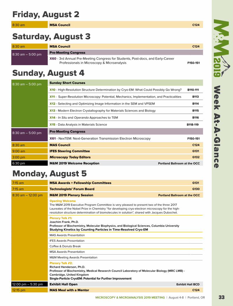
MICROSCOPY & MICROANALYSIS 2019 MEETING | August 4-8 | Portland, OR 33
Week
At-A
-Gla
nce
Friday, August 28:30 am MSA Council C124
Saturday, August 38:30 am MSA Council C124
8:30 am – 5:00 pm Pre-Meeting Congress
X60 - 3rd Annual Pre-Meeting Congress for Students, Post-docs, and Early-Career Professionals in Microscopy & Microanalysis F150-151
Sunday, August 48:30 am – 5:00 pm Sunday Short Courses
X10 - High-Resolution Structure Determination by Cryo-EM: What Could Possibly Go Wrong? B110-111
X11 - Super-Resolution Microscopy: Potential, Mechanics, Implementation, and Practicalities B113
X12 - Selecting and Optimizing Image Information in the SEM and VPSEM B114
X13 - Modern Electron Crystallography for Materials Sciences and Biology B115
X14 - In Situ and Operando Approaches to TEM B116
X15 - Data Analysis in Materials Science B118-119
8:30 am – 5:00 pm Pre-Meeting Congress
X61 - NexTEM: Next-Generation Transmission Electron Microscopy F150-151
8:30 am MAS Council C124
9:00 am IFES Steering Committee G131
3:00 pm Microscopy Today Editors G132
6:30 pm M&M 2019 Welcome Reception Portland Ballroom at the OCC
Monday, August 57:15 am MSA Awards + Fellowship Committees G131
7:15 am Technologists’ Forum Board G130
8:30 am – 12:00 pm M&M 2019 Plenary Session Portland Ballroom at the OCC
Opening WelcomeThe M&M 2019 Executive Program Committee is very pleased to present two of the three 2017 Laureates of the Nobel Prize in Chemistry “for developing cryo-electron microscopy for the high-resolution structure determination of biomolecules in solution”, shared with Jacques Dubochet.
Plenary Talk #1:Joachim Frank, Ph.D. Professor of Biochemistry, Molecular Biophysics, and Biological Sciences, Columbia UniversityStudying Kinetics by Counting Particles in Time-Resolved Cryo-EM
MAS Awards Presentation
IFES Awards Presentation
Coffee & Donuts Break
MSA Awards Presentation
M&M Meeting Awards Presentation
Plenary Talk #2:Richard Henderson, Ph.D. Professor of Biochemistry, Medical Research Council Laboratory of Molecular Biology (MRC LMB) - Cambridge, United KingdomSingle-Particle CryoEM: Potential for Further Improvement
12:00 pm – 5:30 pm Exhibit Hall Open Exhibit Hall BCD
12:15 pm MAS Meal with a Mentor C124
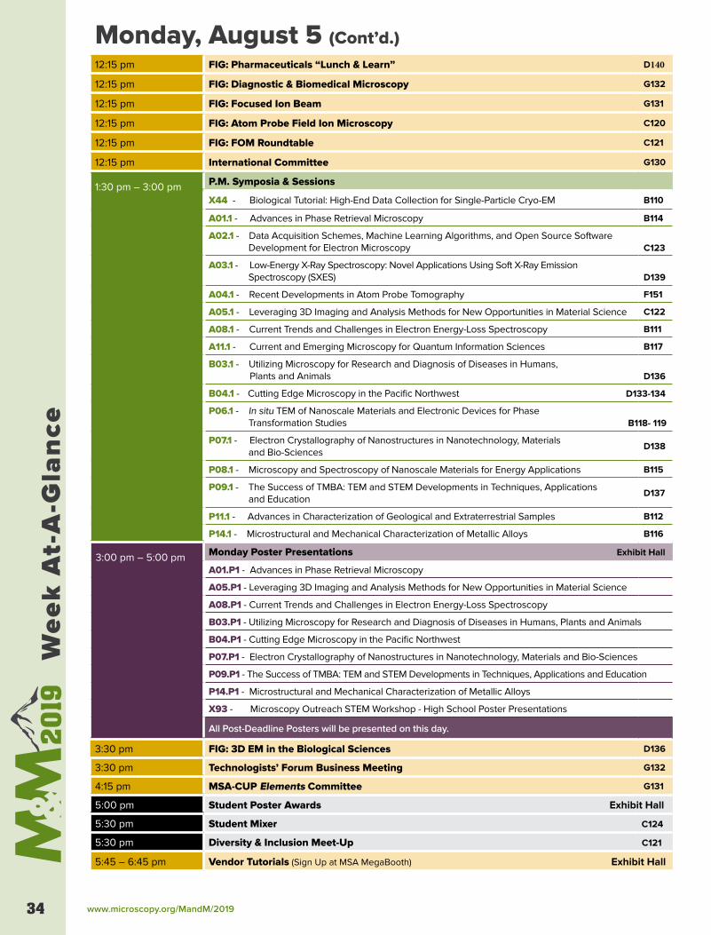
www.microscopy.org/MandM/201934
Wee
k A
t-A
-Gla
nce
Monday, August 5 (Cont’d.)12:15 pm FIG: Pharmaceuticals “Lunch & Learn” D140
12:15 pm FIG: Diagnostic & Biomedical Microscopy G132
12:15 pm FIG: Focused Ion Beam G131
12:15 pm FIG: Atom Probe Field Ion Microscopy C120
12:15 pm FIG: FOM Roundtable C121
12:15 pm International Committee G130
1:30 pm – 3:00 pm P.M. Symposia & Sessions
X44 - Biological Tutorial: High-End Data Collection for Single-Particle Cryo-EM B110
A01.1 - Advances in Phase Retrieval Microscopy B114
A02.1 - Data Acquisition Schemes, Machine Learning Algorithms, and Open Source Software Development for Electron Microscopy C123
A03.1 - Low-Energy X-Ray Spectroscopy: Novel Applications Using Soft X-Ray Emission Spectroscopy (SXES) D139
A04.1 - Recent Developments in Atom Probe Tomography F151
A05.1 - Leveraging 3D Imaging and Analysis Methods for New Opportunities in Material Science C122
A08.1 - Current Trends and Challenges in Electron Energy-Loss Spectroscopy B111
A11.1 - Current and Emerging Microscopy for Quantum Information Sciences B117
B03.1 - Utilizing Microscopy for Research and Diagnosis of Diseases in Humans, Plants and Animals D136
B04.1 - Cutting Edge Microscopy in the Pacific Northwest D133-134
P06.1 - In situ TEM of Nanoscale Materials and Electronic Devices for Phase Transformation Studies B118- 119
P07.1 - Electron Crystallography of Nanostructures in Nanotechnology, Materials and Bio-Sciences
D138
P08.1 - Microscopy and Spectroscopy of Nanoscale Materials for Energy Applications B115
P09.1 - The Success of TMBA: TEM and STEM Developments in Techniques, Applications and Education
D137
P11.1 - Advances in Characterization of Geological and Extraterrestrial Samples B112
P14.1 - Microstructural and Mechanical Characterization of Metallic Alloys B116
3:00 pm – 5:00 pm Monday Poster Presentations Exhibit Hall
A01.P1 - Advances in Phase Retrieval Microscopy
A05.P1 - Leveraging 3D Imaging and Analysis Methods for New Opportunities in Material Science
A08.P1 - Current Trends and Challenges in Electron Energy-Loss Spectroscopy
B03.P1 - Utilizing Microscopy for Research and Diagnosis of Diseases in Humans, Plants and Animals
B04.P1 - Cutting Edge Microscopy in the Pacific Northwest
P07.P1 - Electron Crystallography of Nanostructures in Nanotechnology, Materials and Bio-Sciences
P09.P1 - The Success of TMBA: TEM and STEM Developments in Techniques, Applications and Education
P14.P1 - Microstructural and Mechanical Characterization of Metallic Alloys
X93 - Microscopy Outreach STEM Workshop - High School Poster Presentations
All Post-Deadline Posters will be presented on this day.
3:30 pm FIG: 3D EM in the Biological Sciences D136
3:30 pm Technologists’ Forum Business Meeting G132
4:15 pm MSA-CUP Elements Committee G131
5:00 pm Student Poster Awards Exhibit Hall
5:30 pm Student Mixer C124
5:30 pm Diversity & Inclusion Meet-Up C121
5:45 – 6:45 pm Vendor Tutorials (Sign Up at MSA MegaBooth) Exhibit Hall
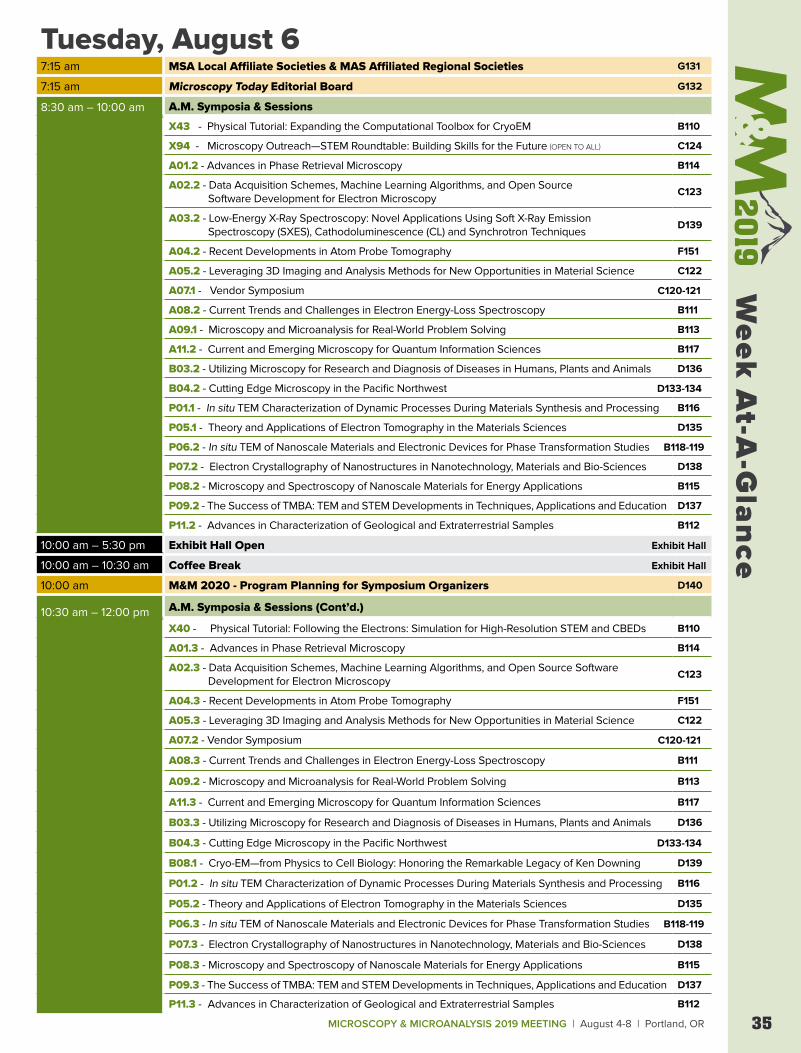
MICROSCOPY & MICROANALYSIS 2019 MEETING | August 4-8 | Portland, OR 35
Week
At-A
-Gla
nce
Tuesday, August 6 7:15 am MSA Local Affiliate Societies & MAS Affiliated Regional Societies G131
7:15 am Microscopy Today Editorial Board G132
8:30 am – 10:00 am A.M. Symposia & Sessions
X43 - Physical Tutorial: Expanding the Computational Toolbox for CryoEM B110
X94 - Microscopy Outreach—STEM Roundtable: Building Skills for the Future (OPEN TO ALL) C124
A01.2 - Advances in Phase Retrieval Microscopy B114
A02.2 - Data Acquisition Schemes, Machine Learning Algorithms, and Open Source Software Development for Electron Microscopy
C123
A03.2 - Low-Energy X-Ray Spectroscopy: Novel Applications Using Soft X-Ray Emission Spectroscopy (SXES), Cathodoluminescence (CL) and Synchrotron Techniques
D139
A04.2 - Recent Developments in Atom Probe Tomography F151
A05.2 - Leveraging 3D Imaging and Analysis Methods for New Opportunities in Material Science C122
A07.1 - Vendor Symposium C120-121
A08.2 - Current Trends and Challenges in Electron Energy-Loss Spectroscopy B111
A09.1 - Microscopy and Microanalysis for Real-World Problem Solving B113
A11.2 - Current and Emerging Microscopy for Quantum Information Sciences B117
B03.2 - Utilizing Microscopy for Research and Diagnosis of Diseases in Humans, Plants and Animals D136
B04.2 - Cutting Edge Microscopy in the Pacific Northwest D133-134
P01.1 - In situ TEM Characterization of Dynamic Processes During Materials Synthesis and Processing B116
P05.1 - Theory and Applications of Electron Tomography in the Materials Sciences D135
P06.2 - In situ TEM of Nanoscale Materials and Electronic Devices for Phase Transformation Studies B118-119
P07.2 - Electron Crystallography of Nanostructures in Nanotechnology, Materials and Bio-Sciences D138
P08.2 - Microscopy and Spectroscopy of Nanoscale Materials for Energy Applications B115
P09.2 - The Success of TMBA: TEM and STEM Developments in Techniques, Applications and Education D137
P11.2 - Advances in Characterization of Geological and Extraterrestrial Samples B112
10:00 am – 5:30 pm Exhibit Hall Open Exhibit Hall
10:00 am – 10:30 am Coffee Break Exhibit Hall
10:00 am M&M 2020 - Program Planning for Symposium Organizers D140
10:30 am – 12:00 pm A.M. Symposia & Sessions (Cont’d.)
X40 - Physical Tutorial: Following the Electrons: Simulation for High-Resolution STEM and CBEDs B110
A01.3 - Advances in Phase Retrieval Microscopy B114
A02.3 - Data Acquisition Schemes, Machine Learning Algorithms, and Open Source Software Development for Electron Microscopy
C123
A04.3 - Recent Developments in Atom Probe Tomography F151
A05.3 - Leveraging 3D Imaging and Analysis Methods for New Opportunities in Material Science C122
A07.2 - Vendor Symposium C120-121
A08.3 - Current Trends and Challenges in Electron Energy-Loss Spectroscopy B111
A09.2 - Microscopy and Microanalysis for Real-World Problem Solving B113
A11.3 - Current and Emerging Microscopy for Quantum Information Sciences B117
B03.3 - Utilizing Microscopy for Research and Diagnosis of Diseases in Humans, Plants and Animals D136
B04.3 - Cutting Edge Microscopy in the Pacific Northwest D133-134
B08.1 - Cryo-EM—from Physics to Cell Biology: Honoring the Remarkable Legacy of Ken Downing D139
P01.2 - In situ TEM Characterization of Dynamic Processes During Materials Synthesis and Processing B116
P05.2 - Theory and Applications of Electron Tomography in the Materials Sciences D135
P06.3 - In situ TEM of Nanoscale Materials and Electronic Devices for Phase Transformation Studies B118-119
P07.3 - Electron Crystallography of Nanostructures in Nanotechnology, Materials and Bio-Sciences D138
P08.3 - Microscopy and Spectroscopy of Nanoscale Materials for Energy Applications B115
P09.3 - The Success of TMBA: TEM and STEM Developments in Techniques, Applications and Education D137
P11.3 - Advances in Characterization of Geological and Extraterrestrial Samples B112
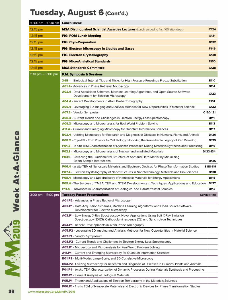
www.microscopy.org/MandM/201936
Wee
k A
t-A
-Gla
nce
Tuesday, August 6 (Cont’d.)
10:00 am – 10:30 am Lunch Break
12:15 pm MSA Distinguished Scientist Awardee Lectures (Lunch served to first 100 attendees) C124
12:15 pm FIG: FOM Lunch Meeting G131
12:15 pm FIG: Cryo-Preparation G132
12:15 pm FIG: Electron Microscopy in Liquids and Gases F149
12:15 pm FIG: Electron Crystallography G130
12:15 pm FIG: MicroAnalytical Standards F150
12:15 pm MSA Standards Committee C128
1:30 pm – 3:00 pm P.M. Symposia & Sessions
X45 - Biological Tutorial: Tips and Tricks for High-Pressure Freezing / Freeze Substitution B110
A01.4 - Advances in Phase Retrieval Microscopy B114
A02.4 - Data Acquisition Schemes, Machine Learning Algorithms, and Open Source Software Development for Electron Microscopy
C123
A04.4 - Recent Developments in Atom Probe Tomography F151
A05.4 - Leveraging 3D Imaging and Analysis Methods for New Opportunities in Material Science C122
A07.3 - Vendor Symposium C120-121
A08.4 - Current Trends and Challenges in Electron Energy-Loss Spectroscopy B111
A09.3 - Microscopy and Microanalysis for Real-World Problem Solving B113
A11.4 - Current and Emerging Microscopy for Quantum Information Sciences B117
B03.4 - Utilizing Microscopy for Research and Diagnosis of Diseases in Humans, Plants and Animals D136
B08.2 - Cryo-EM - from Physics to Cell Biology: Honoring the Remarkable Legacy of Ken Downing D139
P01.3 - In situ TEM Characterization of Dynamic Processes During Materials Synthesis and Processing B116
P02.1 - Microscopy and Microanalysis of Nuclear and Irradiated Materials D133-134
P03.1 - Revealing the Fundamental Structure of Soft and Hard Matter by Minimizing Beam-Sample Interactions D135
P06.4 - In situ TEM of Nanoscale Materials and Electronic Devices for Phase Transformation Studies B118-119
P07.4 - Electron Crystallography of Nanostructures in Nanotechnology, Materials and Bio-Sciences D138
P08.4 - Microscopy and Spectroscopy of Nanoscale Materials for Energy Applications B115
P09.4 - The Success of TMBA: TEM and STEM Developments in Techniques, Applications and Education D137
P11.4 - Advances in Characterization of Geological and Extraterrestrial Samples B112
3:00 pm – 5:00 pm Tuesday Poster Presentations Exhibit Hall
A01.P2 - Advances in Phase Retrieval Microscopy
A02.P1 - Data Acquisition Schemes, Machine Learning Algorithms, and Open Source Software Development for Electron Microscopy
A03.P1 - Low-Energy X-Ray Spectroscopy: Novel Applications Using Soft X-Ray Emission Spectroscopy (SXES), Cathodoluminescence (CL) and Synchrotron Techniques
A04.P1 - Recent Developments in Atom Probe Tomography
A05.P2 - Leveraging 3D Imaging and Analysis Methods for New Opportunities in Material Science
A07.P1 - Vendor Symposium
A08.P2 - Current Trends and Challenges in Electron Energy-Loss Spectroscopy
A09.P1 - Microscopy and Microanalysis for Real-World Problem Solving
A11.P1 - Current and Emerging Microscopy for Quantum Information Sciences
B01.P1 - Multi-Modal, Large-Scale, and 3D Correlative Microscopy
B03.P2 - Utilizing Microscopy for Research and Diagnosis of Diseases in Humans, Plants and Animals
P01.P1 - In situ TEM Characterization of Dynamic Processes During Materials Synthesis and Processing
P02.P1 - Element Analysis of Biological Materials
P05.P1 - Theory and Applications of Electron Tomography in the Materials Sciences
P06.P1 - In situ TEM of Nanoscale Materials and Electronic Devices for Phase Transformation Studies
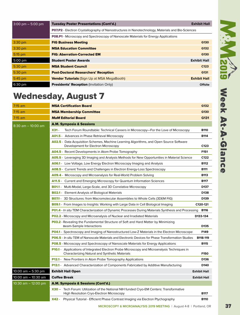
MICROSCOPY & MICROANALYSIS 2019 MEETING | August 4-8 | Portland, OR 37
Week
At-A
-Gla
nce
3:00 pm – 5:00 pm Tuesday Poster Presentations (Cont’d.) Exhibit Hall
P07.P2 - Electron Crystallography of Nanostructures in Nanotechnology, Materials and Bio-Sciences
P08.P1 - Microscopy and Spectroscopy of Nanoscale Materials for Energy Applications
3:30 pm FIG Business Meeting G130
3:30 pm MSA Education Committee G132
5:15 pm FIG: Aberration-Corrected EM G130
5:00 pm Student Poster Awards Exhibit Hall
5:30 pm MSA Student Council C123
5:30 pm Post-Doctoral Researchers’ Reception G131
5:45 pm Vendor Tutorials (Sign Up at MSA MegaBooth) Exhibit Hall
6:30 pm Presidents’ Reception (Invitation Only) Offsite
Wednesday, August 77:15 am MSA Certification Board G132
7:15 am MSA Membership Committee G130
7:15 am MaM Editorial Board G131
8:30 am – 10:00 am A.M. Symposia & Sessions
X31 - Tech Forum Roundtable: Technical Careers in Microscopy—For the Love of Microscopy B110
A01.5 - Advances in Phase Retrieval Microscopy B114
A02.5 - Data Acquisition Schemes, Machine Learning Algorithms, and Open Source Software Development for Electron Microscopy C123
A04.5 - Recent Developments in Atom Probe Tomography F151
A05.5 - Leveraging 3D Imaging and Analysis Methods for New Opportunities in Material Science C122
A06.1 - Low Voltage, Low Energy Electron Microscopy Imaging and Analysis B112
A08.5 - Current Trends and Challenges in Electron Energy-Loss Spectroscopy B111
A09.4 - Microscopy and Microanalysis for Real-World Problem Solving B113
A11.5 - Current and Emerging Microscopy for Quantum Information Sciences B117
B01.1 - Multi-Modal, Large-Scale, and 3D Correlative Microscopy D137
B02.1 - Element Analysis of Biological Materials D138
B07.1 - 3D Structures: from Macromolecular Assemblies to Whole Cells (3DEM FIG) D139
B09.1 - From Images to Insights: Working with Large Data in Cell Biological Imaging C120-121
P01.4 - In situ TEM Characterization of Dynamic Processes During Materials Synthesis and Processing B116
P02.2 - Microscopy and Microanalysis of Nuclear and Irradiated Materials D133-134
P03.2 - Revealing the Fundamental Structure of Soft and Hard Matter by Minimizing Beam-Sample Interactions D135
P04.1 - Spectroscopy and Imaging of Nanostructured Low-Z Materials in the Electron Microscope F149
P06.5 - In situ TEM of Nanoscale Materials and Electronic Devices for Phase Transformation Studies B118-119
P08.5 - Microscopy and Spectroscopy of Nanoscale Materials for Energy Applications B115
P10.1 - Applications of Integrated Electron Probe Microscopy and Microanalysis Techniques in Characterizing Natural and Synthetic Materials F150
P12.1 - New Frontiers in Atom Probe Tomography Applications D136
P13.1 - Advanced Characterization of Components Fabricated by Additive Manufacturing D140
10:00 am – 5:30 pm Exhibit Hall Open Exhibit Hall
10:00 am – 10:30 am Coffee Break Exhibit Hall
10:30 am – 12:00 pm A.M. Symposia & Sessions (Cont’d.)
X30 – Tech Forum: Utilization of the National NIH funded Cryo-EM Centers: Transformative High Resolution Cryo-Electron Microscopy B117
X42 - Physical Tutorial - Efficient Phase Contrast Imaging via Electron Ptychography B110
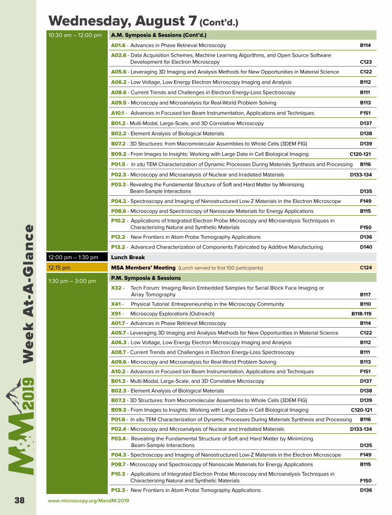
www.microscopy.org/MandM/201938
Wee
k A
t-A
-Gla
nce
Wednesday, August 7 (Cont’d.)10:30 am – 12:00 pm A.M. Symposia & Sessions (Cont’d.)
A01.6 - Advances in Phase Retrieval Microscopy B114
A02.6 - Data Acquisition Schemes, Machine Learning Algorithms, and Open Source Software Development for Electron Microscopy C123
A05.6 - Leveraging 3D Imaging and Analysis Methods for New Opportunities in Material Science C122
A06.2 - Low Voltage, Low Energy Electron Microscopy Imaging and Analysis B112
A08.6 - Current Trends and Challenges in Electron Energy-Loss Spectroscopy B111
A09.5 - Microscopy and Microanalysis for Real-World Problem Solving B113
A10.1 - Advances in Focused Ion Beam Instrumentation, Applications and Techniques F151
B01.2 - Multi-Modal, Large-Scale, and 3D Correlative Microscopy D137
B02.2 - Element Analysis of Biological Materials D138
B07.2 - 3D Structures: from Macromolecular Assemblies to Whole Cells (3DEM FIG) D139
B09.2 - From Images to Insights: Working with Large Data in Cell Biological Imaging C120-121
P01.5 - In situ TEM Characterization of Dynamic Processes During Materials Synthesis and Processing B116
P02.3 - Microscopy and Microanalysis of Nuclear and Irradiated Materials D133-134
P03.3 - Revealing the Fundamental Structure of Soft and Hard Matter by Minimizing Beam-Sample Interactions D135
P04.2 - Spectroscopy and Imaging of Nanostructured Low-Z Materials in the Electron Microscope F149
P08.6 - Microscopy and Spectroscopy of Nanoscale Materials for Energy Applications B115
P10.2 - Applications of Integrated Electron Probe Microscopy and Microanalysis Techniques in Characterizing Natural and Synthetic Materials F150
P12.2 - New Frontiers in Atom Probe Tomography Applications D136
P13.2 - Advanced Characterization of Components Fabricated by Additive Manufacturing D140
12:00 pm – 1:30 pm Lunch Break
12:15 pm MSA Members’ Meeting (Lunch served to first 100 participants) C124
1:30 pm – 3:00 pm P.M. Symposia & Sessions
X32 - Tech Forum: Imaging Resin Embedded Samples for Serial Block Face Imaging or Array Tomography B117
X41 - Physical Tutorial: Entrepreneurship in the Microscopy Community B110
X91 - Microscopy Explorations (Outreach) B118-119
A01.7 - Advances in Phase Retrieval Microscopy B114
A05.7 - Leveraging 3D Imaging and Analysis Methods for New Opportunities in Material Science C122
A06.3 - Low Voltage, Low Energy Electron Microscopy Imaging and Analysis B112
A08.7 - Current Trends and Challenges in Electron Energy-Loss Spectroscopy B111
A09.6 - Microscopy and Microanalysis for Real-World Problem Solving B113
A10.2 - Advances in Focused Ion Beam Instrumentation, Applications and Techniques F151
B01.3 - Multi-Modal, Large-Scale, and 3D Correlative Microscopy D137
B02.3 - Element Analysis of Biological Materials D138
B07.3 - 3D Structures: from Macromolecular Assemblies to Whole Cells (3DEM FIG) D139
B09.3 - From Images to Insights: Working with Large Data in Cell Biological Imaging C120-121
P01.6 - In situ TEM Characterization of Dynamic Processes During Materials Synthesis and Processing B116
P02.4 - Microscopy and Microanalysis of Nuclear and Irradiated Materials D133-134
P03.4 - Revealing the Fundamental Structure of Soft and Hard Matter by Minimizing Beam-Sample Interactions D135
P04.3 - Spectroscopy and Imaging of Nanostructured Low-Z Materials in the Electron Microscope F149
P08.7 - Microscopy and Spectroscopy of Nanoscale Materials for Energy Applications B115
P10.3 - Applications of Integrated Electron Probe Microscopy and Microanalysis Techniques in Characterizing Natural and Synthetic Materials F150
P12.3 - New Frontiers in Atom Probe Tomography Applications D136
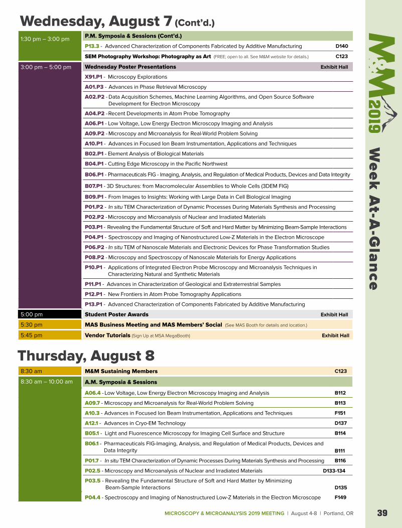
MICROSCOPY & MICROANALYSIS 2019 MEETING | August 4-8 | Portland, OR 39
Week
At-A
-Gla
nce
1:30 pm – 3:00 pm P.M. Symposia & Sessions (Cont’d.)
P13.3 - Advanced Characterization of Components Fabricated by Additive Manufacturing D140
SEM Photography Workshop: Photography as Art (FREE; open to all. See M&M website for details.) C123
3:00 pm – 5:00 pm Wednesday Poster Presentations Exhibit Hall
X91.P1 - Microscopy Explorations
A01.P3 - Advances in Phase Retrieval Microscopy
A02.P2 - Data Acquisition Schemes, Machine Learning Algorithms, and Open Source Software Development for Electron Microscopy
A04.P2 - Recent Developments in Atom Probe Tomography
A06.P1 - Low Voltage, Low Energy Electron Microscopy Imaging and Analysis
A09.P2 - Microscopy and Microanalysis for Real-World Problem Solving
A10.P1 - Advances in Focused Ion Beam Instrumentation, Applications and Techniques
B02.P1 - Element Analysis of Biological Materials
B04.P1 - Cutting Edge Microscopy in the Pacific Northwest
B06.P1 - Pharmaceuticals FIG - Imaging, Analysis, and Regulation of Medical Products, Devices and Data Integrity
B07.P1 - 3D Structures: from Macromolecular Assemblies to Whole Cells (3DEM FIG)
B09.P1 - From Images to Insights: Working with Large Data in Cell Biological Imaging
P01.P2 - In situ TEM Characterization of Dynamic Processes During Materials Synthesis and Processing
P02.P2 - Microscopy and Microanalysis of Nuclear and Irradiated Materials
P03.P1 - Revealing the Fundamental Structure of Soft and Hard Matter by Minimizing Beam-Sample Interactions
P04.P1 - Spectroscopy and Imaging of Nanostructured Low-Z Materials in the Electron Microscope
P06.P2 - In situ TEM of Nanoscale Materials and Electronic Devices for Phase Transformation Studies
P08.P2 - Microscopy and Spectroscopy of Nanoscale Materials for Energy Applications
P10.P1 - Applications of Integrated Electron Probe Microscopy and Microanalysis Techniques in Characterizing Natural and Synthetic Materials
P11.P1 - Advances in Characterization of Geological and Extraterrestrial Samples
P12.P1 - New Frontiers in Atom Probe Tomography Applications
P13.P1 - Advanced Characterization of Components Fabricated by Additive Manufacturing
5:00 pm Student Poster Awards Exhibit Hall
5:30 pm MAS Business Meeting and MAS Members’ Social (See MAS Booth for details and location.)
5:45 pm Vendor Tutorials (Sign Up at MSA MegaBooth) Exhibit Hall
Wednesday, August 7 (Cont’d.)
Thursday, August 88:30 am M&M Sustaining Members C123
8:30 am – 10:00 am A.M. Symposia & Sessions
A06.4 - Low Voltage, Low Energy Electron Microscopy Imaging and Analysis B112
A09.7 - Microscopy and Microanalysis for Real-World Problem Solving B113
A10.3 - Advances in Focused Ion Beam Instrumentation, Applications and Techniques F151
A12.1 - Advances in Cryo-EM Technology D137
B05.1 - Light and Fluorescence Microscopy for Imaging Cell Surface and Structure B114
B06.1 - Pharmaceuticals FIG-Imaging, Analysis, and Regulation of Medical Products, Devices and Data Integrity B111
P01.7 - In situ TEM Characterization of Dynamic Processes During Materials Synthesis and Processing B116
P02.5 - Microscopy and Microanalysis of Nuclear and Irradiated Materials D133-134
P03.5 - Revealing the Fundamental Structure of Soft and Hard Matter by Minimizing Beam-Sample Interactions D135
P04.4 - Spectroscopy and Imaging of Nanostructured Low-Z Materials in the Electron Microscope F149
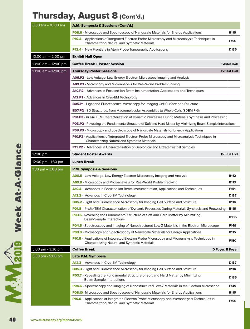
www.microscopy.org/MandM/201940
Wee
k A
t-A
-Gla
nce
Thursday, August 8 (Cont’d.)8:30 am – 10:00 am A.M. Symposia & Sessions (Cont’d.)
P08.8 - Microscopy and Spectroscopy of Nanoscale Materials for Energy Applications B115
P10.4 - Applications of Integrated Electron Probe Microscopy and Microanalysis Techniques in Characterizing Natural and Synthetic Materials
F150
P12.4 - New Frontiers in Atom Probe Tomography Applications D136
10:00 am – 2:00 pm Exhibit Hall Open
10:00 am – 12:00 pm Coffee Break + Poster Session Exhibit Hall
10:00 am – 12:00 pm Thursday Poster Sessions Exhibit Hall
A06.P2 - Low Voltage, Low Energy Electron Microscopy Imaging and Analysis
A09.P3 - Microscopy and Microanalysis for Real-World Problem Solving
A10.P2 - Advances in Focused Ion Beam Instrumentation, Applications and Techniques
A12.P1 - Advances in Cryo-EM Technology
B05.P1 - Light and Fluorescence Microscopy for Imaging Cell Surface and Structure
B07.P2 - 3D Structures: from Macromolecular Assemblies to Whole Cells (3DEM FIG)
P01.P3 - In situ TEM Characterization of Dynamic Processes During Materials Synthesis and Processing
P03.P2 - Revealing the Fundamental Structure of Soft and Hard Matter by Minimizing Beam-Sample Interactions
P08.P3 - Microscopy and Spectroscopy of Nanoscale Materials for Energy Applications
P10.P2 - Applications of Integrated Electron Probe Microscopy and Microanalysis Techniques in Characterizing Natural and Synthetic Materials
P11.P2 - Advances in Characterization of Geological and Extraterrestrial Samples
12:00 pm Student Poster Awards Exhibit Hall
12:00 pm - 1:30 pm Lunch Break
1:30 pm – 3:00 pm P.M. Symposia & Sessions
A06.5 - Low Voltage, Low Energy Electron Microscopy Imaging and Analysis B112
A09.8 - Microscopy and Microanalysis for Real-World Problem Solving B113
A10.4 - Advances in Focused Ion Beam Instrumentation, Applications and Techniques F151
A12.2 - Advances in Cryo-EM Technology D137
B05.2 - Light and Fluorescence Microscopy for Imaging Cell Surface and Structure B114
P01.8 - In situ TEM Characterization of Dynamic Processes During Materials Synthesis and Processing B116
P03.6 - Revealing the Fundamental Structure of Soft and Hard Matter by Minimizing Beam-Sample Interactions
D135
P04.5 - Spectroscopy and Imaging of Nanostructured Low-Z Materials in the Electron Microscope F149
P08.9 - Microscopy and Spectroscopy of Nanoscale Materials for Energy Applications B115
P10.5 - Applications of Integrated Electron Probe Microscopy and Microanalysis Techniques in Characterizing Natural and Synthetic Materials
F150
3:00 pm - 3:30 pm Coffee Break D Foyer; B Foyer
3:30 pm - 5:00 pm Late P.M. Symposia
A12.3 - Advances in Cryo-EM Technology D137
B05.3 - Light and Fluorescence Microscopy for Imaging Cell Surface and Structure B114
P03.7 - Revealing the Fundamental Structure of Soft and Hard Matter by Minimizing Beam-Sample Interactions
D135
P04.6 - Spectroscopy and Imaging of Nanostructured Low-Z Materials in the Electron Microscope F149
P08.10 - Microscopy and Spectroscopy of Nanoscale Materials for Energy Applications B115
P10.6 - Applications of Integrated Electron Probe Microscopy and Microanalysis Techniques in Characterizing Natural and Synthetic Materials
F150
MICROSCOPY & MICROANALYSIS 2019 MEETING | August 4-8 | Portland, OR 41
Week
At-A
-Gla
nce
On behalf of the Microscopy Society of America (MSA), the Microanalysis Society (MAS), and the International Field Emission Society (IFES), we are excited to welcome you to Microscopy & Microanalysis 2019 in Portland, Oregon.
As many of you who have previously visited Portland know, this city is a terrific location for our meeting: set between Mt. Hood and the Pacific coast, Portland (PDX to locals) presents a variety of attractions for everybody. Infused with the awesome Pacific Northwest culture of environmental conservation and organic local food sourcing, you will find a lot of wonderful restaurants here, plus a myriad of microbreweries, distilleries and wineries! Don’t forget to visit famous PDX staples like Powell’s Books and the charismatic Voodoo Donuts. Don’t forget your hiking shoes or beachwear if you’re planning to explore the great outdoors like the Columbia Gorge area or the Oregon Coast beaches.
The meeting itself will be preceded by our usual array of Sunday Short Courses, two Pre-Meeting Congresses, and our Sunday evening Opening Welcome Reception. We will once again hold a Pre-Meeting Congress featuring outstanding work by students and early-career researchers.
The technical program will kick off with our annual Monday morning plenary session, featuring the major awards ceremonies for the sponsoring societies, the M&M meeting awards, and two exciting plenary talks of the 2017 Nobel Prize laureates Joachim Frank and Richard Henderson.
Immediately following the plenary session, the largest microscopy/ microanalysis instrument exhibition in the world opens to participants. Plan to visit the more than 100 companies displaying their latest technology and services. The social activities of the opening reception, and daily “happy hours” accompanying each day’s poster and awards sessions, have become “can’t-miss” events of learning and fellowship.
For a complete description of all the symposia, contributed sessions, educational opportunities and the multiple award possibilities from the three organizing societies, please visit: http://www.microscopy.org/MandM/2019/.
Be sure to download the M&M 2019 mobile app! (“M&M Annual Meeting” in App Store or Google Play.)
Alice Dohnalkova Huolin Xin Assel Aitkaliyeva Baptiste Gault PROGRAM CHAIR PROGRAM CO-CHAIR MAS CO-CHAIR IFES CO-CHAIR
WELCOME FROM THE PROGRAM CHAIRS
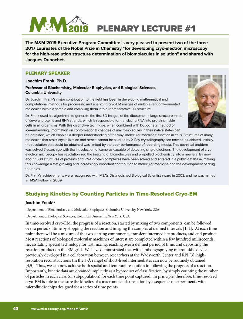
www.microscopy.org/MandM/2019 42
PLENARY LECTURE #1The M&M 2019 Executive Program Committee is very pleased to present two of the three 2017 Laureates of the Nobel Prize in Chemistry "for developing cryo-electron microscopy for the high-resolution structure determination of biomolecules in solution" and shared with Jacques Dubochet.
PLENARY SPEAKER
Joachim Frank, Ph.D.
Professor of Biochemistry, Molecular Biophysics, and Biological Sciences, Columbia University
Dr. Joachim Frank's major contribution to the field has been in developing mathematical and computational methods for processing and analyzing cryo-EM images of multiple randomly-oriented molecules within a sample and compiling them into a representative 3D structure.
Dr. Frank used his algorithms to generate the first 3D images of the ribosome - a large structure made of several proteins and RNA strands, which is responsible for translating RNA into proteins inside cells in all organisms. With this distinctive technique, when combined with Dubochet's method of ice-embedding, information on conformational changes of macromolecules in their native states can be obtained, which enables a deeper understanding of the way 'molecular machines' function in cells. Structures of many molecules that resist crystallization and hence cannot be studied by X-Ray crystallography can now be elucidated. Initially, the resolution that could be obtained was limited by the poor performance of recording media. This technical problem was solved 7 years ago with the introduction of cameras capable of detecting single electrons. The development of cryo-electron microscopy has revolutionized the imaging of biomolecules and propelled biochemistry into a new era. By now, about 1500 structures of proteins and RNA-protein complexes have been solved and entered in a public database, making this knowledge a fast growing and increasingly important contribution to molecular medicine and the development of drug therapies.
Dr. Frank's achievements were recognized with MSA's Distinguished Biological Scientist award in 2003, and he was named an MSA Fellow in 2009.
Studying Kinetics by Counting Particles in Time-Resolved Cryo-EMJoachim Frank1,2
1Department of Biochemistry and Molecular Biophysics, Columbia University, New York, USA2Department of Biological Sciences, Columbia University, New York, USA
In time-resolved cryo-EM, the progress of a reaction, started by mixing of two components, can be followed over a period of time by stopping the reaction and imaging the samples at defined intervals [1, 2]. At each time point there will be a mixture of the two starting components, transient intermediate products, and end product. Most reactions of biological molecular machines of interest are completed within a few hundred milliseconds, necessitating special technology for fast mixing, reacting over a defined period of time, and depositing the reaction product on the EM grid. We have demonstrated that with a mixing/spraying microfluidic device previously developed in a collaboration between researchers at the Wadsworth Center and RPI [3], high-resolution reconstructions (in the 3-Å range) of short-lived intermediates can now be routinely obtained [4,5]. Thus, we can now achieve both spatial and temporal resolution in following the progress of a reaction. Importantly, kinetic data are obtained implicitly as a byproduct of classification: by simply counting the number of particles in each class (or subpopulation) for each time point captured. In principle, therefore, time-resolved cryo-EM is able to measure the kinetics of a macromolecular reaction by a sequence of experiments with microfluidic chips designed for a series of time points.
MICROSCOPY & MICROANALYSIS 2019 MEETING | August 4-8 | Portland, OR 43
Here I would like to raise the question to what extent this method of kinetic measurement can be used in practice. This is an important question since a reliable method of measuring reaction kinetics of macromolecules by counting particles in micrographs collected by cryo-EM would be a significant addition to the tool chest of Biophysics.However, the precise quantification of the numbers of particles in such experiments faces several obstacles: there is evidence that a fraction of molecules suffer damage as they are prepared for cryo-EM. This damage is attributed to denaturing interactions with the air-water interface [6], where the majority of the molecules reside according to recent electron tomographic evidence [7]. Even if there were a reliable way of sorting out damaged from undamaged molecules, then a bias in counting could still be created if the propensity to damage is different for different reaction products. Clearly a remedy to this problem would require a different preparation method that ensures all molecules are fully encased in ice. Next is the difficulty of unbiased particle selection. At the particle-picking step, it is possible that different molecule species are picked with different error rates. This problem requires a systematic study of particle-picking algorithms Using mixtures of different species with defined ratios. Third on the list is the question if the classification algorithm is able to make the right decision in sorting the images into a pre-given number of classes, especially in the presence of “noise images”, or false positives in the particle picking. Here it is useful to do tests on a given dataset with different fractions of fake particles (i.e., simulated false positives of particle picking) admixed, taking for as fake particles, for example, data excised from the background [4]. Fourth is the reproducibility of classification results from the same dataset with different seeds, which for a dataset of ribosome particles turned out extremely good, within a very small margin of error. In all, these questions define an entire program of validation tests for different kinds of molecules. These tests do not necessarily all require time-resolved experiments, but can be parceled into separate studies all involving conventional cryo-EM [8].
References[1] [1] Chen, B., and Frank, J. (2015). Two promising future developments of cryo-EM: capturing short-lived states and mapping a continuum of states of a macromolecule. Microscopy 65, 69-79.
[2] Frank, J. (2017). Time-resolved cryo-electron microscopy: Recent progress. J. Struct. Biol. 200, 303-306.
[3] Lu, Z., Shaikh, T.R., Barnard, D., Meng, X., Mohamed, H., Yassin, A., Mannella, C.A., Agrawal, R.K., Lu, T.-M., and Wagenknecht, T. (2009) Monolithic microfluidic mixing-spraying devices for time-resolved cryo-electron microscopy. J. Struct. Biol. 168, 388-395.
[4] Fu, Z., Indrisiunaite, G., Kaledhonkar, S., Shah, B., Sun, M., Chen, B., Grassucci, R.A., Ehrenberg, M., and Frank, J. (2019). The structural basis for release factor activation during translation termination revealed by time-resolved cryogenic electron microscopy. Nature Communications, in press.
[5] Kaledhonkar, S., Fu, Z., Caban, K., Li, W., Chen, B., Sun, M., Gonzalez, R.L. and Frank, J. (2019). Late steps in bacterial translation initiation visualized Using time-resolved cryo-EM. Nature, in press.
[6] Noble, A.J., Dandey, V.P., Wei, H., Brasch, J., Chase, J., Acharya, P., Tan, Y.Z., Zhang, Z., Kim, L.Y., Scapin, G., Rapp, M., Eng, E.T., Rice, W.J., Cheng, A., Negro, C.J., Shapiro, L., Kwong, P.D., Jeruzalmi, D., Amedee des Georges, A., Potter, C.S., Carragher, B. (2018). Routine single particle cryoEM sample and grid characterization by tomography. eLife https://doi.org/10.7554/eLife.34257.001.
[7] Glaeser, R.M. and Han, B.-G. (2017). Opinion: hazards faced by macromolecules when confined to thin aqueous films. Biophys. Rep. 3, 1-7.
[8] This work was supported by NIH R01 GM55440.
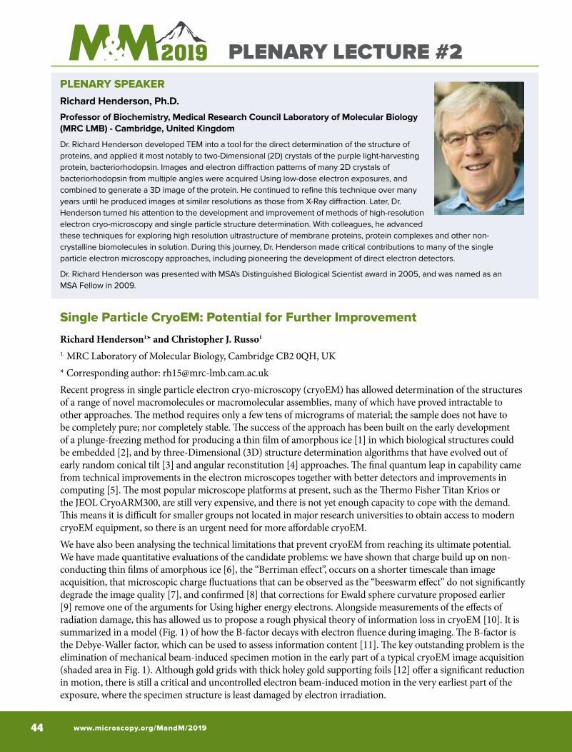
www.microscopy.org/MandM/2019 44
PLENARY LECTURE #2PLENARY SPEAKERRichard Henderson, Ph.D.
Professor of Biochemistry, Medical Research Council Laboratory of Molecular Biology (MRC LMB) - Cambridge, United Kingdom
Dr. Richard Henderson developed TEM into a tool for the direct determination of the structure of proteins, and applied it most notably to two-Dimensional (2D) crystals of the purple light-harvesting protein, bacteriorhodopsin. Images and electron diffraction patterns of many 2D crystals of bacteriorhodopsin from multiple angles were acquired Using low-dose electron exposures, and combined to generate a 3D image of the protein. He continued to refine this technique over many years until he produced images at similar resolutions as those from X-Ray diffraction. Later, Dr. Henderson turned his attention to the development and improvement of methods of high-resolution electron cryo-microscopy and single particle structure determination. With colleagues, he advanced these techniques for exploring high resolution ultrastructure of membrane proteins, protein complexes and other non-crystalline biomolecules in solution. During this journey, Dr. Henderson made critical contributions to many of the single particle electron microscopy approaches, including pioneering the development of direct electron detectors.
Dr. Richard Henderson was presented with MSA's Distinguished Biological Scientist award in 2005, and was named as an MSA Fellow in 2009.
Single Particle CryoEM: Potential for Further Improvement
Richard Henderson1* and Christopher J. Russo1
1. MRC Laboratory of Molecular Biology, Cambridge CB2 0QH, UK* Corresponding author: [email protected] progress in single particle electron cryo-microscopy (cryoEM) has allowed determination of the structures of a range of novel macromolecules or macromolecular assemblies, many of which have proved intractable to other approaches. The method requires only a few tens of micrograms of material; the sample does not have to be completely pure; nor completely stable. The success of the approach has been built on the early development of a plunge-freezing method for producing a thin film of amorphous ice [1] in which biological structures could be embedded [2], and by three-Dimensional (3D) structure determination algorithms that have evolved out of early random conical tilt [3] and angular reconstitution [4] approaches. The final quantum leap in capability came from technical improvements in the electron microscopes together with better detectors and improvements in computing [5]. The most popular microscope platforms at present, such as the Thermo Fisher Titan Krios or the JEOL CryoARM300, are still very expensive, and there is not yet enough capacity to cope with the demand. This means it is difficult for smaller groups not located in major research universities to obtain access to modern cryoEM equipment, so there is an urgent need for more affordable cryoEM.We have also been analysing the technical limitations that prevent cryoEM from reaching its ultimate potential. We have made quantitative evaluations of the candidate problems: we have shown that charge build up on non-conducting thin films of amorphous ice [6], the “Berriman effect”, occurs on a shorter timescale than image acquisition, that microscopic charge fluctuations that can be observed as the “beeswarm effect” do not significantly degrade the image quality [7], and confirmed [8] that corrections for Ewald sphere curvature proposed earlier [9] remove one of the arguments for Using higher energy electrons. Alongside measurements of the effects ofradiation damage, this has allowed us to propose a rough physical theory of information loss in cryoEM [10]. It issummarized in a model (Fig. 1) of how the B-factor decays with electron fluence during imaging. The B-factor isthe Debye-Waller factor, which can be used to assess information content [11]. The key outstanding problem is theelimination of mechanical beam-induced specimen motion in the early part of a typical cryoEM image acquisition(shaded area in Fig. 1). Although gold grids with thick holey gold supporting foils [12] offer a significant reductionin motion, there is still a critical and uncontrolled electron beam-induced motion in the very earliest part of theexposure, where the specimen structure is least damaged by electron irradiation.
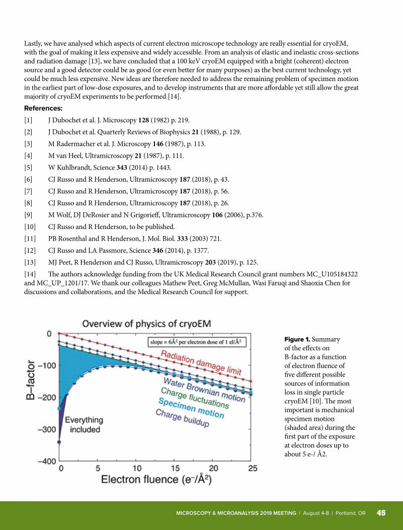
MICROSCOPY & MICROANALYSIS 2019 MEETING | August 4-8 | Portland, OR 45
Lastly, we have analysed which aspects of current electron microscope technology are really essential for cryoEM, with the goal of making it less expensive and widely accessible. From an analysis of elastic and inelastic cross-sections and radiation damage [13], we have concluded that a 100 keV cryoEM equipped with a bright (coherent) electron source and a good detector could be as good (or even better for many purposes) as the best current technology, yet could be much less expensive. New ideas are therefore needed to address the remaining problem of specimen motion in the earliest part of low-dose exposures, and to develop instruments that are more affordable yet still allow the great majority of cryoEM experiments to be performed [14].References:
[1] J Dubochet et al. J. Microscopy 128 (1982) p. 219.[2] J Dubochet et al. Quarterly Reviews of Biophysics 21 (1988), p. 129.[3] M Radermacher et al. J. Microscopy 146 (1987), p. 113.[4] M van Heel, Ultramicroscopy 21 (1987), p. 111.[5] W Kuhlbrandt, Science 343 (2014) p. 1443.[6] CJ Russo and R Henderson, Ultramicroscopy 187 (2018), p. 43.[7] CJ Russo and R Henderson, Ultramicroscopy 187 (2018), p. 56.[8] CJ Russo and R Henderson, Ultramicroscopy 187 (2018), p. 26.[9] M Wolf, DJ DeRosier and N Grigorieff, Ultramicroscopy 106 (2006), p.376.[10] CJ Russo and R Henderson, to be published.[11] PB Rosenthal and R Henderson, J. Mol. Biol. 333 (2003) 721.[12] CJ Russo and LA Passmore, Science 346 (2014), p. 1377.[13] MJ Peet, R Henderson and CJ Russo, Ultramicroscopy 203 (2019), p. 125.[14] The authors acknowledge funding from the UK Medical Research Council grant numbers MC_U105184322 and MC_UP_1201/17. We thank our colleagues Mathew Peet, Greg McMullan, Wasi Faruqi and Shaoxia Chen for discussions and collaborations, and the Medical Research Council for support.
Figure 1. Summary of the effects on B-factor as a function of electron fluence of five different possible sources of information loss in single particle cryoEM [10]. The most important is mechanical specimen motion (shaded area) during the first part of the exposure at electron doses up to about 5 e-/ Å2.
www.microscopy.org/MandM/2019 46
SUNDAY SHORT COURSESORGANIZER: Mike Marko, Wadsworth Center
• These full-day courses run from 8:30 AM to 5:00 PM on Sunday, August 4th, 2019.
• A certificate of participation will be issued to each requesting participant, following the conclusion of the M&M 2019 meeting.
• Two (2) Continuing Microscopy Education Units are available (registration fee $10 for members; sign up at Registration Desk).
• Morning and afternoon coffee breaks are included (breakfast and lunch are on your own).
• Separate registration with additional fees is required (visit Registration Desk to sign up).
X-10 High-Resolution Structure Determination by Cryo-EM: What Could Possibly Go Wrong? Room B110-111
LEAD INSTRUCTORS:Anchi Cheng, New York Structural Biology Center
Steve Ludtke, Baylor College of Medicine
• Specimen preparation and plunge-freezing choices and considerations
• Data collection - Camera and TEM parameters, TEM automation
• Initial image processing from raw data• Single-particle reconstruction, choices and validation• Structure modeling and results presentation
In this one-day course, it is not possible to explain and cover all the methods required for successfully determining a structure. However, an overview of the steps, and particularly advice on pitfalls to be avoided, will be especially of interest to those who will wish to employ this method. We identify the range of methods, instrumentation, and software available, and we will illustrate by means of recent successful results.
X-11 Super-Resolution Microscopy: Potential, Mechanics, Implementation, and Practicalities Room B113
LEAD INSTRUCTORS:Bryan Millis, Vanderbilt University
Simon Watkins, University of Pittsburgh
• What is “super-resolution microscopy” and do you need it?• What are the various approaches available and how does
each work?• What are the strengths and weaknesses of each method?• Practicalities of running super-resolution imaging in a multi-
user facility.• How practical is live-cell super-resolution microscopy?• Common pitfalls of super-resolution microscopy.
Super-resolution microscopy is becoming a mainstream approach in both individual and core biomedical research laboratories. The diversity and overall capabilities of these techniques are evolving rapidly. As such, the term “super-resolution microscopy” can describe very different fundamental approaches, all quite distinct with respect to implementation, and each with specific strengths and weaknesses. For the researcher looking to get into super-resolution microscopy, the aforementioned can be overwhelming. This workshop primarily attempts to clarify many of the questions that typically go along with getting started with super-resolution microscopy, as well as pairing the right technology with the biological question of interest.
X-12 Selecting and Optimizing Image Information in the SEM and VPSEM Room B114
LEAD INSTRUCTOR:
(NEW) Bradley Thiel, SUNY-Polytechnic Institute• Understanding the imaging options for materials and
biological application-Specific SEM use• Determining specific conditions for the most-relevant sample
imaging• Modern electron detection systems and stage/column
variables for SEM/VPSEM• Tools for measurement and resolution determination
Below-lens, through-lens and above-lens detection and imaging systems provide complex and exciting imaging options. Selection of the optimal option can be prohibitively time-consuming. To help simplify or speed up this up, each option will be explained and compared by means of a set of standard samples. This will include the effects of angular selectivity of annular backscattered electron detection. Effects of the stage and chamber on the detected signals will be followed by a comparative overview of VPSEM imaging. Stage biasing, beam deceleration, stage isolation and the effects of contact-protection systems will discussed. Brief presentations on their detection systems by Invited manufacturers will be followed by a general discussion. Effects of sample types, coatings and cleaning will be reviewed. Techniques for resolution measurement, and sourcing of suitable samples, will be presented as a student practical, Using software provided in the course notes. Questions are encouraged throughout the day and can also be submitted prior to the course, following enrolment.
MICROSCOPY & MICROANALYSIS 2019 MEETING | August 4-8 | Portland, OR 47
SUNDAY SHORT COURSESX-13 Modern Electron Crystallography for
Materials Sciences and Biology Room B115
LEAD INSTRUCTORS:Sergei Rouvimov, University of Notre Dame
Peter Moeck, Portland State University
• Recent developments in electron crystallography for nanomaterials including soft and biological materials
• Basics of scanning electron diffraction methods for microstructure analysis including bio-crystals
• Electron crystallography applications for structural biology, including protein crystals
• Cryo-electron crystallography, including single-particle cryo-EM• New experimental and computer-simulation techniques to
improve the speed and reliability of structure characterization
Electron crystallography has many applications in nanotechnology, materials and biological sciences. Due to recent developments in TEM instrumentation, electron crystallography became readily available to study nanostructures and biological materials at the nanoscale. This course addresses recent developments in instrumentation and electron diffraction methods, both experimental techniques and computer-Based data processing, that allow research of a wide range of materials, including Beam-Sensitive materials. This course brings together and provides updates for researchers from both materials and biological sciences on novel techniques for a wide range of materials systems, and new data analyses and computational methods including their application for cryo-EM.
X-14 In Situ and Operando Approaches to TEM Room B116
LEAD INSTRUCTORS:Robert Sinclair, Stanford University
Peter Crozier, Arizona State University
This course introduces the fundamental concepts for in situ electron microscopy, and includes:• Hot stages• Gas cells• Liquid cells• Biasing holders• Magnetic field• Light illumination
In situ and operando transmission electron microscopy are becoming increasingly important in advanced materials characterization. Being able to observe materials in state that are similar to real world applications is now recognize as vital for relating structure to functionality. The ability to perform atomic level analysis while the sample is exposed to different stimuli/environments such as heat, strain, gas, liquids, electric field, magnetic field, light, etc.…is a key part of the in situ approach. Operando approach require simultaneous measurement of some technologically relevant functionality such as current flow, deformation or catalysis.
X-15 Data Analysis in Materials Science Room B118-119
INSTRUCTORS:Duncan Johnstone, University of Cambridge, United Kingdom
Katherine E. MacArthur, Forschungszentrum Jülich, Germany
Magnus Nord, University of Antwerp, Belgium
Francisco de la Peña, University of Lille, France
Eric Prestat, University of Manchester, United Kingdom
Joshua Taillon, National Institute of Standards and Technology
• Introduction to HyperSpy and related Python libraries for multi-Dimensional image and spectra processing and analysis
• Machine learning• Big data analysis strategies• Curve fitting of multi-Dimensional datasets• EELS and EDS analysis• Atomic resolution image analysis
This short course introduces the use of HyperSpy and related Python libraries (atomap, pixStem, pyXem) for analysis of microscopy datasets. No prior Python knowledge is required. Attendees learn how to perform basic machine learning, multi-Dimensional curve fitting for EELS and EDS quantification, atomic resolution image analysis and big data processing (such as 4D STEM) on desktop computers.
All M&M 2019 symposia, workshops, meetings, and official events are held at the Oregon Convention Center unless otherwise indicated.
www.microscopy.org/MandM/2019 48
PRE-MEETING CONGRESSES
ProtochipsQuantifiably Better™
X60 Third Annual Pre-Meeting Congress for Students, Post-Docs, and Early-Career Professionals in Microscopy and Microanalysis
Saturday, August 3, 2019 • 8:20 AM–5:00 PM Oregon Convention Center, Room F150-151
INCLUDED IN REGISTRATION FEE:Friday Evening Welcome Reception; Breakfast; AM Break; Lunch; PM Break; Saturday Evening Banquet
Organized by
PROGRAM CHAIR: Ethan Lawrence, Arizona State University
BIOLOGICAL SCIENCES CO-CHAIR:Mike Casasanta, Pennsylvania State University
PHYSICAL SCIENCES CO-CHAIR:Eric Hoglund, University of Virginia
The Pre-meeting Congress X60 (PMCx60) is an annual meeting organized by the MSA Student Council (StC) for Students, Postdocs, and Early-career Professionals in Microscopy and Microanalysis. The PMCx60 is held in conjunction with the annual Microscopy and Microanalysis (M&M) Meeting.
In addition to the premier technical program, the PMCx60 features informal get-togethers on Friday and Saturday nights so participants can meet in a relaxed setting. The PMCx60 is scheduled to allow attendees to participate in Sunday Short Courses or Focused Interest Group (FIG) sponsored PMCs, or to enjoy a free day with new friends.
The PMCx60 is organized by, and tailored to, students and early-career professionals, however all are welcome to attend. The PMCx60 offers a highly interactive forum for participants to discuss their cutting-edge research, and to network and engage with peers ahead of the main meeting. Invited speakers are selected from the awardees to be honored at M&M, giving attendees a unique opportunity to sample the best research from their peers in biological science, physical science, analytical science, and instrumentation. Contributed posters also give attendees an opportunity to share their work with peers in an intimate and casual setting.
Additional professional development activities include a luncheon featuring a panel of recent graduates currently working in industry, academia, policy, and government labs. The PMCx60 and M&M are wonderful opportunities for students (undergraduate and graduate), postdocs, and early-career professionals to network with peers, members of academia and industry, as well as vendors from a variety of top companies who are invited to exhibit their latest developments during the PMCx60 Poster and Extended Networking Sessions.
Thank you to our sponsors and supporters!
MICROSCOPY & MICROANALYSIS 2019 MEETING | August 4-8 | Portland, OR 49
Separate registration fee required. See the Registration Desk if you wish to register.Please review individual PMC listings below for information on meals and breaks.
X61 NexTEM: Next-Generation Transmission Electron Microscopy
Sunday, August 4, 2019 • 8:30 AM–5:00 PM Oregon Convention Center, F149-150
INCLUDED IN REGISTRATION FEE:Breakfast, AM Break, Lunch, PM Break, Late Afternoon Reception
Supported by the Aberration Corrected Electron Microscopy FIG
ORGANIZERS: Steven R. Spurgeon, Pacific Northwest National Laboratory
Mitra L. Taheri, Drexel University
Demie Kepaptsoglou, SuperSTEM, United Kingdom
The second annual Next-Generation Transmission Electron Microscopy (NexTEM) workshop brings together researchers from diverse backgrounds to present the state-of-the-art in cutting edge electron microscopy tools and related applications.
TOPICS
Advanced Detector and Spectroscopy Developments • Design and use of novel detectors to investigate material structure and functionality, including 4D STEM and
ptychography. • Vibrational and phonon spectroscopies at unprecedented spatial and energy resolution. • Methods to conduct high-resolution imaging and spectroscopy of Beam-Sensitive samples. • Examination of materials structure and chemistry at cryogenic temperatures.
Frontiers of In Situ / Operando Microscopy• Advances in S/TEM methods and instrumentation to capture the dynamics of complex materials systems, including alloys,
thin films, nanoparticles, and liquids. • Investigation of materials under stimulus across a range of sample environments and temperatures. • New workflows for in situ experimentation to ensure reliability, reproducibility, and improve data quality.
Data-Driven Microscopy and Analysis • Machine learning-Based analysis of materials structure, dynamics, and defects. • Integration of multiple large-scale imaging and spectroscopic data streams to elucidate physical descriptors of complex
systems and phenomena. • High-throughput simulation approaches to guide the interpretation of experimental datasets.
INVITED SPEAKERSDavid Muller, Cornell UniversityNaoya Shibata, University of TokyoStig Helveg, Haldor TopsøeQuentin Ramasse, SuperSTEMLuiz Tizei, Université Paris-SudPaul Voyles, University of Wisconsin-MadisonHamish Brown, Lawrence Berkeley National LaboratoryRama Vasudevan, Oak Ridge National LaboratoryChongmin Wang, Pacific Northwest National Laboratory
We are grateful to our sponsors for their generous support:
ProtochipsQuantifiably Better™
www.microscopy.org/MandM/2019 50
ANALYTICAL SCIENCES SYMPOSIAA
All symposia are held in the Oregon Convention Center.
A01 Advances in Phase Retrieval Microscopy ORGANIZERS:Kai He, Clemson University
Charudatta Phatak, Argonne National Laboratory
Martha McCartney, Arizona State University
Toshiaki Tanigaki, Hitachi, Ltd., Japan
Room B114
Check mobile app for session & poster days
In the past few years, there have been significant advances in new detectors, computational algorithms and experimental implementations for phase retrieval down to atomic level in electron and X-Ray microscopy, such as 4D-STEM, holography, ptychography, differential phase contrast, etc. This symposium focuses on latest theory, algorithms, and instrumentation developments in phase retrieval microscopy using coherent electrons and X-Rays, as well as their applications in quantitative and functional imaging of local strain, polarity, electric and magnetic fields for functional materials and biological matters.
A02 Data Acquisition Schemes, Machine Learning Algorithms, and Open Source Software Development for Electron Microscopy
ORGANIZERS:Eric Prestat, University of Manchester and SuperSTEM, United Kingdom
Francisco de la Peña, University of Lille, France
Philippe T. Pinard, Oxford Instruments NanoAnalysis, United Kingdom
Room C123
Check mobile app for session & poster days
Cutting-edge microscopy does not only require state-of-the-art instruments and detectors but also innovative approaches and programs to collect and analyze data. Recent advances in instrumentation and computing capabilities enable the application of machine learning for the processing of microscopy datasets. Furthermore, the development of sustainable, open-source and user-friendly software is of paramount importance to make these algorithms and workflows available widely in the scientific community and to promote reproducible research. This symposium features recent progress in data acquisition scheme, processing workflow, algorithm and software in electron microscopy with a focus on, but not limited to, open-source software and machine learning.
A03 Low-Energy X-Ray Spectroscopy: Novel Applications Using Soft X-Ray Emission Spectroscopy (SXES), Cathodoluminescence (CL) and Synchrotron Techniques
ORGANIZERS:Anette von der Handt, University of Minnesota
Emma Bullock, Carnegie Institution for Science
Juliane Gross, Rutgers University
Zach Gainsforth, University of California, Berkeley
Room D139
Check mobile app for session & poster days
This symposium discusses applications and advances in low-energy X-Ray spectroscopy utilizing techniques such as soft X-Ray emission spectroscopy (SXES), cathodoluminescence (CL), electron Energy-Loss Spectroscopy (EELS), X-Ray photoelectron spectroscopy (XPS), synchrotron-Based analyses (including both absorption and emission spectroscopy), and the software designed to simulate, fit and model electron and X-Ray interactions. Applications to be discussed include but are not limited to geological and extraterrestrial materials, semiconductors, biological materials, ceramics, and metals.
A04 Recent Developments in Atom Probe Tomography
ORGANIZERS:Ty Prosa, CAMECA Instruments, Inc.
Baptiste Gault, Max-Planck-Institut für Eisenforschung, Germany
David J. Larson, CAMECA Instruments, Inc.
Room F151
Check mobile app for session & poster days
The field of atom probe tomography sees current, extensive research to understand field evaporation of different material systems, to establish theories and models, and also to develop methods that address opportunities for improving the technique (e.g. decreased reconstruction artifacts). New instrumentation developments in APT and field ion microscopy (FIM) are contributing to the expanding range of materials that can be analyzed by these techniques. This symposium focuses on key areas of recent theoretical and methodological developments and highlight the contributions on topics related, but not restricted to:• New developments in field evaporation theories
and mechanisms• Advances in APT instrumentation and technique
development (including FIM)• Reconstruction improvements and future directions• Standards development for Atom Probe Tomography
MICROSCOPY & MICROANALYSIS 2019 MEETING | August 4-8 | Portland, OR 51
A05 Leveraging 3D Imaging and Analysis Methods for New Opportunities in Material Science
ORGANIZERS:Ashwin Shahani, University of Michigan
Roland Brunner, Materials Center Leoben Forschung GmbH, Germany
Wil Harris, Carl Zeiss Microscopy
Erdmann Spiecker, Universität Erlangen-Nürnberg, Germany or
Friedrich-Alexander-Universität Erlangen-Nürnberg
Room C122
Check mobile app for session & poster days
In this symposium, contributions from researchers developing or leveraging 3D imaging and analysis methods to drive new discoveries in materials science are presented. This encompasses data acquisition with a variety of microscopy methods (optical, electron, ion, X-Ray, neutron, and more) including 3D and multiscale modal correlative workflows; as well as challenges associated with big data handling, image processing analysis, and linking imaging data with computational methods (FEM, CFD, etc.). Examples of relevant classes of materials include, but are certainly not limited to, porous materials, complex intermetallics, energy storage and conversion devices, catalysts, bio-scaffolds, fiber-reinforced composites, hierarchical biomaterials, and additively manufactured components.
A06 Low Voltage, Low Energy Electron Microscopy Imaging and Analysis
ORGANIZERS:David C. Bell, Harvard University
Natasha Erdman, JEOL USA, Inc.
Hector Calderon, Instituto Politécnico Nacional, Mexico OR National
Polytechnic Institute of Mexico
Room B112
Check mobile app for session & poster days
This symposium covers the physical and instrumental aspects as well as the application of Low Voltage SEM, TEM and STEM, including Low Dose applications to hard and soft materials imaging and analysis. With this symposium we attempt to find out if there are optimum energies/strategies when working with beam sensitive materials and what are the limitations with respect of resolution, applicable dose, achievable contrast and specimen preparation. Use of novel electron column design, beam deceleration techniques and new detector technologies for improvements of both imaging and microanalysis at low voltages are covered. Analytical aspects of operating at low electron energies are also discussed from both materials science and biological perspectives.
A07 Vendor Symposium
ORGANIZERS:Elizabeth Dickey, North Carolina State University
Deborah Kelly, Virginia Carilion Research Institute
Room C120-121
Check mobile app for session & poster days
This symposium is a forum for vendors to highlight advances in the development and improvement of their products. It covers new methods and technologies that advance the fields of microscopy and microanalysis for both physical and biological sciences, and provides a forum for exchange of ideas and best practices.
A08 Current Trends and Challenges in Electron Energy-Loss Spectroscopy
ORGANIZERS:Matthieu Bugnet, University of Lyon - CNRS, France
Patricia Abellan, SuperSTEM Laboratory, United Kingdom
Xiaoqing Pan, University of California-Irvine
Peter Crozier, Arizona State University
Room B111
Check mobile app for session & poster days
Electron Energy-Loss Spectroscopy (EELS) has shed light on countless scientific challenges where correlated (sub)nanometer structural information was key for the elucidation of the local electronic properties and chemistry of materials. This symposium brings together the scientific community for exchanges on the latest research progress in acquisition, processing, and interpretation of low-loss and core-loss EELS. Topics covered include (not limited to): low-energy excitations, q-dependence, aloof EELS, elemental/fine structure mapping, temperature effects and non-destructive analytical characterization of materials by EELS. Studies where EELS is combined with complementary imaging and spectroscopy techniques are also presented.
ANALYTICAL SCIENCES SYMPOSIA continuedA

www.microscopy.org/MandM/2019 52
A09 Microscopy and Microanalysis for Real-World Problem Solving
ORGANIZERS:Janet H. Woodward, Buckman
Ke-Bin Low, BASF Corporation
Xiaofeng Zhang, Nanosys Inc.
Room B113
Check mobile app for session & poster days
Microscopy and microanalysis of real-world samples present special challenges. Non-ideal samples may not lend themselves to established methodologies for preparation and analysis. Sample amounts and background information about the material and the problem may be limited, and the time frame for producing results may be very short. This symposium focuses on ways in which biologists and physical scientists develop unique and creative solutions for sample preparation, data acquisition, analysis, and providing meaningful results to solve problems in the real world.
A10 Advances in Focused Ion Beam Instrumentation, Applications and Techniques
ORGANIZERS:Suzy Vitale, Carnegie Institution of Washington
Joshua Sugar, Sandia National Laboratories
Bruce Arey, Pacific Northwest National Laboratory
Alan Bahm, Thermo Fisher Scientific
Room F151
Check mobile app for session & poster days
The MSA Focused Ion Beam (FIB) Focused Interest Group (FIG) seeks to promote continuous advancement in the instrumentation, applications and techniques involved in focused ion beam work. The objective of this FIG-sponsored symposium provides an overview of recent developments of focused ion beam instrumentation, as well as a platform wherein FIB users share and discuss new, novel applications and techniques across multiple scientific disciplines. Our emphasis is on innovative approaches to sample preparation, fabrication, and analytics that go beyond conventional methods.
A11 Current and Emerging Microscopy for Quantum Information Sciences
ORGANIZERS:Miaofang Chi, Oak Ridge National Laboratory
Sonia Conesa-Boj, Delft University of Technology, Netherlands
Lena F. Kourkoutis, Cornell University
Room B117
Check mobile app for session & poster days
Urgent needs for transformative developments in information and energy sciences require the understanding of unusual phenomena at the nanoscale. For example, the strong correlations between electrons, spins, and atoms in many materials, and how they respond to external stimuli (T, E or B) or are modified by atomic structural and chemical defects must be elucidated. However, current experimental tools that can enable visualization, understanding, and control of the electrons, spin states, and ions, are insufficient. This symposium highlights new technique developments, applications, and breakthroughs in characterizing quantum materials and their functionality.
A12 Advances in Cryo-EM Technology
ORGANIZERS:Mike Marko, Wadsworth Center
Anchi Cheng, NY Structural Biology Center
Radostin Danev, Tokyo University, Japan
Room D137
Check mobile app for session & poster days
Cryo-EM provides single-particle maps with resolution in the sub-3Å range, and sub-tomogram-averaged maps in the 1 nm range, all with the sample in a near-native, hydrated state. This symposium highlights new technology for specimen preparation, new instrumentation and software that improves the image quality, and processing advancement that pushes the resolution boundary. The symposium has invited speakers who are key to the latest developments, and includes contributed papers from participants in this exciting field.
ANALYTICAL SCIENCES SYMPOSIA continuedA
MICROSCOPY & MICROANALYSIS 2019 MEETING | August 4-8 | Portland, OR 53
B01 Multi-Modal, Large-Scale, and 3D Correlative Microscopy
ORGANIZERS:James Fitzpatrick, Washington University School of Medicine
Ben Giepmans, University Medical Center Groningen, Netherlands
Jacob Hoogenboom, Delft University of Technology, Netherlands
Room D137
Check mobile app for session & poster days
Imaging using electron microscopy has evolved from taking structural 'snapshot' images to generating large and complex multi-Dimensional datasets. Correlative Microscopy is a unique approach to locate rare events and has evolved into a semi-routine technique. However, data acquisition, navigation, molecular recognition, and interpretation of biological function still remains a challenge. We highlight scientific innovations such as correlating 3D structural to 3D functional data, multi-color EM to "fingerprint" biomolecules, and volume-EM approaches. By disseminating these methods, as well as initiatives to share volume-EM data we enable researchers to infer insights into the regulation of organelles, cells and tissues.
B02 Element Analysis of Biological Materials
ORGANIZERS:Peta Clode, University of Western Australia
Stefan Vogt, Argonne National Laboratory
Nicole Hondow, University of Leeds, United Kingdom
Room D138
Check mobile app for session & poster days
This session encompasses all aspects of elemental and isotopic analysis of cells, tissues, and biominerals. Techniques include (but not be limited to) electron, ion, and X-Ray based analytical platforms. Papers are expected to cover a wide range of applications from the biomedical and biological sciences and extend into bio-nanotechnology applications. Submissions will be encouraged from experts through to students, with the view to inspiring interaction between biologists, materials scientists, and microanalysts at all levels.
B03 Utilizing Microscopy for Research and Diagnosis of Diseases in Humans, Plants and Animals
ORGANIZERS:Ru-ching Hsia, University of Maryland, Baltimore
Marcela Redigolo, West Virginia University
Han Chen, Penn State College of Medicine
Room D136
Check mobile app for session & poster days
Microscopy is critically important in the ongoing research, detection, diagnosis and treatment of diseases. Advances that improve rapid and accurate detection and treatment often involve the use of various microscopic techniques in basic and clinical practice and research. These varied techniques provide us with an improved ability to diagnose and study the mechanism, development and pathology of diseases in human, plant and animal specimens. This symposium shares information on the investigation of pathogenic cells, tissues and entire organisms in clinical, diagnostic and research laboratories. Emphasis is placed on using latest microscopy in both clinical and research laboratories.
B04 Cutting Edge Microscopy in the Pacific Northwest ORGANIZERS:Claudia Lopez, Oregon Health & Science University /Pacific Northwest
Center for Cryo-EM
Douglas Keene, Shriners Hospital for Children
Room D133-134
Check mobile app for session & poster days
The Pacific Northwest has a diverse scientific community crossing many life science disciplines. Each discipline has unique resources to offer the regional community that many researchers from other scientific focuses may not be aware of. On a national scale and as part of the NIH Transformative High Resolution Cryo-Electron Microscopy program, the "Pacific Northwest Center for Cryo-EM" will be introduced to the community. Researchers present available resources to foster collaborations and pool resources. This symposium discusses different advances in both fluorescence and electron microscopy, the use of analytical tools to maximize data information and also visualization.
BIOLOGICAL SCIENCES SYMPOSIA B
All symposia are held in the Oregon Convention Center.
www.microscopy.org/MandM/2019 54
B05 Light and Fluorescence Microscopy for Imaging Cell Surface and Structure
ORGANIZERS:Justin Taraska, National Institutes of Health - Laboratory of Molecular & Cellular Imaging
David Zenisek, Yale University School of MedicineDavid Perrais, CNRS UMR, Institut interdisciplinaire de Neurosciences, France
Xiaolin Nan, Oregon Health & Sciences University
Room B114
Check mobile app for session & poster days
Advances in light and laser fluorescence resolution, particularly those seen in confocal instruments and total internal reflection fluorescence microscopy, continue to reveal both structural diversity and 4D complexity in cell biology. This symposium reports on new developments in instrumentation for light, fluorescence, total internal reflection, light sheet and expansion fluorescence microscopy, and correlative microscopies for light, laser and electron instruments. Presentations include advances in exocytosis and endocytosis and membrane remodeling, and resolution of cell signaling structures and pathways. Student posters and platform presentations on emerging research in these modalities and methodologies are also included in this symposium.
B06 Pharmaceuticals FIG—Imaging, Analysis, and Regulation of Medical Products, Devices and Data Integrity
ORGANIZERS:Gianpiero Torraca, Amgen, Inc.
Daniel Skomski, Merck & Co.
Room B111
Check mobile app for session & poster days
This symposium presents diverse content related to the manufacturing and use of pharmaceuticals, medical products/devices and data integrity. Content features the use of advanced techniques to address the unique problems that arise during drug discovery, vaccine research, formulation, biocompatibility, production, product life cycle, and eventual patient use. In-depth technical presentations describe the development of methods specially optimized for use with these real-world materials and biological systems, including hybrid and correlative techniques. Additionally, there is a focus on data integrity, 21 CFR part 11 and understanding regulations as they specifically apply to the pharmaceutical industry.
B07 3D Structures: From Macromolecular Assemblies to Whole Cells (3DEM FIG)
ORGANIZERS:Melanie Ohi, University of Michigan Life Sciences Institute
Elitza Tocheva, University of British Columbia, Canada
Teresa Ruiz, University of Vermont
Room D139
Check mobile app for session & poster days
Our understanding of the 3D structure and functional subtleties of complex biological systems has skyrocketed due to recent advances in EM imaging technology and hybrid methodologies. This symposium highlights structural studies of macromolecules, microorganisms, cells, and tissues using state-of-the-art high-resolution techniques. These techniques include electron crystallography, single particle cryo-EM, helical reconstruction, STEM; AFM, X-Ray crystallography, and molecular modeling. Biological topics of interest include: cellular architecture, metabolism, trafficking, communication, and division; gene regulation, transcription, and translation; host-pathogen interactions and virus structure; In situ studies using TEM and SEM, and all aspects of structure-function studies of biological assemblies.
B08 Cryo-EM—from Physics to Cell Biology: Honoring the Remarkable Legacy of Ken Downing
ORGANIZERS:Melanie Ohi, University of Michigan Life Sciences Institute
Eva Nogales, University of California, Berkeley, Lawrence Berkeley
National Laboratory
Room D139
Check mobile app for session & poster days
This symposium honors Dr. Ken Downing's legacy by showcasing work in single particle cryo-EM, cryo-electron tomography, and microscopy methods that build on his seminal discoveries. Dr. Downing, MSA Fellow and former president, was a titan in the field of electron microscopy (EM). His scientific accomplishments span an amazing range of widely-recognized techniques and methods. Work in his laboratory led to the first high-resolution structure of tubulin using electron crystallography and he made important contributions in the current "resolution revolution" in single particle cryo-EM. Perhaps even more importantly, Ken Downing was a wonderful mentor to the next generation of scientists.
B BIOLOGICAL SCIENCES SYMPOSIA continued
MICROSCOPY & MICROANALYSIS 2019 MEETING | August 4-8 | Portland, OR 55
BBIOLOGICAL SCIENCES SYMPOSIA continued
B09 From Images to Insights: Working with Large Data in Cell Biological Imaging
ORGANIZERS:Kedar Narayan, National Institutes of Health-National Cancer Institute
Camenzind Robinson, St. Jude Children's Research Hospital
Jonathan Lefman, NVIDIA Corporation
Room C120-121
Check mobile app for session & poster days
"So you've collected these massive image data. Now what?" Technological advances in cell biological imaging have resulted in the generation of massive amounts of data, yet the ability to handle (store, move, access, use, share) these data often lags far behind, especially in smaller labs and core facilities. Appropriate integration and correlation of disparate data streams at various scales, automated feature extraction, and portability of automated solutions remain daunting. This symposium addresses issues and solutions stemming from large image data in cell biology, including image processing, correlation and segmentation, as well as archiving, annotating and distributing large data especially in the context of available or open-source options.
www.microscopy.org/MandM/2019 56
P01 In Situ TEM Characterization of Dynamic Processes During Materials Synthesis and Processing
ORGANIZERS:Dongsheng Li, Pacific Northwest National Laboratory
Haimei Zheng, Lawrence Berkeley National Laboratory and University of California-Berkeley
Benjamin Bammes, Direct Electron
Yu Han, King Abdullah University of Science and Technology, Saudi Arabia
Room B116
Check mobile app for session & poster days
In situ imaging and spectroscopy techniques have emerged as primary tools for characterizing the dynamics of materials formation. The development of in situ capabilities for TEM has led to rapid advances in our understanding of nucleation, growth, assembly in colloidal, electrochemical, organic, semiconductor, and other systems. The symposium covers a broad range of topics including particle nucleation, crystal growth, phase transformations, polymeric and organic/inorganic self-assembly, electrochemical processes, and interface dynamics in gases and liquids. This symposium aims to provide a platform of discussion to understand the physics and chemistry of materials formation for researchers from various fields.
P02 Microscopy and Microanalysis of Nuclear and Irradiated Materials
ORGANIZERS:Chad Parish, Oak Ridge National Laboratory
Khalid Hattar, Sandia National Laboratories
Pater Hosemann, University of California-Berkeley
Assel Aitkaliyeva, University of Florida
Room D133-134
Check mobile app for session & poster days
Materials for modern and future nuclear energy systems will be subject to high radiation damage doses, high temperatures, severe corrosion, and other extreme conditions. Designing materials to withstand these conditions and understanding the response of materials to service or irradiation testing, requires high fidelity microstructural characterization. This symposium brings together experts in nuclear materials science and advanced microscopy, with an emphasis on application of latest-generation methods of microscopy and microanalysis, such as atom probe tomography, Aberration-Corrected microscopy, transmission Kikuchi diffraction, plasma FIB, advanced data analytics, scanned probe microscopy and nano-mechanics, in situ microscopy, and other new methods, to problems in both traditional and cutting-edge nuclear and irradiated materials.
P03 Revealing the Fundamental Structure of Soft and Hard Matter by Minimizing Beam-Sample Interactions
ORGANIZERS:Joerg Jinschek, The Ohio State University
David Flannigan, University of Minnesota
Dalaver H. Anjum, King Abdullah University of Science & Technology (KAUST), Saudi Arabia
Stig Helveg, Haldor Topsøe A/S, Denmark
Room D135
Check mobile app for session & poster days
Atomic-Scale studies performed on Beam-Sensitive soft and hard matter, including metal-organic frameworks, zeolites, polymers, catalysts, liquid crystals, emulsions, etc. often require novel electron microscopy characterization approaches. It is thus of critical importance to develop new techniques and concepts that control the probing beam in both space and time, to optimize the detection of every scattering event, and to understand the role of sample temperature and environment on irradiation-induced defect accumulation. Submissions involving the development or optimization of EM imaging techniques as well as electron detection tools and schemes that aim for characterizing matter in its genuine atomic state are presented.
PHYSICAL SCIENCES SYMPOSIAP
All symposia are held in the Oregon Convention Center.
MICROSCOPY & MICROANALYSIS 2019 MEETING | August 4-8 | Portland, OR 57
PHYSICAL SCIENCES SYMPOSIA continuedP
P04 Spectroscopy and Imaging of Nanostructured Low-Z Materials in the Electron Microscope
ORGANIZERS:Dan Hodoroaba, Federal Institute for Materials Research and Testing (BAM), Germany
Andrew Stewart, University of Limerick, Ireland
Meiken Falke, Bruker Nano GmbH, Germany
Room F149
Check mobile app for session & poster days
Accurate morphological, structural and chemical analysis of low-Z materials at the nanoscale is possible by high-resolution electron microscopy and related spectroscopies/microscopies, but often presents substantial challenges. The symposium includes qualitative and quantitative analysis of nanostructured light element materials, be that organic, inorganic, bio-, bio-mimetic materials, nano-inclusions or the challenge of quantifying light elements in a heavy matrix. We present a wide range of techniques. High-Resolution SEM, TEM, STEM in SEM combined with EDS, EELS, CL, or in combination with Raman or Auger electron spectroscopy when used to improve the quantification of the analysis of low-Z materials are also presented.
P05 Theory and Applications of Electron Tomography in the Materials Sciences
ORGANIZERS:Peter Ercius, Lawrence Berkeley National Laboratory
Robert Hovden, University of Michigan
Sandra Van Aert, University of Antwerp, Belgium
Room D135
Check mobile app for session & poster days
Transmission electron microscopy (TEM) and scanning TEM (STEM) reveal the structure of materials across the Nano-Scale with achievable resolutions below one Ångstrom. However, these are only two-Dimensional (2D) projections of complex three-Dimensional (3D) structures. 3D morphology and composition are critical to determine the function of Nanostructures used across many fields—such as energy, catalysis, and electronics. The success of electron tomography has fueled rapid innovation in quantitative, sub-nanoscale 3D analysis in electron microscopy that include recent achievements of atomic resolution and methods to determine the 3D structure from few or even a single projection image.
P06 In Situ TEM of Nanoscale Materials and Electronic Devices for Phase Transformation Studies
ORGANIZERS:Leopoldo Molina-Luna, Technische Universität Darmstadt, Germany
Lin Zhou, Ames Laboratory
Judy Cha, Yale University
Thomas Pekin, University of California-Berkeley
Room B118-119
Check mobile app for session & poster days
Phase transformations of nanoscale materials, and corresponding changes in material properties and functionalities, are critical for fundamental science and device applications. In situ transmission electron microscopic (TEM) observations of such phase transformations provide atomic scale information to illuminate the transformation mechanisms and processes, and reveal complexities associated with the phase transformations. This symposium presents in situ (S)TEM experiments that apply heating, cooling, electrical biasing, and mechanical testing to induce and probe phase transformations of functional materials and devices at the nanoscale.
P07 Electron Crystallography of Nano- Structures in Nanotechnology, Materials and Bio-Sciences
ORGANIZERS:Sergei Rouvimov, University of Notre Dame
Roberto Reis, Northwestern University
Alex Eggeman, University of Manchester, United Kingdom
Room D138
Check mobile app for session & poster days
Electron crystallography is a powerful tool for studying the atomic arrangement of Nano-Scale structures and their organization into micro-structures and objects, applicable to man-made as well as naturally occurring (biological) materials. This symposium addresses recent developments in electron diffraction, both experimental (including the growth of scanning diffraction approaches) as well as new detectors allowing the study of materials that were traditionally unsuitable for electron microscopy. This symposium brings together researchers from physical and biological sciences to share understanding with particular focus on novel materials/systems, new data analyses and computational methods applied to such rich structural data.
www.microscopy.org/MandM/2019 58
P08 Microscopy and Spectroscopy of Nanoscale Materials for Energy Applications
ORGANIZERS:Chongmin Wang, Pacific Northwest National Laboratory
Matthew T. McDowell, Georgia Institute of Technology
Yuanyuan Zhu, University of Connecticut
Room B115
Check mobile app for session & poster days
We have witnessed significant progress in the development of new microscopic and spectroscopic techniques based on photons, electrons and ions that have improved spatial and temporal resolution. This has resulted in the unprecedented ability to investigate local structure and chemistry coupled with charge and mass transport in energy materials and devices. This symposium focuses on recent advances in microscopy imaging, diffraction and spectroscopy methods and their application to probe structural and chemical properties of materials with different dimensionalities, such as 2D materials, for energy applications, including batteries, fuel cells, catalyst, photovoltaics and thermoelectric systems.
P09 The Success of TMBA: TEM and STEM Developments in Techniques, Applications and Education
ORGANIZERS:Masashi Watanabe, Lehigh University
Joseph Michael, Sandia National Laboratories
Paul Kotula, Sandia National Laboratories
Room D137
Check mobile app for session & poster days
Techniques and applications of TEM and STEM are developing at an increasing rate. Reasons include computerization and automation, aberration correction, cryo-microscopy and others. TEM and STEM users can focus more on the data that is available from the instrument and less on the actual operations. Initially, users were educated in the laboratory, more recently, education is through textbooks and lecture courses. This symposium includes developments in TEM and STEM and to link these to improvements in the way TEM and STEM knowledge is transferred by TMBA (successful Textbooks of Microscopy from Basics to Advances, a.k.a. Too Many Bloody Acronyms!).
P10 Applications of Integrated Electron Probe Microscopy and Microanalysis Techniques in Characterizing Natural and Synthetic Materials
ORGANIZERS:Donggao Zhao, University of Missouri-Kansas City
Minghua Ren, University of Nevada-Las Vegas
Owen Neill, University of Michigan
Room F150
Check mobile app for session & poster days
Electron microbeam techniques, such as SEM/ESEM, EPMA and TEM/STEM, use a focused electron probe or a small parallel electron beam to bombard a specimen and generate signals at a scale from micrometer down to Angstrom level. These signals include secondary electron (SE), backscattered electron (BSE), characteristic X-Ray, Cathodoluminescence (CL), transmitted electron, diffracted or scattered electron, etc. Information acquired using these signals includes image, chemistry and crystal structure of a specimen at micrometer, nanometer and sub-Angstrom levels. These symposium sessions cover applications of integrated electron probe microscopy and microanalysis techniques in characterizing natural and synthetic materials.
P11 Advances in Characterization of Geological and Extraterrestrial Samples
ORGANIZERS:Bradley De Gregorio, U.S. Naval Research Laboratory
Kultaransingh (Bobby) Hooghan, Weatherford Laboratories
Lori Hathon, University of Houston
Room B112
Check mobile app for session & poster days
This symposium is a forum for the exchange of knowledge regarding the formation, history, and use of geological and extraterrestrial samples enabled by advances in microscopy and microanalytical techniques. Invited papers include investigations of natural samples where microscale and nanoscale imaging and analytical techniques are essential for characterization. Of particular interest are novel applications of advanced microscopy and microanalysis to geological problems and innovative solutions to long-standing technical challenges for sample preparation and characterization. Attendees include scientists and engineers with backgrounds in a broad range of imaging and analytical techniques, but with research interests related to geological and extraterrestrial samples.
PHYSICAL SCIENCES SYMPOSIA continuedP
MICROSCOPY & MICROANALYSIS 2019 MEETING | August 4-8 | Portland, OR 59
PHYSICAL SCIENCES SYMPOSIA continuedP
P12 New Frontiers in Atom Probe Tomography Applications
ORGANIZERS:Baishakhi Mazumder, University at Buffalo
Arun Devaraj, Pacific Northwest National Laboratory
Room D136
Check mobile app for session & poster days
This symposium focuses on the key areas of where atom probe tomography (APT) is being deployed to provide near-atomic scale compositional analyses and relate this information to physical properties of biological activities. Indeed, APT is used for characterizing an ever-increasing diverse range of material systems starting from wide variety of alloys, semiconductors, large band gap insulators and more recently different minerals and biomaterials. Its use in biology and medicine is also being pushed, following early efforts over 30 years ago. This symposium covers the use of APT to provide a unique characterization of all types of materials.
P13 Advanced Characterization of Components Fabricated by Additive Manufacturing
ORGANIZERS:Isabella van Rooyen, Idaho National Laboratory
Mukesh Bachhav, Idaho National Laboratory
Federico Sciammarella, Northern Illinois University
Cesar Terrazas, The University of Texas-El Paso
Room D140
Check mobile app for session & poster days
Additive manufacturing (AM) has emerged as a global disruptive technology in industries such as advanced transportation, nuclear, aerospace for manufacturing complex three-Dimensional components by the deposition of ceramic, alloy, or metal precursors. AM techniques provide a unique advantage for multiple industries due to the shortened development and fabrication times, quality of the product, and repeatability of the process. This symposium intends to be an information exchange forum for cutting-edge microscopy and microanalysis techniques to assess the microstructural design aspect of existing materials and novel materials by various AM method types.
P14 Microstructural and Mechanical Characterization of Metallic Alloys
ORGANIZERS:Philipp Kurnsteiner, Max-Planck-Institute für Eisenforschung GmbH, Germany
Baptiste Gault, Max-Planck-Institute für Eisenforschung GmbH, Germany, and Imperial College London, United Kingdom
Room B116
Check mobile app for session & poster days
www.microscopy.org/MandM/2019 60
X91 Microscopy Explorations for Attendees & Families/Friends, and Kids of All Ages
ORGANIZERS:Elaine Humphrey, University of Victoria, Canada
Pat Connelly, National Institutes of Health
Wednesday, August 7 n 1:30 PM–3:00 PMRoom B118-119
• All session participants will receive a foldscope (an origami microscope) to put together in the session.
• Have fun with “Microscopy Activities” adapted for different age groups (From 6 to 106 years), including how to make a LEGO microscope for use with your smart phone and how to use the foldscope effectively.
• Come and try many different microscopes suitable for home or school and determine which ones you like best. You can bring your own samples taken from your blooming garden, your favorite pet fur, or your own mouth cells.
• A visit to the vendor hall to put a “Solve the Mystery” sample on a scanning electron microscope.
X92 Microscopy Outreach – ProjectMICRO
ORGANIZERS:Elaine Humphrey, University of Victoria, Canada
Janet Schwarz, University of Vermont
Pat Connelly, National Institutes of Health
Monday-Thursday n Exhibit Hall HoursMSA MegaBooth (#502)
Visit the Outreach booth every day to see how to set up different stations in a classroom and share your experiences with how you have fun with microscopy outreach. See different microscope systems for use in a classroom, in action; peruse the books suitable for elementary school age children; and put your name into a draw for the daily door prize.
MICROSCOPY OUTREACHO
X93 Microscopy Outreach – High School & Undergraduate STEM Student Workshop
ORGANIZERS:Eric Sanchez, Portland State University
Josh Silverstein, Pacific Northwest National Laboratory
Jennifer Wells, Center for Research in Environmental Science and Technology (CREST)
High school and undergraduate STEM students from the Portland area attend a day-long workshop at Portland State University, then attend M&M 2019 for a day of professional learning and experiences. The students are presenting original research during the poster presentations on Monday in the exhibit hall.
X94 Microscopy Outreach – STEM Roundtable: Building Skills for the Future
ORGANIZERS:Lori Harvey, Hitachi High Technologies America
Mario Cuellar, Hitachi High Technologies America
Tuesday, August 6 n 8:30 – 10:00 AMRoom C124
There is a lot of exciting work being done around STEM and there is a shared optimism that the time is right to explore ways in which we can collectively propel efforts. This can be done by exploring how working together to integrate varied resources can have broader, sustainable impacts, bridge K-12 outreach and undergraduate and graduate education, understand the importance of diversity and inclusion and that “STEM is for all, not some” will lead to a stronger workforce for us all.
**Session is open to all! No separate registration required.**
We welcome and encourage participants in Academia; Education; all levels of Government; Business/Industry; and Change Leaders within Science, Technology, Engineering, and Math Sectors
Special Educational Opportunities. All symposia are held in the Oregon Convention Center.
MICROSCOPY & MICROANALYSIS 2019 MEETING | August 4-8 | Portland, OR 61
The Technologists’ Forum is a committee dedicated to the growth and development of technologists within the Microscopy Society of America. The Forum organizes a symposium, special topics/lecture workshops, and roundtable discussion, and staffs an exhibit booth at the annual M&M meeting. A semi-annual newsletter, website, and bulk e-mailer are used to increase contact among its members and expand their participation within MSA. Forum services that are available to members include the Microscopy Facilities Directory. The Forum also sponsors the Professional Technical Staff Awards which is a competitive program to encourage participation of the technologists at the annual meeting.
X30 Utilization of the National NIH Funded Cryo-EM Centers: Transformative High Resolution Cryo-Electron Microscopy
ORGANIZERS:Claudia Lopez, Oregon Health & Science University
Janice G. Pennington, University of Wisconsin-Madison
Wednesday, August 7 n 10:30 AM
Room B117
• Sample Preparation "Do's & Don'ts"• Best approaches for data collection• Direct Electron detectors: uses and preferences• Data processing and handling• Best practices in a national laboratory• "Personalities" of different centers
X31 Roundtable: Technical Careers in Microscopy—For the Love of Microscopy
ORGANIZERS:Phoebe J. Doss, University of Texas Southwestern Medical Center
Janice G. Pennington, University of Wisconsin-Madison
Wednesday, August 7 n 8:30 AM
Room B110
A panel of technologists with diverse backgrounds discuss their careers in microscopy. Learn how they found out about microscopy, why they chose it, and how their career has evolved through the years. The panel includes early and late career technologists from academia and industry. Learn how to become a Certified Electron Microscopy Technologist (CEMT) and what it can do for you to promote your career. Join in the conversation and share your story!• Technologists from diverse backgrounds in
microscopy will speak about their careers.• How did they find out about microscopy as a career?
Why did they choose that instead of all the other options available?
• How has their career developed through the years and what advice do they have for technologists new to the field?
X32 Imaging Resin Embedded Samples for Serial Block Face Imaging or Array Tomography
ORGANIZERS:Janice G. Pennington, University of Wisconsin-Madison
Phoebe J. Doss, University of Texas Southwestern Medical Center
Wednesday, August 7 n 1:30 PM
Room B117
Array tomography, a technique for imaging serial sections for 3D reconstruction, will be compared with SBFSEM and FIB SEM.• Tips for resin embedding of samples for SEM imaging• Tips for preparing serial sections for array tomography• Techniques for correlative light and electron mic
TECHNOLOGISTS’ FORUM SESSIONS TF
www.microscopy.org/MandM/2019 62
TUTORIALST
The physical sciences and biological sciences tutorials offer to a select number of researchers the opportunity to give extended lectures on practical aspects of certain microscopy techniques, methods, and computations. Generally focused on cutting-edge and/or immediately relevant microscopy, these tutorials give speakers opportunities to venture well beyond the cursory introductory material of platform presentations, which results in providing attendees with an in-depth and practical understanding of a given technique.
One Continuing Education Microscopy Unit (CEMU) is available for each Tutorial attended. Inquire at the registration desk about CEMU fees.
PHYSICAL SCIENCES TUTORIALS Room B110
ORGANIZER:Donovan Leonard, Oak Ridge National Laboratory
X40 Following the Electrons: Simulation for High-Resolution STEM and CBEDs
INSTRUCTOR:Mark P. Oxley, Oak Ridge National Laboratory
Mark Oxley is a research scientist in the Materials Science and Technology Division at Oak Ridge National Laboratory. His expertise is the simulation and quantification of scanning transmission electron microscopy images and spectroscopy. He is also working on the accurate simulation of 4D STEM data sets to be used as training sets for deep learning algorithms.• Introduction to basic STEM simulation techniques and the
requirement for convergence• Simulation of electron Energy-Loss Spectroscopy for core
and low loss excitations• The importance of including the contribution of electrons
that have undergone thermal diffuse scattering• Convergent beam diffraction patterns: requirements for
quantitative simulation
X41 Entrepreneurship in the Microscopy Community
INSTRUCTOR:Daniel Masiel, Integrated Dynamic Electron Solutions
Dan founded Integrated Dynamic Electron Solutions (IDES) fresh out of grad school. IDES allows researchers to illuminate nanoscale dynamics with its line of time-resolved imaging products spanning femtosecond to millisecond time scales.• Instrumentation development and commercialization• Practical steps to take when starting your own business• Business start-up best practices• Financing a scientific instrumentation company
X42 Efficient Phase Contrast Imaging via Electron Ptychography
INSTRUCTOR:Timothy J. Pennycook, Max Planck Institute for Solid State Research, Germany
Timothy Pennycook is a Scientist at the Max Planck Institute for Solid State Research. His research focuses on developing methods to extract the maximum information out of samples, including Using dose efficient 4D STEM methods such as ptychography to see beam sensitive materials more clearly before they are destroyed. He programmed the first implementation of single side band ptychography which has now evolved into ptychoSTEM, a free and open source package for performing ptychography.• Introduction to ptychography• Hardware considerations; fast cameras• Introduction to the free and open source
ptychoSTEM package• Processing the data and performing post collection
aberration correction and optical sectioning
MICROSCOPY & MICROANALYSIS 2019 MEETING | August 4-8 | Portland, OR 63
TUTORIALS continuedT
BIOLOGICAL SCIENCES TUTORIALSRoom B110
ORGANIZER:Tommi White, University of Missouri
X43 Expanding the Computational Toolbox for CryoEM
INSTRUCTOR:Alberto Bartesaghi, Duke University
Alberto Bartesaghi, PhD is currently an Associate Professor of Computer Science, Electrical Engineering and Biochemistry. He pushed the resolution of cryoEM protein structure determination during his tenure as a post-doctoral fellow and staff scientist at the National Cancer Institute in the Sriram Subramaniam lab.• Robust strategies for particle picking and sorting• Per-particle frame alignment for high-resolution
cryoEM• Data-driven approaches for optimal exposure
weighting• Unsupervised image sorting Using Machine
Learning algorithms• Towards fully automated cryoEM workflows
X44 High-End Data Collection for Single- Particle Cryo-EM
INSTRUCTOR:Felix Weis, The European Molecular Biology Laboratory - Heidelburg, Germany
Felix Weis joined EMBL Heidelberg in April 2017. He is a cryo-EM support scientist taking care of the EMBL platform, providing help, training and support to internal users as well as to external visitors of the facility. His research interests cover many aspects of cryo-EM, including sample preparation, imaging and processing.• Sample optimization• Acquisition strategies• Automation and throughput• Alternatives for difficult samples
X45 Tips and Tricks for High-Pressure Freezing / Freeze Substitution
INSTRUCTOR:Martin Schauflinger, University of Missouri
Martin Schauflinger, PhD is currently a Senior Research Specialist at University of Missouri's Electron Microscopy Core. Martin has been refining advanced biological specimen preparative techniques, with his major focus on obtaining optimal contrast of high pressure frozen cellular membranes upon freeze substitution.• Sample preparation for high-pressure freezing• Sample loading into a high-pressure freezer• Modifying freeze substitution solutions• Quick freeze substitution
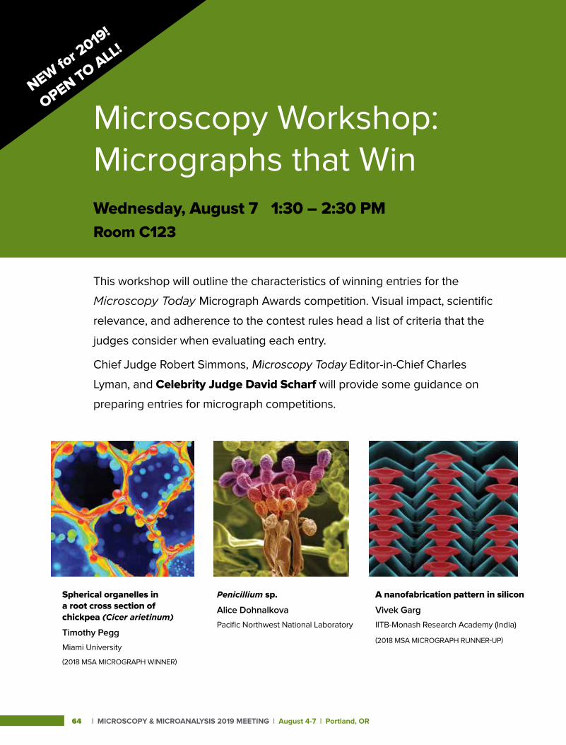
| MICROSCOPY & MICROANALYSIS 2019 MEETING | August 4-7 | Portland, OR64
Microscopy Workshop: Micrographs that WinWednesday, August 7 1:30 – 2:30 PMRoom C123
This workshop will outline the characteristics of winning entries for the
Microscopy Today Micrograph Awards competition. Visual impact, scientific
relevance, and adherence to the contest rules head a list of criteria that the
judges consider when evaluating each entry.
Chief Judge Robert Simmons, Microscopy Today Editor-in-Chief Charles
Lyman, and Celebrity Judge David Scharf will provide some guidance on
preparing entries for micrograph competitions.
A nanofabrication pattern in silicon
Vivek Garg
IITB-Monash Research Academy (India)
(2018 MSA MICROGRAPH RUNNER-UP)
Spherical organelles in a root cross section of chickpea (Cicer arietinum)
Timothy Pegg
Miami University
(2018 MSA MICROGRAPH WINNER)
Penicillium sp.
Alice Dohnalkova
Pacific Northwest National Laboratory
NEW for 2
019!
OPEN TO ALL!
Monday P
rogram
Scientific Program Information Monday, August 5, 2019

Mon
day
Pro
gram
http://microscopy.org/MandM/2019 | 65
Monday, A
ugust 5Scientific Program
PLENARY LECTURES AND AWARDSSESSION CHAIRS:Paul Kotula, President, Microscopy Society of AmericaRhonda Stroud, President, Microanalysis SocietyDavid Larson, President, International Field Emission SocietyAlice Dohnalkova, M&M 2019 Program Chair
MONDAY 8:30 AM • Room: Portland Ballroom
OPENING WELCOME:Paul Kotula, MSA PresidentRhonda Stroud, MAS PresidentDavid Larson, IFES President
8:45 AM 1 (Invited) Studying Kinetics by Counting Particles in
9:45 AM
10:00 AM
10:05 AM
10:30 AM
10:45 AM
Time-Resolved Cryo-EM; Joachim Frank; Professor of Biochemistry, Molecular Biophysics, and Biological Sciences, Columbia University
MAS Awards Presentation
IFES Awards Presentation
Coffee Break
MSA Awards Presentation
M&M Meeting Awards Presentation11:00 AM 2 (Invited) Single-Particle CryoEM: Potential
for Further Improvement; Richard Henderson, Professor of Biochemistry, Medical Research Council Laboratory of Molecular Biology (MRC LMB) - Cambridge, United Kingdom
A Analytical Sciences Symposia – Monday Afternoon
A01.1 Advances in Phase Retrieval Microscopy
SESSION CHAIRS: Kai He; Clemson UniversityXiaoqing Pan; University of California-Irvine
PLATFORM SESSIONMonday 1:30 PM • Room: B114
1:30 PM 3 (Invited) Phase Imaging Beyond the Diffraction Limit with Electron Ptychography; DA Muller, Z Chen, Y Jiang, M Odstrcil, Y Han, P Purohit, MW Tate, SM Gruner, V Elser
2:00 PM 4 Three-Dimensional Electron Ptychography of Catalyst Nanoparticles Using Combined HAADF STEM and Atom Counting; E Liberti, GT Martinez, CM O’Leary, PD Nellist, AI Kirkland
2:15 PM 5 (Invited) Advanced Phase Reconstruction Methods Enabled by Four-Dimensional Scanning Transmission Electron Microscopy; C Ophus, TR Harvey, FS Yasin, HG Brown, PM Pelz, BH Savitzky, J Ciston, BJ McMorran
2:45 PM 6 Unsupervised Machine Learning to Distill Structural-Property Insights from 4D-STEM; X Li, O Dyck, M Oxley, A Lupini, S Jesse, SV Kalinin
A02.1 Data Acquisition Schemes, Machine Learning Algorithms, and Open Source Software Development for Electron Microscopy
SESSION CHAIRS: Francisco de la Peña; Université Lille, FrancePhilippe T. Pinard; Oxford Instruments, United KingdomEric Prestat; Manchester University, United Kingdom
PLATFORM SESSIONMonday 1:30 PM • Room: C123
1:30 PM 7 (Invited) Nion Swift: Open Source Image Processing Software for Instrument Control, Data Acquisition, Organization, Visualization, and Analysis Using Python; CE Meyer, N Dellby, JA Hachtel, T Lovejoy, A Mittelberger, O Krivanek
2:00 PM 8 py4DSTEM: Open Source Software for 4D-STEM Data Analysis; BH Savitzky, L Hughes, KC Bustillo, HD Deng, NL Jin, EG Lomeli, WC Chueh, P Herring, A Minor, C Ophus
2:15 PM 9 Distinguishability of Structures via Principal Component Analysis: Application to 4D STEM; M Oxley, SV Kalinin, RK Vasudevan
2:30 PM 10 (STUDENT) Atomic Resolution Convergent Beam Electron Diffraction Analysis Using Convolutional Neural Networks; C Zhang, J Feng, LR DaCosta, PM Voyles
2:45 PM 11 Statistical Physics-Based Framework and Bayesian Inference for Model Selection and Uncertainty Quantification; L Vlcek, S Yang, M Ziatdinov, SV Kalinin, RK Vasudevan
A03.1 Low-Energy X-Ray Analysis: Novel Applications Using Soft X-Ray Emission Spectroscopy (SXES), Cathodoluminescence (CL), and Synchrotron Techniques
SESSION CHAIRS: Anette von der Handt; University of MinnesotaZack Gainsforth; University of California-Berkeley
PLATFORM SESSIONMonday 1:30 PM • Room: D139
1:30 PM 12 (Invited) Fast Hyperspectral Soft X-Ray Mapping; CM MacRae, NC Wilson, A Torpy, AE Hughes
2:00 PM 13 Information of Valence Charge of 3D Transition Metal Elements Observed in L-emission Spectra; M Terauchi
2:15 PM 14 (STUDENT) The Impact of Phase on Mass Absorption Coefficients Using Soft X-Ray Emission Spectrometry; S Rudinsky, CM MacRea, NC Wilson, H Demers, R Gauvin
2:30 PM 15 (Invited) An EPMA Study of the Soft Fe Lα-Lβ X-Ray lines in Fe-silicide, Olivine and Fe-sulfide Minerals by SXES and WDS; A Moy, AV der Handt, JH Fournelle
| MICROSCOPY & MICROANALYSIS 2019 MEETING | August 4-7 | Portland, OR66
Scientific ProgramM
onda
y, A
ugus
t 5 A04.1 Recent Developments in Atom
Probe Tomography SESSION CHAIRS: Ty J. Prosa; CAMECA Instruments Inc.Baptiste Gault; Max Planck Institute, Germany
PLATFORM SESSIONMonday 1:30 PM • Room: F151
1:30 PM 16 (Invited) Exploring New Science Domains with Atom Probe Tomography Enabled by an Environmental Transfer Hub; DE Perea, DK Schreiber, A Devaraj, DD Reilly, SV Lambeets, EJ Kautz, TG Lach, MG Wirth, JE Evans
2:00 PM 17 A Toolchain for the Analysis of Hydrogen in Materials at the Atomic Scale; PJ Felfer
2:15 PM 18 Comprehensive Analysis of Hydrogen, Deuterium, Tritium and Isotopic Ratios of Other Light Elements in Neutron Irradiated TPBAR Components; A Devaraj, B Matthews, B Arey, EJ Kautz, G Sevigny, D Senor
2:30 PM 19 (Invited) Building Laboratory-on-Tip for Imaging Biological Samples with Atom Probe Tomography; J Fu
A05.1 Leveraging 3D Imaging and Analysis Methods for New Opportunities in Material Science SESSION CHAIR: Will Harris; Carl Zeiss Microscopy USA
PLATFORM SESSIONMonday 1:30 PM • Room: C122
1:30 PM 20 3D Reconstruction of an Additive Manufactured IN625 Tensile Sample Using Serial Sectioning and Multi-Modal Characterization; MG Chapman, MD Uchic, JM Scott, MN Shah, SP Donegan, PA Shade, WD Musinski, M Obstalecki, MA Groeber, D Menasche, ME Cox, EJ Schwalbach
1:45 PM 21 (STUDENT) Correlative 3D X-Ray, Laser Ablation, and SEM/EDS Mapping Establishing Access Point for FIB Tomography of Defects in Multi-Layer Ceramic Capacitors; J Favata, V Ray, S Shahbazmohamadi
2:00 PM 22 (Invited) Combining EBSD with Serial Sectioning for the 3D Analysis of Materials; DJ Rowenhorst, RW Fonda
2:30 PM 23 (STUDENT) 3D Characterization of Porous Copper Thin Films; A Wijaya, B Eichinger, M Mischitz, R Brunner
2:45 PM 24 Plasma FIB Spin Milling for Large Volume Serial Sectioning Tomography; B Winiarski, C Rue, PJ Withers
A08.1 Current Trends and Challenges in Electron Energy-Loss Spectroscopy SESSION CHAIRS: Peter Crozier; Arizona State UniversityXiaoqing Pan; University of California-Irvine
PLATFORM SESSIONMonday 1:30 PM • Room: B111
1:30 PM 25 (Invited) Atomic-Plane Resolved Electron Magnetic Circular Dichroism by Achromatic Spatially-Resolved Electron Energy-Loss Spectroscopy; X Zhong
2:00 PM 26 Rapid Simulation of Elemental Maps in Core-Loss Electron Energy-Loss Spectroscopy; HG Brown, SD Findlay, LJ Allen, J Ciston, C Ophus
2:15 PM 27 Development of Monochromatic Analytical Electron Microscope Equipped with Higher-Order Aberration Corrector; M Mukai, E Okunishi, A Nakamura, T Ishikawa, H Sawada
2:30 PM 28 (STUDENT) Understanding the Complex Structure of CeO2/TiO2 Nanocatalyst. Key Contributions of the Combined Use of HAADF, X-EDS and EELS Spectroscopies; R Manzorro, JA Perez-Omil, JJ Calvino, S Trasobares
2:45 PM 29 (STUDENT) (M&M Meeting Scholar Award) Harnessing Local Sample Variations to Generate Self-Consistent EELS References for Stoichiometry Quantification; BH Goodge, HP Nair, JP Ruf, NJ Schreiber, KM Shen, DG Schlom, LF Kourkoutis
A11.1 Current and Emerging Microscopy for Quantum Information Sciences SESSION CHAIRS: Miaofang Chi; Oak Ridge National Laboratory
PLATFORM SESSIONMonday 1:30 PM • Room: B117
1:30 PM 30 Emerging Microscopy for Quantum Information Sciences; Q Qiao, L Wu, J Li, S Cheng, M Han, J Garlow, SD Pollard, X Fu, J Tao, Y Zhu, J Wall
2:00 PM 31 (STUDENT) Atomic Resolution CryoSTEM Across Continuously Variable Temperatures; BH Goodge, E Bianco, HW Zandbergen, LF Kourkoutis
2:15 PM 32 New Advanced Electron Microscopy to Discover New Quantum Materials; DC Bell, C Ozsoy-Keskinbora, F VonCube, J Checkelsky, RW Westervelt
2:30 PM 33 (Invited) (STUDENT) Low Temperature Electron Microscopy of “Charge-Ordered” Phases; I El Baggari, DJ Baek, BH Savitzky, MJ Zachman, R Hovden, LF Kourkoutis
A Analytical Sciences Symposia – Monday Afternoon cont.
http://microscopy.org/MandM/2019 | 67
Monday, A
ugust 5Scientific Program
B03.1 Utilizing Microscopy for Research and Diagnosis of Diseases in Humans, Plants and Animals SESSION CHAIRS: Ru-ching Hsia; University of Maryland-BaltimoreMarcela Redigolo; West Virginia UniversityHan Chen; Penn State College of Medicine
PLATFORM SESSIONMonday 1:30 PM • Room: D136
1:30 PM 34 Analysis of Ultrastructural Properties Reveals SP-D Protective Roles in Lung and Pancreatic Injury of Severe Acute Pancreatitis in Mouse Model; H Chen, J Yu, G Wang
1:45 PM 35 (Invited) Isolation and Characterization of Cell- and Size-Specific Extracellular Vesicles by Nanoscale High Resolution Flow Cytometry; TK Morgan
2:15 PM 36 Scanning Electron Microscopy as a New Tool for Diagnostics in Pathology; T Cohen Hyams, MC Killingsworth
2:30 PM 37 (Invited) Multi-Scale Imaging of Tissues and Organoid Models by Backscattered Electron Scanning Microscopy after Pre- and Post-Embedding Immuno-Labeling; M Reichelt, D Mandikian, A Lopez, AK Katakam, D Smith, W Meilandt, J Imperio, M Sagolla
B04.1 Cutting-Edge Microscopy in the Pacific Northwest SESSION CHAIRS: Doug Keene; Shriners Hospitals for ChildrenClaudia López; Oregon Health & Science University
PLATFORM SESSIONMonday 1:30 PM • Room: D133-134
1:30 PM 38 (Invited) 3DEM Data Collection of Cancer Biopsies: Comparing FIB-SEM and SBF-SEM; JL Riesterer, K Loftis, K Stoltz, E Stempinski, M Williams, CS Lopez, G Thibault, JW Gray
2:00 PM 39 (Invited) Computer Vision Techniques for Cancerous Cells Analysis in FIB-SEM Images; G Thibault, J Riesterer, K Stoltz, K Loftis, G Schau, E Stempinski, C Lopez, YH Chang, J Gray
2:15 PM 40 (Invited) (STUDENT) (M&M MEETING SCHOLAR AWARD) Multiscale Cardiac Imaging: From Whole Heart Images to Cardiac Ultrastructure; G Rykiel, CS Lopez, JL Riesterer, M Williams, K Courchaine, A Maloyan, K Thornburg, S Rugonyi
2:45 PM 41 ELVIS: A Correlated Light-Field and Digital Holographic Microscope for Field and Laboratory Investigations; J Nadeau, M Schadegg, C Snyder, I Hanczarek
P Physical Sciences Symposia – Monday Afternoon
P06.1 In situ TEM of Nanoscale Materials and Electronic Devices for Phase Transformation Studies SESSION CHAIRS: Thomas C. Pekin; Humboldt Universität zu Berlin, GermanyJudy Cha; Yale University
PLATFORM SESSIONMonday 1:30 PM • Room: B118-119
1:30 PM 42 (Invited) In Situ TEM on Formation of Monatomic Metallic Glasses Through Ultrafast Liquid Quenching; S Mao
2:00 PM 43 Electric Field-Induced Surface Melting of Gold Observed In Situ at Room Temperature and at Atomic Resolution Using TEM; L de Knoop, M Juhani Kuisma, J Löfgren, K Lodewijks, M Thuvander, P Erhart, A Dmitriev, E Olsson
2:15 PM 44 (STUDENT) In situ TEM Investigation of the Electroplasticity Phenomenon in Metals; X Li, AM Minor
2:30 PM 45 (Invited) Microstructure and Defect Characterization Using Advanced STEM Techniques: 4D-STEM and WB DF STEM; J Miao, L Casalena, J Ciston, T Pekin, M Ghazisaeidi, MJ Mills
P07.1 Electron Crystallography of Nanostructures in Nanotechnology, Materials and Bio-Sciences SESSION CHAIRS: Roberto Reis; Northwestern UniversitySergei Rouvimov; University of Notre Dame
PLATFORM SESSIONMonday 1:30 PM • Room: D138
1:30 PM 46 (Invited) Scanning Precession Electron Diffraction to aid Aluminum Alloy Development; JK Sunde, E Christiansen, E Thronsen, S Wenner, CD Marioara, AT van Helvoort, R Holmestad
2:00 PM 47 Revealing Embedded Crystals through their Diffracting Signals in Transmission Electron Microscopy; EF Rauch, M Veron
2:15 PM 48 (STUDENT) MicrostructureEvolution and Its Effect on Magnetic Properties of Mn-Al Alloy Fabricated via End Milling Machining Process; J Jo, B Vishwanadh, R Shankar, J Wiezorek
2:30 PM 49 Advantages of Precession Electron Diffraction in the Study of Small Metallic Nanoparticles; C Fernando, M Hoque, J Reyes Gasga, A Ponce
2:45 PM 50 Dynamical Diffraction Calculations of Incommensurate Modulations in Crystals; L Wu, S Deng, Y Zhu
B Biological Sciences Symposia – Monday Afternoon
| MICROSCOPY & MICROANALYSIS 2019 MEETING | August 4-7 | Portland, OR68
Scientific ProgramM
onda
y, A
ugus
t 5 P08.1 Microscopy and Spectroscopy of
Nanoscale Materials for Energy Applications SESSION CHAIRS: Chongmin Wang; Pacific Northwest National LaboratoryYuanyuan Zhu; University of Connecticut
PLATFORM SESSIONMonday 1:30 PM • Room: B115
1:30 PM 51 (Invited) Designing Energy Materials via Atomic-resolution Microscopy and Spectroscopy; S Pennycook, X Zhao, J Lu, W Zang, H Wu, J Wang
2:00 PM 52 Ultrafast Laser-Pump Electron-Probe Microscopy for Imaging Semiconductor Carrier Dynamics; MWH Garming, GIGC Weppelman, P Kruit, JP Hoogenboom
2:15 PM 53 (STUDENT) Direct Imaging of Incoherent-to-Coherent Structural Dynamics in Plasmonic Nanorods with Ultrafast Electron Microscopy; R Gnabasik, D Valley, M Quan, V Ferry, D Flannigan
2:30 PM 54 In Situ Tracking of Picoscale Atomic Displacements with Millisecond Time-Resolution During Exchange and Diffusion Processes in Energy Materials in TEM; BD Levin, EL Lawrence, P Haluai, PA Crozier
2:45 PM 55 (STUDENT) Exploring the Connection between Coherent Acoustic Phonons and Bright-Field Contrast in Ultrafast Electron Microscopy; DX Du, DJ Flannigan
P09.1 The Success of TMBA: TEM and STEM Developments in Techniques, Applications and Education SESSION CHAIRS: Paul Kotula; Sandia National LaboratoriesJoe Michael; Sandia National LaboratoriesMasashi Watanabe; Lehigh University
PLATFORM SESSIONMonday 1:30 PM • Room: D137
1:30 PM 56 Transmission Microscopy: Beginning Automation; CB Carter, DB Williams
1:45 PM 57 High-Throughput Identification of Crystal Structures Via Machine Learning; K Kaufmann, C Zhu, AS Rosengarten, D Maryanovsky, T Harrington, E Marin, KS Vecchio
2:00 PM 58 The Power of AEM and Data Analytics: 20 Years of Teaching MSA at M&M; P Kotula
2:15 PM 59 (STUDENT) Ion Beam Induced Artifacts in Lead Based Chalcogenides; X Zhang, S Hao, G Tan, X Hu, EW Roth, MG Kanatzidis, C Wolverton, VP Dravid
2:30 PM 60 (Invited) Analytical Electron Microscopy: Rivaling / Complementing Atom Probe Tomography; M Burke, JJH Lim
P11.1 Advances in Characterization of Geological and Extraterrestrial Samples SESSION CHAIRS: Lori Hathon; University of HoustonKultaransingh Hooghan; Sandia National Laboratories
PLATFORM SESSIONMonday 1:30 PM • Room: B112
1:30 PM 61 Microns to Millimeters: Multi-range Hyperspectral Imaging of Organic and Inorganic Reservoir Rock Samples; TD Jobe, C Sandu, SL Eichmann, L Stout
1:45 PM 62 Innovative Mapping of Mineral and Organic Matter of Source Rocks at the Nanometer Scale Using Back Scatter Electron and Energy Dispersive Spectroscopy; DJ Jacobi, Q Sun, JM Longo, J Brothers, J Kone
2:00 PM 63 Digital Rock Evaluation of the Thermal Conductivity and Electrical Resistivity of Source Rocks; SL Eichmann, A Kotb, M Sengupta
2:15 PM 64 Correlative Petrographic and Spectroscopic Characterization of Boquillas Shale Samples Before and After Hydrous Pyrolysis: Understanding Diagenetic and Thermogenesis as it Relates to Storage and Expulsion; AR Boehlke, J Hatcherian, P Hackley, J Birdwell, A Jubb, B Valentine
2:30 PM 65 Optimization of Specimen Preparation Methods for Cryo Electron Microscopy of Oil-and-Water Emulsions; DH Anjum, P Guida, AR Behzad, S Saxena, WL Roberts
2:45 PM 66 New Insights to Characterize Fluid/Carbonate Rock Interfaces Using Transmission Electron Microscopy with Liquid Cell Holder; D Cha, M Alotaibi, S Enezi, AA Yousef, K Song, X Li, Y Han
P14.1 Microstructural and Mechanical Characterization of Metallic Alloys SESSION CHAIRS: Philipp Kürnsteiner; Max Planck Institute, GermanyBaptiste Gault; Max Planck Institute, Germany
PLATFORM SESSIONMonday 1:30 PM • Room: B116
1:30 PM 67 (STUDENT) (M&M MEETING SCHOLAR AWARD)
Characterization of Misfit Dislocations at Heterovalent II-VI/III-V Interfaces; BS McKeon, X Liu, JK Furdyna, DJ Smith
1:45 PM 68 Structure, Morphology and Coarsening Behavior of MX (NbC) Nanoprecipitates in Fe-Ni-Cr Based Alloys; M Heczko, BD Esser, R Gröger, L Feng, V Mazánová, Y Wang, MJ Mills
2:00 PM 69 (STUDENT) Characterization of Complex Retained Austenite on a TRIP800 Automotive Steel; N-I Makris, S Deligiannis, A Alexandratou, P Tsakiridis, G Fourlaris
P Physical Sciences Symposia – Monday Afternoon cont.
http://microscopy.org/MandM/2019 | 69
Monday, A
ugust 5Scientific Program
2:15 PM 70 (STUDENT) Effect of High-Temperature Impact on the Formation of Adiabatic Shear Bands; A Eliasu, S Boakye- Yiadom
2:30 PM 71 Ternary Fe-Al-Si Alloys Prepared by Mechanical Alloying and Spark Plasma Sintering; J Kopeček, F Laufek, M Karlík, P Haušild, J Čech, A Školáková, K Nová, B Severa, J Šesták, P Novák, F Průša
T Tutorials – Monday Afternoon
X44.1 Electron Optics for CryoEM: Facts and Myths SESSION CHAIRS: Tommi White; University of Missouri
PLATFORM SESSIONMonday 2:00 PM • Room: B110
2:00 AM 72 (Invited) High-End Data Collection for Single-Particle Cryo-EM; F Weis
A Analytical Sciences Poster Sessions– Monday
A01.P1 Advances in Phase Retrieval Microscopy
POSTER SESSIONMonday 3:00 PM • Room: Exhibit Hall
POSTER # 1
3:00 PM 73 Electromagnetic Field Reconstructions of 4D-STEM Datasets Using Ptychography and Differential Phase Contrast Imaging; S Cao, M Chi, KL More
POSTER # 2
3:00 PM 74 Effectively Synchronizing 4D-STEM Detectors with Probe Movement; B Bammes, C Zhang, R Bilhorn, PM Voyles
POSTER # 3
3:00 PM 75 Assessing the Phase Accuracy of ePIE Reconstructions of Crystalline Materials; AM Blackburn, RA McLeod
POSTER # 4
3:00 PM 76 Electron Ptychography of Single Biological Macromolecules; PM Pelz, R Bücker, G Ramm, H Venugopal, G Kassier, D Eggert, P-H Lu, RE Dunin-Borkowski, RJD Miller
POSTER # 5
3:00 PM 77 Three-Dimensional Electrostatic Field at an Electron Nano-Emitter Determined by Differential Phase Contrast in Scanning Transmission Electron Microscopy; M Wu, A Tafel, P Hommelhoff, E Spiecker
POSTER # 6
3:00 PM 78 (M&M POST-DOCTORAL RESEARCHER AWARD) Structure Retrieval of Strongly Scattering Materials in the Transmission Electron Microscope; HG Brown, Z Chen, M Weyland, C Ophus, J Ciston, LJ Allen, SD Findlay
POSTER # 7
3:00 PM 79 Spatial and Temporal Incoherence in Scanning Diffraction and Differential Phase Contrast; MP Oxley
POSTER # 8
3:00 PM 80 (STUDENT) Nanoscale Quantification of Jahn-Teller Distortion in LaMnO3; B Wang, BD Esser, N Bagués, J Yan, DW McComb
POSTER # 9
3:00 PM 81 Local Phase Curvature Measurement in STEM With a Pixelated Detector; M Krajnak, DM Paganin, K Fallon, S McVitie, J Etheridge
POSTER # 10
3:00 PM 82 Phase Plate STEM Imaging Using Two-Dimensional Electron Detector; M Tsubouchi, H Minoda
POSTER # 11
3:00 PM 83 (STUDENT) Large Field of View Strain Characterization in a Scanning Transmission Electron Microscope Using a Designed Coherent Sampler; A Pofelski, S Ghanad-Tavakoli, D Thompson, G Botton
| MICROSCOPY & MICROANALYSIS 2019 MEETING | August 4-7 | Portland, OR70
Scientific ProgramM
onda
y, A
ugus
t 5 A05.P1 Leveraging 3D Imaging and Analysis
Methods for New Opportunities in Material Science POSTER SESSIONMonday 3:00 PM • Room: Exhibit Hall
POSTER # 12
3:00 PM 84 Mapping the Evolution of Grains in Strontium Titanate through Laboratory-Based 4D Diffraction Contrast Tomography; A Krause, H Bale, J Sun, W Harris, E Lauridsen, C Marvel, C Krill, M Harmer
POSTER # 13
3:00 PM 85 Exploration of TATB Grain Structure on Detonation Sensitivity Using X-Ray CT; L Kuettner, LG Hill, J Yeager, K Henderson, BM Patterson
POSTER # 14
3:00 PM 86 What Lies Beneath: 3D vs 2D Correlative Imaging Challenges and How to Overcome Them; RL Mitchell, S Freitag, T Volkenandt, J Russell, P Davies, C Pleydell-Pearce, R Johnston
POSTER # 15
3:00 PM 87 (STUDENT) A Correlative and Multi-Modal Approach to Analyzing Microscopic Particulate Contaminants in Carbon Black; A Poozhikunnath, J Favata, B Ahmadi, J Xiong, S Shahbazmohamadi, L Bonville, R Maric
POSTER # 16
3:00 PM 88 Bubbles in the Bullion: Micro-CT Imaging of the Internal Structure of Ancient Coins; LL Van Loon, AJ Nelson, U Wartenberg Kagan, K Barron, NR Banerjee
POSTER # 17
3:00 PM 89 (STUDENT) Structural Analysis of Liquid Metal Catalysts in Porous Silica Utilizing Nano-CT and Analytical Transmission Electron Microscopy; J Wirth, S Englisch, C Wiktor, N Taccardi, B Apeleo Zubiri, P Wasserscheid, E Spiecker
POSTER # 18
3:00 PM 90 The Dynamics of Abnormal Grain Growth in a Particle-Containing System: Integration of 3D Experimental Data into a Capillarity Driven Model; N Lu, J Kang, N Senabulya, N Gueninchault, A Shahani
A08.P1 Current Trends and Challenges in Electron Energy-Loss Spectroscopy POSTER SESSIONMonday 3:00 PM • Room: Exhibit Hall
POSTER # 19
3:00 PM 91 (STUDENT) Low Loss EELS of Lateral MoS2/WS2 Heterostructures; S Susarla, LHG Tizei, SY Woo, A Zobelli, O Stephan, PM Ajayan
POSTER # 20
3:00 PM 92 (STUDENT) Localization of Plasmon Resonance Using Momentum-Resolved EELS; ER Hoglund, JM Howe
POSTER # 21
3:00 PM 93 (STUDENT) Nanoscale Probing of Adsorbates on Pt/CeO2 with Aloof-beam Vibrational Electron Energy-Loss Spectroscopy; K Venkatraman, J Vincent, K March, P Rez, PA Crozier
POSTER # 22
3:00 PM 94 (STUDENT) Manipulation of Optical Phonon Polaritons in Patterned SiO2 Thin-Films; K Venkatraman, S Situ, K March, P Rez, PA Crozier
POSTER # 23
3:00 PM 95 Model-Based EELS Quantification and ELNES Phase Mapping Using Experimentally Measured Cross-Sections; B Schaffer, L Spillane, PJ Thomas
POSTER # 24
3:00 PM 96 (STUDENT) Simultaneous Multi-Region Background Subtraction for EELS Spectra; JT Held, H Yun, KA Mkhoyan
POSTER # 25
3:00 PM 97 (STUDENT) Effects of Beam-Induced Carbon Deposition on Electron Energy-Loss Spectroscopy Analysis of Compositional Fluctuations in InGaN/GaN Quantum Well LEDs; SA Goodman, GJ Syaranamual, JY Chung, Z Li, A Singh, K Kisslinger, R Armitage, I Wildeson, D Su, P Deb, E Stach, S Gradecak
POSTER # 26
3:00 PM 98 (STUDENT) Towards Quantitative Nanomagnetism in Transmission Electron Microscope by the Use of Patterned Apertures; H Ali, T Warnatz, L Xie, B Hjörvarsson, K Leifer
POSTER # 27
3:00 PM 99 (STUDENT) Decomposition of Core-Loss EEL Edges into a Linear Combination of Refined ‘Pure’ Spectra; JT Held, K Hunter, UR Kortshagen, KA Mkhoyan
POSTER # 28
3:00 PM 100 Determination of Mn Valences in Li1-xMgxMn2O4 Using Monochromated EELS in an Aberration-Corrected STEM; X Zhan, J-M Zuo, W Chen, Q Chen
A Analytical Sciences Poster Sessions– Monday cont.
http://microscopy.org/MandM/2019 | 71
Monday, A
ugust 5Scientific Program
B Biological Sciences Poster Sessions– Monday
B03.P1 Utilizing Microscopy for Research and Diagnosis of Diseases in Humans, Plants, and Animals POSTER SESSIONMonday 3:00 PM • Room: Exhibit Hall
POSTER # 29
3:00 PM 101 Histopathology and in situ Hybridization of Coatomer (CopI) Knockdown in Western Corn Rootworm (Diabrotica virgifera virgifera); AJ Bowling, HE Pence, E Fishilevich
POSTER # 30
3:00 PM 102 Scanning Confocal Microscopy for Study of the Neuromuscular Junction (NMJ) Acethylcholine Receptors(nAChRs): One Model of the Peripheral Nerve Injury Recovered After Fibrin Sealant Use; SMM Matheus, CG Pinto, APS Leite, AA Sartor, FC Tibúrcio, RS Ferreira Junior, AL Filadelpho, SC de Carvalho
POSTER # 31
3:00 PM 103 The Examination on Corneal Nerve in Diabetic WBN/Kob Rat is Useful for Detecting Peripheral Sensory Neuropathy; K Ozaki, Y Terayama, T Matsuura
POSTER # 32
3:00 PM 104 (STUDENT) The Role of Tyramine within the Mouse Ovary; SM Peck, SB Obayomi, DP Baluch
POSTER # 33
3:00 PM 105 (STUDENT) Morphologic Analysis of Multiple Tissues in Caveolin-1 Null Mice; B Qu, L Li
POSTER # 34
3:00 PM 106 Correlative Structural, Biochemical, and Behavioral Study in Neuroprotection with Palmitoylehanolamide after Perinatal Asphyxia in the Immature Brain; MI Herrera, T Kobiec, L Udovin, N Toro-Urrego, CF Kusnier, JP Luaces, R Kolliker, F Capani
POSTER # 35
3:00 PM 107 (STUDENT) Ultrastructural Modifications of the Mouse Uterus upon Biogenic Amine Stimulation; SB Obayomi, S Peck, DP Baluch
POSTER # 36
3:00 PM 108 (STUDENT) Transient Exposure to 450 nm Light Alters Phagocytosis by Retinal Pigment Epithelium Without Compromising Viability; J Blaize, JV Fabozzi
POSTER # 37
3:00 PM 109 Chronic Oxidative Stress and Comorbidities in the HIV-1 Transgenic Rat; F Denaro, M Worthington, F Benedetti, S Curreli, D Zella, J Bryant
POSTER # 38
3:00 PM 110 (M&M POST-DOCTORAL RESEARCHER AWARD) Localization of the Herpesvirus Portal; MH Buch, J Heymann, WW Newcomb, DC Winkler, AC Steven
POSTER # 39
3:00 PM 111 Using Electron Microscopy to Enhance the Knowledge of Biological Systems; K Ishikawa, Z Wang, C Burks, M Larion, N de Val, J Schneider
POSTER # 40
3:00 PM 112 (STUDENT) Microscopy Techniques Used to Visualize Immune-Labeled Chlamydial Inclusion Membrane Proteins in 2D and 3D; B Crews, ER Fischer, FH Hoyt, BT Hansen, T Hackstadt
POSTER # 41
3:00 PM 113 (STUDENT) Using Correlative Light and Electron Microscopy (CLEM) To Understand Ultrastructural Changes Induced by Salmonella typhimurium Infection in a Calf Ileum Loop Model; S Antonioli-Schmit, A Chong, BT Hansen, M Pulliam, V Nair
B04.P1 Cutting-Edge Microscopy in the Pacific Northwest POSTER SESSIONMonday 3:00 PM • Room: Exhibit Hall
POSTER # 42
3:00 PM 114 Studying Ras Nanocluster Formation on the Cell Membrane with Correlative Superresolution and Electron Microscopies; Y Zhang, K Tao, L Wu, Y Lee, YH Chang, X Nan
POSTER # 43
3:00 PM 115 #Protein Lights Up in Cryo-EM; J ChenPOSTER # 44
3:00 PM 116 Structural Characterization of Immune Responses to HPV Infection and HPV Vaccination; S Singh, J Carter, K Cohen, J McElrath, D Galloway, M Pancera
| MICROSCOPY & MICROANALYSIS 2019 MEETING | August 4-7 | Portland, OR72
Scientific ProgramM
onda
y, A
ugus
t 5
P Physical Sciences Poster Sessions– Monday
P07.P1 Electron Crystallography of Nanostructures in Nanotechnology, Materials and Bio-Sciences POSTER SESSIONMonday 3:00 PM • Room: Exhibit Hall
POSTER # 45
3:00 PM 117 High Throughput Grain Mapping with Sub-Nanometer Resolution by 4D-STEM; FI Allen, TC Pekin, A Persaud, SJ Rozeveld, GF Meyers, J Ciston, C Ophus, AM Minor
POSTER # 46
3:00 PM 118 (STUDENT) Indexing Electron Backscatter Diffraction Patterns with a Refined Template Matching Approach; A Foden, DM Collins, AJ Wilkinson, TB Britton
POSTER # 47
3:00 PM 119 When Ion or Electron Channeling Meets Crystal Orientation Mapping; C Langlois, C Lafond, T Douillard, S Dubail, S Cazottes, J Silvent, A Delobbe
POSTER # 48
3:00 PM 120 The role of Cluster Analysis in Automated Serial Electron Crystallography; S Smeets, B Wang, X Zou
POSTER # 49
3:00 PM 121 Two Beam Toy Model for Dislocation Contrast in ECCI; E Pascal, B Hourahine, C Trager-Cowan, M De Graef
POSTER # 50
3:00 PM 122 Nano-Scale Crystallographic Aspects of Discontinuous Precipitation and Coarsening Reactions in Cu-10% Co alloy; NM Suguihiro, G Solórzano
POSTER # 51
3:00 PM 123 Determination of Crystallinity in Li1-xMgxMn2O4 Nanocrystals Based on Diffraction Patterns Correlation Analysis and Strain Mapping; X Zhan, R Yuan, W Chen, Q Chen, J-M Zuo
POSTER # 52
3:00 PM 124 The Structural Changes in the Human Teeth during Their Calcination From 25 To 1200 °C by Electron Microscopy; J Reyes-Gasga, NV Becerril
POSTER # 53
3:00 PM 125 (STUDENT) Effect of Magnetic Stirring on the Green Synthesis of Silver Nanoparticles Using Lavandula angustifolia Plant Extract; MA Villalpando, GA Rosas
POSTER # 54
3:00 PM 126 (STUDENT) Study of 0D, 1D and 3D Zinc Oxide Morphologies by SEM and TEM; JE Morales-Mendoza, F Paraguay-Delgado, E Guerrero, D Lardizabal, L de la Torre
POSTER # 55
3:00 PM 127 An Electron Microscopy Collaboratory for Correlative Imaging Sciences; DE Huber, FJ Scheltens, RE Williams, DB Williams, DW McComb
POSTER # 56
3:00 PM 128 An Interactive and Adaptive Online Electron Microscopy Education Platform; N Freed, D Butler, A Cairns, R Hugo, J Jiao, Z Chen
POSTER # 57
3:00 PM 129 Evolution of Microstructures and Interfaces in Compositionally Graded Mixed Oxide Thin Films for Nanoelectronics and Energy; A Singh Pal, A Singh, RK Mandal, J Basu
POSTER # 58
3:00 PM 130 A TD-DFT Treatment of Electronic Excitations in the STEM Spanning Dipole and Impact Scattering Regimes; DB Lingerfelt, J Jakowski, P Ganesh, B Sumpter
POSTER # 59
3:00 PM 131 In situ Cathodoluminescence and Monitoring Electronic Structure Change Using Optical TEM Holder; M Xu, S Dai, W Gao, T Blum, X Pan
P14.P1 Microstructural and Mechanical Characterization of Metallic Alloys POSTER SESSIONMonday 3:00 PM • Room: Exhibit Hall
POSTER # 60
3:00 PM 132 Effect of Ni Addition and T6 Heat Treatment on Microstructure and Microhardness of Hot Plastic Deformed (Al-Si- Mg) Alloys; HM Medrano Prieto, CG Garay Reyes, I Estrada Guel, JM Mendoza Duarte, JC Guía Tello, JS Castro Carmona, H Camacho Montes, MC Maldonado Orozco, R Martínez Sánchez
POSTER # 61
3:00 PM 133 Improvement of Mechanical Properties of an Aluminum Alloy 7075 by Adding Reduced Graphene Oxide Trough Mechanical Milling; E Cuadros Lugo, I Estrada Guel, JM Herrera Ramirez, C López Meléndez, C Carreño Gallardo
POSTER # 62
3:00 PM 134 Exploring the Reinforcing Effect of AgcNP and Al2O3NP in Aluminum Alloy 2024 Matrix Composites; C Carreño Gallardo, E Cuadros Lugo, I Estrada Guel, C López Meléndez, JM Herrera Ramirez
POSTER # 63
3:00 PM 135 Effect of the Age-hardering Time on the Microstructure of Cold Rolled Al2024 Alloy; JC Guía Tello, CG Garay Reyes, HM Medrano Prieto, MA Ruiz-Esparza Rodiguez, MC Maldonado Orozco, G Rodriguez Cabriales, R Martínez Sánchez
POSTER # 64
3:00 PM 136 (STUDENT) Wear Behavior in Al-Cu-Mg Alloy Reinforced With WC Particles Fabricated by Mechanical Alloying; G Rodriguez-Cabriales, CG Garay-Reyes, I Estrada-Guel, MC Maldonado-Orozco, R Martínez-Sánchez
http://microscopy.org/MandM/2019 | 73
Monday, A
ugust 5Scientific Program
POSTER # 65
3:00 PM 137 (STUDENT) Scanning Electron Microscopy of the Electrochemical Corrosion Products of a Titanium based Metallic Foam; L Béjar, E Huape, A Medina, AA Mejía, C Aguilar, I Alfonso
POSTER # 66
3:00 PM 138 Evaluation of Mechanical Properties of Ti/Al/Zr-N and Zr/Al/Ti-N Thin Films; CD Gómez-Esparza, RP Talamantes- Soto, A Duarte-Moller
POSTER # 67
3:00 PM 139 (STUDENT) Microstructural Evolution of Cold-Rolled β Metastable Ti-29Nb-2Mo-6Zr Alloy; AR Vieira Nunes, S Borborema, L Malet, J Dille, LH de Almeida
POSTER # 68
3:00 PM 140 (STUDENT) Α Comparative ΤΕΜ Study of Nanoprecipitate Formation in Waspaloy® Welds; A Alexandratou, S Deligiannis, NI Makris, PE Tsakiridis, G Fourlaris
POSTER # 69
3:00 PM 141 (STUDENT) Rationalization of Thermo-Mechanical Instabilities in Transient Additive Manufacturing of Ni-Based Superalloys; B Lim, XC Cui, SP Ringer
POSTER # 70
3:00 PM 142 Microstructural Behavior of AA319 Aluminum Alloy Modified with Nickel; J Camarillo-Cisneros, R Martinez- Sanchez, H Arcos-Gutierrez, IE Garduño-Olvera, R Pérez-Bustamante
POSTER # 71
3:00 PM 143 Microstructural and Hardness Behavior of AlCoCuFeNi and AlCoCuFeNiCr High-Entropy Alloys Synthesized by Milling and Arc Melting; CD Gomez-Esparza, M Hernandez-Hernandez, VH Mercado-Lemus, R Pérez-Bustamante
POSTER # 72
3:00 PM 144 (STUDENT) Densification, Microhardness and Microestructural Evolution by Fast Low-Temperature Consolidation of AlxCoCrFeMnNi High Entropy Alloy; MA Ruiz Esparza Rodriguez, CG Garay Reyes, JM Mendoza Duarte, I Estrada Guel, R Martinez Sanchez
POSTER # 73
3:00 PM 145 Microstructural Study and Antibacterial Response of an AlCoCrCuFeMoNi High-Entropy Alloy; CD Gómez- Esparza, CA Ramírez-Valdespino, I Estrada-Guel, A Duarte-Moller
POSTER # 74
3:00 PM 146 Microstructural Study of Fe-Cr-Al-X (X=Nd,Pr) Alloys for High Temperature Structural Applications; MA Encinas Ocejo, O Hernández-Negrete, G Tiburcio Munive, F Brown, A Valenzuela Soto, HE Esparza Ponce
POSTER # 75
3:00 PM 147 (STUDENT) Discontinuous Precipitation of α-Cr Phase in Alloy 33 (Cr-Fe-Ni-N); JC Spadotto, MG Burke, IG Solórzano
POSTER # 76
3:00 PM 148 (STUDENT) Influence of the Powder Pack Boriding Mixture on the FeB and Fe2B Layers; M Ortiz Domínguez, G Ares de Parga, A Arenas Flores, I Morgado Gonzalez, OA Gómez Vargas, J Zuno Silva, A Cruz Avilés
POSTER # 77
3:00 PM 149 Investigation of Digital Imaging Processing in Determining Nano-Particle Size Distribution Based on Scanning Electron Microscopic Image; BC Park, MJ Kwak
POSTER # 78
3:00 PM 150 (STUDENT) Effect of Finishing and Coiling Temperatures on the Nanoprecipitate Dispersion in Novel HSLA Steels; S Deligiannis, A Alexandratou, NI Makris, P Tsakiridis, G Fourlaris

Notes
Tuesday Program
Scientific Program Information Tuesday, August 6, 2019
Tues
day
Pro
gram
75
Scientific ProgramTuesday, A
ugust 6
http://microscopy.org/MandM/2019 |
A Analytical Sciences Symposia – Tuesday Morning
A01.2 Advances in Phase Retrieval Microscopy
SESSION CHAIRS: Jian-Min Zuo; University of Illinois–Urbana-ChampaignToshiaki Tanigaki; Hitachi Ltd.
PLATFORM SESSIONTuesday 8:30 AM • Room: B114
8:30 AM 151 (Invited) Differential Phase Contrast Scanning Transmission Electron Microscopy at Atomic Resolution; N Shibata
9:00 AM 152 (M&M POST-DOCTORAL RESEARCHER AWARD) Measuring Charge State at the Single-Atomic-Column-Base with Four-Dimensional Scanning Transmission Electron Microscopy; W Gao, C Addiego, H Huyan, X Pan
9:15 AM 153 (STUDENT) Charge Density Mapping via Scanning Diffraction in Scanning Transmission Electron Microscopy; C Addiego, W Gao, X Pan
A02.2 Data Acquisition Schemes, Machine Learning Algorithms, and Open Source Software Development for Electron Microscopy
SESSION CHAIRS: Francisco de la Peña; Université Lille, FrancePhilippe T. Pinard; Oxford Instruments, United KingdomEric Prestat; Manchester University, United Kingdom
PLATFORM SESSIONTuesday 8:30 AM • Room: C123
8:30 AM 155 (Invited) Scientific Python: A Mature Computational Ecosystem for Microscopy; SJ VanderWalt
9:00 AM 156 Fuzzy Clustering to Merge EDS and EBSD Datasets with Crystallographic Ambiguity; CM Parish
9:15 AM 157 (STUDENT) Utilizing an Open-Source Workflow for the Analysis of Atom Dynamics in Two-Dimensional Materials; EN O’Connell, M Hennessy, E Courtney, QM Ramasse, B Kardynal, H Hofsaess, M Ghorbani-Asl, A Krasheninnikov, U Bangert
9:30 AM 158 Open Source Development Tools for Robust and Reproducible Electron Microscopy Data Analysis; M Nord, J Verbeeck
9:45 AM 159 Harvesting Microscopy Experimental Context with a Configurable Laboratory Information Management System; JA Taillon, RF Devers, RL Plante, MW Newrock, JW Lau, G Greene
A03.2 Low-Energy X-Ray Analysis: Novel Applications Using Soft X-Ray Emission Spectroscopy (SXES), Cathodoluminescence (CL), and Synchrotron Techniques
SESSION CHAIRS: Emma Bullock; Carnegie ScienceJuliane Gross; Rutgers University
PLATFORM SESSIONTuesday 8:30 AM • Room: D139
8:30 AM 160 (Invited) Spatially Resolved Soft X-Ray Spectroscopy in Scanning X-Ray Microscopes; AP Hitchcock, P Krüger, C Bittencourt, GD Swerhone, JR Lawrence
9:00 AM 161 Total Electron Yield Mapping of Electronic Device Features via Measurement of X-Ray Beam-Induced Currents; WA Hubbard, JJ Lodico, B Zutter, D Shapiro, YH Lo, A Rana, D Morrill, C Gentry, HL Chan, BC Regan
9:15 AM 162 XAS between the Stars; Z Gainsforth, AL Butterworth, JA Tomsick, AJ Westphal
9:30 AM 163 Barite Cathodoluminescence as a Potential Indicator of Undiscovered Ore Deposits; H Lowers, D Olinger, D Adams
9:45 AM 164 Development of LVSEM-EDS Analyzer Utilizing Superconducting X-Ray Detector Toward Nanometer-scale Elemental Mapping of CFRP/Adhesive Interface; G Fujii, M Ukibe, S Shiki, M Ohkubo
A04.2 Recent Developments in Atom Probe Tomography
SESSION CHAIRS: David J. Larson; CAMECA Instruments Inc. Daniel Perea; Pacific Northwest National Laboratory
PLATFORM SESSIONTuesday 8:30 AM • Room: F151
8:30 AM 165 (Invited) Atomistic-Simulation Based Modeling of Atom Probe Tomography; C Oberdorfer, T Withrow, EA Marquis, W Windl
9:00 AM 166 A Mesoscopic Field Evaporation Model; C Hatzoglou, F Vurpillot
9:15 AM 167 (STUDENT) (M&M MEETING SCHOLAR AWARD) Fast Continuum Models for Atom Probe Simulation and Reconstruction; CEA Fletcher, MP Moody, D Haley
9:30 AM 168 Interpreting Voids in Atom Probe Tomography Data via Experiment and Theory; J Poplawsky, X Wang, C Hatzouglou, W Guo, K Jin, H Bei, Y Wang, W Weber, Y Zhang, F Vurpillot, K More
9:45 AM 169 (STUDENT) Improving Spatial Accuracy in Atom Probe Tomography through a Crystallography-Mediated Reconstruction (CMR); AC Day, AV Ceguerra, SP Ringer
Please see mobile app for 9:30 INVITED talk #154 in A01.2.
See above
76
Scientific ProgramTu
esda
y, A
ugus
t 6
| MICROSCOPY & MICROANALYSIS 2019 MEETING | August 4-7 | Portland, OR
A05.2 Leveraging 3D Imaging and Analysis Methods for New Opportunities in Material Science SESSION CHAIR: Rich Johnston, Swansea University, United Kingdom
PLATFORM SESSIONTuesday 8:30 AM • Room: C122
8:30 AM 170 Femtosecond Laser-Enabled TriBeam as a Platform for Analysis of Thermally- and Charge-Sensitive Materials; S Randolph, R Geurts, J Wang, B Winiarski, C Rue
8:45 AM 171 (Invited) Acquisition, Reconstruction and Curation of Multimodal 3D TriBeam Data; AT Polonsky, M- A Charpagne, MP Echlin, BS Manjunath, TM Pollack
9:15 AM 172 3D Microstructure Characterization of a Silicon Based Anode Material on Different Length Scales Suitable for Storage Applications; T Vorauer, P Kumar, F Chamasemani, J Rosc, B Fuchsbichler, S Koller, L Helfen, P-H Jouneau, S Lyonnard, R Brunner
9:30 AM 173 (STUDENT) Multi-Method 3D Characterization of Different Tissue Types in Healing Bone; V Schemenz, FF Chamasemani, A Gjardy, P Roschger, P Fratzl, L Helfen, R Weinkamer, R Brunner, BM Willie, W Wagermaier
9:45 AM 174 Evolution of Material Properties and Macropores in Thermally Crept Nuclear Graphite; JJ Kane, AC Matthews, T Yoder, WE Windes
A07.1 Vendor Symposium SESSION CHAIRS: Elizabeth C. Dickey; North Carolina State UniversityDeb Kelly; Pennsylvania State University
PLATFORM SESSIONTuesday 8:30 AM • Room: C120-121
8:30 AM 175 Advances in STEM and EELS: New Operation Modes, Detectors and Software; AL Bleloch, NJ Bacon, GJ Corbin, N Dellby, MV Hoffman, MT Hotz, P Hrncirik, N Johnson, TC Lovejoy, CE Meyer, A Mittelberger, B Plotkin-Swing, GS Stone, ZS Szilagyi, OL Krivanek
8:45 AM 176 Improvement of Spatial Resolution in Z Direction with Improved Energy Spread Measured Using Aberration Corrected STEM with Cold Field Emission Gun; H Hashiguchi, R Sagawa, N Endo, E Okunishi, Y Kondo
9:00 AM 177 News on Silicon Drift Detectors for High Resolution EDX Imaging and Spectroscopy; A Niculae, T Barros, A Bechteler, R Lackner, K Hermenau, K Heizinger, T Mönninghoff, H Soltau, L Strüder
9:15 AM 178 BX-1: A New Detector Window for Microanalysis Applications; J Abbott, J Sommer, B Law, J Wong
9:30 AM 179 Use of Wavelength- and Angle-Resolved Cathodoluminescence for Spectroscopic Analysis of the Emission Pattern of a Nitride Semiconductor Micro Pillar Array; J Lee, M Bertilson, DJ Stowe, T Worsley, JA Hunt
A08.2 Current Trends and Challenges in Electron Energy-Loss Spectroscopy SESSION CHAIRS: Matthieu Bugnet; University of Lyon – CNRS, FrancePeter Crozier; Arizona State University
PLATFORM SESSIONTuesday 8:30 AM • Room: B111
8:30 AM 180 (Invited) Atomic-Resolution Spectroscopy of Quantum Materials at Cryogenic Temperatures; BN Goodge, DJ Baek, I El Baggari, MJ Zachman, LF Kourkoutis
9:00 AM 181 (STUDENT) Direct Detection EELS at High Energy: Elemental Mapping and EXELFS; JL Hart, AC Lang, RB Cummings, I MacLaren, ML Taheri
9:15 AM 182 The Performance of Electron Counting Direct Detection in Electron Energy-Loss Spectroscopy; S Cheng, P Longo, RD Twesten, GA Botton
9:30 AM 183 Unveiling the Ferroelectric Behavior of HfO2 Thin Films Using Fast DualEELS Analysis; P Longo, P Favia, M Popovici, B Vermeulen, L Spillane, RD Twesten
9:45 AM 184 Atomic-Scale Characterization of Ferro-Electric Domains in Lithium Niobate - Revealing the Electronic Properties of Domain Walls; M Conroy, K Moore, EN O’Connell, JPV McConville, H Lu, P Chaudhary, A Lipatov, A Sinitskii, A Gruverman, JM Gregg, U Bangert
A09.1 Microscopy and Microanalysis for Real-World Problem Solving SESSION CHAIRS: Janet H. Woodward; BuckmanKe-Bin Low; BASF Xiaofeng Zhang; Nanosys, Inc.
PLATFORM SESSIONTuesday 8:30 AM • Room: B113
8:30 AM 185 (Invited) Sample Preparation Challenges in Advanced Semiconductor Test Structures; JJ Demarest, L Tierney, Y Lu, K Dovidenko
9:00 AM 186 Improved Sample Preparation of Beam-Sensitive Ultra-Thin Cuprate Films; V Srot, Y Wang, U Salzberger, B Fenk, M Kelsch, M Minola, B Keimer, PA van Aken, M Salluzo, GM De Luca
9:15 AM 187 Combining Emerging Sample Preparation Methods, SEM, and TEM investigations for Microelectronics Device Characterization at Multiple Scales; P Nowakowski, C Bonifacio, M Ray, P Fischione
9:30 AM 188 (Invited) Industrial Applications of Electron Microscopy: A Shared Laboratory Perspective; P Zhang, M Salmon, S Wang, J Zhang, M Izquierdo, J Sun
A Analytical Sciences Symposia – Tuesday Morning cont.
77
Scientific ProgramTuesday, A
ugust 6
http://microscopy.org/MandM/2019 |
A11.2 Current and Emerging Microscopy for Quantum Information Sciences
SESSION CHAIR: Lena F. Kourkoutis; Cornell University
PLATFORM SESSIONTuesday 8:30 AM • Room: B117
8:30 AM 189 (Invited) In situ Materials Characterization of 2-Dim Materials at High Energy and Spatial Resolution; X Hu, JR Jokisaari, S Ogut, RF Klie
9:00 AM 190 Electron-beam Manipulation of Lattice Impurities in Graphene and Single-Walled Carbon Nanotubes; T Susi, M Tripathi, K Mustonen, A Markevich, C Mangler, C Su, J Li, JC Idrobo, J Meyer, J Kotakoski
9:15 AM 191 Towards Atomic Scale Quantum Structure Fabrication in 2D Materials; O Dyck, A Lupini, M Ziatdinov, X Li, SV Kalinin, S Jesse
9:30 AM 192 Atomic-Scale Study of Intrinsic Defects Suppressing the Thermal Conductivity of Boron Arsenide; Q Zheng, CA Polanco, M-H Du, LR Lindsay, M Chi, J Yan, BC Sales
9:45 AM 193 Double-slit Electron Interference Experiment with Zero Propagation Distance Using Electron Biprism; K Harada, T Kodama, T Akashi, Y Takahashi, K Shimada, YA Ono, D Shindo, H Shinada, S Mori
B Biological Sciences Symposia – Tuesday Morning
B03.2 Utilizing Microscopy for Research and Diagnosis of Diseases in Humans, Plants and Animals SESSION CHAIRS: Ru-ching Hsia; University of Maryland-BaltimoreMarcela Redigolo; West Virginia UniversityHan Chen; Penn State College of Medicine
PLATFORM SESSIONTuesday 8:30 AM • Room: D136
8:30 AM 194 (STUDENT) Real-Time Observation of Ferritin Biomineralization Using Graphene Liquid Cells Electron Microscopy; S Narayanan, R Shahbazian-Yassar, T Shokuhfar
8:45 AM 195 (Invited) Quantitative Anatomical Approaches to Examining Plasticity in Neural Circuits; DM Hegarty, AE Gonzales, JH Harkness, BA Sorg, SA Aicher
9:15 AM 196 (Invited) Optical Clarity and Cataract: The Contributions of Intermediate Filaments to the Biology of the Ocular Lens; P FitzGerald, B Shibata, N Sun
9:45 AM 197 Investigating the Mucoid Switch of Pseudomonas aeruginosa at Oil-Water Interfaces; SD House, SR Balmuri, JC Yang, TH Niepa
B04.2 Cutting-Edge Microscopy in the Pacific Northwest SESSION CHAIRS: Doug Keene; Shriners Hospitals for Children Claudia López; Oregon Health & Science University
PLATFORM SESSIONTuesday 8:30 AM • Room: D133-134
8:30 AM 198 (Invited) Simple Chemical Stains for Feature-Rich Super-Resolution and Cleared-Tissue Microscopy; C Mao, MY Lee, J-R Jhan, J Vaughan
9:00 AM 199 (Invited) (STUDENT) (M&M MEETING SCHOLAR AWARD) VIPER: a Genetically-Encoded Tag for Fluorescence and Electron Microscopy; JK Doh, JD White, HK Zane, YH Chang, CS Lόpez, CA Enns, KE Beatty
9:15 AM 200 (STUDENT) Antibody Conjugated Oligonucleotides as a Platform for Cyclic Immunofluorescent Staining; N McMahon, J Jones, A Solanki, J Eng, S Kwon, Y-H Chang, G Thibault, K Chin, M Nederlof, J Gray, SL Gibbs
9:30 AM 201 (Invited) The Not-Boring Talk; EC Humphrey
NEW! X94 STEM Roundtable: Building
Skills for the Future Tuesday, August 6 • Room C1248:30 AM – 10:00 AM
There is a lot of exciting work being done around STEM and there is a shared optimism that the time is right to explore ways in which we can collectively propel efforts. This can be done by exploring how working together to integrate varied resources can have broader, sustainable impacts, bridge K-12 outreach and undergraduate and graduate education, understand the importance of diversity and inclusion and that “STEM is for all, not some” will lead to a stronger workforce for us all.
**Session is open to all! No separate registration required.**
The organizers welcome and encourage participants in Academia; Education; all levels of Government; Business/Industry; and Change Leaders within Science, Technology, Engineering, and Math Sectors
O Microscopy Outreach Session – Tuesday Morning
78
Scientific ProgramTu
esda
y, A
ugus
t 6
| MICROSCOPY & MICROANALYSIS 2019 MEETING | August 4-7 | Portland, OR
P Physical Sciences Symposia – Tuesday Morning
P01.1 In situ TEM Characterization of Dynamic Processes during Materials Synthesis and Processing
SESSION CHAIRS: Benjamin Bammes; Direct ElectronYu Han; King Abdullah University of Science and Technology, Saudi Arabia
PLATFORM SESSIONTuesday 8:30 AM • Room: B116
8:30 AM 202 (Invited) Mapping Electronic State Changes with STEM EBIC; B Regan, T Joshi, JJ Lodico, BT Zutter, HL Chan, M Mecklenburg, D Lederman, WA Hubbard
9:00 AM 203 Atomic-Scale Chemical Manipulation of Materials in the Scanning Transmission Electron Microscope under Controlled Atmospheres; GT Leuthner, C Mangler, JC Meyer, T Susi, J Kotakoski
9:15 AM 204 Site Dependent Oxidation and Reduction Mechanisms in Nanoparticles Investigated via Environmental Scanning Transmission Electron Microscopy; AP LaGrow, DC LLoyd, ED Boyes, PL Gai
9:30 AM 205 (Invited) Cryo-Focused Ion Beam Applications in High Resolution Electron Microscopy Studies of Beam Sensitive Crystals; D Zhang, N Wei, L Liu, K Song, A Behzad, A Genovese, Y Han
P05.1 Theory and Applications of Electron Tomography in the Materials Sciences
SESSION CHAIRS: Peter Ercius; Lawrence Berkeley National LaboratorRobert Hovden; University of Michigan
PLATFORM SESSIONTuesday 8:30 AM • Room: D135
8:30 AM 206 (Invited) 3D Electron Ptychography; S Gao, Z Ding, X Pan, AI Kirkland, P Wang
9:00 AM 207 (Invited) Engineering Chiral Structures Through Strain Release: Electron Tomography Study of Twisted Nanowires; X Song, A Bruefach, PM Pelz, H Devyldere, M Scott
9:30 AM 208 (Invited) Reconstruction of Projected and 3D Magnetization Distributions from Electron-Optical Phase Images Using an Iterative Model-Based Algorithm; J Caron, J Ungermann, A Kovacs, P Diehle, RE Dunin-Borkowski
P06.2 In situ TEM of Nanoscale Materials and Electronic Devices for Phase Transformation Studies
SESSION CHAIRS: Leopoldo Molina-Luna; Technische Universität Darmstadt, GermanyLin Zhou; Ames Laboratory
PLATFORM SESSIONTuesday 8:30 AM • Room: B118-119
8:30 AM 209 Differential Phase Contrast Imaging of the Magnetostructural Transition and Phase Boundary Motion in Uniform and Gradient-Doped FeRh-Based Thin Films; TP Almeida, L Clark, R Temple, J Massey, M Thomas, D McGrouther, C Marrows, S McVitie
8:45 AM 210 (STUDENT) (M&M MEETING SCHOLAR AWARD)
situ Ferroelectric Domain Dynamics Probed with Differential Phase Contrast Imaging; A Kumar, R Dhall, JM LeBeau
9:00 AM 211 Real-Space Observation of a Transformation from Antiskyrmion to Skrymion by Lorentz TEM: L Peng, R Takagi, W Koshibae, K Shibata, T Arima, N Nagaosa, Y Tokura, S Seki, X Yu
9:30 AM 213 In situ Electric Field Manipulation of Ferroelectric Vortices; CT Nelson, Z Hong, C Zhang, AK Yadav, S Das, S- L Hsu, M Chi, PD Rack, L-Q Chen, LW Martin, R Ramesh
9:45 AM 214 (STUDENT) Mapping Ferroelectricity in Hafnia Thin Films with STEM EBIC; HL Chan, WA Hubbard, JJ Lodico, BC Regan
P07.2 Electron Crystallography of Nanostructures in Nanotechnology, Materials and Bio-Sciences
SESSION CHAIR: Alexander Eggeman; University of Manchester, United Kingdom
PLATFORM SESSIONTuesday 8:30 AM • Room: D138
8:30 AM 215 (Invited) The 4D Camera: Very High-speed Electron Counting for 4D-STEM; J Ciston, IJ Johnson, BR Draney, P Ercius, E Fong, A Goldschmidt, JM Joseph, JR Lee, A Mueller, C Ophus, A Selvarajan, DE Skinner, T Stezelberger, CS Tindall, AM Minor, P Denes
9:00 AM 216 k-Scanning: an Alternative to 4D-STEM; A Barnum
9:15 AM 217 (STUDENT) (M&M MEETING SCHOLAR AWARD) Sub-nm Resolution, Sub-pm Precision Structure Mapping Robust to Thickness and Tilt Variations by Cepstral Analysis of Scanning Nanodiffraction 4D-STEM; P Cueva, E Padgett, DA Muller
9:00 AM 212 Correlation of Structural Modifications by Multiscale Phase Mapping in Filamentary Type HfO2-based RRAM: Towards a Component-specific in situ TEM Investigation, A Zintler, R Eilhardt, S Petzold, N Kaiser, S Ulhas, L Alff, L Molina-Luna
79
Scientific ProgramTuesday, A
ugust 6
http://microscopy.org/MandM/2019 |
9:30 AM 218 Crystallographic Symmetry Classifications of Noisy 2D Periodic Images in the Presence of Pseudo-Symmetries of the Fedorov Type; P Moeck, A Dempsey
9:45 AM 219 Decoupling Polarization, Crystal Tilt and Symmetry in Epitaxially-Strained Ferroelectric Thin Films Using 4D-STEM; Y-T Shao, Z Chen, A Mei, M Holtz, E Padgett, D Schlom, DA Muller
P08.2 Microscopy and Spectroscopy of Nanoscale Materials for Energy Applications
SESSION CHAIRS: Thomas Hansen ; Denmark Technical University, DenmarkYaobin Xu; Pacific Northwest National Laboratory
PLATFORM SESSIONTuesday 8:30 AM • Room: B115
8:30 AM 220 Operando Transmission Electron Microscopy of Noble Metal Nano-catalysts during CO Oxidation; SW Chee, U Mirsaidov
8:45 AM 221 (STUDENT) Atomic Structure of Extended Defects in GaAs-Based Heterostructures; A Gangopadhyay, A Maros, N Faleev, RR King, DJ Smith
9:00 AM 222 Room Temperature Reduction of Carbon Dioxide to Carbon Monoxide Initialed by Localized Surface Plasmon Resonance of Aluminum; C Wang, C Park, TJ Woehl
9:15 AM 223 (STUDENT) Visualizing Platinum Supraparticle Formation with Liquid Cell Electron Microscopy and Correlative Investigation of Catalytic Activity; M Wang, C Park, TJ Woehl
9:30 AM 224 (Invited) High-resolution Electron Imaging and Spectroscopy of Reactive Materials and Liquid-Solid Interfaces in Energy Storage Devices; M Zachman, Z Tu, S Choudhury, T Moon, Q Zhao, L Archer, L Kourkoutis
P09.2 The Success of TMBA: TEM and STEM Developments in Techniques, Applications and Education
SESSION CHAIRS: Joe Michael; Sandia National LaboratoriesMasashi Watanabe; Lehigh UniversitPaul Kotula; Sandia National Laboratories
PLATFORM SESSIONTuesday 8:30 AM • Room: D137
8:30 AM 225 Teaching Microscopy Beats Analysis; DB Williams, CB Carter
8:45 AM 226 Teaching Electron Microscopy; A Variety of Options for a Diverse Range of Students; S McKernan
9:15 AM 228 TEM Characterization of a Refractory HEA Synthesized by High Energy Milling; JA Smeltzer, CJ Marvel, BC Hornbuckle, AJ Roberts, JM Marisco, AK Giri, KA Darling, JM Rickman, HM Chan, MP Harmer
9:30 AM 229 (Invited) Some Reflections on Collaborative Research in the Education of Young Scientists; J Bentley
P11.2 Advances in Characterization of Geological and Extraterrestrial Samples
SESSION CHAIRS: Lori Hathon; University of Houston Bradley De Gregorio; U.S. Naval Research Laboratory
PLATFORM SESSIONTuesday 8:30 AM • Room: B112
8:30 AM 230 (Invited) A Multi-Platform Microanalysis Approach to Unravelling Geological Problems: A Case Study From Olympic Dam South Australia; AK Netting, BP Wade, MM Verdugo-Ihl, L Courtney-Davies, C Ciobanu, N Cook, KJ Ehrig, A Slattery, S Gilbert, K Neubauer
9:00 AM 231 Ice Nucleation Properties of Soil Derived Mineral and Soil Organic Particles; S China, G Kulkarni, K-P Tseng, N Sharma
9:15 AM 232 A Correlative Bimodal Surface Imaging Method to Assess Hyphae-Rock Interactions; O Qafoku, RA Lybrand, V Shutthanandan, RE Gallery, JC Austin, PA Schroeder, J Fedenko, E Rooney, DG Zaharescu
9:30 AM 233 Mineral-Organic Interface on Clay Minerals: Imaging and Analytical Approaches; A Dohnalkova, M Smith, S Bell, L Kovarik, M Bowden, K Hofmockel
9:45 AM 234 (STUDENT) (M&M MEETING SCHOLAR AWARD) Nanoscale Analysis of Manganeous Oxide Rock Varnish on the Smithsonian Castle, Washington, DC; MC Sharps, EP Vicenzi, CA Grissom
80
Scientific ProgramTu
esda
y, A
ugus
t 6
| MICROSCOPY & MICROANALYSIS 2019 MEETING | August 4-7 | Portland, OR
T Tutorials – Tuesday Morning
X43.1 (Biological Sciences) Expanding the Computational Toolbox for CryoEM SESSION CHAIRS: Tommi White; University of Missouri
PLATFORM SESSIONTuesday 9:00 AM • Room: B110
9:00 AM 235 (Invited) Expanding the Computational Toolbox for Single-Particle Cryo-Electron Microscopy; A Bartesaghi
A Analytical Sciences Symposia – Tuesday Late Morning
A01.3 Advances in Phase Retrieval Microscopy SESSION CHAIRS: Rafal Dunin-Borkowski; Ernst Ruska-Centre Jülich, GermanyKai He; Clemson University
PLATFORM SESSIONTuesday 10:30 AM • Room: B114
10:30 AM 236 (Invited) Quantitative Analysis of Topological, Chiral Spin Textures Stabilized by the Dzyaloshinskii–Moriya Interaction in Co/Pd Multilayers; Y Zhu, J Garlow, S Pollard, M Han, X Fu, L Wu, H Yang, Y Zhu
11:00 AM 237 (STUDENT) Lorentz TEM Imaging of Topological Magnetic Features in Asymmetric [Pt/(Co/Ni)M/Ir]N Based Multi- layers; M Li, D Lau, M De Graef, V Sokalski
11:15 AM 238 Understanding Curvature Effects on Magnetic Domains in 3D Nanostructures; C Phatak, CS Miller, Z Thompson, A Petford-Long
11:30 AM 239 (Invited) Imaging Magnetic Vortices Including Skyrmions by Lorentz TEM and Differential Phase-contrast Microscopy; X Yu
A02.3 Data Acquisition Schemes, Machine Learning Algorithms, and Open Source Software Development for Electron Microscopy
SESSION CHAIRS: Francisco de la Peña; Université Lille, FrancePhilippe T. Pinard; Oxford Instruments, United KingdomEric Prestat; Manchester University, United Kingdom
PLATFORM SESSIONTuesday 10:30 AM • Room: C123
10:30 AM 240 (Invited) ImageJ for the Next Generation of Scientific Image Data; CT Rueden, KW Eliceiri
11:00 AM 241 Semantic SEM Image Segmentation of Concrete with Contextual Labels; P Bajcsy, S Feldman, M Brady, K Snyder
11:15 AM 242 Enhancement for MAINMAST, de Novo Main-Chain Tracing Method: Symmetric Multi-Chain Modeling, Local Refinement, and Graphical User Interface; G Terashi, Y Zha, D Kihara
11:30 AM 243 Comparison of Techniques for Fine Alignment of Image Stacks in Serial Block-Face Electron Microscopy; Q He, M Guay, G Zhang, R Leapman
11:45 AM 244 (STUDENT) (M&M MEETING SCHOLAR) Machine Learning for High Throughput HRTEM Analysis; C Groshner, C Choi, D Nguyen, C Ophus, M Scott
A04.3 Recent Developments in Atom Probe Tomography SESSION CHAIRS: Baptiste Gault; Max Planck Institute, GermanyFrédéric De Geuser; University Grenoble Alpes, France
PLATFORM SESSIONTuesday 10:30 AM • Room: F151
10:30 AM 245 (Invited) Bridging Reciprocal and Direct Space by Using the Small-Angle Scattering Formalism for Atom Probe Tomography; F De Geuser
11:00 AM 246 (STUDENT) Multiplicity vs. Composition Study to Understand the Field Evaporation of Polar AlxGa1-xN Heterostructures: A New Approach; B Mazumder, OG Licata
11:15 AM 247 On Strong Scaling Open Source Tools for Mining Atom Probe Tomography Data; M Kühbach, P Bajaj, A Breen, EA Jägle, B Gault
11:30 AM 248 Modularity Optimization for Fast Automated Detection of Solute Clusters in Atom Probe Tomography; A Mukherjee, S Broderick, T Zhang, K Rajan
11:45 AM 249 Improved Data Analysis with IVAS 4 and AP Suite; DA Reinhard, TR Payne, EM Strennen, E Oltman, BP Geiser, GS Sobering, J Mandt
81
Scientific ProgramTuesday, A
ugust 6
http://microscopy.org/MandM/2019 |
A05.3 Leveraging 3D Imaging and Analysis Methods for New Opportunities in Material Science
SESSION CHAIRS: Ashwin J. Shahani; University of Michigan
PLATFORM SESSIONTuesday 10:30 AM • Room: C122
10:30 AM 250 Multiscale 3D Investigation of Damage in Angle-Interlocked Ceramic Matrix Composite under in situ Loading; A Badran, H Bale, S Kelly, D Marshall
10:45 AM 251 (Invited) In situ Four Dimensional (4D) X-Ray Microtomography of the Compressive Behavior of eTPU Foam for High Performance Footware; A Singararelu, JJ Williams, J Walter, C Holmes, M Henderson, N Chawla
11:15 AM 252 Mapping Grain Morphology and Grain Orientations by Laboratory Diffraction Contrast Tomography; J Sun, H Bale, F Bachmann, J Oddershede, ST Kelly, W Harris, EM Lauridsen
11:30 AM 253 (STUDENT) The Dynamics of Abnormal Grain Growth in a Particle Containing System: New Insights from Multimodal Three-Dimensional X-Ray Imaging; J Kang, N Lu, N Senabulya, N Gueninchault, AJ Shahani
11:45 AM 254 Three-Dimensional in situ Reconstructions of Microstructures with Bimodal Grain Size Distributions; A Bucsek, L Casalena, D Pagan, P Paul, Y Chumlyakov, M Mills, A Stebner
A07.2 Vendor Symposium
SESSION CHAIRS: Deb Kelly; Pennsylvania State UniversityElizabeth C. Dickey; North Carolina State University
PLATFORM SESSIONTuesday 10:30 AM • Room: C120-121
10:30 AM 255 Atom Probe Tomography Productivity Enhancements; DA Reinhard, TR Payne, EM Strennen, E Oltman, BP Geiser, GS Sobering, DR Lenz, J Mandt, GA Groth, DJ Larson, RM Ulfig, TJ Prosa
10:45 AM 256 Helios 5 – New Generation DualBeam Technology for Materials Science; M Dutka, B Van Leer
11:00 AM 257 ZEISS ORION NanoFab: New SIMS Spectrometer; JA Notte, D Runt, F Khanom, B Lewis, S Sijbrandij, C Guillermier, D Dowsett
11:15 AM 258 Cryogenic UHV Specimen Preparation for APT: A Transfer Solution; KP Rice, RM Ulfig, U Maier, RG Passey
11:30 AM 259 ZEISS Orion NanoFab New Features: “Shuttle and Find” and Automation; A Morin, J Notte, D Runt
A08.3 Current Trends and Challenges in Electron Energy-Loss Spectroscopy SESSION CHAIRS: Matthieu Bugnet; University of Lyon – CNRS, FranceXiaoqing Pan; University of California-Irvine
PLATFORM SESSIONTuesday 10:30 AM • Room: B111
10:30 AM 260 (Invited) Atomically Resolved Vibrational Spectroscopy in the Electron Microscope; FS Hage, D Kepaptsoglou, QM Ramasse, LJ Allen
11:00 AM 261 Prospects for Spatially-Resolved EELS of Atomic Point Defects; C Dwyer, D Kordahl, W Xu, SLY Chang
11:15 AM 262 (STUDENT) Atomic Resolution Vibrational Spectroscopy with On-Axis Detector Geometry; K Venkatraman, BDA Levin, K March, P Rez, PA Crozier
11:30 AM 263 Probing Phonon and Infrared-Plasmons in Nanoscale Interfaces; MJ Lagos, U Hohenester, A Trugler, V Amarasinghe, LC Feldman, PE Batson, GA Botton
11:45 AM 264 Characterization of Sub-Bandgap Plasmon Excitations in Transparent Conducting Oxides with Electron Energy- Loss Spectroscopy; JI Deitz, JM Walls, JW Bowers, TJ Grassman, DW McComb
A09.2 Microscopy and Microanalysis for Real-World Problem Solving SESSION CHAIRS: Janet H. Woodward; BuckmanKe-Bin Low; BASFXiaofeng Zhang; Nanosys, Inc.
PLATFORM SESSIONTuesday 10:30 AM • Room: B113
10:30 AM 265 (Invited) Unique Challenges, Solutions and Methodologies for Planetary Sample Analyses; ZU Rahman, LP Keller, A Nguyen, CJ Snead, SA Messanger
11:00 AM 266 Evaluation of High-Resolution STEM Imaging Advancement under Gas-Environment with Open Window MEMS Holder and Gas Injection System; A Hanawa, Y Kubo, H Kikuchi, K Nakamura, M Shirai, H Inada, H Matsumoto, M Kawasaki
11:15 AM 267 Simultaneous Topographical and Electrochemical Mapping Using Scanning Ion Conductance Microscopy – Scanning Electrochemical Microscopy (SICM-SECM); G Mendoza, B Kim, K Lee
11:30 AM 268 Controlled Environments from Sample Preparation to Electron Microscopy Characterization; CS Bonifacio, P Nowakowski, K Costello, ML Ray, R Morrison, PE Fischione
11:45 AM 269 Flight Readiness of Mochii S: Portable Spectroscopic Scanning Electron Microscope Facility on the International Space Station; JE Martinez, CS Own, MP Galeano, G Weppelman, DR Pettit
82
Scientific ProgramTu
esda
y, A
ugus
t 6
| MICROSCOPY & MICROANALYSIS 2019 MEETING | August 4-7 | Portland, OR
A11.3 Current and Emerging Microscopy for Quantum Information Sciences
SESSION CHAIRS: Conesa-Boj, Sonia; Delft University of Technology, Netherlands
PLATFORM SESSIONTuesday 10:30 AM • Room: B117
10:30 AM 270 (Invited) High Spatial and Energy Resolution Analytical Scanning Transmission Electron Microscopy for Quantum Materials; D Kepaptsoglou, K Holsgrove, F Azough, B Kuerbanjiang, V Lazarov, R Freer, M Arredondo, Q Ramasse
11:00 AM 271 Microstructure Effect on LaPtBi Superconductivity; C Ozsoy Keskinbora, Y Ou, D Nandi, K Huang, S Kraemer, P Kim, A Yacoby, J Moodera, DC Bell
11:15 AM 272 (STUDENT) Correlating Atomic-Scale Lattice and Nanoscale Structural Dynamics with Ultrafast Electron Microscopy; SA Reisbick, DJ Flannigan
11:30 AM 273 (Invited) Tracking Quantum Effects at the Nanometer Scale with EELS and Cathodoluminescence; M kociak, L Galvao-Tizei, H Lourenço-Martins, A Campos, S Meuret, A Zobelli, M Hillenkamp, O Stéphan, J-D Blazit, M Tencé
B Biological Sciences Symposia – Tuesday Late Morning
B03.3 Utilizing Microscopy for Research and Diagnosis of Diseases in Humans, Plants and Animals SESSION CHAIRS: Ru-ching Hsia; University of Maryland-BaltimoreMarcela Redigolo; West Virginia UniversityHan Chen; Penn State College of Medicine
PLATFORM SESSIONTuesday 10:30 AM • Room: D136
10:30 AM 274 (Invited) Imaging Neuromodulatory Signaling Events at Single Cell Resolution in Behaving Animal; L Ma, BC Jongbloets, W-H Xiong, H Zhong, T Mao
11:00 AM 275 (Invited) Utilizing Correlated Light and Electron Microscopy to Examine Alpha-Synuclein Pathology in a Mouse Model of Parkinson’s Disease; AJ Schaser, CS López, VK Unni
11:30 AM 276 (STUDENT) A Correlative Imaging Approach for Extracellular Matrix Characterization in Mice; Y Liu, Y-U Lee, T Yi, K Wu, C Bouchet-Marquis, H Chan, CK Breuer, DW McComb
11:45 AM 277 (STUDENT) (M&M MEETING SCHOLAR AWARD); Indirect CLEM Identifies Proarrhythmic Remodeling of
Intercalated Disk Nanodomains in Murine Atria Following Acute VEGF Treatment; L Mezache, H Struckman, A Greer-Short, TJ Hund, R Veeraraghavan
B04.3 Cutting-Edge Microscopy in the Pacific Northwest
SESSION CHAIRS: Doug Keene; Shriners Hospitals for ChildrenClaudia López; Oregon Health & Science University
PLATFORM SESSIONTuesday 10:30 AM • Room: D133-134
10:30 AM 278 (Invited) Seeing with Phase: Interferometric Electron Microscopy for Magnetic Materials and Biological Specimens; BJ McMorran
11:00 AM 279 (M&M POST-DOCTORAL RESEARCHER AWARD) HOTSPUR: A Real-Time Interactive Preprocessing System for Cryo-EM Data; J Elferich, R Posert, C Yoshioka, E Gouaux
11:15 AM 280 (Invited) Dual Native MS and Cryo-EM Approach to Resolve Heteromeric Protein Assemblies and Subunit Stoichiometry; I Novikova, M Zhou, J Shaw, H Hellmann, JE Evans
11:30 AM 281 (Invited) (STUDENT) Visualization of Protein-Lipid Interactions in Connexin-46/50 Intercellular Communication Channels at 2.1 Å Resolution; JA Flores, KA Dolan, BG Haddad, JB Myers, CC Yoshioka, DM Zuckerman, SL Reichow
11:45 AM 282 NMDA Receptors’ Structural Asymmetry; F Jalali-Yazdi, E Gouaux
B08.1 Cryo-EM—from Physics to Cell Biology: Honoring the Remarkable Legacy of Ken Downing
SESSION CHAIRS: Eva Nogales; University of California-BerkeleyMelanie Ohi; University of Michigan
PLATFORM SESSIONTuesday 10:30 AM • Room: D139
10:30 AM 283 (Invited) Taking the Road Less Travelled – The Downing Legacy; SG Wolf
11:00 AM 284 (Invited) Microscale Fluid Mechanics of Making Thin Specimens for Cryo-EM; RM Glaeser, M Armstrong, B- G Han, DA Fletcher
11:30 AM 285 (Invited) Cryo-EM Studies of Respiratory Complexes in a Hyperthermophilic Archaeon Pyrococcus furiosus Suggest an Evolutionary Path to Modern-Day Complex I; H Yu, GJ Schut, C-H Wu, DK Haja, MWW Adams, H Li
A Analytical Sciences Symposia – Tuesday Late Morning cont.-
83
Scientific ProgramTuesday, A
ugust 6
http://microscopy.org/MandM/2019 |
P Physical Sciences Symposia – Tuesday Late Morning
P01.2 In situ TEM Characterization of Dynamic Processes during Materials Synthesis and Processing
SESSION CHAIRS: B.C. Regan; University of California-Los AngelesBenjamin Bammes; Direct Electron
PLATFORM SESSIONTuesday 10:30 AM • Room: B116
10:30 AM 286 (Invited) Colloidal Nanostructures: In situ Electron Microscopy of Plasmon-Mediated Synthesis, Chemistry and Self-Assembly; E Sutter
11:00 AM 287 Liquid Pockets Encapsulated in MoS2 Liquid Cells; J Yang, MK Choi, P Ercius, H Zheng
11:15 AM 288 Nucleation and Growth of Pharmaceutical Crystals in situ Using Liquid Cell Electron Microscopy; J Cookman, V Hamilton, S Hall, U Bangert
11:30 AM 289 Supercluster-Coupled Crystal Growth in Metallic Glass Forming Liquids; Y Xie, S Sohn, M Wang, H Xin, Y Jung, M Shattuck, C O’Hern, J Schroers, J Cha
11:45 AM 290 (STUDENT) In situ TEM Etching of Gold Nanocrystals: Elucidating the Shape Transformation Mechanisms and Chemistry of the Graphene Liquid Cell; MR Hauwiller, JC Ondry, AP Alivisatos
P05.2 Theory and Applications of Electron Tomography in the Materials Sciences SESSION CHAIRS: Peter Ercius; Lawrence Berkeley National LaboratoryRobert Hovden; University of Michigan
PLATFORM SESSIONTuesday 10:30 AM • Room: D135
10:30 AM 291 (Invited) Quantification of 3D Atomic Structures and Their Dynamics by Atom-Counting from an ADF STEM Image; A De Backer, A De wael, I Lobato, T Altantzis, A Béché, L Jones, PD Nellist, S Bals, S Van Aert
11:00 AM 292 (STUDENT) (M&M MEETING SCHOLAR AWARD) Defining Theoretical Limits for Aberration-Corrected Electron Tomography: New Limits of Resolution, Object Size, and Dose; R Yalisove, SH Sung, R Hovden
11:15 AM 293 STEM/EDS Tomography of Cometary Dust; Z Gainsforth, P Ercius, K Bustillo, AL Butterworth, CE Jilly-Rehak, AJ Westphal
11:30 AM 294 (Invited) 4D Atomic Electron Tomography; J Miao, J Zhou, Y Yang, Y Yang, D Kim, A Yuan, X Tian, C Ophus, F Sun, A Schmid, M Nathanson, H Heinz, Q An, H Zeng, P Ercius
P06.3 In situ TEM of Nanoscale Materials and Electronic Devices for Phase Transformation Studies SESSION CHAIRS: Leopoldo Molina-Luna; Technische Universität Darmstadt, GermanyLin Zhou; Ames Laboratory
PLATFORM SESSIONTuesday 10:30 AM • Room: B118-119
10:30 AM 295 (Invited) Direct Observation of Field-induced Modulation of Two-Dimensional Electron Gas at Oxide Interfaces; J Seo, B Park, CT Koch, H Lee, J Lee, C-B Eom, SH Oh
11:00 AM 296 Atomic Resolution Probing of Phase Transformations and Domain Evolution during Large Superelastic Deformation in Ferroelectrics with in situ TEM; Y Deng, C Gammer, J Ciston, P Ercius, C Ophus, KC Bustillo, C Song, R Zhang, AM Minor
11:15 AM 297 Unraveling the Relationship between Layer Stacking and Magnetic Order in Nb3X8 Systems via Controlled- Temperature Cryo-STEM; EF Bianco, I El Baggari, BH Goodge, C Pasco, TM McQueen, LF Kourkoutis
11:30 AM 298 (Invited) Developing Multifunctional and High-resolution in situ TEM Holders; S Cai, M Xu, S Dai, P Wang, X Pan
P07.3 Electron Crystallography of Nanostructures in Nanotechnology, Materials and Bio-Sciences
SESSION CHAIR: Jim Ciston; Lawrence Berkeley National Laboratory
PLATFORM SESSIONTuesday 10:30 AM • Room: D138
10:30 AM 299 (Invited) Advances in the Structure Analysis from Electron Diffraction Data: Determination of Absolute Structure in Organic Crystals; L Palatinus, P Brazda
11:00 AM 300 Multidimensional Electron Diffraction-Microscopy of Cabotegravir Nanocrystals; DN Johnstone, RCB Copley, RG Graves, J Brum, PA Midgley
11:15 AM 301 4D-STEM Pair Distribution Function Mapping of the Morphology and Structure of Amorphous Organic Materials; X Mu, A Mazilkin, C Sprau, A Colsmann, C Kuebel
11:30 AM 302 In situ Electron Diffraction Using Lliquid-Electrochemical TEM for Monitoring Structural Transformation in Single Crystals of Cathode Materials for Li-Ion Batteries; OM Karakulina, W Dachraoui, AM Abakumov, J Hadermann, A Demortière
11:45 AM 303 Electron Crystallography in the Age of Pixelated Detectors: From Crystalline/Amorphous Organic Structural Models and Phase Mapping of Beam-Sensitive Materials; S Nicolopoulos, A Gomez Perez, PP Das
84
Scientific ProgramTu
esda
y, A
ugus
t 6
| MICROSCOPY & MICROANALYSIS 2019 MEETING | August 4-7 | Portland, OR
P08.3 Microscopy and Spectroscopy of Nanoscale Materials for Energy Applications SESSION CHAIRS: Stephen Pennycook; National University of Singapore, SingaporeFeng Wang; Brookhaven National Laboratory
PLATFORM SESSIONTuesday 10:30 AM • Room: B115
10:30 AM 304 (Invited) Using in situ Methods to Characterize Phase Changes in Charged Lithium Nickel Cobalt Aluminum Oxide Cathode Materials; EA Stach, S Hwang, K Karki, SM Kim, W Chang, K-Y Chung, G Zhou, Q-X Yang, MS Whittingham
11:00 AM 305 In situ Studies for Understanding Intragranular Nanopore Evolution In Ni-Rich Layered Oxide Cathode Material; A Pokle, S Ahmed, A Beyer, S Schweidler, M Bianchini, P Hartmann, T Brezesinski, J Janek, K Volz
11:15 AM 306 Understanding the Structure of LiMn2O4 by Differential Phase Contrast; S Calderon, JD Guimarães, R Ribeiro, PJ Ferreira
11:30 AM 307 (STUDENT) Determination of the Crystal Structure of Gamma-Alumina by Electron Diffraction and Electron Energy-Loss Spectroscopy; H Ayoola, CS Bonifacio, MT Curnan, SD House, M Li, JJ Kas, JJ Rehr, E Stach, WA Saidi, JC Yang
11:45 AM 308 (STUDENT) Mechanical and Electrical Failure of Silver Nanowire Electrodes: A Scale Bridging in situ Electron Microscopy Study; N Schrenker, P Schweizer, M Moninger, N Karpstein, M Mackovic, GD Spyropoulos, M Göbelt, S Christiansen, CJ Brabec, E Spiecker
P09.3 The Success of TMBA: TEM and STEM Developments in Techniques, Applications, and Education SESSION CHAIRS: Masashi Watanabe; Lehigh UniversityPaul Kotula; Sandia National LaboratoriesJoe Michael; Sandia National Laboratories
PLATFORM SESSIONTuesday 10:30 AM • Room: D137
10:30 AM 309 (Invited) Exploration of Novel Nano-Scale Instabilities in Metastable Beta Titanium Alloys Using Transmission Electron Microscopy and Aberration-Corrected Scanning Transmission Electron Microscopy; Y Zheng, R Banerjee, H Fraser
11:00 AM 310 Structure of TiN/CrN Interface in Nanolaminate Coatings with Enhanced Mechanical and Tribological Properties; S Yarmolenko, M Konchady, S Neralla, A Kvit, Z Xu, J Sankar
11:15 AM 311 Phase Formation and Microstructural Evaluation in V-Ti-Cr System Using Advanced Microscopy Analysis; C Ghosh, J Basu, R Divakar
11:30 AM 312 (Invited) Electron Microscopy Informed Catalyst Design; CJ Kiely, L Lu, S Althahban, Q He, GJ Hutchings
P11.3 Advances in Characterization of Geological and Extraterrestrial Samples SESSION CHAIRS: Bradley De Gregorio; U.S. Naval Research LaboratoryKultaransingh Hooghan; Sandia National Laboratories
PLATFORM SESSIONTuesday 10:30 AM • Room: B112
10:30 AM 313 (Invited) Hayabusa2: Sample Acquisition at a Near-Earth-Type Asteroid Ryugu and Analysis Plan of Returned Samples; S Tachibana
11:00 AM 314 Evaluation of Various Methods for Determining Bulk Compositions of Chondrules and Other Objects in Petrographic Thin Sections; DK Ross, JI Simon
11:15 AM 315 Space Rocks and Optimizing Scanning Electron Channeling Contrast; B Britton, D Goran, V Tong
11:30 AM 316 A Nanoscale Record of Impact-Induced Pb Mobility in Lunar Zircon; TB Blum, DA Reinhard, MA Coble, MJ Spicuzza, Y Chen, AJ Cavosie, L Nasdala, C Chanmuang N, TJ Prosa, DJ Larson, JW Valley
11:45 AM 317 Structure Determination of Donwilhelmsite by Electron Diffraction Tomography; M Klementova, L Palatinus, J Fritz, A Greshake, R Wirth, V Assis Fernandes, L Ferrière
T Tutorials – Tuesday Late Morning
X40.1 Following the Electrons: Simulation for High-Resolution STEM and CBEDs
SESSION CHAIR: Donovan Leonard; Oak Ridge National Laboratory
PLATFORM SESSIONTuesday 11:00 AM • Room: B110
11:00 AM 318 (Invited) Following the Electrons: Simulation for High-Resolution STEM and CBED; MP Oxley
P Physical Sciences Symposia – Tuesday Late Morning cont.
85
Scientific ProgramTuesday, A
ugust 6
http://microscopy.org/MandM/2019 |
A Analytical Sciences Symposia – Tuesday Afternoon
A01.4 Advances in Phase Retrieval Microscopy SESSION CHAIRS: Toshiaki Tanigaki; Hitachi Ltd.Kazuo Yamamoto; Japan Fine Ceramics Center
PLATFORM SESSIONTuesday 1:30 PM • Room: B114
1:30 PM 319 (Invited) Scaling and Channelling Behavior of Helical and Skyrmion Spin Textures in Thin Films of Te-Doped Cu2OSeO3; M-G Han, J Garlow, Y Kharkov, L Camacho, G Vats, K Kisslinger, K Kato
2:00 PM 320 Resolving Internal Magnetic Structures of Skyrmions by Lorentz Electron Ptychography; Z Chen, E Turgut, Y Jiang, KX Nguyen, GC Correa, MJ Stolt, S Jin, DC Ralph, GD Fuchs, DA Muller
2:15 PM 321 In situ Lorentz Electron Microscopy Imaging of Skyrmions in Geometric Confined Structures; N Bagués, BD Esser, AS Ahmed, J Rowland, JQ Yan, DE Huber, M Randeria, DW McComb
2:30 PM 322 (Invited) Formation and Relaxation Dynamics of Magnetic Skyrmion; T Kim, L Peng, Y Zhang, L Ke, B Jensen, XG Zhang, M Kramer, L Zhou
A02.4 Data Acquisition Schemes, Machine Learning Algorithms, and Open Source Software Development for Electron Microscopy
SESSION CHAIRS: Francisco de la Peña; Université Lille, FrancePhilippe T. Pinard; Oxford Instruments, United KingdomEric Prestat; Manchester University, United Kingdom
PLATFORM SESSIONTuesday 1:30 PM • Room: C123
1:30 PM 323 (STUDENT) Performance Analysis of Interaction- Free-Measurement-Based Electron Microscopy; A Agarwal, V Goyal, KK Berggren
1:45 PM 324 An Adaptive Sparse Sampling Scheme for Scanning Electron Microscopy Using Delauney Triangulation; T Dahmen, P Trampert
2:00 PM 325 (STUDENT) Statistical Machine Learning and Compressed Sensing Approaches for Analytical Electron Tomography-Application to Phase Change Materials; M Jacob, L El Gueddari, G Navarro, M-C Cyrille, P Bayle-Guillemaud, P Ciuciu, Z Saghi
2:15 PM 326 Deep Learning for Sparse Scanning Electron Microscopy; P Trampert, S Schlabach, T Dahmen, P Slusallek
2:30 PM 327 (STUDENT) Maximal Resolution from the Ronchigram: Human vs. Deep Learning; N Schnitzer, SH Sung, R Hovden
2:45 PM 328 Dynamic Random Scan Approach of Spectrum Imaging for Temporal Evolution of Spectroscopic Signals; A Zobelli, SY Woo, LH Tizei, N Brun, X Li, A Tararan, O Stéphan, M Kociak, M Tencé
A04.4 Recent Developments in Atom Probe Tomography SESSION CHAIRS: Ann Chiaramonti; National Institute of Standards and TechnologyDavid J. Larson; CAMECA Instruments Inc.
PLATFORM SESSIONTuesday 1:30 PM • Room: F151
1:30 PM 329 (Invited) An Atomic Renaissance for Pulsed Field Ion Microscopy; S Katnagallu, I Mouton, F Oliveira, B Gault, D Raabe, LT Stephenson
2:00 PM 330 (STUDENT) (M&M MEETING SCHOLAR AWARD) An Integrated Workflow To Investigate Electrocatalytic Surfaces By Correlative X-Ray Photoemission Spectroscopy, Scanning Photoemission Electron Microscopy and Atom Probe Tomography; K Schweinar, O Kasian, RL Nicholls, CR Rajamathi, P Zeller, M Amati, L Gregoratti, D Raabe, M Greiner, B Gault
2:15 PM 331 In situ HR-TEM and Simulation of Si Field Emitter Tips under Field Evaporation; Y He, DE Perea, SX Mao, C Wang, T Withrow, C Oberdorfer, W Windl
MSA Distinguished Scientist Awards & TalksTUESDAY, AUGUST 6, 2019, 12:15 PM | Oregon Convention Center, C124 | Complimentary box lunch served to first 100 attendees.
DISTINGUISHED SCIENTIST – PHYSICAL SCIENCES
Philip Batson, Rutgers University, Rutgers, NJ
Mapping Quantum Excitations in Space-Time Using Electron Energy-Loss Spectroscopy
DISTINGUISHED SCIENTIST – BIOLOGICAL SCIENCES
Bridget Carragher, New York Structural Biological Center, New York City, NY
Fun with Automation, and Some Thoughts About What Happens Next
86
Scientific ProgramTu
esda
y, A
ugus
t 6
| MICROSCOPY & MICROANALYSIS 2019 MEETING | August 4-7 | Portland, OR
2:30 PM 332 (STUDENT) Understanding Fundamental Processes on Au-Ag Catalysts during Oxidation Reactions: A Correlative Microscopy Approach; L Jacobs, C Barroo, B von Boehn, M Homann, R Imbihl, TV de Bocarmé
A05.4 Leveraging 3D Imaging and Analysis Methods for New Opportunities in Material Science
SESSION CHAIR: Ashwin J. Shahani; University of Michigan
PLATFORM SESSIONTuesday 1:30 PM • Room: C122
1:30 PM 333 Correlating Microstructure to in situ Micromechanical Behaviour and Toughening Strategies in Biological Materials; RE Johnston, RL Mitchell, C Pleydell-Pearce, M Coleman, L North, D LaBonte, M Oyen, R Board, EC Pope, H Arora, D Howells
1:45 PM 334 (Invited) Probing Material Morphology and Deformation as a Response to in situ Loading Using X-Ray Tomography; BM Patterson, L Kuettner, N Cordes, K Henderson, M Herman, C Welch, J Carpenter, C Montgomery, T Sun, K Fezzaa, X Xiao, J Williams, N Chawla
2:15 PM 335 X-Ray CT Reconstruction of Additively Manufactured Parts Using 2.5D Deep Learning MBIR; AK Ziabari, M Kirka, V Paquit, P Bingham, S Venkatakrishnan
2:30 PM 336 Formation of Faceted Spirals during Directional Eutectic Solidification; S Moniri, H Bale, T Volkenandt, S Kelly, A Shahani
2:45 PM 337 Opportunities for Time-resolved Dynamic CT Imaging in the Laboratory; AP Merkle, M Boone, D Van Loo, J Dewanckele, F Coppens
A07.3 Vendor Symposium SESSION CHAIRS: Deb Kelly; Pennsylvania State UniversityElizabeth C. Dickey; North Carolina State University
PLATFORM SESSIONTuesday 1:30 PM • Room: C120-121
1:30 PM 338 New Developments and Applications of Electron Beam Absorbed Current in SEM; G Moldovan
1:45 PM 339 Elastic and Plastic Strain Measurement Using Electron Backscatter Diffraction Technique: The Influence of Sample Preparation; P Nowakowski, J Wiezorek, I Spinelli, M Ray, P Fischione
2:00 PM 340 AZtecICE: A New Dawn in EBSD Data Processing; J Goulden, P Trimby, K Thomsen, K Mehnert
2:15 PM 341 Optimizing Workflow in Electron Microscopes with Fast BSE/STEM Diodes and Preamplifier Modules; M Schmid, A Liebel, H Soltau
2:30 PM 342 Visualizing Evactron® Turbo PlasmaTM Cleaning in nanoflight® Movies; B Armbruster, S Diller, J Grande, R Vane
2:45 PM 343 Advancements in Plasma-Based Decontamination Equipment and Related Metrology; CA Moore, GJ Collins
A08.4 Current Trends and Challenges in Electron Energy-Loss Spectroscopy SESSION CHAIRS: Patricia Abellan; SuperSTEM, United KingdomXiaoqing Pan; University of California-Irvine
PLATFORM SESSIONTuesday 1:30 PM • Room: B111
1:30 PM 344 (Invited) First-Principles Modeling of Vibrational Electron Energy Loss Spectra; G Radtke, D Taverna, M Lazzeri, E Balan, FS Hage, QM Ramasse, OL Krivanek, TC Lovejoy
2:00 PM 345 Luminescence from Isolated Tb-Based Metallacrown Molecular Complexes on h-BN; SY Woo, T Mallah, V Pecoraro, M Kociak, O Stéphan, A Zobelli
2:15 PM 346 Local Coordination in Metal-Organic Frameworks Probed in the Vibrational and Optical Regime by EELS; SM Collins, DM Kepaptsoglou, K Butler, L Longley, J Hou, TD Bennett, QM Ramasse, PA Midgley
2:30 PM 347 (STUDENT) Harnessing Shape Effects for Adsorbate Signal Enhancement in Vibrational EELS; D Kordahl, C Dwyer
2:45 PM 348 (STUDENT) (M&M MEETING SCHOLAR AWARD) Optimizing Nanostructure Size to Yield High Raman Signal Enhancement by Electron Energy-Loss Spectroscopy; Y Zeng, S Madsen, A Yankovich, E Olsson, R Sinclair
A09.3 Microscopy and Microanalysis for Real-World Problem Solving SESSION CHAIRS: Janet H. Woodward; BuckmanKe-Bin Low; BASFXiaofeng Zhang; Nanosys, Inc.
PLATFORM SESSIONTuesday 1:30 PM • Room: B113
1:30 PM 349 Routine Microscopy in Quantum Dot Industry; X Zhang
1:45 PM 350 (STUDENT) Quantitative Nanomechanical Property Mapping and in situ SPM Imaging of Polyetherimide Nanocomposites; F Syed, S Zainuddin, I Wilson, M Elafandi, S Jeelani
2:00 PM 351 Quantifying the Dispersion of Nanoparticles by Electron Microscopy; N Hondow, M Ilett, J Wills, S Micklethwaite, R Brydson, A Brown
A Analytical Sciences Symposia – Tuesday Afternoon cont.
87
Scientific ProgramTuesday, A
ugust 6
http://microscopy.org/MandM/2019 |
2:15 PM 352 Quantification and Precision in Particle Analysis Using SEM and EDS; S Mu, J Rafaelsen
2:30 PM 353 (STUDENT) TEM Investigation on the Relationship between Catalytic Activity and Structure in Rh/Al2O3 Catalysts; C-H Li, J Wu, C Chae, JR Jinschek
2:45 PM 354 Understanding the Composition of Dispersed Phases in Silicon-Rich Metal-Silicon Alloys by Parsing SEM/EDS Hyperspectral Images; JM Beebe, MA Gave, JR Sootsman, AA Klele, JR Young, VA Shamamian
A11.4 Current and Emerging Microscopy for Quantum Information Sciences
SESSION CHAIR: Ismail El Baggari; Cornell University
PLATFORM SESSIONTuesday 1:30 PM • Room: B117
1:30 PM 355 (Invited) Advanced Scanning Transmission Electron Microscopy as a Tool for Direct Real-Space Visualization and Artificial Control of Quantum Spin Textures; T Matsumoto, Y Ikuhara, N Shibata
2:00 PM 356 STEM Study of Structure and Local Short-Range Orders in the Fe5-xGeTe2 Crystals with Ferromagnetism Near Room Temperature; Q Zheng, MA McGuire, AF May
2:15 PM 357 (STUDENT) Investigation of Spin Manipulation in Pt/CoFe2O4 via Scanning Transmission Electron Microscopy; DW McComb, IV Pinchuk, REA Williams, RK Kawakami, W Amamou, AH Trout
2:30 PM 358 (Invited) Mapping Local Structural and Electronic Properties of 2D Materials by Multi-Dimensional STEM; MJ Zachman, M Chi
B Biological Sciences Symposia – Tuesday Afternoon
B03.4 Utilizing Microscopy for Research and Diagnosis of Diseases in Humans, Plants and Animals SESSION CHAIRS: Ru-ching Hsia; University of Maryland-BaltimoreMarcela Redigolo; West Virginia UniversityHan Chen; Penn State College of Medicine
PLATFORM SESSIONTuesday 1:30 PM • Room: D136
1:30 PM 359 (Invited) Segmentation, Modeling and Quantification of Electron Cryotomographic Datasets; R Hylton, V Seader, MT Swulius
2:00 PM 360 (Invited) Microscopy Assists Understanding Important Aspects of Bioenergy Grasses; G Sarath, LM Baird, H Chen, NA Palmer, SJ Edme, RB Mitchell
2:30 PM 361 (Invited) Passion of a Different Kind: Working Career as an Electron Microscopist; MB Ard
B08.2 Cryo-EM—from Physics to Cell Biology: Honoring the Remarkable Legacy of Ken Downing
SESSION CHAIRS: Eva Nogales; University of California-BerkeleyMelanie Ohi; University of Michigan
PLATFORM SESSIONTuesday 1:30 PM • Room: D139
1:30 PM 362 Imaging of Polypeptoid Nanosheets with Atomic Scale Precision (In Honor of Ken Downing); X Jiang, S Xuan, RK Spencer, RN Zuckermann, KH Downing, NP Balsara
1:45 PM 363 (STUDENT) Reconstitution of Tubulin Methylation by SETD2; S Kearns, M Cianfrocco, K Verhey
2:00 PM 364 (Invited) Electron Microscopy of the Microtubule Framework in Primary Cilia; S Sun, RL Fisher, BT Pentecost, H Sui
2:30 PM 365 (Invited) Severed Actin and Microtubules with Motors Walking All Over Them: Cryo-EM Studies of Seriously Perturbed Helical Assemblies; G Debs, A Huehn, M Cha, X Liu, WA Elam, W Cao, EM DeLa Cruz, CV Sindelar
P Physical Sciences Symposia – Tuesday Afternoon
P01.3 In situ TEM Characterization of Dynamic Processes during Materials Synthesis and Processing SESSION CHAIRS: Eli Sutter; University of Nebraska-LincolnB.C. Regan; University of California-Los Angeles
PLATFORM SESSIONTuesday 1:30 PM • Room: B116
1:30 PM 366 (Invited) Liquid-Phase TEM Imaging of Self-Assembly Pathways of Anisotropic Nanoparticles; Z Ou, B Luo, C Liu, Q Chen
2:00 PM 367 Real-Time Investigation of Nanoparticle Self-Assembly Mechanisms and Its Controlling Factors; J Lee, E Nakouzi, J Chun, D Li
2:15 PM 368 Radiation-Induced Dissolution of a Recalcitrant Aluminum Oxyhydroxide in Liquid Cell TEM; JA Soltis, TR Graham, X Zhang, GA Kimmel
2:30 PM 369 Characterization and Modeling of Coarsening Mechanisms in Supported Nanoparticle Ensemble; DN Zakharov, A Tkachenko, X Qu, H Wang, Y Lin, JP Horwath, S Yoo, EA Stach
2:45 PM 370 Spatially Mapping Heterogeneous Nucleation Kinetics of Silver Nanocrystals with Liquid Cell Scanning Transmission Electron Microscopy; M Wang, TU Dissanayake, C Park, TJ Woehl
88
Scientific ProgramTu
esda
y, A
ugus
t 6
| MICROSCOPY & MICROANALYSIS 2019 MEETING | August 4-7 | Portland, OR
P02.1 Microscopy and Microanalysis of Nuclear and Irradiated Materials SESSION CHAIRS: Rachel Seibert; Oak Ridge National LaboratoryAssel Aitkaliyeva; University of Florida
PLATFORM SESSIONTuesday 1:30 PM • Room: D133-134
1:30 PM 371 (Invited) Depth Resolved Measurements of Atomic Scale Defects in Ion Irradiated Fe; F Selim, S Agarwal, MO Liedke, M Butterling, A Wagner, P Hosemann, N Li, Y Wang, B Uberuaga
2:00 PM 372 Towards the Synthesis of Mixed Actinide Particulate Reference Materials: Microscopy and Spectroscopic Characterization of U/Ce-Containing Specimens; T Williamson, M DeVore, T Tenner, R Smith, L Inabinet, J Mershon, J Hiller, M Wellons
2:15 PM 373 Spectroscopic Ellipsometry and White-Light Interferometry Investigation into Time-Dependent Oxidation Rates of Uranium in Pure Oxygen; Y Idell, W Siekhaus, W McLean II
2:30 PM 374 (STUDENT) Electron Probe Microanalysis of U and U-Alloys… How Hard Can It Be?; MB Matthews, SL Kearns, B Buse
2:45 PM 375 Using Atom Probe Tomography as a Forensic Tool to Determine Burnup from Nuclear Fuels; M Bachhav, J Gan, J Giglio, D Wachs, D Keiser
P03.1 Revealing the Fundamental Structure of Soft and Hard Matter by Minimizing Beam-Sample Interactions SESSION CHAIRS: Joerg R Jinschek; The Ohio State UniversityDavid Flannigan; University of Minnesota
PLATFORM SESSIONTuesday 1:30 PM • Room: D135
1:30 PM 376 (Invited) Temporally Resolved HyperSpectral Analysis vs Radiation Damage in Hard and Soft Matter; NJ Zaluzec
2:00 PM 377 (STUDENT) Mapping Non-Crystalline Nanostructure in Beam Sensitive Systems with Low-dose Scanning Electron Pair Distribution Function Analysis; JEM Laulainen, DN Johnstone, I Bogachev, SM Collins, L Longley, TD Bennett, PA Midgley
2:15 PM 378 2D Materials Characterization: Should We Rely on HR STEM Imaging?; S Lopatin, V Roddatis, T Meyer, A Aljarb, L-J Li, V Tung
2:30 PM 379 Challenges in Determining Structure of Supported Subnano Metal Clusters; J Liu
2:45 PM 380 (STUDENT) Atomic Scale Debye-Waller Thermometry; M Zhu, J Johnson, J Hwang
P06.4 In situ TEM of Nanoscale Materials and Electronic Devices for Phase Transformation Studies SESSION CHAIRS: Lin Zhou; Ames LaboratoryLeopoldo Molina-Luna; Technische Universität Darmstadt, Germany
PLATFORM SESSIONTuesday 1:30 PM • Room: B118-119
1:30 PM 381 (Invited) In situ Analytical Electron Microscopy and Cryogenic Electron Microscopy for Characterizing Nanoscale Materials in Electrochemical Process; M Zhang, X Wang, YS Meng
2:00 PM 382 Investigation of the Oxidation Reaction of LiFePO4 Cathode Material Using Environmental TEM; M Bugnet, T Epicier, M Duchamp, N Recham, T Lombard, C Masquelier, A Demortière
2:15 PM 383 (STUDENT) Facile in situ Lithiation and Sodiation Observation in TEM Employing MF (M=Li, Na); JY Park, JH Chang, SJ Kim, HK Seo, JM Yuk
2:30 PM 384 (Invited) In situ Electron Microscopy for Electrically Induced Charge Transport and Phase Transformation; K He
P07.4 Electron Crystallography of Nanostructures in Nanotechnology, Materials and Bio-Sciences SESSION CHAIRS: Sergei Rouvimov; University of Notre DameRoberto Reis; Northwestern University
PLATFORM SESSIONTuesday 1:30 PM • Room: D138
1:30 PM 385 (Invited) Understanding Deformation and Failure Mechanisms via Multimodal and Multiscale Electron Diffraction Analysis; J Kacher, YSJ Yoo
2:00 PM 386 In situ Bending and Structural Characterization of Penta-Twinned Silver Nanowires; A Eggeman, H Zhao, B Derby
2:15 PM 387 Gazing at Crystal Balls – Electron Backscatter Diffraction Indexing and Cross Correlation on a Sphere; R Hielscher, F Bartel, B Britton
2:30 PM 388 (STUDENT) Diffraction Mapping with a Pixelated Detector to Quantify Crystal Orientation in 3D Structures Made from 2D Materials; MC Cao, J Suh, Z Chen, E Padgett, C Park, J Park, D Muller
2:45 PM 389 (STUDENT) Improved 4D-STEM Strain Mapping Precision Using Patterned Probes; SE Zeltmann, A Müller, KC Bustillo, BH Savitzky, AM Minor, C Ophus
P Physical Sciences Symposia – Tuesday Afternoon cont.
89
Scientific ProgramTuesday, A
ugust 6
http://microscopy.org/MandM/2019 |
P08.4 Microscopy and Spectroscopy of Nanoscale Materials for Energy Applications SESSION CHAIRS: Lin Gu; Institute of Physics, ChinaRaymond Unocic; Oak Ridge National Laboratory
PLATFORM SESSIONTuesday 1:30 PM • Room: B115
1:30 PM 390 (Invited) The Light Years: Combined Optical and Environmental Electron Microscopy to Visualize Photonic Processes with Atomic-Scale Resolution; JA Dionne, F Hayee, M Vadai, D Angell, A Saleh, K Sytwu
2:00 PM 391 Understanding Structure Changes during Cycling of MoS2-Based Mg Batteries; X Mu, Z Li, Z Zhao-Karger, M Fichtner, C Kubel
2:15 PM 392 (STUDENT) Strained Phase Boundaries in Li-Rich Cathodes; An Atomic Resolution Study; S Sharifi-Asl, A Gutierrez, J Croy, M Balasubramanian, R Shahbazian-Yassar
2:30 PM 393 (Invited) In situ Scanning/Transmission Electron Microscopy and Spectroscopy of Local Ionic Transport in Battery Electrodes; F Wang
P09.4 The Success of TMBA: TEM and STEM Developments in Techniques, Applications and Education SESSION CHAIRS: Joe Michael; Sandia National LaboratoriesPaul Kotula; Sandia National LaboratoriesMasashi Watanabe; Lehigh University
PLATFORM SESSIONTuesday 1:30 PM • Room: D137
1:30 PM 394 (Invited) TMBA: DBW @70, STEM EELS SI @30, PANS TBD; JA Hunt
2:00 PM 395 The Role of Simulation of Valence Electron Energy-Loss Spectroscopy (EELS) for Understanding Electronic Structure and Optical Properties of Materials; VJ Keast
2:15 PM 396 The Major Developments In Camera And Electron Energy Loss Spectrometer Technology Since The Turn Of The Century; A Papworth, L Houben
2:30 PM 397 A STEM-Based Path Towards Atomic-Scale Silicon-Based Devices; BM Hudak, S Jesse, J Song, A Borisevich, PC Snijders, SV Kalinin, AR Lupini
2:45 PM 398 Toward More Brilliant Quantitative X-Ray Analysis in (S)TEM; M Watanabe
P11.4 Advances in Characterization of Geological and Extraterrestrial Samples SESSION CHAIRS: Bradley De Gregorio; U.S. Naval Research LaboratoryLori Hathon; University of Houston
PLATFORM SESSIONTuesday 1:30 PM • Room: B112
1:30 PM 399 (Invited) Amorphous and Nano-Crystalline Materials in Pristine Carbonaceous Chondrite Meteorites; NM Abreu, CM Corrigan, DC Hezel, LP Keller, KT Howard, BT De Gregorio, KL Crispin, J Gray, H Wang
2:00 PM 400 In situ Ion Irradiation and Heating Experiments in the Transmission Electron Microscope: Simulations of Dust Processing in Circumstellar Environments; P Haenecour, MS Thompson, TJ Zega, JY Howe, W-Y Chen
2:15 PM 401 Analysis of in situ Nanodiamonds in Organic Matter from Primitive Meteorites with Electron Energy-Loss Spectroscopy and Energy Dispersive X-Ray Spectroscopy; BT De Gregorio, CMO Alexander, RM Stroud
2:30 PM 402 (STUDENT) Understanding the Origin and Evolution of Meteoritic Refractory Minerals Through Transmission Electron Microscopy; T Ramprasad, P Haenecour, L Seifert, TJ Zega
2:45 PM 403 Non-Invasive 3D Crystallography of Geological Media in the Laboratory; M Andrew, H Bale, N Gueninchault, J Sun, R Hanna, J Maisano, R Ketcham, M Pankhurst, M Zolensky
T Tutorials – Tuesday Afternoon
X45.1 Biological Sciences Tutorial: Tips and Tricks for High-Pressure Freezing / Freeze Substitution
SESSION CHAIR: Tommi White; University of Missouri
PLATFORM SESSIONTuesday 2:00 PM • Room: B110
2:00 PM 404 (Invited) Tips and Tricks for High-pressure Freezing / Freeze Substitution; M Schauflinger
90
Scientific ProgramTu
esda
y, A
ugus
t 6
| MICROSCOPY & MICROANALYSIS 2019 MEETING | August 4-7 | Portland, OR
A Analytical Sciences Poster Sessions – Tuesday
A01.P2 Advances in Phase Retrieval Microscopy POSTER SESSIONTuesday 3:00 PM • Room: Exhibit Hall
POSTER # 79
3:00 PM 405 (Invited) (STUDENT) High-Angular Splitting Electron Vortex Beams Generation by Topological Defects; X Zhong, S Kao, J Lin, Z Liao, J Zhu, X Huang, R Zhang, HL Xin
POSTER # 80
3:00 PM 406 (STUDENT) (M&M MEETING SCHOLAR AWARD) Feasibility of an Electron Orbital Angular Momentum Sorter; W Parker, B McMorran
POSTER # 81
3:00 PM 407 (STUDENT) Potentially Programmable Virtual Phase Plate for Electron Beams; MA Krielaart, P Kruit
POSTER # 82
3:00 PM 408 (STUDENT) Transforming a Thermionic Transmission Electron Microscope into an Electron Interferometer; G Carrillo, RM Haynes, F Yasin, BJ McMorran
POSTER # 83
3:00 PM 409 (STUDENT) (M&M MEETING SCHOLAR AWARD) Lorentz Implementation of STEM Holography; A Greenberg, F Yasin, C Johnson, B McMorran
POSTER # 84
3:00 PM 410 (STUDENT) Direct Correction of Residual Symmetric Aberrations in Electron Holograms of Weak Phase Objects; F Kern, M Linck, D Wolf, T Niermann, H Arora, N Alem, A Erbe, S Gemming, A Lubk
POSTER # 85
3:00 PM 411 Improvement of Accuracy in Particle Extraction from Reconstructed Phase Image of Electron Holograms; Y Asari, S Terada, T Tanigaki, Y Takahashi, H Shinada, H Nakajima, A Sato, Y Murakami
POSTER # 86
3:00 PM 412 Electron Holography on Fraunhofer Diffraction Using Double Slit; K Harada, K Niitsu, K Shimada, T Kodama, T Akashi, YA Ono, D Shindo, H Shinada, S Mori
A02.P1 Data Acquisition Schemes, Machine Learning Algorithms, and Open Source Software Development for Electron Microscopy POSTER SESSIONTuesday 3:00 PM • Room: Exhibit Hall
POSTER # 87
3:00 PM 413 (STUDENT) Iterative Machine Learning Method for Pore-Back Artifact Mitigation in High Porosity Membrane FIB- SEM Image Segmentation; J Tracey, S Lin, J Jankovic, A Zhu, S Zhang
POSTER # 88
3:00 PM 414 Two-Stage Neural Architecture Search for Microscopy Image Segmentation; M Guay, Z Emam, R Leapman
POSTER # 89
3:00 PM 415 Deep Learning as a Tool for Image Denoising and Drift Correction; RK Vasudevan, S Jesse
POSTER # 90
3:00 PM 416 Towards Fast and Direct Memory Read-out by Multi-beam Scanning Electron Microscopy and Deep Learning Image Classification; K Crosby, T Garbowski, S Nickell
POSTER # 91
3:00 PM 417 Machine Learning Approaches for Analysis of Multiscale Imaging Data for Atmospheric and Soil Particles; S China, S Colby, AK Battu, T Varga
POSTER # 92
3:00 PM 418 Sparse Scanning Electron Microscopy and Deep Learning for Imaging and Segmentation of Neuron Structures; T Dahmen, P Potocek, P Trampert, M Peemen, R Schoenmakers
POSTER # 93
3:00 PM 419 (STUDENT) Machine Learning and Computer Vision for the Classification of Carbon Nanotube and Nanofiber Structures from Transmission Electron Microscopy Data; T Matson, M Farfel, N Levin, E Holm, C Wang
POSTER # 94
3:00 PM 420 Image Registration of Low-Signal-to-Noise STEM Data with Open Source Software; BH Savitzky, I El Baggari, C Clement, E Waite, R Hovden, LF Kourkoutis
POSTER # 95
3:00 PM 421 (STUDENT) Quantitative Characterization of Misfit Dislocations at GaP/Si Heteroepitaxial Interfaces via Electron Channeling Contrast Imaging and Semi-Automated Image Analysis; AN Blumer, JT Boyer, JI Deitz, FA Rodriguez, TJ Grassman
POSTER # 96
3:00 PM 422 A Combination of Unsupervised and Supervised Machine Learning Method for Auto-thresholding Scanning Electron Microscopy Images; B Fu, T Parson, A Das, M Deangelo, E Appiah-Amponsah
POSTER # 97
3:00 PM 423 Optimizing Reflector Selection for Indexing of EBSD Patterns via Dynamic Pattern Simulation; SI Wright, M De Graef, S Singh
91
Scientific ProgramTuesday, A
ugust 6
http://microscopy.org/MandM/2019 |
POSTER # 98
3:00 PM 424 (STUDENT) Application of Forward Modelling and Dictionary Indexing to EBSD Orientation Data as a Means of Quantifying Dislocation Substructure Formation in FCC Metals; DL Foley, C Pate, K Matthews, X Zhao, N Savino, M DeGraef, L Lamberson, ML Taheri
POSTER # 99
3:00 PM 425 (STUDENT) Simulating Thermal Noise for S/TEM Images from Atom Coordinate Data; J Tessmer, M De Graef
POSTER # 100
3:00 PM 426 (STUDENT) Expanding the Dimensions of a High Dynamic Range Detector with a Limited Number of Pixels via Scripting; X Chen, T Eldred, M Abdelhamid, JM LeBeau
POSTER # 101
3:00 PM 427 3D Platform for Machine Learning-Based Segmentation and Visualization Using FIB-SEM Imagery; R Pelapur, MM Heck
POSTER # 102
3:00 PM 428 Using Neural Networks to Identify Atoms in HRTEM Images; J Schiøtz, J Madsen, BJG Østergaard, AS Dreisig, P Liu, S Helveg, O Winther, J Kling, JB Wagner, TW Hansen
POSTER # 103
3:00 PM 429 A Workflow for Imaging 2D Materials Using 4D STEM-in-SEM; BW Caplins, RM White, JD Holm, RR Keller
A03.P1 Low-Energy X-Ray Analysis: Novel Applications Using Soft X-Ray Emission Spectroscopy (SXES), Cathodoluminescence (CL), and Synchrotron Techniques POSTER SESSIONTuesday 3:00 PM • Room: Exhibit Hall
POSTER # 104
3:00 PM 430 Detection of Rare Earth Elements in Appalachian Coal Fly Ash by Cathodoluminescence Spectroscopy; A Falcon, JA Poston, S Montross, C Verba
POSTER # 105
3:00 PM 431 A Combined WDS, EDS and Cathodoluminescence Study of Carbonate Grains in Water-Rich Meteorites; ES Bullock
POSTER # 106
3:00 PM 432 Ultrafast SEM; P Hlavenka, E Kieft, EJ Vesseur
POSTER # 107
3:00 PM 433 Soft X-Ray Emission Spectroscopy of Borosilicate Glasses and Minerals by SXES and EPMA-WDS; A von der Handt, J Mosenfelder
POSTER # 108
3:00 PM 434 Detection of Low Energy X-Rays with High Efficiency and Spectral Resolution; L Strüder, H Soltau, A Niculae, S Aschauer, R Hartmann, J Davis
POSTER # 109
3:00 PM 435 Innovative μXRF Mapping of Ore Samples: A Comparison of Novel Synchrotron- and Lab-Based Technology; LL Van Loon, NR Banerjee, JM Stromberg, A Woll, W Yun, B Stripe, S McIntyre
A04.P1 Recent Developments in Atom Probe Tomography POSTER SESSIONTuesday 3:00 PM • Room: Exhibit Hall
POSTER # 110
3:00 PM 436 Mechanical Precision Preparation of Atom Probe Tips; A Bachmaier, GB Rathmayr, P Felfer
POSTER # 111
3:00 PM 437 Imaging Belgian Chocolate by Field Ion Microscopy; L Jacobs, TV de Bocarmé, C Barroo
POSTER # 112
3:00 PM 438 Hough Transform Based Accurate Composition Extractions from Correlation Histograms in Atom Probe Tomography; M Fathidoost, L Stephenson, D Raabe, B Gault, S Katnagallu
POSTER # 113
3:00 PM 439 (STUDENT) Three-Dimensional Electric Field Mapping of an Electrically-biased Atom Probe Needle Using Off-axis Electron Holography; F Zheng, V Migunov, J Caron, H Du, G Pozzi, RE Dunin-Borkowski
POSTER # 114
3:00 PM 440 Investigation of Microstructure and Dispersoids/Precipitates in Additively Manufactured Aluminum Alloys; L Zhou, H Hyer, S Park, Y Sohn, KP Rice, Y Chen
POSTER # 115
3:00 PM 441 Surface/Subsurface Interactions during Rh Oxidation Revealed by Atom Probe Tomography and Microscopy; SVM Lambeets, T Visart-de-Bocarmé, N Kruse, DE Perea
A05.P2 Leveraging 3D Imaging and Analysis Methods for New Opportunities in Material Science POSTER SESSIONTuesday 3:00 PM • Room: Exhibit Hall
POSTER # 116
3:00 PM 442 Quantifying the 3D Distribution of Pd Nanocatalysts Supported on Mesoporous Carbon for Furfural Hydrogenation; W Wang, A Villa, D Wang, C Kuebel
POSTER # 117
3:00 PM 443 Correlated Analyses of Corroded Aluminum Alloy with a New SPM and FIB-SEM Linkage System; A Morikawa, T Aiso, M Tyoki, T Yamaoka, T Sat, H Suzuki, C Kamiva
92
Scientific ProgramTu
esda
y, A
ugus
t 6
| MICROSCOPY & MICROANALYSIS 2019 MEETING | August 4-7 | Portland, OR
POSTER # 118
3:00 PM 444 Correlative Probe and Electron Microscopy CPEM—The Novel Technology for 3D Material Surface Analysis; J Neuman, Z Novacek, M Pavera, V Novotna
POSTER # 119
3:00 PM 445 Three-Dimensional (3D) Analysis of Inclusions in Structural Alloys with Automated Serial Sectioning; S Ganti, R Reed, W Davis, V Sundar
POSTER # 120
3:00 PM 446 Structural and Elemental Three-Dimensional Analysis of Dentin Occlusion; S Xu, L Pan
POSTER # 121
3:00 PM 447 Beyond Volumes: Extraction of Non-manifold Topologies From 3D Microscopy Data; PJ Felfer
POSTER # 122
3:00 PM 448 Microstructures and Properties of As-Cast Al2.7CrFeMnV, Al2.7CrFeTiV, and Al2.7CrMnTiV High Entropy Alloys; KE Knipling, R Michi, D Beaudry
POSTER # 123
3:00 PM 449 Application of Missing Wedge Inpainting in Material Science; P Trampert, T Dahmen, P Slusallek
A07.P1 Vendor Symposium POSTER SESSIONTuesday 3:00 PM • Room: Exhibit Hall
POSTER # 124
3:00 PM 450 Electron Counting Mode with Fiber-Optically Coupled Camera System; R Ghadimi, D Tietz, M Oster, A Wisnet, H Tietz
POSTER # 125
3:00 PM 451 Digital Camera System in Transmission Electron Microscope; C Han, J-M Jeung, J-G Kim, S-C Lee
POSTER # 126
3:00 PM 452 In situ Low Energy Argon Ion Source for Artifact Free High Resolution STEM Imaging; M Dutka, R Isaacs, A Prokhodtseva, T Vystavel
POSTER # 127
3:00 PM 453 20th Anniversary of Evactron® Plasma Cleaners for SEMs and FIBs; R Vane, B Armbruster, M Cable, E Kosmowska, G Safar
POSTER # 128
3:00 PM 454 Development of STEM Imaging in SEM Using Photon Detector; K Hosoya, Y Dan, A Muto
POSTER # 129
3:00 PM 455 AutoTEM 5 – Fully Automated TEM Sample Preparation For Materials Science; M Dutka, A Prokhodtseva
POSTER # 130
3:00 PM 456 Advancements of Evactron® Plasma Cleaning of Moxtek® X-Ray Windows; E Kosmowska, M Almond, J Wong, B Law, B Armbruster, R Vane
POSTER # 131
3:00 PM 457 Latest Generation of Silicon Drift Detectors and Readout Electronics; A Pahlke, F Dams, R Fojt, M Fraczek, J Knobloch, C Luckey, N Miyakawa, N Willems
POSTER # 132
3:00 PM 458 Complementary Standardless Quantitative Methods with EDS; F Eggert
POSTER # 133
3:00 PM 459 Live Color SEM Imaging; EJR Vesseur
POSTER # 134
3:00 PM 460 Development of in situ Imaging Capabilities in SEM and Their Applications; Y Hashimoto, A Muto, K Shigeto, H Ito, H Itabashi, M Ohno, T Saito, S Tachibana
POSTER # 135
3:00 PM 461 TESCAN Cryo FIB-SEM as a Flexible Tool for Advanced Sample Analysis; S Zachej, K Rosikova, J Javurek, T Novacek, J Havrankova, R Vana
POSTER # 136
3:00 PM 462 A 331-Beam Scanning Electron Microscope; S Nickell, D Zeidler
POSTER # 137
3:00 PM 463 Latest Developments in Multiple Ion Species Plasma FIB Technology; B Van Leer, M Dutka
A08.P2 Current Trends and Challenges in Electron Energy-Loss Spectroscopy POSTER SESSIONTuesday 3:00 PM • Room: Exhibit Hall
POSTER # 138
3:00 PM 464 (STUDENT) Defect-Induced Electronic Structure Changes in Cesium Lead Halide Nanocrystals; AS Thind, JA Hachtel, G Luo, MV Morrell, Y Xing, JC Idrobo, R Mishra
POSTER # 139
3:00 PM 465 (STUDENT) Calculation of Chemical Shift for Ti via EELS White-Line-Ratio Method; LR Slick, Y-J Chang, T Zega
POSTER # 140
3:00 PM 466 Atomic Scale Near-Edge Structures of a Structurally Abrupt Ni-SrTiO3 Interface; G Radtke, M Bugnet, QM Ramasse, M Hennes, X Weng, D Demaille, Y Zheng, F Vidal
POSTER # 141
3:00 PM 467 (STUDENT) Electronic Structure Characterization of the Bi-YIG by EELS-STEM; N Herrera-Pineda, M García- Guaderrama, GM Herrera-Pérez, ME Fuentes-Montero, JM Nápoles-Duarte, JP Palomares-Baez, C Ornelas-Gutiérrez, R Ochoa- Gamboa, W Antunez-Flores
POSTER # 143
3:00 PM 469 Single Atom Identification of Barium by HAADF-STEM for the new Enriched Xenon Observatory (nEXO); JB Warren, M Chiu, D Su
A Analytical Sciences Poster Sessions – Tuesday cont.
93
Scientific ProgramTuesday, A
ugust 6
http://microscopy.org/MandM/2019 |
POSTER # 144
3:00 PM 470 (STUDENT) Observation of an Emerging Charged Domain Wall at a Non-ferroelectric Heterointerface with Aberration-Corrected STEM; M Li, C Li, C Tang, SJ Pennycook
POSTER # 145
3:00 PM 471 Background Modelling for Quantitative Analysis in Vibrational EELS; BDA Levin, K Venkatraman, DM Haiber, K March, PA Crozier
POSTER # 146
3:00 PM 472 Measurement of the Point Spread Function for Low-Loss Inelastic Scattering; RF Egerton, AM Blackburn, RA Herring, L Wu, Y Zhu
POSTER # 147
3:00 PM 473 (STUDENT) (M&M MEETING SCHOLAR AWARD) Plasmonic Metalattices: A Correlated Monochromated Electron Energy Loss Study and Theoretical Calculations; P Moradifar, L Kang, P Mahale, Y Liu, NN Nova, A Glaid, TE Mallouk, J Badding, D Werner, N Alem
POSTER # 148
3:00 PM 474 Benefits of a High Speed Low Point Spread Detector for Monochromated Electron Energy-Loss Spectroscopy; L Spillane, A Yankovich, P Longo, RD Twesten, E Olsson
POSTER # 149
3:00 PM 475 (STUDENT) Microstructural Characterization of Boride Layers on Gray Cast Iron and Ductil Cast Iron by Dehydrated Paste-pack Boriding; M Ortiz, A Cruz, I Morgado, E Cardoso, OAG Vargas, J Solis, VA Castellanos, EC Guerra
A09.P1 Microscopy and Microanalysis for Real-World Problem Solving POSTER SESSIONTuesday 3:00 PM • Room: Exhibit Hall
POSTER # 150
3:00 PM 476 TSP Analysis Performed by SEM-EDS to Air Quality Studies; R Ramirez-Leal
POSTER # 151
3:00 PM 477 TiN and Boride Layers Obtained by Dehydrated Paste-pack Boriding and PVD Treatments Formed on AISI M2 Steel; OA Gómez, M Ortiz, A Cruz, I Morgado, J Solis, VA Castellanos, EC Guerra, E Cardoso
POSTER # 152
3:00 PM 478 A BKM to Measure BEOL Liner Thickness from XEDS Mapping with Accuracy Within 1%; WW Zhao
POSTER # 153
3:00 PM 479 Development of Automatic TEM/SEM Specimen Preparation Instrument for Nanomaterial Dispersed in Liquid; S Akai, K Kumagai, N Handa, A Kurokawa, Y Sasaki, N Kikuchi, S-I Kitamura, H Manabe
POSTER # 154
3:00 PM 480 Understanding Chemical States of Copper Nanoparticles in Wood Structures by Electron Energy-Loss Spectroscopy; C Wang, A Dozier, P Kulkarni
POSTER # 155
3:00 PM 481 Microscopy Investigation of Surface Contamination Effect on Fatigue Fracture of Al60 (GlidCop) Wire; RE Goddard, VJ Toplosky
POSTER # 156
3:00 PM 482 SEM and EDS Study in Bidirectional Fabrics of Composites Materials for Aeronautical Applications; E Vera Cardenas, J Mendoza Mendoza, E Avila Davila, S Ledesma Ledesma, A Martinez Perez, M Moreno Rios
POSTER # 157
3:00 PM 483 Emerging Contaminants: Extracting Micro & Nanomaterial Data from Soil Samples; K Varner
POSTER # 158
3:00 PM 484 Interfacial Phases in a Graphene-Doped Aluminum/B4C MMC; HO Colijn, D Huber, S Polat
POSTER # 159
3:00 PM 485 Bridging Industry to Beamline Through an Advanced Laboratory-Based Characterisation Facility; RE Johnston, C Pleydell-Pearce, A Clarke, K Mouzakitis, L Wechie, L Xu, R Allott
POSTER # 160
3:00 PM 486 Revealing the Microstructural Information of the Quasi-plastic Zone in a Boron Carbide Using the Advanced Precession Electron Diffraction Technique; S Xiang, L Ma, B Yang, C Hwang, KJ Hemker, RA Haber, KY Xie
POSTER # 161
3:00 AM 487 TEM Sample Preparation of Patterned Quantum Dots; H Wang, V Srot, B Fenk, G Laskin, J Mannhart, P van Aken
POSTER # 162
3:00 PM 488 In situ Mass Thickness Calibrations Using MWCNTs; M Malac, D Homeniuk, M Hayashida, T Fujii, T Yaguchi, R Egerton
A11.P1 Current and Emerging Microscopy for Quantum Information Sciences POSTER SESSIONTuesday 3:00 PM • Room: Exhibit Hall
POSTER # 163
3:00 PM 489 Study of Sub-atomic Channeling in SrTiO3 Crystal Along <100> Direction Using Multislice Simulations; JS Jeong, KA Mkhoyan
POSTER # 164
3:00 PM 490 (STUDENT) A Versatile Common Platform for Quantum Transport Measurements in Fluidic, Cryogenic, and In situ Electron Microscopy Environments; J Swett, I Kravchenko, S Jesse, O Dyck, J Mol
POSTER # 165
3:00 PM 491 (STUDENT) Rapid Fourier Masked Domain Mapping to Reveal Head to Head Charged Domain Walls in Lead Titanate; K Moore, U Bangert, E O’Connell, M Conroy, M Gregg
94
Scientific ProgramTu
esda
y, A
ugus
t 6
| MICROSCOPY & MICROANALYSIS 2019 MEETING | August 4-7 | Portland, OR
POSTER # 166
3:00 PM 492 (STUDENT) Probing the Local Lattice Distortions in Doped SrTiO3 Using Quantitative STEM; S Salmani-Rezaie, H Kim, K Ahadi, S Stemmer
POSTER # 167
3:00 PM 493 (STUDENT) Metal Thio/Selenophosphates: A Novel Two-Dimensional Materials System; M Cheng, RD Reis, D Chica, MG Kanatzidis, VP Dravid
POSTER # 168
3:00 PM 494 (STUDENT) (M&M MEETING SCHOLAR AWARD) Visualization of Misfit Dislocation Network at the BaSnO3-LaAlO3 Interface; H Yun, A Prakash, B Jalan, JS Jeong, K Mkhoyan
B Biological Sciences Poster Sessions – Tuesday
B01.P1 Multi-Modal, Large-Scale and 3D Correlative Microscopy POSTER SESSIONTuesday 3:00 PM • Room: Exhibit Hall
POSTER # 169
3:00 PM 495 FIB-SEM 3D CLEM of Cultured Cells; Y Wu, S Yu, P De Camilli, T Melia, X Liu
POSTER # 170
3:00 PM 496 A Versatile En bloc Staining Procedure for Large Tissue Sample Imaging; A Graff-Meyer, B Titze, C Genoud
POSTER # 171
3:00 PM 497 Improved Ultrastructural Preservation of the Drosophila Neuromuscular Junction by a Combination of Chemical Fixation, High Pressure Freezing and Freeze Substitution; NA Iyer, PK Rivlin
POSTER # 172
3:00 PM 498 (STUDENT) Use of Negative Bias Potential for High Throughput Array Tomography in an Integrated Light- Electron Microscope; R Lane, Y Vos, P de Boer, BN Giepmans, JP Hoogenboom
POSTER # 173
3:00 PM 499 Correlative Synchrotron Micro-CT and FIB-SEM Imaging for the Analysis of Multifocal Pathologies; P Parlanti, R Rigolio, G Tromba, C Manzini, A Popratiloff, V Cappello
POSTER # 174
3:00 PM 500 (Technologist/Staff - Bio) Correlative Light and Electron Microscopy Technique Using Commercially Available Reagents to Facilitate Immunolocalization via Epi-fluorescence and TEM; LG Cummins, V Tu, LM Weiss, F Macaluso
POSTER # 175
3:00 PM 501 (STUDENT) (M&M MEETING SCHOLAR AWARD) High-Contrast Imaging of Nanodiamonds in Cells by Energy filtered and Correlative Light-Electron Microscopy: Towards a Quantitative Nanoparticle-Cell Analysis; S Han, M Raabe, L Hodgson, J Mantell, P Verkade, T Lasser, K Landfester, T Weil, I Lieberwirth
POSTER # 176
3:00 PM 502 (STUDENT) Studying the Bioactivity of Tissue Engineering Scaffolds Derived from Egg and Sea Shell Waste Using SEM, EDS, TEM; V Hembrick-Holloman, V Rangari, T Samuel, S Jeelani
POSTER # 177
3:00 PM 503 Multi-Color Electron Microscopy of Cellular Ultrastructure Using 4D-STEM; B Bammes, R Ramachandra, MR Mackey, R Bilhorn, MH Ellisman
POSTER # 178
3:00 PM 504 Characterization of Biogenic Nanoparticles Via in situ Correlative Secondary Electron Helium Microscopy and Secondary Ion Mass Spectrometry; C Guillermier, D Medina Cruz, J-N Audinot, T Wirtz
POSTER # 179
3:00 PM 505 Photoinduced Thermal Desorption on an Atomic Force Microscope Platform Coupled with Mass Spectrometry for Multimodal Imaging; M Lorenz, R Wagner, R Proksch, OS Ovchinnikova
POSTER # 180
3:00 PM 506 Advancing TEM Based Biomedical Nanoparticle Characterization: GMP Compliant TEM Workflow In a BSL2 Environment and Automation Using MiniTEM; M Colomb-Delsuc, J Härmark, B Eriksson, R Nordström, V Carvalho, G Kylberg, I- M Sintorn
B03.P2 Utilizing Microscopy for Research and Diagnosis of Diseases in Humans, Plants and Animals POSTER SESSIONTuesday 3:00 PM • Room: Exhibit Hall
POSTER # 181
3:00 PM 507 Co-Registered Application of Matrix Assisted Laser Desorption/Ionization Mass Spectrometry and Time-of-Flight Secondary Ion Mass Spectrometry Images for Visualizing Signaling Molecules; M Lorenz, ST King, N Borodinov, CA Steed, J Chae, AV Ievlev, OS Ovchinnikova
POSTER # 182
3:00 PM 508 (STUDENT) Vesicle Structural Changes Control Content Release of Transmitters and Hormones; G Arpino, W Shin, O Shupliakov, L-G Wu
POSTER # 183
3:00 PM 509 Use of Electron Microscopy for Detecting the Environmental Contamination by Asbestos: Analysis of Sentinel Animal Lung Tissue; A Campopiano, A Cannizzaro, A Olori, F Angelosanto, MR Bruno, BM Bruni, F Casalinuovo, S Iavicoli
A Analytical Sciences Poster Sessions – Tuesday cont.
95
Scientific ProgramTuesday, A
ugust 6
http://microscopy.org/MandM/2019 |
POSTER # 184
3:00 PM 510 Identifying Artifacts from Real Changes in Electron Microscopy of Molecular Era; G Ning
POSTER # 185
3:00 PM 511 Phaechromacytoma Associated with Fatal Myocardial Infarction; S Siew, JE Richards
POSTER # 186
3:00 PM 512 Scanning Electron Microscopy Preparation Method for Coolia sp., a Delicate Dinoflagellate; LE Gómez- Lizárraga, LM Durán-Riveroll, HI Pérez-López
POSTER # 187
3:00 PM 513 Rapid Automated Preparation for Serial Block Face Scanning Electron Microscopy; SL Goodman, EK Benson, JM Campbell, GJ Kidd
POSTER # 188
3:00 PM 514 Automated Preparation of Core Needle Biopsy Specimens for TEM Imaging; TE Strader, BK August
POSTER # 189
3:00 PM 515 (STUDENT) Gold Nanorings Encapsulated in PNIPAM Nanoparticles; SA Tovar-Cabrera, A Lucero-Acuna, R Esquivel
POSTER # 190
3:00 PM 516 Novel Approaches for Treating Cardiac Disease; A Kumar, D DiPette, J Potts
POSTER # 191
3:00 PM 517 Microlens Enhancement in Respiratory Infection Diagnosis; F Teng, X Wu, T Chou, M Libera
POSTER # 192
3:00 PM 518 (STUDENT) Analysis of the Density and Distribution of HER2 In Breast Cancer Cell Subpopulations and Their Response to Anti-Cancer Drugs on the Single Cell Level Using Liquid-Phase Electron Microscopy; P Blach, F Weinberg, D Peckys, N de Jonge
P Physical Sciences Poster Sessions – Tuesday
P01.P1 In situ TEM Characterization of Dynamic Processes during Materials Synthesis and Processing POSTER SESSIONTuesday 3:00 PM • Room: Exhibit Hall
POSTER # 193
3:00 PM 519 Engineering and Modifying Two-Dimensional Materials via Electron Beams; X Zhao, J Dan, W Zhou, KP Loh, SJ Pennycook
POSTER # 194
3:00 PM 520 (STUDENT) Quantifying Structural Transformations from Redox Reactions in TiO2; B Shindel, P Haluai, Q Liu, BD Levin, T Boland, PA Crozier
POSTER # 195
3:00 PM 521 A Scanning Tunneling Microscopy Study on Surface-Supported Imine-Based Covalent Organic Frameworks: a New Design for Robust 2D Materials; N Bilbao, D Waghray, T Janssen, S De Feyter
POSTER # 196
3:00 PM 522 Electron-Beam-Induced Nucleation in an Antisolvent; T Yamazaki, Y Kimura
POSTER # 197
3:00 PM 523 (STUDENT) (M&M MEETING SCHOLAR AWARD) In situ Negative Cs HRTEM Imaging of Topotactic Phase Transformation from Perovskite SrFeO3 to Brownmillerite SrFeO2.5; YL Xing, B Park, Z Wang, KT Kang, J Seo, JC Kim, HY Jeong, WS Choi, SH Oh
POSTER # 198
3:00 PM 524 (STUDENT) In situ TEM Investigation on Rotation and Coalescence Behaviors of Au Nanoparticles on h-BN Substrate; B Song, Y Yuan, R Rojaee, R Shahbazian-Yassar
POSTER # 199
3:00 PM 525 (STUDENT) In situ Observations of Abnormal Pore Size Changes of a Zirconium Based Metal-Organic Framework Using High Resolution S/TEM and EELS; P Tieu, C Gadre, W Gao, X Yan, M Li, Z Xu, X Pan
POSTER # 200
3:00 PM 526 (STUDENT) Quantifying the Atomic Ordering of Binary Intermetallic Nanocatalysts Using in situ Heating STEM and XRD; Y Yang, Y Xiong, E Padgett, H Joress, V Yarlagadda, A Kongkanand, JD Brock, FJ DiSalvo, HD Abruña, DA Muller
POSTER # 201
3:00 PM 527 In situ Analysis of nm-Scale Alpha Formation in Titanium Alloys; AK Ackerman, C Ophus, M Danaie, P Karamched, AJ Knowles, A Wilkinson, D Rugg, D Dye
POSTER # 202
3:00 PM 528 (STUDENT) In situ Liquid Phase Transmission Electron Microscopy Study for Phase Evolution of α-Fe2O3 Nnanorods upon Lithiation/Delithiation Process; MW Pin, Y Kim, SJ Ahn, J-H Kwon
POSTER # 203
3:00 PM 529 Atomic Scale Dynamic Process of Cu Oxidation Revealed By Correlated in situ Environmental TEM and DFT Simulations; M Li, MT Curnan, MA Cresh-Sill, SD House, WA Saidi, JC Yang
POSTER # 204
3:00 PM 530 (STUDENT) Observation of Void Formation in Cubic NaYF4 Nanocrystaals Using in situ Heating Transmission Electron Microscopy; AB Bard, MB Lim, X Zhou, JA Rodríguez Manzo, DH Alsem, PJ Pauzauskie
96
Scientific ProgramTu
esda
y, A
ugus
t 6
| MICROSCOPY & MICROANALYSIS 2019 MEETING | August 4-7 | Portland, OR
P02.P1 Microscopy and Microanalysis of Nuclear and Irradiated Materials
POSTER SESSIONTuesday 3:00 PM • Room: Exhibit Hall
POSTER # 205
3:00 PM 531 Development of the in situ Ion Irradiation SEM
POSTER # 206
at Sandia National Laboratories; SA Briggs, A Monterrosa, N Heckman, CM Barr, L Treadwell, BL Boyce, K HattarNanoscale Quantification of Interstitial Oxygen in Hyperstoichiometric UO2+x, S Spurgeon, M Sassi, C Ophus, J Stubbs, E Ilton, E BuckPOSTER # 207
3:00 PM 533 Tungsten Microstructural Results from the Gadolinium-Shielded 19J Irradiation Experiment; CM Parish, L Garrison, E Lang, J-P Allain, Y Katoh
POSTER # 208
3:00 PM 534 (STUDENT) Analysis Microstructural on Gray Cast Iron Boriding and Hydrogen of Permeation; I Lopez Velazquez, N Lopez Perrusquia, MA Doñu-Ruiz, ED Garcia Bustos
POSTER # 209
3:00 PM 535 Microscopy and Elemental Analysis on a Haynes-25 Crushed Weld Ring; AD Wall, TG Holesinger, JP Romero, TF Cousins, WL Stellwag, J Phillips, A Sandford
POSTER # 210
3:00 PM 536 Removal of FIB Damage Using Flash Electropolishing for Artifact-free TEM Foils; A Schemer-Kohrn, J Wang, MB Toloczko, DJ Edwards, Y Zhu
POSTER # 211
3:00 PM 537 TEM Specimen Preparation Using Low-energy Ion Beam for Nuclear Metallic Materials; JJH Lim, E Prestat, Q Ramasse, MG Burke
POSTER # 213
3:00 PM 539 Micro- and Nano- Characterization of Neutron Irradiated TRISO Coated Particles; I Vanrooyen, Y Yang, K Wright, T Lillo, S Meher, Z Fu
POSTER # 214
3:00 PM 540 Probing the Irradiation Defects in Enhanced 2G High Temperature Superconducting Wire; Y Zhang, M Rupich, A Goyal
POSTER # 215
3:00 PM 541 Dynamic Oxygen Motion in Irradiated-Annealed High Temperature Superconducting Wire; Y Zhang, MW Rupich, A Goyal
P05.P1 Theory and Applications of Electron Tomography in the Materials Sciences
POSTER SESSIONTuesday 3:00 PM • Room: Exhibit Hall
POSTER # 216
3:00 PM 542 Data Acquisition in 4D Atomic Electron Tomography; J Zhou, Y Yang, Y Yang, C Ophus, F Sun, A Schmid, H Zeng, P Ercius, J Miao
POSTER # 217
3:00 PM 543 Characterisation and Categorisation Strategies for Anisotropic Gold Nanoparticles for Applications in Biology; J Cookman, JM de Araujo, KA Dawson
POSTER # 218
3:00 PM 544 Towards Routine EDX Tomography in Semiconductor Failure Analysis; FH Baumann, B Popielarski, T Mitchell, Y Lu
POSTER # 219
3:00 PM 545 Structure-dependent Conducting Properties of Phosphonated Polypeptoid Electrolyte Membranes Revealed by Cryogenic Electron Tomography; X Jiang, J Sun, RN Zuckermann, NP Balsara
POSTER # 220
3:00 PM 546 Shape Determination in Lithium-ion Battery Cathode Materials Using Electron Diffraction-assisted Electron Tomography; AK Shukla, C Ophus, J Mendoza, L Yedra, S Estrade, Q Ramasse
POSTER # 221
3:00 PM 547 Three-Dimensional Analysis of Non-stained Polymer Alloy Using Differential Phase Contrast-STEM Tomography; S Inamoto, A Yoshida, Y Otsuka
P06.P1 In situ TEM of Nanoscale Materials and Electronic Devices for Phase Transformation Studies
POSTER SESSIONTuesday 3:00 PM • Room: Exhibit Hall
POSTER # 222
3:00 PM 548 (STUDENT) In situ Observation of Oxygen Vacancy Order-Disorder Transition in NdBaCo2O5.5 Layered Perovskite Oxide; O Kwon, YI Kim, JC Kim, H Jeong, G Kim, Y-M Kim, HY Jeong
POSTER # 223
3:00 PM 549 Tracking Metastable Phase Selection during Devitrification in a Metallic Glass; L Zhou, F Meng, S Zhou, K Sun, T Kim, R Ott, R Napolitano, M Kramer
POSTER # 224
3:00 PM 550 (STUDENT) High Resolution S/Transmission Electron Microscopy Investigation of Ca3Mn2O7 Phase Transformation under in situ Heating Condition; L Miao, P Moradifar, D Mukherjee, R Hu, S-W Cheong, N Alem
P Physical Sciences Poster Sessions – Tuesday cont.
97
Scientific ProgramTuesday, A
ugust 6
http://microscopy.org/MandM/2019 |
POSTER # 225
3:00 PM 551 Advanced and in situ Electron Microscopy Investigation of Phase Composition and Phase Transformation in Ga-Rh Liquid Metal Catalysts; M Wu, M Grabau, N Taccardi, C Papp, H-P Steinrück, P Wasserscheid, E Spiecker
POSTER # 226
3:00 PM 552 (STUDENT) Nanoscale Deformation Processes Revealed in Nacre of Pinna Nobilis Mollusk Shells; J Gim, N Schnitzer, Y Cui, LM Otter, SE Wolf, DE Jacob, A Misra, R Hovden
POSTER # 227
3:00 PM 553 Investigating Ferroelectric Domain and Domain Wall Dynamics at Atomic Resolution by TEM/STEM in situ Heating and Biasing; M Conroy, K Moore, E O’Connell, E Courtney, A Harvey, C Cochard, J Guy, R McQuaid, L Jones, C Downing, R Whatmore, M Gregg, U Bangert
POSTER # 228
3:00 PM 554 In situ STEM Mechanical Experiments at Atomic-resolution Using a MEMS Device; E Tochigi, T Sato, N Shibata, H Fujita, Y Ikuhara
POSTER # 230
3:00 PM 556 The in situ Studies on the Anomalous Domain Switching Caused by Trace Amount of Oxygen Vacancies; S Cheng, Q Meng, M-G Han, S Deng, X Li, GA Botton, Y Zhu
POSTER # 231
3:00 PM 557 (STUDENT) In situ Transmission Electron Microscopy Annealing for Crystallization and Phase Stability Studies in the Ga2O3-In2O3 System; C Wouters, T Markurt, O Bierwagen, C Sutton, M Albrecht
POSTER # 232
3:00 PM 558 (STUDENT) (M&M MEETING SCHOLAR AWARD) Understanding the Slip Planarity and Residual Strain Field in Ti-6Al Using Nanobeam Electron Diffraction and First Principles Calculations; R Zhang, S Zhao, T Pekin, E Rothchild, M Asta, D Chrzan, A Minor
POSTER # 233
3:00 PM 559 Atomic and Electronic Reconstruction at the a-LAO/ STO Interface by e-beam Induced Crystallization; G Lee, S- I Kim, G Han, Y-M Kim, S-H Baek, DH Kim, HJ Chang
POSTER # 234
3:00 PM 560 (STUDENT) In situ Phase Transformation of Monodisperse Manganese Oxide Nanoparticles; B Deljoo, SL Suib, M Aindow
POSTER # 235
3:00 PM 561 In situ TEM Study of Friction and Wear of Olivine; S Bhowmick, E Hintsala, D Stauffer, S Asif
POSTER # 236
3:00 PM 562 Size Effect of Charge Density Wave Phase Transformation in EuGa4 Compound Nanoparticles; H Yasuda, X Zhou
POSTER # 237
3:00 PM 563 (STUDENT) Accelerated Electromigration Study of Cobalt Thin Films by in situ TEM; B Engler, R Hull
POSTER # 238
3:00 PM 564 (STUDENT) (M&M MEETING SCHOLAR AWARD) Electronic Biasing of Monolayer Transition Metal Dichalcogenides in a TEM; AA Murthy, TK Stanev, R dos Reis, NP Stern, VP Dravid
POSTER # 239
3:00 PM 565 (STUDENT) Operando TEM Investigation of Sintering Kinetics of Nanocatalysts on MoS2 in Hydrogen Environment; B Song, Y Yuan, S Sharifi-Asl, Y Liu, R Shahbazian-Yassar
POSTER # 240
3:00 PM 566 Electron Beam Manipulated Nanoscale Reaction; H Sheng, J Wang, J Wen
POSTER # 241
3:00 PM 567 Direct Visualization of Polar Nanoregions in BaTiO3-Based Ferroelectrics above Curie Temperature; G Drazic, A Bencan, T Rojac, D Damjanovic
POSTER # 242
3:00 PM 568 In situ Observation of Concurrent Oxidation and Mechanical Deformation in Al and Zr; Y Yang, A Kushima, H Xin, P Hosemann, J Li
POSTER # 243
3:00 PM 569 (STUDENT) Phase Stability of Iron Oxide Evaluated Through Selected Area Electron Diffraction during in situ Heating Experiments; B Qu, AM Thron, K van Benthem
POSTER # 244
3:00 PM 570 Characterisation of Misfit Dislocations at Semicoherent Interfaces in Biphasic Functional Heusler Intermetallics; YM Eggeler, EE Levin, F Wang, R Seshadri, TM Pollock, DS Gianola
POSTER # 245
3:00 PM 571 (STUDENT) Electron Microscopy and Spectroscopy Characterization of the Effects of Annealing on the Cu/Graphene/Si Multilayer Thin Films; D Shrestha, N Ries, O Zeitz, J Barnes, A Goforths, Z Chen, J Jiao
98
Scientific ProgramTu
esda
y, A
ugus
t 6
| MICROSCOPY & MICROANALYSIS 2019 MEETING | August 4-7 | Portland, OR
P07.P2 Electron Crystallography of Nanostructures in Nanotechnology, Materials and Bio-Sciences POSTER SESSIONTuesday 3:00 PM • Room: Exhibit Hall
POSTER # 246
3:00 PM 572 Investigation of Strain in core@shell Electrocatalysts with ADF-STEM and 4D STEM Scanning Nanodiffraction; D Mukherjee, JT Gamler, X Sang, S Somnath, SE Skrabalak, RR Unocic
POSTER # 247
3:00 PM 573 Ni(111) Thin Layers Recrystallization Studied by SEM, EBSD and AFM; D Teklinska, I Jozwik, P Knyps
POSTER # 248
3:00 PM 574 Structural and Microstructural Analysis for CuO Nanoparticles Prepared by Precipitation Method; JS Salas-Leiva, GM Herrera-Pérez, LE Palma-Cano, G Rojas-George, C Ornelas-Gutiérrez, MA Luna-Velasco
POSTER # 249
3:00 PM 575 (STUDENT) Coexistence of Tetragonal Spinel Nanowires and Cubic Spinel Nanopillars during Gold-assisted Growth; M-H Lin, Y Liu, F Liu, G-Z Zhu
POSTER # 250
3:00 PM 576 (STUDENT) Crystallization of Amorphous Alumina Whiskers on Carbon Nanotubes under Electron Beam Irradiation; AM Jasim, X He, T White, Y Xing
POSTER # 251
3:00 PM 577 (STUDENT) Structure and Morphology Study of Copper/Copper Oxides Nanoparticles; LA Hermida-Montero, N Pariona, AI Martínez-Enríquez, F Paraguay Delgado
POSTER # 252
3:00 PM 578 Probing Crystalline Defects Using an EBSD-Based Virtual Dark-field Method; S Zhao, R Zhang, T Pekin, A Minor
POSTER # 253
3:00 PM 579 (STUDENT) Cu2ZnSnS4 Agglomeration Nanoparticles Study by TEM; JD Crisobal, F Paraguay, A Arteaga, E Guerrero, D Lardizabal, L De la Torre
POSTER # 254
3:00 PM 580 Local Structural Study of Ferroelectric Domain Boundaries Using STEM-CBED with a Fast Pixelated STEM Detector; K Tsuda, R Sagawa, H Hashiguchi, Y Kondo
P08.P1 Microscopy and Spectroscopy of Nanoscale Materials for Energy Applications POSTER SESSIONTuesday 3:00 PM • Room: Exhibit Hall
POSTER # 255
3:00 PM 581 Operando Scanning Transmission X-Ray Microscopy of Co(OH)2 Oxygen Evolution Electrocatalysts; JT Mefford, K Karki, DH Alsem, D Shapiro, N Salmon, WC Chueh
POSTER # 256
3:00 PM 582 (STUDENT) Understanding the Ordering of Charged Nanoparticles in Water; L Bhatt, N Rosenmann, JR Jokisaari, M Schlossman, RF Klie
POSTER # 257
3:00 PM 583 Microstructure Study on Initial Lithiation/Delithiation Cycle of Crystalline Silicon Wafer; J Liu, T Yoon, C Ban, M Al-Jassim
POSTER # 258
3:00 PM 584 (STUDENT) Valence State Transformation of Rh on CeO2NR/γ-Al2O3 Composite Support; Y Wang, Z Liu, R Wang
POSTER # 259
3:00 PM 585 STXM Study on Layered Nanomaterials; J Park
POSTER # 260
3:00 PM 586 Synthesis and Structural Characterization of Co3O4 Electrocatalysts on Carbon Fiber Cloth with Tunable Morphologies and Electrochemical Properties; D Chen, J Yu, Z Cui, Q Zhang, L Yu, L Dong
POSTER # 261
3:00 PM 587 (STUDENT) Identification of Anion Sites in BiCuXO (X= Se, S) Heteroanionic Materials; C Zhang, HJ Jung, X Hu, RD Reis, A Murthy, KR Poeppelmeier, VP Dravid
POSTER # 262
3:00 PM 588 Operando Electrochemical TEM of Solid-State Energy Storage Materials Using a Probe-Based Biasing Holder; N Singh, J Horwath, A Foucher, TS Arthur, JA Rodríguez Manzo, DH Alsem, EA Stach
POSTER # 263
3:00 PM 589 (STUDENT) Qualification of La Dopant Level in La: SrSnO3/SrSnO3/BaSnO3 Heterostructures with STEM-ELLS; H Yun, J Held, A Prakash, T Wang, B Jalan, KA Mkhoyan
POSTER # 264
3:00 PM 590 Structural Analysis of HR-TEM Images of Ni-Doped Zirconia Nanoparticles; C Angeles Chavez, DA Prado Chay, MA Cortes Jacome, JA Toledo Antonio
POSTER # 265
3:00 PM 591 (STUDENT) TEM Imaging and Electron Diffraction of Vertically Stacked Graphene/h-BN with Fine Control of Twist Angle; S Lee, Y Lee, J-Y Yoon, K Kim
POSTER # 266
3:00 PM 592 TEM Study of MoOx/Ni and MoOx/Al Contacts for Silicon Solar Cells; H Ali, G Gregory, KO Davis
P Physical Sciences Poster Sessions – Tuesday cont.
99
Scientific ProgramTuesday, A
ugust 6
http://microscopy.org/MandM/2019 |
POSTER # 267
3:00 PM 593 Analytical Electron Microscopy Characterization of a Temperature-Stable Relaxor Ferroelectric Ceramic; T Roncal-Herrero, J Harrington, A Zeb, SJ Milne, AP Brown
POSTER # 268
3:00 PM 594 Atomic Scale Recognition of Structure in the Intercalation of Sodium by Aberration-Corrected Scanning Transmission Electron Microscopy; X Shen, X Wang, S Hao, Y Gao, M Tian, R Yu, Z Wang, L Chen
POSTER # 269
3:00 PM 595 (STUDENT) Lattice Tetragonality and Local Strain Depending on Shape of Gold Nanoparticles; K Aso, J Maebe, T Yamamoto, S Matsumura
POSTER # 270
3:00 PM 596 (STUDENT) Effect of Metal-Assisted Catalytic Etching (MACE) on Single-Crystal Si Wafers with Faceted Macropores; AT Ernst, KW Kolasinski, BA Unger, M Aindow
POSTER # 271
3:00 PM 597 (STUDENT) Effect of Graphene on Wear Properties of Aluminum and Alumina Matrix Nanocomposites; SH Duntu, M Islam, S Boakye-Yiadom
POSTER # 272
3:00 PM 598 (STUDENT) (M&M MEETING SCHOLAR AWARD) Direct Imaging of Localized Anisotropic Acoustic-Phonon Dynamics in MoS2; Y Zhang, DJ Flannigan
POSTER # 273
3:00 PM 599 Multi-Modal Analytical Insights into Li-Ion Battery Aging with XFC; PM Attia, ST Kelly, W Harris, J Liao, W Huang, Y Cui, WC Chueh
POSTER # 274
3:00 PM 600 EBSD Analysis of Undesired Phase Development in Solid Oxide Fuel Cell (SOFC) Lanthanum Strontium Manganese Oxide (LSM) / 8 mol% Yttrium Zirconium Oxide (8YSZ) Cathodes during Long-Term Thermal Anneal; N Canfield, J Hardy, C Coyle, J Stevenson

Notes
Wednesday P
rogram
Scientific Program Information Wednesday, August 7, 2019
Wed
nesd
ay P
rogr
am
Scientific ProgramW
ednesday, August 7
http://microscopy.org/MandM/2018 | 101
A Analytical Sciences Symposia – Wednesday Morning
A01.5 Advances in Phase Retrieval Microscopy SESSION CHAIRS: Myung-Geun Han; Brookhaven National Laboratory Charudatta Phatak; Argonne National Laboratory
PLATFORM SESSIONWednesday 8:30 AM • Room: B114
8:30 AM 601 (Invited) Femtosecond Lorentz Microscopy for the Mapping of Ultrafast Magnetization Dynamics; N Rubiano, M Möller, A Feist, H Ulrichs, C Ropers, S Schäfer
9:00 AM 602 Ultrafast Ptychography with 7500 Frames per Second; M Huth, R Ritz, CM O´Leary, I Griffiths, P Nellist, H Soltau
9:15 AM 603 (STUDENT) Full-Field Stroboscopic Imaging of Acoustic and Thermal Dynamics in Isolated Nanostructures Using Tabletop EUV Coherent Imaging; B Wang, R Karl, G Mancini, J Knobloch, T Frazer, BA Mayor, M Tanksalvala, C Bevis, H Kapteyn, JN Hernandez-Charpak, M Murnane
9:30 AM 604 Live Measurement of Electrical Charge Density in Materials Using Off-Axis Electron Holography; E Voelkl, F Zheng, V Migunov, M Beleggia, R Dunin-Borkowski
9:45 AM 605 High-speed and Large Field-of-view Imaging via X-Ray Fly-Scan Ptychography; Y Jiang, J Deng, JA Klug, Y Yao, C Preissner, C Roehrig, Z Cai, B Lai, S Vogt
A02.5 Data Acquisition Schemes, Machine Learning Algorithms, and Open Source Software Development for Electron Microscopy SESSION CHAIRS: Francisco de la Peña; Université Lille, FrancePhilippe T. Pinard; Oxford Instruments, United KingdomEric Prestat; Manchester University, United Kingdom
PLATFORM SESSIONWednesday 8:30 AM • Room: C123
8:30 AM 606 DefectNet – A Deep Convolutional Neural Network for Semantic Segmentation of Crystallographic Defects in Advanced Microscopy Images; G Roberts, R Sainju, B Hutchinson, MB Toloczko, DJ Edwards, Y Zhu
8:45 AM 607 Automated Real-Time Analysis of Atomic-Resolution STEM Images; J Madsen, A Postl, T Susi
9:00 AM 608 Merging Deep Learning, Chemistry, and Diffraction for High-Throughput Material Structure Prediction; M Gong, B Miller, RR Unocic, K Hattar, B Reed, D Maisel, T Tasdizen, JA Aguiar
9:15 AM 609 FerroNet: Machine Learning Flow for Analysis of Ferroelectric and Ferroelastic Materials; M Ziatdinov, C Nelson, SV Kalinin
9:30 AM 610 (STUDENT) Deep Learning-Enabled Measurements of Single-Atom Defects in 2D Transition Metal Dichalcogenides with Sub-Picometer Precision; C-H Lee, C Shi, D Luo, A Khan, BE Janicek, S Kang, W Zhu, BK Clark, PY Huang
9:45 AM 611 (STUDENT) (M&M MEETING SCHOLAR AWARD); Removing Stripes, Scratches, and Curtaining with Non-Recoverable Compressed Sensing; J Schwartz, Y Jiang, Y wang, A Aiello, P Bhattacharya, H Yuan, Z Mi, N Bassim, R Hovden
A04.5 Recent Developments in Atom Probe Tomography SESSION CHAIRS: Leigh Stephenson; Max Planck Institute, GermanyTy J. Prosa; CAMECA Instruments Inc.
PLATFORM SESSIONWednesday 8:30 AM • Room: F151
8:30 AM 612 (Invited) Opportunities and Challenges in APT Metrology for Semiconductor Applications; C Fleischmann, R Cuduvally, R Morris, D Melkonyan, JO de Beeck, I Makhotkin, P van der Heide, W Vandervorst
9:00 AM 613 (Invited) Atom Probe Tomography Using a Wavelength-Tunable Femtosecond-Pulsed Coherent Extreme Ultraviolet Light Source; AN Chiaramonti, L Miaja-Avila, PT Blanchard, DR Diercks, BP Gorman, NA Sanford
9:30 AM 614 Using a Plasma FIB Equipped with Xe, N2, O2 and Ar for Atom Probe Sample Preparation – Ion Implantation and Success Rates; K Eder, V Bhatia, B Van Leer, JM Cairney
9:45 AM 615 Direct Observation of Zirconium Alloy Oxidation at the Nanoscale; EJ Kautz, S Lambeets, B Gwalani, D Perea, L Kovarik, A Devaraj
A05.5 Leveraging 3D Imaging and Analysis Methods for New Opportunities in Material Science
SESSION CHAIR: Roland Brunner; Materials Center Leoben Forschung GmbH, Germany
PLATFORM SESSIONWednesday 8:30 AM • Room: C122
8:30 AM 616 Nondestructive 3D Nanoscale X-Ray Imaging of Solid Oxide Fuel Cells in the Laboratory; ST Kelly, S Ricote, P Weddle, A Dubois, B Kee, W Harris, J Berger, RJ Kee
Wed
nesd
ay, A
ugus
t 7
102 | MICROSCOPY & MICROANALYSIS 2019 MEETING | August 4-7 | Portland, OR
Scientific Program
8:45 AM 617 (Invited) High Resolution 3D and 4D Characterization of Microstructure Formation in Novel Ti Alloys for Additive Manufacturing; G Requena, P Barriobero-Vila, K Bugelnig, J Haubrich, J Gussone, U Hecht, F Sket, JC da Silva, J Villanova
9:15 AM 618 Full-field X-Ray Imaging, a Workhorse Microscopy Beamline at NSLS II for Material Science Researches; X Xiao, M Ge, C Yin, F Wang, L Zou, F Chen, Y-C Chen-Wiegart, W-K Lee
9:30 AM 619 Energy Tunability in Laboratory 3D Nano-XRM; J Gelb, D Vine, T Lancon, S Lewis, S Lau, W Yun
9:45 AM 620 (STUDENT) (M&M MEETING SCHOLAR AWARD) Correlative 3D Characterization of High Temperature Oxide Scales on Co-Base Superalloys Using Nano-CT and FIB/SEM Tomography; J Wirth, M Lenz, S Englisch, J Rosiwal, N Buchinger, M Weiser, B Apeleo Zubiri, S Virtanen, E Spiecker
A06.1 Low Voltage, Low Energy Electron Microscopy Imaging and Analysis SESSION CHAIRS:Natasha Erdman; JEOL USAHector Calderon; IPN
PLATFORM SESSIONWednesday 8:30 AM • Room: B112
8:30 AM 621 (Invited) Ultra Low Voltage Reflected Electron Energy-Loss Spectroscopy; Y Sakuda, S Asahina, N Erdman, T Togashi, M Kurihara, O Terasaki
9:00 AM 622 Surface Imaging with UHV SLEEM and SEM LEEM; S Mikmekova, P Jansky, V Kolarik, I Mullerova
9:15 AM 623 Using the EDS Clues: Peak Fitting Residual Spectrum and Analytical Total; DE Newbury, NWM Ritchie
9:30 AM 624 Imaging Li–Ion Battery Material with Low Voltage Backscattered Electrons – Comparison of a Field Emission SEM Crossbeam540/Merlin with the DELTA SEM; U Golla-Schindler, I Wacker, B Schindler, T Bernthaler, G Schneider, RR Schröder
9:45 AM 625 Low-Energy Nano Diffraction (LEND) – Bringing True Diffraction to SEM; P Schweizer, P Denninger, C Dolle, S Rechberger, E Spiecker
A08.5 Current Trends and Challenges in Electron Energy-Loss Spectroscopy
SESSION CHAIRS: Matthieu Bugnet; University of Lyon – CNRS, FrancePatricia Abellan; SuperSTEM, United Kingdom
PLATFORM SESSIONWednesday 8:30 AM • Room: B111
8:30 AM 626 (Invited) Nanoscale Vibrational Spectroscopy of Graphene by Large-q EELS; R Senga, K Suenaga, S Morishita, P Barone, F Mauri, T Pichler
9:00 AM 627 (STUDENT) Bosonic Collective Modes in Quantum Materials Studied with meV-Resolved, Momentum-Resolved EELS; AA Husain, M Mitrano, M Rak, S Rubeck, A Kogar, S Vig, J Chen, P Abbamonte
9:15 AM 628 (Invited) Theory for High Energy Resolution EELS of Vibrational and Defect States; P Rez, BDA Levin, A Singh, C Dwyer
9:45 AM 629 Nature of the Vibrational-Loss EELS Peaks Measured from Ionic Specimens; RF Egerton, K March, K Venkatraman, PA Crozier
A09.4 Microscopy and Microanalysis for Real-World Problem Solving SESSION CHAIRS: Janet H. Woodward; BuckmanKe-Bin Low; BASFXiaofeng Zhang; Nanosys, Inc.
PLATFORM SESSIONWednesday 8:30 AM • Room: B113
8:30 AM 630 (Invited) Transforming Samples into Data – Experimental Design and Sample Preparation for Electron Microscopy; A Liang, C Petzold, K Dancel-Manning, Y Grobler, J Sall, R Lehmann, C Zhou, PH Ren
9:00 AM 631 (STUDENT) (M&M MEETING SCHOLAR AWARD) Microscopy on Drigs: Characterization and Quantification of Pt-Based Pharmaceuticals Using the STEM; AA Scheader, G Vizcay-Barrena, RA Fleck, SJL Flatters, PD Nellist
9:15 AM 632 Microscopic Evaluation of Fungal Cleaning Protocols for Aircraft Coatings; TT Brown, JS Lee
9:30 AM 633 Microscopy Techniques for Characterization of Hydration in Dairy Powders; VA Maidannyk, N McCarthy, MA Auty
9:45 AM 634 Scanning Electron Microscopy for Fabrication and Imaging of Hydrogel Composites; A Kolmakov, T Gupta
A Analytical Sciences Symposia – Wednesday Morning cont.
Scientific ProgramW
ednesday, August 7
http://microscopy.org/MandM/2018 | 103
A11.5 Current and Emerging Microscopy for Quantum Information Sciences SESSION CHAIR: Michael Zachman; Oak Ridge National Laboratory
PLATFORM SESSIONWednesday 8:30 AM • Room: B117
8:30 AM 635 (Invited) Structure-Property Relationships of Topological Insulator Nanomaterials; P Liu, J Cha
9:00 AM 636 (STUDENT) Aberration-Corrected STEM Observations on the Interfacial Structure and Strain Fields of Patterned SrRuO3 Artificial Atoms; H Wang, V Srot, G Laskin, H Boschker, J Mannhart, PA van Aken
9:15 AM 637 Structural and Compositional Effects in Epitaxially-Strained Vanadate Thin Films; DT Alexander, H Meley, B Mundet, J-M Triscone, S Gariglio
9:30 AM 638 (Invited) Pushing the Limits of Absolute Scale Energy Dispersive X-Ray Quantification; KE MacArthur, SM Collins, HG Brown, SD Findlay, LJ Allen
B Biological Sciences Symposia – Wednesday Morning
B01.1 Multi-Modal, Large-Scale and 3D Correlative Microscopy SESSION CHAIR: Jacob P. Hoogenboom; Delft University of Technology, The Netherlands
PLATFORM SESSIONWednesday 8:30 AM • Room: D137
8:30 AM 639 (Invited) Smart Microscopy: Automation of CLEM Using in situ Fluorescence Detection; LM Collinson
9:00 AM 640 Automatic Registration of Correlative Microscopies with Error Assessment and Applications for the Optimization of Multimodal Acquisitions; A Achaibou, G Potier, R Capoulade, J Merot, F Lavancier, X Heiligenstein, J Salamero, P Paul-Gilloteaux
9:15 AM 641 (Invited) Synapse to Circuit—Correlative Microscopy Workflows for Functional Analysis of the Brain; N Kamasawa, CI Thomas, MA Ryan, D Guerrero-Given
9:45 AM 642 Spectral Map Reconstruction Using Pan-Sharpening Algorithm: Enhancing Chemical Imaging with AFM-IR; N Borodinov, N Bilkey, M Foston, AV Ievlev, A Belianinov, S Jesse, RK Vasudevan, SV Kalinin, OS Ovchinnikova
B02.1 Element Analysis of Biological Materials SESSION CHAIRS: Stefan Vogt; Argonne National Laboratory, Peta Clode; University of Western Australia, Australia
PLATFORM SESSIONWednesday 8:30 AM • Room: D138
8:30 AM 643 (Invited) X-Ray Microscopy and Spectroscopy Combine to Probe Selenium Biology; HH Harris, MJ Ceko, CM Weekely, PK Witting, RJ Rodgers
9:00 AM 644 Multiscale Multimodal Multicolor Microscopy; B Giepmans
9:15 AM 645 (M&M POST-DOCTORAL RESEARCHER AWARD) Selective Ion Accumulation in Biomineralizing Marine Acantharia; V Merk, J Decelle, S Chen, A Lanzirotti, M Newville, O Antipova, D Joester
9:30 AM 646 (Invited) Visualization of the Ionome in Planktonic Symbioses; J Decelle, G Veronesi, B Gallet, H Stryhanyuk, S Marro, R Tucoulou, N Musat
B07.1 3D Structures: from Macromolecular Assemblies to Whole Cells (3DEM FIG) SESSION CHAIRS: Melanie Ohi; University of Michigan Life Sciences InstituteElitza Tocheva; University of British Columbia, CanadaTeresa Ruiz; University of Vermont
PLATFORM SESSIONWednesday 8:30 AM • Room: D139
8:30 AM 647 (Invited) Towards a Mechanistic Understanding of P Element Transposition Using Single-Particle Cryo-EM; EH Kellogg, G Ghanim, E Nogales, DC Rio
9:00 AM 648 (STUDENT) (M&M MEETING SCHOLAR AWARD) Structural Analysis of Helicobacter pylori VacA Reveals Insights into Oligomerization; AL Erwin, M Su, AM Campbell, DL Akey, DB Lacy, TL Cover, MD Ohi
9:15 AM 649 (Invited) Computational Methods to Process Highly Heterogeneous Cryo-EM Samples; J Gomez-Blanco, S Kaur, J Ortega, J Vargas
9:45 AM 650 (STUDENT) (M&M MEETING SCHOLAR AWARD) Cryo-EM Reveals IMP Dehydrogenase I Filaments Adopt Diverse Architectures; AL Burrell, M Said, C Nie, MC Johnson, JM Kollman
Wed
nesd
ay, A
ugus
t 7
104 | MICROSCOPY & MICROANALYSIS 2019 MEETING | August 4-7 | Portland, OR
Scientific Program
B09.1 From Images to Insights: Working with Large Data in Cell Biological Imaging SESSION CHAIRS: Camenzind Robinson; St. Jude Children’s Research HospitalKedar Narayan; Frederick National Laboratory for Cancer Research
PLATFORM SESSIONWednesday 8:30 AM • Room: C120-121
8:30 AM 651 (Invited) Automated Reconstruction of a Serial-Section EM Drosophila Brain with Flood-Filling Networks and Local Realignment; PH Li, LF Lindsey, M Januszewski, M Tyka, J Maitin-Shepard, T Blakely, V Jain
9:00 AM 652 Building a Data-Driven Workflow to Streamline Cryo-EM Data Processing; Y Li, MA Cianfrocco
9:15 AM 653 A FAIR Principal Data Model for Focused Ion Beam Scanning Electron Microscopy (FIB-SEM) and the Frederick National Laboratory Data Coordinating Center; P Aiyetan, K Narayan, D Mott, R Kuchipudi, C Zeitler, D Hope, U Mudunuri, A Quong
9:30 AM 654 Scalable Imaging Science Tools to Support Increasingly Efficient Workflows for Research Tasks on Massive Images; C Goetze, CT Zugates, T Ruth, P Boenisch, A Brady-Myerov
9:45 AM 655 Harnessing the Power of the Crowd for Bioimage Analysis; M Jones, H Songhurst, C Peddie, A Weston, H Spiers, C Lintott, LM Collinson
P Physical Sciences Symposia – Wednesday Morning
P01.4 In situ TEM Characterization of Dynamic Processes During Materials Synthesis and Processing SESSION CHAIRS: Qian Chen; University of IllinoisEli Sutter; University of Nebraska-Lincoln
PLATFORM SESSIONWednesday 8:30 AM • Room: B116
8:30 AM 656 (Invited) In situ TEM Approaches to Controlling the Growth of Semiconductors on 2D Materials; P Periwal, JD Thomsen, MC Reuter, D Zakharov, L Gignac, T Booth, S Hofmann, FM Ross
9:00 AM 657 Using in situ Gas Heating TEM to Investigate Compound Nanowire Growth Mechanisms; M Song, J Lee, D Li
9:15 AM 658 Tracing Oxygen Transport Pathways with in situ STEM and Theory; AY Birenbaum, VR Cooper, AY Borisevich
9:30 AM 659 Composition Analysis by EDS at Elevated Temperatures and More; M Falke, I Nemeth
9:45 AM 660 (STUDENT) In situ Heating to Investigate Phase Transformations in Individual Powder Particles of a Gas- Atomized Icosahedral-Phase-Strengthened Al Alloy; HR Leonard, S Rommel, S Vijayan, TJ Watson, T Policandriotes, M Aindow
P02.2 Microscopy and Microanalysis of Nuclear and Irradiated Materials SESSION CHAIRS: Khalid Hattar; Sandia National LaboratoriesPeter Hosemann; University of California-Berkeley
PLATFORM SESSIONWednesday 8:30 AM • Room: D133-134
8:30 AM 661 (Invited) Cavities Imaged by In-Line Electron Holography in Irradiated Aluminium Alloy; J Ribis, P Donnadieu, C Flament, M Loyer-Prost, F Leprêtre
9:00 AM 662 Investigating Effects of Alloy Chemical Complexity on Helium Bubble Formation by Accurate Segregation Measurements Using Atom Probe Tomography; X Wang, K Jin, D Chen, H Bei, Y Wang, WJ Weber, Y Zhang, J Poplawsky, KL More
9:15 AM 663 Mapping Cation Disorder in Irradiated Gd2Ti2O7 Pyrochlore by 4D-STEM; MT Janish, MM Schneider, C Ophus, J Ciston, JA Valdez, KJ McClellan, DD Byler, D Chen, Y Wang, TG Holesinger, BP Uberuaga
9:30 AM 664 Atomic-Scale Mechanisms for Interfacial Radiation Damage Resistance of Thin Film Oxide Heterostructures; SR Spurgeon, M Sassi, T Kaspar, W Jiang, V Shutthanandan
B Biological Sciences Symposia – Wednesday Morning cont.
Scientific ProgramW
ednesday, August 7
http://microscopy.org/MandM/2018 | 105
9:45 AM 665 (STUDENT) Automated Quantitative Analysis of Extended Irradiation Defects - Dislocations, Voids and Precipitates in Neutron Irradiated HT-9 Steel; R Sainju, C Ophus, MB Toloczko, DJ Edwards, Y Zhu
P03.2 Revealing the Fundamental Structure of Soft and Hard Matter by Minimizing Beam-Sample Interactions SESSION CHAIRS: Stig Helveg; Haldor Topsoe A/S, DenmarkJoerg R Jinschek; The Ohio State University
PLATFORM SESSIONWednesday 8:30 AM • Room: D135
8:30 AM 666 (Invited) Development of a Dynamic Environment Transmission Electron Microscope for the Study of Fast Phenomena in Nanoscale Materials; R vanderVeen
9:00 AM 667 (STUDENT) Reducing Radiation Damage Using Pulsed Electron Beams in the TEM; EJ VandenBussche, DJ Flannigan
9:15 AM 668 Nanoscale Structural Dynamics Probed by Coherent Ultrafast TEM; A Feist, T Danz, NR da Silva, S Vogelgesang, N Bach, T Domröse, S Schäfer, C Ropers
9:30 AM 669 (Invited) Anisotropic Structural Dynamics of Few-Layer Black Phosphorus Revealed by Ultrafast Electron Microscopy; Y-J Kim, O-H Kwon
P04.1 Spectroscopy and Imaging of Nanostructured Low-Z Materials in the Electron Microscope SESSION CHAIRS: Meiken Falke; Bruker, GermanyDan Hodoroaba; Federal Institute for Materials Research and Testing, Germany
PLATFORM SESSIONWednesday 8:30 AM • Room: F149
8:30 AM 670 (Invited) Imaging and Spectroscopy of Low-Dimensional Low-Z Materials by 20-300kV TEM; UKaiser
9:00 AM 671 (STUDENT) New Metastable Carbon Phases Observed by HRTEM; D Luo, L Yang, H Xie, H-K Mao, J Wen
9:15 AM 672 (STUDENT) Probing the Mechanical Properties of Few-Layer Graphene with Aberration-Corrected, Low-Voltage STEM; E Han, J Yu, K Nguyen, E Ertekin, A van-der Zande, PY Huang
9:30 AM 673 (Invited) Imaging Low Z Materials in Crystalline Environments via Scanning Transmission Electron Microscopy; SD Findlay, N Shibata, Y Ikuhara, L Clark, HG Brown, TC Petersen, DM Paganin, MJ Morgan
P06.5 In situ TEM of Nanoscale Materials and Electronic Devices for Phase Transformation Studies SESSION CHAIRS: Judy Cha; Yale UniversityThomas C. Pekin; Humboldt Universität zu Berlin, Germany
PLATFORM SESSIONWednesday 8:30 AM • Room: B118-119
8:30 AM 674 (Invited) In situ S/TEM Heating Experiments to Study the Effects of Cyclic Thermal Gradients in Additive Manufacturing Build Processes; JR Jinschek, S Vijayan, M Shao, C-H Li
9:00 AM 675 In situ Atomic Scale Observation of Cu2O reduction under Methanol; M Li, H Chi, MT Curnan, MA Cresh-Sill, SD House, WA Saidi, G Veser, JC Yang
9:15 AM 676 (Invited) TiO2 Phase Transformation Mechanisms at Atomic Scale Under Heating and Electron Beam; M Song, D Li
9:45 AM 677 Self-Healing of a Confined Phase Change Memory Device with a Metallic Surfactant Layer; Y Xie, W Kim, Y Kim, S Kim, J Gonsalves, M BrightSky, C Lam, Y Zhu, J Cha
P08.5 Microscopy and Spectroscopy of Nanoscale Materials for Energy Applications SESSION CHAIRS: Katherine Jungjohann; Sandia National LaboratoriesRuoqian Lin; Brookhaven National Laboratory
PLATFORM SESSIONWednesday 8:30 AM • Room: B115
8:30 AM 678 (Invited) Multi-Modal Characterization Approach to Understand Proton Transport Mechanisms in Solid Oxide Fuel Cells; R Unocic, J Ding, J Balachandran, X Sang, W Guo, JD Poplawsky, G Veith, C Bridges, N Bassiri-Gharb, P Ganesh
9:00 AM 679 (STUDENT) (M&M MEETING SCHOLAR AWARD) Real-Time Imaging of Surface Dynamics on CeO2 Nanoparticles Using Time-Resolved Aberration-Corrected TEM; EL Lawrence, BD Levin, BK Miller, PA Crozier
9:15 AM 680 (STUDENT) Identical Location STEM analysis on La1-xSrxCoO3 Oxygen-Evolution Catalysts; X Rui, D Chung, P Papa Lopes, H Zheng, J Mitchell, NM Markovic, RF Klie
9:30 AM 681 (Invited) Transmission Electron Microscopy of Catalytic Nanomaterials at Atomic Resolution; S Dai, X Yan, W Gao, P Tieu, GW Graham, X Pan
Wed
nesd
ay, A
ugus
t 7
106 | MICROSCOPY & MICROANALYSIS 2019 MEETING | August 4-7 | Portland, OR
Scientific Program
P10.1 Applications of Integrated Electron Probe Microscopy and Microanalysis Techniques in Characterizing Natural and Synthetic Materials SESSION CHAIRS: Donggao Zhao; University of Missouri-Kansas CityMinghua Ren; University of Nevada-Las Vegas
PLATFORM SESSIONWednesday 8:30 AM • Room: F150
8:30 AM 682 (Invited) Electron Probe Microanalysis of Actinide Elements: Our Past, Present, and Future; KE Wright
9:00 AM 683 Beyond Rules-of-Thumb: Optimizing EPMA Measurements; NWM Ritchie
9:15 AM 684 Quantitative Compositional Stage Mapping and Cluster Analysis Applied to a Vanadian Slag Sample; PK Carpenter
9:30 AM 685 (Invited) Standard Reference Materials of Microanalysis in China; Z Chen, J Zhou
P12.1 New Frontiers in Atom Probe Tomography Applications SESSION CHAIRS: Baishakhi Mazumder; University at BuffaloArun Devaruj; Pacific Northwest National Laboratory
PLATFORM SESSIONWednesday 8:30 AM • Room: D136
8:30 AM 686 (Invited) Correlative UHV-Cryo Transfer Suite: Connecting Atom Probe, SEM-FIB, Transmission Electron Microscopy via an Environmentally-Controlled Glovebox; J Cairney, I McCarroll, Y-S Chen, K Eder, T Sato, Z Liu, A Rosenthal, R Wepf
9:00 AM 687 Local Electrode Atom Probe Tomography of Carbon Fibre; RK Marceau, AS Taylor, T Sato, SP Ringer, BL Fox, N Stanford, LC Henderson
9:15 AM 688 New Applications to Atom Probe Tomography: Insights on Trace Element Diffusion in Naturally Deformed Minerals; R Dubosq, B Gault, A Rogowitz, K Schweinar, S Zaefferer, D Schneider
9:30 AM 689 Three-Dimensional Microstructural Characterization of Novel Chalcogenide Nanocomposites for Gradient Refractive Index Applications; HG Francois-Saint-Cyr, M Kang, I Martin, S Antonov, TJ Prosa, K Richardson
9:45 AM 690 Correlative Analysis in the Semiconductor Industry; DJ Larson, TJ Prosa, I Martin, A-S Robbes, A Merkulov, N Bernier, V Delaye, P vander Heide, O Dulac, D Reinhard, R Ulfig
P13.1 Advanced Characterization of Components Fabricated by Additive Manufacturing SESSION CHAIRS: Isabella van Rooyen; Idaho National LaboratoryMukesh Bachhav; Idaho National LaboratoryFederico Sciammarella; Northern Illinois UniversityCesar Terrazas; University of Texas
PLATFORM SESSIONWednesday 8:30 AM • Room: D140
8:30 AM 691 (Invited) (STUDENT) Porosity Formation and Meltpool Geometry Analysis Using High-speed, in situ Imaging of Directed Energy Deposition; S Webster, S Wolff, J Bennett, T Sun, J Cao, K Ehmann
8:45 AM 692 High Resolution X-Ray Microscopy for 3D Characterization and Qualification of AM Materials; WM Harris, H Bale, ST Kelly
9:00 AM 693 Understanding the High Strength of L-PBF Metals Using In and Ex Situ Characterization by TEM and Synchrotron XRD; T Voisin, J-B Forien, JT McKeown, MJ Matthews, YM Wang
9:15 AM 694 Melt Pool and Microstructure Characterization for AM Model Development; JT McKeown, JD Roehling, A Perron, J-L Fattebert, JM Wiezorek, AJ Clarke, DJ Bober, JN Florando, M Kumar, SA Khairallah, MJ Matthews
9:30 AM 695 Rapid Analysis as an Integral Part of Additive Manufacturing; J Goulden, P Trimby, J Porter, M Hiscock
9:45 AM 696 In situ Characterization of Laser Powder Bed Fusion Using High-speed Synchrotron X-Ray Imaging Technique; N Parab, C Zhao, R Cunningham, L Escano, K Fezzaa, A Rollett, L Chen, T Sun
P Physical Sciences Symposia – Wednesday Morning cont.
Scientific ProgramW
ednesday, August 7
http://microscopy.org/MandM/2018 | 107
T Tutorials – Wednesday Morning
X31.1 Technologists’ Forum Roundtable: Technical Careers in Microscopy – For the Love of Microscopy SESSION CHAIRSPhoebe Doss; University of Texas SouthwesternJanice G Pennington; University of Wisconsin
PLATFORM SESSIONWednesday 8:30 AM • Room: B110
8:30 AM 697 Tech Forum Roundtable: Technical Careers in Microscopy – For the Love of Microscopy; F Macaluso, RL Martens, T Nylese, L Cohen-Gould, FR Villalovoz
698 (Invited) Career Advancement: Microscopy Technician to Core Facility Director; FP Macaluso
699 (Invited) My Life as a Microscopist; Two Dual Beams and an Atom Probe; RL Martens, TF Kelly, GB Thompson
700 (Invited) A Career with Passion in The Sciences; T Nylese
701 (Invited) The MSA Certification Program for Biological Electron Microscopy; L Cohen-Gould
702 (Invited) A Review of Fifty Years of Light and Electron Microscopy at a Two-Year Community College; FR Villalovoz, CJ Davis
A Analytical Sciences Symposia – Wednesday Late Morning
A01.6 Advances in Phase Retrieval Microscopy SESSION CHAIRS: Martha R. McCartney; Arizona State UniversityBen McMorran; University of Oregon
PLATFORM SESSIONWednesday 10:30 AM • Room: B114
10:30 AM 703 (Invited) Model-Based Iterative Reconstruction of Charge Density in Nanoscale Materials Using Off-Axis Electron Holography; F Zheng, J Caron, V Migunov, G Pozzi, RE Dunin-Borkowski
11:00 AM 704 Precise Measurement of Carrier Concentrations in n-Type GaN by Phase-Shifting Electron Holography; K Yamamoto, K Nakano, A Tanaka, Y Honda, Y Ando, M Ogura, M Matsumoto, S Anada, Y Ishikawa, H Amano, T Hirayama
11:15 AM 705 Electron Hologram Denoising via Sparse Coding and Dictionary Learning; S Anada, Y Nomura, T Hirayama, K Yamamoto
11:30 AM 706 In-Plane Magnetic Field Evaluation with 0.47-nm Resolution by Aberration-Corrected 1.2-MV Holography Electron Microscope; T Tanigaki, T Akashi, T Yoshida, K Harada, K Ishizuka, M Ichimura, Y Murakami, K Mitsuishi, Y Tomioka, D Shindo, X Yu, Y Tokura, H Shinada
11:45 AM 707 Focused Electron-Beam Induced Deposition, In situ TEM and Off-Axis Electron Holography Investigation of Bi- Magnetic Core-Shell Nanostructures; TP Almeida, D McGrouther, A Kovács, RE Dunin-Borkowski, S McVitie
A02.6 Data Acquisition Schemes, Machine Learning Algorithms, and Open Source Software Development for Electron Microscopy SESSION CHAIRS:Francisco de la Peña; Universite Lille, FrancePhilippe T. Pinard; Oxford Instruments, United KingdomEric Prestat; Manchester University, United Kingdom
PLATFORM SESSIONWednesday 10:30 AM • Room: C123
10:30 AM 708 Advancements to the Digital Representation Environment for Analysis of Materials in Three Dimensions—DREAM.3D; MA Jackson, DM Dimiduk, MA Groeber, SP Donegan
11:00 AM 709 (STUDENT) Using Deep Learning to Deconvolute Complex Spectra for Hyperspectral Imaging Applications; S Rudinsky, Y Yuan, F Lavoie, R Gauvin, R Gosselin, N Braidy, N Piché, M Marsh
11:15 AM 710 (STUDENT) Machine Learning for Challenging EELS and EDS Spectral Decomposition; TF Blum, M Chi, X Pan, J Graves, M Zachman, R Kannan
FREE & OPEN TO ALL!Microscopy Workshop: Micrographs that Win 1:30 – 2:30 PM • Room C123
See page 64 for details!
Wed
nesd
ay, A
ugus
t 7
108 | MICROSCOPY & MICROANALYSIS 2019 MEETING | August 4-7 | Portland, OR
Scientific Program
11:30 AM 711 Deep-Learning Based Autofocus Score Prediction of Scanning Electron Microscope; H Kim, M Oh, H Lee, J Jang, MU Kim, HJ Yang, M Ryoo, J Lee
11:45 AM 712 Information Theory-Based Crystallographic Symmetry Classifications of a Noisy 2D Periodic Scanning Tunneling Microscope Image; P Moeck, A Dempsey, C Shu
A05.6 Leveraging 3D Imaging and Analysis Methods for New Opportunities in Material Science SESSION CHAIR: Erdmann Spiecker; Universität Erlangen-Nürnberg, Germany
PLATFORM SESSIONWednesday 10:30 AM • Room: C122
10:30 AM 713 Scale-Bridging 3D-Analysis of Colloidal Clusters Using 360° Electron Tomography and X-Ray Nano-CT; S Englisch, J Wirth, T Przybilla, B Apeleo Zubiri, J Wang, N Vogel, E Spiecker
10:45 AM 714 (Invited) 3D Imaging Using HAADF-STEM and HRTEM Atomic Electron Tomography; C Ophus, D Ren, J Zhou, H Devyldere, M Chen, PM Pelz, P Ercius, J Miao, M Scott, L Waller
11:15 AM 715 (M&M POST-DOCTORAL RESEARCHER AWARD) Scale-Bridging 3D Analysis of Micro-/Macroporous Zeolite Particles Using X-Ray Nano-Tomography and Electron Tomography; B Apeleo Zubiri, T Weissenberger, T Przybilla, J Wirth, S Englisch, D Drobek, W Schwieger, E Spiecker
11:30 AM 716 (STUDENT) 3D Structure Determination of Pt-Based Nanocatalysts at Atomic Resolution; Y Yang, J Zhou, Y Yang, Z Zhao, DS Kim, X Tian, C Ophus, J Ciston, P Ercius, Y Huang, J Miao
11:45 AM 717 (STUDENT) Three-Dimensional Imaging of Nanoparticle Chemistry Using Spectroscopic Single Particle Reconstruction; Y-C Wang, TJA Slater, GM Leteba, AM Roseman, CP Race, NP Young, AI Kirkland, CI Lang, SJ Haigh
A06.2 Low Voltage, Low Energy Electron Microscopy Imaging and Analysis SESSION CHAIRS: David Bell; Harvard UniversityQuentin Ramasse; SuperSTEM, United Kingdom
PLATFORM SESSIONWednesday 10:30 AM • Room: B112
10:30 AM 718 (Invited) Influence of Electron Beam Energy and Dose on in situ Electron Microscopy Studies for Direct Correlation between Structure and Properties; E Olsson
11:00 AM 719 Quantifying Elastic and Inelastic Electron Irradiation Damage in Transmission Electron Microscopy of 2D Materials; T Susi, T Lehnert, U Kaiser, J Meyer, J Kotakoski
11:15 AM 720 Semiconductor and Soft Material Analysis with Low-kV TEM; T Yaguchi, KT Tamura, T Kubo, Y Nodera, K Igarashi, A Wakui, T Kanemura, M Konomi
11:30 AM 721 Trinity Detection System for SEM and FIB/SEM; P Wandrol
11:45 AM 722 Low Voltage Imaging of Quantum Materials imaging the Surface Plasmon Polaritons in Chalcogenides; C Ozsoy- Keskinbora, K Chaudhary, M Tamagnone, Y Ou, A Devarakonda, T Suzuki, JG Checkelsky, F Capasso, J Moodera, DC Bell
A08.6 Current Trends and Challenges in Electron Energy-Loss Spectroscopy SESSION CHAIRS: Patricia Abellan; SuperSTEM, United KingdomMatthieu Bugne University of Lyon – CNRS, France
PLATFORM SESSIONWednesday 10:30 AM • Room: B111
10:30 AM 723 (Invited) EELS in STEM: the “Swiss Army Knife” of Spectroscopy; JC Idrobo, A Konečná, J Gazquez, JA Hachtel, TC Lovejoy, N Dellby, FJ Garcia de Abajo, M Kociak, J Aizpurua, OL Krivanek
11:00 AM 724 Probing Thermal-induced Phonon Energy Shift of SiC in Nanoscale by in situ Vibrational Spectroscopy; X Yan, C Liu, CA Gadre, R Wu, X Pan
11:15 AM 725 (M&M POST-DOCTORAL RESEARCHER AWARD) Towards Plasmon-Exciton Hybridization at the Nanoscale Using STEM EELS; AB Yankovich, B Munkhbat, DG Baranov, J Cuadra, E Olsén, H Lourenço-Martins, LHJ Tizei, M Kociak, E Olsson, T Shegai
11:30 AM 726 (STUDENT) Mapping the Nanoscale Redshift of Optical Phonon Modes in a Strained Quantum Dot System; CA Gadre, X Yan, T Aoki, S-W Lee, X Pan
11:45 AM 727 Progress in Ultra-High Energy Resolution EELS; TC Lovejoy, GJ Corbin, N Dellby, N Johnson, MV Hoffman, A Mittelberger, OL Krivanek
A09.5 Microscopy and Microanalysis for Real-World Problem Solving SESSION CHAIRS: Janet H. Woodward; BuckmanKe-Bin Low; BASFXiaofeng Zhang; Nanosys, Inc.
PLATFORM SESSIONWednesday 10:30 AM • Room: B113
10:30 AM 728 (Invited) Failure Analysis of Trace Elements and Surfaces Using Hitachi Dedicated STEM; C Vartuli, B Tracy, J Gu
A Analytical Sciences Symposia – Wednesday Late Morning cont.
Scientific ProgramW
ednesday, August 7
http://microscopy.org/MandM/2018 | 109
11:00 AM 729 A Simple Method to Decouple Redeposition-Related Artifacts from Real Defects in the Failure Analysis of Silicon Photonics Modules; F Khatkhatay, PS Pichumani
11:15 AM 730 (TECHNOLOGIST/STAFF - PHYSICAL) Failure Analysis of Polyethylene Products Using Microscopy; AF Johnson, P Boopalachandran, E Garcia-Meitin, SR Athreya
11:30 AM 731 Radiation Damage on Liquid Electrolyte during Spatially Resolved Soft X-Ray Photoemission Measurements; CM Arble, H Gou, B Hoskins, P Zeller, M Amati, L Gregoratti, A Kolmakov
11:45 AM 732 Structural Assessment of (Sub-)Monolayer Coatings in Device Processing at High Spatial Resolving Power by TOF-SIMS Tandem MS Imaging; GL Fisher
A10.1 Advances in Focused Ion Beam Instrumentation and Techniques SESSION CHAIRS: Suzy Vitale; Carnegie Institution for ScienceJoshua Sugar; Sandia National LaboratoriesBruce Arey; Pacific Northwest National LaboratoryAlan Bahm; Thermo Fisher Scientific
PLATFORM SESSIONWednesday 10:30 AM • Room: F151
10:30 AM 733 (Invited) Avoiding FIB Damage Using the “Umbrella” Method; C Tasan, T Vermeij, E Plancher
11:00 AM 734 (STUDENT) Ion Induced Bidirectional Bending for Controlled Manipulation at Nanoscale; V Garg, RG Mote, T Chou, A Liu, AD Marco, B Kamaliya, S Qiu, J Fu
11:15 AM 735 Integrated Focused Ion Beam-Ultramicrotomy Method for TEM Specimen Preparation of Porous Fine-Grained Materials; KK Ohtaki, J Bradley, H Ishii
11:30 AM 736 Too Close for Comfort: Creating Two TEM Samples From Areas Separated By 270 Nanometers; N Wang, G Perreault
11:45 AM 737 Two Methods for Measuring Lamellae Thicknesses in situ for Improved FIB Specimen Preparation; AM Rakowski, E Tillotson, AP Rooney, SJ Haigh
B Biological Sciences Symposia – Wednesday Late Morning
B01.2 Multi-Modal, Large-Scale and 3D Correlative Microscopy SESSION CHAIR: James A.J. Fitzpatrick; Washington University School of Medicine
PLATFORM SESSIONWednesday 10:30 AM • Room: D137
10:30 AM 738 (Invited) In situ Correlative Helium Ion Microscopy and Secondary Ion Mass Spectrometry for High-Resolution Nano-Analytics in Life Sciences; J Lovric, J-N Audinot, T Wirtz
11:00 AM 739 Correlative Live-Cell, Electron Microscopy and Nanoscale Single Ion Mass Spectrometry Elucidates the Mechanism for the Release of Cholesterol-Rich Particles from the Plasma Membrane of Macrophages; TA Weston, X Hu, C He, RS Jung, LG Fong, H Jiang, SG Young
11:15 AM 740 (Invited) Correlative X-Ray Ptychographic and Fluorescence Imaging at the Advanced Photon Source; J Deng, S Chen, Q Jin, E Vacek, C Jacobsen, B Lai, S Vogt
11:45 AM 741 Using 3D X-Ray Microscopy to Study Crown Root Development and Primary Root Tip Growth in Diverse Maize (Zea mays L.) Lines; KE Duncan, AL Bray, TG Dowd, CN Topp
B02.2 Element Analysis of Biological Materials SESSION CHAIRS: Peta Clode; University of Western Australia, AustraliaNicole Hondow; University of Leeds, United Kingdom
PLATFORM SESSIONWednesday 10:30 AM • Room: D138
10:30 AM 742 (Invited) Challenges in Biological X-Ray Microanalysis in the AEM; NJ Zaluzec, J Seeler, R Bleher, T O’Halloran
11:00 AM 743 (STUDENT) Intracellular Elemental Mapping Using Simultaneous EELS and EDS: A Combined Approach to Quantifying Na, K and Ca; AA Sheader, G Vizcay-Barrena, RA Fleck, SJL Flatters, PD Nellist
11:15 AM 744 Analysing Cell Level Allocation of Calcium and Phosphorus in Leaves of Proteaceae from South-Western Australia; PE Hayes, PL Clode, C Guilherme Pereira, H Lambers
11:30 AM 745 (Invited) Assessing Membrane Micro-Domain Physiology from the Inside-Out Using Confocal Microscopy; TR Cully, RM Murphy, I Jayasinghe, GG Rodney, BS Launikonis
Wed
nesd
ay, A
ugus
t 7
110 | MICROSCOPY & MICROANALYSIS 2019 MEETING | August 4-7 | Portland, OR
Scientific Program
B07.2 3D Structures: from Macromolecular Assemblies to Whole Cells (3DEM FIG)
SESSION CHAIRS: Elitza Tocheva; University of British Columbia, CanadaMelanie Ohi; University of MichiganTeresa Ruiz; University of Vermont
PLATFORM SESSIONWednesday 10:30 AM • Room: D139
10:30 AM 746 (STUDENT) (M&M MEETING SCHOLAR AWARD)
CryoEM Reveals Multi-step Activation of yTubulin and Assemblies to Optimize Nicrotubule Nucleation; AF Brilot, DA Agard
10:45 AM 747 (M&M POST-DOCTORAL RESEARCHER AWARD)
Structural Studies of Coronavirus Fusion Proteins; AC Walls, MA Tortorici, X Xiong, J Snijder, B Frenz, B-J Bosch, F DiMaio, D Corti, FA Rey, D Veesler
11:00 AM 748 (Invited) Structural Studies of Coronavirus Fusion Proteins; AC Walls, MA Tortorici, X Xiong, J Snijder, B Frenz, B-J Bosch, F DiMaio, D Corti, FA Rey, D Veesler
B09.2 From Images to Insights: Working with Large Data in Cell Biological Imaging
SESSION CHAIRS: Jonathan Lefman; NVIDIA CorporationCamenzind Robinson; St. Jude Children’s Research Hospital
PLATFORM SESSIONWednesday 10:30 AM • Room: C120-121
10:30 AM 751 (Invited) Visualizing and Interacting with Large Imaging Data; E Perlman
11:00 AM 752 Deep Learning Based Segmentation of Nuclei from Fluorescence Microscopy Images; P Gudla, G Zaki, S Shachar, T Misteli, G Pegoraro
11:15 AM 753 Good Practices in Data Management Significantly Reduce Costs in Cryo-EM; MJ Borgnia, A Bartesaghi
11:30 AM 754 Maturation Times of Pancreatic Beta Cell Secretory Granules Estimated from Serial Block-Face Electron Microscopy; RD Leapman, MA Aronova, A Rao, EL McBride, G Zhang, H Xu, AL Notkins, T Cai
11:45 AM 755 Processing Volumetric Data for Correlative Analysis: an Anecdote from a Core Facility; CI Thomas, N Kamasawa
P Physical Sciences Symposia – Wednesday Late Morning
P01.5 In situ TEM Characterization of Dynamic Processes during Materials Synthesis and Processing
SESSION CHAIRS:Frances Ross; Massachusetts Institute of TechnologyQian Chen; University of Illinois
PLATFORM SESSIONWednesday 10:30 AM • Room: B116
10:30 AM 756 (Invited) In situ TEM Investigation on Reaction Mechanisms of Conversion Electrode Materials for Batteries; D Su
11:00 AM 757 (STUDENT) Dynamic Observation of Li-Ion Movement in a Solid-State Li-Ion Battery; Y Nomura, K Yamamoto, T Hirayama, E Igaki, K Saitoh
11:15 AM 758 Operando Liquid-Electrochemical TEM for Monitoring Growth and Dissolution Steps of NaO2 Cubes in Na-O2 Battery; W Dachraoui, L Lutz, LR Johnson, PG Bruce, J-M Tarascon, A Grimaud, A Demortière
11:30 AM 759 Can the Environmental TEM Confirm Atomistic Models of Adsorbed Molecules at Surfaces of Solids?; M Bugnet, I Pershukov, O Matz, M Calatavud, T Epicier
11:45 AM 760 The Utility of Xe-Plasma FIB for Preparing Aluminum Alloy Specimens for MEMS-Based in situ Double-Tilt Heating Experiments; LF Allard, DN Leonard, JD Poplawsky, MF Chisholm, BD Eckhart, A Shyam, FS Walden, BB Larson, R Kelley, A Stokes, WC Bigelow
P02.3 Microscopy and Microanalysis of Nuclear and Irradiated Materials
SESSION CHAIRS: Khalid Hattar; Sandia National LaboratoriesFarida Selim; Bowling Green State University
PLATFORM SESSIONWednesday 10:30 AM • Room: D133-134
10:30 AM 761 (Invited) In situ TEM Measurements of Ion Irradiation Induced Creep; SJ Dillon, G Jawaharram, C Barr, PM Price, K Hattar
11:00 AM 762 (STUDENT) The Temperature Dependence of Defect Evolution in Irradiated Graphite; SD Johns, JJ Kane, WE Windes, R Ubic, K Bustillo, K Chinnathambi
11:15 AM 763 Investigation of Helium Behavior in Multilayered Hydride Structures Through in situ TEM Ion Implantation; CA Taylor, K Hattar, B Arey, BE Matthews, D Zschiesche, R Goeke
11:30 AM 764 In situ Ion Irradiation and Recrystallization in Highly Structured Materials; JA Aguiar, A Monterrosa, B Reed, D Masiel, S Kwon, M Gong, T Tasdizen, B Coryell, K Jungjohann, K Hattar, E Luther, HT Hartman
B Biological Sciences Symposia – Wednesday Late Morning cont.
NOTE: Two talks in B07.2 were inadvertently omitted from the printed program: #749 (N.Gulati-11:30 am) and #750 (Z.Wang-11:45 am). They are both correctly shown in the M&M mobile app.
SEE ABOVE
Scientific ProgramW
ednesday, August 7
http://microscopy.org/MandM/2018 | 111
11:45 AM 765 (STUDENT) (M&M MEETING SCHOLAR AWARD) Substitutional Si Doping of Graphene and Nanotubes through Ion Irradiation-Induced Vacancies; H Inani, K Mustonen, A Markevich, E-X Ding, M Tripathi, A Hussian, C Mangler, EI Kauppinen, T Susi, J Kotakoski
P03.3 Revealing the Fundamental Structure of Soft and Hard Matter by Minimizing Beam-Sample Interactions SESSION CHAIRS:David Flannigan; University of MinnesotaStig Helveg; Haldor Topsoe A/S, Denmark
PLATFORM SESSIONWednesday 10:30 AM • Room: D135
10:30 AM 766 (Invited) Towards Atomic Resolution Electron Microscopy with Structured Temporal Electron Illumination of Picosecond Time Resolution; C Kisielowski, P Specht, B Freitag, E Kieft, W Verhoeven, J van Rens, P Mutsaers, J Luiten, S Rozeveld, J Kang, A McKenna, P Nickias, D Yancey
11:00 AM 767 Measuring Single Electrons – What Does it Mean?; H Ryll, M Huth, R Ritz, CM O´Leary, I Griffiths, P Nellist, R Sagawa, Y Kondo, H Soltau, L Strüder
11:15 AM 768 Secondary-Electron Electron-Beam-Induced Current Measurements at Lattice Resolution; M Mecklenburg, WA Hubbard, JJ Lodico, BC Regan
11:30 AM 769 (Invited) Laser-free GHz Stroboscopic TEM: Construction, Deployment, and Benchmarking; JW Lau, KB Schliep, MB Katz, VJ Dokhale, JJ Gorman, A Liu, Y Zhao, C Jing, A KJanareykin, X Fu, Y Zhu
P04.2 Spectroscopy and Imaging of Nanostructured Low-Z Materials in the Electron Microscope SESSION CHAIRS:Andrew Stewart; University of Limerick, IrelandMeiken Falke; Bruker, Germany
PLATFORM SESSIONWednesday 10:30 AM • Room: F149
10:30 AM 770 (Invited) Imaging and Analysis of Low Atomic Number Materials in the STEM; JA Alexander, M Chilcote, E Johnston-Halperin, DW McComb
11:00 AM 771 Aberration-Corrected STEM Analysis of Impurities in Cosmic Nanodiamonds and Synthetic Analogs; RM Stroud
11:15 AM 772 Scan Strategies for Electron Energy-Loss Spectroscopy at Optical and Vibrational Energies in Perylene Diimide Nanobelts; SM Collins, DM Kepaptsoglou, DN Johnstone, T Willhammar, R Pandya, J Gorman, R Friend, A Rao, PA Midgley, QM Ramasse
11:30 AM 773 Aggregation Behavior of Detonation Nanodiamond in Solution; SLY Chang, D Williams, M Roldan Gutierrez, C Dwyer, AS Barnard
11:45 AM 774 Transmission Electron Microscopy of Gas-Supersaturated Water Encapsulated in Graphene Liquid Cells; W- H Hsu, I-S Hwang
P08.6 Microscopy and Spectroscopy of Nanoscale Materials for Energy Applications SESSION CHAIRS: Chaoying Ni; University of Delaware Jennifer Dionne; Stanford University
PLATFORM SESSIONWednesday 10:30 AM • Room: B115
10:30 AM 775 (Invited) Diagnostic Study of Lithium-Rich Cathode Materials at Primary and Sub-Primary Particle Level by Using Chemical-Sensitive STEM Tomography, Aberration-Corrected Imaging and EELS; R Lin, X-Q Yang, H Xin
11:00 AM 776 Probing the Origin of Microcracks in Layered Oxide Cathodes via Electron Microscopy; X Liu, X Zhan, Z Hood, M Chi
11:15 AM 777 (STUDENT) STEM of a Single Crystal Lithium Ion Battery Anode during Electrochemical Cycling; J Lodico, M Mecklenburg, HL Chan, W Hubbard, BC Regan
11:30 AM 778 (Invited) Structure and Electronic Structure of Functional Materials under Symmetric Breaking; L Gu
P10.2 Applications of Integrated Electron Probe Microscopy and Microanalysis Techniques in Characterizing Natural and Synthetic Materials SESSION CHAIRS: Owen Neill; University of MichiganMinghua Ren; University of Nevada-Las Vegas
PLATFORM SESSIONWednesday 10:30 AM • Room: F150
10:30 AM 779 (Invited) Electron microprobe analysis of minor and trace elements in beam sensitive materials: how far can we go?; JM Allaz, R-G Popa, E Reusser, L Martin
11:00 AM 780 Average Atomic Number and Electron Backscattering in Compounds; JJ Donovan, J Fellowes, B McMorran
11:15 AM 781 SD-WDS: Bremsstrahlung Determination by Theoretical Calculation and Subsequent Estimation of Mass Attenuation Coefficients; K Moran, R Wuhrer
11:30 AM 782 (Invited) An Improved EPMA Method for Gaining High Precision and High Accuracy Determination of Trace Elements in Quartz; S-Y Yang, J-Q Cui, S-Y Jiang, J Xie
Wed
nesd
ay, A
ugus
t 7
112 | MICROSCOPY & MICROANALYSIS 2019 MEETING | August 4-7 | Portland, OR
Scientific Program
P12.2 New Frontiers in Atom Probe Tomography Applications SESSION CHAIRS: Arun Devaraj; Pacific Northwest LaboratoryStephan Gerstl; ETH Zurich, Switzerland
PLATFORM SESSIONWednesday 10:30 AM • Room: D136
10:30 AM 783 (Invited) APT Tip Shape Modifications During Analysis, Its Implications, and the Potential to Measure Tip Shapes in Real Time via Soft X-Ray Ptychography; PA van der Heide, I Mathotkin, W Vandervost, C Fleischmann
11:00 AM 784 Hydride Growth Mechanism in Zircaloy-4: Investigation of the Partitioning of Alloying Elements; I Mouton, Y Chang, S Wang, AJ Breen, A Szczepaniak, LT Stephenson, D Raabe, B Britton, B Gault
11:15 AM 785 Understanding the Growth Mechanism of β-(AlxGa1-x)2O3 by Atom Probe Tomography; J Sarker, Y Zhang, M Zhu, S Rajan, J Hwang, B Mazumder
11:30 AM 786 Multimodal Atomic-Scale Characterization of Structural and Compositional Changes during Shear Deformation of Materials; B Gwalani, EJ Kautz, T Kaspar, L Kovarik, VV Joshi, SN Mathaudhu, A Rohatgi, P Sushko, A Devaraj
11:45 AM 787 (STUDENT) Quantification of Solute Deuterium in Titanium Deuteride by Atom Probe Tomography with Both Laser Pulsing and High-Voltage Pulsing: Influence of the Global and Local Surface Electric Field; Y Chang, I Mouton, L Stephenson, M Ashton, G Zhang, A Szczpaniak, D Ponge, D Raabe, B Gault
P13.2 Advanced Characterization of Components Fabricated by Additive Manufacturing SESSION CHAIRS: Mukesh Bachhav; Idaho National LaboratoryCesar Terrazas; University of Texas-El PasoFederico Sciammarella ; Northern Illinois University
PLATFORM SESSIONWednesday 10:30 AM • Room: D140
10:30 AM 788 (STUDENT) Effect of Cyclic Thermal Loadings on the Microstructural Evolution of a Cantor Alloy in 3D Printing Processes; H Wang, Z Zhu, H Chen, SM Nai, X An, R Zheng, S Primig, SP Ringer, X Liao
10:45 AM 789 (STUDENT) Microstructural Analysis of Additively Manufactured Corrosion Resistant Duplex Stainless Steel Clads on Carbon Steel Substrate; PV Murkute, S Pasebani, OB Isgor
11:00 AM 790 Effect of Part Placement Strategy on the Microstructure of Additively Manufactured 17-4PH Stainless Steel Thin-Wall Parts; Y Sun, R Hebert, M Aindow
11:15 AM 791 Contribution of Microstructural Features at Various Length Scales to the Strength of Additively Manufactured Austenitic Stainless Steels; JD Sugar, TR Smith, C San Marchi
11:30 AM 792 (STUDENT) Microstructural Evolution during Heat Treatment of 3D Printed Maraging Steel; F Tetteh, S Boakye- Yiadom
11:45 AM 793 (STUDENT) Additive Manufacturing of Ni-Mn-Cu-Ga: Influence of Sintering Temperature on Magnetocaloric Effect and Microstructure; E Stevens, D Salazar, K Kimes, R Rodriguez, V Chernenko, M Chmielus
TF Technologists’ Forum Sessions – Wednesday Late Morning
X30.1 Tech Forum Session: Utilization of the National NIH-Funded Cryo-EM Centers: Transformative High Resolution Cryo-Electron Microscopy SESSION CHAIRS: Claudia Lopez; Oregon Science and Health UniversityJanice G Pennington; University of Wisconsin
PLATFORM SESSIONWednesday 10:30 AM • Room: B117
10:30 AM 794 (Invited) Stanford-SLAC Cryo-EM Center (S2C2); W Chiu, YT Li, D Bushnell, MF Schmid, G Skiniotis, B Bedman
11:00 AM 795 (Invited) Introducing The Pacific Northwest Center for Cryo-EM; CK Yoshioka
11:30 AM 796 (Invited) Best Practices at the National Center for CryoEM Access and Training (NCCAT); ET Eng, WJ Rice, A Cheng, B Carragher, CS Potter
T Tutorials – Wednesday Late Morning
X42.1 Efficient Phase Contrast Imaging via Electron Ptychography SESSION CHAIR: Donovan Leonard; Oak Ridge National Laboratory
PLATFORM SESSIONWednesday 11:00 AM • Room: B110
11:00 AM 797 (Invited) Efficient Phase Contrast Imaging via Elec-tron Ptychography, a Tutorial; TJ Pennycook, GT Martinez, CM O’Leary, H Yang, PD Nellist
P Physical Sciences Symposia – Wednesday Late Morning cont.
Scientific ProgramW
ednesday, August 7
http://microscopy.org/MandM/2018 | 113
A Analytical Sciences Symposia – Wednesday Afternoon
A01.7 Advances in Phase Retrieval Microscopy SESSION CHAIRS: Charudatta Phatak; Argonne National LaboratoryTrevor Almeida; University of Glasgow
PLATFORM SESSIONWednesday 1:30 PM • Room: B114
1:30 PM 798 (Invited) Towards Ptychography with Structured Illumination, and a Derivative-Based Reconstruction Algorithm; W Van-den-Broek, M Schloz, TC Pekin, PM Pelz, P-H Lu, M Kruth, V Grillo, RE Dunin-Borkowski, RJD Miller, CT Koch
2:00 PM 799 Iterative Algorithm of Atomic Potential Reconstruction Based on DPC Signal from Thick Specimens; T Seki, Y Ikuhara, N Shibata
2:15 PM 800 (Invited) Learning Phase Retrieval with Backpropagation; YSG Nashed, S Kandel, M Du, C Jacobsen
2:45 PM 801 (STUDENT) Contrast Transfer and Noise Minimization in Electron Ptychography; CM O’Leary, GT Martinez, E Liberti, MJ Humphry, AI Kirkland, PD Nellist
A05.7 Leveraging 3D Imaging and Analysis Methods for New Opportunities in Material Science SESSION CHAIR: Erdmann Spiecker; Universität Erlangen-Nürnberg, Germany
PLATFORM SESSIONWednesday 1:30 PM • Room: C122
1:30 PM 802 Simplifying and Streamlining Large-Scale Materials Image Processing with Wizard-Driven and Scalable Deep Learning; B Provencher, N Piché, M Marsh
1:45 PM 803 Determining the 3D atomic Coordinates and Crystal Defects in 2D Materials with Picometer Precision; X Tian, DS Kim, S Yang, CJ Ciccarino, Y Gong, Y Yang, Y Yang, B Duschatko, Y Yuan, PM Ajayan, J-C Idrobo, P Narang, J Miao
2:00 PM 804 (Invited) Understanding Hindered Dffusion & Flow in Hierarchical Porous Networks Combining Electron Tomography and Pore-Scale Simulations; W Wang, S Schlabach, S-J Reich, A Svidrytski, D Hlushkou, D Stoeckel, A Hoeltzel, U Tallarek, C Kuebel
2:30 PM 805 Tomviz: Open Source Platform Connecting Image Processing Pipelines to GPU Accelerated 3D Visualization; MD Hanwell, CJ Harris, A Genova, J Schwartz, Y Jiang, R Hovden
2:45 PM 806 (STUDENT) Projecting into the Third Dimension: 3D Ore Mineralogy via Machine Learning of Automated Mineralogy and X-Ray Microscopy; MR Ball, JF Einsle, M Andrew, DD McNamara, RJM Taylor, RJ Harrison
A06.3 Low Voltage, Low Energy Electron Microscopy Imaging and Analysis SESSION CHAIRS: Hector Calderon; Instituto Politécnico Nacional, MexicoDavid Bell; Harvard University
PLATFORM SESSIONWednesday 1:30 PM • Room: B112
1:30 PM 807 (Invited) Atomic Resolution Dynamics of NiO Nano-Particle Studied by Low Dose In-line 3D Holography; F- R Chen, D Van Dyck, C Kisielowski, H Calderon
2:00 PM 808 (STUDENT) Differential Electron Scattering Cross-Section at Low Electron Energies: The Influence of the Screening Parameter; M Čalkovský, M Hugenschmidt, E Müller, D Gerthsen
2:15 PM 809 Application of Low kV EELS to Problematic Samples; EV Formo, JY Howe, T Sunaoshi, A Muto, J Kilcrease, TT Salguero
2:30 PM 810 (Invited) High Spatial Resolution Low-Voltage Electron Imaging and Spectroscopy of Two-Dimensional Materials and Semiconductor Nanostructures; X Pan, X Yan, CA Gadre, T Aoki
A08.7 Current Trends and Challenges in Electron Energy-Loss Spectroscopy SESSION CHAIRS: Patricia Abellan; SuperSTEM, United KingdomPeter Crozier; Arizona State University
PLATFORM SESSIONWednesday 1:30 PM • Room: B111
1:30 PM 811 (Invited) High Spectral Resolution EELS to Probe Optics at the Nanometer Scale; LHG Tizei, H Saito, H Lourenço-Martins, M Tencé, J-D Blazit, X Li, A Zobelli, L Bocher, A Gloter, O Stéphan, M Kociak
2:00 PM 812 (STUDENT) Inelastic Momentum Transfer Measurements on Plasmonic Structures in the (S)TEM; J Schultz, J Krehl, G Guzzinati, P Potapov, FS Nogueira, A Lubk
2:15 PM 813 STEM-EELS Imaging of Resonant Modes in Dielectric Silicon Nanostructures; V Flauraud, DTL Alexander
2:30 PM 814 (STUDENT) Surface Plasmon Resonance Mode Behaviour in Sierpinski Fractal Triangles and New Plasmonic Materials; IC Bicket, EP Bellido, D McRae, V Kapetanovic, F Lagugné-Labarthet, GA Botton
2:45 PM 815 Electron Energy-Loss Spectroscopy for Designing Plasmonic Catalysts; R Sharma, A Bruma, W-CD Yang, C Wang
Wed
nesd
ay, A
ugus
t 7
114 | MICROSCOPY & MICROANALYSIS 2019 MEETING | August 4-7 | Portland, OR
Scientific Program
A09.6 Microscopy and Microanalysis for Real-World Problem Solving SESSION CHAIRS: Janet H. Woodward; Buckman,Ke-Bin Low; BASFXiaofeng Zhang; Nanosys, Inc.
PLATFORM SESSIONWednesday 1:30 PM • Room: B113
1:30 PM 816 Utilizing Correlative Microscopy for the Analysis of Native Tissues and the Development of Novel Tissue Constructs for Regenerative Therapies; I Robinson, J Meiss, N Sopko, N Baetz
1:45 PM 817 Microscopy: The Gateway to Bio-Inspired Design Innovation; S Okerstrom
2:00 PM 818 Petrography Analysis of Northern White and Texas Brown Sands to Understand Its Crush Resistance Behavior for Hydraulic Fracturing Operations; MH Haque, D Jacobi, MA Sayed, RK Saini
2:15 PM 819 New Possibilities of Complex Forensic Analysis in the Field of Cultural Heritage Protection and Counterfeits Identification; M Kotrly, I Turkova
2:30 PM 820 SEM-EDS of Artifacts from the HMAV Bounty Discovered at Pitcairn Island; AP Lindstrom, NWM Ritchie
2:45 PM 821 Characterisation Techniques for the Identification of Composite Cladding Materials; R Wuhrer, L George
A10.2 Advances in Focused Ion Beam Instrumentation and Techniques SESSION CHAIRS: Suzy Vitale; Carnegie Institution for ScienceJoshua Sugar; Sandia National LaboratoriesBruce Arey; Pacific Northwest National LaboratoryAlan Bahm; Thermo Fisher Scientific
PLATFORM SESSIONWednesday 1:30 PM • Room: F151
1:30 PM 822 (Invited) Focused Ion Beams of Xe+, Ar+, O+, and N+: Sputter Rate Trends, Chemical Interactions, and Emerging Applications; C Rue, J Wang, S Randolph, G Mitchson
2:00 PM 823 Exploring Heterogeneity in Li Battery Electrodes Using FIB-SEM Integrated with Raman and TOF-SIMS; DJ Miller, D Zapotok, J Hiller, Z Juracka, U Schmidt, T Ezell, N Senabulya, B Kerns, M Fonseca Rodrigues, V Maroni, D Abraham
2:15 PM 824 Focused Ion Beams in Biology: How the Helium Ion Microscope and FIB/SEMs Help Reveal Nature’s Tiniest Structures; A Wolff, N Klingner, W Thompson, Y Zhou, J Lin, Y Peng, J Ramshaw, Y Xiao
2:30 PM 825 Focused Ne+ Beam for Improved SIMS Analysis of Lithium Ion Batteries; F Khanom, UG Schindler, T Bernthaler, G Schneider, B Lewis
2:45 PM 826 Surface Analysis of Polymers Using Helium Ion Microscopy Coupled with Secondary Ion Mass Spectrometry (HIM- SIMS); AA Trofimov, M Lorenz, A Ievlev, ST King, OS Ovchinnikova, A Belianinov
B Biological Sciences Symposia – Wednesday Afternoon
B01.3 Multi-Modal, Large-Scale and 3D Correlative Microscopy SESSION CHAIR: Ben N.G. Giepmans; University Medical Center Groningen, Netherlands
PLATFORM SESSIONWednesday 1:30 PM • Room: D137
1:30 PM 827 A Dedicated Multi-Beam SEM for Transmission Imaging of Thin Samples; P Kruit, W Zuidema
1:45 PM 828 (STUDENT) Using MicroCT to Evaluate Stain Penetration and Establish Fiducial Marks for High Throughput Electron Microscopy; J Buchanan, MM Takeno, AL Bodor, DJ Bumbarger, AA Bleckert, E Froudarakis, J Reimer, AS Tolias, RC Reid, NM da Costa
2:00 PM 829 (STUDENT) Integrated Array Tomography for High Throughput Electron Microscopy; R Lane, P de Boer, BN Giepmans, JP Hoogenboom
2:15 PM 830 Multi-Order Scaling of High-Throughput Transmission Electron Microscopy; CS Own, T DeRego, LS Own, G Weppelman, AA Wanner, S Ströh, E Hammerschmith, A Vishwanathan, SH Seung
2:30 PM 831 (Invited) New Molecular Tools for Protein Detection and Dynamics in Electron Microscopy; D Boassa, JT Ngo, MR Mackey, R Ramachandra, TJ Deerinck, MH Ellisman, SR Adams
B02.3 Element Analysis of Biological Materials SESSION CHAIRS: Nicole Hondow; University of Leeds, United KingdomStefan Vogt; Argonne National Laboratory
PLATFORM SESSIONWednesday 1:30 PM • Room: D138
1:30 PM 832 (Invited) Elemental Analysis and Cryo-STEM Tomography of Vitrified Cells; SG Wolf, M Elbaum
2:00 PM 833 Progress on Cryogenic Analytical STEM of Nanomaterials; M Ilett, T Roncal-Herrero, R Brydson, A Brown, N Hondow
2:15 PM 834 Damage-Free Nanoscale Isotopic Analysis of Biological Materials with Vibrational Electron Spectroscopy; JA Hachtel, J Huang, I Popovs, S Jansone-Popova, J Keum, J Jakowski, T Lovejoy, N Dellby, O Krivanek, JC Idrobo
A Analytical Sciences Symposia – Wednesday Afternoon cont.
Scientific ProgramW
ednesday, August 7
http://microscopy.org/MandM/2018 | 115
2:30 PM 835 Optimization of Sample Preparation Methods and SEM Imaging Conditions Enables High Resolution X-Ray Mapping of Essential Elements in Biological Specimens; L Hughes, P Trimby, J McKenna, C Hawes
2:45 PM 836 Quick 3D Elemental Mapping of Biological Tissues Using Super High Solid Angle EDS; Y Yamaguchi, H Nishioka, Y Moriya, C Nakayama, T Haruta, J Gilbert, S Asahina
B07.3 3D Structures: from Macromolecular Assemblies to Whole Cells (3DEM FIG) SESSION CHAIRS: Melanie Ohi; University of MichiganElitza Tocheva; University of British Columbia, CanadaTeresa Ruiz; University of Vermont
PLATFORM SESSIONWednesday 1:30 PM • Room: D139
1:30 PM 837 (Invited) Development of an Intact Mammalian System for High-Resolution Imaging by Cryo-Electron Tomography; X Li, D Park, Y Chang, A Radhakrishnan, H Wu, J Liu
2:00 PM 838 Hunting for the Adhesion Molecule, Retinoschisin, in Retina Using CEMOVIS; JB Heymann, CKE Bleck, RN Fariss, C Vijayasarathy, DC Winkler, R Huang, AD Dearborn, A Smirnov, PA Sieving, AC Steven
2:15 PM 839 Customizable Cryo-EM Chips Improve 3D Analysis of Macromolecules; C Varano, NA Alden, WJ Dearnaley, MA Casasanta, J Damiano, J McConnell, M Dukes, DF Kelly
2:30 PM 840 (Invited) Structural Studies αvβ8 Integrin in Complex with a Fab that Blocks Ligand Binding; MG Campbell, A Cormier, S Ito, S Wu, J Lou, J Marks, JL Baron, SL Nishimura, Y Cheng
B09.3 From Images to Insights: Working with Large Data in Cell Biological Imaging SESSION CHAIRS: Kedar Narayan; Frederick National Laboratory for Cancer ResearchJonathan Lefman; NVIDIA Corporation
PLATFORM SESSIONWednesday 1:30 PM • Room: C120-121
1:30 PM 841 (Invited) Data Fabric Infrastructure for Heterogeneous Cell Biology Image Data; JHJ Scott
2:00 PM 842 Collaborative Editing and Distributing Large Image-Based Data for Connectomics; WT Katz, SE Berg, SK Plaza
2:15 PM 843 Workflow Automation and Portability Enable High-throughput Image Processing and Segmentation for Cell Biology Systems; B Provencher, R Makovetsky, E Yen, N Piché, M Marsh
2:30 PM 844 Roundtable Discussion: The Current Status and Future Directions of Large Data in Biological Imaging; K Narayan
P Physical Sciences Symposia – Wednesday Afternoon
P01.6 In situ TEM Characterization of Dynamic Processes During Materials Synthesis and Processing SESSION CHAIRS: Dong Su; Brookhaven National LaboratoryFrances Ross; Massachusetts Institute of Technology
PLATFORM SESSIONWednesday 1:30 PM • Room: B116
1:30 PM 845 (Invited) Development of a Method to Characterize Active Sites in Photocatalysis Using in situ Transmission Electron Microscopy; N Glachman, N Geller, A Shea, VA Verret, K Karki, J Rodriguez Manzo, NJ Salmon, DH Alsem, D Jariwala, E Stach
2:00 PM 846 Environmental TEM Study of Oxidation Processes of Catalytic Nanoparticles; L Kovarik, Z Wei, C Peden, J Szanyi
2:15 PM 847 Multimodal Operando Electron Microscopy Approach to Study Pt Catalyst During CO Oxidation Reaction; M Plodinec, E Stotz, L Sandoval-Diaz, R Schlögl, T Lunkenbein
2:30 PM 848 (M&M POST-DOCTORAL RESEARCHER AWARD)
Application of Electron-beam-excited Localized Surface Plasmon Resonance to Unveil Catalytically Active Sites on Au Nanoparticles; W-CD Yang, C Wang, LA Fredin, PA Lin, L Shimomoto, HJ Lezec, R Sharma
2:45 PM 849 (STUDENT) In situ Measurements of Single Walled Carbon Nanotube Growth Reveal the Structures of Active and Inactive Catalyst Nanoparticles; H-Y Chao, H Jiang, J Cumings, R Sharma
P02.4 Microscopy and Microanalysis of Nuclear and Irradiated Materials SESSION CHAIRS: Peter Hosemann; University of California-BerkeleyAssel Aitkaliyeva; University of Florida
PLATFORM SESSIONWednesday 1:30 PM • Room: D133-134
1:30 PM 850 (Invited) Direct Comparison of Tungsten Nanoparticles and Foils under Helium Irradiation at High Temperatures Studied via in situ Transmission Electron Microscopy; E Aradi, J Lewis-Fell, RW Harrison, G Greaves, AH Mir, SE Donnelly, JA Hinks
2:00 PM 851 In Operando SEM Imaging of Electrochemical Oxidation of UO2 in Liquid; X-Y Yu, J Yao, E Buck
2:15 PM 852 Focused Ion Beam Serial Sectioning and 3D-Reconstruction of Nuclear Fuel; RL Seibert, JD Arregui-Mena, TJ Gerczak, C McKinney, G Helmreich, KA Terrani
Wed
nesd
ay, A
ugus
t 7
116 | MICROSCOPY & MICROANALYSIS 2019 MEETING | August 4-7 | Portland, OR
Scientific Program
2:30 PM 853 Analysis of Radioactive Microparticles Emitted from the Fukushima Nuclear Plant by Using Advanced STEM Techniques; T Okumura, T Hara, T Kogure
2:45 PM 854 Micromanipulation, FIB, STEM, EDS and EELS of UF4Particles; LA Giannuzzi, M DeVore II, M Summer, M Wellons
P03.4 Revealing the Fundamental Structure of Soft and Hard Matter by Minimizing Beam-Sample Interactions
SESSION CHAIRS: Joerg R Jinschek; The Ohio State UniversityDalaver Anjum; King Abdullah University of Science and Technology, Saudi Arabia
PLATFORM SESSIONWednesday 1:30 PM • Room: D135
1:30 PM 855 (Invited) Cryogenic STEM Imaging and Spectroscopy of Electron Beam Sensitive Materials; K Spoth, M Zachman, D Muller, L Kourkoutis
2:00 PM 856 (STUDENT) (M&M MEETING SCHOLAR AWARD) Electron Ptychography Using Fast Binary 4D STEM Data; CM O’Leary, E Liberti, SM Collins, DN Johnstone, M Rothmann, J Hou, CS Allen, JS Kim, TD Bennett, PA Midgley, AI Kirkland, PD Nellist
2:15 PM 857 Phase Contrast in Quantum Electron Microscopy; P Kruit, M Krielaart, Y van Staaden
2:30 PM 858 (Invited) Resonant RF Cavity Based Beam Chopping for Precise Control over Electron Arrival Time Distribution; E Kieft, J van Rens, W Verhoeven, P Mutsaers, J Luiten
P04.3 Spectroscopy and Imaging of Nanostructured Low-Z Materials in the Electron Microscope
SESSION CHAIRS: Andrew Stewart; University of Limerick, IrelandMeiken Falke; Bruker, Germany
PLATFORM SESSIONWednesday 1:30 PM • Room: F149
1:30 PM 859 (Invited) “Hybrid” Microscopy: Multimodal, Correlative and Dynamic Characterization of Soft and Hybrid Structures; VP Dravid
2:00 PM 860 Low-Dose Scanning Electron Diffraction Microscopy of Mechanochemically Nanostructured Pharmaceuticals; DN Johnstone, CS Allen, M Danaie, RCB Copley, J Brum, AI Kirkland, PA Midgley
2:15 PM 861 (STUDENT) (M&M MEETING SCHOLAR AWARD) Explaining the Magnetic Properties of Oxygen Deficient LSMO Thin Films by iDPC; AN Penn, R Trappen, N Mottaghi, C-Y Huang, A Kumar, M Holcomb, JM LeBeau
2:30 PM 862 Combined Imaging and Aanalytical STEM of Ultra-Thin Cuprate Films; V Srot, Y Wang, M Minola, U Salzberger, M Salluzzo, GM De Luca, B Keimer, PA van Aken
2:45 PM 863 4D-STEM Characterization of Molecular Ordering in Organic Semiconductors; G Calderon, L Dou, J Hwang
P08.7 Microscopy and Spectroscopy of Nanoscale Materials for Energy Applications
SESSION CHAIRS: Yuanyuan Zhu; University of ConnecticutXiaoqing Pan; University of California-Irvine
PLATFORM SESSIONWednesday 1:30 PM • Room: B115
1:30 PM 864 (Invited) SEM, TEM, and Cryo-EM of Electrodeposited Li, Mg, and Ca Metal Electrodes; K Jungjohann, K Harrison, S Goriparti, P Cullier, R Fischer, B Boyce, Z Casias, N Hahn, B Perdue, K Zavadil
2:00 PM 865 Resolving Active Sites in Atomically Dispersed Electrocatalysts for Energy Conversion Applications; DA Cullen, KL More, K Neyerlin, HT Chung, P Zelenay, DJ Myers
2:15 PM 866 Relationship between Mechanical Strain and Chemical Cmposition in LiFePO4 Via 4D-scanning Transmission Electron Microscopy and Scanning Transmission X-Ray Microscopy; LA Hughes, BH Savitzky, HD Deng, NL Jin, EG Lomeli, WC Chueh, P Herring, C Ophus, AM Minor
2:30 PM 867 (STUDENT) The Correlation of Optical Transmittance with Structural Evolution in Fluorozirconate Glass (ZLANI) Thin Films as a Function of Thermal Annealing; Y Jin, C Bond, R Leonard, Y Liu, J Johnson, A Petford-Long
2:45 PM 868 (STUDENT) Transmission Electron Microscopy (TEM) Studies on Nickel and Molybdenum Nitrides as Oxygen Reduction Reaction Catalysts; Y Liu, ME Kreider, MB Stevens, L King, T Jaramillo, R Sinclair
P Physical Sciences Symposia – Wednesday Afternoon cont.
Scientific ProgramW
ednesday, August 7
http://microscopy.org/MandM/2018 | 117
P10.3 Applications of Integrated Electron Probe Microscopy and Microanalysis Techniques in Characterizing Natural and Synthetic Materials SESSION CHAIRS: Minghua Ren; University of Nevada-Las VegasOwen Neill; University of Michigan
PLATFORM SESSIONWednesday 1:30 PM • Room: F150
1:30 PM 869 (Invited) Recent Advances in the Analysis of Nitrogen by EPMA; A von der Handt, J Mosenfelder, C Dalou, M Hirschmann
2:00 PM 870 Accessory Mineral Analysis of Alkali-Rich Granite from Gejiu Tin District; M Ren, X Wang
2:15 PM 871 A Synergistic Materials Characterization Approach for the Evaluation of Particles and Talc Minerals in Cosmetic Powders ; AC Bohórquez, K Schepker, GW Scheiffele, LA Dempere
2:30 PM 872 SEM Applied on Magnetic Materials: Magnetic Contrast and Morphology Imaging; U Golla-Schindler, R Loeffler, T Groß, J Laukart, D Goll, G Schneider
2:45 PM 873 Towards Accurate Analysis of Particle Size Distribution for Non-Spherically Shaped Nanoparticles as Quality Control Materials; U Mansfeld, F Pellegrino, V Maurino, S Marguet, F Testard, O Tache, V-D Hodoroaba
P12.3 New Frontiers in Atom Probe Tomography Applications
SESSION CHAIRS: Stephan Gerstl; ETH Zurich, SwitzerlandBaishakhi Mazumder; University at Buffalo
PLATFORM SESSIONWednesday 1:30 PM • Room: D136
1:30 PM 874 (Invited) (STUDENT) Application of Atom Probe Tomography to Complex Microstructures of Laser Additively Manufactured Samples; P Kürnsteiner, A Hariharan, HY Jung, N Peter, MB Wilms, A Weisheit, P Barriobero-Vila, B Gault, D Raabe, EA Jägle
2:00 PM 875 (STUDENT) Combined APT, TEM and SAXS Characterisation of Nanometre-Scale Precipitates in Titanium Alloys; FF Dear, P Kontis, B Gault, J Ilavsky, H Gardner, PA Bagot, MP Moody, D Rugg, D Dye
2:15 PM 876 Nanoscale Spatially Resolved Mapping of Uranium Enrichment in Actinide-Bearing Materials; EJ Kautz, TG Lach, D Reilly, V Joshi, C Lavender, A Devaraj
2:30 PM 877 Atom Probe Tomography Analyses of Solute Segregation in Self-Ion Irradiated Electron-Beam Welded SA508 Grade 3 Reactor Pressure Vessel Steels; JO Douglas, PAJ Bagot, MG Burke, MP Moody
2:45 PM 878 Direct Observation of Hydrogen in Cold-Drawn Pearlitic Steel Wires Using Cryogenic Atom Probe Tomography; AJ Breen, Y Li, LT Stephenson, B Gault, M Herbig
P13.3 Advanced Characterization of Components Fabricated by Additive Manufacturing
SESSION CHAIRS: Cesar Terrazas; University of Texas-El PasoMukesh Bachhav; Idaho National LaboratoryFederico Sciammarella ; Northern Illinois University
PLATFORM SESSIONWednesday 1:30 PM • Room: D140
1:30 PM 879 (Invited) (STUDENT) Corrosion-resistant Microstructure Design Using Mesoscale Modeling of Additive Manufacturing; X Yan, A Samei, B Mercer, P Priyia, S Chaudhuri
2:00 PM 880 (STUDENT) Processing-Microstructure Relationships From 3D Characterization of Additively Manufactured Metals; AT Polonsky, N Raghavan, MP Echlin, WC Lenthe, MM Kirka, RR Dehoff, TM Pollock
2:15 PM 881 2D/3D Characterization of AM IN625 For Calibrating and Validating AM Modeling Techniques; MG Chapman, MD Uchic, MA Groeber, EJ Schwalbach, SP Donegan, ME Cox
2:30 PM 882 (STUDENT) Multiscale Characterization of Microstructure and Residual Strain Distribution in Additively Manufactured Inconel 625; KA Small, ML Taheri
2:45 PM 883 TEM Study of Additively Manufactured Metallic Alloys: Nickel Aluminum Bronze; B Shalchi Amirkhiz, D Chalasani, M Mohammadi
Wed
nesd
ay, A
ugus
t 7
118 | MICROSCOPY & MICROANALYSIS 2019 MEETING | August 4-7 | Portland, OR
Scientific Program
TF Technologists’ Forum Sessions – Wednesday Afternoon
X32.1 Tech Forum Session: Imaging Resin- Embedded Samples for Serial Block Face Imaging or Array Tomography SESSION CHAIRS: Claudia Lopez; Oregon Science and Health UniversityJanice G Pennington; University of Wisconsin
PLATFORM SESSIONWednesday 1:30 PM • Room: B117
1:30 PM 884 (Invited) Essentials of Specimen Preparation for High Performance Volume Imaging by SEM; TJ Deerinck
2:00 PM 885 (Invited) Tools for 3D Electron Microscopy in Life Sciences – Generate Meaningful Statistics from 3DEM Data; J Mancuso, K Czymmek, A Ellil
2:30 PM 886 (Invited) Correlation Of Imaging Technologies: Methodologies; CS López, K Loftis, G Thibault, S Kwon, E Stempinski, JL Riesterer, JW Gray
T Physical Sciences Tutorials – Wednesday Afternoon
X41.1 Entrepreneurship in the Microscopy Community
SESSION CHAIR: Donovan Leonard; Oak Ridge National Laboratory
PLATFORM SESSIONWednesday 2:00 PM • Room: B110
2:00 PM 887 (Invited) Entrepreneurship in Microscopy: Identifying and Addressing Needs in the Microscopy Market; D Masiel
Scientific ProgramW
ednesday, August 7
http://microscopy.org/MandM/2018 | 119
A Analytical Sciences Poster Sessions – Wednesday
A01.P3 Advances in Phase Retrieval Microscopy POSTER SESSIONWednesday 3:00 PM • Room: Exhibit Hall
POSTER # 275
3:00 PM 888 Dislocation Core Phase Imaging by DBI; RA Herring, S Penkova
POSTER # 276
3:00 PM 889 Quick Evaluation of Potential Difference on Al/Al3Fe Interface in a Conventional Transmission Electron Microscope; K Sasaki, H Sasaki, Y Yamamoto, Y Oya
POSTER # 277
3:00 PM 890 (STUDENT) (M&M MEETING SCHOLAR AWARD) A Semi-Quantitative Predictive Model for SnO2 Adatom Diffusion & Its Application to Exit Wave Reconstruction; AN Moya, OA Makgae, E Liberti, AI Kirkland
POSTER # 278
3:00 PM 891 Measurements of Amorphous (a)-C, Au + a-C and a-Metal TEM Specimen Stabilities for HRTEM Studies; RA Herring
POSTER # 279
3:00 PM 892 SQUARREL: Scattering Quotient Analysis to Retrieve the Ratio of Elements in X-Ray Ptychography; C-T Liao, YH Lo, J Zhou, A Rana, CS Bevis, G Gui, B Enders, K Cannon, D Shapiro, C Bennett, H Kapteyn, R Falcone, J Miao, M Murnane
POSTER # 280
3:00 PM 893 (STUDENT) Correlative 3D X-Ray Fluorescence and Ptychographic Tomography of Frozen-Hydrated Green Algae; YH Lo, J Deng, M Gallagher-Jones, S Chen, A Pryor, Q Jin, HP Hong, YSG Nashed, S Vogt, C Jacobsen, J Miao
POSTER # 281
3:00 PM 894 (STUDENT) Ptychographic Complex Imaging Reflectometry for Spatially-Resolved Dopant Profiling Using a Tabletop EUV Source; Y Esashi, CL Porter, M Tanksalvala, GP Miley, N Horiguchi, JL Knobloch, J Zhou, RM Karl, P Johnsen, CS Bevis, B Wang, SL Cousin, M Gerrity, HC Kapteyn, MM Murnane
POSTER # 282
3:00 PM 895 (STUDENT) Generalized Proximal Smoothing for Phase Retrieval; M Pham, P Yin, A Rana, S Osher, J Miao
POSTER # 283
3:00 PM 896 Hollow-Cone Foucault Imaging Method for Magnetic Structure Observations; K Harada, A Kawaguchi, A Kotani, Y Fujibayashi, K Shimada, S Mori
A02.P2 Data Acquisition Schemes, Machine Learning Algorithms, and Open Source Software Development for Electron Microscopy POSTER SESSIONWednesday 3:00 PM • Room: Exhibit Hall
POSTER # 284
3:00 PM 897 USID and Pycroscopy – Open Source Frameworks for Storing and Analyzing Imaging and Spectroscopy Data; S Somnaths, CR Smith, N Laanait, RK Vasudevan, S Jesse
POSTER # 285
3:00 PM 898 (STUDENT) Extending Monte Carlo Simulations of Electron Microscopy Images and Hyperspectral Images in a User-Friendly Framework; S Rudinsky, Y Yuan, R Gauvin, N Piché, M Marsh
POSTER # 286
3:00 PM 899 (STUDENT) Scanning Electron Microscope 3D Surface Reconstruction via Optimization; Y Sartipi, A Ross, W Zhang, S Norris, H El-Sherif, C Anand, N Bassim
POSTER # 287
3:00 PM 900 (STUDENT) First Auto-Magnifier Platform for Hardware Assurance and Reverse Engineering Integrated Circuits; R Wilson, N Asadizanjani, D Forte, DL Woodard
POSTER # 288
3:00 PM 901 (STUDENT) Processing Structurally Heterogeneous Cryo-EM Data Using Atomic Models; G Reggiano, F DiMaio
POSTER # 289
3:00 PM 902 (STUDENT) The Effect of Nonuniform Pixel Responses in CCD on Quantitative Analysis; B Wang, BD Esser, N Bagués, J Yan, AN Blumer, DW McComb
POSTER # 290
3:00 PM 903 Towards Reproducible and Transparent Science of (Big) Electron Microscopy Data Using Version Control; M Nord, J Verbeeck
POSTER # 291
3:00 PM 904 Real-Time Data Processing Using Python in DigitalMicrograph; BK Miller, S Mick
POSTER # 292
3:00 PM 905 Novel SEM and DualBeam experiments with Python; R Geurts, E-J Vesseur
POSTER # 293
3:00 PM 906 Workshop App: the Data Analysis Design Pattern of the Enabler Framework; M Kundmann
POSTER # 294
3:00 PM 907 How Does the Internal Structure of Cardiac Muscle Cells Regulate Cellular Metabolism?; S Ghosh, E Hanssen, EJ Crampin, V Rajagopal
POSTER # 295
3:00 PM 908 Evaluation of Crystal Lattice Rotation Around a Stress-Induced Twin in a Step-Graded SiGe / Si (110) Using STEM Moiré Observation and Its Image Analysis; J Yamanaka, C Yamamoto, M Shirakura, KO Hara, K Arimoto, K Nakagawa, A Ishizuka, K Ishizuka
POSTER # 296
3:00 PM 909 Measurand-Optimized, Content-Aware Scanning Electron Microscopy; AE Vladar, BN Damazo
Wed
nesd
ay, A
ugus
t 7
120 | MICROSCOPY & MICROANALYSIS 2019 MEETING | August 4-7 | Portland, OR
Scientific Program
A04.P2 Recent Developments in Atom Probe Tomography POSTER SESSIONWednesday 3:00 PM • Room: Exhibit Hall
POSTER # 297
3:00 PM 910 (STUDENT) Interpreting the Simplified Multicomponent Short-Range Order Parameter; WJ Davids, AV Ceguerra, SP Ringer
POSTER # 298
3:00 PM 911 (STUDENT) Mass Spectrometry Inside Single Proteins by Atom Probe Tomography; S Qiu, C Zheng, V Garg, J Li, RK Marceau, J Fu
POSTER # 299
3:00 PM 912 Reconstruction Metrics in Atom Probe Tomography; BP Geiser, DA Reinhard, JH Bunton, TR Payne, KP Rice, Y Chen, DJ Larson
POSTER # 300
3:00 PM 913 (STUDENT) Recent Developments in APT Analysis Automation and Support for User-Defined Custom Analysis Procedures in IVAS 4; AC Day, H Francois-Saint-Cyr, BP Geiser, T Payne, E Oltman, SP Ringer, DA Reinhard
POSTER # 301
3:00 PM 914 New Science with Atom Probe Tomography Enabled via an Environmental Transfer Hub; MG Wirth, DE Perea
A06.P1 Low Voltage, Low Energy Electron Microscopy Imaging and Analysis POSTER SESSIONWednesday 3:00 PM • Room: Exhibit Hall
POSTER # 302
3:00 PM 915 Characterization of Nopal Nanoparticles by Electron Microscopy Techniques; J Madrigal, JJ Chanona, , Manuel, HA Calderón, IC Romero
POSTER # 303
3:00 PM 916 Microstructural Characterization of Casein by Microscopy and Spectroscopy Techniques; LE Rojas, HA Calderón, JJ Chanona, JV Méndez, CA Resendiz, F Cervantes
POSTER # 304
3:00 PM 917 Electron Microscopy of Sulfur Selenium Alloy with High –K Dielectric Properties; HA Calderón, FC Robles- Hernandez, PM Ajayan
POSTER # 305
3:00 PM 918 Visualizing Electron-Molecule Dynamics with in situ Fluorescence in a Few eV-SEM; Y Vos, KW Hagen, J Hoogenboom
POSTER # 306
3:00 PM 919 (STUDENT) Soft Microscopy Of Macromolecules: Correlative Imaging and Enhancing Contrast; KA Parker, S Bobbala, IV Drachuk, PA Mirau, JL Chavez, LF Drummy, EA Scott, VP Dravid
POSTER # 307
3:00 PM 920 Graded Microstructure of Additive Manufactured Ti-6Al-4V via Electron Beam Melting; H Chen, C Cooper, C Durrant, Y Li, S Primig, X Liao, SS Babu, S Ringer
POSTER # 308
3:00 PM 921 Contamination Mitigation Strategy for Ultra-Low Energy Electron Microscopy and Spectroscopy; E Materna Mikmekova, L Frank, I Konvalina, I Mullerova, T Zhang, T Asefa
POSTER # 309
3:00 PM 922 A Compact Aberration Corrector for SEMs with Electrostatic-field formed by Annular and Circular Electrodes; T Kawasaki, R Yoshida, T Kato, T Nomaguchi, S Motomura, K Nishinaka, T Agemura, T Ikuta
A09.P2 Microscopy and Microanalysis for Real-World Problem Solving POSTER SESSIONWednesday 3:00 PM • Room: Exhibit Hall
POSTER # 310
3:00 PM 923 SEM, EDS and XRD Study of Heavy-Duty Asbestos Brake Pads; A Martinez Perez, G Luis Raya, G Villagomez Garcia, E Vera Cardenas, R Cabrera, M Villanueva Ibanez, M Moreno Rios, E Avila Davila
POSTER # 311
3:00 PM 924 (STUDENT) Duplex Surface Treatment of an ARMCO Pure Iron by Dehydrated Paste-pack Boriding and Powder- pack Nitriding; OA Gómez, M Ortiz, J Solis, A Arenas, I Morgado, J Zuno, FR Barrientos, J Medina
POSTER # 312
3:00 PM 925 (STUDENT) Removal of Humic Acid from Natural Water by ZVI/H2O2 Process; N dos Santos, J Spadotto, G Burke, G Solórzano, L Campos, L Teixeira
POSTER # 313
3:00 PM 926 Application of Innovative Geochemical and Mineralogical Techniques to Understanding the La Victoria Epithermal Gold Deposit, Peru; K Rozon, NR Banerjee, LL Van Loon, B Pearson
POSTER # 314
3:00 PM 927 Application of Synchrotron Spectroscopy to Understanding Gold Mineralization at the Monument Bay Project, Stull Lake Greenstone Belt, Manitoba, Canada; HE Cavallin, NR Banerjee, LL Van Loon
POSTER # 315
3:00 PM 928 Long Element Profile of Layered Coastal Sediments by SEM/EDS; JF Konopka
POSTER # 316
3:00 PM 929 An Integrated in SEM Multi-Purpose AFM Instrument Utilizing an Active Cantilever; M Holz, A Reum, A Ahmad, T Ivanov, M Hofmann, I Rangelow, C Reuter
A Analytical Sciences Poster Sessions – Wednesday cont.

Scientific ProgramW
ednesday, August 7
http://microscopy.org/MandM/2018 | 121
POSTER # 317
3:00 PM 930 Microscopy Analysis in CFRP and GFRP Composites Manufactured by LCM and Autoclave Techniques; M Moreno- Rios, J Mendoza-Mendoza, EO Avila-Davila, EE Vera Cardenas, S Ledesma Ledesma, A Martinez Perez
POSTER # 318
3:00 PM 931 (STUDENT) Effect of Terbium Doping on the Microstructure of Fe81Al19 Alloys; JS Uribe-Chavira, CR Santillán- Rodríguez, RJ Sáenz-Hernández, GM Herrera-Pérez, JA Matutes-Aquino, MC Grijalva-Castillo
POSTER # 319
3:00 PM 932 Layered Structures of Ti-6Al-4V Alloy and Metal Matrix Composites on Its Base Joint by Diffusion Bonding and Friction Welding; SV Prikhodko, DG Savvakin, PE Markovsky, OO Stasiuk, E Norbert, F Deley, B Flipo, AA Shirzadi, HM Davies, PD Davies, J Penney, K Bozhilov, OM Ivasishin
POSTER # 320
3:00 PM 933 Study of Groove Firmation in InP Surface During Zn Diffusion; I Grimberg, A Giladi, M Nitzani, I Lukomsky, Y Furst
POSTER # 321
3:00 PM 934 Zeolite Crystal Growth Analysis by Dissolution; KN Bozhilov, V Valtchev
POSTER # 322
3:00 PM 935 (STUDENT) Gold-Assisted Growth of Pyramid-shape SrTiO3 Bases during Thermal Dewetting; A Yi, F Liu, M Lin, G Zhu
POSTER # 323
3:00 PM 936 Sequential Collective Microanalysis (SCM) Applied on the Characterization of Atmospheric Carbonaceous Particulate Materials Collected in the Metropolitan Area of Monterrey, in México; LT González, FE Longoria, C Leyva, IA Estrada, Y Mancilla, A Mendoza
A10.P1 Advances in Focused Ion Beam Instrumentation and Techniques POSTER SESSIONWednesday 3:00 PM • Room: Exhibit Hall
POSTER # 324
3:00 PM 937 Characterization of Materials for Energy Storage and Production by Helium Ion Microscopy Coupled to Secondary Ion Mass Spectrometry; J-N Audinot, J Lovric, T Wirtz
POSTER # 325
3:00 PM 938 (STUDENT) Analytics on the FIB: ORION-SIMS and the Discovery of a Unique, Chondrite-like, Precambrian Impactor; MR Ball, RJM Taylor, JF Einsle, J-N Audinot, RJ Harrison
POSTER # 326
3:00 PM 939 Understanding Microstructural Evolution in ZrC Inoculated Zr47.5Cu45.5Al5Co2 Via High Resolution SIMS; B Lewis, MMA Rafique, F Khanom
POSTER # 327
3:00 PM 940 Semi-Inverted Sample Preparation of Meteorites for High Resolution Analytical Electron Microscopy Using Correlative Raman Spectroscopy and Xe Plasma FIB; S Vitale, A Steele, LG Benning, R Wirth
POSTER # 328
3:00 PM 941 Novel FIB Torus Milling on Diamond Anvils with FIB gasket Fabrication Enabling 5+ Megabar High Pressures Studies; NE Teslich, ZS Jenei, EF O’Bannon
POSTER # 329
3:00 PM 942 (STUDENT) “Stand-Out”: A Novel Approach for Preparing Sub-100 nm Samples Through in situ Ion Induced Bending; V Garg, S Zhang, RG Mote, Y Chen, L Cao, J Fu
POSTER # 330
3:00 PM 943 Detailed Investigation of Silicone Nitride Phase Plates Prepared by Focused Ion Beam Milling; A Müller, S Zhang, RG Mote, Y Chen, L Cao, J Fu
POSTER # 331
3:00 PM 944 Application of Ar Ion Beam Milling on Sectioning of Cells for SEM Observations; S Tanaka, Y Ohmi
POSTER # 332
3:00 PM 945 Xe Plasma vs Gallium FIB Delayering; S Sharang, P Anzalone, J Obona
POSTER # 333
3:00 PM 946 Light Ion Beams Interacting with Thin Films; D Xia, J Notte
B Biological Sciences Poster Sessions – Wednesday
B02.P1 Element Analysis of Biological Materials POSTER SESSIONWednesday 3:00 PM • Room: Exhibit Hall
POSTER # 334
3:00 PM 947 Visualizing the Hyperaccumulation of Metals in Fucus gardneri From Howe Sound, British Columbia, Canada, Using Synchrotron X-Ray Fluorescence Mapping; LL Van Loon, NR Banerjee, D Mitchell, C Dunn
POSTER # 335
3:00 PM 948 (STUDENT) Investigation of Morphology and Surface Structure of Stenocara eburnea, Namib Desert Beetle; JJ Lee, D-Y Kim
POSTER # 336
3:00 PM 949 Determining and Characterizing if Deer Placenta Stem Cells are Present in Commercial Food Supplement Capsules: Utilizing Microscopy, Elemental Analysis, Cytology, Histology, Immunohistochemistry and Flow Cytometry; L Santos, S Gune, MC Killingsworth, T Cohen-Hyams, R Wuhrer, M Harvey, N McNamara, L Nguyan, S Sabapathy, C Evangelista, JLC Yong
POSTER # 337
3:00 PM 950 (STUDENT) AFM Structural Analysis of Colletotrichum gloeosporioides Interacting with ZnO Nanoparticles; M Rodríguez-Nieto, NM Gomez-Ortiz, G Espinosa, P Martínez-Torres, JL Menchaca, SC de-la-Rosa-García, S Gomez-Cornelio
Wed
nesd
ay, A
ugus
t 7
122 | MICROSCOPY & MICROANALYSIS 2019 MEETING | August 4-7 | Portland, OR
Scientific Program
POSTER # 338
3:00 PM 951 (STUDENT) Green Synthesis and Characterization of Ag@Au Core-Shell Bimetallic Nanoparticles Using the Extract of Hamelia Patens Plant; KDC Chávez, GA Rosas
POSTER # 339
3:00 PM 952 The Application of SEM-Based EDS Microanalysis to the Study of Congenital Dyserythropoietic Anaemia Type-1 (CDA-I); E Johnson, P Trimby, C Scott, J Brown, C Babbs, A-A Olijnik, V Buckle
POSTER # 340
3:00 PM 953 Orientation of Organic Fibers and the Presence of Amorphous Calcium Phosphate in Elongated Crustacean Skeletal Elements; M Vittori, V Srot, B Bussmann, F Predel, PA van Aken, J Štrus
POSTER # 341
3:00 PM 954 Freezing and Sublimation Effects on Cryo-SEM Imaging and Microanalysis; J Liang, X Xiao, T Chou, M Libera
POSTER # 342
3:00 PM 955 Metallic Pollutants in Sargassum of Cancun, Quintana Roo, Mexico Coast; G Gonzalez-Mancera, S Tehuacanero-Cuapa, E Chavira
POSTER # 343
3:00 PM 956 (STUDENT) SEM/EDS as a Tool to Investigate Pyrolysis-induced Transformations in Chicken Feather Fibers; Z Mohammed, S Jeelani, VK Rangari
B06.P1 Pharmaceuticals FIG – Imaging, Analysis, and Regulation of Medical Products, Devices and Data Integrity POSTER SESSIONWednesday 3:00 PM • Room: Exhibit Hall
POSTER # 344
3:00 PM 957 (STUDENT) Fluorescent Ligands of Kv1 Channels on the Basis of Hongotoxin: Atto488-Hongotoxin; N Orlov, O Nekrasova, A Feofanov
POSTER # 345
3:00 PM 958 (STUDENT) Effect of Arginine and Lysine Substituents on Intracellular Localization and Photocytotoxicity of Dipropoxy-Bacteriopurpurinimide; E Dyakova, A Efremenko, P Ostroverkhov, M Grin, A Feofanov
POSTER # 346
3:00 PM 959 (STUDENT) 7-Methylguanine Traps PARP-1 on Nucleosomes: spFRET Microscopy Study; N Maluchenko, D Nilov, A Feofanov, A Lys, M Kutuzov, N Gerasimova, V Studitsky
POSTER # 347
3:00 PM 960 Ultrastructure of Human Pancreatic Cancer Cells Treated with a TBK1 Inhibitor; L Yasui, N Baker, H Savage, C Der
POSTER # 348
3:00 PM 961 Application of Cryogenic-Electron Microscopy (cryo-EM) for Morphological Characterization of Complex Drug Products; J Zheng, Y Wu
B07.P1 3D Structures: from Macromolecular Assemblies to Whole Cells (3DEM FIG) POSTER SESSIONWednesday 3:00 PM • Room: Exhibit Hall
POSTER # 349
3:00 PM 962 Building Cryo-EM at Genentech to Enable Research and Drug Discovery; A Estevez, C Arthur, L Rouge, M Kschonsak, A Rohou, C Ciferri
POSTER # 350
3:00 PM 963 Imaging and Reconstructing Microglia in 3 Dimensions Using FIB-SEM; JC Savage, SW Novak, M-E Tremblay
POSTER # 351
3:00 PM 964 Structural Organization of the Guinea Pig αA-Crystallin and αA66-80 Peptide Complex; PK Yadav, M Su, S Jeyarajan, FJ Giblin, MD Ohi
POSTER # 352
3:00 PM 965 Elucidating Molecular Mechanisms of Mitochondrial Fission Machinery via Helical Studies of Drp1 and Its Partner Proteins; KT Whiddon, RW Clinton, JA Mears
POSTER # 353
3:00 PM 966 (STUDENT) Determining the Solution Structure of the Drp1 and Its Role in the Assembly of the Mitochondrial Fission Machinery; K Rochon, JA Mears
POSTER # 354
3:00 PM 967 Structure and Function of the Proteasome Activator PA28 of the Malaria Parasite Plasmodium falciparum; R Metcalf, E Hanssen, SC Xie, D Gillett, A Leis, C Morton, MW Parker, W Wong, M Griffin, L Tilley
POSTER # 355
3:00 PM 968 Structure of the Cyanobacterial NAD(P)H Dehydrogenase-Like Complex of Oxygenic Photosynthesis; TG Laughlin, AN Bayne, J-F Trempe, DF Savage, KM Davies
POSTER # 356
3:00 PM 969 (STUDENT) (M&M MEETING SCHOLAR AWARD) Cryo-EM Structure of Nipah Virus Fusion Glycoprotein in Complex with a Monoclonal Antibody Reveals Mechanism of Neutralization; HV Dang, Y-P Chan, Y-J Park, CC Broder, D Veesler
POSTER # 357
3:00 PM 970 The Three-Dimensional Reconstruction of a Full-length Tankyrase 1; A Chemeris, AM Gautreau, OS Sokolova
POSTER # 358
3:00 PM 971 The Three-Dimensional Structure of (+39) RNA-Polymerase Elongation Complex Determined by Cryo-Electron Microscopy; O Chertkov, M Karlova, V Studitsky, OS Sokolova
B Biological Sciences Poster Sessions – Wednesday cont.
Scientific ProgramW
ednesday, August 7
http://microscopy.org/MandM/2018 | 123
POSTER # 359
3:00 PM 972 ATP-bound Conformation of OBP Chaperonin; T Stanishneva-Konovalova, E Pichkur, P Semenuyk, LP Kurochkona, OS Sokolova
B09.P1 From Images to Insights: Working with Large Data in Cell Biological Imaging POSTER SESSIONWednesday 3:00 PM • Room: Exhibit Hall
POSTER # 360
3:00 PM 973 (STUDENT) Large Data Considerations in Digital Holographic Microscopy; C Snyder, D Cohoe, M Schadegg, J Nadeau
POSTER # 361
3:00 PM 974 Large Area SEM Backscatter Imaging of GABA Post-Embedding Immunogold, Web-Based Crowdsourcing Segmentation and Local Image Processing for Rigorous Quantification; C Clarkson, CT Zugates, CA Brantner, M Rust, A Popratiloff
POSTER # 362
3:00 PM 975 Strategies for Data Flow and Storage for High Throughput, High Resolution Cryo-EM Data Collection; WJ Rice, A Cheng, S Dallakyan, S Bhatkar, S Krit, ET Eng, B Carragher, CS Potter
P Physical Sciences Poster Sessions – Wednesday
P01.P2 In situ TEM Characterization of Dynamic Processes During Materials Synthesis and Processing POSTER SESSIONWednesday 3:00 PM • Room: Exhibit Hall
POSTER # 365
3:00 PM 976 (STUDENT) Real-Time Electron Microscopy of Protein Nanodiscs in Graphene Liquid Cells; JW Smith, LN Carnevale, A Das, Q Chen
POSTER # 366
3:00 PM 977 (STUDENT) (M&M MEETING SCHOLAR AWARD) Liquid-Phase STEM-EDS in Graphene and Silicon Nitride Cells; DJ Kelly, N Clark, M Zhou, M Lindley, EA Lewis, MG Burke, RV Gorbachev, SJ Haigh
POSTER # 367
3:00 PM 978 In situ Liquid Cell Transmission Electron Microscopy Study of Hydroxyapatite Mineralization Process; K He, C Liu, Y Yuan, B Song, Y-P Lu, T Shokuhfar, R Shahbazian-Yassar
POSTER # 368
3:00 PM 979 Liquid Phase Electron Microscopy Study on the Growth Mechanism of Gold Nanoparticles: In Scanning Electron Microscope; IH Kang, MD Kim, YH Kim
POSTER # 369
3:00 PM 980 (STUDENT) Exploring the Link between Amorphous Structure and Crystallization Behavior of Titania Thin Films by Electron-Based Pair Distribution Functions and in situ TEM; J Mangum, LM Garten, V Jacobson, DS Ginley, BP Gorman
POSTER # 370
3:00 PM 981 (STUDENT) Operando Insight into Oxygen Transfer at Pt/CeO2 Interfaces during CO Oxidation; JL Vincent, PA Crozier
POSTER # 371
3:00 PM 982 Probing the Functional Activity Limits of Biomolecules Under Electron Irradiation; T Moser, J Evans
POSTER # 372
3:00 PM 983 In situ Heating TEM Study of Phase Change Material; J Li, L Hong, H Wang
POSTER # 373
3:00 PM 984 Live EDS Mapping of the Precipitation and Annealing Cycle of Alloys in the TEM Generated Through in situ Heating; S Marks, P Pinard, H Kotadia, G West
POSTER # 374
3:00 PM 985 Lattice Registry and Evidence for Surface Reconstructions of Metal Films on Suspended 2D Membranes Following Annealing; TH Brintlinger, JF Vega, JC Culbertson, M Zalalutdinov, RM Stroud, JT Robinson
POSTER # 375
3:00 PM 986 Initiation of Grain Growth Observed Using Electrostatic-Subframing; BW Reed, AM Monterrosa, AA Moghadam, RS Bloom, ST Park, SA Briggs, PM Price, CM Barr, JT McKeown, D Masiel, K Hattar
POSTER # 376
3:00 PM 987 Electron Correlation Microscopy for Studying Fluctuating Systems in situ; D Chatterjee, P Zhang, PM Voyles
P02.P2 Microscopy and Microanalysis of Nuclear and Irradiated Materials POSTER SESSIONWednesday 3:00 PM • Room: Exhibit Hall
POSTER # 377
3:00 PM 988 (STUDENT) (M&M MEETING SCHOLAR AWARD) Inducing Electrically-Active Defects in a Gallium Arsenide Nanowire with an Electron Beam; BT Zutter, H Kim, W Hubbard, D Ren, J Lodico, T-Y Chang, D Huffaker, C Regan
POSTER # 378
3:00 PM 989 TEM Investigations of Ion-Irradiated Cerium Oxide Thin Film; S Rouvimov, KV Manukyan
POSTER # 379
3:00 PM 990 (STUDENT) Synthesis of Defect-Fluorite Pyrochlore Sodium Niobate Nanoparticles and Characterization of their Tolerance to Neutron Radiation; RF Ali, JS Ovens, K Starosta, BD Gates
Wed
nesd
ay, A
ugus
t 7
124 | MICROSCOPY & MICROANALYSIS 2019 MEETING | August 4-7 | Portland, OR
Scientific Program
POSTER # 380
3:00 PM 991 Radiation Stability of Ti2InC (M2AX) Nanolaminates Under He Ions Irradiation – Evaluation Through STEM Microscopy; S Bakardjieva, J Vacik, A Cannavo, A Michalcova, R Klie, X Rui
POSTER # 381
3:00 PM 992 Ion Beam Sputtering for Controlled Synthesis of Thin MAX (MXene) Phases; P Horak, J Vacik, S Bakardjieva, A Cannovo, G Ceccio, J Kupcik, R Klie
POSTER # 382
3:00 PM 993 Ti-Based MXenes: Preparation by Ion Beam Sputtering and Microstructural Evolution by Ion Irradiation; P Horak, S Bakardjieva, J Vacik, J Plocek, R Klie
POSTER # 383
3:00 PM 994 Ti2SnC and Ti2InC Nanolaminates by Low Energy Ion Facility (LEIF) and Their Resistance Towards Ar+ Ion Bombardment; S Bakardjieva, J Vacik, R Klie, X Rui
POSTER # 384
3:00 PM 995 Radiation Resistant Layered Ti3AlC2 Ceramics Prepared by LEIF; S Bakardjieva, J Vacik, R Klie, X Rui
P03.P1 Revealing the Fundamental Structure of Soft and Hard Matter by Minimizing Beam-Sample Interactions POSTER SESSIONWednesday 3:00 PM • Room: Exhibit Hall
POSTER # 385
3:00 PM 996 Investigation of Image Contrast in Biological Samples by Pixelated STEM Detector; R Sagawa, H Hashiguchi, C Hamamoto, R Ritz, M Simson, M Huth, H Soltau, Y Kondo
POSTER # 386
3:00 PM 997 (STUDENT) Atomic-Resolution TEM Imaging of Phosphorene Protected by Graphene; Y Lee, J-Y Yoon, HY Jeong, K Kim
POSTER # 387
3:00 PM 998 Electron Beam-Induced Effects on Bi6S2O15 Nanowires: An Insight into Stability and Applications; H Qian, J Chen
POSTER # 388
3:00 PM 999 (STUDENT) Sandwich Structure in Al-Cu(-Au) Alloys—Characterization by Atomic-Resolution HAADF-STEM and EDXS-STEM; Y Zheng, Y Liu, N Wilson, S Liu, X Zhao, H Chen, J Li, Z Zheng, L Bourgeois, J-F Nie
POSTER # 389
3:00 PM 1000 (STUDENT) The Effect of the Ion Beam Energy on M-Plane IngaN Layer Preparation for STEM; T Nguyen, B Dzuba, Y Cao, A Senichev, R Diaz, G Gardner, M Manfra, O Malis
POSTER # 390
3:00 PM 1001 (STUDENT) Electron-Beam-Damage in Metal Organic Frameworks in the TEM; S Ghosh, P Kumar, S Conrad, M Tsapatsis, KA Mkhoyan
POSTER # 391
3:00 PM 1002 Analytical Cryo-Electron Microscopy for Characterization of Pickering Emulsions; T Roncal-Herrero, S Micklethwaite, M Ilett, J Hitchcock, O Cayre, N Hondow
P04.P1 Spectroscopy and Imaging of Nanostructured Low-Z Materials in the Electron Microscope POSTER SESSIONWednesday 3:00 PM • Room: Exhibit Hall
POSTER # 392
3:00 PM 1003 Graphite Nanoplatelets in Elastomer Composites; IA Estrada-Moreno, C Leyva-Porras, M Mendoza-Duarte, S Flores Gallardo, JL Rivera-Armenta
POSTER # 393
3:00 PM 1004 A Case-Study of Bubble Formation Mechanism by Analytical TEM during Evaluation of an Incoming Spin-On- Hardmask at Wafer-Foundries; WW Zhao, F Baumann
POSTER # 394
3:00 PM 1005 Analysis of Elemental Composition of Fe1-xNix and Si1-xGex Alloy Thin Films by EPMA and µ-XRF; V- D Hodoroaba, R Terborg, S Böhm, KJ Kim
POSTER # 395
3:00 PM 1006 (STUDENT) HAADF-STEM Study of Filament Material in Hot Filament CVD Diamond Films; J Anderson, R Ahmed, A Siddique, EL Piner
POSTER # 396
3:00 PM 1007 (STUDENT) Synthesis and Analysis Microstructural of CeO2 Nanoparticles Using Chelating Agents; ML Camacho-Rios, K Campos-Venegas, RA Ochoa-Gamboa, D Lardizabal-Gutierrez, JD Cristóbal-García, I Estrada-Guel, R Martínez-Sánchez
POSTER # 397
3:00 PM 1008 (STUDENT) Hydrothermal Synthesis of Nanohybrid Gr-CeO2; ML Camacho-Ríos, RA Ochoa-Gamboa, W Antunez-Flores, D Lardizabal-Gutierrez, JD Cristóbal-García, I Estrada-Guel, R Martínez-Sánchez
POSTER # 398
3:00 PM 1009 (STUDENT) Microstructural, Structural and Mechanical Behavior of WC-Based Hardmetals Bonded with High and Medium Entropy Alloys; MA Ruiz-Esparza-Rodriguez, CG Garay-Reyes, I Estrada-Guel, R Martinez-Sanchez
POSTER # 399
3:00 PM 1010 Raman Characterization of LiNbO3 Nanofibers Doped with Mn ; MC Maldonado, R Narro, CG Nava, JP Flores, MT Ochoa, F Espinosa
POSTER # 400
3:00 PM 1011 (STUDENT) Energy Dispersive X-Ray Spectroscopic Analysis of Al-Cu-Fe Quasicrystalline Thin Film Layer; A Canter, K Smith, A Baker, C Li
P Physical Sciences Poster Sessions – Wednesday cont.
Scientific ProgramW
ednesday, August 7
http://microscopy.org/MandM/2018 | 125
POSTER # 401
3:00 PM 1012 Correlative STEM and SEM Imaging of PTB7:PC71BM-Based Photoactive Layers in a Scanning Electron Microsope; Y Li, E Müller, C Sprau, A Colsmann, D Gerthsen
P08.P2 Microscopy and Spectroscopy of Nanoscale Materials for Energy Applications POSTER SESSIONWednesday 3:00 PM • Room: Exhibit Hall
POSTER # 403
3:00 PM 1014 Nanostructured TiO2 Microrods with 3D Nanovoids for Green Photocatalysis - PEC Water Splitting; S Bakardjieva, M Caplovicova, F Mamon, R Fajgar, V Jandova, T Brovdyova
POSTER # 404
3:00 PM 1015 (STUDENT) Metal and 2D Material Interaction Investigated via HAADF STEM; E Courtney, E O’Connell, M Conroy, R Nair, U Bangert
POSTER # 405
3:00 PM 1016 Synthesis of ZnO Nanoparticles at Low Temperature by Hydrothermal Treatment Using the Sol-Gel Method; B Murguía Martínez, SE Borjas García, A Medina Flores, L Béjar Gómez, PG Martínez Torres
POSTER # 406
3:00 PM 1017 Plasmonic-Photonic Hybrids by Freeze-Casting: Ag decorated 1D and 2D TiO2 Hollow Patterned Nanostructures for Green Photocatalysis; J Mares, J Tolasz, MS Slusna, T Brovdyova, B Komarkova, S Bakardjieva, R Fajgar
POSTER # 407
3:00 PM 1018 (STUDENT) Surface Species in Graphene Liquid Cells for Transmission Electron Microscopy; ND Rosenmann, L Bhatt, J Jokisaari, M Schlossman, RF Klie
POSTER # 408
3:00 PM 1019 Strain and Sn Distribution in Ge/Ge1-xSnx Core-Shell Nanowires; AF Marshall, A Meng, M Braun, A Pakzad, H Cheng, PC Mcintyre
POSTER # 409
3:00 PM 1020 (STUDENT) Investigation of Cobalt Oxidation States in PtCo Nanoparticles Using STEM-EELS; J Sode, RJ Nicholls, PD Nellist, T Tran, D Ozkaya
POSTER # 410
3:00 PM 1021 (STUDENT) Study of Effects of Cl and Se in CdSeTe Solar Cells Using Scanning Transmission Electron Microscopy; J Guo, FG Sen, A Mannodi-Kannakithodi, ES Barnard, W Sampath, A Munshi, MKY Chan, RF Klie
POSTER # 411
3:00 PM 1022 Atomic Doping Site and Dielectric Property in Perovskite Oxide Nanosheets; H Choi, G Lee, Y Kim, H Yim, J-W Choi, JW Roh, HJ Chang
POSTER # 412
3:00 PM 1023 Growth Processes of Cu2ZnSn(S1-x,Sex)4-Au Nanocomposites: Microanalysis and Corresponding Impacts on Photovoltaic Properties; S Ma, J Wang, F Xia, Y Chen, H Dong, L Yu, L Dong
POSTER # 413
3:00 PM 1024 SPION-Graphene Nanocomposites for Electrochemical Energy Storage and Conversion De; VN Joshi, A- R Joshi
POSTER # 414
3:00 PM 1025 Out-of-Focus STEM Method for Imaging Misfit Dislocations in a Hetero Interface; S Kim, JJ Kim
POSTER # 415
3:00 PM 1026 Transmission Electron Microscopy Study of Epitaxial Li-Mn-O Films Grown by Pulsed Laser Deposition: The Effect of Temperature on Formation of Phases; H Tan, KK Bharathi, I Takeuchi, LA Bendersky
POSTER # 416
3:00 PM 1027 Tuning the Composition and Porosity of Structured Catalytic Supports for Electrocatalysts Used in Fuel Cells; BD Gates, MT Paul, B Kinkead, RF Ali (presenter)
POSTER # 417
3:00 PM 1028 In situ S/TEM Probing of the Coupling among Electrochemical, Thermal, and Mechanical Effect in Rechargeable Batteries; C Wang
POSTER # 418
3:00 PM 1029 Structural and Compositional Properties of Recrystallized CdS/CdTe Thin-Films Grown on Oxidized Silicon Substrates; D Magginetti, J Aguiar, H Yoon
POSTER # 419
3:00 PM 1030 (STUDENT) EELS Monitoring of Beam-Induced Dynamic Transformation of Lithium Materials at 30 keV; F Voisard, H Demers, N Brodusch, ML Trudeau, K Zaghib, R Gauvin
POSTER # 420
3:00 PM 1031 Engineered Sulfur-MoS2-Graphene Heterostructure Cathodes for Diagnostics of Nanoscale Electrochemical Processes in High Energy Density Li-S Batteries; VP Oleshko, AJ Kriz, W McGehee, S Takeuchi, S Zhang, A Kolmakov, JJ McClelland, CL Soles
POSTER # 421
3:00 PM 1032 Revealing the Complex Structural Intergrowth within Ternary W-Nb-O Oxide; X Hu, Q Rong, RD Reis, VP Dravid
POSTER # 422
3:00 PM 1033 (STUDENT) Probing Thermal and Acoustic Dynamics of Inverse Silicon Metallatices; CS Bevis, B Abad, JL Knobloch, TD Frazer, A Adak, J Hernández-Charpak, H Cheng, A Grede, N Giebink, N Nova, T Mallouk, P Mahale, W Chen, Y Xiong, I Dabo, V Crespi, D Talreja, HC Kapteyn, V Gopalan, J Badding, MM Murnane
POSTER # 423
3:00 PM 1034 (STUDENT) Electron Microscopy Investigation of CeO2 Nanofibers Supported Noble Metal (Pt, Pd and Ru) Catalysts for CO Oxidation; Z Liu, Y Lu, J Li, Y Wang, EK Wujcik, R Wang
Wed
nesd
ay, A
ugus
t 7
126 | MICROSCOPY & MICROANALYSIS 2019 MEETING | August 4-7 | Portland, OR
Scientific Program
P10.P1 Applications of Integrated Electron Probe Microscopy and Microanalysis Techniques in Characterizing Natural and Synthetic Materials POSTER SESSIONWednesday 3:00 PM • Room: Exhibit Hall
POSTER # 424
3:00 PM 1035 Uncertainties in Secondary Fluorescence Correction in EPMA; M Procop, V-D Hodoroaba
POSTER # 425
3:00 PM 1036 (STUDENT) Effect of Beam Diameter on the Accurate Analysis of Silicate Glasses Using Electron Probe Microanalysis; R-X Zhang, S-Y Yang, S-Y Jiang, J Xie
POSTER # 426
3:00 PM 1037 (STUDENT) Multipoint Background Analysis for Chemical Dating of Monazite; J-Q Cui, S-Y Yang, S-Y Jiang, J Xie
POSTER # 427
3:00 PM 1038 (STUDENT) Electrospun Rare-Earth Orthovanadate Scintillator Nanofibers Analyzed by EPMA; J Guffie, CD Posada, G George, S Han, Z Luo
POSTER # 428
3:00 PM 1039 (STUDENT) Cathodoluminescence and EPMA Analysis of Alkaline Earth Fluoride Nanowires; JI Hayes, RD Jeffery, G George, J Davis, D Ila, Z Luo
POSTER # 429
3:00 PM 1040 (STUDENT) Compositional Analysis of Electrospun Zn-Doped Ferrite Nanofibers Using an Electron Probe Microanalyzer; CN Collins, G George, Z Luo
POSTER # 430
3:00 PM 1041 (STUDENT) Green Synthesis of Silver Nanoparticles Using the Mixture of Aqueous Extracts of Sedum praealtum and Origanum vulgare; JL Landeros, GA Rosas
POSTER # 431
3:00 PM 1042 Green Synthesis of Gold Microspheres Using Schinus Molle Leaf Extract; F Mares-Briones, SE Borjas, JL Lopez- Miranda, R Esparza, GA Rosas
POSTER # 432
3:00 PM 1043 Green Synthesis of Silver Nanoparticles Using Solar Radiation and Loeselia Mexicana Leaf Extract; R Herrero, SE Borjas, GA Rosas
POSTER # 433
3:00 PM 1044 Green Synthesis of Gold Nanoparticles Using Origanum Vulgare; MDS Aguilar, F Mares, SE Borjas, GA Rosas
POSTER # 434
3:00 PM 1045 (STUDENT) Comparison between Commercial and Aquatic Lily Lignins; M Mendoza-Duarte, J Espinoza- Acosta, C Leyva-Porras, I Estrada-Moreno
POSTER # 435
3:00 PM 1046 (STUDENT) Controlled Growth of Silicon Dioxide Nanospheres by Regulation in the Addition Rate of the Precursor; I López-Miranda, E Ortiz-Rascón, L Baez-Castillo, RC Carrillo-Torres, ME Álvarez-Ramos
POSTER # 436
3:00 PM 1047 (STUDENT) Study by SEM of Carbon Nanotubes Deposited by CVD Using Al2O3 and TiO2 as Catalysts; L Béjar, E Huape, A Medina, A Mejía, C Aguilar, C Parra, I Alfonso
POSTER # 437
3:00 PM 1048 Graphite Exfoliation by an Alternative Mechano-Chemical Process Using Sulfur as Exfoliating Agent; G Tarango- Rivero, CG Garay-Reyes, I Estrada-Guel, R Martínez-Sánchez
POSTER # 438
3:00 PM 1049 Preparation of an Exfoliated Graphite by Mechano-Chemical Processing, in the Presence of Sulfur; G Tarango- Rivero, CD Gómez-Esparza, D Lardizabal-Gutierrez, C Carreño-Gallardo, I Estrada-Guel, R Martínez-Sánchez
P11.P1 Advances in Characterization of Geological and Extraterrestrial Samples POSTER SESSIONWednesday 3:00 PM • Room: Exhibit Hall
POSTER # 439
3:00 PM 1050 (STUDENT) Three-Dimensional Rock Microstructure Modeling Using Two-Dimensional SEM Micrographs; C Zhang
POSTER # 440
3:00 PM 1051 Mineral Surface Transformations by Ice Nucleation; DP Veghte, R Lybrand, S China, D Zaharescu, O Qafoku
POSTER # 441
3:00 PM 1052 Correlative Microscopy of Diverse Filamentous Microfossils from 850 Ma Rocks; D Wacey, M Saunders, A McPherson, S Gain, E Sirantoine, K Eiloart
POSTER # 442
3:00 PM 1053 Automated Feature Analysis of Complex Geological Materials; M Hiscock, S Burgess, J Zhang, A Stavropoulou
POSTER # 443
3:00 PM 1054 Microanalytical Characterization of REE Phases in a Glauconitic Sandstone; J Yang, C Verba, S Montross, J Moore, R McDowell, M McKoy
POSTER # 444
3:00 PM 1055 (STUDENT) Low-dose HRTEM Study of Interstratified Clay Minerals; G Wang, H Wang, L Zhang, H Dong, KM Kemner, J Wen
POSTER # 445
3:00 PM 1056 (STUDENT) STEM EDS/EELS for Phase Analysis of Deep-Mantle Rock Assemblages Supported by Machine Learning; H Chen, F Nabiei, J Badro, DT Alexander, C Hébert
P Physical Sciences Poster Sessions – Wednesday cont.
Scientific ProgramW
ednesday, August 7
http://microscopy.org/MandM/2018 | 127
POSTER # 446
3:00 PM 1057 Addressing the Difficulties of Differentiating Members of the Feldspar Solid Solution Series via Electron Backscatter Diffraction; SW Wallace, SI Wright
POSTER # 447
3:00 PM 1058 Application of Combined Mean Atomic Number Backgrounds and Blank Corrections to EPMA Trace Element Measurements: Successes and Challenges; OK Neill, NL La Cruz, D Hernández Uribe, JS Jolles, JR Boro, JJ Donovan
P12.P1 New Frontiers in Atom Probe Tomography Applications POSTER SESSIONWednesday 3:00 PM • Room: Exhibit Hall
POSTER # 448
3:00 PM 1059 Atom Probe Tomography of Oxidised Grain Boundaries in Highly Irradiated SS316; K Lindgren, A Jenssen, O Tengstrand, P Ekström, P Efsing, M Thuvander
POSTER # 449
3:00 PM 1060 (STUDENT) Nanoindentation, EPMA and Atom Probe Tomography Characterisation of Oxygen-rich Layer formed on a Titanium Jet Engine Component; HM Gardner, A Radecka, D Rugg, D Armstrong, M Moody, P Bagot
POSTER # 450
3:00 PM 1061 Spatial Reconstruction of Atom Probe Data from Zircon; DW Saxey, D Fougerouse, WDA Rickard, SM Reddy
POSTER # 451
3:00 PM 1062 Deformation of Borides in Nickel-Based Superalloys: a Study of Segregation at Dislocations; L Lilensten, S Antonov, D Raabe, S Tin, B Gault, P Kontis
POSTER # 452
3:00 PM 1063 (STUDENT) Characterization of p-type Doping in Silicon Nanocrystals Embedded in Si02; R Demoulin, M Roussel, S Duguay, D Muller, D Mathiot, P Pareige, E Talbot
POSTER # 453
3:00 PM 1064 (STUDENT) Influence of Composition and Structure on Measured H Concentration in beta-Ti Alloys via Atom Probe Tomography; J Ballor, E Kautz, B Gwalani, C Boehlert, A Devaraj
POSTER # 454
3:00 PM 1065 Complementary SEM-EDS / FIB-SEM Sample Preparation Techniques for Atom Probe Tomography of nanophase-Fe0 in Apollo 16 Regolith Sample 61501,22; P Gopon, JO Douglas, J Wade, MP Moody
POSTER # 455
3:00 PM 1066 Atom Probe Tomography Investigation on the Effect of Ni Additions on the Site Occupation and Partitioning Behavior in Co-Based Superalloys; S Antonov, A Day, H Francois-Saint-Cyr, S Lu, W Li, Q Feng
POSTER # 456
3:00 PM 1067 In Situ Crystallization of Metallic Glass in an Atom Probe Instrument; DR Diercks, R Jha, CV Ciobanu, AP Stebner
POSTER # 457
3:00 PM 1068 Performance of Ultra-Violet Laser Pulsing with a Wire-Geometry, Moderately Focused Atom Probe System (EIKOS); RM Ulfig, J Bunton, D Lenz, Y Chen, KP Rice, TJ Prosa, PH Clifton, DJ Larson
POSTER # 458
3:00 PM 1069 An Atom-Probe Tomography Study of the Stability of Oxide-Dispersion Strengthening Particles in 14YWT Exposed to Neutron Irradiation; K Kruska, DJ Edwards, J Wang, T Yamamoto, CH Henager, RJ Kurtz, GR Odette
POSTER # 459
3:00 PM 1070 Post-FIB Specimen Preparation of Atom Probe Specimens under Controlled Environments for Correlative Microscopy; CS Bonifacio, P Nowakowski, K Costello, ML Ray, R Morrison, PE Fischione
P13.P1 Advanced Characterization of Components Fabricated by Additive Manufacturing POSTER SESSIONWednesday 3:00 PM • Room: Exhibit Hall
POSTER # 460
3:00 PM 1071 (STUDENT) Using Microscopy and Image Analysis to Show Density and Property Variations in Additive Manufactured Ti-6Al-4V; E Stevens, S Schloder, E Bono, D Schmidt, M Chmielus
POSTER # 461
3:00 PM 1072 (TECHNOLOGIST/STAFF - PHYSICAL) Identification of Twin Formation in Additively Manufactured Stainless Steel by in situ EBSD; SM Dickens, PJ Noell, JM Rodelas, D Wilson
POSTER # 462
3:00 PM 1073 (STUDENT) Microstructural Analysis of Additively Manufactured 304L Stainless Steel Oxide Dispersion Strengthened Alloy; M Ghayoor, K Lee, Y He, C-H Chang, BK Paul, S Pasebani
POSTER # 463
3:00 PM 1074 Understanding Microstructural Properties of AlxCrCoFeNiCu High Entropy Alloy by Advanced Scanning Transmission Electron Microscopy; P Lu, A Kustas, N Argibay, M Chandross, S Whetten
POSTER # 464
3:00 PM 1075 (STUDENT) Quantitative Study on the Effect of Thermal Gradients on the Microstructure of Additively Manufactured Ti-6Al-4V Builds; M Shao, C Blackwell, S Vijayan, S Kumar, SS Babu, J Jinschek
POSTER # 465
3:00 PM 1076 (STUDENT) Binder Jet 3D Printing of 316L Stainless Steel: Effects of HIP on Fatigue; K Kimes, K Myers, A Klein, M Ahlfors, E Stevens, M Chmielus
Wed
nesd
ay, A
ugus
t 7
128 | MICROSCOPY & MICROANALYSIS 2019 MEETING | August 4-7 | Portland, OR
Scientific Program
POSTER # 466
3:00 PM 1077 Hardness Variation of Microstructural Heterogeneities in Directed Energy Deposited 304L Stainless Steel; H Vega, TR Smith, CW San Marchi, RW Friddle, JD Sugar
POSTER # 467
3:00 PM 1078 (STUDENT) Microstructural Characterization of WC-Co Cemented Carbide Processed Using Selective Laser Sintering; J Agyapong, A Czekanski, S Boakye-Yiadom
POSTER # 468
3:00 PM 1079 (STUDENT) Materials Characterization of Thin Films Printed with Ge20Se80 Ink; A-A Ahmed Simon, SM Rahmot Ullah, B Badamchi, H Subbaraman, M Mitkova
POSTER # 469
3:00 PM 1080 (STUDENT) Studies and Analysis of GexSe100-x -Based, Spin-Coated Chalcogenide Thin Films; SMR Ullah, A- A Ahmed Simon, M Mitkova
O Microscopy Outreach Poster Session – Wednesday
X91.P1 Microscopy Explorations POSTER SESSIONWednesday 3:00 PM • Room: Exhibit Hall
POSTER # 363
3:00 PM 1081 Suitability of Martian Regolith as Material for Future Dwellings—An Investigation by Middle School Students in Collaboration with MIT and JEOL, USA; N Miller, M Richmond, B Meagan, S Richardson, O Akubude, W Bao, R Li, K Dinner, H Griffin, D Shattuck, F Martin Martinez, Z Qin, M Buehler, K Kupwade-Patil, V Robertson
POSTER # 364
3:00 PM 1082 The Morgan University STEM EXPO: A STEM Microscopy Workshop Offered at the Community Level; F Denaro, L Givens, D Hill-Bartlett, J Ross, C Sampson, S Sampson, T Smarton, S Nyaga
P Physical Sciences Poster Sessions – Wednesday cont.
Scientific Program Information Thursday, August 8, 2019
Thursday P
rogram
Thur
sday
Pro
gram
Scientific ProgramTh
ursday, August 8
http://microscopy.org/MandM/2019 | 129
A Analytical Sciences Symposia – Thursday Morning
A06.4 Low Voltage, Low Energy Electron Microscopy Imaging and Analysis SESSION CHAIRS: David Bell; Harvard UniversityNatasha Erdman; JEOL USA
PLATFORM SESSIONThursday 8:30 AM • Room: B112
8:30 AM 1083 (Invited) Understanding your Material Better—Low Voltage Imaging, Analysis and X-Ray Mapping— Applications and Points to Consider; R Wuhrer, K Moran
9:00 AM 1084 Applications of Low and Ultra-Low Energy Scanning Electron Microscopy; I Müllerová, L Frank, Š Mikmeková, E Materna-Mikmeková
9:15 AM 1085 Reduce Charging Effects on Beam Sensitive Material by Optimized Scanning Routines in SEM; M Schmid, A Liebel, F Michael, G Moldovan, H Soltau
9:30 AM 1086 (STUDENT) Visualization of AMPA Receptor Distribution during Long Term Potentiation with Correlative Light and Electron Microscopy; Y Sun, N Kamasawa, R Yasuda
9:45 AM 1087 Beam Broadening Measured in Transmission Mode at Low Electron Energies in a Scanning Electron Microscope; M Hugenschmidt, E Müller, D Gerthsen
A09.7 Microscopy and Microanalysis for Real-World Problem Solving SESSION CHAIRS: Janet H. Woodward; BuckmanKe-Bin Low; BASFXiaofeng Zhang; Nanosys, Inc.
PLATFORM SESSIONThursday 8:30 AM • Room: B113
8:30 AM 1088 (STUDENT) Microstructure Evolution and Corrosion Properties of Electrodeposited SnZn Coatings containing Graphene-Oxide; R M Y, C Srivastava
8:45 AM 1089 Correlative Microscopy Observation (3D EBSD + APT + TEM) on Intergranular Corrosion Behaviors in 316L Stainless Steel; S-P Tsai, P Konijnenberg, S Zaefferer, N Peter, B Gault, K Kawano-Miyata, D An, T Griffiths
9:00 AM 1090 (STUDENT) High Corrosion Resistance Offered by Carbon Nanotubes Directly Grown Over Mild Steel Substrate; S Arora, R M Y, A Gupta, C Srivastava
9:15 AM 1091 (STUDENT) Localized Corrosion Phenomena in Powder-Processed Icosahedral-Phase-Strengthened Aluminum Alloys; S Rommel, HR Leonard, TJ Watson, T Policandriotes, M Aindow
9:30 AM 1092 Surface Oxide Nanopillars Formed by Atmospheric Plasma; DN Leonard, H Meyer, MP Brady, PJ Yancey
9:45 AM 1093 Ta/Cu1-x Ndx/NiFe/Ta Layers Characterized Using TEM/Microanalysis Techniques; J-G Zheng, Q Chen, Y Zhai
A10.3 Advances in Focused Ion Beam Instrumentation and Techniques SESSION CHAIRS: Suzy Vitale; Carnegie Institution for ScienceJoshua Sugar; Sandia National LaboratoriesBruce Arey; Pacific Northwest National LaboratoryAlan Bahm; Thermo Fisher Scientific
PLATFORM SESSIONThursday 8:30 AM • Room: F151
8:30 AM 1094 (Invited) Advances in Multi-Beam and Multi-Ion FIB-SEM for 3D Correlative Microscopy; B Winiarski, R Geurts, SJ Randolph, R Gannon, G Pyka, T Varslot, PJ Withers
9:00 AM 1095 Tri-Beam FIB Sample Preparation to Study Alterations in Ancient Glass from Broborg, a Vitrified Swedish Hillfort; BE Matthews, B Arey, C Pearce, A Kruger
9:15 AM 1096 Combining a Nanoprobing Setup with PFIB Sample Deprocessing; AJ Smith, A Rummel, M Kemmler, S Kleindiek
9:30 AM 1097 Three-Dimensional Time-of-Flight Secondary Ion Mass Spectrometry and DualBeam FIB/SEM Imaging of Lithium-ion Battery Cathode; C Jiao, L Pillatsch, J Mulders, D Wall
A12.1 Advances in Cryo-EM Technology SESSION CHAIR: Mike Marko; Wadsworth Center
PLATFORM SESSIONThursday 8:30 AM • Room: D137
8:30 AM 1098 (Invited) Laser-Based Phase Contrast for Transmission Electron Microscopy; O Schwartz, J Axelrod, S Campbell, C Turnbaugh, A Herman, E Planz, R Glaeser, H Mueller
9:00 AM 1099 Optimizing the Electron Energy for Cryomicroscopy; MJ Peet, R Henderson, CJ Russo
9:15 AM 1100 TOPAZ: A Positive-Unlabeled Convolutional Neural Network CryoEM Particle Picker that can Pick Any Size and Shape Particle; T Bepler, A Morin, M Rapp, J Brasch, L Shapiro, AJ Noble, B Berger
9:30 AM 1101 (Invited) Development of Rapid Tilt-Series Collection for Electron Cryo-Tomography; G Chreifi, S Chen, LA Metskas, M Kaplan, C Oikonomou, G Jensen
Scientific ProgramTh
ursd
ay, A
ugus
t 8
130 | MICROSCOPY & MICROANALYSIS 2019 MEETING | August 4-7 | Portland, OR
B Biological Sciences Symposia – Thursday Morning
B05.1 Light and Fluorescence Microscopy for Imaging Cell Surface and Cell Structure SESSION CHAIRS: Randall Smith; Portland State UniversityDavid Zenisek; Yale University School of MedicineXiaolin Nan; Oregon Health & Science University
PLATFORM SESSIONThursday 8:30 AM • Room: D138
8:30 AM 1102 (Invited) Small Volumes in Biology; D Axelrod, JJ Axelrod, RW Holz, NL Thompson
9:00 AM 1103 (STUDENT) Generative Adversarial Networks Enable Cross-Modality Super-Resolution in Fluorescence Microscopy; H Wang, Y Rivenson, Y Jin, Z Wei, R Gao, H Günaydın, LA Bentolila, C Kural, A Ozcan
9:15 AM 1104 (STUDENT) Numerical Analysis of Tightly Focused Spot for Confocal Microscopy Illumination by a Real Lens System; R Shi, S Zhang, C Hellmann, F Wyrowski
9:30 AM 1105 (STUDENT) A Proposed Method for Optimizing the Spectral Discernibility of Engineered Point-spread Functions for Localization Microscopy; JT Martineau, R Menon, EM Jorgensen, J Gerton
B06.1 Pharmaceuticals FIG – Imaging, Analysis, and Regulation of Medical Products, Devices and Data Integrity SESSION CHAIRS: Gianpiero Torraca; Amgen, Inc.Daniel Skomski; Merck & Co., Inc
PLATFORM SESSIONThursday 8:30 AM • Room: D139
8:30 AM 1107 (Invited) Opportunities for Advanced Imaging Techniques in Pharmaceutical Formulation Development; MS Lamm
9:00 AM 1108 Comprehensive Identification of the Particulate Matter Population Present in Medical Products and their Manufacturing Processes; X Gu, JR Mantei
9:15 AM 1109 Significance of Cryo-scanning Electron Microscopy (cryo-SEM) in Evaluating the Morphology of Multivesicular Lposomes; S Manna, Y Wu, B Koo, X Xu, S Choi, Y Wang, D Kozak, J Zheng
9:30 AM 1110 Characterization of Encapsulated Liposomal Irinotecan; JM Noble, L Chen, R Jog, D Kozak, J Zheng
9:45 AM 1111 The Use of ‘Live Chemical Imaging’ to Enhance and Increase Productivity in SEM/EDS Investigation of Pharmaceutical Samples, While Still Conforming to 21 CFR part 11 Regulations; A Hyde, S Burgess, J Goulden
P Physical Sciences Symposia – Thursday Morning
P01.7 In situ TEM Characterization of Dynamic Processes During Materials Synthesis and Processing SESSION CHAIRS: Eric Stach; University of PennsylvaniaDong Su; Brookhaven National Laboratory
PLATFORM SESSIONThursday 8:30 AM • Room: B116
8:30 AM 1112 (Invited) Low-Dose and High-Speed Observations of Battery Processes by Operando STEM; B Mehdi, ND Browning, J Lee, H Amari, N Johnson, D Nicholls, AJ Stevens
9:00 AM 1113 Surface Topotactic Growth of Edge-Terminated MoS2 Catalysts; C Dahl-Petersen, M Saric, M Brorson, LP Hansen, PG Moses, J Rossmeissl, JV Lauritsen, S Helveg
9:15 AM 1114 Environmental TEM Studies Reveal Catalyst/Support Registry on 2D Zeolites; J Carpena-Núñez, D Zakharov, RS Rao, D Kim, JA Boscoboinik, EA Stach, M Tsapatsis, DJ Stacchiola, B Maruyama
9:30 AM 1115 In situ S/TEM Reactions of Ag/ZrO2/SBA-16 Catalysts for Single-Step Conversion of Ethanol to Rutadiene; KA Unocic, V Lebarbier Dagle, RA Dagle, EC Wegener, J Kropf, TR Krause, DA Ruddy, LF Allard, SE Habas
9:45 AM 1116 Influence of Water Vapor on NiAl Oxidation Using in situ STEM; KA Unocic, FS Walden II, LF Allard
P02.5 Microscopy and Microanalysis of Nuclear and Irradiated Materials SESSION CHAIRS: Chad Parish; Oak Ridge National LaboratoryRachel Seibert; Oak Ridge National Laboratory
PLATFORM SESSIONThursday 8:30 AM • Room: D133-134
8:30 AM 1117 (Invited) Coupling Multi-Scale Mechanical Testing Techniques with FIB and TEM Characterization to Reveal Mechanisms of Embrittlement of High Dose Inconel X-750; CD Judge, C Howard, V Bhakhri, C Dixon, C Mayhew, S Hoendermis
9:00 AM 1118 Microstructure and Formation Mechanisms of δ-Hydrides in Variable Grain Size Zircaloy-4 Studied by Electron Backscatter Diffraction; S Wang, F Giuliani, B Britton
9:15 AM 1119 Integrated SEM, EDS, EBSD and EPMA Characterization of Aluminide Coatings on Stainless Steel; BR Johnson, NL Canfield, BE Matthews, J Silverstein
9:30 AM 1120 Electron Microscopy Characterization of Copper Coatings for Canada’s Used Nuclear Fuel Containers; J Tam, B Yu, W Li, JD Giallonardo, JY Howe, U Erb
Scientific ProgramTh
ursday, August 8
http://microscopy.org/MandM/2019 | 131
9:45 AM 1121 Scanning Transmission Electron Microscopy Characterization of the Native Surfaces Oxides in High Density Ceramic Fuels; PD Edmondson, QB Smith, JW Werden, D Skiff, C Hobbs, JT White, JT Dunwoody, KA Terrani, AT Nelson
P03.5 Revealing the Fundamental Structure of Soft and Hard Matter by Minimizing Beam-Sample Interactions SESSION CHAIRS: Dalaver Anjum; King Abdullah University of Science and Technology, Saudi ArabiaStig Helveg; Haldor Topsoe A/S, Denmark
PLATFORM SESSIONThursday 8:30 AM • Room: D135
8:30 AM 1122 (Invited) Challenges in Revealing Soft Matter Structural Dynamics by Liquid-phase Electron Microscopy; J Patterson
9:00 AM 1123 Impact of Electron Energy and Dose on Particle Dynamics Imaging in the Scanning Electron Microscope; Y Gao, S Srivastava, PY Kim, DA Hoagland, TP Russell, AE Ribbe
9:15 AM 1124 (STUDENT) A Fluorescence Microscopy Assay for Assessing Beam Damage to Nanoparticle Capping Ligands during Liquid Cell Electron Microscopy; TU Dissanayake, M Wang, TJ Woehl
9:30 AM 1125 (Invited) Damage by Induced Electric Field versus Radiolysis; N Jiang
P04.4 Spectroscopy and Imaging of Nanostructured Low-Z Materials in the Electron Microscope SESSION CHAIRS: Dan Hodoroaba; Federal Institute for Materials Research and Testing, GermanyAndrew Stewart; University of Limerick, Ireland
PLATFORM SESSIONThursday 8:30 AM • Room: F149
8:30 AM 1126 (Invited) Chemical State Mapping of Amorphous Carbon Films by Soft X-Ray Emission Spectroscopy; M Terauchi, Y Sato
9:00 AM 1127 Microstructural and Analytic Characterisation of Li-Ion Batteries by Correlative EDS, SIMS, Light-, Raman-, Ion- and Electron Microscopy; U Golla-Schindler, L Han, P Gnauck, F Khanom, T Bernthaler, G Schneider
9:15 AM 1128 The Benefits of Correlative Scanning Electron - and Confocal Raman Microscopy for the Characterization of Polymers, 2D Materials and Lithium Oxides; U Schmidt, W Liu, D Steinmetz, S Freitag
9:30 AM 1129 Imaging Extended Defects in low Z materials Using Electron Channelling Contrast Imaging—New Approaches and Challenges; NK Gunasekar, A Alasmari, B Hourahine, C Trager-Cowan
9:45 AM 1130 High-Quality Experimental Data in Electron Microscopy and Microanalysis—What Can, and Should We Jointly Do?; V-D Hodoroaba
P08.8 Microscopy and Spectroscopy of Nanoscale Materials for Energy Applications SESSION CHAIRS: Linze Li; Pacific Northwest National LaboratorChongmin Wang; Pacific Northwest National Laboratory
PLATFORM SESSIONThursday 8:30 AM • Room: B115
8:30 AM 1131 (Invited) Structural Evolution of Reaction Bonded Interfacial Zone of Diamond/SiC; Y Zhang, J Wynn, P Karandikar, C Ni
9:00 AM 1132 (STUDENT) Multi-Model Imaging of Local Chemistry and Ferroic Properties of Hybrid Organic-Inorganic Perovskites; Y Liu, AV Ievlev, L Collins, A Belianinov, S Kim, B Doughty, S Jesse, M Ahmadi, ST Retterer, K Xiao, BG Sumpter, SV Kalinin, B Hu, OS Ovchinnikova
9:15 AM 1133 Effect of Chemical Treatment on the Surface Structure of Li1-x[Mn2]O4; CD Amos, MA Roldan, JB Goodenough, PJ Ferreira
9:30 AM 1134 (Invited) In situ Probing of Nanostructure Surfaces; TW Hansen, MR Nielsen, P Liu, P Schlexer, J Madsen, AB Andersen, J Schiøtz, B Sebök, I Chorkendorff, JB Wagner
P10.4 Applications of Integrated Electron Probe Microscopy and Microanalysis Techniquest in Characterizing Natural and Synthetic Materials SESSION CHAIRS: Donggao Zhao; University of Missouri-Kansas City\Owen Neill; University of Michigan
PLATFORM SESSIONThursday 8:30 AM • Room: F150
8:30 AM 1135 (Invited) Effect of Natural Polyphenols from Various Botanical Sources on Dentin Collagen Crosslinking and Stabilization Studied by Electron Microscopy; H Liu, J Guo, D Zhao, Y Wang
9:00 AM 1136 STEM Characterization of Metal Dusting Corrosion in Ni-Based Alloy 600 and Fe-Based Alloy 800H Exposed to a High Pressure Environment; W-T Chen, B Li, M Galetz, B Gleeson
9:15 AM 1137 Microstructural and Chemical Studies of Zirconia/Silicate Glass Interfaces Following Long-term Exposure to Solid Oxide Fuel Cell Environments; Q Guo, KA Unocic, E Lara-Curzio
Scientific ProgramTh
ursd
ay, A
ugus
t 8
132 | MICROSCOPY & MICROANALYSIS 2019 MEETING | August 4-7 | Portland, OR
9:30 AM 1138 EDS X-Ray Characterization of S7 Tool Steel Laser Alloyed with Boron and Silicon; JD Morrow, SM Seddio, FE Pfefferkorn
9:45 AM 1139 Microstructural Characterization the Effect of Cementation Treatment on Microalloyed Steel Boronizing; N Lopez-Perrusquia, MA Doñu-Ruiz, GJ Perez-Mendoza, CR Torres-Sanmiguel
P12.4 New Frontiers in Atom Probe Tomography Applications SESSION CHAIRS: Baishakhi Mazumder; University at BuffaloArun Devaraj; Pacific Northwest National Laboratory
PLATFORM SESSIONThursday 8:30 AM • Room: D136
8:30 AM 1140 (Invited) Isotopic Composition and Origin of Meteoritic Nanodiamonds Studied by Atom-Probe Tomography and Complementary Techniques; JB Lewis, C Floss, D Isheim, TL Daulton, DN Seidman, RC Ogliore
9:00 AM 1141 Novel Synthesis and Multi-technique Characterisation of Au-Cu Nanoparticles; Q Yang, M Danaie, V Broadley, DE Joyce, TL Martin, E Marceau, MP Moody, PAJ Bagot
9:15 AM 1142 Atomic-Scale Observation of Hydroxyapatite Nanoparticle; Y-S Chen, DS Mosiman, L Yang, TH Pham, B Hawkett, BJ Mariñas, JM Cairney
9:30 AM 1143 (Invited) Atomic-Scale Characterisation of Catalyst Nanoparticles in Ionic Liquids by Atom Probe Tomography; T Li, H Meyer, A Ludwig
A Analytical Sciences Poster Sessions – Thursday Late Morning
A06.P2 Low Voltage, Low Energy Electron Microscopy Imaging and Analysis POSTER SESSIONThursday 10:00 AM • Room: Exhibit Hall
POSTER # 470
10:00 AM 1144 Electron Channelling Contrast Imaging in a Low Voltage Scanning Electron Microscope; G Naresh Kumar, C Trager-Cowan, K Mingard
POSTER # 471
10:00 AM 1145 Electronic Structure and Coupling of Re Clusters in Monolayer MoS2; S Yang; P Manchada, Y Gong, ST Pentelides, W Zhou, MF Chisholm
POSTER # 472
10:00 AM 1146 Sample Orientation for Electron Channeling Contrast Imaging; T Vystavěl, L Strakoš, A Prokhodtseva, H Han, J Stárek, P Stejskal, T Hantschel
POSTER # 473
10:00 AM 1147 Imaging Reaction Dynamics on Inverse Model Catalyst Surfaces by In Situ Environmental SEM; C Barroo, Z- J Wang, M-G Willinger
A09.P3 Microscopy and Microanalysis for Real-World Problem Solving POSTER SESSIONThursday 10:00 AM • Room: Exhibit Hall
POSTER # 474
10:00 AM 1148 (STUDENT) Analysis of Denton E-Beam 2 Nanofabricated Thin Films by Metrology; JF Al-Sharab, R Reilford
POSTER # 475
10:00 AM 1149 Analyses of the Morphological Stability of γ' Particles in Turbine Blades Removed from Service Manufactured from a Ni-Based Superalloy; EO Avila-Davila, VM Lopez-Hirata, ML Saucedo-Muñoz, M Moreno-Ríos, EE Vera-Cardenas, N Cayetano-Castro, IA Martinez-Perez
POSTER # 476
10:00 AM 1150 Time-Resolved in situ Raman Spectroscopic Observations of a Biomineralization Model System; B Wingender, E Tong, J Emery, L Gower, R Kröger
POSTER # 477
10:00 AM 1151 Characterization of Localized Elemental Composition of Core-Shell Pd/C Catalysts by FESEM/EDS and STEM/EDS; YC Wang, M Kulzick
POSTER # 478
10:00 AM 1152 Micro structural Evolution of the Boiler Tubes after Long-Term Service; L Xu, Y He, S-Y Bae, K Shin
POSTER # 479
10:00 AM 1153 Microanalysis of Antigorite in Brake Pads; AI Martinez Perez, EE Vera Cardenas, G Luis Raya, G Villagomez Garcia, M Villanueva Ibañez, F Mejia Hernandez
P Physical Sciences Symposia – Thursday Morning cont.
Scientific ProgramTh
ursday, August 8
http://microscopy.org/MandM/2019 | 133
POSTER # 480
10:00 AM 1154 New Insights into Gold Mineralization at the Yellowknife City Gold Project, Northwest Territories from Synchrotron micro-XRF and PXRD; RJ Botor, HE Cavallin, TJ Flynn, LL Van Loon, A Sexton, NR Banerjee
POSTER # 481
10:00 AM 1155 A Comparative Evaluation of the Intensities, Spectral Resolution, and Overall Time of Acquisition Achievable by SEM-Based Parallel Beam WDS and SEM-Based Rowland Circle WDS; SM Seddio
POSTER # 482
10:00 AM 1156 In situ Nanoscale Characterization of Polymer Melting and Crystallization via Multimodal Chemical Imaging; AV Ievlev, M Lorenz, O Ovchinnikova
POSTER # 483
10:00 AM 1157 SEM Study of Growth of SnO2 Nanoflowers in Thin films by Spray Pyrolysis; A Faudoa-Arzate, HJ Morales- Rodriguez, M Sanchez-Carrillo, A González-Vázquez, PR Realyvazquez-Guevara, JA Rojas, WM Chávez-Montes
POSTER # 484
10:00 AM 1158 Probing Threading Dislocations in a Micrometer-Thick GaN Film by High-Voltage Scanning Transmission Electron Microscopy; K Sato, H Yasuda
POSTER # 485
10:00 AM 1159 Mechanical Synthesis of Fullerene-Graphene/Morphed Graphene Architectures; HA Calderon, O Velazquez Meraz, L Echegoyen, FC Robles Hernandez
POSTER # 486
10:00 AM 1160 Correcting an Aberration with a Wire Corrector for SEM; T Nakano, Y Yamazawa
POSTER # 487
10:00 AM 1161 (STUDENT) In situ Measurement of Diamond/SiC Interfacial Strength; Y Zhang, C-Y Hsu, Y Zhao, P Karandikar, C Ni
A10.P2 Advances in Focused Ion Beam Instrumentation and Techniques POSTER SESSIONThursday 10:00 AM • Room: Exhibit Hall
POSTER # 488
10:00 AM 1162 Helium Ion Microscopy Imaging of Bottlebrush Copolymers; N Borodinov, D Chang, A Trofimov, K Hong, B Sumpter, O Ovchinnikova, A Belianinov
POSTER # 489
10:00 AM 1163 A Method for FIB Liftout of Particles in Epoxy Resin; W York, D Robinson, J Sugar
POSTER # 490
10:00 AM 1164 3D-Printed Lift Outs For EXLO and INLO Practice and Training; ZA Giannuzzi, KA Gehoski, WJ Mahoney, LA Giannuzzi
POSTER # 491
10:00 AM 1165 FIB Sample Preparation for In-Depth EDS Analysis; R Garcia, FA Stevie, L Giannuzzi
POSTER # 492
10:00 AM 1166 In situ Analysis of Cryoformed Metals by STEM; RS Namur, DE Huber, AJ Ramirez, OM Cintho
POSTER # 493
10:00 AM 1167 A Fast and Accurate Workflow for Analytic 3D FIB-SEM Tomography; T Volkenandt, F Pérez-Willard, F Bauer, N Stephen, J Goulden, P Trimby, K Larsen
POSTER # 494
10:00 AM 1168 Microstructural and Defect Characterization of Al-Si alloy Using PFIB and EMPAD; RE Williams
POSTER # 495
10:00 AM 1169 (STUDENT) Demonstration of High Efficiency Diffractive Optics for Electrons Fabricated with Ion Beam Gas- Assisted Etching; C Johnson, B McMorran
POSTER # 496
10:00 AM 1170 Channeling Contrast Simulation of Secondary Electron Images in Scanning Ion Microscopes; K Ohya
POSTER # 497
10:00 AM 1171 (STUDENT) Using Plasma Focused Ion Beam Microscopy to Characterize 3D Structure and Porosity of OPC Mortar; P Dong, A Allahverdi, H Yuan, ND Bassim
A12.P1 Advances in Cryo-EM TechnologyPOSTER SESSIONThursday 10:00 AM • Room: Exhibit Hall
POSTER # 498
10:00 AM 1172 (Invited) (STUDENT) (M&M MEETING SCHOLAR
AWARD) Cryo-EM Study of Chaperonin Mm-Cpn’s Conformational Heterogeneity under Different ATP Conditions; Y Zhao, W Chiu
POSTER # 499
10:00 AM 1173 The NCI National Cryo-EM Facility; U Baxa, TJ Edwards, AD Wier, H Wang, TM Hutchison, JE Finney, S Subramaniam
POSTER # 500
10:00 AM 1174 Spotiton and Chameleon Viitrification Robots; H Wei, V Dandey, M Kopylov, E Eng, P Kahn, C Potter, B Carragher
POSTER # 501
10:00 AM 1175 Advances In Single Particle Analysis Data Acquisition; S Konings, M Kuijper, J Keizer, F Grollios, T Spanjer, P Tiemeijer
POSTER # 502
10:00 AM 1176 Determining the Differences in Image-Resolutions of Single-Particle CryoTEM Datasets Acquired with Indirect-Electron and Direct-Electron CMOS Cameras; DH Anjum, MA Sobhy, AR Behzad, F Rashid, R Sougrat, SM Hamdan
POSTER # 503
10:00 AM 1177 Cryo-FIB Workflow for Fabricating Thin Lamellae Using TESCAN S8000G; S Zachej, T Heuser, Z Hovádková, J Havrankova, K Rosikova, R Vana
Scientific ProgramTh
ursd
ay, A
ugus
t 8
134 | MICROSCOPY & MICROANALYSIS 2019 MEETING | August 4-7 | Portland, OR
B Biological Sciences Poster Sessions – Thursday Late Morning
B05.P1 Light and Fluorescence Microscopy for Imaging Cell Surface and Cell Structure POSTER SESSIONThursday 10:00 AM • Room: Exhibit Hall
POSTER # 504
10:00 AM 1178 (STUDENT) (M&M MEETING SCHOLAR AWARD) Immunoprofiling of Cell Wall Carbohydrate Modifications During Aerenchyma Formation in Fabaceae Roots; TJ Pegg, RE Edelmann, DK Gladish
POSTER # 505
10:00 AM 1179 Optimizing F-actin Labeling at the Leading Edge of Cells Using Multiple Actin Probes, Fixation Methods and Imaging Techniques; V DesMarais, RJ Eddy, VP Sharma, O Stone, JS Condeelis
POSTER # 506
10:00 AM 1180 3D Confocal Fluorescence Microscopy Analysis of Skeletal Muscle Myogenesis in Self Assembled 3D Microtissues; GP Ahnrud, J Morgan, K O’Fallon, P Marek
POSTER # 507
10:00 AM 1181 (STUDENT) An Alpha-synuclein Overexpression Model of Vocal Symptoms in Parkinson’s Disease; A Badwal, CA Medina, SJ Munger, JE Miller
POSTER # 508
10:00 AM 1182 (TECHNOLOGIST/STAFF AWARD-BIOLOGICAL SCIENCES) Multiplex Immunolabeling and Imaging of Functionally Essential Kidney Structures in X-CLARITY-Cleared Tissue; PK Mochama, R Tyshynsky, MA Sanders
POSTER # 509
10:00 AM 1183 Fluorescent Ligands on the Basis of Hongotoxin 1: eGFP-Hongotoxin 1; MV Savelieva, KS Kudryashova, OV Nekrasova, AV Feofanov
POSTER # 510
10:00 AM 1184 Method for Analysis of Excitation-Emission Spectral Scans to Characterize Fluorescent Probes in Confocal Microscopy; V Cmiel, J Skopalik, L Baiazitova, I Provaznik
POSTER # 511
10:00 AM 1185 Strategy for Compositional Analysis of the Hair Cell Mechanotransduction Complex Using TIRF Microscopy; S Clark, J Elferich, J Gai, A Goehring, J Mitra, T Ha, E Gouaux
B07.P2 3D Structures: from Macromolecular Assemblies to Whole Cells (3DEM FIG) POSTER SESSIONThursday 10:00 AM • Room: Exhibit Hall
POSTER # 512
3:00 PM 1186 Electron Microscopy of Yeast FACT Protein Complex; MG Karlova, ME Valieva, Y Danilova, AL Kozlova, VM Studitsky, OS Sokolova
POSTER # 513
3:00 PM 1187 SMA-Based Extraction of the Yeast Cell Wall Integrity Sensor Mid2 from Native Membranes of Saccharomyces cerevisiae – Electron Microscopy Study; N Voskoboynikova, M Karlova, R Kurre, J Heinisch, H-J Steinhoff, KV Shaitan, OS Sokolova
POSTER # 514
3:00 PM 1188 (STUDENT) Interconnection of Actin-Binding Proteins and Septins in Asymmetric Cell Division; AV Vakhrusheva, TB Stanishneva-Konovalova, OS Sokolova
POSTER # 515
3:00 PM 1189 Atomic Force Microscopy Investigation of Influenza A Virus Nuclear Export Protein Aggregation; EV Dubrovin, ON Koroleva, AO Golovko, NV Kuzmina, DV Klinov, VL Drutsa
POSTER # 516
3:00 PM 1190 Nanometer-Scale Structures of Neurons Differ between Individuals and Those Differences Become Extraordinary in Schizophrenia; R Saiga, V De Andrade, F De Carlo, A Takeuchi, K Uesugi, Y Terada, Y Suzuki, S Takekoshi, C Inomoto, N Nakamura, Y Torii, I Kushima, S Iritani, N Ozaki, K Oshima, M Itokawa, M Arai, R Mizutani
POSTER # 517
3:00 PM 1191 Hierarchical Spidroin Micellar Nanoparticles as the Precursors of Spider Silks; LR Parent, D Onofrei, D Xu, D Stengel, JD Roehling, JB Addison, C Forman, SA Amin, BR Cherry, JL Yarger, NC Gianneschi, GP Holland
POSTER # 518
3:00 PM 1192 Hair-Structure Mystery Solved by Datamining Two Decades of Electron Tomograms; DP Harland, V Novotná, M Richena, M Bostina, S Velamoor, AJ McKinnon
Scientific ProgramTh
ursday, August 8
http://microscopy.org/MandM/2019 | 135
P Physical Sciences Poster Sessions – Thursday Late Morning
P01.P3 In situ TEM Characterization of Dynamic Processes During Materials Synthesis and Processing POSTER SESSIONThursday 10:00 AM • Room: Exhibit Hall
POSTER # 519
10:00 AM 1193 In situ Transmission Electron Microscopic Investigation of Coalescence Dynamics of Au Nanoparticles Embedded in Solid Potassium Bromide; W Xin
POSTER # 520
10:00 AM 1194 Application of in situ TEM Nanoscale Quantitative Mechanical Testing to Elastomers; CM Barr, D Qu, WM Mook, B Korth, JM Kropka, C Burkhart, K Hattar
POSTER # 521
10:00 AM 1195 In situ Observation of Catalytic Reaction in Gas Atmosphere Using an Aberration Corrected STEM; M Shirai, A Hanawa, H Kikuchi, H Inada, H Matsumoto
POSTER # 522
10:00 AM 1196 HAADF and Analytical TEM of the Metastable α-Al and θ’-Al2Cu Phases in a Rapidly Solidified Hypo-Eutectic Al- Cu Alloy; B Vishwanadh, JT McKeown, JM Wiezorek
POSTER # 523
10:00 AM 1197 (STUDENT) Patterned Ultra-Thin Gold Nanostructures on Graphene; G Zagler, C Mangler, J Kotakoski
POSTER # 524
10:00 AM 1198 (STUDENT) Morphology Change Study of Molybdenum Oxide from 3D to 2D Particles; C Ornelas, J Lara- Romero, C Campos-Valdez, C Leyva-Porras, F Paraguay-Delgado
POSTER # 525
10:00 AM 1199 (STUDENT) In situ TEM Observation of Nanoparticles Formation during Carbothermal Shock; Z Huang, Y Yao, L Hu, R Shahbazian-Yassar
POSTER # 526
10:00 AM 1200 (STUDENT) Early Stages of Secondary Phase Formation in Multicomponent Alloys Using an in situ TEM Heating Approach; EA Anber, E Lass, AC Lang, P Suri, D Scotto D’Antuono, H Diao, R Feng, P Liaw, ML Taheri
POSTER # 527
10:00 AM 1201 (STUDENT) (M&M MEETING SCHOLAR AWARD) Quantifying Atomic-Scale Quantum Dot Superlattice Behavior Upon in situ Heating; MA Smeaton, DM Balazs, T Hanrath, LF Kourkoutis
POSTER # 528
10:00 AM 1202 Optimized High-Temperature in situ Transmission Electron Microscopy Double-Tilt Sample Heating Platform; DH Alsem, J Horwath, JA Rodríguez Manzo, K Karki, EA Stach
POSTER # 529
10:00 AM 1203 In situ Transmission Electron Microscopy Investigation on the Vapor-Solid Growth of ZnO Nanowires; X Li, S Cheng, J Zhu, Q Chen
POSTER # 530
10:00 AM 1204 Insights into the Formation of Bicontinuous, Porous CuZn nano/micro Particles by in situ (S)TEM; S Kundu, KA Mkhoyan
P03.P2 Revealing the Fundamental Structure of Soft and Hard Matter by Minimizing Beam-Sample Interactions POSTER SESSIONThursday 10:00 AM • Room: Exhibit Hall
POSTER # 531
10:00 AM 1205 High Resolution TEM Imaging of Polymer Crystals Using Low Dose Techniques; I Lieberwirth, F Wurm, K Wagener, O Suraeva
POSTER # 532
10:00 AM 1206 Co-evolution of Microstructure and Magnetic Properties in Magnetically Aligned MnBi-Bi Composites; SR Boona, B Wooten, K Vandaele, JP Heremans
POSTER # 533
10:00 AM 1207 (STUDENT) Single Electron Interferometry: A Step Toward Quantum Electron Microscopy; AE Turner, F Yasin, C Johnson, B McMorran
POSTER # 534
10:00 AM 1208 Low Dose-Rate High-Resolution Transmission Electron Microscopy Of Group III – Nitride Electronic Device Structures Using A Direct Electron Detector; P Specht, R Kirste, Z Sitar, C Kisielowski
POSTER # 535
10:00 AM 1209 Low Dose TEM on the Degradation of the MAPbI3 Perovskite; HA Calderon, J Bao, Y Wang, V Hadjiev, FC Robles Hernandez
POSTER # 536
10:00 AM 1210 Real-Time Electron Counting for Continuous TEM Imaging of Sensitive Samples; BK Miller, A Pakzad, S Mick
POSTER # 537
10:00 AM 1211 Imaging Chiral Materials with Photon-Induced Near-Field Electron Microscopy; TR Harvey, J-W Henke, O Kfir, A Feist, C Ropers
POSTER # 538
10:00 AM 1212 (M&M POST-DOCTORAL RESEARCHER AWARD) Atomic-Scale Characterization Reveals Core-Shell Structure of Enamel Crystallites; PJ Smeets, K DeRocher, MJ Zachman, BH Goodge, LF Kourkoutis, D Joester
POSTER # 539
10:00 AM 1213 Imaging Metal−Organic Frameworks (MOFs) Using Cryo-TEM and Direct Electron-Detection Camera; L Xing, H Yu, J-G Zheng
Scientific ProgramTh
ursd
ay, A
ugus
t 8
136 | MICROSCOPY & MICROANALYSIS 2019 MEETING | August 4-7 | Portland, OR
P08.P3 Microscopy and Spectroscopy ofNanoscale Materials for Energy Applications
POSTER SESSIONThursday 10:00 AM • Room: Exhibit Hall
POSTER # 540
10:00 AM 1214 Effect of Trace Ce/La Addition on the Microstructure and Microhardness of Nanostructured Nickel-Based Superalloy Inconel 718; HM Medrano-Prieto, CG Garay-Reyes, MA Ruiz Esparza-Rodriguez, I Estrada-Guel, JM Silva-Aceves, JS Castro-Carmona, H Camacho-Montes, R Martínez-Sánchez
POSTER # 541
10:00 AM 1215 (STUDENT) Investigating Surface Structure, Chemistry, and Thickness of NMC Cathodes Blended with LFO Using EELS; CJ Villa, X Hu, W Dose, CS Johnson, VP Dravid
POSTER # 542
10:00 AM 1216 Interfacial Analysis in AZ31B Magnesium Alloy Reinforced with Multiwalled Carbon Nanotubes; CA Isaza Merino, Y Cardona-Maya, JM Herrera-Ramirez, C Carreno-Gallardo, JM Meza
POSTER # 543
10:00 AM 1217 SEM Study of Zn/ZnO Transition Obtained by Mechanical Milling; HJ Morales-Rodriguez, A Faudoa-Arzate, WM Chávez-Montes, M Sanchez-Carrillo, OA Chávez-Montes, JA Rojas
POSTER # 544
10:00 AM 1218 Investigation of the Atomic and Electronic Structure of β-(Al0.2Ga0.8)2O3 Alloys by STEM-EELS; AE Chmielewski, P Moradifar, L Miao, KA Lopez, Y Zhang, A Mauze, JS Speck, N Alem
POSTER # 545
10:00 AM 1219 Precipitation Hardening via Chemically Ordered L12 Precipitates in Al-Sc-Zr Alloys— New Insights Using Combined STEM and EDS Study; Y Yang, P Mukherjee, P Sanders
POSTER # 546
10:00 AM 1220 Structural Analysis of MoS2 Layers in a 3D Assembly; JE Samaniego-Benitez, JJ Velazquez-Salazar, R Mendoza- Cruz, L Bazan-Diaz, JE Ortega, G Plascencia-Villa, MJ Arellano-Jimenez, A Garcia-Garcia, J Willars-Rodriguez, JF Perez, SE Favela- Camacho, M Jose Yacaman
POSTER # 547
10:00 AM 1221 (STUDENT) The Atomic-level Characterization of Platinum Nanoparticles in γ-Alumina Using TEM and DFT; A Clauser, KO Sarfo, C Ophus, R Giulian, L Arnadottir, MK Santala
POSTER # 548
10:00 AM 1222 TEM Studies of Fe1-xNix Nanowires by Magnetic-Field-Induced Synthesis; S-C Liou, K Krajewski, W-A Chiou, M Tokarczyk, G Kowalski
POSTER # 549
10:00 AM 1223 Atomic Resolution Characterization of Cr Thin Films Produced from Cr3+ Electrolytes; FC Robles Hernandez, K Ahmadi, A Stokes, J McNeil, SR Brankovic
POSTER # 550
10:00 AM 1224 (STUDENT) Ruthenium Diffusion on Different CeO2 Surfaces: Support Shape Effect; J Li, Z Liu, DA Cullen, R Wang
POSTER # 551
10:00 AM 1225 Grain Boundaries and Anti-Phase Boundaries in Ba1.015Zr0.8-xCe0.2YxO3 Proton Conductors; D Zhou, W Sigle, Y Huang, R Merkel, PA van Aken, J Maier
POSTER # 552
10:00 AM 1226 (STUDENT) Structure-Activity Relationships in Pt-Functionalized Graphitic Carbon Nitride Photocatalysts; DM Haiber, PA Crozier
POSTER # 553
10:00 AM 1227 Probing the Active Sites of ZnO Nanowire Supported Ir Species for CO Oxidation; J Xu, Y Cai, X Li, J Liu
POSTER # 554
10:00 AM 1228 Reconstructions in Atomic and Electronic Structures at Gold-Oxide Interfaces; G-Z Zhu, M-H Lin, F Liu, DY Xie, S-Y Yao, A Yi
POSTER # 556
10:00 AM 1230 Two-Dimensional Polycrystalline Co3O4 Supported High-Number-Density Metal Single Atoms and Clusters; J Xu, D Wang, Y Cai, J Liu
POSTER # 55810:00 AM 1232 Electron Microscopy Study of TiO2 Nanoparticle in Mesoporous Silica, K Vibulyaseak, W-A Chiou, M Ogawa
POSTER # 557
10:00 AM 1231 (STUDENT) Effects of Wavelength Variation on Localized Photoemission in Triangular Gold Antennas; CM Scheffler, RC Word, R Könenkamp
POSTER # 559
10:00 AM 1233 (STUDENT) Photoemission Electron Microscopy for Analysis of Plasmonic Responses of Silver Nanoparticles; T Stenmark, R Könenkamp
POSTER # 560
10:00 AM 1234 Development of a Laboratory-Based X-Ray Absorption System for Energy Material Research; R Qiao, S Seshadri, S Lewis, S Lau, W Yun
POSTER # 561
10:00 AM 1235 Atomic Structure of Electrochemically Deposited Lithium Metal and Its Solid Electrolyte Interphases Revealed by Cryo–electron Microscopy; Y Xu, Y He, H Wu, W Xu, C Wang
POSTER # 562
10:00 AM 1236 (STUDENT) Synthesis and Structural Characterization of SnO2 Electron Transport Layer in Perovskite Solar Cells; H Dong, C Gao, X Bao, L Yu, L Dong
POSTER # 563
10:00 AM 1237 Microscopy Study of the Morphology of Carbonized ZIF-67 Tailored by CTAB; C Feng, J Sui, Y Xia, J Yu, L Yu, L Dong
P Physical Sciences Poster Sessions – Thursday Late Morning cont.
Scientific ProgramTh
ursday, August 8
http://microscopy.org/MandM/2019 | 137
POSTER # 564
10:00 AM 1238 Synthesis of Mesoporous Zinc Oxide Without Template; B Murguía-Martínez, A Medina Flores, SE Borjas- García, V Sayil, PG Martínez-Torres, L Béjar-Gómez
POSTER # 565
10:00 AM 1239 Using Ice Crystals to Template Nanocavities on the Surface of Au Modified 2D TiO2 Nanosheets: Visible Light Driven Water Splitting; M Caplovicova, J Mares, J Tolasz, E Koci, S Bakardjieva, R Fajgar, V Jandova
POSTER # 566
10:00 AM 1240 Effect of La Additive on the Morphology and Photocatalytic Performance of 2D TiO2 Nanosheets: Degradation of 4 Chlorophenol; S Bakardjieva, R Fajgar, VY Zenou, H Bibova, E Chatzisymeon, R Nikolava
POSTER # 567
10:00 AM 1241 Microstructure of Iron (Fe) Nanowires Synthesized by Chemical Reduction of Different Fe Ionic Precursors; S- C Liou, W-A Chiou, Y Zhou, X-X Cai, M-Y Chen, Y-J Chiou, H-M Lin, C-K Lin
POSTER # 568
10:00 AM 1242 Electron Microscopy Characterization of Forest Biomass Soot as Solar Energy Absorption Material; LB López- Sosa, M González-Avilés, LM Hernández-Ramírez, A Medina-Flores, I Santos-Ramos, J Zárate-Medina
POSTER # 569
10:00 AM 1243 (STUDENT) All-Scale Architecturing of Microstructure in Chalcogenide Thermoelectric Materials; S Cai, Y Luo, X Hu, MG Kanatzidis, VP Dravid
POSTER # 570
10:00 AM 1244 (STUDENT) Tuning the Relaxation of Imprinted Polymer Films with Polymer-Grafted Nanoparticles; S Bhadauriya, X Wang, D Raghavan, M Bockstaller, C Stafford, J Douglas, A Karim
POSTER # 571
10:00 AM 1245 (STUDENT) Characterization of Etched and Grown GaN-GaN Schottky Diodes; P Peri, DJ Smith, K Fu, Y Zhao, H Fu
POSTER # 572
10:00 AM 1246 (STUDENT) Effect of Aluminum Concentration and Process Control Agents on synthesis of AlxCoCrFeMnNi Alloys; MA Ruiz Esparza Rodriguez, CG Garay Reyes, I Estrada Guel, R Martinez Sanchez
POSTER # 573
10:00 AM 1247 (STUDENT) Synthesis of Ultrathin-Wall Mesoporous Cu2O Nanotubes for Low-Temperature Carbon Monoxide Oxidation; Y Yu, N Ta, J Liu
POSTER # 574
10:00 AM 1248 Investigation of Precipitation and Segregation of Secondary Phase Byproducts in Intermetallic Superconducting Materials; Y-F Su, S Balachandran, Y Collantes, F Kametani, C Tarantini, PJ Lee, EE Hellstrom, DC Larbalestier
POSTER # 575
10:00 AM 1249 Growth of Plasmonic Au Nanoparticles on TiO2 Thin Films Prepared by Laser Ablation for Visible-light Water Splitting; M Klementova, A Lancok, R Fajgar, K Koštejn, S Bakardjieva
POSTER # 576
10:00 AM 1250 Numerical Simulations for Plasmonic Nano-Antenna a-Si:H Solar Cell; A Garcia-Barrientos, J Plaza-Castillo, JA Hoyo-Montaño, MA Garcia-Ramirez, R Balderas-Navarro, FR Castillo-Soria, U Pineda-Rico
POSTER # 577
10:00 AM 1251 (STUDENT) Non-Aqueous Synthesis of Graphene Supported Spinel Ferrite Nanoparticles; N Jansen, J Yuzon, E McCardle-Blunk, J Barnes, A Goforth, J Jiao
POSTER # 578
10:00 AM 1252 e-Beam Detection of Buried Open Defects in Semiconductor Device; CC Hun, LE Fernandez, P-C Kuo, K Owusu-Boahen, TH Oo
P10.P2 Applications of Integrated Electron Probe Microscopy and Microanalysis Techniques in Characterizing Natural and Synthetic Materials
POSTER SESSIONThursday 10:00 AM • Room: Exhibit Hall
POSTER # 579
10:00 AM 1253 Improved Water Flux of Polyamide Reverse Osmosis Membrane Ontology Doping with g-C3N4; F Shao, Z Yi, C Xu, L Yu, L Dong
POSTER # 580
10:00 AM 1254 Electron Microscopy and Microanalysis Study of La1.89Ce0.11CuO4/Ba0.5Sr0.5TiO3/ La0.67Sr0.33MnO3 Heterostructure; F Xia, J Wang, S Ma, J Feng, L Dong
POSTER # 581
10:00 AM 1255 The Analysis of Sensitive Materials Using EBSD: The Importance of Beam Conditions and Detector Sensitivity; P Trimby, A Bewick, D Abou-Ras, P Caprioglio, D Neher, L Otter
POSTER # 582
10:00 AM 1256 Microstructural Characterization on AISI H13 Nitrided After a Heat Treatment; MA Doñu Ruiz, N Lopez Perrusquia, VJ Cortés Suárez, MG Buenrostro Arvizu, JA García Sánchez
POSTER # 583
10:00 AM 1257 (STUDENT) A Microstructure Obtained on AISI 1018 and AISI M2 Steel by Powder Paste Pack Boriding Process; GJ Pérez Mendoza, MA Doñu Ruiz, N López Perrusquia, CR Torres Sanmiguel
POSTER # 584
10:00 AM 1258 Characterization of Coatings Obtained by Dehydrated Paste-Pack Boriding Process Formed on AISI A36 Carbon and 304 Alloy Steels; M Ortiz, I Morgado, A Cruz, A Soto, R Trujillo, MDLL Moreno, G Moreno, OA Gómez, J Zuno-Silva
POSTER # 585
10:00 AM 1259 (STUDENT) The Use of Scanning Electron Beam-Based Phase Classification as a Crucial Tool in Alloy Development for Gas Turbine Engine Applications; TP McAuliffe, L Reynolds, I Bantounas, TB Britton, D Dye
Nicole
Text Box
Poster #558 was inadvertently omitted from the printed program, but appears correctly in the mobile app under P08.P3.
Scientific ProgramTh
ursd
ay, A
ugus
t 8
138 | MICROSCOPY & MICROANALYSIS 2019 MEETING | August 4-7 | Portland, OR
POSTER # 586
10:00 AM 1260 Polarity Determination in GaN Nanowires by Electron Backscatter Diffraction; NK Gunasekar, C Trager- Cowan, A Winkelmann
POSTER # 587
10:00 AM 1261 (STUDENT) Novel Microstructures Produced by Flash Sintering LaPO4/Al2O3 Composites; Y Yang, ML Mecartney
POSTER # 588
10:00 AM 1262 Microscopic Studies of Irradiated Wood by UV light; P Svora, SS Pawelkowicz, Z Prošek
POSTER # 589
10:00 AM 1263 (STUDENT) Observation of Dislocation-assisted 2-Dimensional Channels Embedded in Perovskite Thin Films; H Huyan, L Li, Y Zhang, C Heikes, D Schlom, X Pan
POSTER # 590
10:00 AM 1264 (STUDENT) Dispersion of Carbon Nanotubes Using Nonlyphenol Commercial Surfectant; I Santos, GA Rosas, LB López, J Zárate
POSTER # 591
10:00 AM 1265 (STUDENT) Synthesis and Characterization of Silver Nanoparticles Embedded in Silica Matrix; JR Montes- Bojorquez, J Carrillo-Pesqueira, F Rojas-Gonzalez, O Hernández-Negrete, HE Esparza-Ponce, R Carrillo-Torres, J Hernández- Paredes
POSTER # 592
10:00 AM 1266 Phase Composition and Hardness of Series of Nanocrystalline CoCrFeMnMoNiTiW High-Entropy Alloys; C Gómez-Esparaza, R Martínez-Sánchez, A Duarte-Moller, C Rodríguez-González
POSTER # 593
10:00 AM 1267 Precise Measurements of Transmission Attenuation in Mass-Thickness Contrast TEM Images; J Yamasaki, Y Ubata, H Yasuda
P11.P2 Advances in Characterization of Geological and Extraterrestrial Samples POSTER SESSIONThursday 10:00 AM • Room: Exhibit Hall
POSTER # 594
10:00 AM 1268 Can the Source Location of a Coloured Quartz Gemstone be Determined from Non-destructive Chemical Analysis?; A Halfpenny
POSTER # 595
10:00 AM 1269 Examination of Heritage and Geological Materials Using Correlated Electron- and X-Ray-Beam Microanalysis in the SEM; EP Vicenzi, T Lam
POSTER # 596
10:00 AM 1270 Chromium in Corundum: Ultra-high Contents under Reducing Conditions; SE Gain, WL Griffin, M Saunders, V Toledo
POSTER # 597
10:00 AM 1271 A Showcase of Analytical Techniques: V Metal in Hibonite; SE Gain, WL Griffin, M Saunders, J Shaw, V Toledo
POSTER # 598
10:00 AM 1272 Putting Stars in the Gap: In situ Irradiation and Heating of Synthetic SiC and Implications for the Origins of C- rich Circumstellar Materials; TJ Zega, J Bernal, JY Howe, P Haenecour, S Amari, L Ziurys
POSTER # 599
10:00 AM 1273 (STUDENT) Coordinated Analyses of a Supernova Polycrystalline Olivine Aggregate in the CO Chondrite Dominion Range 08006; L Seifert, P Haenecour, T Zega, T Ramprasad
POSTER # 600
10:00 AM 1274 SEM-Based Quantitative Analysis of Lunar Meteorite Northwest Africa 2727; SM Seddio, SN Valencia
P Physical Sciences Poster Sessions – Thursday Late Morning cont.
Scientific ProgramTh
ursday, August 8
http://microscopy.org/MandM/2019 | 139
A Analytical Sciences Symposia – Thursday Afternoon
A06.5 Low Voltage, Low Energy Electron Microscopy Imaging and Analysis SESSION CHAIRS: Hector Calderon; Instituto Politécnico Nacional, MexicoRichard Wuhrer; University of Western Sydney, Australia
PLATFORM SESSIONThursday 1:30 PM • Room: B112
1:30 PM 1275 (Invited) Challenges and Applications of High Spatial and Energy Resolution EELS for Mapping Functional Chemistry in Beam-Sensitive Materials at Low Acceleration Voltages; Q Ramasse, D Kepaptsoglou, C Vollmer, F Hage, P Abellan, J Leitner
2:00 PM 1276 Studying 2D Materials by Means of Microscopy and Spectroscopy with Low Energy Electrons; I Konvalina, B Daniel, M Zouhar, J Piňos, T Radlička, L Frank, I Müllerová, E Materna Mikmeková
2:15 PM 1277 (STUDENT) (M&M MEETING SCHOLAR AWARD) Light Element Imaging Technique at Low Dose Condition by Processing Simultaneously Obtained STEM Images Using a Segmented Detector; K Ooe, T Seki, Y Ikuhara, N Shibata
2:30 PM 1278 Ion-Irradiated Damage in Semiconductors Visualized by Means of Low-kV Scanning Electron Microscopy; I Jozwik, A Barcz, E Dumiszewska, E Dabrowska
A09.8 Microscopy and Microanalysis for Real-World Problem Solving SESSION CHAIRS: Janet H. Woodward; BuckmanKe-Bin Low; BASFXiaofeng Zhang; Nanosys, Inc.
PLATFORM SESSIONThursday 1:30 PM • Room: B113
1:30 PM 1279 Quantitative Approaches to Study Elemental Distribution on Fluid Catalytic Cracking Catalyst; Y Tang, M Allahverdi, P Diddams
1:45 PM 1280 Multi-Functional Resin Coated Sand Proppants: Examination Using Microscopy and Energy Dispersive X-Ray Spectroscopy; A Johnson, A Raghuraman, D Reuschle
2:00 PM 1281 A Micro-Scale Experimental Investigation on the Impact of Varying Stress on Geo-Mechanical Deformation of Proppant-Packed Fractured Shale; MA Sayed, M Arshadi, M Piri
2:15 PM 1282 Meso to Atomic Scale Microstructural Changes During Ageing of NCM Li-Ion Battery Materials; JR Jokisaari, J Guo, J Bareno, D Abraham, RF Klie
2:30 PM 1283 (STUDENT) Strategies to Investigate Grain Boundary Structure and Properties of Bulk Thermoelectric Samples; JA Grovogui, P Yasaei, IT Witting, GJ Snyder, VP Dravid
A10.4 Advances in Focused Ion Beam Instrumentation and Techniques
SESSION CHAIRS: Suzy Vitale; Carnegie Institution for ScienceJoshua Sugar; Sandia National LaboratoriesBruce Arey; Pacific Northwest National LaboratoryAlan Bahm; Thermo Fisher Scientific
PLATFORM SESSIONThursday 1:30 PM • Room: F151
1:30 PM 1284 (Invited) Advanced FIB-Based Preparation of Cryogenically-Prepared Specimens for APT Analysis; DE Perea, DK Schreiber, M Wirth, JE Evans
2:00 PM 1285 Residual Stress in Focused Charged Particle Beam-Deposited Materials; G Mitchson, A Clarke, J Johnson
2:15 PM 1286 Plasma FIB Spin Milling for 3D Residual Stress Measurements; B Winiarski, C Rue, PJ Withers
2:30 PM 1287 Passive Voltage Contrast Applications with Helium Ion Microscopy Imaging; D Xia, S McVey, C Huynh, W Kuehn, D Runt
2:45 PM 1288 Ar+ FIB Milling and Measurement of FIB Damage in Silicon; B Van Leer, H Cheng, M Dutka
A12.2 Advances in Cryo-EM Technology SESSION CHAIR: Anchi Cheng; New York Structural Biology Center
PLATFORM SESSIONThursday 1:30 PM • Room: D137
1:30 PM 1289 (Invited) Streptavidin Affinity Grids for Single-Particle Cryo-EM; RM Glaeser, B-G Han
2:00 PM 1290 (Invited) Exploiting the Dose-Rate Dependence of Radiolysis – a Future for Cryo-STEM ?; R Egerton, H Qian
2:30 PM 1291 chameleon: Next-Generation Sample Preparation for CryoEM-Based on Spotiton; MC Darrow, JP Moore, RJ Walker, K Doering, RS King
2:45 PM 1292 Investigation of the Effects of Low-Energy Plasma Treatment on Amorphous Carbon and Graphene Support Films for Cryo-Electron Microscopy; MJ Campin, MW Stumpf, PE Fischione
Scientific ProgramTh
ursd
ay, A
ugus
t 8
140 | MICROSCOPY & MICROANALYSIS 2019 MEETING | August 4-7 | Portland, OR
B Biological Sciences Symposia – Thursday Afternoon
B05.2 Light and Fluorescence Microscopy for Imaging Cell Surface and Cell Structure SESSION CHAIRS: David Zenisek; Yale University School of MedicineJustin Taraska; National Institutes of HealthXiaolin Nan; Oregon Health & Science University
PLATFORM SESSIONThursday 1:30 PM • Room: D138
1:30 PM 1293 (Invited) Mechanism of Syntaxin Clustering Near Secretory Granules in Live Cells; X Chen, L Wan, W Almers
2:00 PM 1294 Mapping Protein Dynamics during Exocytosis of Single Microvesicles in Neuroendocrine Cells with Evanescent Field Microscopy; JW Taraska, KA Sochacki, A Somasundaram
2:15 PM 1295 (STUDENT) Exchange Dynamics of Dynamin Measured in Living Cells During Endocytic Vesicle Formation; L Claverie, M Rosendale, P Maschalchi, C Butler, N Retailleau, J-B Sibarita, D Choquet, D Perrais
2:30 PM 1296 (Invited) Visualizing the Molecular Dynamics of Adaptive and Innate Immune Signaling at the Cell Surface; Y Mun, F Gerpott, M Chupanova, A Schmidt, M Taylor
P Physical Sciences Symposia – Thursday Afternoon
P01.8 In situ TEM Characterization of Dynamic Processes During Materials Synthesis and Processing SESSION CHAIRS: Layla Mehdi; University of Liverpool, United KingdomHaimei Zheng; Lawrence Berkeley National Laboratory
PLATFORM SESSIONThursday 1:30 PM • Room: B116
1:30 PM 1297 (Invited) Dynamic Restructuring during Processing: Approaches to Higher Temporal Resolution; PA Crozier, EL Lawrence, JL Vincent, BD Levin
2:00 PM 1298 Subsampled Acquisition to Increase Speed and Reduce Data Size for in situ TEM; B Bammes, H Amari, BL Mehdi, R Bilhorn, ND Browning
2:15 PM 1299 Electrostatic Subframing and Compressive Sensing Video in in situ Transmission Electron Microscopy; BW Reed, AA Moghadam, RS Bloom, ST Park, AM Monterrosa, PM Price, CM Barr, SA Briggs, K Hattar, DJ Masiel
2:30 PM 1300 Four Dimensional Scanning Transmission Electron Microscopy during the in situ Annealing of a CuZrAl Bulk Metallic Glass; TC Pekin, C Gammer, C Ophus, RO Ritchie, AM Minor
2:45 PM 1301 Environmental TEM Study of NiMoO4 Nanorods Undergoing Thermal Reduction: Observing the Formation of a Ni–Mo Alloy@oxide Core-shell Catalyst; SD House, RB Patil, A Mantri, JC Yang, JR McKone
P03.6 Revealing the Fundamental Structure of Soft and Hard Matter by Minimizing Beam-Sample Interactions SESSION CHAIRS: Stig Helveg; Haldor Topsoe A/S, DenmarkDavid Flannigan; University of Minnesota
PLATFORM SESSIONThursday 1:30 PM • Room: D135
1:30 PM 1302 (Invited) Advancing Atomic-Resolution TEM of Electron Beam-Sensitive Crystalline Materials from “Impossible” to “Routine”; D Zhang, L Liu, Y Zhu, Y Han
2:00 PM 1303 From Control of the Electron Beam to Control of Single Atoms; AR Lupini, BM Hudak, S Jesse, J Song, OE Dyck, PC Snijders, SV Kalinin
2:15 PM 1304 (STUDENT) Environment-Dependent Electron Beam Reduction of Rutile Nanocrystals at High Temperatures; A Yoon, J-M Zuo
2:30 PM 1305 (Invited) Observing Structural Dynamics and Measuring Chemical Kinetics In Low Dimensional Materials Using High Speed Imaging; AI Kirkland, C Allen, E Besley, C Huang, J Kim, S Skowron, J Warner
P04.5 Spectroscopy and Imaging of Nanostructured Low-Z Materials in the Electron Microscope SESSION CHAIRS: Dan Hodoroaba; Federal Institute for Materials Research and Testing, GermanyAndrew Stewart; University of Limerick, Ireland
PLATFORM SESSIONThursday 1:30 PM • Room: F149
1:30 PM 1306 (Invited) Carbon Analysis in Steels by FE-EPMA: A Challenge or Contradiction?; S Richter, P Pinard, H Farivar, G Achuda
2:00 PM 1307 Analysis and Quantification of Transition Metal Borides with WDS and EDS; R Terborg, S Richter
2:15 PM 1308 Pushing the Measuring Capabilities of Silicon Drift Detectors for EDX Imaging of Low-Z Materials down to Lithium; A Niculae, T Barros, A Bechteler, K Hermenau, K Heinzinger, A Liebel, H Soltau, L Strüder
2:30 PM 1309 (STUDENT) Analysis of Elemental Composition and Porosity of Mesoporous Iridium-titanium Mixed Oxide Thin Films for Energy Application by SEM/EDS; R Sachse, V-D Hodoroaba, A Hertwig, L Kotil, R Kraehnert
Scientific ProgramTh
ursday, August 8
http://microscopy.org/MandM/2019 | 141
P08.9 Microscopy and Spectroscopy of Nanoscale Materials for Energy Applications SESSION CHAIR: Chongmin Wang; Pacific Northwest National Laboratory
PLATFORM SESSIONThursday 1:30 PM • Room: B115
1:30 PM 1310 Silicon Substitution in Monolayer Hexagonal Boron Nitride; MR Ahmadpour Monazam, U Ludacka, H- P Komsa, J Kotakoski
1:45 PM 1311 (STUDENT) Sensing Interfacial Visible Light Absorption in TiO2-Supported CeO2-x Photocatalyst Nanoparticles; DM Haiber, K Venkatraman, T-U Phan, PA Crozier
2:00 PM 1312 (STUDENT) TEM Analysis of Model Li-Ion battery Cathodes Grown by Molecular Beam Epitaxy; B KC, J Jokisaari, R Klie
2:15 PM 1313 Atomic Resolution Analysis of Defect Structures in Multi-Layer Chalcogenide Films; DL Medlin, R Fischer, R Gannon, DM Hamann, DD Johnson
2:30 PM 1314 Correlation Microanalysis of Localized Molecular Structure and Nano-Mechanical Property of PVDF Based Copolymer; W Qian, S Sun, C Nguyen, S Ducharme, JA Turner
2:45 PM 1315 In situ and Ex Situ Energy-Filtered Transmission Electron Microscopy Studies on the Nanomorpholgy Evolution of Organic Bulk Heterojunction Solar Cells; C Harreiß, S Langner, M Berlinghof, S Rechberger, J Will, T Unruh, CJ Brabec, E Spiecker
P10.5 Applications of Integrated Electron Probe Microscopy and Microanalysis Techniques in Characterizing Natural and Synthetic Materials SESSION CHAIRS: Minghua Ren; University of Nevada-Las VegasDonggao Zhao; University of Missouri - Kansas City
PLATFORM SESSIONThursday 1:30 PM • Room: F150
1:30 PM 1316 (Invited) Direct Observation of Heavy Metals and Impurities in Minerals Using Z-Contrast Imaging; H Xu
2:00 PM 1317 Characterization of Iron-Containing Nanoparticle Catalyst Using STEM Techniques; M Xu, Y Yu, J-G Zheng
2:15 PM 1318 (STUDENT) Gold Palladium Thin Films: Multi-Twinned Nanoparticles to Five-Fold Annealing Twins; P Parajuli, A Ponce
2:30 PM 1319 Implications of Point Defects on the Atomic Structure of Domain Walls in BiFeO3; A Bencan Golob, G Drazic, M Makarovic, H Ursic, M Komelj, T Rojac
A Analytical Sciences Symposia – Thursday Late Afternoon
A12.3 Advances in Cryo-EM Technology SESSION CHAIR: Radostin Danev; Tokyo University
PLATFORM SESSIONThursday 3:30 PM • Room: D137
3:30 PM 1321 (Invited) CryoTEM with a Cold Field Emission Gun that Moves Structural Biology into a New Stage; T Kato, F Makino, T Nakane, N Terahara, T Kaneko, Y Shimizu, S Motoki, I Ishikawa, K Yonekura, K Namba
4:00 PM 1322 Initial Results from the CryoARM300 and DE-64 Counting; B Bammes, M Spilman, J Streetley, D Bhella
4:15 PM 1323 Scorpion: Facilitating High-Throughput Electron Microscopy; VP Dandey, P Kahn, H Wei, B Carragher, CS Potter
4:30 PM 1324 (Invited) Charging Dynamics in Low-Dose Cryo-TEM Imaging; MT Schreiber, A Maigné, M Wolf
B Biological Sciences Symposia – Thursday Late Afternoon
B05.3 Light and Fluorescence Microscopy for Imaging Cell Surface and Cell Structure SESSION CHAIRS: Justin Taraska; National Institutes of HealthDavid Perrais; CNRS, University of Bordeaux, FranceXiaolin Nan; Oregon Health & Science University
PLATFORM SESSIONThursday 3:30 PM • Room: D138
3:30 PM 1325 (Invited) Fluorescence Polarization Microscopy Enables Spatial Mapping of the 3D Orientation of Piconewton Integrin Traction Forces; J Brockman, AT Blanchard, K Salaita, AL Mattheyses
4:00 PM 1326 A Novel Anisotropy Imaging Technique for NAD(P)H Autofluorescence; J Varghese Chacko, K Eliceiri
4:15 PM 1327 Probing Membrane Nanodomain Organization with Single-Particle Tracking via Photoactivated Localization Microscopy (spt-PALM); Y Lee, C Phelp, T Huang, B Mostofian, D Zuckerman, X Nan
Scientific ProgramTh
ursd
ay, A
ugus
t 8
142 | MICROSCOPY & MICROANALYSIS 2019 MEETING | August 4-7 | Portland, OR
P Physical Sciences Symposia – Thursday Late Afternoon
P03.7 Revealing the Fundamental Structure of Soft and Hard Matter by Minimizing Beam-Sample Interactions
SESSION CHAIRS: Dalaver Anjum; King Abdullah University of Science and Technology, Saudi ArabiaJoerg R Jinschek; The Ohio State University
PLATFORM SESSIONThursday 3:30 PM • Room: D135
3:30 PM 1329 (Invited) Making Compressive Sensing Accessible in Scientific Imaging; A Stevens, H Amari, C Buurma, B Bammes, D Wije, D Nicholls, N Johnson, R Bilhorn, ND Browning
4:00 PM 1330 Viability of Compressed Sensing as a Dose Reduction Strategy in STEM; W Van-den-Broek, BW Reed, A Béché, J Verbeeck, CT Koch
4:15 PM 1331 Compressive Sensing on Diverse STEM Scans: Real-Time Feedback, Low-Dose and Dynamic Range; X Li, O Dyck, SV Kalinin, S Jesse
4:30 PM 1332 (STUDENT) Probing Local Structures and Disorder in Graphitic Carbon Nitrides; DM Haiber, BD Levin, PA Crozier
4:45 PM 1333 Characterization of Catalyst-Nanomaterials with Myriad Modalities of Transmission Electron Microscopy; DH Anjum, W Almaksoud, RK Rai, BE Al-Sabban, N Morlanes, J-M Basset
P04.6 Spectroscopy and Imaging of Nanostructured Low-Z Materials in the Electron Microscope
SESSION CHAIRS: Meiken Falke; Bruker, GermanyDan Hodoroaba; Federal Institute for Materials Research and Testing, Germany
PLATFORM SESSIONThursday 3:30 PM • Room: F149
3:30 PM 1334 (STUDENT) Quantitative Chemical Mapping of Anisotropic Molecular Distributions on Gold Nanorods; BE Janicek, JG Hinman, JJ Hinman, SH Bae, M Wu, H-H Chang, KS Suslick, CJ Murphy, PY Huang
3:45 PM 1335 Sub-nm Spectroscopic Mapping of Highly Beam Sensitive Species Using Direct Detection Electron Energy-Loss Spectroscopy; MC Akatay
4:00 PM 1336 STEM and Elemental Analysis by EDS and EELS for Two-Dimensional Atomic Structure Containing Au and Cu; H Sawada, KB Borisenko, I Ohnishi, Y Jimbo, E Okunishi, AI Kirkland
4:15 PM 1337 Nanoscale Correlative Imaging of Low-Z Elements by in situ Secondary Ion Mass Spectrometry in a Transmission Electron Microscope; S Eswara, J Lovric, T Wirtz
4:30 PM 1338 (STUDENT) Monolayer-Like Behavior of Bilayer Transition-Metal Dichalcognidies; JH Kim, S-Y Kim, HJ Park, S-Y Kwon, Z Lee
P08.10 Microscopy and Spectroscopy of Nanoscale Materials for Energy Applications SESSION CHAIR: Linze Li; Pacific Northwest National Laboratory
PLATFORM SESSIONThursday 3:30 PM • Room: B115
3:30 PM 1339 (STUDENT) AC-STEM and HRSEM Investigation of Silica Nanoparticle Film Structure; JV Carpenter, S Poges, Z Holman
3:45 PM 1340 Atomic Resolution Investigation of Electric Field Effects on Equilibrium Grain Boundary Configurations in Ceramics; LA Hughes, K van Benthem
4:00 PM 1341 (STUDENT) Structural, Optical and Thermal Behavior Investigation of 2D Bi2Te3/Sb2Te3 In-plane Heterostructures via Aberration Corrected STEM and EELS; P Moradifar, S Bachu, T Sharifi, PM Ajayan, N Alem
4:15 PM 1342 (STUDENT) Finite Element Modeling of Gas and Temperature Distributions during Catalytic Reactions in an Environmental Transmission Electron Microscope; JT Langdon, JL Vincent, PA Crozier
4:30 PM 1343 Domain Structures and PrCo Antisite Point Defects in Double-perovskite PrBaCo2O5+δ; Y Ding, Y Chen, M Liu, Z Wang
4:45 PM 1344 Precession Electron Diffraction (PED) Strain Measurements in Stacked Nanosheet Structures; J Li, S Mochizuki, J Zhang, N Loubet, J Gaudiello, B Haran
Scientific ProgramTh
ursday, August 8
http://microscopy.org/MandM/2019 | 143
P10.6 Applications of Integrated Electron Probe Microscopy and Microanalysis Techniques in Characterizing Natural and Synthetic Materials SESSION CHAIRS: Owen Neill; University of MichiganDonggao Zhao; University of Missouri-Kansas City
PLATFORM SESSIONThursday 3:30 PM • Room: F150
3:30 PM 1345 (Invited) Electron Microscopic Characterization of Mineral-Microbe Interactions; H Dong
4:00 PM 1346 (STUDENT) (M&M MEETING SCHOLAR AWARD) Analytical STEM/EDS Characterization of Elemental Segregation and Solid Solution Formation in Multiphase Ceramics; K Syed, M Xu, D Kok, KK Ohtaki, ML Mecartney
4:00 PM 1346 (STUDENT) (M&M MEETING SCHOLAR AWARD) Analytical STEM/EDS Characterization of Elemental Segregation and Solid Solution Formation in Multiphase Ceramics; K Syed, M Xu, D Kok, KK Ohtaki, ML Mecartney
4:15 PM 1347 Adjusting the STEM Sample Holder Potential for Improved EBIC Contrast; M Mecklenburg, WA Hubbard, HL Chan, BC Regan
4:30 PM 1348 Observation of a Direct Correlation of the Crystallite Morphology and the Optical Properties in Indium Tin Oxide Thin Films; AC Leff, WL Sarney, J Ni, W Zhou
4:45 PM 1349 Template Synthesis of Ternary Hybrid Nanocrystals of CoS/Ag2S-Fe2O3 with Near-Infrared Photoluminescence; Y Chen, Z Shi, J Feng, B Pang, L Yu, L Dong
Author Index
www.microscopy.org/MandM/2019 144
Author Presentation #
Abad, B 1033
Abakumov, AM 302
Abbamonte, P 627
Abbasi Yeganeh, F 746
Abbott, J 178
Abdelhamid, M 426
Abellan, P 1275
Abou-Ras, D 1255
Abraham, D 823, 1282
Abreu, NM 399
Abruña, HD 526
Achaibou, A 640
Achuda, G 1306
Ackerman, AK 527
Adak, A 1033
Adams, D 163
Adams, MWW 285
Adams, SR 831
Addiego, C 152, 153
Addison, JB 1191
Agard, DA 747
Agarwal, A 323
Agarwal, S 371
Agemura, T 922
Aguiar, JA 1029, 764
Aguiar, JA 608
Aguilar, C 137, 1047
Aguilar, MDS 1044
Agyapong, J 1078
Ahadi, K 492
Ahlfors, M 1076
Ahmad, A 929
Ahmadi, B 87
Ahmadi, K 1223
Ahmadi, M 1132
Ahmadpour Monazam, MR
1310
Ahmed Simon, A-A 1079, 1080
Ahmed, AS 321
Ahmed, R 1006
Ahmed, S 305
Ahn, SJ 528
Ahnrud, GP 1180
Aicher, SA 195
Aiello, A 611
Aigouy, L 1013
Aindow, M560, 596, 660,
790, 1091
Aiso, T 443
Aiyetan, P 653
Author Presentation #
Aizpurua, J 723
Ajayan, PM 91, 803, 917, 1341
Akai, S 479
Akashi, T 193, 412, 706
Akatay, MC 1335
Akey, DL 648
Akubude, O 1081
Al-Jassim, M 583
Al-Sabban, BE 1333
Al-Sharab, JF 1148
Alasmari, A 1129
Albrecht, M 557
Alden, NA 839
Alem, N410, 473, 550,
1218, 1341
Alexander, CMO 401
Alexander, DTL227, 637, 1056,
813
Alexander, JA 770
Alexandratou, A 69, 140,150
Alff, L 212
Alfonso, I 137, 1047
Ali, H 98
Ali, H 592
Ali, RF 990
Alivisatos, AP 290
Aljarb, A 378
Allahverdi, A 1171
Allahverdi, M 1279
Allain, J-P 533
Allard, LF 760, 1115, 1116
Allaz, JM 779
Allen, CS154, 1305, 856,
860
Allen, CR 1358
Allen, FI 117, 943
Allen, LJ 26, 78
Allen, LJ 260, 638
Allott, R 485
Almaksoud, W 1333
Almeida, TP 209, 707
Almers, W 1293
Almond, M 456
Alotaibi, M 66
Alsem, DH530, 588, 845,
1202, 581
Altantzis, T 291
Althahban, S 312
Álvarez-Ramos, ME 1046
Amamou, W 357
Author Presentation #
Amano, H 704
Amarasinghe, V 263
Amari, H 1112, 1298, 1329
Amari, S 1272
Amati, M 330, 731
Amin, SA 1191
Amos, CD 1133
An, D 1089
An, Q 294
An, X 788
Anada, S 704, 705
Anand, C 899
Anber, EA 1200
Andersen, AB 1134
Anderson, J 1006
Ando, Y 704
Andrew, M 403, 806
Angeles Chavez, C 590
Angell, D 864
Angelosanto, F 509
Anjum, DH 65, 1176, 1333
Antipova, O 645
Antonioli-Schmit, S 113
Antonov, S 689, 1062, 1066
Antunez-Flores, W 467, 1008
Anzalone, P 945
Aoki, T 726, 810
Apeleo Zubiri, B 89, 620, 713, 715
Appiah-Amponsah, E 422
Aradi, E 850
Arai, M 1190
Arble, CM 731
Archer, L 224
Arcos-Gutierrez, H 142
Ard, MB 361
Arellano-Jimenez, MJ 1220
Arenas Flores, A 148, 924
Ares de Parga, G 148
Arey, B 18, 763, 1095
Argibay, N 1074
Arima, T 211
Arimoto, K 908
Armbruster, B 342, 453, 456
Armitage, R 97
Armstrong, D 1060
Armstrong, M 284
Arnadottir, L 1221
Aronova, MA 754
Arora, H 333
Author Presentation #
Arora, H 410
Arora, S 1090
Arpino, G 508
Arredondo, M 270
Arregui-Mena, JD 852
Arshadi, M 1281
Arteaga, A 579
Arthur, C 962
Arthur, TS 588
Asadizanjani, N 900
Asahina, S 621, 836
Asari, Y 411
Aschauer, S 434
Asefa, T 921
Ashton, M 787
Asif, S 561
Aso, K 595
Assis Fernandes, V 317
Asta, M 558
Athreya, SR 730
Attia, PM 599
Audinot, J-N504, 738, 937,
938
August, BK 514
Austin, JC 232
Auty, MA 633
Avila Davila, EO482, 923, 1149,
930
Axelrod, D 1102
Axelrod, JJ 1098, 1102
Ayoola, H 307
Azough, F 270
Babbs, C 952
Babu, SS 920, 1075
Bach, N 668
Bachhav, M 375
Bachmaier, A 436
Bachmann, F 252
Bachu, S 1341
Bacon, NJ 175
Badamchi, B 1079
Badding, J 473, 1033
Badran, A 250
Badro, J 1056
Badwal, A 1181
Bae, S-Y 1152
Bae, SH 1334
Baek, DJ 33, 180
Baek, S-H 559
Baetz, N 816
145MICROSCOPY & MICROANALYSIS 2019 MEETING | August 4-8 | Portland, OR 145
Author Presentation #
Baez-Castillo, L 1046
Bagot, P (PA) (PAJ)1060, 875, 877,
1141, 80, 321, 902
Baiazitova, L 1184
Baird, LM 360
Bajaj, P 247
Bajcsy, P 241
Bakardjieva, S
991, 992, 993, 994, 995, 1014, 1017, 1239, 1240,
1249
Baker, A 1011
Baker, N 960
Balachandran, J 678
Balachandran, S 1248
Balan, E 344
Balasubramanian, M 392
Balazs, DM 1201
Balderas-Navarro, R 1250
Bale, H84, 250, 252, 336,
403, 692
Ball, MR 806, 938
Ballor, J 1064
Balmuri, SR 197
Bals, S 291
Balsara, NP 362, 545
Baluch, DP 104, 107
Bammes, B74, 503, 1298,
1322, 1329
Ban, C 583
Banerjee, NR88, 435, 926, 927,
947, 1154
Banerjee, R 309
Bangert, U27, 157, 288, 491,
553, 1015
Bantounas, I 1259
Bao, J 1209
Bao, W 1081
Bao, X 1236
Baranov, DG 725
Barcz, A 1278
Bard, AB 530
Bareno, J 1282
Barnard, AS 773
Barnard, ES 1021
Barnes, J 571, 1251
Barnum, A 216
Barocas, VH 1355
Baron, JL 840
Barone, P 626
Barr, CM531, 986, 1194,
1299, 761
Barrientos, FR 924
Author Presentation #
Barriobero-Vila, P 617, 874
Barron, K 88
Barroo, C 332, 437, 1147
Barros, T 177, 1308
Bartel, F 387
Bartesaghi, A 235, 753
Basset, J-M 1333
Bassim, ND 611, 899, 1171
Bassiri-Gharb, N 678
Basu, J 129, 311
Batson, PE 263
Battu, AK 417
Bauer, F 1167
Baumann, FH 1004, 544
Baxa, U 1173
Bayle-Guillemaud, P 325
Bayne, AN 968
Bazan-Diaz, L 1220
Beatty, KE 199
Beaudry, D 448
Becerril, NV 124
Béché, A 291, 1330
Bechteler, A 177, 1308
Bedman, B 794
Beebe, JM 354
Behzad, AR 205, 65, 1176
Bei, H 168, 662
Béjar Gómez, L1016, 1238, 137,
1047
Beleggia, M 604
Belianinov, A642, 826, 1132,
1162
Bell, DC 32, 271, 722
Bell, JM 750
Bell, S 233
Bellido, EP 814
Bencan Golob, A 1319, 567
Bendersky, LA 1026
Benedetti, F 109
Bennett, C 892
Bennett, J 691
Bennett, TD 346, 377, 856
Benning, LG 940
Benson, EK 513
Bentley, J 229
Bentolila, LA 1103
Bepler, T 1100
Berg, SE 842
Berger, B 1100
Berger, J 616
Author Presentation #
Berggren, KK 323
Berlinghof, M 1315
Bernal, J 1272
Bernier, N 690
Bernthaler, T 624, 825, 1127
Bertilson, M 179
Besley, E 1305
Bevis, CS892, 894, 1033, 603
Bewick, A 1255
Beyer, A 305
Bhadauriya, S 1244
Bhakhri, V 1117
Bharathi, KK 1026
Bhatia, V 614
Bhatkar, S 975
Bhatt, L 582, 1018
Bhattacharya, P 611
Bhella, D 1322
Bhowmick, S 561
Bianchini, M 305
Bianco, EF 31, 297
Bibova, H 1240
Bicket, IC 814
Bierwagen, O 557
Bigelow, WC 760
Bilbao, N 521
Bilhorn, R74, 503, 1298,
1329
Bilkey, N 642
Billot, L 1013
Bingham, P 335
Birdwell, J 64
Birenbaum, AY 658
Bittencourt, C 160
Blach, P 518
Blackburn, AM 75, 472
Blackwell, C 1075
Blaize, J 108
Blakely, T 651
Blanchard, AT 1325
Blanchard, PT 613
Blazit, J-D 273, 811
Bleck, CKE 838
Bleckert, AA 828
Bleher, R 742
Bleloch, AL 175
Bloom, RS 986, 1299
Blum, TB 316
Blum, TF 710
Author Presentation #
Blumer, AN 421, 902, 131
Boakye-Yiadom, S 70, 597, 792, 1078
Board, R 333
Boassa, D 831
Bobbala, S 919
Bober, DJ 694
Bocher, L 811
Bockstaller, M 1244
Bodor, AL 828
Boehlert, C 1064
Boehlke, AR 64
Boenisch, P 654
Bogachev, I 377
Böhm, S 1005
Bohórquez, AC 871
Boland, T 520
Bond, C 867
Bonifacio, CS268, 307, 1070,
187
Bono, E 1071
Bonville, L 87
Boona, SR 1206
Boone, M 337
Boopalachandran, P 730
Booth, T 656
Borborema, S 139
Borgnia, MJ 753
Borisenko, KB 1336
Borisevich, AY 397, 658
Borjas García, SE1016, 1238, 1042,
1043, 1044
Boro, JR 1058
Borodinov, N 507, 642, 1162
Bosch, B-J 748
Boschker, H 636
Boscoboinik, JA 1114
Bostina, M 1192
Botor, RJ 1154
Botton, GA182, 263, 556,
814, 83
Bouchet-Marquis, C 276
Bourgeois, L 999
Bowden, M 233
Bowers, JW 264
Bowling, AJ 101
Boyce, BL 390, 531
Boyer, JT 421
Boyes, ED 204
Bozhilov, KN 932, 934
Brabec, CJ 308, 1315
Author Index continued
www.microscopy.org/MandM/2019 146
Author Presentation #
Bradley, J 735
Brady-Myerov, A 654
Brady, M 241
Brady, MP 1092
Braidy, N 709
Brankovic, SR 1223
Brantner, CA 974
Brasch, J 1100
Braun, M 1019
Bray, AL 741
Brazda, P 299
Breen, AJ 247, 784, 878
Breuer, CK 276
Brezesinski, T 305
Bridges, C 678
Briggs, SA 531, 986, 1299
BrightSky, M 677
Brilot, AF 747
Brintlinger, TH 985
Britton, B 315, 387, 784, 1118
Britton, TB 118
Britton, TB 1259
Broadley, V 1141
Brock, JD 526
Brockman, J 1325
Broder, CC 969
Broderick, S 248
Brodusch, N 1030
Brorson, M 1113
Brothers, J 62
Brovdyova, T 1014, 1017
Brown, AP 351, 833, 593
Brown, F 146
Brown, HG5, 26, 78, 638, 673
Brown, J 952
Brown, TT 632
Browning, ND 1112, 1298, 1329
Bruce, PG 758
Bruefach, A 207
Brum, J 300, 860
Bruma, A 222, 815
Brun, N 328
Bruni, BM 509
Brunner, R 23, 172, 173
Bruno, MR 509
Bryant, J 109
Brydson, R 351, 833
Buch, MH 110
Author Presentation #
Buchanan, J 828
Buchinger, N 620
Buck, E 532, 851
Bücker, R 76
Buckle, V 952
Bucsek, A 254, 1353
Buehler, M 1081
Buenrostro Arvizu, MG 1256
Bugelnig, K 617
Bugnet, M 382, 466, 759
Bullock, ES 431
Bumbarger, DJ 828
Bunton, J 1068
Bunton, JH 912
Burgess, S 1053, 1111
Burke, G 925
Burke, MG537, 147, 877,
977, 60
Burkhart, C 1194
Burks, C 111
Burrell, AL 650
Buse, B 374
Bush, B 1351
Bushnell, D 794
Bussmann, B 953
Bustillo, KC8, 296, 389, 943,
293, 762
Butler, C 1295
Butler, D 128
Butler, K 346
Butterling, M 371
Butterworth, AL 162, 293
Buurma, C 1329
Byler, DD 665
Cable, M 453
Cabrera, R 923
Cai, S 298, 1243
Cai, T 754
Cai, X-X 1241
Cai, Y 1227, 1230
Cai, Z 605
Cairney, JM 686, 614, 1142
Cairns, A 128
Calatayud, M 759
Calderon, G 863
Calderon, HA 807
Calderon, HA1159, 1209, 915,
916, 917
Calderon, S 306
Čalkovský, M 808
Calvino, JJ 28
Author Presentation #
Camacho Montes, H 132, 1214
Camacho-Rios, ML 1007, 1008
Camacho, L 319
Camarillo-Cisneros, J 142
Campbell, AM 648
Campbell, JM 513
Campbell, MG 840
Campbell, S 1098
Campin, MJ 1292
Campopiano, A 509
Campos-Valdez, C 1198
Campos-Venegas, K 1007
Campos, A 273
Campos, L 925
Canfield, NL 600, 1119
Cannavo, A 991, 992
Cannizzaro, A 509
Cannon, K 892
Canter, A 1011
Cao, J 691
Cao, L 942
Cao, MC 388
Cao, S 73
Cao, W 365
Cao, Y 1000
Capani, F 106
Capasso, F 722
Caplins, BW 429
Caplovicova, M 1014, 1239
Capoulade, R 640
Cappello, V 499
Caprioglio, P 1255
Cardona-Maya, Y 1216
Cardoso, E 475, 477
Carnevale, LN 976
Caron, J 208, 439, 703
Carpena-Núñez, J 1114
Carpenter, J 334
Carpenter, JV 1339
Carpenter, PK 684
Carragher, B796, 975, 1174,
1323
Carreño Gallardo, C 133, 134, 1216
Carreño-Gallardo, C 1049
Carrillo-Pesqueira, J 1265
Carrillo-Torres, RC 1046, 1265
Carrillo, G 408
Carter, CB 56, 225
Carter, J 116
Author Presentation #
Carvalho, V 506
Casalena, L 45, 254
Casalinuovo, F 509
Casasanta, MA 839
Casias, Z 390
Castellanos, VA 475, 477
Castillo-Soria, FR 1250
Castro Carmona, JS 1,321,214
Cavallin, HE 927, 1154
Cavosie, AJ 316
Cayetano-Castro, N 1149
Cayre, O 1002
Cazottes, S 119
Ceballos, A 555
Ceccio, G 992
Čech, J 71
Ceguerra, AV 169, 910
Ceko, MJ 643
Cervantes, F 916
Cha, D 66
Cha, J 289, 635, 677
Cha, M 365
Chae, C 353
Chae, J 507
Chalasani, D 883
Chamasemani, F 172
Chamasemani, FF 173
Chan, H 276
Chan, HL 161, 202, 214, 777
Chan, HL 1347
Chan, HM 228
Chan, MKY 1021
Chan, Y-P 969
Chandler, EA 1355
Chandross, M 1074
Chang, C-H 1073
Chang, D 1162
Chang, H-H 1334
Chang, HJ 559, 1022
Chang, JH 383
Chang, SLY 261
Chang, SLY 773
Chang, T-Y 988
Chang, W 304
Chang, Y 784, 787
Chang, Y 837
Chang, Y-J 465
Chang, YH 39, 114, 199, 200
Chanmuang N, C 316
147MICROSCOPY & MICROANALYSIS 2019 MEETING | August 4-8 | Portland, OR 147
Author Presentation #
Chanona, JJ 915, 916
Chao, H-Y 849
Chapman, MG 20, 881
Charpagne, M-A 171
Chatterjee, D 987
Chatzisymeon, E 1240
Chaudhary, K 722
Chaudhary, P 27
Chaudhuri, S 879
Chávez-Montes, OA 1217
Chávez-Montes, WM 1157, 1217
Chavez, JL 919
Chávez, KDC 951
Chavira, E 955
Chawla, N 251, 334
Checkelsky, JG 32, 722
Chee, SW 220
Chemeris, A 970
Chen-Wiegart, Y-C 618
Chen, D 586, 662, 665
Chen, F 618
Chen, F-R 807
Chen, H 34, 360
Chen, H 788, 920
Chen, H 999
Chen, H 1056
Chen, J 115
Chen, J 627
Chen, J 998
Chen, L 594
Chen, L 696
Chen, L 1110
Chen, L-Q 213
Chen, M 714
Chen, M 750
Chen, M-Y 1241
Chen, Q 100, 123
Chen, Q 366, 976, 1093
Chen, Q 1203
Chen, S 645, 740, 893
Chen, S 1101
Chen, W 100, 123
Chen, W 1033
Chen, W-T 1136
Chen, W-Y 400
Chen, X 426, 1293
Chen, Y 316, 440, 912
Chen, Y 942, 1343
Chen, Y 1023, 1349
Author Presentation #
Chen, Y 1068
Chen, Y-S 686, 1142
Chen, Z3, 78, 219, 320, 388
Chen, Z 128, 571
Chen, Z 685
Chen, Z 1013
Cheng, A 796, 975
Cheng, H 1019, 1288
Cheng, H 1033
Cheng, M 493
Cheng, S 30
Cheng, S 182, 556, 1203
Cheng, Y 840
Cheong, S-W 550
Chernenko, V 793
Cherry, BR 1191
Chertkov, O 971
Chi, H 675
Chi, M73, 192, 213, 358,
710, 776
Chiaramonti, AN 613
Chica, D 493
Chilcote, M 770
Chin, K 200
China, S 231, 417, 1051
Chinnathambi, K 762
Chiou, W-A1222, 1232, 1241,
1359
Chiou, Y-J 1241
Chisholm, MF 760, 1145
Chiu, M 469
Chiu, W 794, 1172
Chmielewski, AE 1218
Chmielus, M 793, 1071, 1076
Choi, C 244
Choi, H 1022
Choi, J-W 1022
Choi, MK 287
Choi, S 1109
Choi, WS 523
Chong, A 113
Choquet, D 1295
Chorkendorff, I 1134
Chou, T 517, 734, 954
Choudhury, S 224
Chreifi, G 1101
Christiansen, E 46
Christiansen, S 308
Chrzan, D 558
Chueh, WC 8, 581, 599, 866
Author Presentation #
Chumlyakov, Y 254, 1353
Chun, J 367
Chung, D 680
Chung, HT 865
Chung, JY 97
Chung, K-Y 304
Chupanova, M 1296
Cianfrocco, MA 363, 652
Ciccarino, CJ 803
Ciferri, C 962
Cintho, OM 1166
Ciobanu, CV 230
Ciobanu, CV 1067
Ciston, J 5, 26
Ciston, J45, 78, 117, 215, 296, 665, 716
Ciuciu, P 325
Clark, BK 606
Clark, L 209, 673
Clark, N 977
Clark, S 1185
Clarke, A 485
Clarke, A 1285
Clarke, AJ 694
Clarkson, C 974
Clauser, A 1221
Claverie, L 1295
Clement, C 420
Clifton, PH 1068
Clinton, RW 965
Clode, PL 744
Cmiel, V 1184
Coble, MA 316
Cochard, C 553
Cohen Hyams, T 36
Cohen-Gould, L 697
Cohen-Gould, L 701
Cohen-Hyams, T 949
Cohen, K 116
Cohoe, D 973
Colby, S 417
Coleman, M 333
Colijn, HO 484
Collantes, Y 1248
Collins, CN 1040
Collins, DM 118
Collins, GJ 343
Collins, L 1132
Collins, SM346, 377, 638,
772, 856
Author Presentation #
Collinson, LM 639, 655
Colomb-Delsuc, M 506
Colsmann, A 301, 1012
Condeelis, JS 1179
Conrad, S 1001
Conroy, M 27, 491, 553, 1015
Cook, N 230
Cook, P 1353
Cookman, J 288, 543
Cooper, C 920
Cooper, VR 658
Copley, RCB 300, 860
Coppens, F 337
Corbin, GJ 175, 727
Cordes, N 334
Cormier, A 840
Correa, GC 320
Corrigan, CM 399
Cortes Jacome, MA 590
Cortés Suárez, VJ 1256
Corti, D 748
Coryell, B 764
Costello, K 268, 1070
Courchaine, K 40
Courtney-davies, L 230
Courtney, E 157, 553, 1015
Cousin, SL 894
Cousins, TF 535
Cover, TL 648
Cox, ME 20, 881
Coyle, C 600
Crampin, EJ 907
Cresh-Sill, MA 529, 675
Crespi, V 1033
Crews, B 112
Crispin, KL 399
Cristóbal-García, JD 1007, 1008
Cristóbal, JD 579
Crosby, K 416
Croy, J 392
Crozier, PA
54, 93, 94, 262, 471, 520, 629, 679, 981, 1226, 1297, 1311, 1332,
1342
Cruz Avilés, A148, 475, 477,
1258
Cuadra, J 725
Cuadros Lugo, E 133, 134
Cuduvally, R 612
Cueva, P 217
Author Index continued
www.microscopy.org/MandM/2019 148
Author Presentation #
Cui, J-Q 782, 1037
Cui, XC 141
Cui, Y 552
Cui, Y 599
Cui, Z 586
Culbertson, JC 985
Cullen, DA 865, 1224
Cullier, P 390
Cully, TR 745
Cumings, J 849
Cummings, RB 181
Cummins, LG 500
Cunningham, R 696
Curnan, MT 307, 529, 675
Curreli, S 109
Cyrille, M-C 325
Czekanski, A 1078
Czymmek, K 885
da Costa, NM 828
da Silva, JC 617
da Silva, NR 668
Dabo, I 1033
Dabrowska, E 1278
Dachraoui, W 302, 758
DaCosta, LR 10
Dagle, RA 1115
Dahl-Petersen, C 1113
Dahmen, T324, 326, 418, 449
Dai, S 131, 298, 681
Dallakyan, S 975
Dalou, C 869
Damazo, BN 909
Damiano, J 839
Damjanovic, D 567
Dams, F 457
Dan, J 519
Dan, Y 454
Danaie, M 527, 860, 1141
Dancel-Manning, K 630
Dandey, VP 1174, 1323
Dang, HV 969
Daniel, B 1276
Danilova, Y 1186
Danz, T 668
Darling, KA 228
Darrow, MC 1291
Das, A 422
Das, A 976
Author Presentation #
Das, PP 303
Das, S 213
Daulton, TL 1140
Davids, WJ 910
Davies, HM 932
Davies, KM 968
Davies, P 86
Davies, PD 932
Davis, AS 1361
Davis, CJ 702
Davis, J 434
Davis, J 1039
Davis, KO 592
Davis, W 445
Dawson, KA 543
Day, AC 1066, 169, 913
de Almeida, LH 139
De Andrade, V 1190
de Araujo, JM 543
De Backer, A 291
de Beeck, JO 612
de Bocarmé, TV 332, 437
de Boer, P 498, 829
De Camilli, P 495
De Carlo, F 1190
de Carvalho, SC 102
De Feyter, S 521
De Geuser, F 245
De Graef, M 121, 237, 423, 425
De Gregorio, BT 399, 401
de Jonge, N 518
de Knoop, L 43
de la Torre, L 126
De Luca, GM 186, 862
de Oliveira-Lima, K 1013
de Val, N 111
De wael, A 291
de-la-Rosa-García, SC 950
Deangelo, M 422
Dear, FF 875
Dearborn, AD 838
Dearnaley, WJ 839
Deb, P 97
Debs, G 365
Decelle, J 645, 646
Deerinck, TJ 831, 884
DeGraef, M 424
Dehoff, RR 880
Deitz, JI 264, 421
Author Presentation #
DeLa Cruz, EM 365
DeLa Torre, L 579
Delaye, V 690
Deley, F 932
Deligiannis, S 69, 140, 150
Deljoo, B 560
Dellby, N7, 175, 723, 727, 834
Delobbe, A 119
Demaille, D 466
Demarest, JJ 185
Demers, H 14, 1030
Demortière, A 302, 382, 758
Demoulin, R 1063
Dempere, LA 871
Dempsey, A 218, 712
Denaro, F 109, 1082
Denes, P 215
Deng, HD 8, 866
Deng, J 605, 740, 893
Deng, S 50, 556
Deng, Y 296
Denninger, P 625
der Handt, AV 15
Der, C 960
Derby, B 386
DeRego, T 830
DeRocher, K 1212
DesMarais, V 1179
Detlefs, C 1353
Devaraj, A16, 18, 615, 786,
876, 1064
Devarakonda, A 722
Devers, RF 159
DeVore II, M 854
DeVore, M 372
Devyldere, H 207, 714
Dewanckele, J 337
Dhall, R 210
Diao, H 1200
Diaz, R 1000
Dickens, SM 1072
Diddams, P 1279
Diehle, P 208
Diercks, DR 613, 1067
Dille, J 139
Diller, S 342
Dillon, SJ 761
DiMaio, F 748, 901
Dimiduk, DM 708
Author Presentation #
Ding, E-X 765
Ding, J 678
Ding, Y 1343
Ding, Z 154, 206
Dinner, K 1081
Dionne, JA 864
DiPette, D 516
DiSalvo, FJ 526
Dissanayake, TU 370, 1124
Divakar, R 311
Dixon, C 1117
Dmitriev, A 43
Doering, K 1291
Doh, JK 199
Dohnalkova, A 233
Dolan, KA 281
Dolle, C 625
Domröse, T 668
Donegan, SP 20, 708, 881
Dong, H 1023, 1236
Dong, H 1055, 1345
Dong, L586, 1023,
1236, 1237, 1253,1254, 1349
Dong, P 1171
Donnadieu, P 661
Donnelly, SE 850
Donovan, JJ 780, 1058
Doñu Ruiz, MA1256, 1257, 534, 1139
dos Reis, R 564
dos Santos, N 925
Dose, W 1215
Dou, L 863
Doughty, B 1132
Douglas, J 1244
Douglas, JO 877, 1065
Douillard, T 119
Dovidenko, K 185
Dowd, TG 741
Downing, C 553
Downing, KH 362
Dowsett, D 257
Dozier, A 480
Drachuk, IV 919
Draney, BR 215
Dravid, VP59, 493, 564, 587,
859, 919, 1032, 1215, 1243, 1283
Drazic, G 567, 1319
Dreisig, AS 428
149MICROSCOPY & MICROANALYSIS 2019 MEETING | August 4-8 | Portland, OR 149
Author Presentation #
Drobek, D 715
Drummy, LF 919
Drutsa, VL 1189
Du, DX 55
Du, H 439
Du, M 800
Du, M-H 192
Duarte-Moller, A 138, 145, 1266
Dubail, S 119
Dubois, A 616
Dubosq, R 688
Dubrovin, EV 1189
Duchamp, M 382
Ducharme, S 1314
Dufuor, C 538
Duguay, S 1063
Dukes, M 839
Dulac, O 690
Dumiszewska, E 1278
Duncan, KE 741
Dunin-Borkowski, R604, 76, 208, 439,
703, 707, 798
Dunn, C 947
Duntu, SH 597
Dunwoody, JT 1121
Durán-Riveroll, LM 512
Durham, DB 943
Durrant, C 920
Duschatko, B 803
Dutka, M256, 452, 455,
463, 1288
Dwyer, C 261, 347, 628, 773
Dyakova, E 958
Dyck, OE6, 191, 490, 1331, 1303
Dye, D 527, 875, 1259
Dzuba, B 1000
Echegoyen, L 1159
Echlin, MP 171, 880
Eckhart, BD 760
Eddy, RJ 1179
Edelmann, RE 1178
Eder, K 614, 686
Edme, SJ 360
Edmondson, PD 1121
Edwards, DJ536, 610, 663, 1069
Edwards, PP 468
Edwards, TJ 1173
Efremenko, A 958
Efsing, P 1059
Author Presentation #
Egerton, RF488, 1290, 472, 629
Eggeler, YM 570
Eggeman, A 386
Eggert, D 76
Eggert, F 458
Ehmann, K 691
Ehrig, KJ 230
Eichinger, B 23
Eichmann, SL 61, 63
Eilhardt, R 212
Eiloart, K 1052
Einsle, JF 806, 938
Ekström, P 1059
El Baggari, I 33, 180, 297, 420
El Gueddari, L 325
El-Sherif, H 899
Elafandi, M 350
Elam, WA 365
Elbaum, M 832
Eldred, T 426
Elferich, J 279, 1185
Eliasu, A 70
Eliceiri, KW 1326, 240
Ellil, A 885
Ellisman, MH 503, 831
Elser, V 3
Emam, Z 414
Emery, J 1150
Encinas Ocejo, MA 146
Enders, B 892
Endo, N 176
Enezi, S 66
Eng, ET 796, 1174, 975
Eng, J 200
Engler, B 563
Englisch, S 89, 620, 713, 715
Enns, CA 199
Eom, C-B 295
Epicier, T 382, 759
Erb, U 1120
Erbe, A 410
Ercius, P215, 287, 293, 294, 296, 542,
714, 716
Erdman, N 621
Erhart, P 43
Eriksson, B 506
Ernst, AT 596
Ertekin, E 672
Erwin, AL 648
Author Presentation #
Esashi, Y 894
Escano, L 696
Esparza Ponce, HE 146, 1265
Esparza, R 1042
Espinosa, F 1010, 1360
Espinosa, G 950
Espinoza-Acosta, J 1045
Esquivel, R 515
Esser, BD 68, 80, 321, 902
Estevez, A 962
Estrada Guel, I
132, 133, 134, 144, 1246, 136,
145, 1007, 1008, 1009,1048, 1049,
1214
Estrada-Moreno, IA 1003, 1045
Estrada, IA 936
Estrade, S 546
Eswara, S 1337
Etheridge, J 81
Evangelista, C 949
Evans, JE 16, 982, 280, 1284
Ezell, T 823
Fabozzi, JV 108
Fajgar, R1014, 1017, 1239,
1240, 1249
Falcon, A 430
Falcone, R 892
Faleev, N 221
Falke, M 659
Fallon, K 81
Farfel, M 419
Fariss, RN 838
Farivar, H 1306
Fathidoost, M 438
Fattebert, J-L 694
Faudoa-Arzate, A 1157, 1217
Favata, J 21, 87
Favela-Camacho, SE 1220
Favia, P 183
Fedenko, J 232
Feist, A 601, 668, 1211
Feldman, LC 263
Feldman, S 241
Felfer, PJ 17, 436, 447
Fellowes, J 780
Feng, C 1237
Feng, J 10
Feng, J 1254, 1349
Feng, L 68
Feng, Q 1066
Author Presentation #
Feng, R 1200
Fenk, B 186, 487
Feofanov, AV957, 958, 959, 1183
Fernandez, LE 1252
Fernando, C 49
Ferreira Junior, RS 102
Ferreira, PJ 306, 1133
Ferrière, L 317
Ferry, V 53
Fezzaa, K 334, 696
Fichtner, M 391
Filadelpho, AL 102
Findlay, SD 26, 78, 638, 673
Finney, JE 1173
Fischer, ER 112
Fischer, R 390, 1313
Fischione, PE187, 339, 268,
1070, 1292
Fisher, GL 732
Fisher, RL 364
Fishilevich, E 101
FitzGerald, P 196
Flament, C 661
Flannigan, DJ53, 55, 272,
598, 667
Flatters, SJL 631, 743
Flauraud, V 813
Fleck, RA 631, 743
Fleischmann, C 612, 783
Fletcher, CEA 167
Fletcher, DA 284
Flipo, B 932
Florando, JN 694
Flores Gallardo, S 1003
Flores, JA 281
Flores, JP 1010
Floss, C 1140
Flynn, TJ 1154
Foden, A 118
Fojt, R 457
Foley, DL 424
Fonda, RW 22
Fong, E 215
Fong, LG 739
Fonseca Rodrigues, M 823
Forien, J-B 693
Forman, C 1191
Formo, EV 809
Forte, D 900
Fortin, DA 1328
Author Index continued
www.microscopy.org/MandM/2019 150
Author Presentation #
Foston, M 642
Foucher, A 588
Fougerouse, D 1061
Fourlaris, G 140, 69, 150
Fournelle, JH 15
Fox, BL 687
Fraczek, M 457
Francois-Saint-Cyr, H 913, 1066, 698
Frank, J 1
Frank, L 921, 1084, 1276
Fraser, H 309
Fratzl, P 173
Frazer, TD 603, 1033
Fredin, LA 848
Freed, N 128
Freer, R 270
Freitag, B 766
Freitag, S 86, 1128
Frenz, B 748
Friddle, RW 1077
Friend, R 772
Fritz, J 317
Froudarakis, E 828
Fu, B 422
Fu, H 1245
Fu, J 19, 734, 911, 942
Fu, K 1245
Fu, X 30, 236
Fu, X 769
Fu, Z 539
Fuchs, GD 320
Fuchsbichler, B 172
Fuentes-Montero, ME 467
Fujibayashi, Y 896
Fujii, G 164
Fujii, T 488
Fujita, H 554
Furdyna, JK 67
Furst, Y 933
Gadre, CA525, 724, 726,
810
Gai, J 1185
Gai, PL 204
Gain, SE 1052, 1270, 1271
Gainsforth, Z 162, 293
Galeano, MP 269
Galetz, M 1136
Gallagher-Jones, M 893
Gallagher, JR 749
Gallery, RE 232
Author Presentation #
Gallet, B 646
Galloway, D 116
Galvao-Tizei, L 273
Gamler, JT 572
Gammer, C 296, 1300
Gan, J 375
Ganesh, P 130, 678
Gangopadhyay, A 221
Gannon, R 1094, 1313
Ganti, S 445
Gao, C 1236
Gao, R 1103
Gao, S 206
Gao, W131, 152, 153, 525,
681
Gao, Y 594
Gao, Y 1123
Garay Reyes, CG132, 135, 144,
1246, 136, 1009, 1048, 1214
Garbowski, T 416
Garcia Bustos, ED 534
Garcia de Abajo, FJ 723
García Sánchez, JA 1256
Garcia-Barrientos, A 1250
Garcia-Garcia, A 1220
García-Guaderrama, M 467
Garcia-Meitin, E 730
Garcia-Ramirez, MA 1250
Garcia, R 1165
Gardner, G 1000
Gardner, HM 875, 1060
Garduño-Olvera, IE 142
Garg, V 734, 911, 942
Gariglio, S 637
Garlow, J 30, 236, 319
Garming, MWH 52
Garrison, LM 533, 980
Garza, MA 1360
Gates, BD 990, 1027
Gaudiello, J 1344
Gault, B
247, 329, 330, 438, 688, 784, 787, 874, 875,
878, 1062, 1089
Gautreau, AM 970
Gauvin, R 14, 709, 898, 1030
Gave, MA 354
Gazquez, J 723
Ge, M 618
Gehoski, KA 1164
Geiser, BP 249, 255, 912, 913
Author Presentation #
Gelb, J 619
Geller, N 845
Gemming, S 410
Genoud, C 496
Genovese, A 205, 805
Gentry, C 161
George, G 1038, 1039, 1040
George, L 821
Gerasimova, N 959
Gerczak, TJ 852
Gerpott, F 1296
Gerrity, M 894
Gerthsen, D 808, 1012, 1087
Gerton, J 1105
Geurts, R 170, 905, 1094
Ghadimi, R 450
Ghanad-Tavakoli, S 83
Ghanim, G 647
Ghayoor, M 1073
Ghazisaeidi, M 45
Ghorbani-Asl, M 157
Ghosh, C 311
Ghosh, S 907
Ghosh, S 1001
Giallonardo, JD 1120
Gianneschi, NC 1191
Giannuzzi, LA 1165, 854, 1164
Giannuzzi, ZA 1164
Gianola, DS 570
Gibbs, SL 200
Giblin, FJ 964
Giebink, N 1033
Giepmans, BN 644, 498, 829
Giglio, J 375
Gignac, L 656
Giladi, A 933
Gilbert, J 836
Gilbert, S 230
Gillett, D 967
Gim, J 552
Ginley, DS 980
Giri, AK 228
Giulian, R 1221
Giuliani, F 1118
Givens, L 1082
Gjardy, A 173
Glachman, N 845
Gladish, DK 1178
Glaeser, RM 1098, 284, 1289
Author Presentation #
Glaid, A 473
Gleeson, B 1136
Gloter, A 811
Gnabasik, R 53
Gnauck, P 1127
Göbelt, M 308
Godbey, G 1362
Goddard, RE 481
Goehring, A 1185
Goeke, R 763
Goetze, C 654
Goforth, A 1251, 571
Gokhale, VJ 769
Goldschmidt, A 215
Goll, D 872
Golla-Schindler, U 624, 872, 1127
Golovko, AO 1189
Gomez Perez, A 303
Gómez Vargas, OA 148
Gomez-Blanco, J 649
Gomez-Cornelio, S 950
Gómez-Esparza, C 1266
Gomez-Esparza, CD 143, 138, 145, 1049
Gómez-Lizárraga, LE 512
Gomez-Ortiz, NM 950
Gómez, OA 477, 924, 1258
Gong, M 608, 764
Gong, Y 803, 1145
Gong, Y 1362
Gonsalves, J 677
Gonzales, AE 195
González-Avilés, M 1242
Gonzalez-Mancera, G 955
González-Vázquez, A 1157
González, LT 936
Goodenough, JB 1133
Goodge, BH29, 31, 297, 1212, 180
Goodman, SA 97
Goodman, SL 513
Gopalan, V 1033
Gopon, P 1065
Goran, D 315
Gorbachev, RV 977
Goriparti, S 390
Gorman, BP 613, 980
Gorman, J 772
Gorman, JJ 769
Gosselin, R 709
151MICROSCOPY & MICROANALYSIS 2019 MEETING | August 4-8 | Portland, OR 151
Author Presentation #
Gou, H 731
Gouaux, E 279, 282, 1185
Goulden, J 340, 695, 1111, 1167
Gower, L 1150
Goyal, A 540, 541
Goyal, V 323
Grabau, M 551
Gradecak, S 97
Graff-Meyer, A 496
Graham, GW 681
Graham, TR 368
Grande, J 342
Grassman, TJ 264, 421
Graves, J 710
Graves, RG 300
Gray, JW 39, 200, 38, 886
Gray, J 399
Greaves, G 850
Grede, A 1033
Gredin, P 1013
Greenberg, A 409
Greene, G 159
Greer-Short, A 277
Gregg, JM 27
Gregg, M 491, 553
Gregoratti, L 330, 731
Gregory, G 592
Greiner, M 330
Greshake, A 317
Griffin, H 1081
Griffin, M 967
Griffin, WL 1270, 1271
Griffiths, I 602, 767
Griffiths, T 1089
Grijalva-Castillo, MC 931
Grillo, V 798
Grimaud, A 758
Grimberg, I 933
Grin, M 958
Grissom, CA 234
Gritton, J 1362
Grobler, Y 630
Groeber, MA 20, 708, 881
Gröger, R 68
Grollios, F 1175
Groschner, C 244
Groß, T 872
Grossklaus, KA 1229
Author Presentation #
Groth, GA 255
Grovogui, JA 1283
Gruner, SM 3
Gruverman, A 27
Gu, J 728
Gu, L 778
Gu, X 1108
Guay, M 243, 414
Gudla, P 752
Gueninchault, N 90, 253, 403
Guerra, EC 475, 477
Guerrero-Given, D 641
Guerrero, E 126, 579
Guffie, J 1038
Gui, G 892
Guía Tello, JC 132, 135
Guida, P 65
Guilherme Pereira, C 744
Guillermier, C 257, 504
Guimarães, JD 306
Gulati, NM 749
Gunasekar, NK 1129, 1260
Günaydın, H 1103
Gune, S 949
Guo, C 1328
Guo, J 1021, 1282
Guo, J 1135
Guo, Q 1137
Guo, W 168, 678
Gupta, A 1090
Gupta, T 634
Gussone, J 617
Gutierrez, A 392
Guy, J 553
Guzzinati, G 812
Gwalani, B 615, 786, 1064
Ha, T 1185
Habas, SE 1115
Habbiba Hussain, U 538
Haber, RA 486
Hachtel, JA 7, 464, 723, 834
Hackley, P 64
Hackstadt, T 112
Haddad, BG 281
Hadermann, J 302
Hadjiev, V 1209
Hadley, A 538
Haenecour, P400, 402, 1272,
1273
Author Presentation #
Hage, FS 1275, 260, 344
Hagen, KW 918
Hahn, N 390
Haiber, DM471, 1226, 1311,
1332
Haigh, SJ 717, 737, 977
Haja, DK 285
Haley, D 167
Halfpenny, A 1268
Hall, S 288
Haluai, P 54, 520
Hamamoto, C 996
Hamann, DM 1313
Hamann, T 1362
Hamdan, SM 1176
Hamilton, V 288
Hammerschmith, E 830
Han, B-G 284, 1289
Han, C 451
Han, E 672
Han, G 559
Han, H 1146
Han, L 1127
Han, M-G 319, 556, 236, 30
Han, S 501
Han, S 1038
Han, Y 3
Han, Y 66, 205, 1302
Hanawa, A 266, 1195
Hanczarek, I 41
Handa, N 479
Hanna, R 403
Hanrath, T 1201
Hansen, BT 112, 113
Hansen, LP 1113
Hansen, TW 428, 1134
Hanssen, E 907, 967
Hantschel, T 1146
Hanwell, MD 805
Hao, S 59
Hao, S 594
Haque, MH 818
Hara, KO 908
Hara, T 853
Harada, K193, 412, 706,
896, 1357
Haran, B 1344
Hardy, J 600
Hariharan, A 874
Author Presentation #
Harkness, JH 195
Harland, DP 1192
Härmark, J 506
Harmer, MP 84, 228
Harreiß, C 1315
Harrington, J 593
Harrington, T 57
Harris, AK 749
Harris, CJ 805
Harris, HH 643
Harris, WM84, 252, 599, 616,
692
Harrison, K 390
Harrison, RJ 806, 938
Harrison, RW 850
Hart, JL 181
Hartman, HT 764
Hartmann, P 305
Hartmann, R 434
Haruta, T 836
Harvey, A 553
Harvey, M 949
Harvey, TR 5
Harvey, TR 1211
Hashiguchi, H 176, 580, 996
Hashimoto, Y 460
Hatcherian, J 64
Hattar, K531, 608, 761, 763, 764, 986,
1194, 1299
Hatzoglou, C 166, 168
Haubrich, J 617
Haugen, E 1358
Haušild, P 71
Hauwiller, MR 290
Havrankova, J 461, 1177
Hawes, C 835
Hawkett, B 1142
Hayashida, M 488
Hayee, F 864
Hayes, JI 1039
Hayes, PE 744
Haynes, RM 408
He, C 739
He, K 384
He, K 978
He, Q 243
He, Q 312
He, X 576
He, Y 331
Author Index continued
www.microscopy.org/MandM/2019 152
Author Presentation #
He, Y 1073
He, Y 1152
He, Y 1235
Hébert, C 227, 1056
Hebert, R 790
Hecht, U 617
Heck, MM 427
Heckman, N 531
Heczko, M 68
Hegarty, DM 195
Heikes, C 1263
Heiligenstein, X 640
Heinisch, J 1187
Heinz, H 294
Held, JT 589, 96, 99
Helfen, L 172, 173
Hellman, F 555
Hellmann, C 1104
Hellmann, H 280
Hellstrom, EE 1248
Helmreich, G 852
Helveg, S 428, 1113
Hembrick-Holloman, V 502
Hemker, KJ 486
Henager, CH 1069
Henderson, K 85, 334
Henderson, LC 687
Henderson, M 251
Henderson, R 2, 1099
Henke, J-W 1211
Hennes, M 466
Hennessy, M 157
Herbig, M 878
Heremans, JP 1206
Herman, A 1098
Herman, M 334
Hermenau, K 177, 1308
Hermida-Montero, LA 577
Hernández Uribe, D 1058
Hernández-Charpak, JN 1033, 603
Hernandez-Hernandez, M 143
Hernández-Negrete, O 146, 1265
Hernández-Paredes, J 1265
Hernández-Ramírez, LM 1242
Herrera Ramirez, JM 133, 134
Herrera-Pérez, GM 467, 574, 931
Herrera-Pineda, N 467
Herrera-Ramirez, JM 1216
Herrera, MI 106
Author Presentation #
Herrero, R 1043
Herring, P 8, 866
Herring, RA 472, 888, 891
Hertwig, A 1309
Heuser, T 1177
Heymann, JB 838, 110
Hezel, DC 399
Hielscher, R 387
Hill-Bartlett, D 1082
Hill, LG 85
Hillenkamp, M 273
Hiller, J 372, 823
Hinks, JA 850
Hinman, JG 1334
Hinman, JJ 1334
Hintsala, E 561
Hirayama, T 704, 705, 757
Hirschmann, M 869
Hiscock, M 695, 1053
Hitchcock, AP 160
Hitchcock, J 1002
Hitz, G 1362
Hjörvarsson, B 98
Hlavenka, P 432
Hlushkou, D 804
Hoagland, DA 1123
Hobbs, C 1121
Hodgson, L 501
Hodoroaba, V-D873, 1005, 1035,
1130, 1309
Hoeltzel, A 804
Hoendermis, S 1117
Hoffman, MV 727, 175
Hofmann, M 929
Hofmann, S 656
Hofmockel, K 233
Hofsaess, H 157
Hoglund, ER 92
Hohenester, U 263
Holcomb, M 861
Holesinger, TG 535, 665
Holland, GP 1191
Holm, EA 1352, 419
Holm, JD 429
Holman, Z 1339
Holmes, C 251
Holmestad, R 46
Holsgrove, K 270
Holtz, M 219
Author Presentation #
Holz, M 929
Holz, RW 1102
Homann, M 332
Homeniuk, D 488
Hommelhoff, P 77
Honda, Y 704
Hondow, N 351, 833, 1002
Hong, K 1162
Hong, L 983
Hong, YP 893
Hong, Z 213
Hood, Z 776
Hoogenboom, JP 918, 52, 498, 829
Hope, D 653
Hoque, M 49
Horak, P 992, 993
Horiguchi, N 894
Hornbuckle, BC 228
Horwath, JP 588, 1202, 369
Hosemann, P 568, 371
Hoskins, B 731
Hosoya, K 454
Hotz, MT 175
Hou, J 346, 856
Houben, L 396
Hourahine, B 1129, 121
House, SD197, 307, 529,
675, 1301
Hovádková, Z 1177
Hovden, R33, 292, 327, 420,
552, 611, 805
Howard, C 1117
Howard, KT 399
Howe, JM 92
Howe, JY400, 809, 1120, 1272
Howells, D 333
Hoyo-Montaño, JA 1250
Hoyt, FH 112
Hrncirik, P 175
Hsu, C-Y 1161
Hsu, S-L 213
Hsu, W-H 774
Hu, B 1132
Hu, L 1199, 1362
Hu, R 550
Hu, X59, 587, 1032,
1215, 1243
Hu, X 189
Hu, X 739
Hu, Z 1013
Author Presentation #
Huang, C 1305
Huang, C-Y 861
Huang, J 834
Huang, K 271
Huang, PY 606, 672, 1334
Huang, R 838
Huang, T 1327
Huang, W 599
Huang, X 405
Huang, Y 716
Huang, Y 1225
Huang, Z 1199
Huape, E 137, 1047
Hubbard, WA777, 988, 161, 202,
214, 768, 1347
Huber, DE 484, 127, 321, 1166
Hudak, BM 397, 1303
Huehn, A 365
Huffaker, D 988
Hugenschmidt, M 808, 1087
Hughes, AE 12
Hughes, L 8
Hughes, L 835
Hughes, LA 866, 1340
Hugo, R 128
Hull, R 563
Humphrey, EC 201
Humphry, MJ 801
Hun, CC 1252
Hund, TJ 277
Hunt, JA 179, 394
Hunter, K 99
Husain, AA 627
Hussian, A 765
Hutchings, GJ 312
Hutchinson, B 610
Hutchison, TM 1173
Huth, M 602, 767, 996
Huyan, H 152, 1263
Huynh, C 1287
Hwang, C 486
Hwang, I-S 774
Hwang, J 380, 785, 863
Hwang, S 304
Hyde, A 1111
Hyer, H 440
Hylton, R 359
Iavicoli, S 509
Ichimura, M 706
153MICROSCOPY & MICROANALYSIS 2019 MEETING | August 4-8 | Portland, OR 153
Author Presentation #
Idell, Y 373
Idrobo, JC803, 190, 464,
723, 834
Ievlev, AV826, 507, 642,
1132, 1156
Igaki, E 757
Igarashi, K 720
Ikuhara, Y355, 554, 673,
799, 1277
Ikuta, T 922
Ila, D 1039
Ilavsky, J 875
Ilett, M 351, 833, 1002
Ilton, E 532
Imbihl, R 332
Imperio, J 37
Inabinet, L 372
Inada, H 266, 1195
Inamoto, S 547
Inani, H 765
Inomoto, C 1190
Irimia-Dominguez, J 1350
Iritani, S 1190
Isaacs, R 452
Isaza Merino, CA 1216
Isgor, OB 789
Isheim, D 1140
Ishii, H 735
Ishikawa, I 1321
Ishikawa, K 111
Ishikawa, T 184, 704
Ishizuka, A 908
Ishizuka, K 706, 908
Islam, M 597
Itabashi, H 460
Ito, H 460
Ito, S 840
Itokawa, M 1190
Ivanov, T 929
Ivasishin, OM 932
Iyer, NA 497
Iyoki, M 443
Izquierdo, M 188
Jackson, MA 708
Jacob, DE 552
Jacob, M 325
Jacobi, DJ 818, 62
Jacobs, L 332, 437
Jacobsen, C 740, 800, 893
Jacobson, V 980
Jägle, EA 247, 874
Author Presentation #
Jain, V 651
Jakowski, J 130, 834
Jalali-Yazdi, F 282
Jalan, B 494, 589
Jandova, V 1014, 1239
Janek, J 305
Jang, J 711
Janicek, BE 606, 1334
Janish, MT 665
Jankovic, J 413
Jansen, N 1251
Jansky, P 622
Jansone-Popova, S 834
Janssen, T 521
Januszewski, M 651
Jaramillo, T 868
Jariwala, D 845
Jasim, AM 576
Javurek, J 461
Jawaharram, G 761
Jayasinghe, I 745
Jeelani, S 350, 502, 965
Jeffery, RD 1039
Jenei, ZS 941
Jensen, B 322
Jensen, G 1101
Jenssen, A 1059
Jeong, H 548
Jeong, HY 523, 548, 997
Jeong, JS 489, 494
Jesse, S6, 191, 397, 415, 490, 642, 897, 1132, 1303, 1331
Jeung, J-M 451
Jeyarajan, S 964
Jha, R 1067
Jhan, J-R 198
Jiang, H 739
Jiang, H 849
Jiang, N 1125
Jiang, S-Y 782, 1036, 1037
Jiang, W 664
Jiang, W 1350
Jiang, X362, 545, 3, 320,
605, 611, 805
Jiao, C 1097
Jiao, J 128, 571, 1251
Jilly-Rehak, CE 293
Jimbo, Y 1336
Jin, K 168, 662
Author Presentation #
Jin, NL 8, 866
Jin, Q 740, 893
Jin, S 320
Jin, Y 867
Jin, Y 1103
Jing, C 769
Jinschek, JR 1075, 353, 674
Jo, J 48
Jobe, TD 61
Joester, D 645, 1212
Jog, R 1110
Johns, SD 762
Johnsen, P 894
Johnson, AF 1280, 730
Johnson, BR 1119
Johnson, C 409, 1169, 1207
Johnson, CS 1215
Johnson, DD 1313
Johnson, E 952
Johnson, IJ 215
Johnson, J 380
Johnson, J 867
Johnson, J 1285
Johnson, LR 758
Johnson, MC 650
Johnson, N 175
Johnson, N 727
Johnson, N 1112, 1329
Johnston-Halperin, E 770
Johnston, RE 86, 333, 485
Johnstone, DN300, 377, 772,
856, 860
Jokisaari, JR1018, 1312, 189,
582, 1282
Jolles, JS 1058
Jones, J 200
Jones, L 291, 553
Jones, M 655
Jongbloets, BC 274, 1328
Joress, H 526
Jorgensen, EM 1105
Jose Yacaman, M 1220
Joseph, JM 215
Joshi, A-R 1024
Joshi, T 202
Joshi, VV 876, 786
Joshi, VN 1024
Jouneau, P-H 172
Joyce, DE 1141
Author Presentation #
Jozwik, I 573, 1278
Jubb, A 64
Judge, CD 1117
Juhani Kuisma, M 43
Jung, HJ 587
Jung, HY 874
Jung, RS 739
Jung, Y 289
Jungjohann, K 390, 764
Juracka, Z 823
Kacher, J 385
Kahn, P 1174, 1323
Kaiser, N 212
Kaiser, U 670, 719
Kalinin, SV6, 9, 11, 191, 397, 609, 642, 1132,
1303, 1331
Kamaliya, B 734
Kamasawa, N 641, 755, 1086
Kametani, F 1248
Kamiya, C 443
Kanareykin, A 769
Kanatzidis, MG 59, 493, 1243
Kandel, S 800
Kane, JJ 174, 762
Kaneko, T 1321
Kanemura, T 720
Kang, IH 979
Kang, J 90, 253
Kang, J 766
Kang, KT 523
Kang, L 473
Kang, M 689
Kang, S 606
Kannan, R 710
Kao, S 405
Kapetanovic, V 814
Kaplan, M 1101
Kapteyn, HC603, 892, 894,
1033
Karakulina, OM 302
Karamched, P 527
Karandikar, P 1131, 1161
Kardynal, B 157
Karim, A 1244
Karki, K304, 581, 845,
1202
Karl, RM 603, 894
Karlík, M 71
Karlova, MG 971, 1187, 1186
Karpstein, N 308
Author Index continued
www.microscopy.org/MandM/2019 154
Author Presentation #
Kas, JJ 307
Kasian, O 330
Kaspar, T 664, 786
Kassier, G 76
Katakam, AK 37
Katnagallu, S 329, 438
Kato, K 319
Kato, T 922
Kato, T 1321
Katoh, Y 533
Katz, MB 769
Katz, WT 842
Kaufmann, K 57
Kauppinen, EI 765
Kaur, S 649
Kautz, EJ1064, 16, 18, 615,
786, 876
Kawaguchi, A 896
Kawakami, RK 357
Kawano-Miyata, K 1089
Kawasaki, M 266
Kawasaki, T 922
KC, B 1312
Ke, L 322
Kearns, S 363
Kearns, SL 374
Keast, VJ 395
Kee, B 616
Kee, RJ 616
Keimer, B 186, 862
Keiser, D 375
Keizer, J 1175
Keller, LP 265, 399
Keller, RR 429
Kelley, R 760
Kellogg, EH 647
Kelly, DF 839
Kelly, DJ 977
Kelly, ST250, 336, 252,
599, 616, 692, 699
Kelsch, M 186
Kemmler, M 1096
Kemner, KM 1055
Kennedy, ER 555
Kepaptsoglou, DM260, 270, 1275,
346, 772
Kern, F 410
Kerns, B 823
Ketcham, R 403
Keum, J 834
Author Presentation #
Kfir, O 1211
Khairallah, SA 694
Khan, A 606
Khanom, F257, 825, 939,
1127
Kharkov, Y 319
Khatkhatay, F 729
Kidd, GJ 513
Kieft, E 432, 766, 858
Kiely, CJ 312
Kihara, D 242
Kikuchi, H 266, 1195
Kikuchi, N 479
Kilcrease, J 809
Killingsworth, MC 36, 949
Kim, B 267
Kim, D 294
Kim, D 1114
Kim, D-Y 948
Kim, DH 559
Kim, DS 716, 803
Kim, G 548
Kim, H 492
Kim, H 711
Kim, H 988
Kim, J 1305
Kim, J-G 451
Kim, JC 523, 548
Kim, JH 1338
Kim, JJ 1025
Kim, JS 856
Kim, K 591, 997
Kim, KJ 1005
Kim, MD 979
Kim, MU 711
Kim, P 271
Kim, PY 1123
Kim, S 677
Kim, S 1025
Kim, S 1132
Kim, S-I 559
Kim, S-Y 1338
Kim, SJ 383
Kim, SM 304
Kim, T 322, 549
Kim, W 677
Kim, Y 528
Kim, Y 677
Kim, Y 1022
Author Presentation #
Kim, Y-J 669
Kim, Y-M 548, 559
Kim, YH 979
Kim, YI 548
Kimes, K 793, 1076
Kimmel, GA 368
Kimura, Y 522
King, L 868
King, RR 221
King, RS 1291
King, ST 507
King, ST 826
Kinkead, B 1027
Kirka, M 335
Kirka, MM 880
Kirkland, AI4, 154, 206, 468,
717, 801, 856, 860, 890, 1305, 1336
Kirste, R 1208
Kisielowski, C 766, 807, 1208
Kisslinger, K 97, 319
Kitahara, AR 1352
Kitamura, S-I 479
Klein, A 1076
Kleindiek, S 1096
Klele, AA 354
Klementova, M 317, 1249
Klie, R
991, 992, 993, 994, 995, 1312, 189, 582, 680,
1018, 1021, 1282
Kling, J 428
Klingner, N 824
Klinov, DV 1189
Klose, T 1350
Klug, JA 605
Kluth, P 538
Knipling, KE 448
Knobloch, J 457
Knobloch, J 603, 894, 1033
Knowles, AJ 527
Knyps, P 573
Kobiec, T 106
Koch, CT 295, 798, 1330
Koci, E 1239
kociak, M273, 328, 345, 723, 725, 811
Kodama, T 193, 412
Kogar, A 627
Kogure, T 853
Kok, D 1346
Kolarik, V 622
Author Presentation #
Kolasinski, KW 596
Koller, S 172
Kolliker, R 106
Kollman, JM 650
Kolmakov, A 634, 731, 1031
Komarkova, B 1017
Komelj, M 1319
Komsa, H-P 1310
Konchady, M 310
Kondo, Y 176, 580, 767, 996
Kone, J 62
Konečná, A 723
Könenkamp, R 1231, 1233
Kongkanand, A 526
Konijnenberg, P 1089
Konings, S 1175
Konomi, M 720
Konopka, JF 928
Kontis, P 875, 1062
Konvalina, I 921, 1276
Koo, B 1109
Kopeček, J 71
Kopylov, M 1174
Kordahl, D 261, 347
Koroleva, ON 1189
Korth, B 1194
Kortshagen, UR 99
Koshibae, W 211
Kosmowska, E 453, 456
Koštejn, K 1249
Kotadia, H 984
Kotakoski, J190, 203, 719, 765,
1197, 1310
Kotani, A 896
Kotb, A 63
Kotil, L 1309
Kotrly, M 819
Kotula, P 58
Kourkoutis, LF224, 855, 29, 31, 33, 180, 297, 420,
1201, 1212
Kovacs, A 208, 707
Kovarik, L233, 615, 786,
846
Kowalski, G 1222
Kozak, D 1109, 1110
Kozlosky, K 1352
Kozlova, AL 1186
Kraehnert, R 1309
Kraemer2, S 271
Krajewski, K 1222
155MICROSCOPY & MICROANALYSIS 2019 MEETING | August 4-8 | Portland, OR 155
Author Presentation #
Krajnak, M 81
Kramer, M 322, 549
Krasheninnikov, A 157
Krause, A 84
Krause, TR 1115
Kravchenko, I 490
Krehl, J 812
Kreider, ME 868
Kremer, F 538
Krielaart, MA 857, 407
Krill, C 84
Krit, S 975
Krivanek, OL7, 834, 175, 344,
723, 727
Kriz, AJ 1031
Kröger, R 1150
Kropf, J 1115
Kropka, JM 1194
Kruger, A 1095
Krüger, P 160
Kruit, P 52, 407, 827, 857
Kruse, N 441
Kruska, K 1069
Kruth, M 798
Kschonsak, M 962
Kubel, C 391
Kubo, T 720
Kubo, Y 266
Kuchipudi, R 653
Kudryashova, KS 1183
Kuebel, C 301, 442, 804
Kuehn, W 1287
Kuerbanjiang, B 270
Kuettner, L 85, 334
Kühbach, M 247
Kuijper, M 1175
Kulkarni, G 231
Kulkarni, P 480
Kulzick, M 1151
Kumagai, K 479
Kumar, A 210, 516, 861
Kumar, M 694
Kumar, P 1001, 172
Kumar, S 1075
Kundmann, M 906
Kundu, S 1204
Kuo, P-C 1252
Kupcik, J 992
Kupwade-Patil, K 1081
Author Presentation #
Kural, C 1103
Kurihara, M 621
Kürnsteiner, P 874
Kurochkona, LP 972
Kurokawa, A 479
Kurre, R 1187
Kurtz, RJ 1069
Kushima, A 568
Kushima, I 1190
Kusnier, CF 106
Kustas, A 1074
Kutuzov, M 959
Kuzmina, NV 1189
Kvit, A 310
Kwak, MJ 149
Kwon, J-H 528
Kwon, O 548
Kwon, O-H 669
Kwon, S 200, 886
Kwon, S 764
Kwon, S-Y 1338
Kylberg, G 506
La Cruz, NL 1058
Laanait, N 897
LaBonte, D 333
Lach, TG 16, 876
Lackner, R 177
Lacy, DB 648
Lafond, C 119
Lagos, MJ 263
LaGrow, AP 204
Lagugné-Labarthet, F 814
Lai, B 605, 740
Lam, C 677
Lam, T 1269
Lambeets, SVM 615, 16, 441
Lambers, H 744
Lamberson, L 424
Lamm, MS 1107
Lancok, A 1249
Lancon, T 619
Landeros, JL 1041
Landfester, K 501
Lane, R 498, 829
Lang, AC 181, 1200
Lang, CI 717
Lang, E 533
Langdon, JT 1342
Langlois, C 119
Author Presentation #
Langner, S 1315
Lanzirotti, A 645
Lara-Curzio, E 1137
Lara-Romero, J 1198
Larbalestier, DC 1248
Lardizabal-Gutierrez, D 1007, 1008, 1049
Lardizabal, D 126, 579
Larion, M 111
Larsen, K 1167
Larson, BB 760
Larson, DJ255, 316, 690,
912, 1068
Laskin, G 487, 636
Lass, E 1200
Lasser, T 501
Lau, D 237
Lau, JW 159, 769
Lau, S 619, 1234
Laufek, F 71
Laughlin, TG 968
Laukart, J 872
Laulainen, JEM 377
Launikonis, BS 745
Lauridsen, EM 84, 252
Lauritsen, JV 1113
Lavancier, F 640
Lavender, C 876
Lavoie, F 709
Law, B 178, 456
Lawrence, EL 54, 679, 1297
Lawrence, JR 160
Lazarov, V 270
Lazzeri, M 344
Leapman, R 243, 414, 754
Lebarbier Dagle, V 1115
LeBeau, JM 210, 426, 861
Lederman, D 202
Ledesma Ledesma, S 482, 930
Lee, C-H 606
Lee, G 559, 1022
Lee, H 295
Lee, H 711
Lee, J 179
Lee, J 295
Lee, J 367, 657
Lee, J 711
Lee, J 1112
Lee, JJ 948
Lee, JR 215
Lee, JS 632
Author Presentation #
Lee, K 267
Lee, K 1073
Lee, MY 198
Lee, PJ 1248
Lee, S 591
Lee, S-C 451
Lee, S-W 726
Lee, W-K 618
Lee, Y 114
Lee, Y 591, 997
Lee, Y 1327
Lee, Y-U 276
Lee, Z 1338
Leff, AC 1348
Lehmann, R 630
Lehnert, T 719
Leifer, K 98
Leis, A 967
Leite, APS 102
Leitner, J 1275
Lenthe, WC 880
Lenz, D 1068
Lenz, DR 255
Lenz, M 620
Leonard, DN 760, 1092
Leonard, HR 660, 1091
Leonard, R 867
Leprêtre, F 661
Leteba, GM 717
Leuthner, GT 203
Levin, BDA54, 520, 679,
1297, 1332, 262, 471, 628
Levin, EE 570
Levin, N 419
Lewis-Fell, J 850
Lewis, B 257, 825, 939
Lewis, EA 977
Lewis, JB 1140
Lewis, S 619, 1234
Leyva-Porras, C1198, 1003, 1045, 936
Lezec, HJ 848
Li, B 1136
Li, C 470
Li, C 1011
Li, C-H 353, 674
Li, D 367, 657, 676
Li, H 285
Li, J 30
Author Index continued
www.microscopy.org/MandM/2019 156
Author Presentation #
Li, J 190, 568
Li, J 911
Li, J 983
Li, J 999
Li, J 1034, 1224, 1344
Li, K 1350
Li, L 105
Li, L 1263
Li, L-J 378
Li, M 237
Li, M 307
Li, M 470
Li, M 525
Li, M 529, 675
Li, N 371
Li, PH 651
Li, R 1081
Li, T 1143
Li, W 1066
Li, W 1120
Li, X 6, 191, 1331
Li, X 44
Li, X 66
Li, X 328
Li, X 556
Li, X 811
Li, X 837
Li, X 1203
Li, X 1227
Li, Y 652
Li, Y 878
Li, Y 920
Li, Y 1012
Li, YT 794
Li, Z 97
Li, Z 391
Liang, A 630
Liang, J 954
Liao, C-T 892
Liao, J 599
Liao, X 788, 920
Liao, Z 405
Liaw, P 1200
Libera, M 517, 954
Liberti, E4, 468, 801, 856, 890
Licata, OG 246
Liebel, A 341, 1085, 1308
Lieberwirth, I 501, 1205
Author Presentation #
Liedke, MO 371
Lilensten, L 1062
Lillo, T 539
Lim, B 141
Lim, JJH 60, 537
Lim, MB 530
Lin, C-K 1241
Lin, H-M 1241
Lin, J 405
Lin, J 824
Lin, M-H 935, 575, 1228
Lin, PA 848
Lin, R 775
Lin, S 413
Lin, Y 369
Linck, M 410
Lindgren, K 1059
Lindley, M 977
Lindsay, LR 192
Lindsey, LF 651
Lindstrom, AP 820
Lingerfelt, DB 130
Lintott, C 655
Liou, S-C 1222, 1241
Lipatov, A 27
Liu, A 734
Liu, A 769
Liu, C 366
Liu, C 724
Liu, C 978
Liu, F 575, 935, 1228
Liu, H 1135
Liu, J 379
Liu, J 583, 837
Liu, J 1227, 1230, 1247
Liu, L 205, 1302
Liu, M 1343
Liu, P 428 1134
Liu, P 635
Liu, Q 520
Liu, S 999
Liu, W 1128
Liu, X 67
Liu, X 365
Liu, X 495
Liu, X 776
Liu, Y 276
Liu, Y 473, 868
Liu, Y 565, 867
Author Presentation #
Liu, Y 575
Liu, Y 999
Liu, Y 1132
Liu, Z 584
Liu, Z 686
Liu, Z 1034, 1224
LLoyd, DC 204
Lo, YH 161
Lo, YH 892, 893
Lobato, I 291
Lodewijks, K 43
Lodico, JJ777, 988, 161, 202,
214, 768
Loeffler, R 872
Löfgren, J 43
Loftis, K 38, 39, 886
Loh, KP 519
Lombard, T 382
Lomeli, EG 8, 866
Longley, L 346, 377
Longo, JM 62
Longo, P 182, 183, 474
Longoria, FE 936
Lopatin, S 378
López Meléndez, C 133, 134
Lopez Perrusquia, N 534, 1256, 1257
Lopez Velazquez, I 534
Lopez-Hirata, VM 1149
López-Miranda, I 1046
Lopez-Miranda, JL 1042
Lopez-Perrusquia, N 1139
López-Sosa, LB 1242
Lopez, A 37
López, B 1360
Lopez, CS39, 38, 40, 275, 886
Lopez, KA 1218
López, LB 1264
Lorenz, M505, 507, 826, 1156
Lou, J 840
Loubet, N 1344
Lourenço-Martins, H 273, 725, 811
Lovejoy, TC7, 834, 175, 344,
723, 727
Lovric, J 738, 937, 1337
Lowers, H 163
Loyer-Prost, M 661
Lu, H 27
Lu, J 51
Lu, L 312
Author Presentation #
Lu, N 90, 253
Lu, P 1074
Lu, P-H 76, 798
Lu, S 1066
Lu, Y 185, 544
Lu, Y 1034
Lu, Y-P 978
Luaces, JP 106
Lubk, A 410, 812
Lucero-Acuna, A 515
Luckey, C 457
Ludacka, U 1310
Ludtke, SJ 750
Ludwig, A 1143
Luis Raya, G 923, 1153
Luiten, J 766, 858
Lukomsky, I 933
Luna-Velasco, MA 574
Lunkenbein, T 847
Luo, B 366
Luo, D 606
Luo, D 671
Luo, G 464
Luo, Y 1243
Luo, Z 1038, 1039, 1040
Lupini, AR 6, 191, 397, 1303
Luther, E 764
Lutz, L 758
Lybrand, RA 1051, 232
Lyonnard, S 172
Lys, A 959
Lόpez, CS 199
M Y, R 1088, 1090
Ma, L 274
Ma, L 486
Ma, S 1023, 1254
Macaluso, F 500, 697, 698
MacArthur, KE 638
Mackey, MR 503, 831
Mackovic, M 308
MacLaren, I 181
MacRae, CM 12, 14
Madrigal, J 915
Madsen, J 428, 607, 1134
Madsen, S 348
Maebe, J 595
Magginetti, D 1029
Mahale, P 473, 1033
Mahoney, WJ 1164
157MICROSCOPY & MICROANALYSIS 2019 MEETING | August 4-8 | Portland, OR 157
Author Presentation #
Maidannyk, VA 633
Maier, J 1225
Maier, U 258
Maigné, A 1324
Maisano, J 403
Maisel, D 608
Maitin-Shepard, J 651
Makarovic, M 1319
Makgae, OA 468, 890
Makhotkin, I 612
Makino, F 1321
Makovetsky, R 843
Makris, NI 69, 140, 150
Malac, M 488
Maldonado Orozco, MC 132, 135, 136, 1010
Malet, L 139
Malis, O 1000
Mallah, T 345
Mallouk, TE 473, 1033
Maloyan, A 40
Maluchenko, N 959
Mamon, F 1014
Manabe, H 479
Manchanda, P 1145
Mancilla, Y 936
Mancini, G 603
Mancuso, J 885
Mandal, RK 129
Mandikian, D 37
Mandt, J 249, 255
Manfra, M 1000
Mangler, C190, 203, 765,
1197
Mangum, J 980
Manjunath, BS 171
Manna, S 1109
Mannhart, J 487, 636
Mannodi-Kannakithodi, A 1021
Mansfeld, U 873
Mantei, JR 1108
Mantell, J 501
Mantri, A 1301
Manuel, L 915
Manukyan, KV 989
Manzini, C 499
Manzorro, R 28
Mao, C 198
Mao, H-K 671
Mao, SX 42, 331
Author Presentation #
Mao, T 274, 1328
Marceau, E 1141
Marceau, RK 687, 911
March, K93, 94, 262,
471, 629
Marco, AD 734
Marek, P 1180
Mares-Briones, F 1042
Mares, F 1044
Mares, J 1017, 1239
Marguet, S 873
Maric, R 87
Marin, E 57
Mariñas, BJ 1142
Marioara, CD 46
Marisco, JM 228
Markevich, A 190, 765
Markovic, NM 680
Markovsky, PE 932
Marks, J 840
Marks, S 984
Markurt, T 557
Maroni, V 823
Maros, A 221
Marquis, EA 165
Marro, S 646
Marrows, C 209
Marsh, M709, 802, 843,
898
Marshall, AF 1019
Marshall, D 250
Martens, RL 697, 699
Martin Martinez, F 1081
Martin, I 689, 690
Martin, L 779
Martin, LW 213
Martin, TL 1141
Martineau, JT 1105
Martinez Perez, A482, 923, 930,
1153
Martinez Sanchez, R
132, 135, 136, 142, 144, 1007, 1008,
1009, 1048, 1049, 1214, 1246, 1266,
Martínez Torres, PG 950, 1016, 1238
Martínez-Enríquez, AI 577
Martinez-Perez, IA 1149
Martinez, GT 4, 797, 801
Martinez, JE 269
Maruyama, B 1114
Marvel, C 84, 228
Maryanovsky, D 57
Author Presentation #
Maschalchi, P 1295
Masiel, D764, 887, 986,
1299
Masquelier, C 382
Massey, J 209
Masterson, M 1361
Materna Mikmeková, E 921, 1084, 1276
Mathaudhu, SN 786
Matheus, SMM 102
Mathiot, D 1063
Mathotkin, I 783
Matson, T 419
Matsumoto, H 266, 1195
Matsumoto, M 704
Matsumoto, T 355
Matsumura, S 595
Matsuura, T 103
Matthews, AC 174
Matthews, BE 18, 763, 1095, 1119
Matthews, K 424
Matthews, MB 374
Matthews, MJ 693, 694
Mattheyses, AL 1325
Matutes-Aquino, JA 931
Matz, O 759
Mauri, F 626
Maurino, V 873
Mauze, A 1218
May, AF 356
Mayhew, C 1117
Mayor, BA 603
Mazánová, V 68
Mazilkin, A 301
Mazumder, B 246, 785
McAuliffe, TP 1259
McBride, EL 754
McCardle-Blunk, E 1251
McCarroll, I 686
McCarthy, N 633
McClellan, KJ 665
McClelland, JJ 1031
McComb, DW80, 127, 264, 276, 321, 357, 770, 902
McConnell, J 839
McConville, JPV 27
McCraw, DM 749
McDowell, R 1054
McElrath, J 116
McGehee, W 1031
McGrouther, D 209, 707
Author Presentation #
McGuire, MA 356
Mcintyre, PC 1019
McIntyre, S 435
McKenna, A 766
McKenna, J 835
McKeon, BS 67
McKeown, JT693, 694, 986, 1196
McKernan, S 226
McKinney, C 852
McKinnon, AJ 1192
McKone, JR 1301
McKoy, M 1054
McLean II, W 373
McLean, MJ 1351
McLeod, RA 75
McMahon, N 200
McMorran, B5, 278, 406, 408,
409, 780, 1169, 1207, 1357
McNamara, DD 806
McNamara, N 949
McNeil, J 1223
McOwen, D 1362
McPherson, A 1052
McQuaid, R 553
McQueen, TM 297
McRae, D 814
McVey, S 1287
McVitie, S 81, 209, 707
Meagan, B 1081
Mears, JA 965, 966
Mecartney, ML 1261, 1346
Mecklenburg, M202, 768, 777, 1347
Medina Cruz, D 504
Medina Flores, A 1016, 1238, 1242
Medina, A 137, 1047
Medina, CA 1181
Medina, J 924
Medlin, DL 1313
Medrano Prieto, HM 132, 135, 1214
Mefford, JT 581
Mehdi, BL 1112, 1298
Meher, S 539
Mehnert, K 340
Mei, A 219
Meilandt, W 37
Meiss, J 816
Mejia Hernandez, F 1153
Mejía, AA 137, 1047
Author Index continued
www.microscopy.org/MandM/2019 158
Author Presentation #
Melander, JB 1328
Meley, H 637
Melia, T 495
Melkonyan, D 612
Menasche, D 20
Menchaca, JL 950
Méndez, JV 916
Mendoza Duarte, JM 132, 144
Mendoza-Mendoza, J 482, 930
Mendoza-Cruz, R 1220
Mendoza-Duarte, M 1003, 1045
Mendoza, A 936
Mendoza, G 267
Mendoza, J 546
Meng, A 1019
Meng, F 549
Meng, Q 556
Meng, YS 381
Menon, R 1105
Mercado-Lemus, VH 143
Mercer, B 879
Merk, V 645
Merkel, R 1225
Merkle, AP 337
Merkulov, A 690
Merot, J 640
Mershon, J 372
Messanger, SA 265
Metcalf, R 967
Metskas, LA 1101
Meuret, S 273
Meyer, CE 7, 175
Meyer, H 1092
Meyer, H 1143
Meyer, JC 190, 203, 719
Meyer, T 378
Meyers, GF 117
Meza, JM 1216
Mezache, L 277
Mi, Z 611
Miaja-Avila, L 613
Miao, J 45
Miao, J294, 542, 714, 716, 803, 892,
893, 895
Miao, L 550, 1218
Michael, F 1085
Michalcova, A 991
Michi, R 448
Mick, S 904, 1210
Author Presentation #
Micklethwaite, S 351, 1002
Midgley, PA300, 346, 377, 772, 856, 860
Migunov, V 439, 604, 703
Mikmekova, S 622, 1084
Miley, GP 894
Miller, B 608
Miller, BK 679, 904, 1210
Miller, CS 238
Miller, DJ 823
Miller, JE 1181
Miller, N 1081
Miller, RJD 76, 798
Mills, MJ 54, 68, 254
Milne, SJ 593
Mingard, K 1144
Minoda, H 82
Minola, M 186, 862
Minor, AM
8, 44, 117, 215, 296, 389, 558, 578, 866, 943,
1300
Mir, AH 850
Mirau, PA 919
Mirsaidov, U 220
Mischitz, M 23
Mishra, R 464
Misra, A 552
Misteli, T 752
Mitchell, D 947
Mitchell, J 680
Mitchell, RB 360
Mitchell, RL 86, 333
Mitchell, T 544
Mitchson, G 822, 1285
Mitkova, M 1079, 1080
Mitra, J 1185
Mitrano, M 627
Mitsuishi, K 706
Mittelberger, A 7, 175, 727
Miyakawa, N 457
Mizutani, R 1190
Mkhoyan, KA96, 99, 489, 494, 589, 1001, 1204
Mochama, PK 1182
Mochizuki, S 1344
Moeck, P 218, 712
Moeller, AT 1355
Moghadam, AA 986, 1299
Mohammadi, M 883
Mohammed, Z 956
Author Presentation #
Mol, J 490
Moldovan, G 338, 1085
Molina-Luna, L 212
Möller, M 601
Moninger, M 308
Moniri, S 336
Mönninghoff, T 177
Monterrosa, AM531, 764, 986,
1299
Montes-Bojorquez, JR 1265
Montgomery, C 334
Montross, S 430, 1054
Moodera, J 271, 722
Moody, MP167, 875, 877,
1060, 1065, 1141
Mook, WM 1194
Moon, T 224
Moore, CA 343
Moore, J 1054
Moore, JP 1291
Moore, K 27, 491, 553
Moradifar, P473, 550, 1218,
1341
Morales-Mendoza, JE 126
Morales-Rodriguez, HJ 1157
Morales-Rodriguez, HJ 1217
Moran, K 781, 1083
More, K 168
More, KL 73, 662, 865
Moreno Rios, M482, 923, 930,
1149
Moreno, G 1258
Moreno, MDLL 1258
Morgado Gonzalez, I148, 475, 477,
924, 1058
Morgan, J 1180
Morgan, MJ 673
Morgan, TK 35
Mori, S 193, 412, 896
Morikawa, A 443
Morin, A 259, 1100
Morishita, S 626
Moriya, Y 836
Morlanes, N 1333
Morrell, MV 464
Morrill, D 161
Morris, R 612
Morrison, R 268, 1070
Morrow, JD 1138
Mortier, M 1013
Morton, C 967
Mosenfelder, J 433, 869
Author Presentation #
Moser, T 982
Moses, PG 1113
Mosiman, DS 1142
Mostofian, B 1327
Mota-Santiago, P 538
Mote, RG 734, 942
Motoki, S 1321
Motomura, S 922
Mott, D 653
Mottaghi, N 861
Mouton, I 329, 784, 787
Mouzakitis, K 485
Moy, A 15
Moya, AN 468, 890
Mu, S 352
Mu, X 301, 391
Mudunuri, U 653
Mueller, A 215
Mueller, H 1098
Mukai, M 184
Mukherjee, A 248
Mukherjee, D 550, 572
Mukherjee, P 1219
Mulders, J 1097
Müller, A 389, 943
Muller, DA3, 217, 219, 320, 388, 526, 855
Muller, D 1063
Müller, E 808, 1012, 1087
Mullerova, I622, 921, 1084,
1276
Mun, Y 1296
Mundet, B 637
Munger, SJ 1181
Munkhbat, B 725
Munshi, A 1021
Murakami, Y 411, 706
Murguía Martínez, B 1016, 1238
Murkute, PV 789
Murnane, MM603, 892, 894,
1033
Murphy, CJ 1334
Murphy, RM 745
Murthy, AA 564, 587
Musat, N 646
Musinski, WD 20
Mustonen, K 190, 765
Muto, A 454, 460, 809
Mutsaers, P 766, 865
Myers, DJ 865
Myers, JB 281
159MICROSCOPY & MICROANALYSIS 2019 MEETING | August 4-8 | Portland, OR 159
Author Presentation #
Myers, K 1076
Nabiei, F 1056
Nadeau, J 41, 973
Nagaosa, N 211
Nai, SM 788
Nair, HP 29
Nair, R 1015
Nair, V 113
Nakagawa, K 908
Nakajima, H 411
Nakamura, A 184
Nakamura, K 266
Nakamura, N 1190
Nakane, T 1321
Nakano, K 704
Nakano, T 1160
Nakayama, C 836
Nakouzi, E 367
Namba, K 1321
Namur, RS 1166
Nan, X 114, 1327
Nandi, D 271
Nápoles-Duarte, JM 467
Napolitano, R 549
Narang, P 803
Narayan, K 653, 844
Narayanan, S 194
Naresh Kumar, G 1144
Narro, R 1010
Nasdala, L 316
Nashed, YSG 800, 893
Nathanson, M 294
Nava, CG 1010
Navarro, G 325
Nederlof, M 200
Neher, D 1255
Neill, OK 1058
Nekrasova, O 957, 1183
Nellist, PD4, 291, 602, 631, 797, 801, 856,
1020
Nelson, AJ 88, 1121
Nelson, CT 213, 609
Nemeth, I 659
Neralla, S 310
Netting, AK 230
Neubauer, K 230
Neuman, J 444
Newbury, DE 623
Newcomb, WW 110
Author Presentation #
Newrock, MW 159
Newville, M 645
Neyerlin, K 865
Ngo, JT 831
Nguyan, L 949
Nguyen, A 265
Nguyen, C 1314
Nguyen, D 244
Nguyen, KX 320, 672
Nguyen, T 1000
Ni, C 1131, 1161
Ni, J 1348
Nicholls, D 1112, 1329
Nicholls, RJ 1020
Nicholls, RL 330
Nickell, S 416, 462,
Nickias, P 766
Nicolopoulos, S 303
Niculae, A 177, 477, 1308
Nie, C 650
Nie, J-F 999
Nielsen, MR 1134
Niepa, TH 197
Niermann, T 410
Niitsu, K 412
Nikolava, R 1240
Nilov, D 959
Ning, G 510
Nishimura, SL 840
Nishinaka, K 922
Nishioka, H 836
Nitzani, M 933
Noble, AJ 1100
Noble, JM 1110
Nodera, Y 720
Noell, PJ 1072
Nogales, E 647
Nogueira, FS 812
Nomaguchi, T 922
Nomura, Y 705, 757
Norbert, E 932
Nord, M 158, 903
Nordström, R 506
Norris, S 899
North, L 333
Notkins, AL 754
Notte, J 259, 946
Notte, JA 257
Notthoff, C 538
Author Presentation #
Nová, K 71
Nova, NN 473, 1033
Novacek, T 461
Novacek, Z 444
Novák, P 71
Novak, SW 963
Novikova, I 280
Novotna, V 444, 1192
Nowakowski, P187, 268, 339,
1070
Nyaga, S 1082
Nylese, T 697, 700
O´Leary, CM 602, 767
O'Bannon, EF 941
O'Connell, E27, 157, 491, 553,
1015
O'Fallon, K 1180
O’Halloran, T 742
O'Hern, C 289
O'Leary, CM 4, 801, 856, 797
O'Neill, J 1362
Obayomi, SB 104, 107
Oberdorfer, C 165, 331
Obona, J 945
Obstalecki, M 20
Ochoa-Gamboa, RA 467, 1007, 1008
Ochoa, MT 1010, 1360
Oddershede, J 252
Odette, GR 1069
Odstrcil, M 3
Ogawa, M 1232, 1359
Ogliore, RC 1140
Ogura, M 704
Ogut, S 189
Oh, M 711
Oh, SH 295, 523
Ohi, MD 648, 964
Ohkubo, M 164
Ohmi, Y 944
Ohnishi, I 1336
Ohno, M 460
Ohtaki, KK 735, 1346
Ohya, K 1170
Oikonomou, C 1101
Okerstrom, S 817
Okumura, T 853
Okunishi, E 176, 184, 1336
Oleshko, VP 1031
Olijnik, A-A 952
Olinger, D 163
Author Presentation #
Oliveira, F 329
Olori, A 509
Olsén, E 725
Olsson, E43, 348, 474, 718,
725
Oltman, E 249, 255, 913
Ondry, JC 290
Ono, YA 193, 412
Onofrei, D 1191
Oo, TH 1252
Ooe, K 1277
Ophus, C
5, 8, 26, 78, 117, 215, 244, 294, 296, 389, 527, 532, 542, 546, 555, 663, 665,
714, 716, 866, 943, 1221, 1300
Orlov, N 957
Ornelas-Gutiérrez, C 467, 574
Ornelas, C 1198
Ortega, J 649
Ortega, JE 1220
Ortiz Domínguez, M 148
Ortiz-Rascón, E 1046
Ortiz, M475, 477, 924,
1258
Osborn, WA 1351
Osher, S 895
Oshima, K 1190
Oster, M 450
Østergaard, BJG 428
Ostroverkhov, P 958
Otsuka, Y 547
Ott, R 549
Otter, LM 552, 1255
Ou, Y 271, 722
Ou, Z 366
Ovchinnikova, OS505, 507, 642, 826, 1132, 1156,
1162
Ovens, JS 990
Own, CS 269, 830
Own, LS 830
Owusu-Boahen, K 1252
Oxley, M 6, 9, 79, 318
Oya, Y 889
Oyen, M 333
Ozaki, K 103
Ozaki, N 1190
Ozcan, A 1103
Ozkaya, D 1020
Ozsoy Keskinbora, C 32, 271, 722
Author Index continued
www.microscopy.org/MandM/2019 160
Author Presentation #
Padgett, E 217, 219, 388, 526
Pagan, D 254
Paganin, DM 81, 673
Pahlke, A 457
Pakzad, A 1019, 1210
Palatinus, L 299, 317
Palma-Cano, LE 574
Palmer, NA 360
Palomares-Baez, JP 467
Pan, L 446
Pan, X
131, 152, 153, 154, 206, 298, 525,
681, 710, 724, 726, 810, 1263
Pancera, M 116
Pandya, R 772
Pang, B 1349
Pankhurst, M 403
Pantelides, ST 1145
Papa Lopes, P 680
Papp, C 551
Papworth, A 396
Paquit, V 335
Parab, N 696
Paraguay Delgado, F 126, 577, 579, 1198
Parajuli, P 1318
Pareige, P 1063
Parent, LR 1191
Pariona, N 577
Parish, CM 156, 533
Park, B 295, 523
Park, BC 149
Park, C 223, 370
Park, C 388
Park, D 837
Park, HJ 1338
Park, J 388
Park, J 585
Park, JY 383
Park, S 440
Park, ST 986, 1299
Park, Y-J 969
Parker, KA 919
Parker, MW 967
Parker, W 406
Parlanti, P 499
Parra, C 1047
Parson, T 422
Pascal, E 121
Pasco, C 297
Author Presentation #
Pasebani, S 789, 1073
Passey, RG 258
Pate, C 424
Patil, RB 1301
Patterson, BM 85, 334
Patterson, J 1122
Paul-Gilloteaux, P 640
Paul, BK 1073
Paul, MT 1027
Paul, P 254
Pauzauskie, PJ 530
Pavera, M 444
Pawelkowicz, SS 1262
Payne, TR 249, 255, 912, 913
Pearce, C 1095
Pearson, B 926
Peck, SM 104, 107
Peckys, D 518
Pecoraro, V 345
Peddie, C 655
Peden, C 846
Peemen, M 418
Peet, MJ 1099
Pegg, TJ 1178
Pegoraro, G 752
Pekin, TC45, 117, 558, 578,
798, 1300
Pelapur, R 427
Pellegrino, F 873
Pelz, PM5, 76, 207, 714,
798
Pence, HE 101
Peng, L 211, 322
Peng, Y 824
Penkova, S 888
Penn, AN 861
Penney, J 932
Pennycook, SJ 51, 470, 519
Pennycook, TJ 797
Pentecost, BT 364
Perdue, B 390
Perea, D 615
Perea, DE16, 331, 441, 914,
1284
Pérez Mendoza, GJ 1257
Pérez-Bustamante, R 142, 143
Pérez-López, HI 512
Perez-Mendoza, GJ 1139
Perez-Omil, JA 28
Pérez-Willard, F 1167
Author Presentation #
Perez, JF 1220
Pérez, OI 1360
Peri, P 1245
Periwal, P 656
Perlman, E 751
Perrais, D 1295
Perreault, G 736
Perron, A 694
Persaud, A 117
Pershukov, I 759
Peter, N 874, 1089
Petersen, TC 673
Petford-Long, A 238, 867
Petruk, AA 1358
Pettit, DR 269
Petzold, C 630
Petzold, S 212
Pfefferkorn, FE 1138
Pham, M 895
Pham, TH 1142
Phan, T-U 1311
Phatak, C 238
Phelps, C 1327
Phillips, J 535
Piché, N709, 802, 843,
898
Pichkur, E 972
Pichler, T 626
Pichuguin, K 1358
Pichumani, PS 729
Piekarz, KM 1356
Pillatsch, L 1097
Pin, MW 528
Pinard, P 984, 1306
Pinchuk, IV 357
Pineda-Rico, U 1250
Piner, EL 1006
Piñón, MF 1360
Piňos, J 1276
Pinto, CG 102
Piri, M 1281
Plancher, E 733
Plante, RL 159
Planz, E 1098
Plascencia-Villa, G 1220
Plaza-Castillo, J 1250
Plaza, SK 842
Pleydell-Pearce, C 86, 333, 485
Plocek, J 993
Author Presentation #
Plodinec, M 847
Plotkin-Swing, B 175
Poeppelmeier, KR 587
Pofelski, A 83
Poges, S 1339
Pokle, A 305
Polanco, CA 192
Polat, S 484
Policandriotes, T 660, 1091
Pollack, TM 171
Pollard, SD 30, 236
Pollock, TM 570, 880
Polonsky, AT 171, 880
Ponce, A 49, 1318
Ponge, D 787
Poozhikunnath, A 87
Popa, R-G 779
Pope, EC 333
Popielarski, B 544
Poplawsky, JD168, 662, 678,
760
Popovici, M 183
Popovs, I 834
Popratiloff, A 499, 974
Porter, CL 894
Porter, J 695
Posada, CD 1038
Posert, R 279
Postl, A 607
Poston, JA 430
Potapov, P 812
Potier, G 640
Potocek, P 418
Potter, CS796, 975, 1174,
1323
Potts, J 516
Pozzi, G 439, 703
Prado Chay, DA 590
Prakash, A 494, 589
Predel, F 953
Preissner, C 605
Prestat, E 537
Price, PM 761, 986, 1299
Prikhodko, SV 932
Primig, S 788, 920
Priyia, P 879
Procop, M 1035
Prokhodtseva, A 452, 455, 1146
Proksch, R 505
Prosa, TJ255, 316, 689,
690, 1068
161MICROSCOPY & MICROANALYSIS 2019 MEETING | August 4-8 | Portland, OR 161
Author Presentation #
Prošek, Z 1262
Provaznik, I 1184
Provencher, B 802, 843
Průša, F 71
Pryor, A 893
Przybilla, T 713, 715
Pulliam, M 113
Purohit, P 3
Pyka, G 1094
Qafoku, O 232, 1051
Qian, H 998, 1290
Qian, W 1314
Qiao, Q 30
Qiao, R 1234
Qin, Z 1081
Qiu, S 734, 911
Qu, B 105, 569
Qu, D 1194
Qu, X 369
Quan, M 53
Quong, A 653
Raabe, D329, 330, 438, 784, 787, 874,
1062
Raabe, M 501
Race, CP 717
Rack, PD 213
Radecka, A 1060
Radhakrishnan, A 837
Radlička, T 1276
Radtke, G 344, 466
Rafaelsen, J 352
Rafique, MMA 939
Raghavan, D 1244
Raghavan, N 880
Raghuraman, A 1280
Rahman, ZU 265
Rahmani, H 746
Rahmot Ullah, SM 1079
Rai, RK 1333
Rajagopal, V 907
Rajamathi, CR 330
Rajan, K 248
Rajan, S 785
Rak, M 627
Rakowski, AM 737
Ralph, DC 320
Ramachandra, R 503, 831
Ramasse, QM
157, 260, 270, 344, 346, 466, 537, 546, 772,
1275
Author Presentation #
Ramesh, R 213
Ramirez-Leal, R 476
Ramírez-Valdespino, CA 145
Ramirez, AJ 1166
Ramm, G 76
Ramprasad, T 402, 1273
Ramshaw, J 824
Rana, A 161, 892, 895
Randeria, M 321
Randolph, SJ 170, 822, 1094
Rangari, VK 502, 956
Rangelow, I 929
Rao, A 754
Rao, A 772
Rao, RS 1114
Rapp, M 1100
Rashid, F 1176
Rathmayr, GB 436
Rauch, EF 47
Ray, ML187, 268, 339,
1070
Ray, V 21
Realyvazquez-Guevara, PR
1157
Recham, N 382
Rechberger, S 625, 1315
Reddy, SM 1061
Reed, BW608, 764, 986,
1299, 1330
Reed, R 445
Regan, BC161, 202, 214, 768,
777, 1347
Regan, C 988
Reggiano, G 901
Rehr, JJ 307
Reich, S-J 804
Reichelt, M 37
Reichow, SL 281
Reid, RC 828
Reilford, R 1148
Reilly, DD 16, 876
Reimer, J 828
Reinhard, DA249, 255, 316, 690, 912, 913
Reis, RD 493, 587, 1032
Reisbick, SA 272
Ren, D 714
Ren, D 988
Ren, M 870
Ren, PH 630
Requena, G 617
Author Presentation #
Resendiz, CA 916
Retailleau, N 1295
Retterer, ST 1132
Reum, A 929
Reuschle, D 1280
Reusser, E 779
Reuter, C 929
Reuter, MC 656
Rey, FA 748
Reyes Gasga, J 49, 124
Reynolds, L 1259
Rez, P 93, 94, 262, 628
Ribbe, AE 1123
Ribeiro, R 306
Ribis, J 661
Rice, KP258, 440, 912,
1068
Rice, WJ 796, 975
Richards, JE 511
Richardson, K 689
Richardson, S 1081
Richena, M 1192
Richmond, M 1081
Richter, S 1306, 1307
Rickard, WDA 1061
Rickman, JM 228
Ricote, S 616
Ries, N 571
Riesterer, JL 38, 39, 40, 886
Rigolio, R 499
Ringer, SP141, 169, 687, 788,
910, 913, 920
Rio, DC 647
Ritchie, NWM 623, 683, 820
Ritchie, RO 1300
Ritz, R 602, 767, 996
Rivas, N 1358
Rivenson, Y 1103
Rivera-Armenta, JL 1003
Rivlin, PK 497
Rizza, G 538
Robbes, A-S 690
Roberts, AJ 228
Roberts, G 610
Roberts, WL 65
Robertson, V 1081
Robinson, D 1163
Robinson, I 816
Robinson, JT 985
Robles-Hernandez, FC917, 1159, 1209,
1223
Author Presentation #
Rochon, K 966
Roddatis, V 378
Rodelas, JM 1072
Rodgers, RJ 643
Rodney, GG 745
Rodriguez Cabriales, G 135
Rodríguez Manzo, JA530, 588, 845,
1202
Rodriguez-Cabriales, G 136
Rodríguez-González, C 1266
Rodríguez-Nieto, M 950
Rodriguez, FA 421
Rodriguez, R 793
Roehling, JD 694
Roehling, JD 1191
Roehrig, C 605
Rogowitz, A 688
Roh, JW 1022
Rohatgi, A 786
Rohou, A 962
Rojac, T 567, 1319
Rojaee, R 524
Rojas-George, G 574
Rojas-Gonzalez, F 1265
Rojas, JA 1157, 1217
Rojas, LE 916
Roldan Gutierrez, M 773
Roldan, MA 1133
Rollett, A 696
Romero, IC 915
Romero, JP 535
Rommel, S 660, 1091
Roncal-Herrero, T 593, 833, 1002
Rong, Q 1032
Rooney, AP 737
Rooney, E 232
Ropers, C 601, 668, 1211
Rosas, GA125, 951, 1041,
1042, 1043, 1044, 1264
Rosc, J 172
Roschger, P 173
Roseman, AM 717
Rosendale, M 1295
Rosengarten, AS 57
Rosenmann, ND 582, 1018
Rosenthal, A 686
Rosikova, K 461, 1177
Rosiwal, J 620
Ross, A 899
Ross, DK 314
Author Index continued
www.microscopy.org/MandM/2019 162
Author Presentation #
Ross, FM 656
Ross, J 1082
Rossmeissl, J 1113
Roth, EW 59
Rothchild, E 558
Rothmann, M 856
Rouge, L 962
Roussel, M 1063
Rouvimov, S 989
Rowenhorst, DJ 22
Rowland, J 321
Rozeveld, SJ 117, 766
Rozon, K 926
Rubeck, S 627
Rubiano, N 601
Ruddy, DA 1115
Rudinsky, S 14, 709, 898
Rue, C 24, 170, 822, 1286
Rueden, CT 240
Ruf, JP 29
Rugg, D 527, 875, 1060
Rugonyi, S 40
Rui, X680, 991, 994,
995
Ruiz Esparza-Rodriguez, MA
135, 144, 1009, 1214, 1246
Rummel, A 1096
Runt, D 257, 259, 1287
Rupich, MW 540, 541
Russell, J 86
Russell, TP 1123
Russo, CJ 1099
Rust, M 974
Ruth, T 654
Ryan, MA 641
Rykiel, G 40
Ryll, H 767
Ryoo, M 711
Sabapathy, S 949
Sachse, R 1309
Sáenz-Hernández, RJ 931
Safar, G 453
Sagawa, R 176, 580, 767, 996
Saghi, Z 325
Sagolla, M 37
Said, M 650
Saidi, WA 307, 529, 675
Saiga, R 1190
Saini, RK 818
Sainju, R 610, 663
Author Presentation #
Saito, H 811
Saito, T 460
Saitoh, K 757
Sakuda, Y 621
Salaita, K 1325
Salamero, J 640
Salas-Leiva, JS 574
Salazar, D 793
Saleh, A 864
Sales, BC 192
Salguero, TT 809
Sall, J 630
Salluzzo, M 186, 862
Salmani-Rezaie, S 492
Salmon, M 188
Salmon, N 581, 845
Salzberger, U 186, 862
Samaniego-Benitez, JE 1220
Samei, A 879
Sampath, W 1021
Sampson, C 1082
Sampson, S 1082
Samuel, T 502
San Marchi, CW 791, 1077
Sanchez-Carrillo, M 1157, 1217
Sanders, MA 1182
Sanders, P 1219
Sandford, A 535
Sandoval-Diaz, L 847
Sandu, C 61
Sanford, NA 613
Sang, X 572, 678
Sankar, J 310
Santala, MK 1221
Santillán-Rodríguez, CR 931
Santos-Ramos, I 1246, 1264
Santos, L 949
Sarath, G 360
Sarfo, KO 1221
Saric, M 1113
Sarker, J 785
Sarney, WL 1348
Sartipi, Y 899
Sartor, AA 102
Sasaki, H 889
Sasaki, K 889
Sasaki, Y 479
Sassi, M 532, 664
Sato, A 411
Author Presentation #
Sato, K 1158
Sato, T 443
Sato, T 554
Sato, T 686, 687
Sato, Y 1126
Saucedo-Muñoz, ML 1149
Saunders, M 1052, 1270, 1271
Savage, DF 968
Savage, H 960
Savage, JC 963
Savelieva, MV 1183
Savino, N 424
Savitzky, BH5, 8, 33, 389,
420, 866
Savvakin, DG 932
Sawada, H 154, 184, 1336
Saxena, S 65
Saxey, DW 1061
Sayed, MA 818, 1281
Sayil, V 1238
Schadegg, M 41, 973
Schäfer, S 601, 668
Schaffer, B 95
Schaser, AJ 275
Schau, G 39
Schauflinger, M 404
Scheffler, CM 1231
Scheiffele, GW 871
Scheltens, FJ 127
Schemenz, V 173
Schemer-Kohrn, A 536
Schepker, K 871
Schindler, B 624
Schindler, UG 825
Schiøtz, J 428, 1134
Schlabach, S 326, 804
Schlexer, P 1134
Schliep, KB 769
Schloder, S 1071
Schlögl, R 847
Schlom, DG 29, 219, 1263
Schlossman, M 582, 1018
Schloz, M 798
Schmid, A 294, 542
Schmid, M 341, 1085
Schmid, MF 794
Schmidt, A 1296
Schmidt, D 1071
Schmidt, U 823, 1128
Schneider, D 688
Author Presentation #
Schneider, G624, 825, 872,
1127
Schneider, J 111
Schneider, MM 665
Schnitzer, N 327, 552
Schoenauer Sebag, M 1013
Schoenmakers, R 418
Schreiber, DK 16, 1284
Schreiber, MT 1324
Schreiber, NJ 29
Schrenker, N 308
Schröder, RR 624
Schroeder, PA 232
Schroers, J 289
Schuck, CF 1229
Schultz, J 812
Schuster, ME 468
Schut, GJ 285
Schwalbach, EJ 20, 881
Schwartz, J 611, 805
Schwartz, O 1098
Schweidler, S 305
Schweinar, K 330, 688
Schweizer, P 308, 625
Schwieger, W 715
Sciaini, G 1358
Scott, C 952
Scott, C 1320
Scott, EA 919
Scott, JHJ 841
Scott, JM 20
Scott, M 207, 244, 555
Scott, M 714
Scotto D’Antuono, D 1200
Seader, V 359
Sebök, B 1134
Seddio, SM 1138, 1155, 1274
Seeler, J 742
Seibert, RL 852
Seidman, DN 1140
Seifert, L 402, 1273
Seiner, H 1353
Seki, S 211
Seki, T 799, 1277
Selim, F 371
Selvarajan, A 215
Semenuyk, P 972
Sen, FG 1021
Senabulya, N 90, 253, 823
Senga, R 626
163MICROSCOPY & MICROANALYSIS 2019 MEETING | August 4-8 | Portland, OR 163
Author Presentation #
Sengupta, M 63
Senichev, A 1000
Senkow, TL 1355
Senor, D 18
Seo, HK 383
Seo, J 295, 523
Seshadri, R 570
Seshadri, S 1234
Šesták, J 71
Seung, SH 830
Severa, B 71
Sevigny, G 18
Sexton, A 1154
Shachar, S 752
Shade, PA 20
Shah, MN 20
Shahani, AJ 90, 253, 336
Shahbazian-Yassar, R194, 392, 524, 565, 978, 1199
Shahbazmohamadi, S 21, 87
Shaitan, KV 1187
Shalchi Amirkhiz, B 883, 1320
Shamamian, VA 354
Shankar, R 48
Shao, F 1253
Shao, M 674, 1075
Shao, Y-T 219
Shapiro, D 161, 581, 892
Shapiro, L 1100
Sharang, S 945
Sharifi-Asl, S 392, 565
Sharifi, T 1341
Sharma, N 231
Sharma, R222, 815, 848,
849
Sharma, VP 1179
Sharps, MC 234
Shattuck, D 1081
Shattuck, M 289
Shaw, J 280
Shaw, J 1271
Shea, A 845
Sheader, AA 631, 743
Shegai, T 725
Shen, KM 29
Shen, X 594
Sheng, H 566
Shi, C 606
Shi, R 1104
Shi, X 750
Author Presentation #
Shi, Z 1349
Shibata, B 196
Shibata, K 211
Shibata, N151, 355, 554, 673, 799, 1277
Shigeto, K 460
Shiki, S 164
Shimada, K 193, 412, 896
Shimizu, Y 1321
Shimomoto, L 848
Shin, K 1152
Shin, W 508
Shinada, H193, 411, 412, 706,
1357
Shindel, B 520
Shindo, D193, 412, 706,
1357
Shirai, M 266, 1195
Shirakura, M 908
Shirzadi, AA 932
Shokuhfar, T 194, 978
Shrestha, D 571
Shribak, M 1106
Shu, C 712
Shukla, AK 546
Shupliakov, O 508
Shutthanandan, V 232, 664
Shyam, A 760
Sibarita, J-B 1295
Siddique, A 1006
Siekhaus, W 373
Sieving, PA 838
Siew, S 511
Sigle, W 1225
Sijbrandij, S 257
Silva-Aceves, JM 1214
Silvent, J 119
Simmonds, PJ 1229
Simon, JI 314
Simons, H 1353
Simson, M 996
Sinclair, R 348, 868
Sindelar, CV 365
Singararelu, A 251
Singh Pal, A 129
Singh, A 97, 129, 628
Singh, N 588
Singh, S 116
Singh, S 423
Sinitskii, A 27
Sintorn, I-M 506
Author Presentation #
Sirantoine, E 1052
Sitar, Z 1208
Situ, S 94
Sket, F 617
Skiff, D 1121
Skiniotis, G 794
Skinner, DE 215
Školáková, A 71
Skopalik, J 1184
Skowron, S 1305
Skrabalak, SE 572
Slater, TJA 717
Slattery, A 230
Slick, LR 465
Slusallek, P 326, 449
Slusna, MS 1017
Small, KA 882
Smarton, T 1082
Smeaton, MA 1201
Smeets, PJ 1212
Smeets, S 120
Smeltzer, JA 228
Smirnov, A 838
Smith, AJ 1096
Smith, C 1361
Smith, CR 897
Smith, D 37
Smith, DJ 67, 221, 1245
Smith, JW 976
Smith, K 1011
Smith, M 233
Smith, QB 1121
Smith, R 372
Smith, TR 791, 1077
Snead, CJ 265
Snijder, J 748
Snijders, PC 397, 1303
Snyder, C 41, 973
Snyder, GJ 1283
Snyder, K 241
Sobering, GS 249, 255
Sobhy, MA 1176
Sochacki, KA 1294
Sode, J 1020
Sohn, S 289
Sohn, Y 440
Sokalski, V 237
Sokolova, OS970, 971, 972, 1186, 1187, 1188
Solanki, A 200
Author Presentation #
Soles, CL 1031
Solis, J 475, 477, 924
Solórzano, IG 122, 147, 925
Soltau, H177, 341, 434, 602,
767, 996, 1085, 1308
Soltis, JA 368
Somasundaram, A 1294
Sommer, J 178
Somnath, S 572, 897
Song, B154, 524, 565,
978
Song, C 296
Song, J 397, 1303
Song, K 66, 205
Song, M 657, 676
Song, X 207
Songhurst, H 655
Sootsman, JR 354
Sopko, N 816
Sorg, BA 195
Soto, A 1258
Sougrat, R 1176
Spadotto, JC 147, 925
Spanjer, T 1175
Specht, P 766, 1208
Speck, JS 1218
Spencer, RK 362
Spicuzza, MJ 316
Spiecker, E77, 89, 308, 551, 620, 625, 713,
715, 1315
Spiers, H 655
Spillane, L 95, 183, 474
Spilman, M 1322
Spinelli, I 339
Spoth, K 855
Sprau, C 301, 1012
Spurgeon, SR 532, 664
Spyropoulos, GD 308
Srivastava, C 1088, 1090
Srivastava, S 1123
Srot, V186, 487, 636,
862, 963
Stacchiola, DJ 1114
Stach, EA97, 304, 307,
369, 588, 845, 1114, 1202
Stafford, C 1244
Stanev, TK 564
Stanford, N 687
Stanishneva-Konovalo-va, TB
972, 1188
Author Index continued
www.microscopy.org/MandM/2019 164
Author Presentation #
Stárek, J 1146
Starosta, K 990
Stasiuk, OO 932
Stauffer, D 561
Stavropoulou, A 1053
Stebner, AP 254, 1067, 1353
Steed, CA 507
Steele, A 940
Steinhoff, H-J 1187
Steinmetz, D 1128
Steinrück, H-P 551
Stejskal, P 1146
Stellwag, WL 535
Stemmer, S 492
Stempinski, E 38, 39, 886
Stengel, D 1191
Stenmark, T 1233
Stephan, O91, 273, 328,
345, 811
Stephen, N 1167
Stephenson, LT438, 564, 787,
894, 878
Stern, NP 564
Steven, AC 110, 838
Stevens, AJ 1112, 1329
Stevens, E 793, 1071, 1076
Stevens, MB 868
Stevenson, J 600
Stevie, FA 1165
Stezelberger, T 215
Stoeckel, D 804
Stokes, A 760, 1223
Stolt, MJ 320
Stoltz, K 38, 39
Stone, GS 175
Stone, O 1179
Stotz, E 847
Stout, L 61
Stowe, DJ 179
Strader, TE 514
Strakoš, L 1146
Streetley, J 1322
Strennen, EM 249, 255
Stripe, B 435
Ströh, S 830
Stromberg, JM 435
Stroud, RM 401, 771, 985
Struckman, H 277
Strüder, L 177, 434, 767, 1308
Štrus, J 953
Author Presentation #
Stryhanyuk, H 646
Stubbs, J 532
Studitsky, VM 959, 971, 1186
Stumpf, MW 1292
Su, C 190
Su, D 97, 469, 756
Su, M 648, 964
Su, Y-F 1248
Subbaraman, H 1079
Subramaniam, S 1173
Suenaga, K 626
Sugar, JD 791, 1077, 1163
Suguihiro, NM 122
Suh, J 388
Sui, H 364
Sui, J 1237
Suib, SL 560
Summer, M 854
Summerill, C 746
Sumpter, BG 130, 1132, 1162
Sun, C 1350
Sun, F 294, 542
Sun, J 84
Sun, J 188
Sun, J 252, 403
Sun, J 545
Sun, K 549
Sun, N 196
Sun, Q 62
Sun, S 364
Sun, S 1314
Sun, T 334, 691, 696
Sun, Y 790
Sun, Y 1086
Sunaoshi, T 809
Sundar, V 445
Sunde, JK 46
Sung, SH 292. 327
Suraeva, O 1205
Suri, P 1200
Susarla, S 91
Sushko, P 786
Susi, T190, 203, 607,
719, 765
Suslick, KS 1334
Sutter, E 286
Sutton, C 557
Suzuki, H 443
Suzuki, T 722
Suzuki, Y 1190
Author Presentation #
Svidrytski, A 804
Svora, P 1262
Swerhone, GD 160
Swett, J 490
Swulius, MT 359
Syaranamual, GJ 97
Syed, F 350
Syed, K 1346
Sytwu, K 864
Szanyi, J 846
Szczepaniak, A 784, 787
Szilagyi, ZS 175
Ta, N 1247
Taccardi, N 89, 551
Tache, O 873
Tachibana, S 313
Tachibana, S 460
Tafel, A 77
Taheri, ML181, 424, 882,
1200
Taillon, JA 159
Takagi, R 211
Takahashi, Y 193, 411
Takekoshi, S 1190
Takeno, MM 828
Takeuchi, A 1190
Takeuchi, I 1026
Takeuchi, S 1031
Talamantes-Soto, RP 138
Talbot, E 1063
Tallarek, U 804
Talreja, D 1033
Tam, J 1120
Tamagnone, M 722
Tamura, KT 720
Tan, G 59
Tan, H 1026
Tanaka, A 704
Tanaka, S 944
Tang, C 470
Tang, Y 1279
Tanigaki, T 411, 706, 1357
Tanksalvala, M 603, 894
Tao, J 30
Tao, K 114
Tarango-Rivero, G 1048, 1049
Tarantini, C 1248
Tararan, A 328
Tarascon, J-M 758
Author Presentation #
Taraska, JW 1294
Tasan, C 733
Tasdizen, T 608, 764
Tate, MW 3
Taverna, D 344
Taylor, AS 687
Taylor, CA 763
Taylor, D 746
Taylor, K 746
Taylor, M 1296
Taylor, RJM 806, 938
Tehuacanero-Cuapa, S 955
Teixeira, L 925
Teklinska, D 573
Temple, R 209
Tencé, M 273, 328, 811
Teng, F 517
Tengstrand, O 1059
Tenner, T 372
Terada, S 411
Terada, Y 1190
Terahara, N 1321
Terasaki, O 621
Terashi, G 242
Terauchi, M 13, 1126
Terayama, Y 103
Terborg, R 1005, 1307
Terrani, KA 852, 1121
Teslich, NE 941
Tessmer, J 425
Testard, F 873
Tetteh, F 792
Thibault, G 38, 39, 200, 886
Thind, AS 464
Thomas, CI 641, 755
Thomas, M 209
Thomas, PJ 95
Thompson, D 83
Thompson, GB 699
Thompson, MS 400
Thompson, NL 1102
Thompson, W 824
Thompson, Z 238
Thomsen, JD 656
Thomsen, K 340
Thornburg, K 40
Thron, AM 569
Thronsen, E 46
Thuvander, M 43, 1059
165MICROSCOPY & MICROANALYSIS 2019 MEETING | August 4-8 | Portland, OR 165
Author Presentation #
Tian, M 594
Tian, X 294, 716, 803
Tiburcio Munive, G 146
Tibúrcio, FC 102
Tiemeijer, P 1175
Tierney, L 185
Tietz, D 450
Tietz, H 450
Tieu, P 525, 681
Tilley, L 967
Tillotson, E 737
Tin, S 1062
Tindall, CS 215
Titze, B 496
Tizei, LHG 91, 328, 725, 811
Tkachenko, A 369
Tochigi, E 554
Togashi, T 621
Tokarczyk, M 1222
Tokura, Y 211, 706
Tolasz, J 1017, 1239
Toledo Antonio, JA 590
Toledo, V 1270, 1271
Tolias, AS 828
Toloczko, MB 536, 610, 663
Tomioka, Y 706
Tomsick, JA 162
Tong, E 1150
Tong, V 315
Toplosky, VJ 481
Topp, CN 741
Torii, Y 1190
Toro-Urrego, N 106
Torpy, A 12
Torres-Sanmiguel, CR 1139, 1257
Tortorici, MA 748
Tovar-Cabrera, SA 515
Tracey, J 413
Tracy, B 728
Trager-Cowan, C121, 1129, 1144,
1260
Trampert, P324, 326, 418,
449
Tran, T 1020
Trappen, R 861
Trasobares, S 28
Treadwell, L 531
Tremblay, M-E 963
Trempe, J-F 968
Trimby, P340, 695, 835, 952, 1167, 1255
Author Presentation #
Tripathi, M 190, 765
Triscone, J-M 637
Trofimov, AA 826, 1162
Tromba, G 499
Trout, AH 357
Trudeau, ML 1030
Trugler, A 263
Trujillo, R 1258
Tsai, S-P 1089
Tsakiridis, PE 69, 140, 150
Tsapatsis, M 1001, 1114
Tseng, K-P 231
Tsubouchi, M 82
Tsuda, K 580
Tu, V 500
Tu, Z 224
Tucoulou, R 646
Tung, V 378
Turgut, E 320
Turkova, I 819
Turnbaugh, C 1098
Turner, AE 1207
Turner, JA 1314
Twesten, RD 182, 183, 474
Tyka, M 651
Tyshynsky, R 1182
Ubata, Y 1267
Uberuaga, BP 371, 665
Ubic, R 762
Uchic, MD 20, 881
Udovin, L 106
Uesugi, K 1190
Ukibe, M 164
Ulfig, RM255, 258, 690,
1068
Ulhas, S 212
Ullah, SMR 1080
Ulrichs, H 601
Unger, BA 596
Ungermann, J 208
Unni, VK 275
Unocic, KA 1115, 1116, 1137
Unocic, RR 572, 608, 678
Unruh, T 1315
Uribe-Chavira, JS 931
Ursic, H 1319
Vacek, E 740
Vacik, J991, 992, 993,
994, 995
Vadai, M 864
Author Presentation #
Vago, FS 1350
Vakhrusheva, AV 1188
Valdez, JA 665
Valencia, SN 1274
Valentine, B 64
Valenzuela Soto, A 146
Valieva, ME 1186
Valley, D 53
Valley, JW 316
Valtchev, V 934
Van Aert, S 291
van Aken, PA186, 487, 636,
862, 953, 1225
van Benthem, K 569, 1340
van der Heide, PA 612, 783
Van Dyck, D 807
van Helvoort, AT 46
Van Leer, B256, 463, 614,
1288
Van Loo, D 337
Van Loon, LL88, 435, 926, 927,
947, 1154
Van Remmen, H 1356
van Rens, J 766, 858
van Staaden, Y 857
Van den Broek, W 798, 1330
van der Zande, A 672
Vana, R 461, 1177
Vandaele, K 1206
vanden Bussche, EJ 667
vander Heide, P 690
vander Veen, R 666
Vandervelde, TE 1229
Vandervorst, W 612, 783
VanderWalt, SJ 155
Vane, R 342, 453, 456
Vanrooyen, I 539
Varano, C 839
Varga, T 417
Vargas, J 649
Vargas, OAG 475
Varghese Chacko, J 1326
Varner, K 483
Varslot, T 1094
Vartuli, C 728
Vasudevan, RK9, 11, 415, 642, 897
Vats, G 319
Vaughan, J 198
Vecchio, KS 57
Veeraraghavan, R 277
Author Presentation #
Veesler, D 748, 969
Vega, H 1077
Vega, JF 985
Veghte, DP 1051
Veith, G 678
Velamoor, S 1192
Velazquez Meraz, O 1159
Velazquez-Salazar, JJ 1220
Venkatakrishnan, S 335
Venkatraman, K93, 94, 262, 471,
629, 1311
Venugopal, H 76
Vera Cardenas, EE482, 923, 930,
1149, 1153
Verba, C 430, 1054
Verbeeck, J 158, 903, 1330
Verdugo-Ihl, MM 230
Verhey, K 363
Verhoeven, W 766, 858
Verkade, P 501
Vermeij, T 733
Vermeulen, B 183
Veron, M 47
Veronesi, G 646
Verret, VA 845
Veser, G 675
Vesseur, EJR 432, 459, 905
Vibulyaseak, K 1232, 1359
Vicenzi, EP 234, 1269
Vidal, F 466
Vidal, R 1350
Vieira Nunes, AR 139
Vig, S 627
Vijayan, S 660, 674, 1075
Vijayasarathy, C 838
Villa, A 442
Villa, CJ 1215
Villagomez Garcia, G 923, 1153
Villalovoz, FR 697, 702
Villalpando, MA 125
Villanova, J 617
Villanueva Ibanez, M 923, 1153
Vincent, JL93, 981, 1297,
1342
Vine, D 619
Virtanen, S 620
Visart-de-Bocarmé, T 441
Vishwanadh, B 48, 1196
Vishwanathan, A 830
Author Index continued
www.microscopy.org/MandM/2019 166
Author Presentation #
Vitale, S 940
Vittori, M 953
Vizcay-Barrena, G 631, 743
Vladar, AE 909
Vlcek, L 11
Voelkl, E 604
Vogel, N 713
Vogelgesang, S 668
Vogt, S 605, 740, 893
Voisard, F 1030
Voisin, T 693
Volkenandt, T 86, 336, 1167
Vollmer, C 1275
Volz, K 305
von Boehn, B 332
von der Handt, A 433, 869
VonCube, F 32
Vorauer, T 172
Vos, Y 498, 918
Voskoboynikova, N 1187
Voyles, PM 10, 74, 987
Vurpillot, F 166, 168
Vystavěl, T 452, 1146
Wacey, D 1052
Wachs, D 375
Wachsman, E 1362
Wacker, I 624
Wade, BP 230
Wade, J 1065
Wagener, K 1205
Wagermaier, W 173
Waghray, D 521
Wagner, A 371
Wagner, JB 428, 1134
Wagner, R 505
Waite, E 420
Wakui, A 720
Walden II, FS 760, 1116
Walker, RJ 1291
Wall, AD 535
Wall, D 1097
Wall, J 30
Wallace, SW 1057
Waller, L 714
Walls, AC 748
Walls, JM 264
Walter, J 251
Wan, L 1293
Wandrol, P 721
Author Presentation #
Wang, B80, 120, 603,
894, 902
Wang, C 222
Wang, C 331
Wang, C 419, 480
Wang, C 815, 848
Wang, C 1028, 1235
Wang, D 442
Wang, D 1230
Wang, F 393
Wang, F 570
Wang, F 618
Wang, G 34
Wang, G 1055
Wang, H 369
Wang, H 399
Wang, H 487, 636
Wang, H 788
Wang, H 983
Wang, H 1055
Wang, H 1103
Wang, H 1173
Wang, J 51
Wang, J 170, 536
Wang, J 566
Wang, J 713
Wang, J 822
Wang, J 1023, 1254
Wang, J 1069
Wang, M 223, 370, 11242
Wang, M 289
Wang, N 736
Wang, P 154, 206, 298
Wang, R 584, 1034, 1224
Wang, S 188
Wang, S 784, 1118
Wang, T 589
Wang, W 442, 804
Wang, X 168
Wang, X 381, 594
Wang, X 662
Wang, X 870
Wang, X 1244
Wang, Y 68
Wang, Y 168
Wang, Y 186
Wang, Y 371
Wang, Y 584
Wang, Y 611
Wang, Y 662, 665
Author Presentation #
Wang, Y 862
Wang, Y 1034
Wang, Y 1109
Wang, Y 1135
Wang, Y 1209
Wang, YC 717, 1151
Wang, YM 693
Wang, Z 111
Wang, Z 523
Wang, Z 594
Wang, Z 750
Wang, Z 1343
Wang, Z-J 1147
Wanner, AA 830
Warnatz, T 98
Warner, J 1305
Warren, JB 469
Wartenberg Kagan, U 88
Wasserscheid, P 89, 551
Watanabe, M 398
Watson, TJ 660, 1091
Weber, W 168
Weber, WJ 662
Webster, S 691
Wechie, L 485
Weddle, P 616
Weekely, CM 643
Wegener, EC 1115
Wei, H 1174, 1323
Wei, N 205
Wei, Z 846
Wei, Z 1103
Weil, T 501
Weinberg, F 518
Weinkamer, R 173
Weis, F 72
Weiser, M 620
Weisheit, A 874
Weiss, LM 500
Weissenberger, T 715
Welch, C 334
Wellons, M 372, 854
Wen, J 566, 671, 1055
Weng, X 466
Wenner, S 46
Wepf, R 686
Weppelman, G 52, 269, 830
Werden, JW 1121
Werner, D 473
Author Presentation #
West, G 984
Westervelt, RW 32
Weston, A 655
Weston, TA 739
Westphal, AJ 162, 293
Weyland, M 78
Whatmore, R 553
Whetten, S 1074
Whiddon, KT 965
White, JD 199
White, JT 1121
White, RM 429
White, T 576
Whittingham, MS 304
Wier, AD 1173
Wiezorek, JM 48, 339, 694, 1196
Wijaya, A 23
Wije, D 1329
Wiktor, C 89
Wildeson, I 97
Wilkinson, AJ 118, 527
Will, J 1315
Willars-Rodriguez, J 1220
Willems, N 457
Willhammar, T 772
Williams, D 773
Williams, DB 56, 127, 225
Williams, JJ 251, 334
Williams, M 38, 40
Williams, RE 127, 357, 1168
Williamson, T 372
Willie, BM 173
Willinger, M-G 1147
Wills, J 351
Wilms, MB 874
Wilson, D 1072
Wilson, I 350
Wilson, N 999
Wilson, NC 12, 14
Wilson, R 900
Windes, WE 174, 762
Windl, W 165, 331
Wingender, B 1150
Winiarski, B24, 170, 1094,
1286
Winkelmann, A 1260
Winkler, DC 110, 838
Winther, O 428
Wirth, J 89, 620, 713, 715
167MICROSCOPY & MICROANALYSIS 2019 MEETING | August 4-8 | Portland, OR 167
Author Presentation #
Wirth, MG 16, 914, 1284
Wirth, R 317, 940
Wirtz, T504, 738, 937,
1337
Wisnet, A 450
Withers, PJ 24, 1094, 1286
Withrow, T 165, 331
Witting, IT 1283
Witting, PK 643
Woehl, TJ 223, 370, 1124
Wolf, D 410
Wolf, M 1324
Wolf, SE 552
Wolf, SG 283, 832
Wolff, A 824
Wolff, S 691
Woll, A 435
Wolverton, C 59
Wong, J 178, 456
Wong, W 967
Woo, SY 91, 328, 345
Woodard, DL 900
Wooten, B 1206
Word, RC 1231
Worsley, T 179
Worthington, M 109
Wouters, C 557
Wright, K 539
Wright, KE 682
Wright, SI 423, 1057
Wu, C-H 285
Wu, H 51
Wu, H 837
Wu, H 1235
Wu, J 353
Wu, K 276
Wu, L 30
Wu, L 50
Wu, L 114
Wu, L 236
Wu, L 472
Wu, L-G 508
Wu, M 77, 551
Wu, M 1334
Wu, R 724
Wu, S 840
Wu, X 517
Wu, Y 495
Wu, Y 961
Wu, Y 1109
Author Presentation #
Wuhrer, R781, 821, 949,
1083
Wujcik, EK 1034
Wurm, F 1205
Wynn, J 1131
Wyrowski, F 1104
Xia, D 946, 1287
Xia, F 1023, 1254
Xia, Y 1237
Xiang, S 486
Xiao, K 1132
Xiao, X 334, 618
Xiao, X 954
Xiao, Y 824
Xie, DY 1228
Xie, H 671
Xie, J 782, 1036, 1037
Xie, KY 486
Xie, L 98
Xie, SC 967
Xie, Y 289, 677
Xin, HL289, 405, 568,
775
Xin, W 1193
Xing, L 1213
Xing, Y 464, 576
Xing, YL 523
Xiong, J 87
Xiong, W-H 274, 1328
Xiong, X 748
Xiong, Y 526
Xiong, Y 1033
Xu, C 1253
Xu, D 1191
Xu, H 754
Xu, H 1316
Xu, J 1227, 1230
Xu, L 485
Xu, L 1152
Xu, M131, 298, 1317,
1346
Xu, S 446
Xu, W 261
Xu, W 1235
Xu, X 1109
Xu, Y 1235
Xu, Z 310
Xu, Z 525
Xuan, S 362
Yacoby, A 271
Author Presentation #
Yadav, AK 213
Yadav, PK 964
Yaguchi, T 488, 720
Yalisove, R 292
Yamaguchi, Y 836
Yamamoto, C 908
Yamamoto, K 704, 705, 757
Yamamoto, T 595
Yamamoto, T 1069
Yamamoto, Y 889
Yamanaka, J 908
Yamaoka, T 443
Yamasaki, J 1267
Yamazaki, T 522
Yamazawa, Y 1160
Yan, J 80, 192, 902
Yan, JQ 321
Yan, X525, 681, 724,
726, 810
Yan, X 879
Yancey, D 766
Yancey, PJ 1092
Yang, B 486
Yang, H 236
Yang, H 797
Yang, HJ 711
Yang, J 287
Yang, J 1054
Yang, J 1354
Yang, JC197, 307, 529,
675, 1301
Yang, L 671
Yang, L 1142
Yang, Q 1141
Yang, Q-X 304
Yang, S 11, 803, 1145
Yang, S-Y 782, 1036, 1037
Yang, W-CD 222, 815, 848
Yang, X-Q 775
Yang, Y 294
Yang, Y294, 526, 542,
568, 716
Yang, Y 539, 1219
Yang, Y 542, 716, 803
Yang, Y 1261
Yankovich, AB 348, 474, 725
Yao, B 468
Yao, J 851
Yao, S-Y 1228
Yao, Y 605
Author Presentation #
Yao, Y 1199
Yarasi, SR 1352
Yarger, JL 1191
Yarlagadda, V 526
Yarmolenko, S 310
Yasaei, P 1283
Yasin, FS5, 408, 409, 1207, 1357
Yasuda, H562, 1158, 1267,
1354
Yasuda, R 1086
Yasui, L 960
Yeager, J 85
Yedra, L 546
Yen, E 843
Yi, A 935, 1228
Yi, T 276
Yi, Z 1253
Yildirim, C 1353
Yim, H 1022
Yin, C 618
Yin, P 895
Yoder, T 174
Yonekura, K 1321
Yong, JLC 949
Yoo, S 369
Yoo, YSJ 385
Yoon, A 1304
Yoon, H 1029
Yoon, J-Y 591, 997
Yoon, T 583
York, W 1163
Yoshida, A 547
Yoshida, R 922
Yoshida, T 706
Yoshida, Y 1354
Yoshioka, CC 279, 281, 795
Young, JR 354
Young, NP 717
Young, SG 739
Yousef, AA 66
Yu, B 1120
Yu, H 285
Yu, H 1213
Yu, J 34
Yu, J 586
Yu, J 672
Yu, J 1237
Yu, L586, 1023, 1236, 1237, 1253, 1349
Author Index continued
www.microscopy.org/MandM/2019 168
Author Presentation #
Yu, R 594
Yu, S 495
Yu, X 211, 239, 706
Yu, X-Y 851
Yu, Y 1247
Yu, Y 1317
Yu, Z 750
Yuan, A 294
Yuan, H 611, 1171
Yuan, R 123
Yuan, Y 524, 565, 978
Yuan, Y 709, 898
Yuan, Y 803
Yuk, JM 383
Yun, H 96, 494, 589
Yun, W 435, 619, 1234
Yuzon, J 1251
Zachej, S 461, 1177
Zachman, MJ33, 180, 224, 358,
710, 855, 1212
Zaefferer, S 688, 1089
Zaghib, K 1030
Zagler, G 1197
Zaharescu, DG 232, 1051
Zainuddin, S 350
Zakharov, DN 369, 656, 1114
Zaki, G 752
Zalalutdinov, M 985
Zaluzec, NJ 376, 742
Zandbergen, HW 31
Zane, HK 199
Zang, W 51
Zapotok, D 823
Zárate-Medina, J 1242
Zárate, J 1264
Zavadil, K 390
Zeb, A 593
Zega, TJ400, 402, 465,
1272, 1273
Zeidler, D 462
Zeitler, C 653
Zeitz, O 571
Zelenay, P 865
Zella, D 109
Zeller, P 330, 731
Zeltmann, SE 389
Zeng, H 294, 542
Zeng, Y 348
Zenou, VY 1240
Zha, Y 242
Author Presentation #
Zhai, Y 1093
Zhan, X 100, 123
Zhan, X 776
Zhang, C 10, 74
Zhang, C 213
Zhang, C 587
Zhang, C 1050
Zhang, D 205, 1302
Zhang, G 243, 754
Zhang, G 787
Zhang, J 188
Zhang, J 1053
Zhang, J 1344
Zhang, L 1055
Zhang, L 1362
Zhang, M 381
Zhang, P 188
Zhang, P 987
Zhang, Q 586
Zhang, R 296, 558, 578
Zhang, R 405
Zhang, R-X 1036
Zhang, S 413
Zhang, S 942
Zhang, S 1031
Zhang, S 1104
Zhang, T 248
Zhang, T 921
Zhang, W 899
Zhang, X 59
Zhang, X 349
Zhang, X 368
Zhang, XG 322
Zhang, Y 114
Zhang, Y 168
Zhang, Y 322
Zhang, Y 540, 541
Zhang, Y 598
Zhang, Y 662
Zhang, Y 785
Zhang, Y 1131, 1161
Zhang, Y 1218
Zhang, Y 1263
Zhao-Karger, Z 391
Zhao, C 696
Zhao, D 1135
Zhao, H 386
Zhao, Q 224
Zhao, S 558, 578
Author Presentation #
Zhao, WW 478, 1004
Zhao, X 51
Zhao, X 424
Zhao, X 519
Zhao, X 999
Zhao, Y 769
Zhao, Y 1161
Zhao, Y 1172
Zhao, Y 1245
Zhao, Z 716
Zheng, C 911
Zheng, F 439, 604, 703
Zheng, H 287
Zheng, H 680
Zheng, J 961, 1109, 1110
Zheng, J-G 1093, 1213, 1317
Zheng, Q 192, 356
Zheng, R 788
Zheng, Y 309
Zheng, Y 466
Zheng, Y 999
Zheng, Z 999
Zhong, H 274, 1328
Zhong, X 25, 405
Zhou, C 630
Zhou, D 1225
Zhou, G 304
Zhou, J 294, 542
Zhou, J 685
Zhou, J 714, 716, 892, 894
Zhou, L 322, 549
Zhou, L 440
Zhou, M 280
Zhou, M 977
Zhou, S 549
Zhou, W 519, 1145
Zhou, W 1348
Zhou, X 530
Zhou, X 562
Zhou, Y 824
Zhou, Y 1241
Zhu, A 413
Zhu, C 57
Zhu, G 935
Zhu, G-Z 575, 1228
Zhu, J 405, 1203
Zhu, M 380, 785
Zhu, W 606
Author Presentation #
Zhu, Y30, 50, 236, 472,
556, 769
Zhu, Y 536, 610, 663
Zhu, Y 677
Zhu, Y 1302
Zhu, Z 788
Ziabari, AK 335
Ziatdinov, M 11, 191, 609
Zintler, A 212
Ziurys, L 1272
Zobelli, A91, 273, 328,
345, 811
Zolensky, M 403
Zou, L 618
Zou, X 120
Zouhar, M 1276
Zschiesche, D 763
Zuckerman, DM 281, 1327
Zuckermann, RN 362, 545
Zugates, CT 654, 974
Zuidema, W 827
Zuno Silva, J 148, 1258
Zuno, J 924
Zuo, J-M 100, 123 ,1304
Zutter, BT 161, 202, 988

Notes

Notes
Exhibitor D
irectory
Exhibitor Directory

Exh
ibit
or D
irec
tory
MICROSCOPY & MICROANALYSIS 2019 MEETING | August 4-8 | Portland, OR 175
Exh
ibito
r Directo
ry Exhibitor Directory as of June 1, 2019
3D-Micromac AG #816
Mandy Gebhardt Technologie-Campus 8 Chemnitz Saxony 09126GermanyPhone: 49 371 40043 222Fax: 49 371 40043 40Email: [email protected]
Micromachining Excellence
Advanced Microscopy Techniques Corp. #1437
242 W Cummings Park Woburn MA 01801Phone: 978-774-5550Fax: 978-739-4313Email: [email protected]
Alluxa, Inc. #1150
3660 N Laughlin Rd Santa Rosa CA 95403Phone: 707-284-1063Email: [email protected]
Alluxa offers and manufactures high-performance optical thin films. All of Alluxa's thin-film optical filters and mirrors are hard-coated using a proprietary plasma deposition process on equipment designed by our team. This allows us to repeatably produce the same high-performance optical thin films in our coating chambers.
Analitex #718
Akerbyvagen 186 Taby 18737 SwedenPhone: 46703911598Email: [email protected]
AnaliteX offers: Consulting in HRTEM imaging/diffraction for materials scientists (data collection and processing); Software packages for: • 3D electron diffraction tomography data collection and processing; • Advanced processing of HRTEM images and electron diffraction patterns; • Phase identifications; • Crystallographic computing, visualizations, simulations; • CCD/CMOS acquisition, processing, and visualization.
www.microscopy.org/MandM/2019 176
Exh
ibit
or
Dir
ecto
ry
Exhibitor Directory as of June 1, 2019
Angstrom Scientific Inc. #1452
120 N Central - Ste 3 Ramsey NJ 07446Phone: 201-962-7222Fax: 201-962-8290Email: [email protected]
Angstrom Scientific Inc. provides materials characterization solutions including: Imina Nano-manipulators and probe stations, Hitachi Tabletop-SEM/EDX, FlexSEM, & AFM, Deben Tensile Stages & Microscopy Accessories, Leica EM Sample Prep Equipment, STOE X-Ray systems, Nanopsis Optical microscopy, EMSIS TEM cameras, Molecular Vista PIFM, MelBuild TEM holders, Amsterdam Scientific Imaging Detection. Used SEMs. Visit Booth 1452 and/or www.angstrom.us.
Anton Paar USA #910
10215 Timber Ridge Dr Ashland VA 23005Phone: 804-550-1051Fax: 804-550-1057Email: [email protected]
Anton Paar is the leader in the development of instruments for advanced mechanical surface testing for over thirty years. Our mechanical surface products include Atomic Force Microscope, Indentation & Scratch Testers, and Tribometers of varying load ranges. Our focus on research and development helps us to continue our tradition of cutting-edge technology and superior performance specifications.
Applied Beams LLC #952
14855 SW Murray Scholls Dr Beaverton OR 97007Phone: 503-608-7237Email: [email protected]
At Applied Beams we specialize in revitalizing your FIB and SEM with products that extend and enhance system performance and lifetime. Our high quality yet affordable consumables are form, fit and function equal to or better than the OEM. Our HyperFIB system is the only plasma FIB upgrade on the market for legacy FIB tools. Easy, cost-effective U.S. government transactions on GSA Advantage. Fast shipping, worldwide.
Applied Physics Technologies #1646
1600 NE Miller St McMinnville OR 97128Phone: 503-434-5550Email: [email protected]
APTech specializes in thermionic and field emmission cathodes, including CeBix (cerium hexaboride), LaB6 (lanthanum hexaboride), HfC (hafnium carbide), CFE and ESE sources. Our cathodes have many applications, including X-Ray, microscopy, microanalysis, additive manufacturing, and other industries that rely on effective electron sources. APTech is a valuable partner in many projects involving custom thermal emission, CFEs, and refractory metals.
ASI/Applied Scientific Instrumentation Inc. #713
29391 West Enid Rd, Asi Eugene OR 97402Phone: 541-461-8181Email: [email protected]
ASI manufactures hardware for high resolution & light sheet microscopy including automated XYZ stages for submicron positioning, piezo focUsing devices, automated focus stabilization and tracking, RAMM modular open frame microscopes, C-mount scanners, clarity objectives and systems, and diSPIM / light sheet microscopy systems for long term high resolution imaging.
ASM International #1640
9639 Kinsman Rd Materials Park OH 44073Phone: 440-338-5151Fax: 440-338-4634Email: [email protected]
ASM International is the world’s largest and foremost professional technical society serving materials scientists, engineers, and technicians who develop, test, select, and apply advanced materials. ASM engages and connects members to a global network of peers and provides access to trusted materials information through reference content and data, education courses, international events, and applied research.
MICROSCOPY & MICROANALYSIS 2019 MEETING | August 4-8 | Portland, OR 177
Exh
ibito
r Directo
ry Exhibitor Directory as of June 1, 2019
Barnett Technical Services/Attolight #1030
5050 Laguna Blvd - Ste 112-620 Elk Grove CA 95758Phone: 916-897-2441Email: steve.barnett@ barnett-technical.comwww.barnett-technical.com
Barnett Technical Services is the Distributor/Representative for Attolight cathodoluminescence (CL) - Offering the world’s leading SEM-CL, STEM-CL, and CL wafer measurement tool; Micro Support micromanipulators - Precised sample handling and manipulation including lift-out and operations in glove boxes; Nanoro super-resolution microscopes - Modified benchtop microscope capable of measuring 50 nm spatial resolution under atmospheric conditions.
BioNet Laboratory Inc. #T449
Arubu-Tachikawa Bldg - 2F 2-3-28 Nishiki-chou Tachikawa City, Tokyo 190-0022 JapanPhone: 81-42-512-9021Fax: 81-42-512-9022Email: [email protected]/
BioNet Lab. Inc. supplies the quite new electron microscope image simulation software, elbis which simulates TEM and STEM images by GPU in the fastest time. You can get typical TEM images in a few seconds and STEM images in a few minutes. It also calculates aberration factors. We’ll demonstrate it in M&M.
Bruker Corporation #638
5465 E Cheryl Pkwy Madison WI 53711Phone: 608-276-3000Fax: 608-276-3006Email: [email protected]
Bruker offers a broad range of systems for enhanced analytical Electron Microscopy: EDS and WDS X-Ray spectrometry, EBSD, micro-X-Ray fluorescence, micro computed tomography and nanomechanical characterization on the electron microscope. Also, a range of 2D & 3D surface profiler solutions to answer surface measurement questions with speed, accuracy, and ease.
Cambridge University Press #616
1 Liberty Plaza New York NY 10006Phone: 212-924-3900Fax: 212-691-3239Email: [email protected]/us
Cambridge University Press is a not-for-profit publisher that dates from 1534. We are part of the University of Cambridge and our mission is to unlock people’s potential with the best learning and research solutions. Visit our stand to discuss publishing with us, browse our publications and get a 20% discount.
Cameca #422
5470 Nobel Dr Madison WI 53711Phone: 608-229-1486Email: [email protected]
CAMECA is a world leading supplier of microanalytical and metrology instrumentation. Our instruments measure elemental and isotopic composition in materials at atomic resolution and equip government and university labs as well as industrial companies around the world. We address challenging characterization needs in diverse markets: semi-conductor, nanotechnology, novel materials, nuclear science, energy, biology, environment, mining, and geology.
Carl Zeiss Microscopy, LLC #1117
One Zeiss Dr Thornwood NY 10594Phone: 914-681-7627Email: [email protected]/microscopy
As the world’s only manufacturer of light, X-Ray and electron/ion microscopes, ZEISS offers tailor-made microscope systems for industry, materials research and academia. A well-trained sales force, an extensive support infrastructure and a responsive service team enable customers to use their ZEISS microscope systems to their full potential.
CELLINK #1249
100 Franklin Street Boston MA 02110Phone: 858-531-5438Email: [email protected]
www.microscopy.org/MandM/2019 178
Exh
ibit
or
Dir
ecto
ry
Exhibitor Directory as of June 1, 2019
Clark-MXR Inc #1204
7300 W Huron River Dr Dexter MI 48130Phone: 734-426-2803Email: [email protected]
Made-to-order micromanufactured components and assembly fabrication services, including high level metrology, cleaning and packaging solutions. Nano-to-millimeter-sized machined features: hole drilling, cutting, milling, surface modification, marking, thin-film ablation, cleaning and other. All materials: metals, composites, ceramics, glass, crystals. “Clark-MXR Micromanufacturing: where your imagination of the very small is realized.”
Coxem Co., Ltd #744
#201 Migun Techno World 533 Yongsan-Dong, Yusung-GuDaejeon 34025 Korea (South)Phone: 82 42 861 1686Email: [email protected]
New models of EM-30 Plus Tabletop SEM and CX-200plus Full-Size SEM. New CP-8000 Ion Mill Cross Section Polisher and SP-20 entry level Sputter Coater for sample preparation.
Daeil Systems #840
40, Maengni-ro, Wonsam-myeon, Cheoin-gu Yongin-si Gyeonggi-do Korea (South)Phone: +82-31-339-3375Email: [email protected]
Established in 1984, DAEIL SYSTEMS design and manufacture most advanced active vibration isolation systems Using feedback and feed-forward control algorithms, sensors and actuators to cancel vibration in Real-Time. Our active vibration isolation systems are designed to control low-frequency vibrations for high-performance microscopes requiring accurate Nano-Scale measurements in noisy environments.
Deben UK Limited #1454
Brickfields Business Park, Old Stowmarket RoadWoolpit, Bury St Edmunds Suffolk IP30 9QS United KingdomPhone: +44 0 1359 244870Email: [email protected]
Deben UK Ltd. are a precision engineering company based in the UK, specialising in the field of In Situ tensile testing, motion control and specimen cooling for microscopy applications. Main product groups are motor control systems, In Situ micro-tensile stages, Peltier heating & cooling stages, detectors for SEMs and electrostatic beam blankers. We also make custom and OEM versions of these products to specifically meet customer requirements.
DECTRIS Ltd #1002
Taefernweg 1 Baden-Daettwil 5405 SwitzerlandPhone: 0041 56 500 2100Email: [email protected]
DECTRIS is focused on developing and producing high-quality, high-performance hybrid photon counting (HPC) and direct electron detectors. We are the global leader in HPC detectors for science and industry, and develop our technology to advance new markets such as electron microscopy. DECTRIS provides reliable, easy-to-integrate detectors that enable customers to focus on their unique competitive advantages. Our U.S. offices are in Philadelphia.
Denton Vacuum, LLC #826
1259 N Church St - Bldg #3 Moorestown NJ 08057Phone: 856-439-9100Fax: 856-439-9111Email: [email protected]
DigiM Solution #1254
67 South Bedford StBurlington MA 01803Email: [email protected]
Digital Surf #1352
16 rue Lavoisier Besancon 25000 FrancePhone: 0033381 504800Fax: 0033381 509224Email: [email protected]
Digital Surf provides software solutions for analyzing data from a wide range of instruments including Scanning Electron Microscopes (SEM), Atomic Force Microscopes (AFM) and other Scanning Probe Microscopes (SPM), 3D confocal and interferometric microscopes/profilers and spectrometers. Mountains® software is offered by the majority of microscope and profilometer manufacturers worldwide, embedded in their equipment or available as an option.
Direct Electron, LP #518
13240 Evening Creek Dr S - Ste 311 San Diego CA 92128Phone: 858-384-0291Email: bmonteverde@ directelectron.comwww.directelectron.com
Direct Electron designs, manufactures, and delivers next-generation direct detection cameras for electron microscopy. Our pioneering and award-winning Direct Detection Device (DDD®) sensor technology delivers lower noise, better sensitivity, higher speed, and expanded versatility. Our cameras also deliver full-speed movies to users to enable motion correction, dose filtering, in situ imaging, 4D-STEM data collection and more.
MICROSCOPY & MICROANALYSIS 2019 MEETING | August 4-8 | Portland, OR 179
Exh
ibito
r Directo
ry Exhibitor Directory as of June 1, 2019
Duniway Stockroom Corp. #1347
48501 Milmont Dr Fremont CA 94538Phone: 650-969-8811Fax: 650-965-0764Email: [email protected]
For 43 years, Duniway Stockroom has supplied new and used vacuum equipment to Universities, government laboratories, OEM’s, Fortune 500 corporations and smaller end-users around the world. We manufacture new ion pumps and controllers (Terranova®) as well as new vacuum gauge controllers (Terranova®). Large stock of hardware, supplies, and valves ready for same day shipment as well as new and rebuilt pumps (turbo, ion, mechanical and diffusion).
EDAX #428
91 McKee Dr Mahwah NJ 07430Phone: 201-529-4880Fax: 201-529-3156Email: [email protected]
EDAX is a leading provider of innovative materials characterization systems encompassing Energy Dispersive Spectrometry (EDS), Wavelength Dispersive Spectrometry (WDS), Electron Backscatter Diffraction (EBSD) and Micro X-Ray Fluorescence (µXRF). The company designs, manufactures, distributes and services hardware and software solutions for a broad range of industries, educational institutions and research organizations.
E. Fjeld Co, Inc. #1445
152 Rangeway Rd N Billerica MA 01862Phone: 978-667-1416Fax: 978-667-9059Email: [email protected]
EmCrafts Co., Ltd #1010
254, Docheck-ro, Docheck-myeon Gwangju-si Gyeonggi-do 12814Korea (South)Phone: 82 31 8027 2753Fax: 82 31 763 4467Email: [email protected] www.emcrafts.com
EMS / Diatomed #938
1560 Industry Rd Hatfield PA 19440Phone: 215-412-8400Fax: 215-412-8450Email: [email protected]
Electron microscopy sciences will have on display their complete line of accessories, chemicals, supplies and equipment for all fields of microscopy, biological research and general laboratory requirements. As well as our full line of tools, tweezers and dissecting equipment.
Ephemeron Labs #1447
3525 I St Philadelphia PA 19134Phone: 215-839-9071Email: [email protected]
Ephemeron Labs offers complete Quantitative EBIC, EBAC, and other current and circuit-Based imaging techniques for SEMs. We have complete solutions for EBIC systems from sample holders, custom chip carriers, feed throughs, and stages to epoxy based wire bonding.
EXpressLO LLC #1055
5483 Lee St - Unit 12 Lehigh Acres FL 33971Phone: 321-663-3806Fax: 321-413-0251Email: Lucille.Giannuzzi@ EXpressLO.comwww.EXpressLO.com
EXpressLO is an expert provider of ex situ lift out and micromanipulation systems and instrumentation for FIB prepared specimens, fibers, particulates, powders, CNTs, thin films. Our patented grids and methods enable easy lift out for cross-sections, backside milling, or plan view specimen preparation. Our 3D-printed Praxis™ specimens allowing practice and training of lift out processes.
Exprodo Software Limited #950
125A Broadway Didcot OX11 8AL United KingdomPhone: 562-275-1786Email: [email protected]
Exprodo Software produces Calpendo, an intelligent Core Facility Management System that offers versatility, flexibility and bespoke customization to all users and facilities. Calpendo's seemingly limitless versatility and automation allows Facilities to control every element of their internal processes to ensure that every action produces the exact result required. For further information, please visit www.exprodo.com or email [email protected].
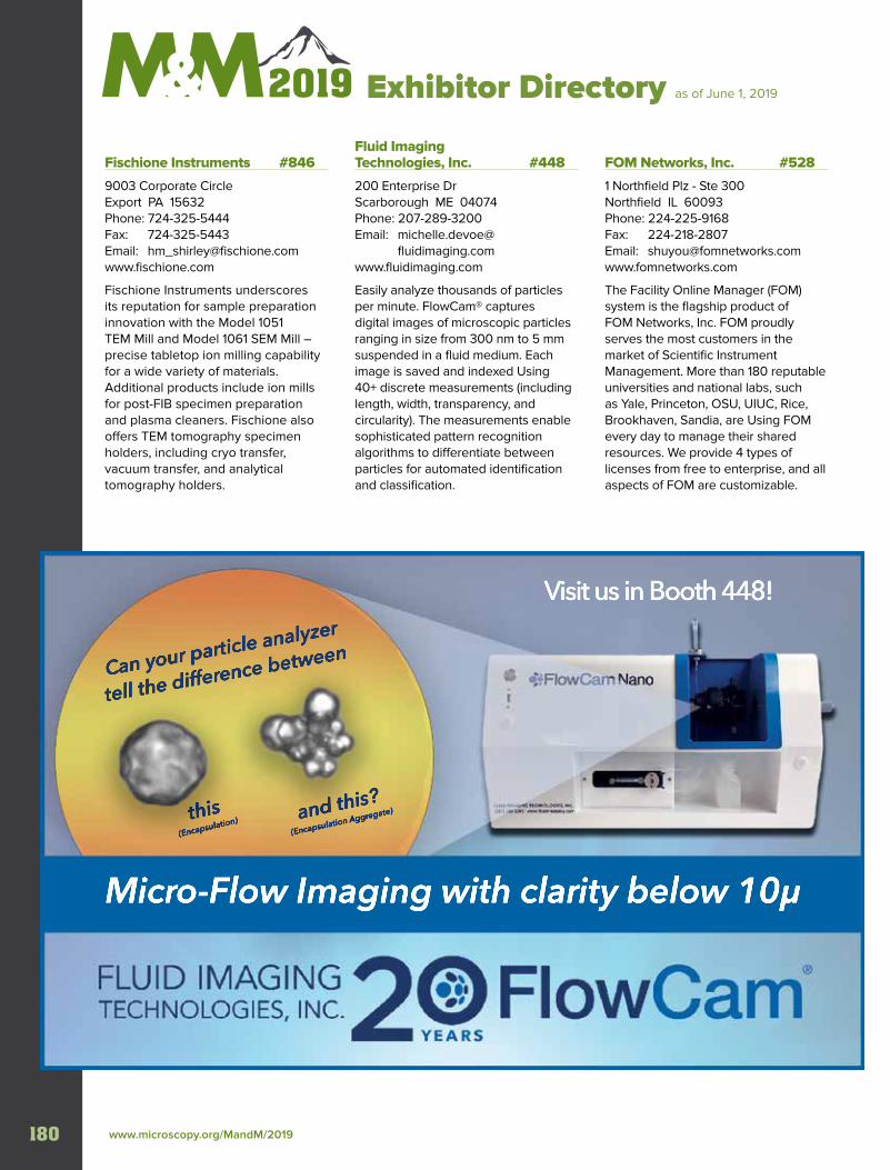
www.microscopy.org/MandM/2019 180
Exhibitor Directory as of June 1, 2019
Fischione Instruments #846
9003 Corporate Circle Export PA 15632Phone: 724-325-5444Fax: 724-325-5443Email: [email protected]
Fischione Instruments underscores its reputation for sample preparation innovation with the Model 1051 TEM Mill and Model 1061 SEM Mill – precise tabletop ion milling capability for a wide variety of materials. Additional products include ion mills for post-FIB specimen preparation and plasma cleaners. Fischione also offers TEM tomography specimen holders, including cryo transfer, vacuum transfer, and analytical tomography holders.
Fluid Imaging Technologies, Inc. #448
200 Enterprise Dr Scarborough ME 04074Phone: 207-289-3200Email: michelle.devoe@ fluidimaging.comwww.fluidimaging.com
Easily analyze thousands of particles per minute. FlowCam® captures digital images of microscopic particles ranging in size from 300 nm to 5 mm suspended in a fluid medium. Each image is saved and indexed Using 40+ discrete measurements (including length, width, transparency, and circularity). The measurements enable sophisticated pattern recognition algorithms to differentiate between particles for automated identification and classification.
FOM Networks, Inc. #528
1 Northfield Plz - Ste 300 Northfield IL 60093Phone: 224-225-9168Fax: 224-218-2807Email: [email protected]
The Facility Online Manager (FOM) system is the flagship product of FOM Networks, Inc. FOM proudly serves the most customers in the market of Scientific Instrument Management. More than 180 reputable universities and national labs, such as Yale, Princeton, OSU, UIUC, Rice, Brookhaven, Sandia, are Using FOM every day to manage their shared resources. We provide 4 types of licenses from free to enterprise, and all aspects of FOM are customizable.
Visit us in Booth 448!
Can your particle analyzer
tell the difference between
this(Encapsulation) and this?
(Encapsulation Aggregate)
Micro-Flow Imaging with clarity below 10µ
MICROSCOPY & MICROANALYSIS 2019 MEETING | August 4-8 | Portland, OR 181
Exh
ibito
r Directo
ry Exhibitor Directory as of June 1, 2019
Gatan, Inc. #928
5794 W Las Positas Blvd Pleasanton CA 94588Phone: 925-463-0200Fax: 925-463-0204Email: [email protected]
Gatan, Inc. is the world’s leading manufacturer of instrumentation and software used to enhance and extend the operation and performance of electron microscopes. Gatan products, which are fully compatible with nearly all electron microscope models, cover the entire range of the research process—from specimen preparation and manipulation to imaging and analysis.
Grace Bio-Labs #650
1015 Emkay Drive Bend OR 97702-1076Phone: 800-813-7339Email: [email protected]
Grace Bio-Labs is a worldwide supplier of advanced microscopy and cell culture tools. For over 30 years, we have produced innovative tools engineered to provide economical and high throughput solutions for a variety of cell biology microscopy workflows from live-cell imaging to optical micro-fluidic flow cells, reagents and accessories.
Henan Zhongjingkeyi Technology Co., Ltd. T452 Room 702, Block A, Free Trade Zone Plaza, NO.296 Kaifeng Henan 475000ChinaPhone: +86-371-25969621Email: [email protected]
Leading company in China with complete line of Electron Microscopy accessories. We manufacture own "EMCN" support films and comprehensive range of SEM mounts, tweezers etc. specimen preparation tools. It is our honor and strength to produce some customized products, like special purpose grids or films, small experimental devices and so on. Further more we offer EM test services and training in China. Waiting for a deeper cooperation with you
Herzan LLC #1248
23042 Alcalde Dr - Ste E Laguna Hills CA 92653Phone: 949-363-2905Fax: 949-340-9751Email: [email protected]
Herzan’s mission is to support researchers around the world in maximizing the quality of data collected by their instrumentation by removing disruptive environmental noise. To achieve this goal, Herzan designs research-grade acoustic enclosures, active vibration isolation platforms, and magnetic field cancellation systems for any precision instrument sensitive to environmental noise.
HIROX-USA, Inc. #1449
100 Commerce Way - Ste 4 Hackensack NJ 07601Phone: 201-342-2600Fax: 201-342-7322Email: [email protected]
Hitachi High Technologies America, Inc. #1438
22610 Gateway Center Dr - Ste 100 Clarksburg MD 20871Phone: 800-253-3053Fax: 301-990-0472Email: [email protected]
Hitachi High Technologies America provides technologically advanced imaging solutions to meet the complex challenges of today’s busy research and industrial labs. Challenging the boundaries of expectations, our expanding and innovative product portfolio includes SEM, TEM, STEM, FIB, Ion Milling instrumentation, AFM and Correlative microscopy, Atmospheric and Tabletop SEM, OES and XRF, Thermal Analysis and microscopy sample preparation systems.
HORIBA Scientific #547
20 Knightsbridge Rd Piscataway NJ 08854Phone: 732-494-8660Fax: 732-549-5125Email: [email protected]
HORIBA Scientific offers Raman microscopes, benchtop Raman spectrometers, AFM-Raman, modular & transmission Raman, In Situ process Raman, and optical components, scientific cameras, light sources & high performance CCDs. Standard Microscope Spectroscopy (SMS) systems enable standard microscopes to be fitted with a spectrometer & detector for Raman, steadystate & time resolved PL/EL, Reflectance/Transmittance, Photocurrent & Dark Field Scattering.
www.microscopy.org/MandM/2019 182
Exh
ibit
or
Dir
ecto
ry
Exhibitor Directory as of June 1, 2019
HREM Research Inc. #537
11-10-503 Nibancho Chiyoda, Tokyo 102-0084 JapanPhone: 81-3 5213 4689Fax: 81-3 5213 4689Email: [email protected]
HREM Research is a leading company developing software for Quantitative Electron Microscopy. Our flagship software is FFT-Multislice HR(S)TEM Image Simulation Package. We also provide a collection of DigitalMicrograph plug-ins: Exit Wave Reconstruction, STEM and EELS Deconvolution, Strain Mapping, Noise Filters for HR(S)TEM, Scan Noise corrector, Multivariate Analysis for SI data, Rocking Beam or Rotated Beam Electron Diffraction acquisition etc.
Hummingbird Scientific #1045
2610 Willamette Dr Lacey WA 98516Phone: 360-252-2737Fax: 360-252-6474Email: daan_alsem@hummingbirdscientific.comwww.hummingbirdscientific.com
Hummingbird Scientific builds products for electron, X-Ray and ion microscopy with an emphasis on transmission electron microscopes (TEM). In close collaboration with our customers, we design and manufacture all aspects of these complex systems, from mechanical, electrical, and software design to fabrication and assembly. We provide pioneering solutions for applications in nanotechnology, materials science, and biology.
ibidi USA, Inc. #438
2935 S. Fish Hatchery Rd - Ste 3, #128 Fitchburg WI 53711Phone: 844-276-6363Email: [email protected]
Cell biology products, Data analysis software, Live Cell Assays, Microscope slides, Microscope slides specialty, Microscopes- optical & accessories, Plastic laboratory ware, Pumps, Reagents
ibss Group, Inc. #1552
111 Anza Blvd - Ste 110Burlingame CA 94010Phone: 650-513-1488Fax: 650-513-1884Email: [email protected]
ibss develops and produces products used successfully in EM and Synchrotron labs around the world. The GV10x Downstream Asher reduces carbon and hydrocarbon contamination 10 to 20x more effectively than traditional methods at vacuum pressure safe for TMP operation. The Mobile Cubic Asher and Chiaro for specimen & In Situ EM cleaning, employ ibss signature GV10x Qwk-Switch source operated via touchscreen panel, fitted into one convenient enclosure
IDES, Inc #1451
5653 Stoneridge Dr - Ste 117 Pleasanton CA 94588Phone: 888-808-4337Email: [email protected]
Integrated Dynamic Electron Solutions (IDES) delivers time resolved electron microscopy imaging solutions. Relativity™ offers up to 10kHz frame rate video Using standard TEM cameras while our Luminary systems allow imaging and spectroscopy at picosecond to nanosecond timescales.
iLab Solutions/Agilent #1651
5301 Stevens Creek Blvd Santa Clara CA 95051Phone: 800-227-9770Email: [email protected]
Agilent is your global partner for sample preparation, chromatography, mass spectrometry, elemental analysis, molecular spectroscopy, laboratory software, support, service, and supplies. Agilent CrossLab enterprise-Based solutions—like the iLab Operations Platform—help you gain insight into your laboratory activities, project management, storeroom management, equipment reservations, spend tracking and more.
Integrated Dynamics Engineering #613
68 Mazzeo Rd Randolph MA 02368Phone: 781-326-5700Fax: 781-326-3004Email: [email protected]
International Centre for Diffraction Data (ICDD) #1350
12 Campus Blvd Newtown Square PA 19073Phone: 610-325-9814Fax: 610-325-9823Email: [email protected]
MICROSCOPY & MICROANALYSIS 2019 MEETING | August 4-8 | Portland, OR 183
Exh
ibito
r Directo
ry Exhibitor Directory as of June 1, 2019
IXRF Systems, Inc. #1638
10421 Old Manchaca Rd - Ste 620 Austin TX 78748Phone: 512-386-6100Fax: 512-386-6105Email: [email protected]
Over the past two decades, IXRF Systems has proven itself as a leader in Microanalysis and X-Ray Fuorescence (XRF) analyzers for bulk and micro applications. The IXRF software platform is high-powered and fully-featured, yet easy-to-use and intuitive. IXRF offers free software upgrades on all products as well as unrivaled service and support.
JASCO #1054
28600 Mary’s Court Easton MD 21601Phone: 410-822-1220Email: [email protected]
JASCO celebrates its 60th anniversary within the academic, pharmaceutical, forensics, biotechnology, and industrial markets worldwide. Our spectroscopy line includes FTIR, portable IR, UV-Vis/NIR, Fluorescence, Raman, Polarimetry, & Circular Dichroism. For chromatography, we offer HPLC, UHPLC and SFC.
JEOL USA, Inc. #623
11 Dearborn Rd Peabody MA 01960Phone: 978-535-5900Fax: 978-536-2205Email: [email protected]
The name JEOL has been synonymous with microscopy and microanalysis for 70 years. Today, we continue to evolve our SEM, TEM, and EPMA technologies through dedication to advancing science and the flow of ideas. JEOL’s innovative solutions instill stability, performance, and productivity in all of our products. Thank you to our customers for being part of our history of leadership in this field and our vision for the future.
I N T E R N A T I O N A L C E N T R E F O R D I F F R A C T I O N D A T A
Serving the Scientific Community
PDF-4+
JADE Pro and the PDF-4+ powder diffraction database are a powerful combination for all of your materials analysis needs. JADE Pro is an All-Inclusive,
Phase ID, Whole Pattern Fitting (WPF) and Rietveld refinement tool.
Atomic & Molecular
Visualization
Selected Area Electron
Diffraction (SAED)
Electron Back-Scatter
Diffraction (EBSD)
1D Diffraction Pattern
2D Ring Diffraction Pattern
[email protected] www.icdd.com • www.materialsdata.com
ICDD, the ICDD logo and PDF are registered in the U.S. Patent and Trademark Office. Powder Diffraction File is a trademark of JCPDS – International Centre for Diffraction Data ©2019JCPDS–International Centre for Diffraction Data – 5/19
��
Visit us at M&M Booth 1350
Better Together
www.microscopy.org/MandM/2019 184
Exh
ibit
or
Dir
ecto
ry
Exhibitor Directory as of June 1, 2019 Kammrath and Weiss #1009
6 Beech Rd Islip NY 11751Phone: 516-313-9742Fax: 631-224-2620Email: george.lanzarotta@ kammrathandweiss.comwww.kammrath-weiss.com
Manufacturer of modules and accessories for microscopy. Specializing in: In-Situ Tensile/Compression testing (from 10uN to 10kN), heating to 1500°C, and cooling to 5K. Precision manipulators, Ultra-fast beam blankers, special sample handling and custom-built SEM stages. CT tensile/compression/torsion modules for X-ray diffraction and complete “Turnkey” systems for Materials research. Contact: [email protected]
Keyence Corporation of America #909
500 Park Blvd Itasca IL 60143Phone: 224-240-5375Fax: 201-930-1883Email: [email protected]/products/microscope/index.jsp
KEYENCE’s microscope and surface measurement systems ensure that our customers can meet increasing quality standards. High resolution imaging, ISO-certified roughness, and 2D/3D measurement is coupled with easy-to-use interfaces for an elevated inspection experience. KEYENCE offers a range of services: free on-site demos/sample testing, training, after sale support, and overnight shipping.
KLA Corporation #1648
1 Technology Dr Milpitas CA 95035Phone: 408-875-3000Email: [email protected]
Kleindiek Nanotechnik #817
Aspenhaustr. 25 Reutlingen 72770 GermanyPhone: 49 7121 345 395 0Fax: 49 7121 345 395 55Email: [email protected]
Kleindiek Nanotechnik specializes in high-precision micromanipulators for integration into SEMs and FIB/SEMs (but also for light microscopy). We provide a wide range of applications from TEM sample liftout to electrical and mechanical characterization at the micro and nanoscale. Nano-assembly and cryo-LiftOut are availble as well as specialized stages for eucentric tilt—e.g. to remove curtaining effects during FIB milling.
MICROSCOPY & MICROANALYSIS 2019 MEETING | August 4-8 | Portland, OR 185
Exh
ibito
r Directo
ry Exhibitor Directory as of June 1, 2019
KMLabs, Inc. #1251
4775 Walnut St - Ste 102 Boulder CO 80301Phone: 303-544-9068Email: [email protected]/
KMLabs pioneered ultrafast fiber and Ti:sapphire laser systems with high reliability, flexibility, and power. The company is a world leader in high harmonic generation with platforms capable of delivering attosecond pulses of EUV and soft X-Ray light, enabling microscopy at the quantum scale. KMLabs also offers systems for biological multiphoton microscopy Using femtosecond, high-power pulses optimized for deeper, gentler 2- and 3-photon imaging.
K-Tek Nanotechnology #842
9755 SW Commerce Circle - Ste B-1 Wilsonville OR 97070Phone: 503-598-9828Email: [email protected]/
K-TEK Nanotechnology supplies a comprehensive portfolio of AFM tips and accessories that cover a wide range of customer demands from basic imaging requirements to the most complex applications. We strive to continually bring new products based on the latest technology. It is our mission to offer only the best products to keep customers on-track and on-budget to achieving reliable and consistent results.
Ladd Research #1014
83 Holly Ct Williston VT 05495Phone: 802-658-4961Fax: 802-660-8859Email: [email protected]
Ladd is the only US manufacturer of quality aperture discs & strips for Ems, FIBs, satellites & other uses. Our clean, burr-free holes range from .1um & up, in a variety of materials. We also provide a wide-range of EM supplies, chemicals, evaporation & sputtering systems. Products include nitrocellulose strips & solution, Mercox, coated grids, specialty adhesives conductive paints, Glutaraldehyde and tungsten/alumina crucibles.
Leica Microsystems #538
1700 Leider Ln Buffalo Grove IL 60089Phone: 847-405-7039Email: vicky.thoene@leica- microsystems.comwww.leica-microsystems.com
Leica Microsystems develops and manufactures microscopes and scientific instruments for the analysis of microstructures and nanostructures. The company is one of the market leaders in compound and stereo microscopy, digital microscopy, confocal laser scanning microscopy, electron microscopy sample preparation, optical coherence tomography, and surgical microscopes.
Linkam Scientific Instruments #551
551 8 Epsom Downs Metro CentreSurrey KT20 5LR United KingdomPhone: +447525591029Email: [email protected]
Linkam makes scientific instruments that help users to characterize materials from polymers to biological tissue. Used for research by the world’s most advanced scientific organisations and companies, solutions are made to interface with other analytical techniques such as Raman and X-Ray spectrometers and in the world of microscopy from AFM to Cryo TEM. Come and see our high performance solutions for CryoCLEM and Cryo plunge freezing.
Lumencor, Inc. #948
14940 NW Greenbrier Pkwy Beaverton OR 97006Phone: 503-455-4005Email: jacqueline.greenwood@ lumencor.comwww.lumencor.com
Lumencor develops, manufactures and markets innovative, solid-state light engines that facilitate imaging and photometric analysis for applications in biomedical and materials sciences. Modern solid-state light engines provide precise electronic control of light output intensity, color and timing, together with reliable, maintenance-free operation.
Mel-Build Corporation #526
2-11-36 Ishimaru Nishi-ku Fukuoka Fukuoka 819-0025 JapanPhone: +81928915111Email: [email protected]
We, Mel-Build, makes TEM holders and SEM stages to make advance in technology. TEM Holder; LN2 Cooling Holder, Vacuum transfer Holder, Tomography Holder, Tensile Holder SEM Stage; Cooling Stage
www.microscopy.org/MandM/2019 186
Exh
ibit
or
Dir
ecto
ry
Exhibitor Directory as of June 1, 2019 MEO Engineering Company #1650
290 Broadway - Ste 298 Methuen MA 01844Phone: 978-305-0479Email: [email protected]
PBS&T service consumables: suppressors, extractors, apertures, and refills of chemical precursors for all models of FIB, SEM, and dual-beam FIB/SEM instruments. Maintenance, support, and service training for users and maintenance technicians. Ion column rebuilding service. GALEX Gas Injection Systems for deposition (GIS-DEP) and gas-assisted etching (GIS-GAE) precursors for FIB/SEM. Peltier cooling and heating stage, vacuum positioning stage.
Microscopy Innovations, LLC #1049
213 Air Park Rd - Ste 101 Marshfield WI 54449-8626Phone: 715-384-3292Email: mark.nelson@ microscopyinnovations.comwww.microscopyinnovations.com
The multifunctional mPrep™ ASP-1000 Auto-Processor offers walk-away convenience for preparing biological EM samples. It easily handles tissue processing, immunogold labeling, grid staining, and 3D EM sample prep. Capsule-Based technology reduces specimen handling, controls reagent use, and cuts hands-on time. Enjoy fast, consistent results: just 45 minutes to process kidney tissue for TEM!
MiTeGen #1152
95 Brown Road, Suite 183 Ithaca NY 14850 Phone: 607-266-8877Email: [email protected]
MiTeGen develops equipment to improve sample preparation and storage. Aware of the pains of storing cryo grid boxes in conical tubes on strings, we launched the first Grid Box Puck System for Cryo-EM. Come learn how you can get your grids organized & easily keep track of your samples.
MIPAR Image Analysis Software #1154
5701 N High St - Ste 204 Worthington OH 43085Phone: 614-407-4510 Email: [email protected]
MPF Products Inc #1050
3046 Bramlett Church Rd Gray Court SC 29645Phone: 864-506-4136Email: [email protected]
MPF Products, Inc. is a leading manufacturer of hermetically sealed, Ultra High Vacuum (UHV) compatible, ceramic-to-metal sealed electrical feedthroughs and viewports. We have been in business nearly 25 years, and have over 100 years of combined engineering and manufacturing expertise. We have over 2,200 standard designs on our website, along with capabilities of producing specialty designs.
MSA Mega Booth #502
11130 Sunrise Valley Dr - Ste 350 Reston VA 20191Phone: 703-234-4115Fax: 703-435-4390Email: AssociationManagement@ microscopy.orgwww.microscopy.org
The MegaBooth provides MSA membership services to meeting attendees. It is comprised of Membership (including LAS and Sustaining Members), Publications (Microscopy and Microanalysis and Microscopy Today), MSA Committees represented are Certification Board, Placement Office, Tech Forum, and Education. This includes Educational Outreach, a Book Display, and Vendor Tutorials.
NanoMEGAS USA #513
1095 W Rio Salado Pkwy - #110 Tempe AZ 85281Phone: 480-389-6816Fax: 480-320-4066Email: [email protected]
NanoMEGAS systems for TEM provide nm resolution orientation-phase maps combined with precession electron diffraction. Applications, including strain mapping (Topspin), ab initio structural determination (ADT-3D), grains statistic (ASTAR), and amorphous short range order bond length (e-PDF) characterization, can all be installed on all new or existing TEM microscopes.
Nanoscience Instruments #1349
10008 S. 51st - ste 110 Phoenix AZ 85044Phone: 480-758-5400Fax: 480-758-5401Email: [email protected]
For over 15 years, Nanoscience Instruments has become a trusted supplier of the most innovative and highest quality analytical solutions to academia and industry. We specialize in surface science and instrumentation for nanoscale applications. Visit booth #1349 to learn about our desktop scanning electron microscopes, ion mills, correlative microscopes, nanoindenters, optical profilometers, QCM, tensiometers, AFMs, and electrospinning equipment.
MICROSCOPY & MICROANALYSIS 2019 MEETING | August 4-8 | Portland, OR 187
Exh
ibito
r Directo
ry Exhibitor Directory as of June 1, 2019
NANOVIZZ #T453
Melkster 23 Gemert Noord Brabant 5422 DJNetherlandsPhone: +31622559937Email: [email protected]
The microscopic world—sometimes surprising, often beautiful, always captivating—is both art and an art. NANOVIZZ was founded with the vision of sharing the art of microscopic imaging through microscopy-inspired designs printed on many day-to-day items, from mugs to art prints. Designs originate from all kinds of microscopy and visualization techniques, from polarised light and fluorescence microscopies to SEM, TEM and field ion microscopy.
NenoVision #750
Purkyňova 649/127 Brno 61200 Czech RepublicEmail: [email protected]/
Nikon Instruments Inc. #941
1300 Walt Whitman Rd Melville NY 11747Phone: 800-526-4566Email: [email protected]
Nikon Instruments Inc. is a world leader in the development and manufacture of optical and digital imaging technology. With over 100 years of optical expertise, Nikon provides individual optical components and complete imaging systems ranging from educational microscopes to confocal, super-resolution and high-content. Visit https://www.microscope.healthcare.nikon.com
Nion Company #1102
11511 NE 118th St Kirkland WA 98034Phone: 425-576-9060Fax: 425-739-0312Email: [email protected]
Nion supplies world-leading Aberration-Corrected STEMs that excel in spatial resolution (0.57 Å at 200 keV), EELS resolution (4 meV with Nion mono-chromator and spectrometer), efficient EDXS (0.7 sr solid angle) and ultra-high sample-level vacuum (10-10 torr range). Two sample stages are offered: ultra-high stability side-insertion cartridge type, or flexible, high-stability side-entry rod type. www.nion.com
Norcada, Inc. #1551
4548 99th St NW Edmonton AB T6E 5H5 CanadaPhone: 780-431-9637Fax: 780-431-9638Email: [email protected]
Object Research Systems #937
760 St Paul St W - Ste 101 Montreal H3C 1M4 CanadaPhone: 514-843-3861Fax: 514-543-5475Email: [email protected]
ORS is the maker of Dragonfly, the engine of scientific imaging. Our software is used by registered users in more than 80 countries. Powered by deep learning, Dragonfly’s eye-popping visualization, defect detection, advanced image segmentation, animation, and other powerful features benefit from endless user extensibility in Python.
Olympus America Inc. #946
136 Turnpike Rd Southborough MA 01772Phone: 781-419-3900Email: Ashley.divine@ olympus-ossa.comwww.olympus.com
Olympus provides an industry-leading portfolio of innovative test, measurement, and imaging instruments. Technologies include remote visual inspection, industrial microscopy, ultrasound, phased array, eddy current, phased array, and optical metrology.
Oxford Instruments #915
300 Baker Ave - Ste 150 Concord MA 01742Phone: 441-483-7703-31Fax: 441-483-7271-93Email: [email protected]
Celebrating 60 years of scientific excellence and innovation, Oxford Instruments is committed to supporting research and industrial applications in order to develop a deeper understanding of the world through Science & Technology. Our innovative key-enabling technologies and expertise empower customers to accelerate their R&D, increase productivity and make new scientific discoveries.
www.microscopy.org/MandM/2019 188
Exh
ibit
or
Dir
ecto
ry
Exhibitor Directory as of June 1, 2019
Pace Technologies #809
3601 E 34th St Tucson AZ 85713Phone: 520-882-6598Fax: 520-882-6599Email: [email protected]/
Pace Technologies is a rapidly growing supplier to the field of metallography. Our robust sample preparation equipment is designed for ease of use and effective results. Pace Technologies also offers high quality consumable products for all of your sample preparation needs. Come stop by our booth to see how Pace Technologies can help improve your sample preparation experience.
Pacific Northwest CryoEM Center #652
2730 S.W. Moody Ave CL-P2M Portland OR 97201Phone: 509-372-4382Email: [email protected]
The Pacific Northwest Center for Cryo-EM is an electron microscopy facility funded by the NIH Common Fund and operated jointly by Oregon Health & Science University and Pacific Northwest National Laboratory. We will be showing cryo-EM related services and training to provide users, at no cost, due to NIH funding. This encompasses everything from sample preparation, to TEM operation, to automated data collection, to data processing and analysis.
Park Systems #710
3040 Olcott Street Santa Clara CA 95054Phone: 518-986-1110Email: [email protected]
Stay Up to Date with the Industry’s Leading Content
WORLDWIDE COVERAGE OF LASERS • OPTICS • POSITIONING • SENSORS & DETECTORS
IMAGING • TEST & MEASUREMENT • SOLAR • LIGHT SOURCES • MICROSCOPY
MACHINE VISION • SPECTROSCOPY • FIBER OPTICS • MATERIALS & COATINGS
®
To subscribe, visit: www.photonics.com/subscribe.
Available in print and digital formats.
www.photonics.com
Visit us in
Booth 849
for the
latest issues.
MICROSCOPY & MICROANALYSIS 2019 MEETING | August 4-8 | Portland, OR 189
Exh
ibito
r Directo
ry Exhibitor Directory as of June 1, 2019
PerkinElmer #1443
68 Elm St Hopkinton MA 01748Phone: 781-663-6900Email: caroline.atkinson@ perkinelmer.comwww.perkinelmer.com
PerkinElmer, Inc. is a global leader committed to innovating for a healthier world. Our dedicated team of about 11,000 employees worldwide is passionate about providing customers with an unmatched experience as they help solve critical issues especially impacting the diagnostics and discovery and analytical solutions markets.
Photonics Media #849
100 West Street, 2nd Fl Pittsfield MA 01201Phone: 413-499-0514Fax: 413-442-3180Email: [email protected]/
Photonics Media brings you the latest research, product information, and business news for the entire photonics industry. Our extensive resources include four magazines -Photonics Spectra, Biophotonics, Industrial Photonics and EuroPhotonics- available in print and digital; the Photonics Buyer’s Guide –available in print and online; e-newsletters; webinars; and our website. Visit www.photonics.com for more information and to subscribe for FREE.
Physical Electronics #812
18725 Lake Dr E Chanhassen MN 55317Phone: 952-828-6100Fax: 952-828-6176Email: [email protected]
Physical Electronics is a subsidiary of ULVAC-PHI, the world’s leading supplier of UHV surface analysis instrumentation used for research & development of advanced materials in a number of high technology fields including nanotechnology, microelectronics, storage media, bio-medical, & basic materials such as metals, polymers, & coatings. PHI’s innovative XPS, AES, and SIMS technologies provide our customers with a full range of instrument types.
PIE Scientific LLC #1348
3209 Whipple Rd Union City CA 94587Phone: 510-936-2063Email: [email protected]
PIE Scientific specializes in developing advanced plasma cleaners for sample cleaning, surface treatment, and SEM/FIB chamber cleaning. EM-KLEEN and SEMI-KLEEN plasma cleaner are the most advanced In Situ plasma cleaners for hydrocarbon contamination removal inside SEM, FIB and XPS systems. Tergeo series table plasma cleaner is designed to handle fragile SEM/TEM samples with unique direct+remote dual-source design and pulsed mode operation.
PNDetector GmbH #647
Otto Hahn Ring 6 81739 Munchen GermanyPhone: 49 89 30908713Fax: 49 89 30908711Email: [email protected]
PNDetector has been developing and manufacturing advanced radiation detectors for X-Ray fluorescence or microanalysis, materials science and quality assurance since 2007. The sensors are fabricated in PNDetector´s own silicon production and packaging line. The emphasis is on Silicon Drift Detectors (SDDs), Backscattered Electron Detectors (BSD) and Charged Coupled Devices (pnCCDs).
PNSensor GmbH #548
Otto - Hahn - Ring 6 D-81739 Munchen GermanyPhone: (49) 89 309087270Fax: (49) 89 309087210Email: [email protected]
The key competence of the scientific research company PNSensor is development and fabrication of detectors and detector systems operating at their physical limits for applications in basic and applied research. The detectors are optimized for high resolution ultrafast spectroscopy and imaging applications based on Active Pixel Sensors (DePFET) and pnCCDs.
www.microscopy.org/MandM/2019 190
Exh
ibit
or
Dir
ecto
ry
Exhibitor Directory as of June 1, 2019 Point electronic GmbH #549
Erich NeuB Weg 15 Halle D-06120 GermanyPhone: 49 345 472256-22Email: [email protected]
point electronic GmbH is an independent supplier of detectors, acquisition and control systems for electron and light microscopy. The company is an established leader in development of electronics and software for advanced techniques, and is a leading supplier of quantitative systems for electrical characterisation, failure analysis and 3D surface reconstruction.
Protochips, Inc. #644
3800 Gateway Centre Blvd - Ste 306 Morrisville NC 27560Phone: 919-377-0800Email: [email protected]
Quantum Design, Inc. #822
10307 Pacific Center Court San Diego CA 92121Phone: 858-481-4400Fax: 858-481-7410Email: [email protected]
Quantum Design manufactures automated cryomagnetic material characterization systems and distributes SPM and Raman solutions for these platforms. They distribute thermal scanning probe and e-beam nano-lithography for SEM/FIB systems, and a leading-edge AFM solution for seamless integration into SEM/FIB, adding 3D topography, mechanical, electric and magnetic characterization at nanometer scales.
Quantum Detectors #954
R104 RAL Harwell Oxford OX11 0QX United KingdomPhone: 44 1235 44 5795Email: [email protected]/merlin-for-em/
Quantum Detectors, founded in 2007, offer detection systems generated out of advanced research and development at globally leading scientific facilities. Our MerlinEM camera - based on Medipix3 technology and with several installations now globally - brings previously unavailable hybrid pixel detector technology to the EM market.
Quorum Technologies #938
Judges House Lewes Rd, LaughtonLewes East Sussex BN8 6BN United KingdomPhone: 01323810981Email: [email protected]
Quorum are market and technology leaders in coating and cryo-SEM for electron microscopy (EM). Providing high performance sample preparation instrumentation worldwide, Quorum’s solutions enable end users to achieve continuously high quality images – time and time again. Visit our stand to see a demo of our new Q Plus range and GloQube Plus.
Raith #1129
1377 Long Island Motor Pkwy Islandia NY 11749Phone: 631-738-9500Fax: 631-738-2055Email: [email protected]
Raith is a leading precision technology manufacturer for focused ion beam (FIB-SEM) nanofabrication, large area scanning electron microscopy (SEM), and electron beam lithography (EBL) instrumentation. With sub-10 nm patterning performance, Raith instruments are enabling researchers in academic and industry environments to continuously advance their applications in applied nanoscale research, development, and device production.
Renishaw Inc. #1011
1001 Wesemann Drive West Dundee IL 60118Phone: 847-286-9953Fax: 847-286-9974Email: [email protected]
Renishaw is a recognized leader in Raman spectroscopy, producing high performance raman systems for a range of applications. We have decades of experience developing flexible Raman systems that give reliable results, even for the most challenging measurements. Whatever your Raman analysis requirements, Renishaw’s teams of scientists and engineers are here to provide you with expert advice, as well as product, technical, and applications support.
Rigaku Americas Corp. #437
9009 New Trails Dr The Woodlands TX 77381Phone: 281-362-2300Fax: 281-364-3628Email: [email protected]
Founded in 1951 in Tokyo (Japan), Rigaku Corporation is a global leader in X-Ray imaging and X-Ray diffraction; X-Ray and Raman spectroscopy; X-Ray crystallography; X-Ray optics; thermal analysis instrumentation; and non-destructive testing. Rigaku employs more than 1,400 people in the development, manufacturing, marketing, and support of its analytical equipment.
MICROSCOPY & MICROANALYSIS 2019 MEETING | August 4-8 | Portland, OR 191
Exh
ibito
r Directo
ry Exhibitor Directory as of June 1, 2019
RMC Boeckeler #828
4650 S Butterfield Dr Tucson AZ 85714Phone: 520-745-0001Fax: 520-745-0004Email: [email protected]
RMC Boeckeler has been proudly manufacturing sample preparation products in Tucson, AZ for over 65 years. The company has evolved into a worldwide provider of instruments for EM and LM sample preparation, specializing in Nano-Scale research instruments such as ultramicrotomes and array tomography systems. RMC products are manufactured, sold and maintained by a team of passionate people, dedicated to research, development and customer support.
Robomet 3D / UES, Inc. #912
4401 Dayton Xenia Rd Dayton OH 45432Phone: 937-426-6900Email: [email protected]
Royal Microscopical Society #1048
37/38 St Clements Oxford OX4 1AJUnited KingdomPhone: 44 1865 254 760Fax: 44 1865 791237Email: [email protected]
The RMS is an international society, at the forefront of new developments in microscopy and imaging. The Society is dedicated to advancing science and developing careers by organising meetings and courses, publishing the Journal of Microscopy and infocus, as well as organising the European Microscopy Congress in Copenhagen, Denmark in 2020.
Sapphire North America #427
1180 E Ellsworth Rd Ann Arbor MI 48108Phone: 734-971-3335Fax: 734-971-3640Email: [email protected] www.caymanchem.com
Sapphire North America offers innovative research tools for immunology, cell biology, cancer, inflammation, and metabolism. We focus on sourcing and distributing unique products from specialty manufacturers operating around the world. Novel fluorescent probes and proteins plus new assay kits will be featured. Visit www.sapphire-usa.com for the latest research products.
Scientific Instruments & Applications, Inc. #719
2773 Heath Ln Duluth GA 30096Phone: 770-232-7785Fax: 770-232-1791Email: [email protected]
sCMOS and CCD TEM camera systems for biomedical and material science applications including dedicated electron diffraction. 4 to 50 megapixels. Choice of mount configurations with large field of view on any port. Modern application software with TEM-camera integration and TEM control for a TEM of any model and any vintage. Affordable instruments without compromise in performance and reliability.
Seiwa Optical America, Inc. #813
3042 Scott BlvdSanta Clara CA 95054Phone: 408-844-8008Email: hitomikimura@ seiwaamerica.comwww.seiwaamerica.com
For over 50 years, Seiwa Optical has been a provider of optical components, systems, and solutions. Our vision systems, such as cameras and microscopes, can be customized for the user's unique application requirements such as wafer inspection and biological inspection. Seiwa Optical invites you to visit our booth to see our atomic force microscope.
SEMTech Solutions, Inc. #530
6 Executive Park Drive North Billerica MA 01862Phone: 978-663-9822Fax: 978-663-9823Email: [email protected]
SEMTech Solutions is a leading supplier of refurbished SEMs and analytical FE-SEM services. Our newest product, SEMView8000 is a universal Win10 SEM operator console, which will be running a FEI XL-30 at the show. In addition, we are also a sales and service representation company that offers new scientific instrumentation based on electron beam technologies. Our growing customer install base ranges from start-ups to Fortune 100 companies.
Sigray, Inc. #524
5750 Imhoff Dr. Ste. I Concord CA 94520Phone: 925-446-4183Email: [email protected]
Sigray, Inc. is a San Francisco Bay Area company founded with the aim to accelerate scientific progress by providing powerful, synchrotron-grade research capabilities in its laboratory X-Ray systems (micro-CT, nano-CT, XAS, and micro-XRF). The breakthrough performance of these systems are uniquely enabled by Sigray’s patented innovations in X-Ray source, optics, and detector technologies.
www.microscopy.org/MandM/2019 192
Exh
ibit
or
Dir
ecto
ry
Exhibitor Directory as of June 1, 2019 SmarAct Inc #1247
2140 Shattuck Ave - Ste 1103 Berkeley CA 94704Phone: 415-766-9006Email: [email protected]
SmarAct develops and produces piezo-Based, high-accuracy positioning and measuring systems for industrial and research applications in the micro- and nanometer scale. Comprehensive positioner systems with multiple degrees of freedom and parallel kinematics, microscopy stages and laser interferometers can be assembled to custom-built robotic systems, and work under extreme conditions, e.g., UHV cryogenic temperatures, and non-magnetic materials.
Spellman High Voltage Electronics Corp #1250
475 Wireless Blvd Hauppauge NY 11788Phone: 631-630-3200Email: [email protected]
Spellman High Voltage Electronics Corp is the world's leading independent supplier of precision DC high voltage power supplies, X-Ray generators and Monoblock® X-Ray sources for medical, industrial & scientific applications. While we offer the broadest, most advanced range of standard & platform products in the industry, we specialize in partnering with OEM’s to develop, produce & support the optimum solution for each system and market segment.
SPI Supplies #1130
206 Garfield Ave West Chester PA 19381Phone: 610-436-5400Fax: 610-436-5755Email: [email protected]
Manufacturer and distributor of light, EM and AFM sample preparation equipment and consumables. Offering a full line of UV and plasma cleaning systems. Traditional and high resolution coating options. Scribing/cleaving tools. Backscatter detectors. Wet Cell II liquid probe system for SEM. See our complete lines of tweezers, grids, substrate, standards and filtration products and talk to our experts about your particular applications.
Springer Nature #1032
233 Spring St New York NY 10013Phone: 201-348-4033Email: [email protected]
Springer Nature is one of the world’s leading global research, educational and professional publishers, home to an array of respected and trusted brands providing quality content through a range of innovative products and services. Springer Nature is the world’s largest academic book publisher and numbers almost 13,000 staff in over 50 countries. www.springernature.com
Syntek Co., Ltd. #T447
Leading Venture Plaza 2-403 75-1 Onocho, Tsurumi-kuYokohama 2300046 JapanPhone: 81-45-500-6603Email: [email protected]/en/
Are you happy with existing diamond knife on your ultramicrotome? If you have no choice but to use that tool you are in an unfortunate status. I can get no satisfaction with this uncompetitive market. Why don’t you try Japanese SYM knife to find new pleasure in your activities?
Technex Lab Co., Ltd. #443
Lofty-Machida, 8-4-4 Naruse, Machida Tokyo 1940044 JapanPhone: +81-42-710-4551Email: [email protected]/
TechNex Co Ltd will display and demonstrate a Tabletop type SEM named “Tiny-SEM” which utilized the permanent magnetic lenses. The characteristic of the permanent magnetic lenses allow us to design “Tiny-SEM” so tiny (column 40 mm diameter) and light weight (column 10 kg). And the lens aberration is minimized thereby expecting to observe outstanding quality images at low voltage (40 nM image resolution at 2 keV). Come see us at booth #443.
Technical Manufacturing Corporation #422
15 Centennial Dr Peabody MA 01960Phone: 978-532-6330Fax: 978-531-8682Email: [email protected]
TMC provides complete environmental control solutions for microscopy: from industry-leading floor vibration isolation tables and active benchtop platforms for optical microscopes to high-performance active piezoelectric vibration cancellation systems for SEMs and TEMs. We also offer advanced active magnetic field cancellation systems and acoustic enclosures, as well as environmental surveys to better determine customer needs.
Technotrade International Inc. #1148
7 Perimeter Rd Manchester NH 03103Phone: 603-622-5011Fax: 603-622-5211Email: [email protected]
MICROSCOPY & MICROANALYSIS 2019 MEETING | August 4-8 | Portland, OR 193
Exh
ibito
r Directo
ry Exhibitor Directory as of June 1, 2019
Ted Pella Inc. #1338
4595 Mountain Lakes Blvd Redding CA 96003Phone: 530-243-2200Fax: 530-243-3761Email: [email protected]
We manufacture and sell instruments and supplies to serve laboratories dedicated to a variety of types of microscopy: Transmission and Scanning Electron Microscopy, Electron Microprobe Analysis, Atomic Force Microscopy, Confocal Laser Microscopy and Light Microscopy.
Tescan USA #1108
765 Commonwealth Dr – Ste 101Warrendale, PA 15086Phone: 724-772-7433Fax: 724-772-7434Email: [email protected]
TESCAN is a globally renowned supplier of Focused Ion Beam workstations, Scanning Electron Microscopes and Optical Microscopes. Our innovative solutions and collaborative nature with its customers have won it a leading position in the world of nano- and micro-technology. We provide our clients with leading-class products in terms of value, quality and reliability.
Thermo Fisher #1038
5350 NW Dawson Creek Hillsboro, OR 97124 Phone: 503-726-7500Email: [email protected]/em
Thermo Fisher Scientific is the world leader in serving science. We supply innovative solutions for electron microscopy, microanalysis and spectroscopy, including Raman and EDS. Our TEMs, DualBeam™ FIB/SEMs, spectrometers and comprehensive portfolio of SEMs, combined with software suites, take customers from questions to usable data by combining high-resolution imaging with physical, chemical, elemental, mechanical and electrical analysis across scales and modes.
Tousimis #819
2211 Lewis Ave Rockville MD 20851Phone: 301-881-2450Fax: 301-881-5374Email: [email protected]
Tousimis is a globally recognized manufacturer of highly reliable CPD systems based in the USA with global sales and service support. Our 45 years of CPD experience in both designing and fabricating reliable CPD systems will benefit your work! Our process reproducibly preserves Micro & Nano 3D structures. Current applications include: Biological, Bio-MEMS, Aerogel, MEMS, Graphene, MOF and others… Please visit us to see what is new this year!
TSS Microscopy, LLC #1441
7431 NE Evergreen Pkwy - Ste 130 Hillsboro OR 97124Phone: 503-616-4710Email: [email protected]
Buying a pre-owned microscope doesn’t need to be risky. For 15 years, TSS has supported customers with a large inventory of meticulously remanufactured electron microscopes, plus accessories, spare parts, and an experienced team of field engineers whose sole concern is your happiness with your “new” microscope. Browse our inventory online or contact us if you need parts or service support for your microscope, even if you didn’t purchase from us.
TTP Labtech #1147
Melbourn Science Park, Melbourn Royston Herts SG8 6EE United KingdomPhone: +44 1223 627555Email: [email protected]
TTP Labtech introduces the chameleon, a next generation system to prepare frozen grids for single particle cryo-EM. Based on Spotiton, chameleon combines ease-of-use, reduced sample volume, precise automation, on-board screening, and rapid dispense-to-plunge time. Self-wicking grids and a pico-litre dispenser enable chameleon to deliver robust and repeatable sample dispensing.
TVIPS GmbH #720
Eremitenweg 1 82131 Gauting GermanyPhone: 498-985-0656-7Fax: 498-985-0848-8Email: [email protected]
TVIPS manufactures high-performance camera systems for Transmission Electron Microscopy with resolutions up to 64 megapixel. Image processing software packages allow seamless integration into any type of microscope. Our TEM cameras are based on custom designed CMOS technology with active pixel sensors, featuring high dynamic range and exceptional acquisition speed.
www.microscopy.org/MandM/2019 194
Exh
ibit
or
Dir
ecto
ry
Exhibitor Directory as of June 1, 2019 Vibration Engineering Consultants #1642
446 Old County Rd - Ste 100-304 Pacifica CA 94044Phone: 831-465-9189Fax: 831-465-9189Email: [email protected]
VEC identifies the origins of all environmental disturbances – usually vibration, acoustic noise and magnetic fields – that degrade performance of microscopes, analytical instruments and high-tech manufacturing equipment. We then provide design and equipment recommendations. VEC distributes Spicer Field Cancellation systems, and provides the innovative monitoring platform Quantridge that allows engineers to respond proactively to disturbances.
Vitatech Electromagnetics LLC #712
115 Juliad Ct - Ste 105 Fredericksburg VA 22406Phone: 540-286-1984Fax: 540-286-1865Email: [email protected]
Vitatech Electromagnetics LLC specializes in full-spectrum electromagnetic field (EMF) and radio frequency (RF) interference engineering services for nano tech, science, research, engineering, and biomedical facilities. Vitatech’s EMI/RFI consulting services encompass strategic planning for technology programs, design services, EMI/RFI testing, simulation modeling, and engineered solutions for magnetic shielding and active compensation systems.
Voxa #431
1001 26th Ave E Seattle WA 98112Phone: 206-288-3230Email: [email protected]
Voxa offers the world’s smallest and most portable scanning electron microscope (SEM), Mochii™. Bring award-winning Mochii in your carry-on luggage and boot up over coffee. Tablet-driven with X-Ray spectroscopy (EDS) in a compact affordable package! Voxa also provides industrial automation tools, including Blade™, an ultra-high-throughput TEM suite based on Voxa’s conveyer-belt GridStage. Come see Blade and Mochii at booth 431. http://voxa.co
Don’t track dirt on your sample! Use Evactron® plasma cleaning*
WWW.EVACTRON.COM 1-650-369-0133
Don’t let contaminated chambers and dirty specimens terminate your microscope session. Dual action turbo plasma cleaning™ removes adventitious hydrocarbons with: Plasma etch plus UV active desorption Hollow cathode plasma radical source No sputter etch damage or debris A compact and efficient plasma source Cleaning in minutes for days of perfect imaging and analysis
Need clean sample surfaces? Let us show you the
“fastest way to pristine”™! *Visit us at
M&M booth #1230 for details.
MICROSCOPY & MICROANALYSIS 2019 MEETING | August 4-8 | Portland, OR 195
Exh
ibito
r Directo
ry Exhibitor Directory as of June 1, 2019
Wiley #442
111 River St Hoboken NJ 07030Phone: 201-748-6000Email: [email protected]
Wiley, a global company, helps people and organizations develop the skills and knowledge they need to succeed. Our online scientific, technical, medical, and scholarly journals, combined with our digital learning, assessment and certification solutions help universities, societies, businesses, governments, and individuals increase the academic and professional impact of their work.
WITec Instruments Corp #717
130G Marketplace Blvd Knoxville TN 37922Phone: 865-984-4445Fax: 865-984-4441Email: witecusa@ witec-instruments.comwww.witec.de
WITec is a manufacturer of high resolution optical and scanning probe microscopy solutions for scientific and industrial applications. A modular product line allows the combination of different microscopy techniques such as Raman, SNOM or AFM in one single instrument for comprehensive and flexible analyses of optical, chemical and structural properties of a sample.
XEI Scientific, Inc. #1230
1755 E Bayshore Rd - Ste 17 Redwood City CA 94061Phone: 650-369-0133Fax: 650-363-1659Email: [email protected]
Evactron® De-Contaminators by XEI Scientific are world leaders in remote RF plasma cleaning of carbon contamination in vacuum chambers. Evactrons use a unique, energy-efficient hollow cathode plasma source to generate oxygen radicals plus UV from nitrogen metastables for dual-action removal of adventitious carbon at turbo molecular pump pressures
YW MEMS(Suzhou) Co.,Ltd. #838
B506,388 Ruoshui Road, Suzhou Industrial Park Suzhou Jiangsu 215124 ChinaPhone: +86-13776017534Email: [email protected]
YW MEMS started CleanSiN brand for our ultra-clean, robust and ultra-flat silicon nitride membranes in 2015. Produced in Class 100 clean room with state-of-the-art MEMS technology, CleanSiN silicon nitride membranes enable researchers to image, characterize and analyze their samples through X-Ray, TEM, SEM etc.Our proven superior quality and performance have been acknowledged by world-wide customers in universities, institutes and companies.
zeroK NanoTech Corporation #715
401 Professional Dr - Ste 125 Gaithersburg MD 20879Phone: 240-702-0081Email: [email protected]
zeroK NanoTech manufacturers focused ion beam (FIB) and SIMS systems with unmatched performance and new capabilities. With our FIB platform you can mill smaller structures than is possible with gallium-Based systems. Our Cs+ focused beam SIMS system enables SIMS elemental analysis at sub 10-nm resolutions, and can perform in domains traditionally served by other techniques like EDX.
Zygo Corporation #614
Laurel Brook Rd Middlefield CT 06455Phone: 860-347-8506Email: [email protected]
For nearly 50 years, Zygo Corporation is a leading global provider of comprehensive metrology solutions, precision optics, and electro-optical design and manufacturing services for the both research and production applications. The company designs and manufactures some of the world’s most advanced noncontact 3D measurement systems providing unmatched performance, versatility, reliability, and value.
Accessories (miscellaneous) Deben UK Limited 1454
EMS / Diatomed 938
Grace Bio-Labs 650
Herzan LLC 1248
ibss Group, Inc. 1552
IDES, Inc 1451
Kammrath and Weiss 1009
Linkam Scientific Instruments 551
Microscopy Innovations, LLC 1049 1049
MPF Products Inc. 1050 1050
NANOVIZZ T453
Norcada, Inc. 1551
PIE Scientific LLC 1348
Technotrade International Inc. 1148
AFM / STM Accessories Herzan LLC 1248
K-Tek Nanotechnology 842
NenoVision 1005
Quantum Design, Inc 822
Seiwa Optical America, Inc. 813
Anti-Contamination Systemsibss Group, Inc. 1552
Mel-Build Corporation 526
PIE Scientific LLC 1348
Atom Probe
3D-Micromac AG 816
Cameca 422
KMLabs, Inc. 1251
K-Tek Nanotechnology 842
Atomic Force Microscopes Angstrom Scientific Inc. 1452
Anton Paar USA 910
Bruker Corporation 638
Digital Surf 1352
Hitachi High Technologies America, Inc. 1438
Kleindiek Nanotechnik 817
Nanoscience Instruments 1349
NenoVision 750
Quantum Design International 822
RMC Boeckeler 828
Seiwa Optical America, Inc. 813
WITec Instruments Corp. 717
Auger Microscopes
JEOL USA, Inc. 623
Backscatter Detectors
Deben UK Limited 1454
EDAX 428
PNDetector GmbH 647
Point electronic GmbH 549
Quantum Detectors 954
SPI Supplies 1130
Books ASM International 1640
Cambridge University Press 616
Royal Microscopical Society 1048
Calibration and Reference Standards / Reference Materials
ASM International 1640
K-Tek Nanotechnology 842
Point electronic GmbH 549
RMC Boeckeler 828
SPI Supplies 1130
Ted Pella Inc. 1338
Camera / Digital Camera Systems - CDC, CMOS, Megapixel
Advanced Microscopy Techniques Corp. 1437
Analitex 718
Angstrom Scientific Inc. 1452
DECTRIS Ltd 1002
Direct Electron, LP 518
Gatan, Inc. 928
HORIBA Scientific 547
IDES, Inc 1451
PNSensor GmbH 548
Quantum Detectors 954
Scientific Instruments & Applications, Inc. 719
Seiwa Optical America, Inc. 813
SEMTech Solutions, Inc. 530
TVIPS GmbH 720
Chemicals EMS / Diatomed 938
Ladd Research 1014
MEO Engineering Company 1650
Pace Technologies 809
Sapphire North America 427
www.microscopy.org/MandM/2019 for up-to-date meeting information196
Exh
ibit
or
Dir
ecto
ry
Exhibitor Categories as of July 1, 2019
Confocal Microscopes ASI/Applied Scientific Instrumentation, Inc. 713
Barnett Technical Services/Attolight 1030
Carl Zeiss Microscopy, LLC 1117
Digital Surf 1352
HORIBA Scientific 547
JASCO 1054
Keyence Corporation of America 909
Leica Microsystems 538
Lumencor, Inc. 948
Nikon Instruments Inc. 941
Rigaku Americas Corp. 437
WITec Instruments Corp. 717
Consulting Analitex 718
FOM Networks, Inc. 528
Herzan LLC 1248
MPF Products Inc. 1050
NANOVIZZ T453
Object Research Systems 937
SEMTech Solutions, Inc. 530
Vibration Engineering Consultants 1642
Courses/Workshops Anton Paar USA 910
ASM International 1640
MEO Engineering Company 1650
Object Research Systems 937
Pacific Northwest CryoEM Center 652
RMC Boeckeler 828
Royal Microscopical Society 1048
Critical Point Dryers Quorum Technologies 938
Tousimis 819
Cryoequipment Angstrom Scientific Inc. 1452
Fischione Instruments 846
Linkam Scientific Instruments 551
MiTeGen 1152
Quorum Technologies 938
RMC Boeckeler 828
SmarAct Inc 1247
Technotrade International Inc. 1148
Crystallographic Mapping Analitex 718
BioNet Laboratory Inc. T449
EDAX 428
Databases Analitex 718
ASM International 1640
BioNet Laboratory Inc. T449
FOM Networks, Inc. 528
International Centre for Diffraction Data (ICDD)
1350
Detectors Analitex 718
Applied Beams LLC 952
Deben UK Limited 1454
DECTRIS Ltd 1002
Gatan, Inc. 928
HORIBA Scientific 547
KMLabs, Inc. 1251
PNDetector GmbH 647
PNSensor GmbH 548
Point electronic GmbH 549
Quantum Detectors 954
Rigaku Americas Corp. 437
Diamond Knives EMS / Diatomed 938
RMC Boeckeler 828
Syntek Co., Ltd. T447
Dual Beam FIB/SEM Applied Beams LLC 952
Cameca 422
Carl Zeiss Microscopy, LLC 1117
Clark-MXR Inc 1204
EXpressLO LLC 1055
Gatan, Inc. 928
Hitachi High Technologies America, Inc. 1438
JEOL USA, Inc. 623
MEO Engineering Company 1650
Point electronic GmbH 549
Raith 1129
Spellman High Voltage Electronics Corp. 1250
TSS Microscopy, LLC 1441
zeroK NanoTech Corporation 715
MICROSCOPY & MICROANALYSIS 2019 MEETING | August 4-8 | Portland, OR 197
Exh
ibito
r Directo
ry Exhibitor Categories as of July 1, 2019
E Beam Lithography JEOL USA, Inc. 623
Quantum Design, Inc 822
Raith 1129
SEMTech Solutions, Inc. 530
Spellman High Voltage Electronics Corp. 1250
EDS Detector Repairs and Upgrades IXRF Systems, Inc. 1638
EDS Detectors & Systems Angstrom Scientific, Inc. 1452
Bruker Corporation 638
EDAX 428
Gatan, Inc. 928
IXRF Systems, Inc. 1638
JEOL USA, Inc. 623
Nanoscience Instruments 1349
Oxford Instruments 915
Physical Electronics 812
PNDetector GmbH 647
PNSensor GmbH 548
SEMTech Solutions, Inc. 530
Voxa 431
Electrical Characterization Angstrom Scientific, Inc. 1452
Barnett Technical Services/Attolight 1030
Ephemeron Labs 1447
Kammrath and Weiss 1009
Kleindiek Nanotechnik 817
Point electronic GmbH 549
Electron Backscattered Diffraction (EBSD) BioNet Laboratory Inc. T449
Bruker Corporation 638
Cameca 422
Direct Electron, LP 518
EDAX 428
International Centre for Diffraction Data (ICDD)
1350
NanoMEGAS USA 513
Oxford Instruments 915
Physical Electronics 812
Electron Microprobes/EPMA Cameca 422
JEOL USA, Inc. 623
Voxa 431
EMI Cancellation Herzan LLC 1248
Vibration Engineering Consultants 1642
Vitatech Electromagnetics LLC 712
Failure Analysis 3D-Micromac AG 816
Angstrom Scientific Inc. 1452
Anton Paar USA 910
Applied Beams LLC 952
ASM International 1640
Barnett Technical Services/Attolight 1030
Clark-MXR Inc 1204
EDAX 428
Gatan, Inc. 928
Kammrath and Weiss 1009
Keyence Corporation of America 909
Kleindiek Nanotechnik 817
KMLabs, Inc. 1251
Leica Microsystems 538
MEO Engineering Company 1650
Nanoscience Instruments 1349
NenoVision 1005
Pace Technologies 809
Physical Electronics 812
Point electronic GmbH 549
Seiwa Optical America, Inc. 813
Technical Manufacturing Corporation 422
zeroK NanoTech Corporation 715
FIB Accessories 3D-Micromac AG 816
Applied Beams LLC 952
Bruker Corporation 638
EXpressLO LLC 1055
Herzan LLC 1248
Kammrath and Weiss 1009
Kleindiek Nanotechnik 817
MEO Engineering Company 1650
Oxford Instruments 915
Protochips, Inc. 644
www.microscopy.org/MandM/2019 for up-to-date meeting information198
Exh
ibit
or
Dir
ecto
ry
Exhibitor Categories as of July 1, 2019
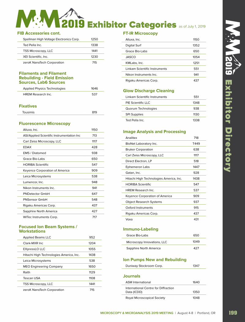
Spellman High Voltage Electronics Corp. 1250
Ted Pella Inc. 1338
TSS Microscopy, LLC 1441
XEI Scientific, Inc. 1230
zeroK NanoTech Corporation 715
Filaments and Filament Rebuilding - Field Emission Sources, Lab6 Sources Applied Physics Technologies 1646
HREM Research Inc. 537
Fixatives Tousimis 819
Fluorescence Microscopy Alluxa, Inc. 1150
ASI/Applied Scientific Instrumentation Inc 713
Carl Zeiss Microscopy, LLC 1117
EDAX 428
EMS / Diatomed 938
Grace Bio-Labs 650
HORIBA Scientific 547
Keyence Corporation of America 909
Leica Microsystems 538
Lumencor, Inc. 948
Nikon Instruments Inc. 941
PNDetector GmbH 647
PNSensor GmbH 548
Rigaku Americas Corp. 437
Sapphire North America 427
WITec Instruments Corp. 717
Focused Ion Beam Systems / Workstations Applied Beams LLC 952
Clark-MXR Inc 1204
EXpressLO LLC 1055
Hitachi High Technologies America, Inc. 1438
Leica Microsystems 538
MEO Engineering Company 1650
Raith 1129
Tescan USA 1108
TSS Microscopy, LLC 1441
zeroK NanoTech Corporation 715
FT-IR Microscopy Alluxa, Inc. 1150
Digital Surf 1352
Grace Bio-Labs 650
JASCO 1054
KMLabs, Inc. 1251
Linkam Scientific Instruments 551
Nikon Instruments Inc. 941
Rigaku Americas Corp. 437
Glow Discharge Cleaning Linkam Scientific Instruments 551
PIE Scientific LLC 1348
Quorum Technologies 938
SPI Supplies 1130
Ted Pella Inc. 1338
Image Analysis and Processing Analitex 718
BioNet Laboratory Inc. T449
Bruker Corporation 638
Carl Zeiss Microscopy, LLC 1117
Direct Electron, LP 518
Ephemeron Labs 1447
Gatan, Inc. 928
Hitachi High Technologies America, Inc. 1438
HORIBA Scientific 547
HREM Research Inc. 537
Keyence Corporation of America 909
Object Research Systems 937
Oxford Instruments 915
Rigaku Americas Corp. 437
Voxa 431
Immuno-Labeling Grace Bio-Labs 650
Microscopy Innovations, LLC 1049
Sapphire North America 427
Ion Pumps New and Rebuilding Duniway Stockroom Corp. 1347
Journals ASM International 1640
International Centre for Diffraction Data (ICDD) 1350
Royal Microscopical Society 1048
MICROSCOPY & MICROANALYSIS 2019 MEETING | August 4-8 | Portland, OR 199
Exh
ibito
r Directo
ry Exhibitor Categories as of July 1, 2019
FIB Accessories cont.
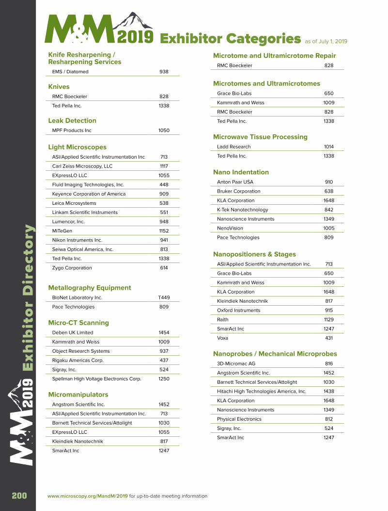
Knife Resharpening / Resharpening Services EMS / Diatomed 938
Knives RMC Boeckeler 828
Ted Pella Inc. 1338
Leak Detection MPF Products Inc 1050
Light Microscopes ASI/Applied Scientific Instrumentation Inc 713
Carl Zeiss Microscopy, LLC 1117
EXpressLO LLC 1055
Fluid Imaging Technologies, Inc. 448
Keyence Corporation of America 909
Leica Microsystems 538
Linkam Scientific Instruments 551
Lumencor, Inc. 948
MiTeGen 1152
Nikon Instruments Inc. 941
Seiwa Optical America, Inc. 813
Ted Pella Inc. 1338
Zygo Corporation 614
Metallography Equipment BioNet Laboratory Inc. T449
Pace Technologies 809
Micro-CT Scanning Deben UK Limited 1454
Kammrath and Weiss 1009
Object Research Systems 937
Rigaku Americas Corp. 437
Sigray, Inc. 524
Spellman High Voltage Electronics Corp. 1250
Micromanipulators Angstrom Scientific Inc. 1452
ASI/Applied Scientific Instrumentation Inc. 713
Barnett Technical Services/Attolight 1030
EXpressLO LLC 1055
Kleindiek Nanotechnik 817
SmarAct Inc 1247
Microtome and Ultramicrotome Repair RMC Boeckeler 828
Microtomes and Ultramicrotomes Grace Bio-Labs 650
Kammrath and Weiss 1009
RMC Boeckeler 828
Ted Pella Inc. 1338
Microwave Tissue Processing Ladd Research 1014
Ted Pella Inc. 1338
Nano Indentation Anton Paar USA 910
Bruker Corporation 638
KLA Corporation 1648
K-Tek Nanotechnology 842
Nanoscience Instruments 1349
NenoVision 1005
Pace Technologies 809
Nanopositioners & Stages ASI/Applied Scientific Instrumentation Inc. 713
Grace Bio-Labs 650
Kammrath and Weiss 1009
KLA Corporation 1648
Kleindiek Nanotechnik 817
Oxford Instruments 915
Raith 1129
SmarAct Inc 1247
Voxa 431
Nanoprobes / Mechanical Microprobes 3D-Micromac AG 816
Angstrom Scientific Inc. 1452
Barnett Technical Services/Attolight 1030
Hitachi High Technologies America, Inc. 1438
KLA Corporation 1648
Nanoscience Instruments 1349
Physical Electronics 812
Sigray, Inc. 524
SmarAct Inc 1247
www.microscopy.org/MandM/2019 for up-to-date meeting information200
Exh
ibit
or
Dir
ecto
ry
Exhibitor Categories as of July 1, 2019
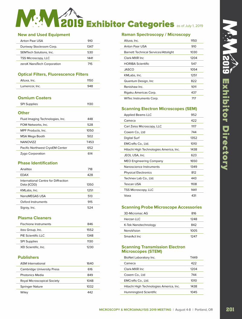
New and Used Equipment Anton Paar USA 910
Duniway Stockroom Corp. 1347
SEMTech Solutions, Inc. 530
TSS Microscopy, LLC 1441
zeroK NanoTech Corporation 715
Optical Filters, Fluorescence Filters Alluxa, Inc. 1150
Lumencor, Inc. 948
Osmium Coaters SPI Supplies 1130
Other Fluid Imaging Technologies, Inc. 448
FOM Networks, Inc. 528
MPF Products, Inc. 1050
MSA Mega Booth 502
NANOVIZZ T453
Pacific Northwest CryoEM Center 652
Zygo Corporation 614
Phase Identification Analitex 718
EDAX 428
International Centre for Diffraction Data (ICDD) 1350
KMLabs, Inc. 1251
NanoMEGAS USA 513
Oxford Instruments 915
Sigray, Inc. 524
Plasma Cleaners Fischione Instruments 846
ibss Group, Inc. 1552
PIE Scientific LLC 1348
SPI Supplies 1130
XEI Scientific, Inc. 1230
Publishers ASM International 1640
Cambridge University Press 616
Photonics Media 849
Royal Microscopical Society 1048
Springer Nature 1032
Wiley 442
Raman Spectroscopy / Microscopy Alluxa, Inc. 1150
Anton Paar USA 910
Barnett Technical Services/Attolight 1030
Clark-MXR Inc 1204
HORIBA Scientific 547
JASCO 1054
KMLabs, Inc. 1251
Quantum Design, Inc 822
Renishaw Inc. 1011
Rigaku Americas Corp. 437
WITec Instruments Corp. 717
Scanning Electron Microscopes (SEM) Applied Beams LLC 952
Cameca 422
Carl Zeiss Microscopy, LLC 1117
Coxem Co., Ltd 744
Digital Surf 1352
EMCrafts Co., Ltd. 1010
Hitachi High Technologies America, Inc. 1438
JEOL USA, Inc. 623
MEO Engineering Company 1650
Nanoscience Instruments 1349
Physical Electronics 812
Technex Lab Co., Ltd. 443
Tescan USA 1108
TSS Microscopy, LLC 1441
Voxa 431
Scanning Probe Microscope Accessories 3D-Micromac AG 816
Herzan LLC 1248
K-Tek Nanotechnology 842
NenoVision 1005
SmarAct Inc 1247
Scanning Transmission Electron Microscopes (STEM) BioNet Laboratory Inc. T449
Cameca 422
Clark-MXR Inc 1204
Coxem Co., Ltd 744
EMCrafts Co., Ltd. 1010
Hitachi High Technologies America, Inc. 1438
Hummingbird Scientific 1045
MICROSCOPY & MICROANALYSIS 2019 MEETING | August 4-8 | Portland, OR 201
Exh
ibito
r Directo
ry Exhibitor Categories as of July 1, 2019
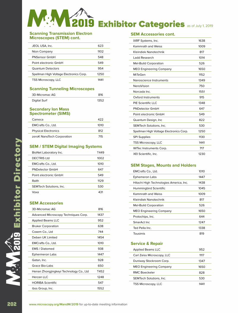
JEOL USA, Inc. 623
Nion Company 1102
PNSensor GmbH 548
Point electronic GmbH 549
Quantum Detectors 954
Spellman High Voltage Electronics Corp. 1250
TSS Microscopy, LLC 1441
Scanning Tunneling Microscopes 3D-Micromac AG 816
Digital Surf 1352
Secondary Ion Mass Spectrometer (SIMS) Cameca 422
EMCrafts Co., Ltd. 1010
Physical Electronics 812
zeroK NanoTech Corporation 715
SEM / STEM Digital Imaging Systems BioNet Laboratory Inc. T449
DECTRIS Ltd 1002
EMCrafts Co., Ltd. 1010
PNDetector GmbH 647
Point electronic GmbH 549
Raith 1129
SEMTech Solutions, Inc. 530
Voxa 431
SEM Accessories 3D-Micromac AG 816
Advanced Microscopy Techniques Corp. 1437
Applied Beams LLC 952
Bruker Corporation 638
Coxem Co., Ltd 744
Deben UK Limited 1454
EMCrafts Co., Ltd. 1010
EMS / Diatomed 938
Ephermeron Labs 1447
Gatan, Inc. 928
Grace Bio-Labs 650
Henan Zhongjingkeyi Technology Co., Ltd T452
Herzan LLC 1248
HORIBA Scientific 547
ibss Group, Inc. 1552
IXRF Systems, Inc. 1638
Kammrath and Weiss 1009
Kleindiek Nanotechnik 817
Ladd Research 1014
Mel-Build Corporation 526
MEO Engineering Company 1650
MiTeGen 1152
Nanoscience Instruments 1349
NenoVision 750
Norcada Inc. 1551
Oxford Instruments 915
PIE Scientific LLC 1348
PNDetector GmbH 647
Point electronic GmbH 549
Quantum Design, Inc 822
SEMTech Solutions, Inc. 530
Spellman High Voltage Electronics Corp. 1250
SPI Supplies 1130
TSS Microscopy, LLC 1441
WITec Instruments Corp. 717
XEI Scientific, Inc. 1230
SEM Stages, Mounts and Holders EMCrafts Co., Ltd. 1010
Ephemeron Labs 1447
Hitachi High Technologies America, Inc. 1438
Hummingbird Scientific 1045
Kammrath and Weiss 1009
Kleindiek Nanotechnik 817
Mel-Build Corporation 526
MEO Engineering Company 1650
Protochips, Inc. 644
SmarAct Inc 1247
Ted Pella Inc. 1338
Tousimis 819
Service & Repair Applied Beams LLC 952
Carl Zeiss Microscopy, LLC 1117
Duniway Stockroom Corp. 1347
MEO Engineering Company 1650
RMC Boeckeler 828
SEMTech Solutions, Inc. 530
TSS Microscopy, LLC 1441
Scanning Transmission Electron Microscopes (STEM) cont.
SEM Accessories cont.
www.microscopy.org/MandM/2019 for up-to-date meeting information202
Exh
ibit
or
Dir
ecto
ry
Exhibitor Categories as of July 1, 2019
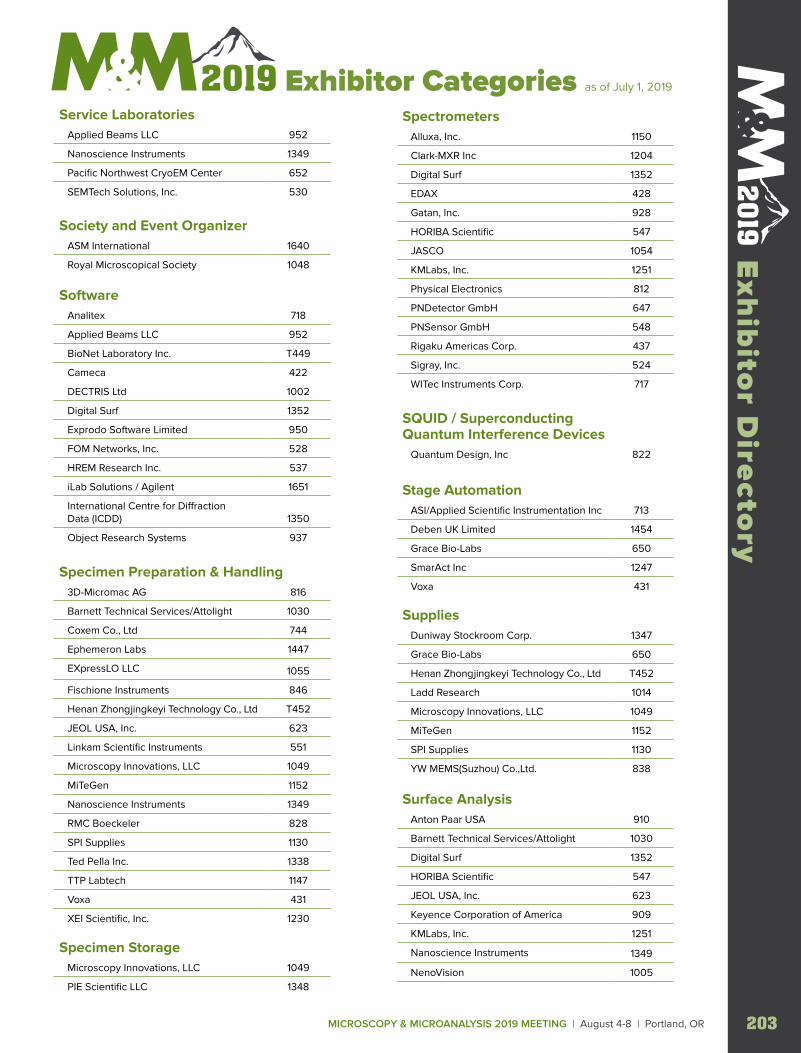
Service Laboratories Applied Beams LLC 952
Nanoscience Instruments 1349
Pacific Northwest CryoEM Center 652
SEMTech Solutions, Inc. 530
Society and Event Organizer ASM International 1640
Royal Microscopical Society 1048
Software Analitex 718
Applied Beams LLC 952
BioNet Laboratory Inc. T449
Cameca 422
DECTRIS Ltd 1002
Digital Surf 1352
Exprodo Software Limited 950
FOM Networks, Inc. 528
HREM Research Inc. 537
iLab Solutions / Agilent 1651
International Centre for Diffraction Data (ICDD) 1350
Object Research Systems 937
Specimen Preparation & Handling 3D-Micromac AG 816
Barnett Technical Services/Attolight 1030
Coxem Co., Ltd 744
Ephemeron Labs 1447
EXpressLO LLC 1055
Fischione Instruments 846
Henan Zhongjingkeyi Technology Co., Ltd T452
JEOL USA, Inc. 623
Linkam Scientific Instruments 551
Microscopy Innovations, LLC 1049
MiTeGen 1152
Nanoscience Instruments 1349
RMC Boeckeler 828
SPI Supplies 1130
Ted Pella Inc. 1338
TTP Labtech 1147
Voxa 431
XEI Scientific, Inc. 1230
Specimen Storage Microscopy Innovations, LLC 1049
PIE Scientific LLC 1348
Spectrometers Alluxa, Inc. 1150
Clark-MXR Inc 1204
Digital Surf 1352
EDAX 428
Gatan, Inc. 928
HORIBA Scientific 547
JASCO 1054
KMLabs, Inc. 1251
Physical Electronics 812
PNDetector GmbH 647
PNSensor GmbH 548
Rigaku Americas Corp. 437
Sigray, Inc. 524
WITec Instruments Corp. 717
SQUID / Superconducting Quantum Interference Devices Quantum Design, Inc 822
Stage Automation ASI/Applied Scientific Instrumentation Inc 713
Deben UK Limited 1454
Grace Bio-Labs 650
SmarAct Inc 1247
Voxa 431
Supplies Duniway Stockroom Corp. 1347
Grace Bio-Labs 650
Henan Zhongjingkeyi Technology Co., Ltd T452
Ladd Research 1014
Microscopy Innovations, LLC 1049
MiTeGen 1152
SPI Supplies 1130
YW MEMS(Suzhou) Co.,Ltd. 838
Surface Analysis Anton Paar USA 910
Barnett Technical Services/Attolight 1030
Digital Surf 1352
HORIBA Scientific 547
JEOL USA, Inc. 623
Keyence Corporation of America 909
KMLabs, Inc. 1251
Nanoscience Instruments 1349
NenoVision 1005
MICROSCOPY & MICROANALYSIS 2019 MEETING | August 4-8 | Portland, OR 203
Exh
ibito
r Directo
ry Exhibitor Categories as of July 1, 2019
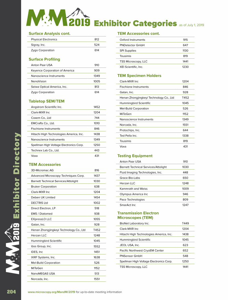
www.microscopy.org/MandM/2019 for up-to-date meeting information204
Exh
ibit
or
Dir
ecto
ry
Exhibitor Categories as of July 1, 2019
Physical Electronics 812
Sigray, Inc. 524
Zygo Corporation 614
Surface Profiling Anton Paar USA 910
Keyence Corporation of America 909
Nanoscience Instruments 1349
NenoVision 1005
Seiwa Optical America, Inc. 813
Zygo Corporation 614
Tabletop SEM/TEM Angstrom Scientific Inc. 1452
Clark-MXR Inc 1204
Coxem Co., Ltd 744
EMCrafts Co., Ltd. 1010
Fischione Instruments 846
Hitachi High Technologies America, Inc. 1438
Nanoscience Instruments 1349
Spellman High Voltage Electronics Corp. 1250
Technex Lab Co., Ltd. 443
Voxa 431
TEM Accessories 3D-Micromac AG 816
Advanced Microscopy Techniques Corp. 1437
Barnett Technical Services/Attolight 1030
Bruker Corporation 638
Clark-MXR Inc 1204
Deben UK Limited 1454
DECTRIS Ltd 1002
Direct Electron, LP 518
EMS / Diatomed 938
EXpressLO LLC 1055
Gatan, Inc. 928
Henan Zhongjingkeyi Technology Co., Ltd T452
Herzan LLC 1248
Hummingbird Scientific 1045
ibss Group, Inc. 1552
IDES, Inc 1451
IXRF Systems, Inc. 1638
Mel-Build Corporation 526
MiTeGen 1152
NanoMEGAS USA 513
Norcada, Inc. 1551
Oxford Instruments 915
PNDetector GmbH 647
SPI Supplies 1130
Tousimis 819
TSS Microscopy, LLC 1441
XEI Scientific, Inc. 1230
TEM Specimen Holders Clark-MXR Inc 1204
Fischione Instruments 846
Gatan, Inc. 928
Henan Zhongjingkeyi Technology Co., Ltd T452
Hummingbird Scientific 1045
Mel-Build Corporation 526
MiTeGen 1152
Nanoscience Instruments 1349
Norcada, Inc. 1551
Protochips, Inc. 644
Ted Pella Inc. 1338
Tousimis 819
Voxa 431
Testing Equipment Anton Paar USA 910
Barnett Technical Services/Attolight 1030
Fluid Imaging Technologies, Inc. 448
Grace Bio-Labs 650
Herzan LLC 1248
Kammrath and Weiss 1009
Olympus America Inc 946
Pace Technologies 809
SmarAct Inc 1247
Transmission Electron Microscopes (TEM) BioNet Laboratory Inc. T449
Clark-MXR Inc 1204
Hitachi High Technologies America, Inc. 1438
Hummingbird Scientific 1045
JEOL USA, Inc. 623
Pacific Northwest CryoEM Center 652
PNSensor GmbH 548
Spellman High Voltage Electronics Corp. 1250
TSS Microscopy, LLC 1441
Surface Analysis cont. TEM Accessories cont.
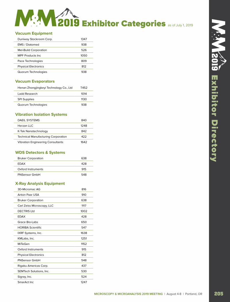
MICROSCOPY & MICROANALYSIS 2019 MEETING | August 4-8 | Portland, OR 205
Exh
ibito
r Directo
ry Exhibitor Categories as of July 1, 2019
Vacuum Equipment Duniway Stockroom Corp. 1347
EMS / Diatomed 938
Mel-Build Corporation 526
MPF Products Inc 1050
Pace Technologies 809
Physical Electronics 812
Quorum Technologies 938
Vacuum Evaporators Henan Zhongjingkeyi Technology Co., Ltd T452
Ladd Research 1014
SPI Supplies 1130
Quorum Technologies 938
Vibration Isolation Systems DAEIL SYSTEMS 840
Herzan LLC 1248
K-Tek Nanotechnology 842
Technical Manufacturing Corporation 422
Vibration Engineering Consultants 1642
WDS Detectors & Systems Bruker Corporation 638
EDAX 428
Oxford Instruments 915
PNSensor GmbH 548
X-Ray Analysis Equipment 3D-Micromac AG 816
Anton Paar USA 910
Bruker Corporation 638
Carl Zeiss Microscopy, LLC 1117
DECTRIS Ltd 1002
EDAX 428
Grace Bio-Labs 650
HORIBA Scientific 547
IXRF Systems, Inc. 1638
KMLabs, Inc. 1251
MiTeGen 1152
Oxford Instruments 915
Physical Electronics 812
PNSensor GmbH 548
Rigaku Americas Corp. 437
SEMTech Solutions, Inc. 530
Sigray, Inc. 524
SmarAct Inc 1247
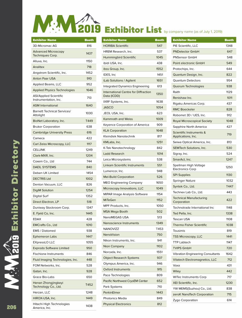
Exhibitor List by company name (as of July 1, 2019) Exhibitor Name Booth
3D-Micromac AG 816
Advanced Microscopy Techniques Corp.
1437
Alluxa, Inc. 1150
Analitex 718
Angstrom Scientific, Inc. 1452
Anton Paar USA 910
Applied Beams, LLC 952
Applied Physics Technologies 1646
ASI/Applied Scientific Instrumentation, Inc.
713
ASM International 1640
Barnett Technical Services/Attolight
1030
BioNet Laboratory, Inc. T449
Bruker Corporation 638
Cambridge University Press 616
Cameca 422
Carl Zeiss Microscopy, LLC 1117
CELLINK 1249
Clark-MXR, Inc. 1204
Coxem Co., Ltd 744
DAEIL SYSTEMS 840
Deben UK Limited 1454
DECTRIS Ltd 1002
Denton Vacuum, LLC 826
DigiM Solution 1254
Digital Surf 1352
Direct Electron, LP 518
Duniway Stockroom Corp. 1347
E. Fjeld Co, Inc. 1445
EDAX 428
EMCrafts Co., Ltd 1010
EMS / Diatomed 938
Ephemeron Labs 1447
EXpressLO LLC 1055
Exprodo Software Limited 950
Fischione Instruments 846
Fluid Imaging Technologies, Inc. 448
FOM Networks, Inc. 528
Gatan, Inc. 928
Grace Bio-Labs 650
Henan Zhongjingkeyi Technology Co., Ltd.
T452
Herzan, LLC 1248
HIROX-USA, Inc. 1449
Hitachi High Technologies America, Inc.
1438
Exhibitor Name Booth
HORIBA Scientific 547
HREM Research, Inc. 537
Hummingbird Scientific 1045
ibidi USA, Inc. 438
ibss Group, Inc. 1552
IDES, Inc 1451
iLab Solutions / Agilent 1651
Integrated Dynamics Engineering 613
International Centre for Diffraction Data (ICDD)
1350
IXRF Systems, Inc. 1638
JASCO 1054
JEOL USA, Inc. 623
Kammrath and Weiss 1009
Keyence Corporation of America 909
KLA Corporation 1648
Kleindiek Nanotechnik 817
KMLabs, Inc. 1251
K-Tek Nanotechnology 842
Ladd Research 1014
Leica Microsystems 538
Linkam Scientific Instruments 551
Lumencor, Inc. 948
Mel-Build Corporation 526
MEO Engineering Company 1650
Microscopy Innovations, LLC 1049
MIPAR Image Analysis Software 1154
MiTeGen 1152
MPF Products, Inc. 1050
MSA Mega Booth 502
NanoMEGAS USA 513
Nanoscience Instruments 1349
NANOVIZZ T453
NenoVision 750
Nikon Instruments, Inc. 941
Nion Company 1102
Norcada, Inc. 1551
Object Research Systems 937
Olympus America, Inc. 946
Oxford Instruments 915
Pace Technologies 809
Pacific Northwest CryoEM Center 652
Park Systems 710
PerkinElmer 1443
Photonics Media 849
Physical Electronics 812
Exhibitor Name Booth
PIE Scientific, LLC 1348
PNDetector GmbH 647
PNSensor GmbH 548
Point electronic GmbH 549
Protochips, Inc. 644
Quantum Design, Inc. 822
Quantum Detectors 954
Quorum Technologies 938
Raith 1129
Renishaw Inc. 1011
Rigaku Americas Corp. 437
RMC Boeckeler 828
Robomet 3D / UES, Inc. 912
Royal Microscopical Society 1048
Sapphire North America 427
Scientific Instruments & Applications, Inc.
719
Seiwa Optical America, Inc. 813
SEMTech Solutions, Inc. 530
Sigray, Inc. 524
SmarAct, Inc. 1247
Spellman High Voltage Electronics Corp
1250
SPI Supplies 1130
Springer Nature 1032
Syntek Co., Ltd. T447
Technex Lab Co., Ltd. 443
Technical Manufacturing Corporation
422
Technotrade International Inc 1148
Ted Pella, Inc. 1338
Tescan USA 1108
Thermo Fisher Scientific 1038
Tousimis 819
TSS Microscopy, LLC 1441
TTP Labtech 1147
TVIPS GmbH 720
Vibration Engineering Consultants 1642
Vitatech Electromagnetics, LLC 712
Voxa 431
Wiley 442
WITec Instruments Corp 717
XEI Scientific, Inc. 1230
YW MEMS(Suzhou) Co., Ltd. 838
zeroK NanoTech Corporation 715
Zygo Corporation 614
206
Exh
ibit
or
Dir
ecto
ry
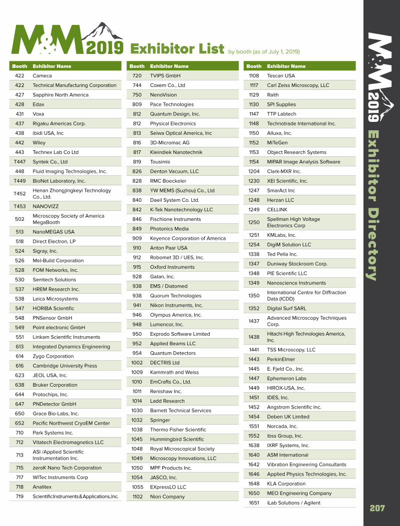
207207
Exh
ibito
r Directo
ry Exhibitor List by booth (as of July 1, 2019)
Booth Exhibitor Name
422 Cameca
422 Technical Manufacturing Corporation
427 Sapphire North America
428 Edax
431 Voxa
437 Rigaku Americas Corp.
438 ibidi USA, Inc
442 Wiley
443 Technex Lab Co Ltd
T447 Syntek Co., Ltd
448 Fluid Imaging Technologies, Inc.
T449 BioNet Laboratory, Inc.
T452Henan Zhongjingkeyi Technology Co., Ltd.
T453 NANOVIZZ
502Microscopy Society of America MegaBooth
513 NanoMEGAS USA
518 Direct Electron, LP
524 Sigray, Inc.
526 Mel-Build Corporation
528 FOM Networks, Inc.
530 Semtech Solutions
537 HREM Research Inc.
538 Leica Microsystems
547 HORIBA Scientific
548 PNSensor GmbH
549 Point electronic GmbH
551 Linkam Scientific Instruments
613 Integrated Dynamics Engineering
614 Zygo Corporation
616 Cambridge University Press
623 JEOL USA, Inc.
638 Bruker Corporation
644 Protochips, Inc.
647 PNDetector GmbH
650 Grace Bio-Labs, Inc.
652 Pacific Northwest CryoEM Center
710 Park Systems Inc.
712 Vitatech Electromagnetics LLC
713ASI /Applied Scientific Instrumentation Inc.
715 zeroK Nano Tech Corporation
717 WITec Instruments Corp
718 Analitex
719 Scientific Instruments & Applications, Inc.
Booth Exhibitor Name
720 TVIPS GmbH
744 Coxem Co., Ltd
750 NenoVision
809 Pace Technologies
812 Quantum Design, Inc.
812 Physical Electronics
813 Seiwa Optical America, Inc
816 3D-Micromac AG
817 Kleindiek Nanotechnik
819 Tousimis
826 Denton Vacuum, LLC
828 RMC Boeckeler
838 YW MEMS (Suzhou) Co., Ltd
840 Daeil System Co. Ltd.
842 K-Tek Nanotechnology LLC
846 Fischione Instruments
849 Photonics Media
909 Keyence Corporation of America
910 Anton Paar USA
912 Robomet 3D / UES, Inc.
915 Oxford Instruments
928 Gatan, Inc.
938 EMS / Diatomed
938 Quorum Technologies
941 Nikon Instruments, Inc.
946 Olympus America, Inc.
948 Lumencor, Inc.
950 Exprodo Software Limited
952 Applied Beams LLC
954 Quantum Detectors
1002 DECTRIS Ltd
1009 Kammrath and Weiss
1010 EmCrafts Co., Ltd.
1011 Renishaw Inc.
1014 Ladd Research
1030 Barnett Technical Services
1032 Springer
1038 Thermo Fisher Scientific
1045 Hummingbird Scientific
1048 Royal Microscopical Society
1049 Microscopy Innovations, LLC
1050 MPF Products Inc.
1054 JASCO, Inc.
1055 EXpressLO LLC
1102 Nion Company
Booth Exhibitor Name
1108 Tescan USA
1117 Carl Zeiss Microscopy, LLC
1129 Raith
1130 SPI Supplies
1147 TTP Labtech
1148 Technotrade International Inc.
1150 Alluxa, Inc.
1152 MiTeGen
1153 Object Research Systems
1154 MIPAR Image Analysis Software
1204 Clark-MXR Inc.
1230 XEI Scientific, Inc.
1247 SmarAct Inc
1248 Herzan LLC
1249 CELLINK
1250Spellman High Voltage Electronics Corp
1251 KMLabs, Inc.
1254 DigiM Solution LLC
1338 Ted Pella Inc.
1347 Duniway Stockroom Corp.
1348 PIE Scientific LLC
1349 Nanoscience Instruments
1350International Centre for Diffraction Data (ICDD)
1352 Digital Surf SARL
1437Advanced Microscopy Techniques Corp.
1438Hitachi High Technologies America, Inc.
1441 TSS Microscopy, LLC
1443 PerkinElmer
1445 E. Fjeld Co., Inc.
1447 Ephemeron Labs
1449 HIROX-USA, Inc.
1451 IDES, Inc.
1452 Angstrom Scientific Inc.
1454 Deben UK Limited
1551 Norcada, Inc.
1552 ibss Group, Inc.
1638 IXRF Systems, Inc.
1640 ASM International
1642 Vibration Engineering Consultants
1646 Applied Physics Technologies, Inc.
1648 KLA Corporation
1650 MEO Engineering Company
1651 iLab Solutions / Agilent
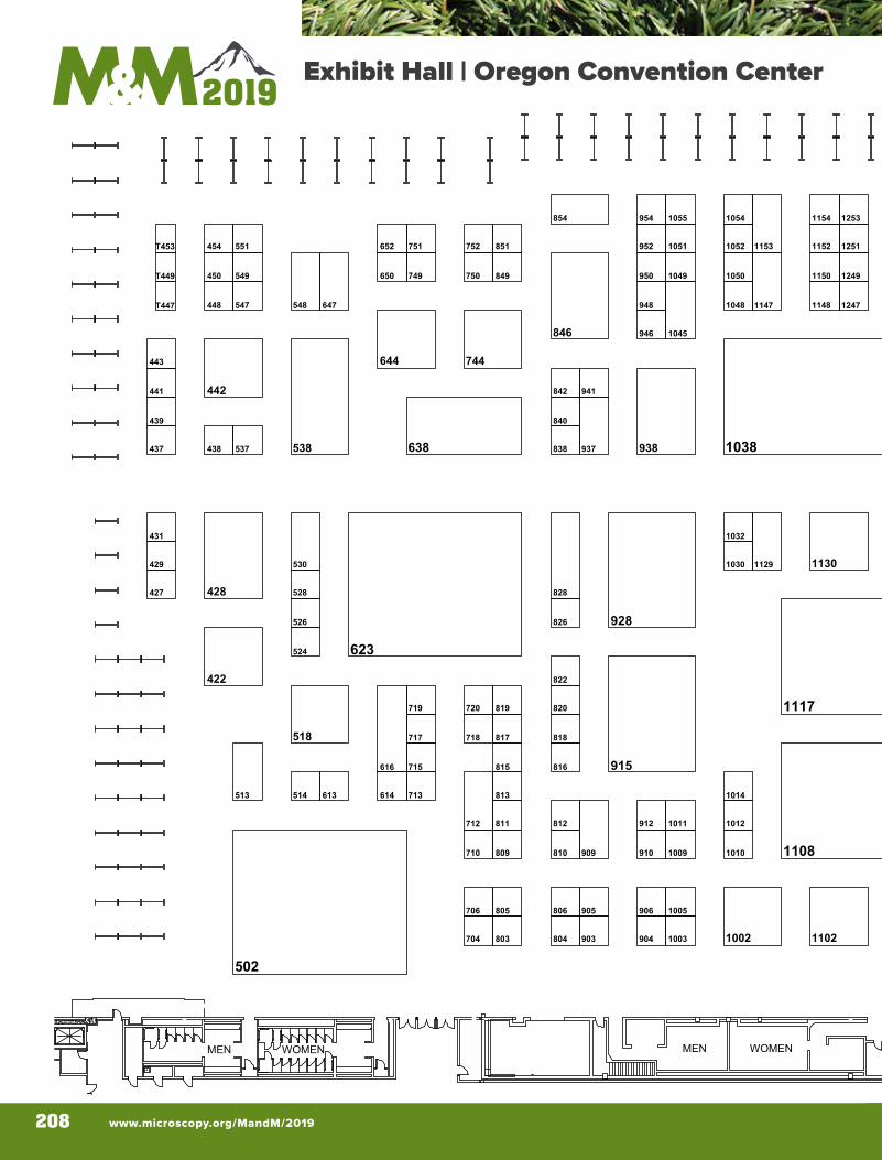
www.microscopy.org/MandM/2019208
C124
C123
C MEETING ROOMS
C125
C126
C122
DISPLA
Y
DIRECTIO
N
C121
C120
B119
B118
FIRSTAID
MEN WOMEN MEN WOMEN
CO
NC
ESSI
ON
S
Entrance
MR 03
MR 02
MR 01
10021003
1005
1009 1010
1011 1012
1014
1030
1032
1038
1045
1048
1049 1050
1051 1052
10541055
1102
1108
1117
1129 1130
1147 1148
1150
11521153
1154
1202
1204
1230
1247 1248
1249 1250
1251 1252
1253 1254
1338
1347 1348
1349 1350
1352
1437 1438
1441
1443
1445
1447
1449
1451 1452
1453 1454
1551 1552
1553
1638
1640
1642
1646
1648
1650
1651 1652
1653 1654
422
427 428
429
431
437 438
439
441 442
443
448
450
454
502
513 514
518
524
526
528
530
537 538
547 548
549
551
613 614
616
623
638
644
647
650
652
704
706
710
712
713
715
717 718
719 720
744
749 750
751 752
803 804
805 806
809 810
811 812
813
815 816
817 818
819 820
822
826
828
838
840
842
846
849
851
854
903 904
905 906
909 910
912
915
928
937 938
941
946
948
950
952
954
T447
T449
T453
3M
3MENTRANCE
UNIT
ENTRANCE
UNIT6ftx2f
t
6ftx2ft
6ftx2ft
6ftx2ft
6ftx2ft
PUB . BIN
S
3Mx1M
6ftx2ft
10x20MAS
8x102020
8x15IF/M
S15
8
8x14S TORAGE
5x25TOTE B
AGS
ADVANCE
REG. ONSITE
REG.
EXHIBITOR
REG.
Microscopy & Microanalysis 2019August 5-8, 2019
Oregon Convention Center Portland, OregonExhibit Hall | Oregon Convention Center
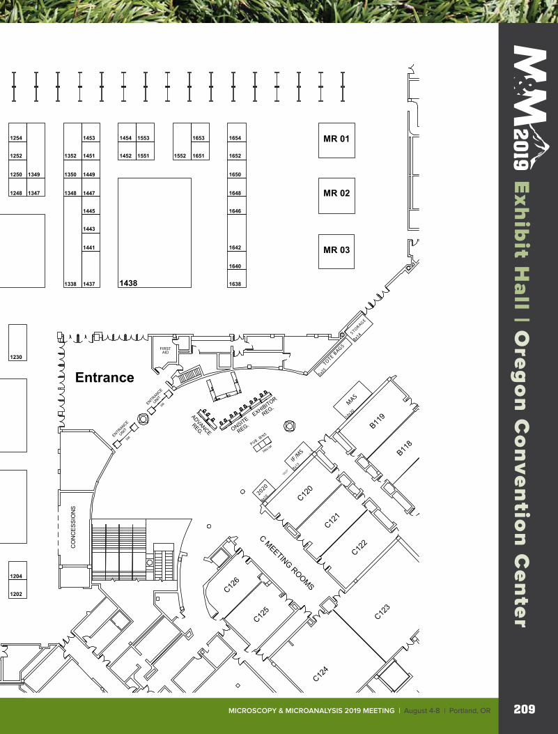
MICROSCOPY & MICROANALYSIS 2019 MEETING | August 4-8 | Portland, OR 209
Exh
ibito
r Directo
ry
MICROSCOPY & MICROANALYSIS 2019 MEETING | August 4-8 | Portland, OR 209
Exh
ibit H
all | O
rego
n C
on
ventio
n C
enter
C124
C123
C MEETING ROOMS
C125
C126
C122
DISPLA
Y
DIRECTIO
N
C121
C120
B119
B118
FIRSTAID
MEN WOMEN MEN WOMEN
CO
NC
ESSI
ON
S
Entrance
MR 03
MR 02
MR 01
10021003
1005
1009 1010
1011 1012
1014
1030
1032
1038
1045
1048
1049 1050
1051 1052
10541055
1102
1108
1117
1129 1130
1147 1148
1150
11521153
1154
1202
1204
1230
1247 1248
1249 1250
1251 1252
1253 1254
1338
1347 1348
1349 1350
1352
1437 1438
1441
1443
1445
1447
1449
1451 1452
1453 1454
1551 1552
1553
1638
1640
1642
1646
1648
1650
1651 1652
1653 1654
422
427 428
429
431
437 438
439
441 442
443
448
450
454
502
513 514
518
524
526
528
530
537 538
547 548
549
551
613 614
616
623
638
644
647
650
652
704
706
710
712
713
715
717 718
719 720
744
749 750
751 752
803 804
805 806
809 810
811 812
813
815 816
817 818
819 820
822
826
828
838
840
842
846
849
851
854
903 904
905 906
909 910
912
915
928
937 938
941
946
948
950
952
954
T447
T449
T453
3M
3MENTRANCE
UNIT
ENTRANCE
UNIT6ftx2f
t
6ftx2ft
6ftx2ft
6ftx2ft
6ftx2ft
PUB . BIN
S
3Mx1M
6ftx2ft
10x20MAS
8x102020
8x15IF/M
S15
8
8x14S TORAGE
5x25TOTE B
AGS
ADVANCE
REG. ONSITE
REG.
EXHIBITOR
REG.
Microscopy & Microanalysis 2019August 5-8, 2019
Oregon Convention Center Portland, OregonExhibit Hall | Oregon Convention Center
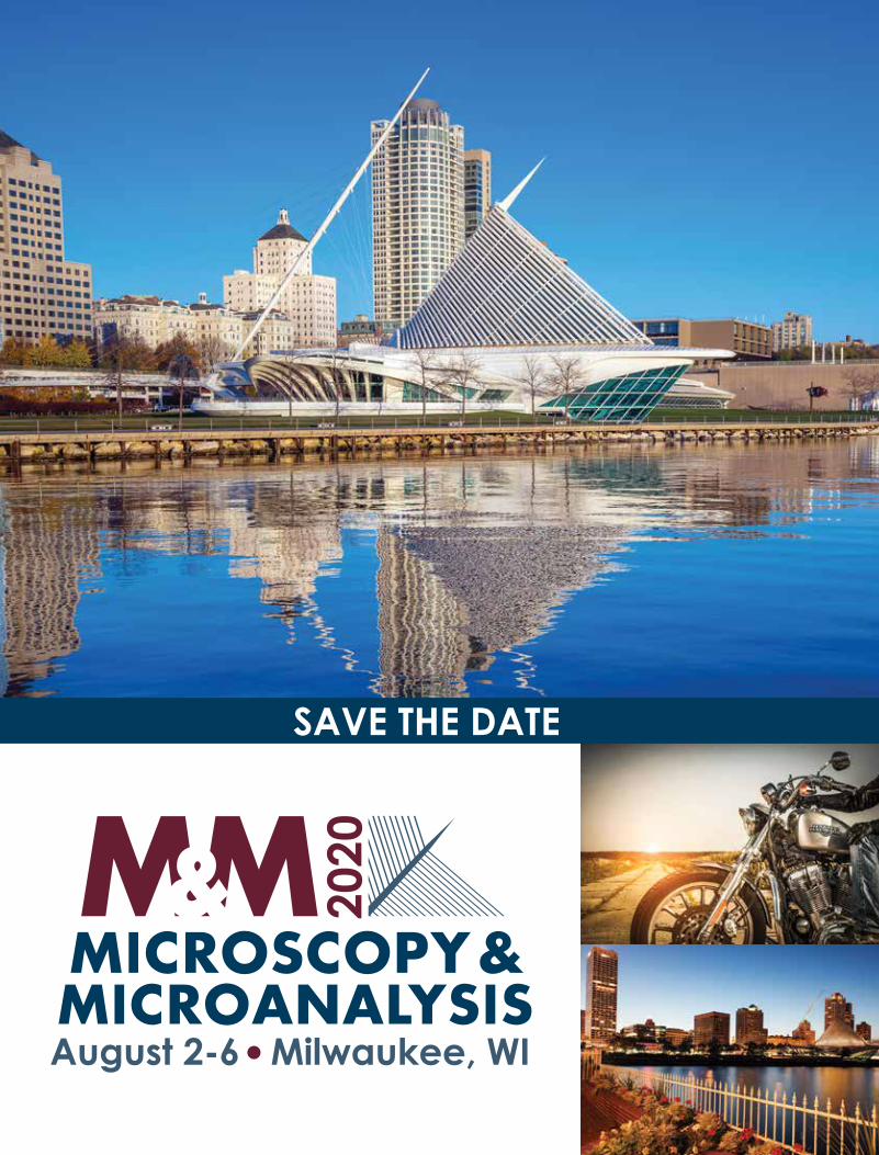
SAVE THE DATE

Find out more at thermofi sher.com/colorSEM
© 2019 Thermo Fisher Scientifi c Inc. All rights reserved. All trademarks are the property of Thermo Fisher Scientifi c and its subsidiaries unless otherwise specifi ed.
ColorSEM Technology: See The DifferenceThe world isn’t greyscale, so why are your electron
microscopy images? New Thermo Scientifi c™ ColorSEM™
Technology makes elemental analysis a matter of routine
thanks to always-on integrated energy-dispersive x-ray
spectroscopy. Obtain elemental information 2–4x faster than
conventional techniques, immediately revealing defects or
imperfections that you might have otherwise missed.
See the difference colo r can make with ColorSEM Technology.
Thermo Scientifi c™ Prisma™ E with ColorSEM technology

Jeol Cityscape Ad.indd 1 5/15/19 8:09 AM