Nuclear Medicine Radiation Detection and...
10
Radiology Residents Basic Science Course Nuclear Medicine Part June 7 th , 2007 Radiation detectors and Counting Statistics Thomas K. Lewellen <[email protected]> 1 Radiation Detection and Measurement May 2007 Tom Lewellen [email protected] Types of radiation relevant to Nuclear Medicine Particle Symbol Mass (MeV/c 2 ) Charge Electron e-, β - 0.511 -1 Positron e+, β+ 0.511 +1 Alpha α 3700 +2 Photon γ no rest mass none • Loses energy in a more or less continuous slowing down process as it travels through matter. • The distance it travels (range) depend only upon its initial energy and its average energy loss rate in the medium. • The range for an α particle emitted in tissue is on the order of μm’ s. α Particle Range in Matter mono-energetic - - - - - - - - - - - - - - - - - - - + + + + + + + + + + + + + + + + + μm’ s α • β particle ranges vary from one electron to the next, even for βs of the same energy in the same material. • This is due to different types of scattering events the β encounters (i.e., scattering events, bremsstrahlung-producing collisions, etc.). • The β range is often given as the maximum distance the most energetic β can travel in the medium. • The range for β particles emitted in tissue is on the order of mm’s. β Particle Range in Matter continuous energy spectrum mm’s - β ± Interactions of Photons with Matter Exponential Penetration: N=N 0 e -λx Photoelectric effect photon is absorbed Compton scattering part of the energy of the photon is absorbed scattered photon continues on with lower energy Pair production positron-electron pair is created requires photons above 1.022 MeV Coherent (Rayleigh) scattering photon deflected with very little energy loss only significant at low photon energies (<50 keV) cm’ s N N 0 x Basic Radiation Detector System Pulse or Current stored to disk incoming radiation Analog -to- digital Amplify & condition
-
Upload
truongxuyen -
Category
Documents
-
view
216 -
download
0
Transcript of Nuclear Medicine Radiation Detection and...
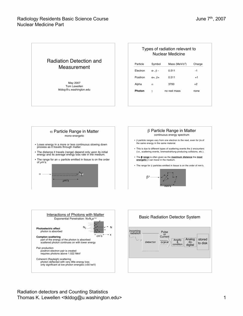
Radiology Residents Basic Science CourseNuclear Medicine Part
June 7th, 2007
Radiation detectors and Counting StatisticsThomas K. Lewellen <[email protected]> 1
Radiation Detection andMeasurement
May 2007Tom Lewellen
Types of radiation relevant toNuclear Medicine
Particle Symbol Mass (MeV/c2) Charge
Electron e-, β - 0.511 -1
Positron e+, β+ 0.511 +1
Alpha α 3700 +2
Photon γ no rest mass none
• Loses energy in a more or less continuous slowing downprocess as it travels through matter.
• The distance it travels (range) depend only upon its initialenergy and its average energy loss rate in the medium.
• The range for an α particle emitted in tissue is on the orderof µm’s.
α Particle Range in Mattermono-energetic
- - - - - - - - - - - - - - - - - - -+ + + + + + + + + + + + + + + + +
µm’s
α
• β particle ranges vary from one electron to the next, even for βs ofthe same energy in the same material.
• This is due to different types of scattering events the β encounters(i.e., scattering events, bremsstrahlung-producing collisions, etc.).
• The β range is often given as the maximum distance the mostenergetic β can travel in the medium.
• The range for β particles emitted in tissue is on the order of mm’s.
β Particle Range in Mattercontinuous energy spectrum
mm’s-
β±
Interactions of Photons with MatterExponential Penetration: N=N0e-λx
Photoelectric effectphoton is absorbed
Compton scatteringpart of the energy of the photon is absorbedscattered photon continues on with lower energy
Pair productionpositron-electron pair is createdrequires photons above 1.022 MeV
Coherent (Rayleigh) scatteringphoton deflected with very little energy lossonly significant at low photon energies (<50 keV)
λ
cm’s
NN0
x
Basic Radiation Detector System
Pulseor
Currentstoredto disk
incoming radiation
Analog-to-
digitalAmplify
&condition
Radiology Residents Basic Science CourseNuclear Medicine Part
June 7th, 2007
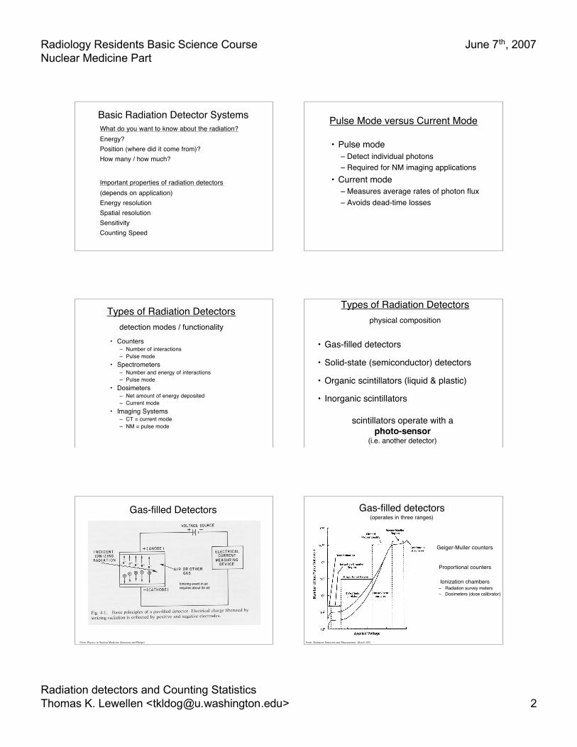
Radiation detectors and Counting StatisticsThomas K. Lewellen <[email protected]> 2
Basic Radiation Detector SystemsWhat do you want to know about the radiation?Energy?Position (where did it come from)?How many / how much?
Important properties of radiation detectors(depends on application)Energy resolutionSpatial resolutionSensitivityCounting Speed
Pulse Mode versus Current Mode
• Pulse mode– Detect individual photons– Required for NM imaging applications
• Current mode– Measures average rates of photon flux– Avoids dead-time losses
Types of Radiation Detectorsdetection modes / functionality
• Counters– Number of interactions– Pulse mode
• Spectrometers– Number and energy of interactions– Pulse mode
• Dosimeters– Net amount of energy deposited– Current mode
• Imaging Systems– CT = current mode– NM = pulse mode
Types of Radiation Detectorsphysical composition
• Gas-filled detectors
• Solid-state (semiconductor) detectors
• Organic scintillators (liquid & plastic)
• Inorganic scintillators
scintillators operate with aphoto-sensor
(i.e. another detector)
Gas-filled Detectors
Ionizing event in airrequires about 34 eV
From: Physics in Nuclear Medicine (Sorenson and Phelps)
Gas-filled detectors(operates in three ranges)
Geiger-Muller counters
Proportional counters
Ionization chambers– Radiation survey meters– Dosimeters (dose calibrator)
From: Radiation Detection and Measurement (Knoll, GF)
Radiology Residents Basic Science CourseNuclear Medicine Part
June 7th, 2007
Radiation detectors and Counting StatisticsThomas K. Lewellen <[email protected]> 3
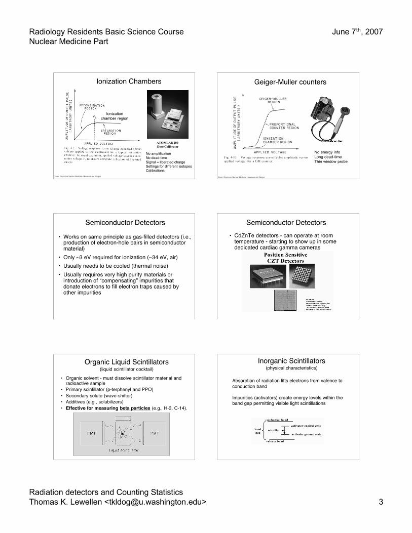
Ionization Chambers
From: Physics in Nuclear Medicine (Sorenson and Phelps)
ATOMLAB 200 Dose Calibrator
No amplificationNo dead-timeSignal = liberated chargeSettings for different isotopesCalibrations
Ionizationchamber region
Geiger-Muller counters
From: Physics in Nuclear Medicine (Sorenson and Phelps)
No energy infoLong dead-timeThin window probe
Semiconductor Detectors
• Works on same principle as gas-filled detectors (i.e.,production of electron-hole pairs in semiconductormaterial)
• Only ~3 eV required for ionization (~34 eV, air)• Usually needs to be cooled (thermal noise)• Usually requires very high purity materials or
introduction of “compensating” impurities thatdonate electrons to fill electron traps caused byother impurities
Semiconductor Detectors• CdZnTe detectors - can operate at room
temperature - starting to show up in somededicated cardiac gamma cameras
Organic Liquid Scintillators(liquid scintillator cocktail)
• Organic solvent - must dissolve scintillator material andradioactive sample
• Primary scintillator (p-terphenyl and PPO)• Secondary solute (wave-shifter)• Additives (e.g., solubilizers)• Effective for measuring beta particles (e.g., H-3, C-14).
Inorganic Scintillators(physical characteristics)
Absorption of radiation lifts electrons from valence toconduction band
Impurities (activators) create energy levels within theband gap permitting visible light scintillations

Radiology Residents Basic Science CourseNuclear Medicine Part
June 7th, 2007
Radiation detectors and Counting StatisticsThomas K. Lewellen <[email protected]> 4
Inorganic Scintillators(physical characteristics)
NaI(Tl) BGO LSO(Ce) GSO(Ce)
Density (gm/cm3) 3.67 7.13 7.4 6.71
EffectiveAtomic Number 51 75 66 59
AttenuationCoefficient(@ 511 keV, cm-1 ) 0.34 0.955 0.833 0.674
Light Output(photons/Mev) 40K ~8K ~30K ~20K
Decay Time 230 ns 300 ns 12 ns 60 ns40 ns
Wavelength 410 nm 480 nm 420 nm 430 nm
Index of Refraction 1.85 2.15 1.82 1.85
Hygroscopy yes no no no
Rugged no yes yes no
sensitivity
energy & spatial resol.counting speed
photo-sensor matchingmanufacturing / cost
relevant detectorproperty
Photomultiplier Tube (PMT) - most common photo-sensor currentlyin use for Nuclear medicine
From: Physics in Nuclear Medicine (Sorenson and Phelps)
photo-sensor needed with scintillators
Sample Spectroscopy SystemHardware
From: The Essential Physics of Medical Imaging (Bushberg, et al)
incoming high-energy gammaray
converted to1000s of visiblephotons
~20%converted toelectrons
electron multiplicationbecomes electricsignal larger current or
voltage
more electrons
more scintillationphotons
higher gammaenergy deposited in
crystal
Interactions of Photons with aSpectrometer
A. PhotoelectricB. Compton + PhotoelectricC. ComptonD. Photoelectric with characteristic
x-ray escapeE. Compton scattered photon from
lead shieldF. Characteristic x-ray from lead
shield
From: The Essential Physics of Medical Imaging (Bushberg, et al)
Sample Spectroscopy SystemOutput
From: Physics in Nuclear Medicine (Sorenson and Phelps)
From: The Essential Physics of Medical Imaging (Bushberg, et al)
counting mode
Ideal Energy Spectrum
Remember - you are looking at theenergy deposited in the detector!
Energy Resolution
From: Physics in Nuclear Medicine (Sorenson and Phelps)
Realistic Energy Spectrum
Radiology Residents Basic Science CourseNuclear Medicine Part
June 7th, 2007
Radiation detectors and Counting StatisticsThomas K. Lewellen <[email protected]> 5
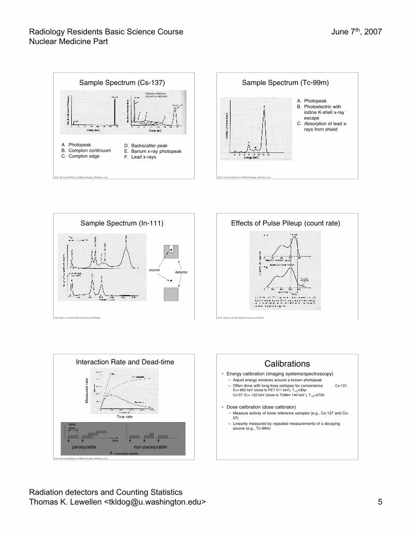
Sample Spectrum (Cs-137)
A. PhotopeakB. Compton continuumC. Compton edge
D. Backscatter peakE. Barium x-ray photopeakF. Lead x-rays
Detection efficiency(32 keV vs. 662 keV)
From: The Essential Physics of Medical Imaging (Bushberg, et al)
Sample Spectrum (Tc-99m)
A. PhotopeakB. Photoelectric with
iodine K-shell x-rayescape
C. Absorption of lead x-rays from shield
From: The Essential Physics of Medical Imaging (Bushberg, et al)
Sample Spectrum (In-111)
source detector
From: Physics in Nuclear Medicine (Sorenson and Phelps)
Effects of Pulse Pileup (count rate)
From: Physics in Nuclear Medicine (Sorenson and Phelps)
Interaction Rate and Dead-time
paralyzable non-paralyzable
From: The Essential Physics of Medical Imaging (Bushberg, et al)
True rate
Mea
sure
d ra
te
time
deadtime
= recorded events
Calibrations• Energy calibration (imaging systems/spectroscopy)
– Adjust energy windows around a known photopeak– Often done with long-lives isotopes for convenience Cs-137:
Eγ= 662 keV (close to PET 511 keV), T1/2=30yrCo-57: Eγ= 122 keV (close to Tc99m 140 keV ), T1/2=272d
• Dose calibration (dose calibrator)– Measure activity of know reference samples (e.g., Cs-137 and Co-
57)– Linearity measured by repeated measurements of a decaying
source (e.g., Tc-99m)
Radiology Residents Basic Science CourseNuclear Medicine Part
June 7th, 2007
Radiation detectors and Counting StatisticsThomas K. Lewellen <[email protected]> 6
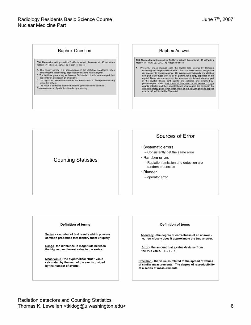
Raphex Question
D58. The window setting used for Tc-99m is set with the center at 140 keV with a width of +/-14 keV i.e., 20%. The reason for this is: A. The energy spread is a consequence of the statistical broadening when
amplifying the initial energy deposition event in the NaI(Tl) crystal. B. The 140 keV gamma ray emission of Tc-99m is not truly monoenergetic but
the center of a spectrum of emissions. C. The higher and lower Gaussian tails are a consequence of compton scattering
within the patient. D. The result of additional scattered photons generated in the collimator. E. A consequence of patient motion during scanning.
Raphex Answer
D58. The window setting used for Tc-99m is set with the center at 140 keV with a width of +/-14 keV i.e., 20%. The reason for this is: A . Photons, which impinge upon the crystal, lose energy by Compton
scattering and the photoelectric effect. Both processes convert the gamma ray energy into electron energy. On average approximately one electron hole pair is produced per 30 eV of g amma ray e nergy deposited in the crystal. These electrons result in the release of visible ligh t when trapped in the crystal. These light quanta are collected and amplified by photomultiplier tubes. The statistical fluctuation in the number of light quanta collected and their amplification is what causes the spread in the detected energy peak, even when most of the Tc-99m photons deposit exactly 140 keV in the NaI(Tl) crystal.
Counting Statistics
Sources of Error
• Systematic errors– Consistently get the same error
• Random errors– Radiation emission and detection are
random processes• Blunder
– operator error
Definition of terms
Series - a number of test results which possess common properties that identify them uniquely.
Range- the difference in magnitude between the highest and lowest value in the series.
Mean Value - the hypothetical “true” value calculated by the sum of the events divided by the number of events.
Definition of terms
Accuracy - the degree of correctness of an answer - ie, how closely does it approximate the true answer.
Error - the amount that a value deviates from the true value. E = X − X
Precision - the value as related to the spread of values of similar measurements. The degree of reproducibility of a series of measurements
Radiology Residents Basic Science CourseNuclear Medicine Part
June 7th, 2007
Radiation detectors and Counting StatisticsThomas K. Lewellen <[email protected]> 7
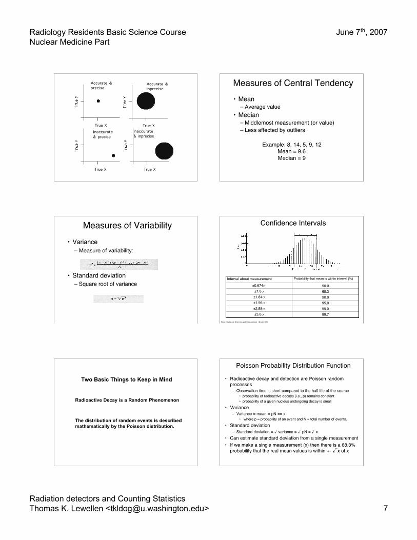
True X True X
True X True X
Accurate &precise
Inaccurate& precise
Inaccurate& inprecise
Accurate &inprecise
Measures of Central Tendency• Mean
– Average value• Median
– Middlemost measurement (or value)– Less affected by outliers
Example: 8, 14, 5, 9, 12Mean = 9.6Median = 9
Measures of Variability• Variance
– Measure of variability:
• Standard deviation– Square root of variance
Confidence Intervals
99.7±3.0σ
99.0±2.58σ
95.0±1.96σ
90.0±1.64σ
68.3±1.0σ
50.0±0.674σ
Probability that mean is within interval (%)Interval about measurement
From: Radiation Detection and Measurement (Knoll, GF)
Radioactive Decay is a Random Phenomenon
The distribution of random events is described mathematically by the Poisson distribution.
Two Basic Things to Keep in Mind
Poisson Probability Distribution Function
• Radioactive decay and detection are Poisson randomprocesses– Observation time is short compared to the half-life of the source
• probability of radioactive decays (i.e., p) remains constant• probability of a given nucleus undergoing decay is small
• Variance– Variance = mean = pN == x
• where p = probability of an event and N = total number of events.
• Standard deviation– Standard deviation = √variance = √pN = √x
• Can estimate standard deviation from a single measurement• If we make a single measurement (x) then there is a 68.3%
probability that the real mean values is within +- √x of x
Radiology Residents Basic Science CourseNuclear Medicine Part
June 7th, 2007
Radiation detectors and Counting StatisticsThomas K. Lewellen <[email protected]> 8
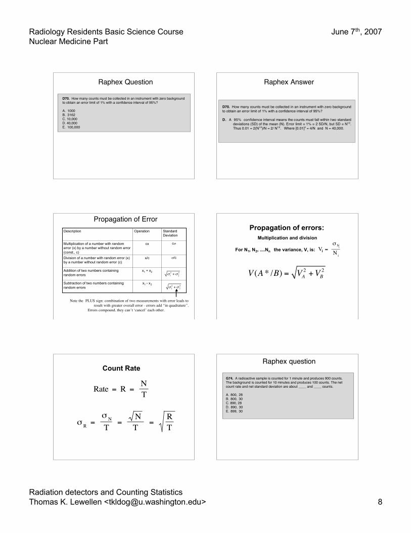
Raphex Question
D70. How many counts must be collected in an instrument with zero background to obtain an error limit of 1% with a confidence interval of 95%? A. 1000 B. 3162 C. 10,000 D. 40,000 E. 100,000
Raphex Answer
D70. How many counts must be collected in an instrument with zero background to obtain an error limit of 1% with a confidence interval of 95%? D. A 95% confidence interval means the counts must fall within two standard
deviations (SD) of the mean (N). Error limit = 1% = 2 SD/N, but SD = N1/2. Thus 0.01 = 2(N1/2)/N = 2/ N1/2. Where [0.01]2 = 4/N and N = 40,000.
Propagation of Error
x1 - x2Subtraction of two numbers containingrandom errors
x1 + x2Addition of two numbers containingrandom errors
σ/cx/cDivision of a number with random error (x)by a number without random error (c)
cσcxMultiplication of a number with randomerror (x) by a number without random error(const., c)
StandardDeviation
OperationDescription
Note the PLUS sign: combination of two measurements with error leads toresult with greater overall error - errors add “in quadrature”.
Errors compound, they can’t ‘cancel’ each other.
σ12 +σ 2
2
σ12 +σ 2
2
Propagation of errors:Multiplication and division
For N1, N2, ....Nn the variance, V, is: V = σ N i
N i i
V (A * /B) = VA2 +VB
2
Count Rate
Rate = R = N T
σ R = σ N
T = N T = R
T
Raphex question
G74. A radioactive sample is counted for 1 minute and produces 900 counts. The background is counted for 10 minutes and produces 100 counts. The net count rate and net standard deviation are about ____ and ____ counts. A. 800, 28 B. 800, 30 C. 890, 28 D. 890, 30 E. 899, 30
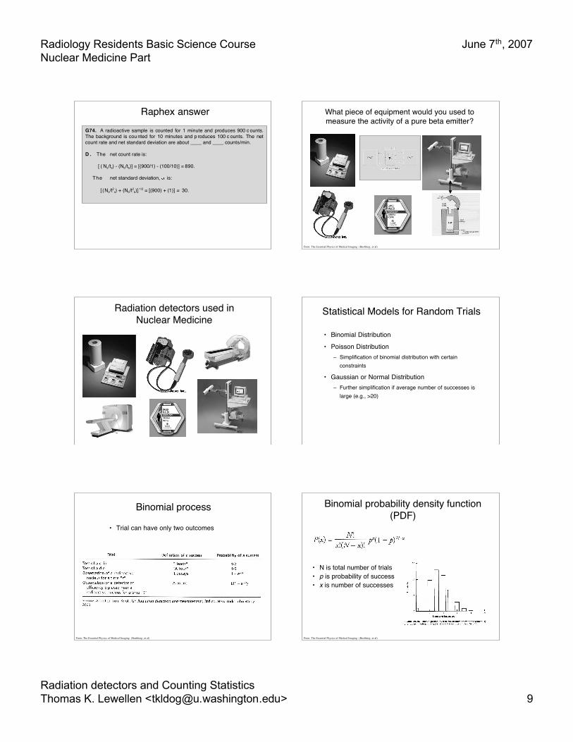
Radiology Residents Basic Science CourseNuclear Medicine Part
June 7th, 2007
Radiation detectors and Counting StatisticsThomas K. Lewellen <[email protected]> 9
Raphex answerG74. A radioactive sample is counted for 1 minute and produces 900 c ounts. The background is cou nted for 10 minutes and p roduces 100 c ounts. The net count rate and net standard deviation are about ____ and ____ counts/min. D . The net count rate is: [ ( Ns/ts) - (Nb/tb)] = [(900/1) - (100/10)] = 890. The net standard deviation, is: [ (Ns/t2
s) + (Nb/t2b)] 1/2 = [(900) + (1)] = 30.
What piece of equipment would you used tomeasure the activity of a pure beta emitter?
From: The Essential Physics of Medical Imaging (Bushberg, et al)
Radiation detectors used inNuclear Medicine
Statistical Models for Random Trials
• Binomial Distribution
• Poisson Distribution– Simplification of binomial distribution with certain
constraints
• Gaussian or Normal Distribution– Further simplification if average number of successes is
large (e.g., >20)
Binomial process
• Trial can have only two outcomes
From: The Essential Physics of Medical Imaging (Bushberg, et al)
Binomial probability density function(PDF)
• N is total number of trials• p is probability of success• x is number of successes
From: The Essential Physics of Medical Imaging (Bushberg, et al)
Radiology Residents Basic Science CourseNuclear Medicine Part
June 7th, 2007
Radiation detectors and Counting StatisticsThomas K. Lewellen <[email protected]> 10
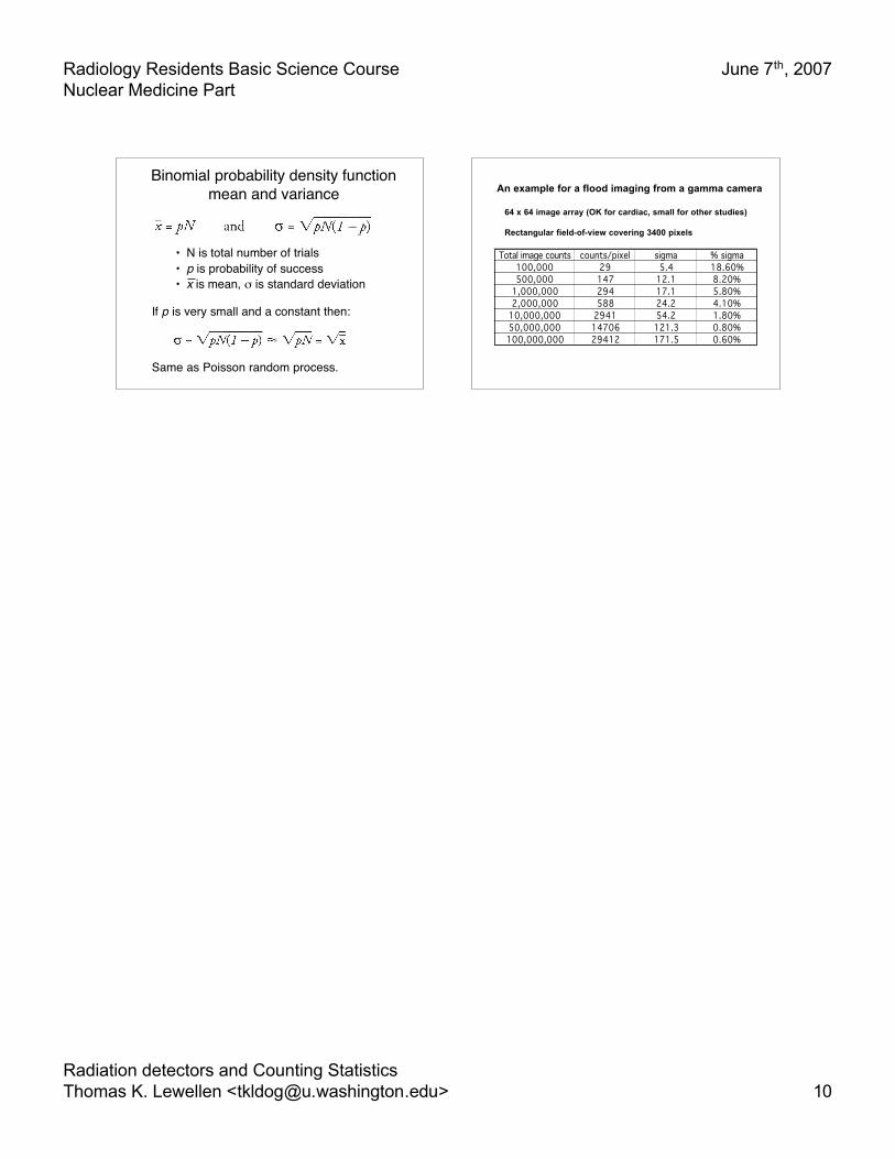
Binomial probability density functionmean and variance
• N is total number of trials• p is probability of success• x is mean, σ is standard deviation
If p is very small and a constant then:
Same as Poisson random process.
An example for a flood imaging from a gamma camera
64 x 64 image array (OK for cardiac, small for other studies)
Rectangular field-of-view covering 3400 pixels
Total image counts counts/pixel sigma % sigma100,000 29 5.4 18.60%500,000 147 12.1 8.20%
1,000,000 294 17.1 5.80%2,000,000 588 24.2 4.10%
10,000,000 2941 54.2 1.80%50,000,000 14706 121.3 0.80%100,000,000 29412 171.5 0.60%