Novel diagnostic methods for rapid characterization of ...
83
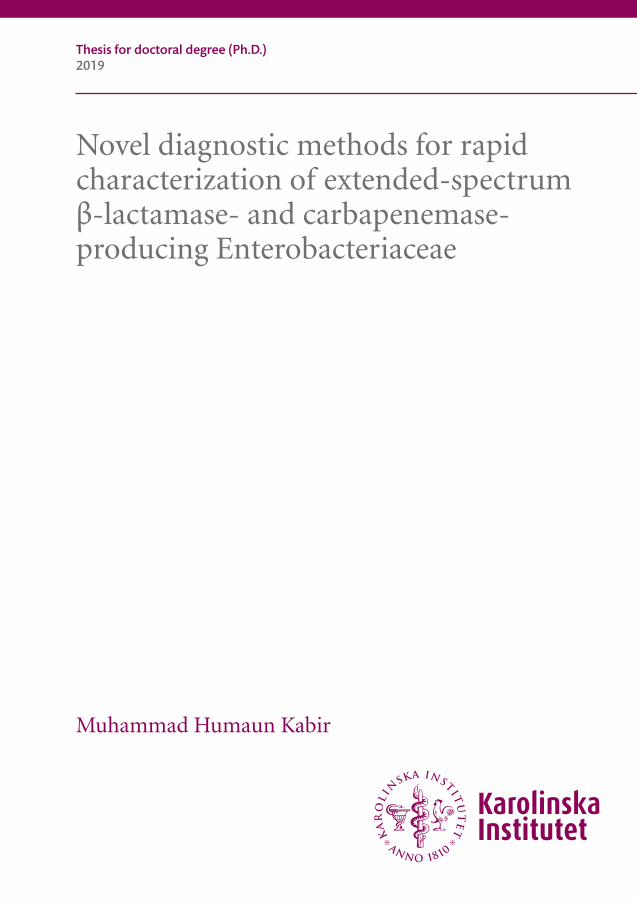
Thesis for doctoral degree (Ph.D.) 2019 Novel diagnostic methods for rapid characterization of extended-spectrum β-lactamase- and carbapenemase- producing Enterobacteriaceae Muhammad Humaun Kabir
Transcript of Novel diagnostic methods for rapid characterization of ...
Thesis for doctoral degree (Ph.D.)2019
Novel diagnostic methods for rapid characterization of extended-spectrum β-lactamase- and carbapenemase- producing Enterobacteriaceae
Muhammad Humaun Kabir
Novel diagn
ostic meth
ods for rapid characterization
of extended-sp
ectrum
β-lactam
ase- and carbap
enem
ase-producin
g En
terobacteriaceae
From Department of Laboratory medicine, Division of Clinical microbiology
Karolinska Institutet, Stockholm, Sweden
NOVEL DIAGNOSTIC METHODS FOR RAPID CHARACTERIZATION OF
EXTENDED-SPECTRUM β-LACTAMASE- AND CARBAPENEMASE-PRODUCING
ENTEROBACTERIACEAE
Muhammad Humaun Kabir
Stockholm 2019

All previously published papers were reproduced with permission from the publisher. Published by Karolinska Institutet. Printed by Arkitektkopia AB, 2019 © Muhammad Humaun Kabir, 2019 ISBN 978-91-7831-512-3
Novel diagnostic methods for rapid characterization of extended-spectrum β-lactamase- and carbapenemase-producing Enterobacteriaceae
THESIS FOR DOCTORAL DEGREE (Ph.D.)
Public defence at Karolinska Institutet, Lecture hall 4z, Alfred Nobels Alle 8, Level 4, Flemingsberg, Huddinge, Stockholm.
Tuesday, October 1, 2019 at 9:00 a.m.
By
Muhammad Humaun Kabir
Principal Supervisor:Professor Christian G. Giske Karolinska Institutet Department of Laboratory medicine Division of Clinical microbiology
Co-supervisors:Dr. Aina Iversen Karolinska Institutet Department of Laboratory medicine Division of Clinical microbiology
Dr. Anni-Maria Örmälä-Odegrip Karolinska Institutet Department of Laboratory medicine Division of Clinical microbiology
Opponent:Dr. Christina Åhrén Gothenburg University Department of Infectious Diseases
Examination Board:Dr. Åsa Sjöling Karolinska Institutet Department of Microbiology Tumor and Cell Biology
Professor Mikael Rhen Karolinska Institutet Department of Microbiology Tumor and Cell Biology Division of Microbial Pathogenesis
Professor Kristian Riesbeck Lund University Department of Translational Medicine Division of Clinical Microbiology


To my teachers, friends and family

ABSTRACTKlebsiella pneumoniae and Escherichia coli are two of the most clinically important pathogens of the family Enterobacteriaceae. Both can cause severe infections includ-ing urinary tract infections (UTI), bloodstream infections (BSI) and pneumonia. Both are also involved in dissemination of antibiotic resistance. The prevalence of extended-spectrum β-lactamase- (ESBL) producing Enterobacteriaceae (EPE) and carbapenemase-producing Enterobacteriaceae (CPE) are increasing worldwide. This is a severe threat to public health, as antibiotic treatment becomes limited. The general aim of the thesis was to investigate the clonal diversity and prevalence of ESBL in invasive isolates of Klebsiella pneumoniae from the Stockholm area, the acquisition of EPE/CPE in healthy Swedish tourists travelling to regions with high prevalence of EPE/CPE, and finally to evaluate rapid detection and characteriza-tion methods for EPE and CPE.
In Paper I, we studied the clonal structure of K. pneumoniae from BSI patients in a well-defined geographical area, as well as the prevalence of ESBL in these strains. We also characterized the clinical isolates to investigate the association between phylogroups, bacterial virulence factors, 30-day mortality and comorbidity. A total of 139 samples were collected from the Department of clinical microbiology at Karolinska University Hospital, Stockholm, Sweden. Only five out of 139 isolates were detected as multidrug-resistant (MDR) and all five isolates were EPE. Only one isolate was detected as a CPE. Further, all isolates were tested for mucoid pheno types, serotypes (K1, K2, K5, K20, K54 and K57) and screened for virulence genes using real-time PCR. To identify risk factors, we retrieved data from medical records including age, sex, time to adequate treatment, hospital acquired infections, comorbidity and mortality. The 30-day mortality was primary end-point. All isolates were further subjected to multilocus sequence typing (MLST) for phylogenetic analysis. We found that all isolates were divided into three distinct phylogroups; KpI (consisting of 96 isolates of K. pneumoniae) followed by phylogroup KpIII (consist-ing of 34 isolates of K. variicola) and phylogroup KpII (consisting of 9 isolates of K. quasipneumoniae). We observed 24/139 (17.3%) overall 30-day mortality. We also observed that K. variicola KpIII (29.4%) were highly associated with 30-day mortality compared to K. pneumoniae KpI isolates (13.5%). We also observed that several comorbidities; malignancy, diabetes mellitus, cirrhosis, biliary tract disor-ders and alcoholism among patients may influence the mortality.
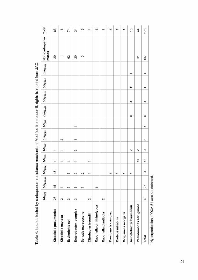
In Paper II, we evaluated a commercial version of RAPIDEC® CARBA NP for rapid detection of carbapenemases against 276 Gram-negative bacilli. According to the manufacturer’s protocol RAPIDEC® CARBA NP assay was performed on 138 carbapenemase-producers and 138 non-carbapenemase-producers. Carbapenemase detection was also performed by using conventional and real-time PCR. A total of 135 out of 138 carbapenemase-producers were detected successfully by the RAPIDEC® CARBA NP assay. The RAPIDEC® CARBA NP assay was unable to detect one OXA-48-producing K. pneumoniae and two Acinetobacter baumannii
producing OXA-23 and OXA-24 carbapenemases. The RAPIDEC® CARBA NP assay successfully detected 135 non-carbapenemase producers. A total of two false positives; one Pseudomonas aeruginosa with OprD loss and efflux, one Enterobacter cloacae with impermeability were detected by the RAPIDEC® CARBA NP assay. The overall sensitivity and specificity of the RAPIDEC® CARBA NP assay was 97.8% and 98.5%, respectively.
In Paper III, we investigated the acquisition of EPE and CPE among healthy Swedish travellers visiting regions with high prevalence of EPE and CPE; Southeast Asia, Indian subcontinent, North Africa and the Middle East. We studied 188 healthy adult travellers and excluded 13 pre-travel colonized tourists from the analysis. Molecular characterization was performed using real-time PCR and survey data were collected for analysis. We detected a total of 67/175 tourist were colonized by EPE strains and most dominant species was E. coli (n=65), followed by K. pneumoniae (n=1) and Citrobacter freundii (n=1). CTX-M type (CTX-M-1 n=49, CTX-M-9 n=12) was the most prevalent EPE type detected in 61 isolates but carbapenemases were absent. Only six travellers had strains with the virulence factor pyelonephritis-associated pili-encoding genes (pap) genes, and all of those had travelled to India. One traveller to Thailand was colonized with colistin resistance gene mcr-1. The highest coloni-zation rate was detected among the travellers to the Indian subcontinent (49%) and northern Africa (44%) whereas lower rates were detected in travellers to Southeast Asia (19%) and Turkey (10%). EPE colonization was associated with diarrhea and antibiotic treatment during the trip but was not associated with sex, age, duration of trip, intake of proton-pump inhibitors, or use of oral cholera vaccine.
In paper IV, we assessed a CRISPR/Cas assisted optical DNA mapping method for rapid identification and characterization of plasmids during an EPE outbreak. We studied a total of 17 neonates initially colonized with ESBL-producing K. pneumoniae (ESBL-KP). In follow-up samples we observed that some of them were colonized with ESBL-producing E. coli. This method successfully detected two plasmids of different sizes; small size (80 kbp) and larger plasmid (162-222 kbp) in all ESBL-KP isolates. Using this method, we also observed that the blaCTX-M-15 gene was located on the smaller size plasmid. In follow-up samples we also observed that the ESBL-KP clone was present and blaCTX-M-15 was stable up to two years. This method demon-strated that plasmids can change over time: three deletions were observed in three plasmids of three isolates with different sizes (~ 5 kbp), (~ 55 kbp) and (~ 31 kbp) and an inversion of 31 kbp in another isolate. This method also confirmed that resistance plasmids were not transferred from one patient to another.
In conclusion, this thesis provides new knowledge on the prevalence of EPE and CPE among BSI patients and healthy Swedish tourists, along with describing the molecular features of ESBL- and carbapenemase-producing Enterobacteriaceae. This thesis also demonstrates the performance of a rapid phenotypic carbapenemase detection assay, as well as a novel CRISPR/Cas-assisted optical DNA mapping method for rapid plasmid characterization.
LIST OF SCIENTIFIC PAPERSI. Makaoui Matallah, Malin Vading, Muhammad Humaun Kabir, Amina
Bakhrouf, Mats Kalin, Pontus Naucler, Sylvain Brisse, Christian G Giske. Klebsiella variicola is a frequent cause of bloodstream infection in the Stockholm area, and associated with higher mortality compared to K. pneu-moniae. PLoS One. 2014;9(11):e113539
II. Muhammad Humaun Kabir, Daniele Meubier, Katie L. Hopkins, Christian G. Giske and Neil Woodford. A two-centre evaluation of RAPIDEC® CARBA NP for carbapenemase detection in Enterobacteriaceae, Pseudomonas aerugi-nosa and Acinetobacter spp. J Antimicrob Chemother. 2015; 71(5):1213-6.
III. Malin Vading, Muhammad Humaun Kabir, Mats Kalin, Aina Iversen, Susanne Wiklund, Pontus Naucler, Christian G. Giske. Frequent acquisition of low-virulent strains of ESBL-producing Escherichia coli in travellers. J Antimicrob Chemother. 2016;71(12):3548-3555.
IV. Santosh K. Bikkarolla, Viveka Nordberg, Fredrika Rajer, Vilhelm Müller, Muhammad Humaun Kabir, Sriram Kesarimangalam, Albertas Dvirnas, Tobias Ambjörnsson, Christian G. Giske, Lars Navér, Linus Sandegren, Fredrik Westerlund. Optical DNA mapping combined with Cas9-targeted resistance gene identification for rapid tracking of resistance plasmids in a neonatal intensive care unit outbreak. MBio 2019;10(4). pii: e00347-19.

CONTENTS
1 Introduction 11.1 Antibiotic resistance 11.2 Mechanisms of antibiotic resistance 21.3 Enterobacteriaceae 5
1.3.1 Klebsiella pneumoniae 51.3.2 Escherichia coli 6
1.4 Antimicrobial resistance in Enterobacteriaceae 61.4.1 ESBL-producing Enterobacteriaceae (EPE) 71.4.2 Carbapenemase-producing Enterobacteriaceae (CPE) 81.4.3 Plasmid-mediatedβ-lactamresistance 101.4.4 Plasmid-mediatednonβ-lactamresistance 10
1.5 Detection of EPE 111.5.1 Phenotypic confirmation methods 121.5.2 Genotypic confirmation 13
1.6 Carbapenemases detection 141.6.1 Phenotypic detection methods 141.6.2 Genotypic methods 16
1.7 CRISPR/Cas9 mediated optical DNA mapping 172 Aims 193 Materials and methods 20
3.1 Sample collection 203.2 Ethical considerations 203.3 Species identification and susceptibility testing 223.4 ESBL Screening and subtyping 223.5 Phenotypic and molecular detection of carbapenemase 233.6 Serotyping and virulence and resistance gene detection 233.7 Phylogenetic analysis 243.8 Clinical data 243.9 Plasmid characterization 25
4 Results 274.1 Paper I 274.2 Paper II 294.3 Paper III 314.4 Paper IV 32
5 Discussion 375.1 Major Findings 375.2 Phylogroups of K. pneumoniae 395.3 Misidentification of K. variicola and K. quasipneumoniae 395.4 Association of phylogroups, bacterial factors, mortality and
comorbidity 40

5.5 Phylogroups of E. coli 415.6 Colonization and duration of EPE and CPE in travellers 415.7 Rapid detection of CPE 425.8 Plasmid characterization using CRISPR/Cas9 assisted optical
DNA mapping 436 Conclusions 447 Future perspectives 458 Acknowledgements 479 References 48
LIST OF ABBREVIATIONSAACs N-acetyltransferasesANTs Aminoglycoside O-nucleotidetransferasesBSI Bloodstream infectionsEPE ESBL-producing EnterobacteriaceaeESBLs Extended-spectrum b-lactamasesHAP Hospital-acquired pneumoniaHUS Hemolytic-uremic syndromeHvKP Hypervirulent K. pneumoniaeKPC Klebsiella pneumoniae carbapenemaseLPS Lipopolysaccharidesmcr-1 Plasmid-mediated colistin resistance geneMDR Multi-drug resistanceNDM New Delhi metallo-b-lactamaseOXA Oxacillinase-type b-lactamasermpA Regulator of the mucoid phenotypeSSI Surgical site infectionUTI Urinary tract infectionsVIM Verona integron-encoded metallo-b-lactamaseWHO World Health Organization

1
1 INTRODUCTION1.1 Antibiotic resistanceIn 1928, Sir Alexander Fleming serendipitously discovered a new drug with remark-able properties; penicillin. Subsequently, related compounds were developed with even broader antimicrobial spectrum – the cephalosporins, the carbapenems, and the monobactams. The Greek words anti- (means against) and bios (means life) are the source of the word antibiotic. Antibiotics are the substances that can kill or inhibit the growth of bacteria. Antibiotics can be chemical agents such as sulfa compounds or can be derived from natural origin with a chemical modification such as Penicillium notatum.
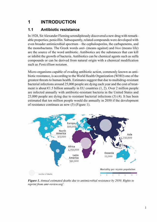
Micro-organisms capable of evading antibiotic action, commonly known as anti-biotic resistance, is according to the World Health Organization (WHO) one of the greatest threats to human health. Estimates suggest that due to multidrug-resistant bacterial infections around 25,000 people are dying each year and the cost of treat-ment is about €1.5 billion annually in EU countries (1, 2). Over 2 million people are infected annually with antibiotic-resistant bacteria in the United States and 23,000 people are dying due to resistant bacterial infections (3) (4). It has been estimated that ten million people would die annually in 2050 if the development of resistance continues as now (5) (Figure 1).
Figure 1.Annualestimateddeathsduetoantimicrobialresistanceby2050.Rightstoreprint from amr-review.org5.
2
1.2 Mechanisms of antibiotic resistance Bacteria exhibit antibiotic resistance intrinsically or they can acquire it. Resistance mechanisms to some common antibiotics are listed in (Table 1) (6). Intrinsic resistance is the ability of a bacterium to resist antibiotic action by its inherent structural or functional characteristics. In comparison to Gram-positive bacteria, intrinsic resistance to a variety of compunds is rather common in Gram-negative due to i) differences in the composition of the cytoplasmic membrane; presence of anionic phospholipids is low in the cytoplasm of Gram-negative bacteria, ii) absence of susceptible targets and iii) inability of the antibiotics to cross outer membrane of Gram-negative bacteria. For example, vancomycin is active against Gram-positive bacteria by inhibiting peptidoglycan crosslinking by binding to D-Ala-D-Ala peptides, but it is not active against Gram-negative bacteria as it cannot reach to peptides in periplasm (7).
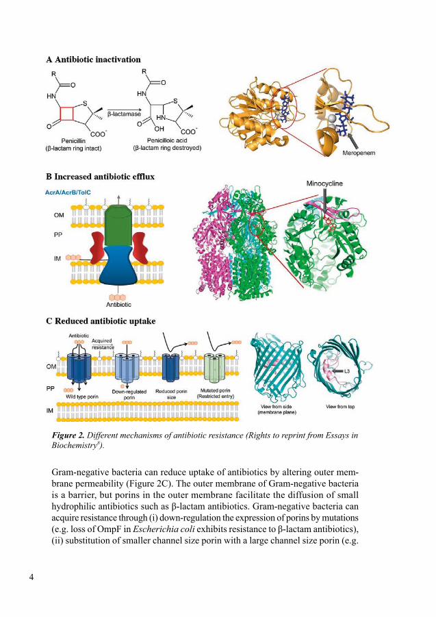
Gram-negative pathogens can also acquire resistance through gene mutations or horizontal gene transfer. The mechanisms of acquired resistance can be classified in four main classes; (i) antibiotic modification/inactivation, (ii) antibiotic target alteration, (iii) increased antibiotic efflux, and (iv) reduced antibiotic uptake (Figure 2) (8).
Antibiotic inactivation can occur by hydrolysis of β-lactam antibiotics. The four-membered β-lactam ring is hydrolyzed by β-lactamase enzymes (Figure 2A). Different groups of β-lactamases have been reported, which can inactivate different groups of antibiotics including, penicillin, cephalosporins and carbapenems (9). Some enzymes such as aminoglycoside N-acetyltransferases (AACs), amino glycoside O-nucleotidetransferases (ANTs) and aminoglycoside O-phosphotransferases (APHs) can modify aminoglycoside antibiotics to exhibit resistance against aminoglycoside antibiotics (10).
Target alteration is another common mechanism of resistance against several classes of antibiotics where the target of antibiotic is changed. One example of such target alteration is DNA-topoisomerase complex. Mutation of the DNA gyrase encoding gene (gyrA) can alter the protein structure which reduces fluoroquinolone-binding affinity leading to resistance (11). Modification of specific nucleotides in the aminoglycoside binding site of 16S rRNA by 16S rRNA methylases can confer high-level aminoglycoside resistance (12).
Bacteria use efflux pumps to force out antibiotics from the cell so that they can lower the antibiotic concentration to sublethal levels (Figure 2B). This is a first line defence and it can force out different compounds, depending on the specificity of the efflux pump. Using efflux pumps, bacteria can develop multidrug resistance, as they can simultaneously export drugs of different drug classes (13).
3
Table 1. Mechanisms of resistance to different classes of antibiotics against Gram-negative bacteria. Modified from Kumar et al 2013 with permission from the publishers6.
Antimicrobial class
Mechanism of Resistance
Specific means to achieve resistance
Examples
b-lactams Degradation of antibiotics
Destruction of b-lactam ring by b-lactamases making it incapable to bind to penicillin binding protein (PBPs)
Resistance to penicillins, cephalosporins and carbapenems
Decreased uptake Decreased porin channel formation
Carbapenem resistance in K. pneumoniae, Enterobacter spp. and P. aeruginosa
Polymyxins Altered target Mutational changes in LPS. Modification of lipid A by enzyme MCR
Colistin resistance in K. pneumoniae and E. coli
Glycylcyclines Efflux Pump tigecycline and eravacycline out of the cell
Tigecycline resistance in K. pneumoniae
Trimethoprim-Sulfamethoxazole
DNA synthesis Acquisition of dfr and sul genes that can circum-vent the inhibitory effect of trimethoprim and sulfa in the synthesis pathway of tetrahydrofolate
Trimethoprim and sulfonamide resistance in E. coli and K. pneumoniae
Permeability barrier and/or efflux
Permeability barrier and/or efflux act against both sul-fonamides and trimethoprim
Trimethoprim and sulfonamide resistance in E. coli and K. pneumoniae
Aminoglycoside Enzymatic modification
AACs, ANTs and APH enzymes modify aminoglycoside
Aminoglycoside resist-ance in Gram-negative bacilli
Efflux Downregulation or alteration of porin channels
Aminoglycoside resist-ance in Gram-negative bacilli
Altered target Modification of ribosomal proteins or 16S rRNA
16S ribosomal RNA (rRNA) methylases in Enterobacteriaceae and P. aeruginosa and Acinetobacter spp.
Fluoroquinolones Decreased uptake and increased excretion
Alteration in the outer mem-brane diminishes uptake of drug. Activation of efflux pumps to excrete the drug
Fluoroquinolone resistant Gram-negative bacteria
Altered targetEnzymatic modification
Changes in DNA gyrase and topoisomerase or target protection (Qnr)AAC6´Ib-CR can confer FQ resistance through enzymatic modification
Fluoroquinolone resistant Gram-negative bacilliFluoroquinolone resistant Gram-negative bacilli
4
Figure 2.Differentmechanismsofantibioticresistance(RightstoreprintfromEssaysinBiochemistry8).
Gram-negative bacteria can reduce uptake of antibiotics by altering outer mem-brane permeability (Figure 2C). The outer membrane of Gram-negative bacteria is a barrier, but porins in the outer membrane facilitate the diffusion of small hydrophilic antibiotics such as β-lactam antibiotics. Gram-negative bacteria can acquire resistance through (i) down-regulation the expression of porins by mutations (e.g. loss of OmpF in Escherichia coli exhibits resistance to β-lactam antibiotics), (ii) substitution of smaller channel size porin with a large channel size porin (e.g.
5
Klebsiella. pneumoniae isolates exhibit resistance against β-lactam antibiotics by substitution of OmpK35 with a smaller channel size OmpK36), (iii) impaired porin function by mutation (e.g. permeation of benzylpenicillin is reduced in PenB porin of Neisseria gonorrhoeae by addition of two negatively charged amino acids in the channel-constricting loop 3) (14-16).
1.3 Enterobacteriaceae Enterobacteriaceae is a family of rod-shaped Gram-negative bacteria, which are commensal inhabitants of the intestinal microbiota, but which are also a common source of both nosocomial and community-acquired infections. Enterobacteriaceae can cause a wide range of infections; cystitis, pyelonephritis, septicemia, pneumonia, peritonitis, meningitis, and device-associated infections. K. pneumoniae and E. coli are the most common human pathogens within the Enterobacteriaceae family.
1.3.1 Klebsiella pneumoniae In 1882, Carl Friedlander first isolated Klebsiella pneumoniae from the lungs of a patient with pneumonia (17). Klebsiella species are abundant in nature, they can be found in plants, animals and humans. They are one of the most important member of family Enterobacteriaceae of Gram-negative pathogenic bacteria. They are non-sporulating, lactose-fermenting, facultatively anaerobic, rod-shaped and non-motile bacteria. They can cause severe infections in humans, including respira-tory tract infections, urinary tract infections, and bloodstream infections (18). They can cause both nosocomial and community-acquired infections. Klebsiella species are the third leading cause of hospital-acquired infections in United States (19) and they are among the leading causes of ventilator-associated pneumonia among intensive care units’ patients (20). They are also considered as the second leading cause of bloodstream infections (BSI) among Gram-negative bacteria (18, 19).
Capsule, lipopolysaccharides (LPS), fimbriae, siderophores (enterobactin, aero-bactin, salmochelin, yersiniabactin) and efflux pumps are considered as the main virulence factors of K. pneumoniae (18). Furthermore, some virulence factors; fimbriae, capsule and enterobactins are ubiquitous in all isolates of K. pneumoniae (21). Hypervirulent K. pneumoniae (HvKP) isolates are associated with a number of putative virulence factors; aerobactin and rmpA (regulator of the mucoid pheno-type) both encoded by large virulence plasmids are present in hypervirulent HvKP isolates (22). Integrative and conjugative elements encoded iron acquisition sys-tem, such as yersiniabactin, and allantoin metabolism associated regions can also be present in HvKP isolates (23, 24). Capsules are composed of polysaccharide forming thick bundle of fibrillous structures that covers bacterium to protect from macrophage-mediated phagocytosis and bactericidal serum factors (25). There are 78 different capsular (K antigen) types, most importantly K1, K2, K5, K20, K54 and K57 are highly associated with invasive disease of K. pneumoniae (26).
6
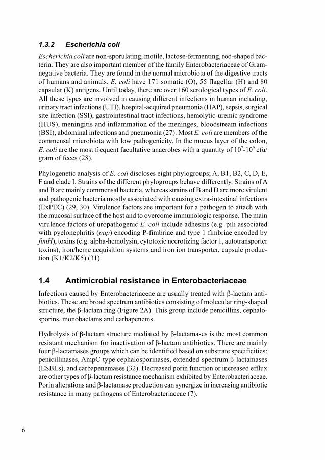
1.3.2 Escherichia coliEscherichia coli are non-sporulating, motile, lactose-fermenting, rod-shaped bac-teria. They are also important member of the family Enterobacteriaceae of Gram-negative bacteria. They are found in the normal microbiota of the digestive tracts of humans and animals. E. coli have 171 somatic (O), 55 flagellar (H) and 80 capsular (K) antigens. Until today, there are over 160 serological types of E. coli. All these types are involved in causing different infections in human including, urinary tract infections (UTI), hospital-acquired pneumonia (HAP), sepsis, surgical site infection (SSI), gastrointestinal tract infections, hemolytic-uremic syndrome (HUS), meningitis and inflammation of the meninges, bloodstream infections (BSI), abdominal infections and pneumonia (27). Most E. coli are members of the commensal microbiota with low pathogenicity. In the mucus layer of the colon, E. coli are the most frequent facultative anaerobes with a quantity of 107-109 cfu/gram of feces (28).
Phylogenetic analysis of E. coli discloses eight phylogroups; A, B1, B2, C, D, E, F and clade I. Strains of the different phylogroups behave differently. Strains of A and B are mainly commensal bacteria, whereas strains of B and D are more virulent and pathogenic bacteria mostly associated with causing extra-intestinal infections (ExPEC) (29, 30). Virulence factors are important for a pathogen to attach with the mucosal surface of the host and to overcome immunologic response. The main virulence factors of uropathogenic E. coli include adhesins (e.g. pili associated with pyelonephritis (pap) encoding P-fimbriae and type 1 fimbriae encoded by fimH), toxins (e.g. alpha-hemolysin, cytotoxic necrotizing factor 1, autotransporter toxins), iron/heme acquisition systems and iron ion transporter, capsule produc-tion (K1/K2/K5) (31).
1.4 Antimicrobial resistance in Enterobacteriaceae Infections caused by Enterobacteriaceae are usually treated with β-lactam anti-biotics. These are broad spectrum antibiotics consisting of molecular ring-shaped structure, the β-lactam ring (Figure 2A). This group include penicillins, cephalo-sporins, monobactams and carbapenems.
Hydrolysis of β-lactam structure mediated by β-lactamases is the most common resistant mechanism for inactivation of β-lactam antibiotics. There are mainly four β-lactamases groups which can be identified based on substrate specificities: penicillinases, AmpC-type cephalosporinases, extended-spectrum β-lactamases (ESBLs), and carbapenemases (32). Decreased porin function or increased efflux are other types of β-lactam resistance mechanism exhibited by Enterobacteriaceae. Porin alterations and β-lactamase production can synergize in increasing antibiotic resistance in many pathogens of Enterobacteriaceae (7).
7
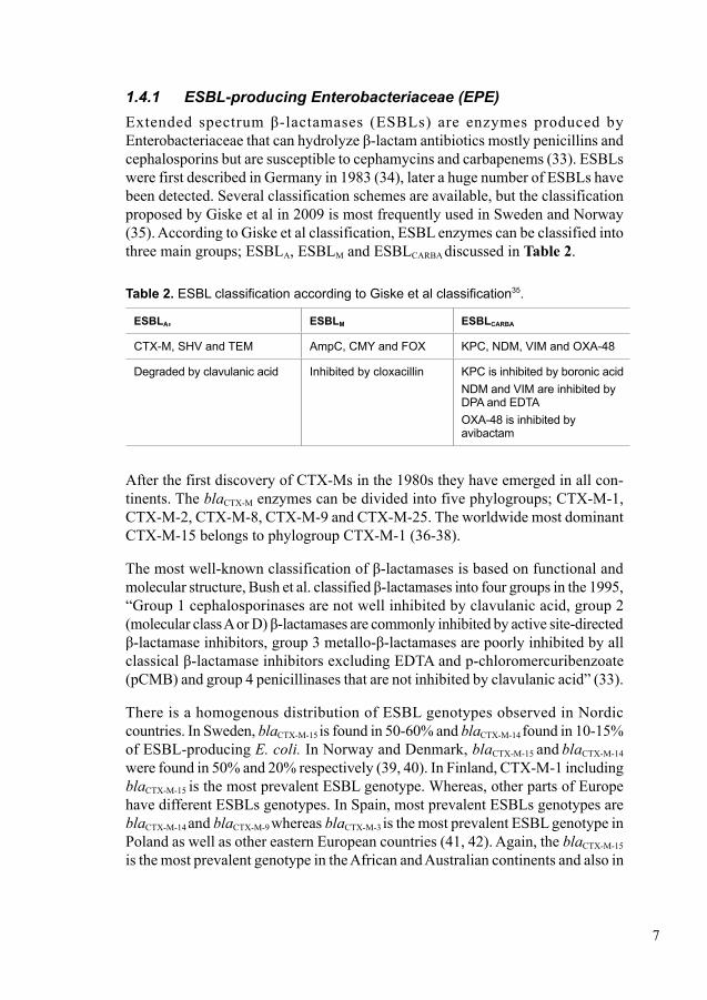
1.4.1 ESBL-producing Enterobacteriaceae (EPE)Extended spectrum β-lactamases (ESBLs) are enzymes produced by Enterobacteriaceae that can hydrolyze β-lactam antibiotics mostly penicillins and cephalosporins but are susceptible to cephamycins and carbapenems (33). ESBLs were first described in Germany in 1983 (34), later a huge number of ESBLs have been detected. Several classification schemes are available, but the classification proposed by Giske et al in 2009 is most frequently used in Sweden and Norway (35). According to Giske et al classification, ESBL enzymes can be classified into three main groups; ESBLA, ESBLM and ESBLCARBA discussed in Table 2.
Table 2. ESBL classification according to Giske et al classification35.
ESBLA, ESBLM ESBLCARBA
CTX-M, SHV and TEM AmpC, CMY and FOX KPC, NDM, VIM and OXA-48
Degraded by clavulanic acid Inhibited by cloxacillin KPC is inhibited by boronic acidNDM and VIM are inhibited by DPA and EDTA OXA-48 is inhibited by avibactam
After the first discovery of CTX-Ms in the 1980s they have emerged in all con-tinents. The blaCTX-M enzymes can be divided into five phylogroups; CTX-M-1, CTX-M-2, CTX-M-8, CTX-M-9 and CTX-M-25. The worldwide most dominant CTX-M-15 belongs to phylogroup CTX-M-1 (36-38).
The most well-known classification of β-lactamases is based on functional and molecular structure, Bush et al. classified β-lactamases into four groups in the 1995, “Group 1 cephalosporinases are not well inhibited by clavulanic acid, group 2 (molecular class A or D) β-lactamases are commonly inhibited by active site-directed β-lactamase inhibitors, group 3 metallo-β-lactamases are poorly inhibited by all classical β-lactamase inhibitors excluding EDTA and p-chloromercuribenzoate (pCMB) and group 4 penicillinases that are not inhibited by clavulanic acid” (33).
There is a homogenous distribution of ESBL genotypes observed in Nordic countries. In Sweden, blaCTX-M-15 is found in 50-60% and blaCTX-M-14 found in 10-15% of ESBL-producing E. coli. In Norway and Denmark, blaCTX-M-15 and blaCTX-M-14 were found in 50% and 20% respectively (39, 40). In Finland, CTX-M-1 including blaCTX-M-15 is the most prevalent ESBL genotype. Whereas, other parts of Europe have different ESBLs genotypes. In Spain, most prevalent ESBLs genotypes are blaCTX-M-14 and blaCTX-M-9 whereas blaCTX-M-3 is the most prevalent ESBL genotype in Poland as well as other eastern European countries (41, 42). Again, the blaCTX-M-15
is the most prevalent genotype in the African and Australian continents and also in
8
Canada. In US the most prevalent ESBL genotype used to be blaSHV about a dec-ade ago, but now blaCTX-M-15 has become the most prevalent ESBL type also there (43-45). Heterogenous distribution of genotypes is observed in Asian continent. In Japan, CTX-M-9 is the most frequently encountered phylogroup whereas in China it is CTX-M-14 and in India CTX-M-15. CTX-M-8 is rare in other continents but it is common in South America along with CTX-M-2 (43, 46-49).
Transmission of EPE ESBL can disseminate either by emerging bacterial clones or by horizontal gene transfer. Multiple factors are involved in dissemination of EPE; including humans, animals and environmental factors. Several studies have reported the transmis-sion between household contacts (50, 51). Dissemination within households was observed in 32% (9/28) during a Norwegian outbreak of infants colonized by EPE at the neonatal intensive care unit (52). EPEs has been found in pets, poultry, cattle and birds (53-56). Dissemination of EPE in the community is very common in countries having high prevalence of intestinal EPE-colonization. Several other reservoirs for EPE are present, for example sewage water and the surface of fruits and vegetables (57, 58). NDM-1, ESBL and carbapenemase gene were reported from drinking water in New Delhi (59).
Prevalence of asymptomatic EPE carriage is not uniform throughout the world. In Sweden, during 2012-2013 the prevalence was 4.7% whereas it was much higher in Thailand 65.7% (60, 61). During 2015 EPE carriage was 19% in a tribal area in India but it was much higher 58.4% in outpatients, which has been reported by a study of rectal swabs in pediatric oncology unit (62, 63). With considerable variation, nearly 50% of the Enterobacteriaceae clinical isolates were reported to show resistance against extended-spectrum cephalosporins in all WHO region by 2014 (64).
In this era of globalization, drug resistant bacteria are spreading rapidly all over the world. EPE colonization is highly associated with international travel and the risk is higher while travelling to prevalent area. The risk of acquisition of EPE is higher while travelling to southeast Asia, especially India. Diarrhea and use of anti-biotics during travel are both independent risk factors for acquiring EPE (65-69).
1.4.2 Carbapenemase-producing Enterobacteriaceae (CPE)Carbapenemase production was detected in Enterobacteriaceae for the first time in 1993, in a clinical isolate of Enterobacter cloacae, in the form of a chromo-somally encoded NmcA (70). Later, a wide variety of carbapenemases have been detected. Carbapenemases can be defined as the group of enzymes that can hydrolyze penicillins, in most cases cephalosporins and to varying degrees car-bapenems and monobactams. The most clinically significant carbapenemases are;
9
Ambler class A, also known as functional group 2f, class B also known as group 3 and class D also known as group 2d. Class A carbapenemases are based on the presence of serine residues in the active site. Enzymes of this class can hydrolyze penicillins, cephalosporins, carbapenems and the monobactam aztreonam, with exception of Guiana extended spectrum (GES) which cannot hydrolyze aztreonam. Clavulanic acid, tazobactam and boronic acid can inhibit most of the members of this group. KPC, GES and IMI are the examples of class A carbapenemases. Class B carbapenemases also known as metallo-β-lactamases (MBL) are a group of carbapenemases who require a zinc ion in the active site for hydrolysis of β-lactams. Ethylenediaminetetraacetic acid (EDTA) can inhibit the activity of this group of enzymes by chelation. MBLs are not inhibited by clavulanic acid or tazobactam. Verona integron-encoded (VIM), German imipenemase (GIM), Seoul imipenemase (SIM) are the examples of this group. Class D carbapenemases are one of the most predominant and diverse group of carbapenemases and have weak hydrolytic activity against carbapenems. Currently, there is a lack of available specific inhibitors for OXA-48 like enzymes; OXA-23, OXA-24 and OXA-58 group. OXA-48 carbapenemases are identified in Enterobacteriaceae but OXA-48 like genes were identified only in Acinetobacter baumannii worldwide (71, 72)
Worldwide dissemination of CPEDissemination of carbapenemase-producing Enterobacteriaceae is still rare in Sweden but it is increasing rapidly. In total 115 CPE were identified in Sweden in 2015, among them 43 were identified in Stockholm. Most of the isolates were identified from fecal screening samples. In 2007-2012, out of the total of 94 iso-lates identified, 24 originated from clinical infections in Sweden (73). The Public Health Agency of Sweden reported a total of 144 cases of CPE in 2018 which is 28 cases more than in 2017 (74). In the United States in 1996, KPC enzymes were first detected from a K. pneumoniae isolate showing resistance to all β-lactams. Epidemic outbreaks of CPE were reported from Greece, the USA and Israel in the early 2000. Now, KPC is the most prevalent carbapenemase among Enterobacteriaceae in Europe and it has spread in South America and China also. KPC is also regarded as endemic in Greece (75, 76). European Antimicrobial Resistance Surveillance Network (EARS-Net) data reported that in Slovakia, Greece and Italy, proportion of carbapenem resistant K. pneumoniae were observed 63%, 57% and 44% respec-tively (77). KPC has been found both in Enterobacteriaceae and Pseudomonas aeruginosa, but is by far most commonly found in K. pneumoniae and is associ-ated with the worldwide disseminated sequence type ST 258 (78). In the end of 1980s, IMP was first isolated from P. aeruginosa in Japan and VIM was first iso-lated from P. aeruginosa in 1997 in Verona, Italy. Later, both were disseminated to K. pneumoniae and other Enterobacteriaceae (79, 80). NDM-1 of Indian origin was detected in 2010 and the prevalence rate is very high in Indian subcontinent. The prevalence of NDM-producing Enterobacteriaceae in Indian and Pakistani
10
hospitals is ranging from 5 to 18.5%. NDM is also found in drinking water and seepage samples in New Delhi (59). As NDM is also found in E. coli, Acinetobacter spp and to some extent in P. aeruginosa, there is a high risk of dissemination of NDM within the community (81, 82). In 2001, the oxacillinase-type β-lactamase (OXA-48) enzyme was first isolated from carbapenem resistant K. pneumoniae in Turkey (83). OXA-48 like enzymes have caused outbreaks in UK, France, Germany, Belgium and the Netherlands and also disseminated in Northern Africa and the Middle East. Still, OXA-48 like enzymes are mostly isolated from K. pneumoniae and E. coli but can be found in other species as well (84).
1.4.3 Plasmid-mediatedβ-lactamresistancePlasmids are circular DNA molecules serving as vectors for transmission of resist-ance genes and facilitating the spreading of ESBLs. Mobile genetic elements; insertion sequences and transposons are acquired by plasmids that mobilize the antimicrobial resistance genes. A plasmid can acquire one to several hundreds of genes encoding traits such as virulence, antibiotic resistance and metabolic func-tions. They are advantageous for bacteria but not vital for survival. Plasmids can acquire multiple physically linked genetic determinants that confer resistance to different classes of antibiotics commonly known as multi-drug resistance (MDR). ESBL-producing Enterobacteriaceae (EPE) show co-resistance most commonly to fluoroquinolones, aminoglycosides and trimethoprim-sulfamethoxazole (85, 86). MDR plasmids are self-conjugative, usually large (>50 kb) and encode sophisticated mechanisms that control their copy number and regulate the rate of replication (87). Plasmid replication is controlled by the replicon; is a DNA molecule, replicates from a single origin. Same replicon sharing plasmids cannot propagate stably within the same cell, which is the principle of the classification of incompatibility groups (Inc). IncFII, IncN and IncI are the predominant groups in Enterobacteriaceae (88).
1.4.4 Plasmid-mediatednonβ-lactamresistanceIn EPE, plasmid-mediated co-resistance to other antibiotic groups are very com-mon. Most clinically important plasmid-mediated non β-lactam resistance are described below.
Plasmid-mediated aminoglycoside resistanceProduction of aminoglycoside modifying enzymes (AMEs) is the most common mechanism of aminoglycoside resistance. Aminoglycoside acetyl-transferase (AAC (6´)) is the most common AMEs in Enterobacteriaceae. In general, AAC (6´) expression confers resistance to both amikacin and gentamicin (89). Plasmid-mediated 16S rRNA methylase exhibits resistance to aminoglycoside antibiotics including gentamicin, amikacin and tobramycin. The most prevalent 16S rRNA methylase-encoding genes are armA, rmtA, rmtC and rmtD (90).
11
Plasmid-mediated quinolone resistancePlasmid-mediated quinolone resistance gene was first identified from a urine sample with growth of K. pneumoniae in 1998 (91). The qnr gene encoding proteins bind with DNA-gyrase or topoisomerase and inhibit the mechanism of quinolones. QepA and oqxAB are plasmid-mediated genes that upregulate efflux pumps. The presence of qnr does not always confer clinical resistance, which has also been reported previously (92).
Plasmid-mediated colistin resistanceThe first report of plasmid-mediated colistin resistance due to the mcr-1 gene came from China in November 2015 (93). Chromosomally mediated colistin resistance gene has also been found in EPE. Changes of charge in lipid A due to modification by the enzyme MCR-1, encoded by gene mcr-1 contributes to less interaction with polymyxin. The mcr gene was found in E. coli isolates from animals, food and patients. E. coli isolate harboring mcr-1 has been reported for co-resistance of CTX-M-55 and an AmpC. Co-resistance was reported after one month of the first detection and the isolate was identified from blood samples in Denmark. In imported chicken meat, mcr-1 was also reported (94). In our project III, we detected mcr-1 for the first time in Sweden from a tourist travelling to Thailand. No gastrointestinal symptoms nor intake of antibiotic during the trip was reported. Later, another five cases of identification of mcr-1 genes have been reported by the Public Health Agency, Sweden (95). It is too early to predict the future impact of mcr-1 but the gene is able to transfer into rapidly disseminating epidemic EPE isolates such as E. coli ST131 and K. pneumoniae ST512 (96). Recently, mcr-1 has been identified from a KPC producer from wound sample (97). Moreover, one clonal outbreak of K. pneumoniae ST45 carrying mcr-1 and blaKPC-3 was reported from Portugal with 16 cases (98). To date eight variants (mcr-1-8) have been reported (96, 99, 100). The clinical significance of CPE acquiring mcr-1 could be life threatening due to the risk of total lack of effective antibiotic treatment against such infections.
1.5 Detection of EPE ESBL-producing Enterobacteriaceae detection and characterization is mandatory for clinical laboratories for infection control purposes. According to CLSI and EUCAST, detection of ESBL mainly consists of two phases. At first, samples are subjected to screen for reduced sensitivity to certain antibiotics such as cefo-taxime, ceftriaxone, ceftazidime and cefpodoxime. Then a confirmatory test is performed with the positive screening samples. According to the guidelines issued by EUCAST and CLSI, >1mg/L is a screening breakpoint for cefotaxime, ceftriaxone, ceftazidime and cefpodoxime, described in detail in Table 3 (101, 102). The most sensitive indicator for detection of ESBL is cefpodoxime, which
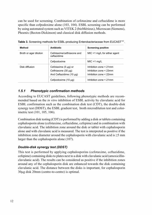
12
can be used for screening. Combination of cefotaxime and ceftazidime is more specific than cefpodoxime alone (103, 104). ESBL screening can be performed by using automated system such as VITEK 2 (bioMérieux), Microscan (Siemens), Phoenix (Becton-Dickinson) and classical disk diffusion methods.
Table 3. Screening methods for ESBL-producing Enterobacteriaceae from EUCAST101.
Method Antibiotic Screening positive
Broth or agar dilution Cefotaxime/ceftriaxone and ceftazidime
MIC >1 mg/L for either agent
Cefpodoxime MIC >1 mg/L
Disk diffusion Cefotaxime (5 µg) orCeftriaxone (30 µg)And Ceftazidime (10 µg)
Inhibition zone < 21mmInhibition zone < 23mmInhibition zone < 22mm
Cefpodoxime (10 µg) Inhibition zone < 21mm
1.5.1 Phenotypic confirmation methodsAccording to EUCAST guidelines, following phenotypic methods are recom-mended based on the in vitro inhibition of ESBL activity by clavulanic acid for ESBL confirmation such as the combination disk test (CDT), the double-disk synergy test (DDST), the ESBL gradient test, broth microdilution test and color-imetric test (101, 105, 106).
Combination disk testing (CDT) is performed by adding a disk or tablets containing cephalosporin alone (cefotaxime, ceftazidime, cefepime) and in combination with clavulanic acid. The inhibition zone around the disk or tablet with cephalosporin alone and with clavulanic acid is measured. The test is interpreted as positive if the inhibition zone diameter around the cephalosporin with clavulanic acid is ≥5 mm larger than the cephalosporin alone (107).
Double-disk synergy test (DDST)This test is performed by applying cephalosporins (cefotaxime, ceftazidime, cefepime) containing disks to plates next to a disk with clavulanic acid (amoxicillin-clavulanic acid). The results can be considered as positive if the inhibition zones around any of the cephalosporin disk are enhanced towards the disk containing clavulanic acid. The distance between the disks is important, for cephalosporin 30µg disk 20mm (centre-to-centre) is optimal.
13
Gradient test methodThe Etest ESBL (bioMérieux) is a commercial kit available to perform ESBL confirmation test. The reading and interpretation can be performed according to manufacturer’s instructions. The test is considered as positive if ≥ 8 fold reduc-tion is found in the MIC of the cephalosporin plus clavulanic acid, compared with the MIC of the cephalosporin alone, or if a deformed ellipse or phantom zone is present. If the strip cannot be read due to growth exceeded to the MIC range of the strip, then the result is considered as indeterminate. The result is considered as negative in all other cases. The gradient test is not reliable for determining the MIC but can be used for confirmation of ESBL only.
Broth microdilutionBroth microdilution can be performed by using Mueller-Hinton broth containing two-fold serial dilution of cefotaxime, ceftazidime and cefepime at concentrations ranging from 0.25 to 512 mg/L with and without clavulanic acid at a concentration of 4 mg/L. Bacterial suspension is added to the each well of the microtitre plate and then incubated at 37°C for 18 to 24 hours. The test is considered as positive if a ≥ 8 fold reduction is found in the MIC of the cephalosporin plus clavulanic acid compared to the cephalosporin alone.
Biochemical (colorimetric) testsIn 2012, the ESBL NDP test was described for the first time. This test was per-formed by using cefotaxime as indicator antimicrobial and tazobactam as inhibitor (108). This test can be performed in 96-well plates or in separate tubes. The test will be considered as positive if the colour changes from red to yellow. It has been reported that the test was performed directly on patient samples (109).
Another, colorimetric test is β-LACTA test which can be carried out by using a chromogenic cephalosporin substrate (HMRZ-86). This test can be performed on isolates and also directly on clinical samples. An excellent sensitivity and specificity for E. coli and K. pneumoniae (96% and 100%, respectively) have been reported by a multicentre study conducted in Belgium and France (110).
1.5.2 Genotypic confirmationPhenotypic confirmation test is not able to identify specific enzymes causing the production of ESBL. Genotypic confirmation is important and can be performed by using polymerase chain reaction (PCR), Eazyplex, ESBL gene sequencing or DNA microarray. Check-Direct ESBL Real-time PCR (Check-Points, Wageningen, The Netherlands) can be used for ESBL detection.
14
1.6 Carbapenemases detection1.6.1 Phenotypic detection methodsMost clinical microbiology laboratories are using established phenotypic methods such as combination disk test, biochemical tests, spectrophotometry, matrix-assisted laser desorption ionization-time of flight mass spectrometry (MALDI-TOF), and lateral flow assays for detection of carbapenemases. Phenotypic detection consists of two steps; screening and confirmation.
Screening of carbapenemasesChromogenic media is used for screening of carbapenemase-producing Entero-bacteriaceae from fecal samples. Chromogenic media contains chromogen that act as substrate for species-specific enzymes and changes colour due to degrada-tion. Species can be identified by the colour of the colonies (111). CHROMagar KPC (bioMérieux, France) is a commercial chromogenic media that can detect VIM and KPC carbapenemases efficiently but OXA-48 with poor sensitivity (112). The highest sensitivity 76% and specificity 75% has been reported for CHROMagar KPC (113, 114). Another chromogenic media, BrillianceTM CRE contains carbapenem and two chromogens can detect carbapenemase-producing K. pneumoniae with higher sensitivity (98%) and specificity (79%) than other members of the Enterobacteriaceae. An overall sensitivity of 92% and specificity of 85% has been reported (111, 113-115). SUPERCARBA is a nonchromogenic media which can detect all class of carbapenemases with higher sensitivity and specificity than CHROMagar KPC. The sensitivity of detection of class A, B and D carbapenemases were 100, 90 and 100% respectively (113). Remel SpectraTM CRE (Thermo Fisher Scientific, Basingstoke, UK) is another type of chromo-genic media having slightly higher sensitivity (97.8%) and specificity (86.4%) than CHROMagar KPC. One day of incubation time is required to obtain result, which is the main disadvantage of these media. In certain strains false negative results can be obtained due to absence of chromogen-specific enzymes (116). In these cases, disk diffusion cut-off for meropenem (<28 mm) and ertapenem (<25 mm) defined by EUCAST for screening of carbapenemaes in clinical isolates can be used (117). Screening of clinical isolates; blood isolates or urine isolates can be performed by disk diffusion method.
Confirmation of carbapenemasesCombination disk testThe commercial version of combination disk test is available from several manu-facturers. MAST, in UK and Rosco, in Denmark were first to introduce the com-mercial combination disk test (118-120). Meropenem and various inhibitors are combined within the disk or tablets. Class A carbapenemases are inhibited by boronic acid, class B carbapenemases are inhibited by dipicolinic acid (DPA)
15
and ethylenediaminetetraacetic acid (EDTA). Avibactam can inhibit OXA-48 carbapenemase (121). Cloxacillin, AmpC β-lactamase inhibitor has also been added to the test. Cloxacillin, can differentiate between AmpC hyperproduction plus porin loss and carbapenemase-production.
Biochemical testsCarba NP and CarbAcineto tests are commonly known as biochemical test having high sensitivity and specificity compared with culture-based methods. Turn-around time is also faster than with the culture-based methods. Nordmann and colleagues developed Carba NP and CarbAcineto tests for the first time in 2012 to identify carbapenemase producers in Enterobacteriaceae, Pseudomonas spp. And A. bau-mannii. The basic principle of these tests is the hydrolysis of imipenem, with a change of colour of a phenol indicator from red to yellow due to acid production from imipenem hydrolysis. High (100%) sensitivity and specificity of Carba NP test has been reported in many studies conducted on Enterobacteriaceae and Pseudomonas spp (122) (123, 124). A commercial version of Carba NP test RAPIDEC® CARBA NP is available (125).
The Blue-Carba test (BCT) is another type of biochemical test for rapid (<2 h) carbapenemase detection. The principle of the test is based on hydrolysis of imi-penem by bacterial colonies which can be detected by changes in pH. Presence of a carbapenemase will change colour of the pH indicator bromothymol blue from blue to green/yellow or from green to yellow (126, 127). An excellent sensitivity for class A and B enzymes but suboptimal sensitivity for OXA-48 enzymes have been reported (128).
Another biochemical test is the β CARBA testTM, which can be performed in < 2h. The test can be carried out by mixing 1 to 3 colonies in the reagents. After 30 min of incubation reading can be carried out. Test results will be considered as positive when colour changes from yellow to orange, red or purple. An excellent performance of β CARBA testTM for detection of CPE has been reported (127).
SpectrophotometrySpectrophotometry is a well-known reference method for identification and dif-ferentiation of carbapenemases in Enterobacteriaceae using imipenem as substrate, but is used to a limited degree in clinical laboratories. Supernatant can be collected from sonication and centrifugation of 24 h-culture cell with imipenem. Absorbance per minute of imipenem and imipenem with bacterial extract were compared. The sensitivity 100% and specificity 98.5% has been reported for this test (129). With exception of class D carbapenemases, this test can detect efficiently other carbapenemases; VIM, IMP and SIM (129).
16
Matrix-assisted laser desorption ionization-time of flight mass spectrometry (MALDI-TOF MS)The MALDI-TOF MS can identify resistance genes, bacterial species and sub- species by using expressed proteins (130, 131). Ionized carbapenem and its deg-radation products can be detected after incubation of carbapenem with extracted bacterial proteins for 1-4 h which formed spectra of the carbapenem and its degradation products. MALDI-TOF MS can detect carbapenemases with 100% sensitivity and specificity (132-134). Incubation time can be reduced by adding sodium dodecyl sulphate (SDS) to the bacterial cell culture before incubation with the meropenem substrate. Using this modification, NDM-1, VIM-1, KPC, OXA-48 and OXA-162 in Enterobacteriaceae were detected with 100% sensitivity and specificity (130).
Lateral flow assaysLateral flow assay, or immunochromatographic assay, has been described for detection of OXA-48 like carbapenemase. In this assay epitopes of OXA-48-like enzymes can be captured immunologically using colloidal gold nanoparticles which is bound to a nitrocellulose membrane within a lateral flow device. In this assay specific capture reagents based on monoclonal anti-OXA-48 antibodies are used to detect OXA-48 like enzymes directly (135). This is a rapid assay and it takes around 4 minutes to perform the assay. This assay can be performed on both clinical isolates and directly on positive blood cultures (136, 137). A recent modi-fication triplex assay has been reported to detect OXA-48, KPC and NDM (138). Recently, one single centre performed a retrospective and prospective evaluation study and has reported highly accurate detection of all five major carbapenemase families (OXA-48, KPC, NDM, VIM, IMP). This study was based on multiplex lateral flow assay in which 296 enterobacterial isolates were examined. Overall, 100% sensitivity and 95.3 (retrospectively) to 100% (prospectively) specificity were reported (139).
1.6.2 Genotypic methodsReal-time PCR can detect and quantify resistance genes simultaneously by using fluorescent probes or dyes. Each gene has unique melting temperature which is used to identify specific resistance mechanisms (140). Multiple primers can be used to detect several genes simultaneously, commonly known as multiplex PCR. In multiplex real-time PCR, amplicon size of the target genes should be differ-ent for easy identification (141). The GeneXpert® (Cepheid, Sunnyvale, CA) is a commercial real-time PCR technique available to detect clinically relevant KPC, IMP, VIM, NDM and OXA-48 carbapenemases. Check-MDR Carba assay (Check-Points) is another commercial real-time multiplex PCR assay to detect KPC, NDM, OXA-48, VIM and IMP from rectal and throat swabs.
17
Eazyplex® SuperBug CRE assay (Amplex Biosystems, GieΒen, Germany) is a commercially available assay that can detect the CTX-M-1-group and CTX-M-9-group of ESBLs and VIM (-1 to -37), NDM (-1 to -7), KPC (-2 to 15) and OXA-48-like (-48, -162, -204 and -244 ) carbapenemases directly from rectal swabs and bacterial culture (142). It is a loop mediated isothermal amplification based assay. One study evaluted this assay directly on urin samples and reported 100% sensitivity and 97.9% specificity (143).
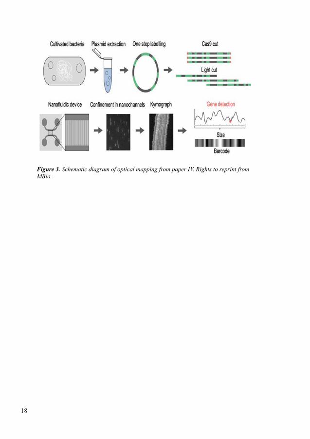
1.7 CRISPR/Cas9 mediated optical DNA mappingOptical mapping is a process of visualizing individual DNA molecules long range sequence information at kbp level. In this process, sequence specific labeled DNA can be imaged after stretching using a fluorescence microscope (Figure 3). Using optical DNA mapping we can study read lengths of DNA, it can be up to 1 Mbp, we can also extract information from single molecules and avoid averaging (144, 145). This method was first described by Schwartz et al in 1993, restriction enzymes were used to cut the DNA molecules at specific sites and the length of different fragments were visualized in an agarose gel (146). After that, a signifi-cant advancement has been observed in the field of optical mapping including different approaches for stretching and labeling were introduced. Most important advancements are stretching of DNA molecules on modified glass surfaces as well as nanofluidic devices (147).
Labeling of DNA molecules can be performed by enzyme based or affinity based techniques. Jo et al (148) introduced nick labeling, in this process a nicking enzyme was used to generate a single stranded break which is known as nick a 4-7 bp long recognition site. Then fluorescently labeled nucleotides were incorporated along the strand using DNA polymerase. DNA molecules were stained using YOYO to determine start and end of each molecule (148). Using CRISPR/Cas9 based approach, a highly adaptable 23 bp recognition site has been reported by McCaffery (149). Another way of labeling DNA is competitive binding that has been reported recently (150, 151). In this process AT specific non-fluorescent molecule netropsin and the fluorescent dye YOYO were used simultaneously. Both molecules will compete for binding site on DNA molecules. If we use accurate concentration, netropsin will bind to AT-site and prevent YOYO to bind to AT-site and YOYO will bind to GC-site. So, GC rich region will be bright and AT rich region will be dark. This process was used to characterize plasmids in the fourth project of this thesis.
18
Figure 3.SchematicdiagramofopticalmappingfrompaperIV.RightstoreprintfromMBio.
19
2 AIMSThe overall aim of this thesis was to investigate the clonality and proportion of ESBL-producers in K. pneumoniae from patients with bloodstream infections, and to study acquisition of EPE in healthy Swedish travellers travelling to high prevalence region of EPE. A second major aim was to evaluate rapid detection and characterization methods for EPE and CPE.
Specific aims
I) To characterize clinical isolates of Klebsiella pneumoniae and investigate the association with phylogroups, virulence factors and mortality during bloodstream infection (BSI) (Paper I)
II) To evaluate a commercial method the RAPIDEC CARBA NP test for detection of carbapenemase-producing Enterobacteriaceae, Pseudomonas aeruginosa and Acinetobacter spp (Paper II)
III) To identify bacterial colonizing characteristics that play key role for travellers to become carriers of ESBL-producing Enterobacteriaceae (EPE) (Paper III)
IV) To characterize risk factors for acquiring EPE in the intestinal microbiota during travel (Paper III)
V) To characterize resistance plasmids during an outbreak with an ESBL-producing strain of K. pneumoniae using optical DNA mapping, and further investigate whether the plasmid was transferred to ESBL-producing E. coli during outbreak (Paper IV)
20
3 MATERIALS AND METHODS3.1 Sample collectionIn Paper I, we studied a total of 139 isolates of K. pneumoniae (blood n=137, or cerebrospinal fluid n=1, or both n=2). All isolates were obtained from 139 adult (≥ 18 years old) patients admitted to Karolinska University Hospital, Solna during the years 2007 to 2009. In Paper II, we studied a total of 276 clinical isolates of Gram-negative bacilli. Detailed strain collection can be found in Table 4. Karolinska University Laboratory (KUL) and Publich Health England’s (PHE) Antimicrobial Resistance and Healthcare Associated Infections (AMRHAI) reference Unit jointly performed this study. In Paper III, we studied a total of 188 healthy adults (≥ 18 years old) Swedish travellers travelling to highly EPE prevalent regions; Southeast Asia, Indian subcontinent, North Africa and the Middle East. We collected rectal swabs and survey data from 188 healthy Swedes travelling to the above-mentioned four high EPE prevalence regions. This study was performed at the Department of Clinical Microbiology, Karolinska University Hospital, Solna, Sweden. Rectal swabs were collected before and after the trip. Travellers, who did not provide both samples were excluded from the study. In Paper IV, we studied fecal samples from 17 neonates during an extended-spectrum β-lactamase (ESBL)-producing Enterobacteriaceae (EPE) outbreak at the NICU at Karolinska University Hospital in Stockholm, Sweden November 2008 and March 2009 which has been published previously (152). During follow-up, we collected fecal samples every second month from 14 surviving neonates from discharge to two years of age. We also conducted a five years’ follow-up on 10 neonates due to drop-outs.
3.2 Ethical considerationsIn this thesis, we used patient and human samples in project I, III and IV. We also used clinical data for analysis. We obtained ethical approval from the Karolinska Institutet Regional Ethics Committee of Stockholm for project I (Paper I) (recordal 2009/1985-31/4) and for project III (Paper III) (recordals 2013/1844-32 and 2015/1620-32). We also obtained ethical approval from regional ethical review board in Stockholm, Sweden (2009/734-31/4 and 2014/491-31/3) for project IV (Paper IV). No ethical approval was required for project II (Paper II) as the study was conducted on clinical strains. Moreover, no clinical data was used for analysis in project II.
The committee approved that we did not need to collect any written or verbal consent for project I from the study subjects as the study was conducted on large number of patients (n=139) and a large number of patients were died before the start of the project. It would not be possible to carry such a study where mortality has been analysed if written or verbal consents were required. Moreover, only
21
Tabl
e 4.
Isol
ates
test
ed b
y ca
rbap
enem
resi
stan
ce m
echa
nism
. Mod
ified
from
pap
er II
, rig
hts
to re
prin
t fro
m J
AC
.
bla K
PCbl
a OXA
-48
bla N
DM
bla V
IMbl
a IM
Pbl
a GES
-5bl
a IM
Ibl
a OXA
-23
bla O
XA-2
4bl
a OXA
-51
bla O
XA-5
8N
on-c
arba
pene
-m
ases
To
tal
Kle
bsie
lla p
neum
onia
e28
1518
11
2083
Kle
bsie
lla o
xyto
ca2
11
12
18
Esch
eric
hia
coli
35
31
6274
Ente
roba
cter
com
plex
33
21
31
120
34
Serr
atia
mar
cesc
ens
12
36
Citr
obac
ter f
reun
dii
21
14
Rao
ulte
lla o
rnith
inol
ytic
a2
2
Rao
ulte
lla p
lant
icol
a2
2
Prov
iden
cia
com
plex
2
2
Prot
eus
mira
bilis
11
Mor
gane
lla m
orga
nii
11
Aci
neto
bact
er b
aum
anni
i1
26
411
115
Pseu
dom
onas
aer
ugin
osa
112
3144
Tota
l 40
2731
169
31
64
11
137
276
1 Hyp
erpr
oduc
tion
of O
XA
-51
was
not
det
ecte
d.
22
limited clinical data were used anonymously in this study. We collected written consent from study subjects for project III. To pursue research project III and IV, most important ethical considerations is emotional impact of study participants. When study participants get to know that they are carrier of antibiotic resistant bacteria some may get psychological reactions and feel worried about being an asymptomatic carrier and which consequences this could have in the future. It is important to provide information to the participants about the implications of being a carrier, and to offer expert consultations to allow the individual to ask questions about fecal carriage and its implications.
3.3 Species identification and susceptibility testing Species were identified by using VITEK2 (bioMérieux, Marcy-I’Etoile, France) in project I & IV and MALDI-TOF Biotyper (Bruker, Billerica, MA, USA) in project III.
In project I and III, antimicrobial susceptibility testing was carried out with the disk diffusion method on Isosensitest agar (Oxoid, Basingstoke, UK). The inter-pretation was performed following the guidelines of Swedish Reference Group for Antibiotics (SRGA) in project I and EUCAST guidelines were followed for interpretation in project III (117) (153). In project II, antimicrobial susceptibility was performed using meropenem and ertapenem disk by disk diffusion method and interpretation was performed following EUCAST guidelines at KUL. At AMRHAI, BSAC agar dilution with AMRHAI’s standard Gram-negative antibiotic panel; ertapenem, imipenem and meropenem was used for susceptibility testing. BSAC breakpoints were used for interpretation.
3.4 ESBL Screening and subtypingESBL- and/or carbapenemase-producing isolates were investigated using Check-MDR (Checkpoints, Wangeningen, The Netherlands) in project I. In project III, screening of EPE was performed using ChromID ESBL agar (bioMérieux, Marcy-I’Etoile, France). Further, broth enrichment was performed using LB broth (Thermo Fisher Scientific, Waltham, USA) for all samples (n=188) and cultivated on an in-house agar on a chromogenic base (Liofilchem, Roseto degli Abruzzi, Italy) supplemented with 200 mg/L cloxacillin and 0.25 mg/L meropenem for an overnight incubation at 35°C. VITEK2 (bioMérieux, Marcy-I’Etoile, France) was also used for ESBL testing. Using Check-MDR (Checkpoints, Wageningen, The Netherlands) we performed ESBL subtyping and Diversilab, an automated rep-PCR system, was used for epidemiological typing.
23
3.5 Phenotypic and molecular detection of carbapenemase
In project II, RAPIDEC® CARBA NP (bioMérieux SA, Marcy l’Etoile, France) was used for phenotypic detection of carbapenemase producers. We followed the manufacturer’s protocol, in brief: isolates were cultivated on Muller Hinton E agar for 18-24 hours. Incubation of RAPIDEC® CARBA NP tests were 30 minutes to two hours at 37°C. Reading was performed by visually comparing colours between test well and control well. Change of colour of the test well from red to yellow or orange was interpreted as positive. In case of positive result, incubation was stopped after 30 minutes otherwise continued to two hours before a negative result was interpreted.
In-house real-time or conventional PCR assays were used for molecular detec-tion of carbapenemases. PCR primers were used to target the following groups of carbapenemases; KPC, NDM, VIM, OXA-23, OXA-24, OXA-48, OXA-58, IMP, GIM, SIM, SPM, GES, IMI and SME. We used antimicrobial susceptibility profile instead of PCR for rarely occurring carbapenemases for all isolates (154-162). False-positive isolates were further investigated using whole-genome sequencing (WGS) using a HiSeq sequencer (Illumina, Little Chesterford, UK). In-house bio-informatics pipeline was used for data analysis. We determined the presence of resistance genes according to previously described methods (163).
3.6 Serotyping and virulence and resistance gene detection
In project I, detection of serotypes and virulence genes were performed using real-time PCR according to previously described methods (164, 165). Community-acquired invasive disease has been associated with six major serotypes; K1, K2, K3, K4, K5, K6 and three virulence genes allS, rmpA, wcaG of K. pneumoniae, which were all investigated (166, 167). Six serotypes and three virulence genes specific primers were used for real-time PCR. In project III, we also investi-gated virulence factors and resistance genes using real-time PCR. PCR primers were specific for fimAMT78, fimH, papAH, papC, papEF, and papG allele I, II, III and armA, rmtA, rmtB, rmtC, rmtD, qnrA, qnrB, qnrC, qnrD, qnrS. We also investigated the presence of plasmid-mediated fluoroquinolones resistance gene aminoglycoside-(6)-N-acetyltransferase (aac(6’)-Ib) and its point mutant aac(6’)-Ib-cr using real-time PCR following previously published protocol (168, 169). We also investigated plasmid-mediated colistin resistance gene mcr-1 against all isolates following previously published protocol (93). One isolate that carried the plasmid-mediated colistin resistance gene mcr-1 was further analysed using Next-generation sequencing technology the Ion Torrent S5 XL system (Themo
24
Fisher Scientific, Waltham, MA, USA). The presence of plasmid-mediated colistin resistance gene mcr-1 was confirmed by analyzing with ResFinder (170). We also confirmed the ST for this isolate following previously described method (171). Plasmid type was confirmed by using PlasmidFinder (172). All isolates of E. coli in project III were subjected to phylogrouping using real-time PCR following previously described protocol (29).
3.7 Phylogenetic analysisIn project I, we performed seven house-keeping genes based MLST for phylo-genetic analysis of all isolates according to previously published methods (166, 173). Seven house-keeping genes were amplified and sequenced using primers and protocol, can be found on the web site (htt://bigsdb.web.pastur.fr). We deter-mined nucleotide sequences for seven house-keeping genes by using ABI Prism 3100 Genetic Analyzer (Applied Biosystems, Foster City, CA). We obtained allelic and sequence type numbers by direct comparison with the K. pneumoniae MLST database (htt://bigsdb.web.pastur.fr). In case of new allele and ST, numbers were assigned by curator of the K. pneumoniae MLST database. All sequences were aligned by using Mega V5 (174). We identified phylogroups using Neighbor-joining trees based on concatenate sequences of all seven MLST loci for all isolates. We used three reference strains; ATCC13883 for phylogroup KpI: K. pneumoniae, SB59 for phylogroup KpII: K. quasipneumoniae and Kp342:ST146 for phylogroup KpIII: K. variicola to identify phylogroups (175). We confirmed phylogroups separately by using Neighbor-joining trees based on each allele. We also used Splits Tree v4 (176) for NeighborNet analysis to confirm phylogroups and recombination events. Nucleotide diversity of each gene for each phylogroup and for all isolates were calculated by using DnaSP (177). With the help of BioNumerics v7.0 (Applied Maths, Sint Maartens-Latem, Belgium), we produced minimal spanning tree (MST) (178). Isolates sharing six loci of their allelic profiles with one another member of the group were defined as clonal complexes. We identified clonal complexes by using eBURST V3 (179).
3.8 Clinical dataIn project I, we retrieved medical data for finding association between phylo-groups, 30-day mortality, patient risk factors, hospital versus community-acquired infections and antibiotic treatment. Assessment of comorbidity was performed by constructing Charlson comorbidity index (180). In case of more than one species were identified from specimens obtained within 24 hours from K. pneumoniae bloodstream infection were considered as polymicrobial infection. If the patient had been to hospital more than 30 days or if the sample was taken >48h after admit-tance, the episode was defined as hospital-acquired. In project III, study subjects
25
returned a filled questionnaire based on personal information and potential risk factors such as underlying chronic diseases, duration of travel, antibiotic treat-ment during travel, getting treatment from local healthcare before and after travel.
3.9 Plasmid characterizationIn paper IV, we conducted bacterial plasmid characterization based on optical DNA mapping (Figure 3). We utilized the emission intensity variation along the DNA by adding fluorescent YOYO-1 and the non-fluorescent AT-selective netropsin, where AT-rich regions are dark and GC-rich region are bright. Visualization of the variation of emission intensity, the DNA barcode can be retrieved by stretch-ing single intact plasmids in nanochannels and imaging them using fluorescence microscopy. Previously, it has been demonstrated that the method can measure the number and size of all large plasmids in a bacterial cell. It has also been dem-onstrated that it can produce a unique barcode for each plasmid which can later be used to identify conjugation of plasmids between different strains and species of bacteria. Latest improvement of the assay was done by adding the CRISPR/Cas9 system, now the method is able to demonstrate a specific (resistance) gene har-bouring plasmid. Plasmids were extracted from 100 ml overnight bacterial cultures in LB broth by using the Macherey-Nagel NucleoBond Xtra Midi preparation kit according to the manufacturer’s instructions. We precipitated eluted plasmid DNA into iso propanol and resuspended it in 1 × TE buffer. Then, we treated plasmid with Cas9 and guide-RNA (gRNA) targeting blaCTX-M group 1 gene according to our previously published protocol (181). To obtain DNA barcode, we stained plasmid and λ-DNA (New England Biolabs) at equal concentration with YOYO-1 (Invitrogen) at a molecular ratio of 1:2 (YOYO-1:bp) and with netropsin (Sigma-Aldrich) at a molar ratio of 100:1(netropsin:YOYO-1). Here, we used λ-DNA for internal size marker (48 500 bp). To obtain uniform staining, we incubated plasmid DNA (2μM) with YOYO-1 and netropsin in 10 μl of 0.5X TBE at 50°C for 30-60 min (182). We used Millipore water to dilute samples in order to obtain a final concentration of 0.05X TBE and 0.2 μM (bp) of DNA. We added 2.5% (v/v) β-mercaptoethanol (BME) to the solution to supress photo nicking and photo bleaching of DNA molecules. We fabricated nanofluidic channels in oxidized silicon, according to previously described methods (183). DNA molecules were stretched through nanochannels of dimensions 100 nm x 150 nm2 and a length of 500 μm. We cleaned nanochannels with 5% w/w sodium hypochlorite solution and then washed a few times with loading buffer (0.05x TBE) before loading samples. This process enhanced the usage of nanofluidic chips more than 50 times. On average we used chips in each section at least 10 times. We placed 15μl of DNA solution to the reservoir and we used nitrogen gas to force DNA molecules into nanochannels. To image DNA molecules we used an inverted microscope (Zeiss AxioObserver.Z1) equipped with either a 63x (with 1.6 times optovar) or 100x
26
oil immersion objective (Zeiss, NA=1.46) and an EMCCD camera (Photometrics Evolve or Andor iXon). We imaged both circular or linear molecules for 200 frames with 100ms exposure by stretching into the nanochannels.
We analyzed data to identify the number of plasmids in each sample, their cor-responding size, their sequence-specific barcode and possible location of a resist-ance gene (blaCTX-M group 1). In brief, we used the internal size marker λ-DNA to calculate the plasmid size. We also used conversion factor 1.8 to estimate whether directly from linearized plasmids, or from plasmids in their circular form (184). We compared linear fragments of similar size (+/- 10%), acquired either by Cas9 linearization, or via extensive light exposure (185). High degree of similar individual barcodes (typically Pearson Cross Correlation (CC) > 0.75) were merged into a consensus barcode to compare between different isolates, rendering a p-value (186). We studied the position of the double strand-breaks of the individual plasmid bar-codes and the consensus barcode to detect the presence of a resistance gene (181).
Qiagen Genomic-tip 100 kit was used to extract complete genomic DNA (chromo-somal DNA and plasmids) according to the manufacturer’s instructions. Long read sequencing was performed using PacBio Sequencing technologies in collaboration with Science for Life Laboratory in Uppsala on a RSII system with one SMRT cell per genome. Sequencing library was prepared using Illumina MiSeq sequencing with two times 300-bp paired-end read lengths and Nextera XT DNA Library preparation kit (Illumina) following manufacturer’s instructions. Overall generated chromosomal coverage was >25-fold for all isolates. The CLC Genomics work-bench v.10 (CLC Bio, Aarhus, Denmark) including the CLC Microbial Genomics Module 3.0 was used to perform de novo assembly for each data set. Illumina reads were assembled using contig sequences from the PacBio sequencing as references. The resulting contigs were analyzed by mate-pair analysis and circularized by detection of redundant end-sequences. De novo assembly of non-matched reads from combined reference assemblies for each dataset was performed to verify that no plasmids were missed between isolates. Remaining contigs were analyzed for gene content by using BLASTn searches. Complete genome annotation was per-formed by using the NCBI Prokaryotic Genome Annotation Pipeline. ResFinder, PlasmidFinder, and VirulenceFinder, were used to detect resistance genes, plasmid replicons and virulence genes respectively (170, 172, 187).
27
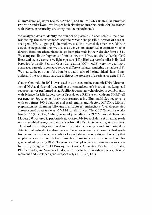
4 RESULTS
4.1 Paper IThe aim of this study was to determine association between phylogroups, virulence factors and mortality in clinical isolates of K. pneumoniae causing BSI. Three dis-tinct phylogroups were disclosed by phylogenetic analysis by multilocus sequence typing (MLST). The most predominant phylogroup was phylogroup KpI (corre-sponding to K. pneumoniae n = 96) followed by phylogroup KpIII (corresponding to K. variicola n = 34) and phylogroup KpII (corresponding to K. quasipneumo-niae n = 9) (Figure 4). The highest mean genetic distance among phylogroups was 4.5% (KpII-KpIII), followed by 4.4% (KpI-III) and 4.0% (KpI-KpII). Thus, we observed that phylogroups KpI-KpII are more closely related compared to phylo-group KpIII. Neighbor-joining tree analysis also supported three phylogroups and suggested a high level of recombination within each group, as well as between the phylogroups. We also observed minor events of insertion/deletion (INDEL) in the tonB locus by aligning seven housekeeping genes. Considering all isolates, highest polymorphic sites were observed in tonB 19.6% and lowest polymorphic sites were observed in gapA 5.1% (Table 5) whereas only 10.7% polymorphic sites were observed in concatenate sequence. Average nucleotide diversity Π of 2.1% was observed for the whole population, whereas Π was much lower within the phylogroups; KpII (1.3%), KpI (0.37%) and KpIII (0.63%). The ratio of the number of non-synonymous substitution per non-synonymous site (Ka) to the number of synonymous substitution per synonymous site (Ks) was observed to be less than one, which indicated that housekeeping genes were evolved predominantly under purifying selection. An extensive haplotypic diversity with 116 distinct STs was observed by the MLST based population analysis. The clonal relatedness among sequence types (STs) were analysed using both MLST and eBURST method. A total of 48 isolates comprised 12 clonal complexes and remaining 91 isolates split into 79 singletons (STs that differed by two or more alleles from another ST).
Only 18 strains of 139 isolates were serotypable for tested capsular serotypes; K1, (1.4%; 2/139), K2 (5.0%; 7/139), K20 (1.4%; 2/139), K54 (2.2%; 3/139) and K57 (2.9%; 4/139). The predominant serotype was K2, whereas K5 was not found in any isolates. A total of 16 out of 18 capsular types were associated with phylogroup KpI and the remaining 2 were associated with KpIII. In this study virulence gene wcaG 8.6% (12/139) was the most frequently detected virulence gene followed by allS 4.3% (6/139) and rmpA 3.6% (5/139). A total of 8 (5.8% 8/139) isolates exhibited mucoid phenotype and among them two isolates were also positive for rmpA. The distribution of isolates harboring virulence genes were KpI (n=13), KpIII (n=5) and KpII (n=3).
28
A total of 17.3% (24/139) with 30-day mortality was observed. Isolates belonging to phylogroup KpIII were highly associated with the highest 30-day mortality of 29.4 % (10/34 cases), whereas isolates belonging to phylogroup KpI and KpII were associated with 30-day mortality of 13.5% (13/96 cases,) and 11.1% (1/9 cases), respectively. Out of the 24 events associated with 30-days mortality, only one was serotypable (K54) and one exhibited mucoid phenotype and/or (wcaG n=1 and wcaG/allS n=1). We observed low level of antibiotic resistance in general but the highest resistance rate 10.8% was observed against trimethoprim/sulfametoxazole followed by ciprofloxacin 8.6%, piperacillin/tazobactam 5.8%, gentamicin 3.6%, ceftazidime 4.3% and cefotaxime 3.6%. Isolates of KpI were more resistant against trimethoprim/sulfametoxazole compared to KpIII isolates. K. variicola (KpIII) isolates were found to be less resistant than isolates of K. pneumoniae (KpI) and K. quasipneumoniae (KpII) for several classes of antibiotics.
Figure 4. Phylogenetic analysis based on Neighbor-joining trees (A), Minimal spanning tree (MST) (B), and NeighborNet analysis (C). Sequence type is represented by each circle. Black lines between STs indicate that they differ in one allele (thick lines), two and three alleles(thin),orfourtosevenalleles(dashed).RightstoreprintfromPLoSOne(PaperI).
29
Table 5. Nucleotide diversity within seven housekeeping genes. N = number of samples subjected for analysis. Sites = Length of sequence after alignments. Net Site = Length of sequence subjected for analysis. S = polymorphic sites. Hap = haplotypes. Hd = haplotypic diversity. Π = nucleotide diversity. θ = average number of nucleotide difference per site. Ka/Ks = ratio of the number of non-synonymous substitution per non-synonymous site (Ka) to the number of synonymous substitution per synonymous site (Ks). Rights to reprint from PLoS One (Paper I).
Alleles n Sites NetSites S Hap Hd Π θ Ka/Ks
gapA 139 450 450 23 18 0.692 0.00819 0.009683 0.0995
infB 139 318 318 28 19 0.756 0.01522 0.017127 0
mdH 139 477 477 59 26 0.805 0.02797 0.023598 0.0324
pgi 139 432 432 54 28 0.693 0.02405 0.023955 0.0144
phoE 139 420 420 41 41 0.939 0.01401 0.020316 0.0064
rpoB 139 501 501 41 27 0.815 0.01447 0.015582 0.023
tonB 139 438 402 79 47 0.952 0.04667 0.040646 0.0693
Concatenate, Total 139 strains
139 3 036 3 000 325 115 0.996 0.02138 0.021363 0.0344
Concatenate, KpI
96 3 024 3 006 115 75 0.993 0.00371 0.007448 0.0778
Concatenate, KpII
9 3 018 3 012 105 9 1 0.0132 0.012949 0.02
Concatenate, KpIII
34 3 018 3 006 155 31 0.995 0.00637 0.012936 0.0125
4.2 Paper IIWe have evaluated a commercial version of RAPIDEC® CARBA NP against a total 276 Gram-negative bacilli consisting of (n=138) carbapenemase producers and (n=138) non-carbapenemase producers. A total of 135 isolates (97.8%) were correctly identified as carbapenemases producers using RAPIDEC® CARBA NP assay. One OXA-48-producing K. pneumoniae, one OXA-23 and one OXA-24-producing Acinetobacter baumannii were not detected in RAPIDEC® CARBA NP assay. Similarly, RAPIDEC® CARBA NP assay confirmed 135 (97.8%) nega-tive isolates those were previously carbapenemases negative. One Pseudomonas aeruginosa with OprD loss and efflux, one ceftazidimase-producing Klebsiella oxytoca and one Enterobacter cloacae with impermeability were false positive in RAPIDEC® CARBA NP assay. Whole genome sequencing (WGS) was used to further analyse the three false positive isolates and known carbapenemases were absent in P. aeruginosa and E. cloacae. However, WGS analysis confirmed the
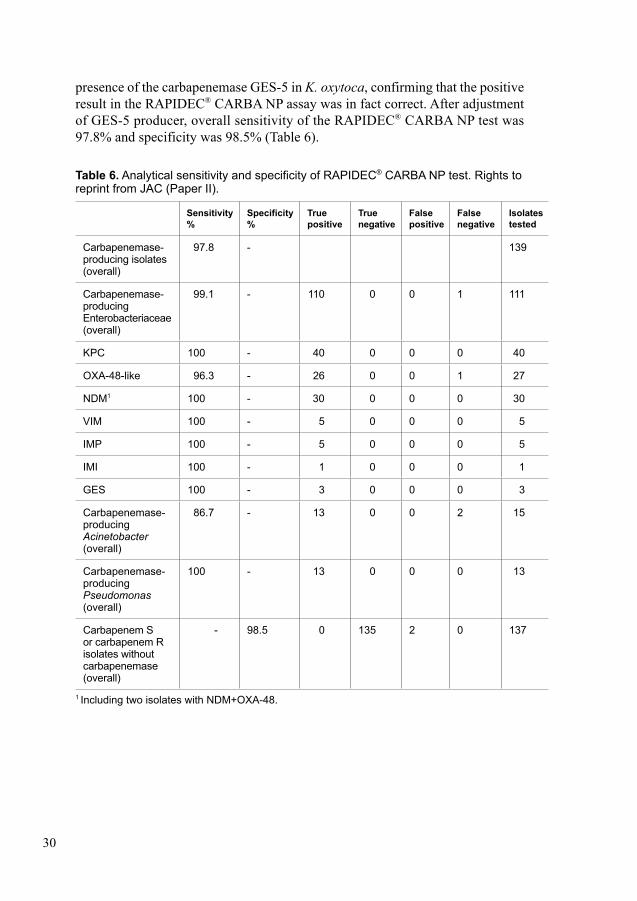
30
presence of the carbapenemase GES-5 in K. oxytoca, confirming that the positive result in the RAPIDEC® CARBA NP assay was in fact correct. After adjustment of GES-5 producer, overall sensitivity of the RAPIDEC® CARBA NP test was 97.8% and specificity was 98.5% (Table 6).
Table 6. Analytical sensitivity and specificity of RAPIDEC® CARBA NP test. Rights to reprint from JAC (Paper II).
Sensitivity %
Specificity %
True positive
True negative
False positive
False negative
Isolates tested
Carbapenemase-producing isolates (overall)
97.8 - 139
Carbapenemase-producing Enterobacteriaceae (overall)
99.1 - 110 0 0 1 111
KPC 100 - 40 0 0 0 40
OXA-48-like 96.3 - 26 0 0 1 27
NDM1 100 - 30 0 0 0 30
VIM 100 - 5 0 0 0 5
IMP 100 - 5 0 0 0 5
IMI 100 - 1 0 0 0 1
GES 100 - 3 0 0 0 3
Carbapenemase-producing Acinetobacter (overall)
86.7 - 13 0 0 2 15
Carbapenemase-producing Pseudomonas (overall)
100 - 13 0 0 0 13
Carbapenem S or carbapenem R isolates without carbapenemase (overall)
- 98.5 0 135 2 0 137
1 Including two isolates with NDM+OXA-48.
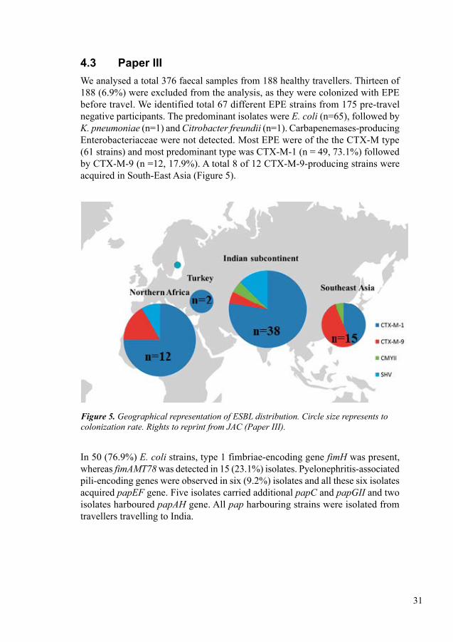
31
4.3 Paper IIIWe analysed a total 376 faecal samples from 188 healthy travellers. Thirteen of 188 (6.9%) were excluded from the analysis, as they were colonized with EPE before travel. We identified total 67 different EPE strains from 175 pre-travel negative participants. The predominant isolates were E. coli (n=65), followed by K. pneumoniae (n=1) and Citrobacter freundii (n=1). Carbapenemases-producing Enterobacteriaceae were not detected. Most EPE were of the the CTX-M type (61 strains) and most predominant type was CTX-M-1 (n = 49, 73.1%) followed by CTX-M-9 (n =12, 17.9%). A total 8 of 12 CTX-M-9-producing strains were acquired in South-East Asia (Figure 5).
Figure 5. Geographical representation of ESBL distribution. Circle size represents to colonizationrate.RightstoreprintfromJAC(PaperIII).
In 50 (76.9%) E. coli strains, type 1 fimbriae-encoding gene fimH was present, whereas fimAMT78 was detected in 15 (23.1%) isolates. Pyelonephritis-associated pili-encoding genes were observed in six (9.2%) isolates and all these six isolates acquired papEF gene. Five isolates carried additional papC and papGII and two isolates harboured papAH gene. All pap harbouring strains were isolated from travellers travelling to India.
32
Phylogroup A (28/65, 43.1%) was the most commonly identified phylogroup, followed by phylogroup D (17/65, 26.2%), phylogroup B1 (12/65, 18.5%) and phylogroup B2 (8/65, 12.3%). Phylogroups B2 and D isolates were more com-monly associated with multi-drug resistant (MDR) than the isolates of phylo-groups A and B1. All isolates were identified as cefotaxime-resistant and 92.5% (62/67) isolates were resistant to ceftazidime. In other antibiotic classes, highest resistance 58.2% (39/67) was observed against trimethoprim/sulfamethoxazole, followed by ciprofloxacin 40.3% (27/67), gentamicin 29.9% (20/67), piperacillin/tazobactam 22.4% (15/67), amikacin 7.5% (5/67) and ertapenem 6.0% (4/67). A total 49.3% (33/67) isolates were detected as MDR strains. A total 46.3% (31/67) isolates harboured plasmid-mediated quinolone and fluoroquinolone resistance genes [qnr and/or aac(6’)-Ib] and among them most commonly detected gene was qnrS, found in 20 isolates followed by aac(6’)-Ib in 10 isolates, qnrB in 4 isolates and qepA in 1 strain. Ten out of 24 qnr harbouring isolates were resistant against ciprofloxacin. Two isolates harbouring 16S methylase gene rmtB were resistant to both gentamicin and amikacin. A CTX-M-55-producing E. coli isolate harbouring plasmid-mediated colistin resistance gene mcr-1 was identified for the first time in Sweden from one traveler had travelled to Thailand in late 2013. The strain had a colistin MIC of 4 mg/L. ResFinder analysis also identified additional two aminoglycoside resistance genes [aadA1A2 and aac(3)-IId], one blaTEM-1B and one fluoroquinolone resistance gene (qnrS1) in this isolate.
EPE colonization rates differed with different geographical region. Highest EPE colonization was observed in Indian subcontinent (49%) followed by northern Africa (44%) and lowest EPE colonization was observed in Turkey (10%) followed by Southeast Asia (19%). In agreement with previous studies, we found that diarrhea and antibiotic treatment during the trip were independent risk factors for EPE acquisition (OR 5.92), (65, 67, 68). EPE colonization was negatively associated with chronic diseases (OR 0.27). Sex, age, duration of trip, intake of proton-pump inhibitors, or use of oral cholera vaccine had no significant association with EPE colonization. The distribution of phylogroups differed between geographical regions. Strains belonging to phylogroup B2 (n=8) were only identified from travellers to Southeast Asia and the Indian subcontinent.
4.4 Paper IVInitially, 17 neonates were colonized with blaCTX-M-15 -producing K. pneumoniae (ESBL-KP), sequence type ST101. After storage, only 16 strains were viable and included in the molecular analysis. Optical mapping results of 16 ESBL-KP ST101 isolates confirmed that each isolate contained two different plasmids with differ-ent sizes. The sizes of the plasmids were approximately 80 kbp and 162–222 kbp.
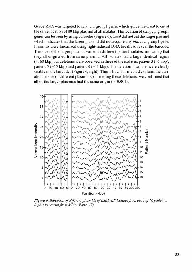
33
Guide RNA was targeted to blaCTX-M- group1 genes which guide the Cas9 to cut at the same location of 80 kbp plasmid of all isolates. The location of blaCTX-M- group1
genes can be seen by using barcodes (Figure 6). Cas9 did not cut the larger plasmid which indicates that the larger plasmid did not acquire any blaCTX-M- group1 gene. Plasmids were linearized using light-induced DNA breaks to reveal the barcode. The size of the larger plasmid varied in different patient isolates, indicating that they all originated from same plasmid. All isolates had a large identical region (~160 kbp) but deletions were observed in three of the isolates; patient 3 (~5 kbp), patient 5 (~55 kbp) and patient 8 (~31 kbp). The deletion locations were clearly visible in the barcodes (Figure 6, right). This is how this method explains the vari-ation in size of different plasmid. Considering these deletions, we confirmed that all of the larger plasmids had the same origin (p<0.001).
Figure 6. Barcodes of different plasmids of ESBL-KP isolates from each of 16 patients. RightstoreprintfromMBio(PaperIV).
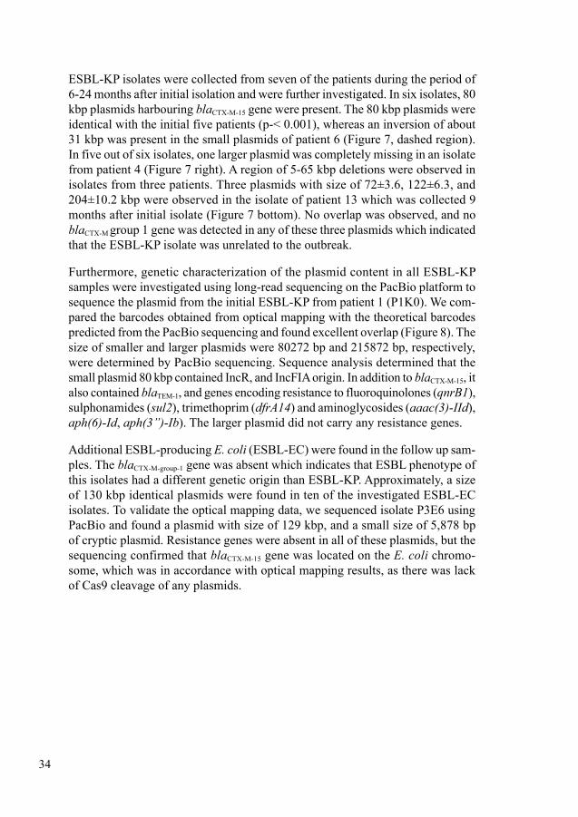
34
ESBL-KP isolates were collected from seven of the patients during the period of 6-24 months after initial isolation and were further investigated. In six isolates, 80 kbp plasmids harbouring blaCTX-M-15 gene were present. The 80 kbp plasmids were identical with the initial five patients (p-< 0.001), whereas an inversion of about 31 kbp was present in the small plasmids of patient 6 (Figure 7, dashed region). In five out of six isolates, one larger plasmid was completely missing in an isolate from patient 4 (Figure 7 right). A region of 5-65 kbp deletions were observed in isolates from three patients. Three plasmids with size of 72±3.6, 122±6.3, and 204±10.2 kbp were observed in the isolate of patient 13 which was collected 9 months after initial isolate (Figure 7 bottom). No overlap was observed, and no blaCTX-M group 1 gene was detected in any of these three plasmids which indicated that the ESBL-KP isolate was unrelated to the outbreak.
Furthermore, genetic characterization of the plasmid content in all ESBL-KP samples were investigated using long-read sequencing on the PacBio platform to sequence the plasmid from the initial ESBL-KP from patient 1 (P1K0). We com-pared the barcodes obtained from optical mapping with the theoretical barcodes predicted from the PacBio sequencing and found excellent overlap (Figure 8). The size of smaller and larger plasmids were 80272 bp and 215872 bp, respectively, were determined by PacBio sequencing. Sequence analysis determined that the small plasmid 80 kbp contained IncR, and IncFIA origin. In addition to blaCTX-M-15, it also contained blaTEM-1, and genes encoding resistance to fluoroquinolones (qnrB1), sulphonamides (sul2), trimethoprim (dfrA14) and aminoglycosides (aaac(3)-IId), aph(6)-Id, aph(3’’)-Ib). The larger plasmid did not carry any resistance genes.
Additional ESBL-producing E. coli (ESBL-EC) were found in the follow up sam-ples. The blaCTX-M-group-1 gene was absent which indicates that ESBL phenotype of this isolates had a different genetic origin than ESBL-KP. Approximately, a size of 130 kbp identical plasmids were found in ten of the investigated ESBL-EC isolates. To validate the optical mapping data, we sequenced isolate P3E6 using PacBio and found a plasmid with size of 129 kbp, and a small size of 5,878 bp of cryptic plasmid. Resistance genes were absent in all of these plasmids, but the sequencing confirmed that blaCTX-M-15 gene was located on the E. coli chromo-some, which was in accordance with optical mapping results, as there was lack of Cas9 cleavage of any plasmids.
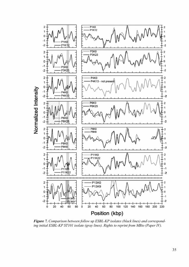
35
Figure 7. Comparison between follow up ESBL-KP isolates (black lines) and correspond-inginitialESBL-KPST101isolate(graylines).RightstoreprintfromMBio(PaperIV).
36
Figure 8. Comparison between experimental optical map (gray) and theoretical optical mapcreatedfromPacBiosequencing(black)forthetwoplasmidsinP1K0.Rightstoreprint from MBio (Paper IV).
37
5 DISCUSSION5.1 Major Findings In the first project, we wanted to analyze clonal diversity and the prevalence of EPE and CPE in invasive isolates of K. pneumoniae isolated from BSI patients in a well-defined geographical area and we collected all samples from the depart-ment of clinical microbiology at Karolinska University Hospital, Stockholm. We observed very low prevalence of antimicrobial resistance among Swedish bloodstream infection (BSI) patients. Only five out of 139 isolates were detected as MDR (resistant to three or more than three antibiotic classes). All of these five isolates were EPE (two isolates CTX-M-1, one isolate CTX-M-1 and SHV, one isolate SHV) but only one isolate was a CPE (carbapenemase VIM), CTX-M-1 and CMYII. Then, we wanted to characterize Klebsiella pneumoniae sensu lato isolated from BSI patients to analyze their population structure. We also wanted to investigate potential linking between phylogroups, bacterial traits, mortality and comorbidity. We observed three distinct phylogroups, the largest phylo group was KpI consisting of (n=96) isolates of K. pneumoniae followed by phylo group KpIII consisting of (n=34) isolates of K. variicola and phylogroup KpII consisting of (n=9) isolates of K. quasipneumoniae. We also observed a high link between 30-day mortality and K. variicola (KpIII) (29.4%) isolates compared to K. pneu-moniae (KpI) (13.5%) isolates. Although, K. variicola (KpIII) isolates had low virulence and resistance profile compared to K. pneumoniae. We observed several comorbidities; malignancy, diabetes mellitus, cirrhosis, biliary tract disorders and alcoholism among patients, which may have influenced the mortality rate.
Secondly, we wanted to investigate the acquisition of EPE and CPE among healthy people those were travelling to high prevalence of EPE and CPE region. We included 188 healthy adult Swedish tourists with planned travel to high EPE and CPE prevalent regions; Southeast Asia, Indian subcontinent, North Africa and the Middle East. We excluded 13 travellers from the analysis who were colonized with EPE before travelling. We found 67 out of 175 healthy Swedish tourists were colonized with different EPE strains; E. coli (n=65), K. pneumoniae (n=1) and Citrobacter freundii (n=1). We also observed that healthy Swedish tourist were not colonized with any carbapenemase producers. The most prevalent EPE type was CTX-M type detected in 61 isolates; 49 isolates were CTX-M-1 and 12 isolates were CTX-M-9. We also observed that Swedish healthy tourists were colonized with low virulence EPE isolates. Only six isolates acquired pyelonephritis-associated pili-encoding genes (pap) genes and travellers were colonized with these six isolates from India. Recently emerging colistin resistance gene mcr-1 harboring E. coli was colonized with a traveler travelling to Thailand. We observed that only 8 out of 65 isolates were belonged to MDR associated phylogroups B2 and all of these 8 isolates were isolated from the travellers had travelled to South-East Asia
38
(n=2) and Indian subcontinent (n=6). As expected, highest colonization rates was observed in Indian subcontinent (49%) and northern Africa (44%) whereas lower rates was observed in Southeast Asia (19%) and Turkey (10%). We also observed that diarrhea and antibiotic treatment during the trip were associated with EPE colonization, but chronic diseases were negatively associated with EPE coloni-zation. We did not find any significant association between sex, age, duration of trip, intake of proton-pump inhibitors, or use of oral cholera vaccine with EPE colonization.
Thirdly, we wanted to evaluate a rapid detection and characterization methods for CPE and EPE due to the urgent need of rapid, simple and reliable phenotypic detection method for low income settings. In low income settings, prevalence of EPE and CPE is high and can be migrated to low prevalence EPE and CPE area like Nordic countries (Showed in travellers study). We evaluated a commercial version of RAPIDEC® CARBA NP, colorimetric liquid-based assay for rapid detection of carbapenemases. The in-house Carba NP assay can be setup but will be challenging for routine laboratories due to high instability of carbapenems. To our knowledge, we reported for the first time two-centre evaluation of RAPIDEC® CARBA NP assay against 276 Gram-negative bacilli. We observed that RAPIDEC® CARBA NP assay can detect carbapenemases in less than 2.5 hours with high sensitivity (97.8%) and specificity (98.5%) without requiring any specialized equipment.
Finally, we evaluated a novel method based on optical DNA mapping assisted with CRISPR/Cas9 to characterize plasmids during EPE outbreak. This method should be equally useful to trace CPE outbreak, although this was not investigated in this paper. The basic of this method is, CTX-M group 1 gene was targeted by guide RNA to guide Cas9 to cut the same location of 80 kbp plasmid of all iso-lates. Using barcodes, location of CTX-M-1 gene (targeted gene) can be seen. We reported that this method successfully can detect different size of plasmids; small size 80 kbp and larger plasmid (162-222) kbp. We also reported that this method can track the changes of plasmid over time. We reported deletions in three plas-mids of three isolates with different size (~5 kbp), (~55 kbp) and (~31 kbp) and inversion of 31 kbp in another isolate.
Despite not being part of the aims of the thesis, it is worth mentioning that rapid antimicrobial susceptibility testing (AST) is urgently needed. Although, it was not the goal of this thesis but to facilitate treatment decision and to prevent spread-ing of antimicrobial/antibiotic resistance resulting from abuse (misuse/overuse) of antibiotics, there is a major demand for development of rapid AST. AST used in this thesis (currently available techniques) require 16-18 hours to read out the results, but with novel methods this could drop to 30 minutes. This will be dis-cussed in detail in the future perspectives section of this thesis.
39
5.2 Phylogroups of K. pneumoniaeOver the years, phylogenetic analysis of K. pneumoniae has resulted in the division into several phylogroups. Phylogroups KpI to KpIV were first reported in the year 2001 (175). Then, two more phylogroups KpV and KpVI were reported in 2017 (188). Another phylogroup KpVII has been reported recently (189). Phylogroups KpI to KpIV and KpVI have taxonomic status. Phylogroups KpI to KpIV and KpVI represent different taxa K. pneumoniae (sensu stricto), K. quasipneumoniae subsp. quasipneumoniae, K. variicola and K. quasipneumoniae subsp. simili-pneumoniae respectively. Phylogroups KpV and KpVII have no taxonomic status but Brisse and colleagues have recently proposed the names K. variicola subsp. tropicalensis for KpV and K. africanensis for KpVII (189). In this thesis, we tested three phylogroups; KpI, KpII and KpIII in project I. Phylogroups KpIV-KpVII were not tested. In fact, KpV and KpVII were not reported during the time we studied project I. We were able to identify all three phylogroups known at the time; KpI (69%), KpII (6%) and KpIII (24%). To our knowledge, this study was the first to find association between phylogroups, virulence factors and mortality data in a well-defined geographical area. In the year 2004, a study based on (n=420) isolates from 26 European hospitals also reported three phylogroups; KpI (82.1%), KpII (6.9%) and KpIII (11%) (190).
5.3 Misidentification of K. variicola and K. quasipneumoniae
The conventional clinical microbiology laboratory techniques are unable to distinguish K. pneumoniae (KpI), K. variicola (KpIII) and K. quasipneumoniae (KpII) due to their closely overlapping biochemical properties (191). K. pneumoniae is adonitol positive but K. variicola and K. quasipneumoniae are adonitol negative. Adonitol fermentation and indole positivity are considered as the classifier but the phenotype is unstable and not consistent among all the isolates of these three species (192). The correct bacterial species identification using molecular diagnostics mainly depend on the availability of the databases. The database of common molecular diagnostics platforms; Verigene, Vitek or MALDI-TOF MS did not include K. variicola and K. quasipneumoniae until recently so either they were not identified or misidentified (193). Previously it has been reported that 2.5% to 10% of K. pneumoniae were misidentified and should have been reported as K. variicola (194-197). The current Bruker MALDI-TOF MS micro-organism database included K. variicola in the year 2015 but still K. quasipneumoniae was not included, causing continuation of misidentifications (193, 194). Previous studies have showed overrepresentation of phylogroup KpI (K. pneumoniae) but in this study phylogroup KpIII (K. variicola) was also relatively common (190, 198). Previously, it has been reported that K. variicola causing bloodstream infection was incorrectly identified as K. pneumoniae using classical and automated identifications but later, correctly identified as K. variicola using next-generation sequencing (199).
40
5.4 Association of phylogroups, bacterial factors, mortality and comorbidity
In the first project of this thesis, to our knowledge we are the first to discuss the possible association with phylogroups, bacterial factors (virulence factors, sero-types), mortality and comorbidity. In accordance with previous studies we observed 17.3% overall 30-day mortality on BSI caused by K. pneumoniae (200, 201). High mortality ranges of 20% to 62% has been observed in Klebsiella bacteremia, reported previously (202). A recent study showed that the 30-day mortality rate was 40.7% in classical K. pneumoniae bacteremia compared with 37.1% hyperviru-lent K. pneumoniae bacteremia (203). One explanation could be that less virulent strains were acquired by severe sick patients and patients died due to underlying diseases. In our study, we also observed that a total of 10 isolates of K. variicola (Phylogroup KpIII), 13 isolates of K. pneumoniae (Phylogroup KpI) and one iso-late of K. quasipneumoniae (Phylogroup KpII) were linked with 30-day mortality. Among them, only 3 isolates out of 24 cases of 30-day mortality were associated with investigated virulence factors; K54 (n=1), mucoid phenotype (n=1) and/or virulence genes (wcaG n=1 and wcaG/allS n=1). According to our knowledge, we reported for the first time that K. variicola bacteremia in adults were associ-ated with higher 30-day mortality than bacteremia caused by K. pneumoniae. A recent study based on an outbreak of multidrug-resistant (MDR) K. variicola in a neonatal unit of Dhaka Medical College at Dhaka, Bangladesh in the year 2016 to 2017 reported that K. variicola is also highly associated with mortality (54.5%) due to BSI in neonates (204). In our study we observed a significant (P=0.037) dif-ference in mortality among KpI and KpIII that has been shown both in univariate analysis and in multivariate analysis after adjustment of age, gender, time to get antibiotic treatment, comorbidity and polymicrobial infections. Clinical condi-tions of patients were more or less similar so that the reason for the difference in mortality between phylogroups remains inexplicable. In this type of study, it was complicated to measure time to appropriate antibiotic treatment and other recom-mended therapy due to the severity of disease. In this study, we also observed very low degree of antibiotic resistance but did not observe any significant changes among survivors and deaths, although 55.4% of the patients received adequate antibiotic treatment within 4 hours and 87.1% patients received adequate anti biotic treatment within 24 hours. Here, we studied BSI and mortality, but BSI may in many cases not be the main cause of death. Previously published reports sug-gested that several comorbidities such as malignancy, diabetes mellitus, cirrhosis, biliary tract disorders and alcoholism may suppress patient immune system and contributed to develop K. pneumoniae infections which may influence mortality rate (203, 205-207). We also found in our study that K. pneumoniae (phylogroup KpI) isolates were more resistant than K. variicola (phylogroup KpIII) isolates, which is in accordance to previously published report (190, 198). In our study we found only five isolates that were considered multidrug-resistant (MDR) belong-
41
ing to phylogroup KpI and all were ESBL-producers. This is in accordance with previously published report, they also found that most of the ESBL-producing isolates belonged to phylogroup KpI (208).
5.5 Phylogroups of E. coliSix phylogroups; A, B1, B2, C, D and E of E. coli were first defined by a study based on multi-locus enzyme electrophoresis on 35 enzyme loci (209). Later, pres-ence of all these above mentioned phylogroups were confirmed by DNA methods; rDNA restriction fragment length polymorphism, random amplified polymorphic DNA, MLST, whole genome sequencing (WGS)(210-212). Another new phylo-group F has been reported recently as a seventh phylogroup of E. coli (213). The most ancient phylogroups of E. coli are B2 and D, then emerged E, followed by the most recently diverged phylogroups C, B1 and A (213). The phylogenetic history of this species and the lifestyle of the strains are associated. The majority of extra-intestinal pathogenic strains belonged to phylogroups B2 and D whereas strains of recently diverged phylogroups C, B1 and A are associated with diarrhoeagenic strains (30). The latest phylogroup G has been reported in 2019 consisting of isolates being an intermediate between the phylogroup F and B2. Isolates of this phylogroup have been found to be associated with extra-intestinal virulence and acquired MDR (30). In this thesis we did not test all eight phylogroups of E. coli, since several were reported after the study was completed. We observed in this thesis that phylogroup A (43.1%) was more predominant than phylogroup B2 (12.3%) which is in accordance with the recently published report (2018), where they also found domination of phylogroup A (73%) than phylogroup B2 (33%) in travellers (214). Our findings and this recent study suggested that colonization of MDR strains were very few in healthy Swedish tourists. Strains of phylogroup A are responsible for causing dysentery, and we found that 28.7% of travellers suf-fered from travellers’ diarrhoea. Based on the virulence profile of E. coli in this thesis we suggest that a majority of healthy travellers are colonized by less virulent strains of E. coli. In a previous study from our research group, we found B2 was the predominant phylogroup in stool samples which is in contrast of this study (215). It could be an explanation that previous study was performed on patients whereas the current study was performed on healthy populations.
5.6 Colonization and duration of EPE and CPE in travellers
To our knowledge, acquisition of CPE colonization is still rare in healthy travel-lers. In our study, we did not observe any CPE colonization in healthy tourists, but we observed that 34.9% of healthy tourists were colonized with CTX-M type EPE. The risk of developing clinical infections by EPE colonizing strains is not yet defined. It has been reported previously that 7.4% individual prior colonized
42
with third generation cephalosporin-resistant Enterobacteriaceae were at risk of acquiring bacteremia (216). In our study all isolates were likely less prone to cause clinical infections, as the majority of the isolates had limited number of virulence determinants and were community-acquired. During 26 months of follow-up of EPE positive travellers we did not observe any clinical infections due to EPE. It suggests that there is a risk of being colonized with EPE while travelling to high EPE regions, but the risk is very low for future clinical infections due to EPE coloniza-tion. However, there is still potential risk of dissemination of resistant strains from healthy travellers to patients, who may have a greater risk of developing disease.
A recently published report mentioned that 113 days and 60 days were the mean and median duration of carriage of CTX-M-EC, respectively. They found 31% of travellers being CTX-M-EC carrier at 6 months after travel. The longest dura-tion of carriage of CTX-M-EC they observed was 11 months after travel (214). Another recent study reported that 11.3% travellers were persistently colonized at 12 months after travel (217) The limitation of our study was that we did not perform long term follow-up ESBL-EC carriage.
5.7 Rapid detection of CPE In this thesis, we have evaluated a commercial version of RAPIDEC® CARBA NP test to detect carbapenemases and we found that this test was highly sensi-tive (97.8%) and specific (98.5%). We detected class A, including weak class A carbapenemase GES-5 and IMI, and class B carbapenemases using commercial version of RAPIDEC® CARBA NP assay. We observed that this commercial version was unable to detect two A. baumannii producing OXA-23 and OXA-24 and one K. pneumoniae producing OXA-48 but these enzymes were detected in other strains by RAPIDEC® CARBA NP thereby this issue was not consistent with this assay. A previous study also reported the failure of detection of some OXA-48 isolates with in-house CARBA NP test (218). The overall sensitivity 92.2% and specificity 100% were reported by a single-centre study (219). They also reported increased sensitivity to 100% by doubling the inoculum of bacteria but we did not follow this approach as in principle specificity could be affected negatively by this approach. However, the developer of original Carba NP test, Poirel and Nordmann also evaluated commercial version of RAPIDEC® CARBA NP assay against 176 isolates and reported 5 out 98 carbapenemase producers were detected as false negative; P. aeruginosa producing GES-2, A. baumannii producing OXA-23, Acinetobacter pittii producing OXA-40, and Proteus mirabilis producing chromosomally encoded OXA-23 (125). They also reported 3 out of 75 non-carbapenemase producers as false positive; two A. baumannii producing chro-mosomal AmpCs and one Proteus vulgaris producing CMY-2. We also observed two false positive results; one Enterobacter cloacae with impermeability and one P. aeruginosa with OprD loss in our study. Any other phenotypic carbapenemase detection methods were not tested in this thesis.
43
5.8 Plasmid characterization using CRISPR/Cas9 assisted optical DNA mapping
In this thesis, a novel method based on optical DNA mapping combined with CRISPR/Cas9 was developed for rapid and detailed characterization of plasmids from bacterial isolates. The purpose of this study was to characterize the ESBL plasmids from an NICU outbreak at Karolinska University Hospital Stockholm, Sweden and investigate whether several different ESBL-producing bacterial spe-cies were present due to horizontal spread of plasmid.
Using Cas9, we were able to identify the exact location blaCTX-M-1 gene on one particular plasmid. Analyzing barcode from optical DNA mapping we were able to confirm the presence of particular plasmid in other isolates during outbreak (if any). We observed, at the beginning of outbreak blaCTX-M-1 gene producing plasmid was identical in all isolates and it persisted within neonates more than a year after discharge from the NICU. DNA mapping also revealed that outbreak ESBL-KP plasmid was different from the ones observed in ESBL-EC and in one strain of ESBL-KP. This suggests that no horizontal transfer had occurred between these species. We also observed a small second chain of transmission in three patients as we found the same ESBL-EC plasmid in isolates from three patients. The blaCTX-
M-1 gene was located on the chromosome in these strains. We could conclude that additional plasmid-mediated dissemination of resistance was not taking place in this outbreak. The comparison between long-read PacBio sequencing and theoretical barcodes suggest that the prediction of the location of the blaCTX-M-15 gene by Cas9 restriction was correct.
44
6 CONCLUSIONSPaper I1. Population structure of K. pneumoniae revealed three well-defined phylogroups;
K. pneumoniae (KpI), K. quasipneumoniae (KpII) and K. variicola (KpIII). Phylogroup KpIII isolates were highly associated with 30-day mortality. High level of comorbidity was observed among the isolates of phylogroup KpI and KpIII.
2. Highly virulent K. pneumoniae clones are still rare in Stockholm area. Isolates belonging to phylogroup KpIII were more often associated with mortality than phylogroup KpI and KpII but were not associated with known virulence factors.
Paper II3. The RAPIDEC® CARBA NP assay can detect carbapenemases within less than
2.5 hours with high sensitivity and specificity without requiring any specific equipment.
4. RAPIDEC® CARBA NP assay is a rapid, simple, and reliable tool for detection of carbapenemases.
Paper III 5. Isolates belonging to phylogroup B2 were only found in Southeast Asia and
Indian-subcontinent but were absent in North Africa and Middle East.
6. Carbapenemases were absent, but we found the mcr-1 gene encoding colistin resistance in an ESBL-producing strain from one of the travellers.
7. Antibiotic treatment during travel can increase the risk of EPE colonization.
8. The risk of colonization of EPE is higher than the risk of colonization of CPE while traveling to high-prevalence areas.
Paper IV9. Optical DNA mapping combined with CRISPR/Cas9 method can readily
demonstrate the number of plasmids, their size, and the location of resistance genes of interest
10. During an outbreak the plasmids change to a limited degree (e.g. deletions and inversions), and the plasmid typing can serve as an indirect tool for detecting strain outbreaks.
11. The findings with optical mapping were confirmed with long-read sequencing (PacBio), confirming the reliability of the novel technique
12. Optical DNA mapping could be a clinically useful novel molecular tool to characterize plasmids during hospital outbreaks with ESBL-producing Enterobacteriaceae.
45
7 FUTURE PERSPECTIVESAntibiotic resistance is a severe threat to public health. It has been estimated that 10 million deaths and more than one trillion dollars in total economic impact would be the cause of antimicrobial resistance by 2050 if no action is taken. It is important for health care settings to determine antimicrobial susceptibility rapidly to be able to facilitate treatment decisions and avoid unnecessary broad-spectrum therapy. No phenotypic AST method has so far been available to obtain results within 30 minutes directly from clinical samples. An overnight culture is required to perform AST using currently available methods and all AST used in this thesis were performed following currently available methods. Depending on time and sample type (pure isolates/ direct samples) the following methods could be used in the near future to develop rapid phenotypic antibiotic susceptibility testing methods for clinical laboratories.
Atomic force microscopy (AFM) cantilever: The bacteria can accumulate on the surface of the cantilever. The movement of the bacteria can increase the amplitude of the cantilever fluctuations. These fluctuations can be sensed by using a sensing chamber. If bacteria are susceptible to antibiotics they will demonstrate decreased movement. Thus, the fluctuation of the cantilever will decrease and vise-versa. This can be used to identify MIC. It has been reported previously that MIC can be determined within 15 minutes after antibiotics treatment (220). Pure culture isolates may be required to perform the test, for which reason it may have to be combined with traditional culture before AST can be done from colonies.
Microfluidic agarose channel (MAC) system: In this method, AST can be deter-mined by using immobilized bacteria in the agarose media. The mixture of bacteria and liquid agarose gel will be injected into the microfluidic channel and bacteria will be immobilized within the channel. Bacterial growth media and antibiotics will be introduced into the agarose by using a capillary valve. Antibiotic will diffuse into the agarose matrix that can be monitored in real time by using microscopy. Using the single bacterial time lapse images, growth of bacteria can be determined in different concentrations of antibiotics (221). This method may also require pure bacterial colonies and could be challenging to use directly on samples.
Isothermal micro calorimetry (IMC): In this method, bacterial growth can be determined by measuring the heat flowrate of a given bacterial suspension. In prin-ciple, produced heat is proportional to the reaction rate in the suspension. Using this data, MIC can be determined (222). This method can also distinguish between bacteriostatic and bactericidal effect of antibiotics. Using this method, antibiotic can be grouped into different mode of action; cell wall synthesis inhibitors, DNA/protein synthesis inhibitors. This method can also be used for classification of new antibiotics. This method normally takes ~24 hours to obtain MIC results. Thus far, it has only been used on cultivated bacterial colonies.
46
Digital loop-mediated isothermal amplification (dLAMP): A recent published article reported that they have developed this ultrafast method for performing AST within 30 minutes from clinical urine samples, published 2017. It has been men-tioned in this report that AST can be determined using digital nucleic acid quan-tification to measure the phenotypic response after antibiotic treatment to E. coli containing urine samples. In this report it has also been described that they used digital quantification of a DNA marker to lower the antibiotic exposure time to 15 minutes. Then, they showed digital AST (dAST) can distinguish the response of target pathogen and commensals. Finally, a rapid, high-resolution measurement method for quantifying NA targets was developed and optimized to reduce the measurement step to less than 10 minutes (222, 223). This method, like many other methods may work best on single colonies, and it is not clear whether it would work directly on complex samples.
Finally, advancement of development of rapid AST methods will provide health care providers with a powerful tool to tailor antimicrobial treatment, and thus to fight antimicrobial resistance.
47
8 ACKNOWLEDGEMENTSAt first, I would like to thank all the travellers who participated in the project III. Without their participation, we could not complete our project III. I would also like to thank all the parents of neonates for allowing us to collect fecal samples from neonates. Without their contribution, we would have been unable to com-plete project IV.
My heartiest thanks to my main supervisor Christian G Giske. You are a very positive, enthusiastic and amazing person. Whenever I derailed, you were there to get me back on track. I am really grateful to you for your countless efforts and continued trust on me.
Aina Iversen, my co-supervisor, many thanks to you for your all efforts during my PhD studies. I enjoyed your supervision and guidance.
Anni Maria Örmälä-Odegrip, my co-supervisor I also would like to thank you for your immense support during my PhD studies.
Besides my supervisors, I would like to thank all the members of Christian Giske research group for ensuring a friendly environment. Specially, I would like to thank Badrul, Chaitanya and Hissa for their motivation and friendly behavior.
I also would like to thank all the co-authors and collaborators for their contributions. A special thanks to Malin Vading for your contribution to project I and project III.
I also would like to thank all the staff of the department of Laboratory Medicine, Division of Clinical Microbiology.
Last but not least, I would like to thank my family; my parents, two brothers, two sisters and a lovely wife for their spiritual and emotional support. My wife, I am really thankful to you for being passionate, even though I could not stay with you during your pregnancy.
48
9 REFERENCES1. Davies SC, Fowler T, Watson J, Livermore DM, Walker D. Annual Report of
the Chief Medical Officer: infection and the rise of antimicrobial resistance. Lancet. 2013;381(9878):1606-9.
2. European Medicines Agency; Antimicrobial resistance 2017. Available from: https://www.ema.europa.eu/human-regulatory/overview/public-health-threats/antimicrobial-resistance.
3. World Health Organization. Antimicrobial Resistance: Global Report on Surveillance 2014
4. Hampton T. Report reveals scope of US antibiotic resistance threat. JAMA. 2013;310(16):1661-3.
5. O’Neill J. Tackling drug-resistant infections globally: Final report and recom-mendations 2016. Available from: https://amr-review.org/.
6. Kumar SaS, B. R. An Overview of Mechanisms and Emergence of Anti-microbials Drug Resistance. Advances in Animal and Veterinary Science. 2013;1:7-14.
7. Blair JM, Webber MA, Baylay AJ, Ogbolu DO, Piddock LJ. Molecular mecha-nisms of antibiotic resistance. Nat Rev Microbiol. 2015;13(1):42-51.
8. Arzanlou M, Chai WC, Venter H. Intrinsic, adaptive and acquired antimicrobial resistance in Gram-negative bacteria. Essays Biochem. 2017;61(1):49-59.
9. Yong D, Toleman MA, Giske CG, Cho HS, Sundman K, Lee K, et al. Charac-terization of a new metallo-beta-lactamase gene, bla(NDM-1), and a novel erythromycin esterase gene carried on a unique genetic structure in Klebsiella pneumoniae sequence type 14 from India. Antimicrob Agents Chemother. 2009;53(12):5046-54.
10. Garneau-Tsodikova S, Labby KJ. Mechanisms of Resistance to Aminoglycoside Antibiotics: Overview and Perspectives. Medchemcomm. 2016;7(1):11-27.
11. Aldred KJ, Kerns RJ, Osheroff N. Mechanism of quinolone action and resist-ance. Biochemistry. 2014;53(10):1565-74.
12. Usui M, Kajino A, Kon M, Fukuda A, Sato T, Shirakawa T, et al. Prevalence of 16S rRNA methylases in Gram-negative bacteria derived from companion animals and livestock in Japan. J Vet Med Sci. 2019;81(6):874-8.
13. Poole K. Efflux-mediated multiresistance in Gram-negative bacteria. Clin Microbiol Infect. 2004;10(1):12-26.
49
14. Nikaido H. Molecular basis of bacterial outer membrane permeability revis-ited. Microbiol Mol Biol Rev. 2003;67(4):593-656.
15. Delcour AH. Outer membrane permeability and antibiotic resistance. Biochim Biophys Acta. 2009;1794(5):808-16.
16. Pagès JM, James CE, Winterhalter M. The porin and the permeating antibiotic: a selective diffusion barrier in Gram-negative bacteria. Nat Rev Microbiol. 2008;6(12):893-903.
17. Martin RM, Bachman MA. Colonization, Infection, and the Accessory Genome of. Front Cell Infect Microbiol. 2018;8:4.
18. Podschun R, Ullmann U. Klebsiella spp. as nosocomial pathogens: epidemi-ology, taxonomy, typing methods, and pathogenicity factors. Clin Microbiol Rev. 1998;11(4):589-603.
19. Magill SS, Edwards JR, Bamberg W, Beldavs ZG, Dumyati G, Kainer MA, et al. Multistate point-prevalence survey of health care-associated infections. N Engl J Med. 2014;370(13):1198-208.
20. Kalanuria AA, Ziai W, Zai W, Mirski M. Ventilator-associated pneumonia in the ICU. Crit Care. 2014;18(2):208.
21. Bialek-Davenet S, Criscuolo A, Ailloud F, Passet V, Jones L, Delannoy-Vieillard AS, et al. Genomic definition of hypervirulent and multidrug-resistant Klebsiella pneumoniae clonal groups. Emerg Infect Dis. 2014;20(11):1812-20.
22. Chen YT, Chang HY, Lai YC, Pan CC, Tsai SF, Peng HL. Sequencing and analysis of the large virulence plasmid pLVPK of Klebsiella pneumoniae CG43. Gene. 2004;337:189-98.
23. Lin TL, Lee CZ, Hsieh PF, Tsai SF, Wang JT. Characterization of integra-tive and conjugative element ICEKp1-associated genomic heterogeneity in a Klebsiella pneumoniae strain isolated from a primary liver abscess. J Bacteriol. 2008;190(2):515-26.
24. Chou HC, Lee CZ, Ma LC, Fang CT, Chang SC, Wang JT. Isolation of a chromosomal region of Klebsiella pneumoniae associated with allantoin metabolism and liver infection. Infect Immun. 2004;72(7):3783-92.
25. Cortés G, Borrell N, de Astorza B, Gómez C, Sauleda J, Albertí S. Molecular analysis of the contribution of the capsular polysaccharide and the lipopoly-saccharide O side chain to the virulence of Klebsiella pneumoniae in a murine model of pneumonia. Infect Immun. 2002;70(5):2583-90.
26. Pan YJ, Fang HC, Yang HC, Lin TL, Hsieh PF, Tsai FC, et al. Capsular poly-saccharide synthesis regions in Klebsiella pneumoniae serotype K57 and a new capsular serotype. J Clin Microbiol. 2008;46(7):2231-40.
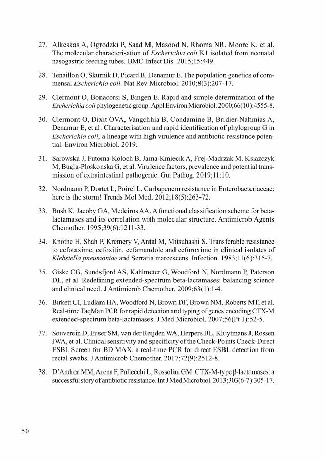
50
27. Alkeskas A, Ogrodzki P, Saad M, Masood N, Rhoma NR, Moore K, et al. The molecular characterisation of Escherichia coli K1 isolated from neonatal nasogastric feeding tubes. BMC Infect Dis. 2015;15:449.
28. Tenaillon O, Skurnik D, Picard B, Denamur E. The population genetics of com-mensal Escherichia coli. Nat Rev Microbiol. 2010;8(3):207-17.
29. Clermont O, Bonacorsi S, Bingen E. Rapid and simple determination of the Escherichia coli phylogenetic group. Appl Environ Microbiol. 2000;66(10):4555-8.
30. Clermont O, Dixit OVA, Vangchhia B, Condamine B, Bridier-Nahmias A, Denamur E, et al. Characterisation and rapid identification of phylogroup G in Escherichia coli, a lineage with high virulence and antibiotic resistance poten-tial. Environ Microbiol. 2019.
31. Sarowska J, Futoma-Koloch B, Jama-Kmiecik A, Frej-Madrzak M, Ksiazczyk M, Bugla-Ploskonska G, et al. Virulence factors, prevalence and potential trans-mission of extraintestinal pathogenic. Gut Pathog. 2019;11:10.
32. Nordmann P, Dortet L, Poirel L. Carbapenem resistance in Enterobacteriaceae: here is the storm! Trends Mol Med. 2012;18(5):263-72.
33. Bush K, Jacoby GA, Medeiros AA. A functional classification scheme for beta-lactamases and its correlation with molecular structure. Antimicrob Agents Chemother. 1995;39(6):1211-33.
34. Knothe H, Shah P, Krcmery V, Antal M, Mitsuhashi S. Transferable resistance to cefotaxime, cefoxitin, cefamandole and cefuroxime in clinical isolates of Klebsiella pneumoniae and Serratia marcescens. Infection. 1983;11(6):315-7.
35. Giske CG, Sundsfjord AS, Kahlmeter G, Woodford N, Nordmann P, Paterson DL, et al. Redefining extended-spectrum beta-lactamases: balancing science and clinical need. J Antimicrob Chemother. 2009;63(1):1-4.
36. Birkett CI, Ludlam HA, Woodford N, Brown DF, Brown NM, Roberts MT, et al. Real-time TaqMan PCR for rapid detection and typing of genes encoding CTX-M extended-spectrum beta-lactamases. J Med Microbiol. 2007;56(Pt 1):52-5.
37. Souverein D, Euser SM, van der Reijden WA, Herpers BL, Kluytmans J, Rossen JWA, et al. Clinical sensitivity and specificity of the Check-Points Check-Direct ESBL Screen for BD MAX, a real-time PCR for direct ESBL detection from rectal swabs. J Antimicrob Chemother. 2017;72(9):2512-8.
38. D’Andrea MM, Arena F, Pallecchi L, Rossolini GM. CTX-M-type β-lactamases: a successful story of antibiotic resistance. Int J Med Microbiol. 2013;303(6-7):305-17.
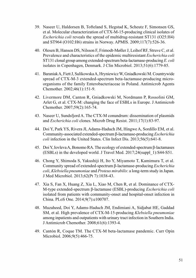
51
39. Naseer U, Haldorsen B, Tofteland S, Hegstad K, Scheutz F, Simonsen GS, et al. Molecular characterization of CTX-M-15-producing clinical isolates of Escherichia coli reveals the spread of multidrug-resistant ST131 (O25:H4) and ST964 (O102:H6) strains in Norway. APMIS. 2009;117(7):526-36.
40. Olesen B, Hansen DS, Nilsson F, Frimodt-Møller J, Leihof RF, Struve C, et al. Prevalence and characteristics of the epidemic multiresistant Escherichia coli ST131 clonal group among extended-spectrum beta-lactamase-producing E. coli isolates in Copenhagen, Denmark. J Clin Microbiol. 2013;51(6):1779-85.
41. Baraniak A, Fiett J, Sulikowska A, Hryniewicz W, Gniadkowski M. Countrywide spread of CTX-M-3 extended-spectrum beta-lactamase-producing micro-organisms of the family Enterobacteriaceae in Poland. Antimicrob Agents Chemother. 2002;46(1):151-9.
42. Livermore DM, Canton R, Gniadkowski M, Nordmann P, Rossolini GM, Arlet G, et al. CTX-M: changing the face of ESBLs in Europe. J Antimicrob Chemother. 2007;59(2):165-74.
43. Naseer U, Sundsfjord A. The CTX-M conundrum: dissemination of plasmids and Escherichia coli clones. Microb Drug Resist. 2011;17(1):83-97.
44. Doi Y, Park YS, Rivera JI, Adams-Haduch JM, Hingwe A, Sordillo EM, et al. Community-associated extended-spectrum β-lactamase-producing Escherichia coli infection in the United States. Clin Infect Dis. 2013;56(5):641-8.
45. Doi Y, Iovleva A, Bonomo RA. The ecology of extended-spectrum β-lactamases (ESBLs) in the developed world. J Travel Med. 2017;24(suppl_1):S44-S51.
46. Chong Y, Shimoda S, Yakushiji H, Ito Y, Miyamoto T, Kamimura T, et al. Community spread of extended-spectrum β-lactamase-producing Escherichia coli, Klebsiella pneumoniae and Proteus mirabilis: a long-term study in Japan. J Med Microbiol. 2013;62(Pt 7):1038-43.
47. Xia S, Fan X, Huang Z, Xia L, Xiao M, Chen R, et al. Dominance of CTX-M-type extended-spectrum β-lactamase (ESBL)-producing Escherichia coli isolated from patients with community-onset and hospital-onset infection in China. PLoS One. 2014;9(7):e100707.
48. Muzaheed, Doi Y, Adams-Haduch JM, Endimiani A, Sidjabat HE, Gaddad SM, et al. High prevalence of CTX-M-15-producing Klebsiella pneumoniae among inpatients and outpatients with urinary tract infection in Southern India. J Antimicrob Chemother. 2008;61(6):1393-4.
49. Cantón R, Coque TM. The CTX-M beta-lactamase pandemic. Curr Opin Microbiol. 2006;9(5):466-75.
52
50. Valverde A, Grill F, Coque TM, Pintado V, Baquero F, Cantón R, et al. High rate of intestinal colonization with extended-spectrum-beta-lactamase- producing organisms in household contacts of infected community patients. J Clin Microbiol. 2008;46(8):2796-9.
51. Lo WU, Ho PL, Chow KH, Lai EL, Yeung F, Chiu SS. Fecal carriage of CTXM type extended-spectrum beta-lactamase-producing organisms by children and their household contacts. J Infect. 2010;60(4):286-92.
52. Löhr IH, Rettedal S, Natås OB, Naseer U, Oymar K, Sundsfjord A. Long-term faecal carriage in infants and intra-household transmission of CTX-M-15-producing Klebsiella pneumoniae following a nosocomial outbreak. J Antimicrob Chemother. 2013;68(5):1043-8.
53. Rocha-Gracia RC, Cortés-Cortés G, Lozano-Zarain P, Bello F, Martínez-Laguna Y, Torres C. Faecal Escherichia coli isolates from healthy dogs harbour CTX-M-15 and CMY-2 β-lactamases. Vet J. 2015;203(3):315-9.
54. Sallem RB, Gharsa H, Slama KB, Rojo-Bezares B, Estepa V, Porres-Osante N, et al. First detection of CTX-M-1, CMY-2, and QnrB19 resistance mechanisms in fecal Escherichia coli isolates from healthy pets in Tunisia. Vector Borne Zoonotic Dis. 2013;13(2):98-102.
55. Horton RA, Randall LP, Snary EL, Cockrem H, Lotz S, Wearing H, et al. Fecal carriage and shedding density of CTX-M extended-spectrum {beta}-lactamase-producing Escherichia coli in cattle, chickens, and pigs: implica-tions for environmental contamination and food production. Appl Environ Microbiol. 2011;77(11):3715-9.
56. Alcalá L, Alonso CA, Simón C, González-Esteban C, Orós J, Rezusta A, et al. Wild Birds, Frequent Carriers of Extended-Spectrum β-Lactamase (ESBL) Producing Escherichia coli of CTX-M and SHV-12 Types. Microb Ecol. 2016;72(4):861-9.
57. Korzeniewska E, Harnisz M. Extended-spectrum beta-lactamase (ESBL)-positive Enterobacteriaceae in municipal sewage and their emission to the environment. J Environ Manage. 2013;128:904-11.
58. Müller A, Stephan R, Nüesch-Inderbinen M. Distribution of virulence factors in ESBL-producing Escherichia coli isolated from the environment, livestock, food and humans. Sci Total Environ. 2016;541:667-72.
59. Walsh TR, Weeks J, Livermore DM, Toleman MA. Dissemination of NDM-1 positive bacteria in the New Delhi environment and its implications for human health: an environmental point prevalence study. Lancet Infect Dis. 2011;11(5):355-62.
53
60. The public Health Agency of Sweden. Report on ESBL, 2014. [Available from: www.folkhalsomyndigheten.se/documents/nyheter-press/nyheter2014/MSB-ESBL-slutrapport.
61. Luvsansharav UO, Hirai I, Nakata A, Imura K, Yamauchi K, Niki M, et al. Prevalence of and risk factors associated with faecal carriage of CTX-M β-lactamase-producing Enterobacteriaceae in rural Thai communities. J Antimicrob Chemother. 2012;67(7):1769-74.
62. Mathai D, Kumar VA, Paul B, Sugumar M, John KR, Manoharan A, et al. Fecal carriage rates of extended-spectrum β-lactamase-producing Escherichia coli among antibiotic naive healthy human volunteers. Microb Drug Resist. 2015;21(1):59-64.
63. Thacker N, Pereira N, Banavali SD, Narula G, Vora T, Chinnaswamy G, et al. Alarming prevalence of community-acquired multidrug-resistant organisms colonization in children with cancer and implications for therapy: A prospec-tive study. Indian J Cancer. 2014;51(4):442-6.
64. World Health Organization. Antimicrobial Resistance: Global Report on Surveillance 2014.
65. Tängdén T, Cars O, Melhus A, Löwdin E. Foreign travel is a major risk factor for colonization with Escherichia coli producing CTX-M-type extended-spectrum beta-lactamases: a prospective study with Swedish volunteers. Antimicrob Agents Chemother. 2010;54(9):3564-8.
66. Ruppé E, Armand-Lefèvre L, Estellat C, Consigny PH, El Mniai A, Boussadia Y, et al. High Rate of Acquisition but Short Duration of Carriage of Multidrug-Resistant Enterobacteriaceae After Travel to the Tropics. Clin Infect Dis. 2015;61(4):593-600.
67. Ostholm-Balkhed A, Tärnberg M, Nilsson M, Nilsson LE, Hanberger H, Hällgren A, et al. Travel-associated faecal colonization with ESBL-producing Enterobacteriaceae: incidence and risk factors. J Antimicrob Chemother. 2013;68(9):2144-53.
68. Kantele A, Lääveri T, Mero S, Vilkman K, Pakkanen SH, Ollgren J, et al. Antimicrobials increase travelers’ risk of colonization by extended-spectrum betalactamase-producing Enterobacteriaceae. Clin Infect Dis. 2015;60(6):837-46.
69. Angelin M, Forsell J, Granlund M, Evengård B, Palmgren H, Johansson A. Risk factors for colonization with extended-spectrum beta-lactamase produc-ing Enterobacteriaceae in healthcare students on clinical assignment abroad: A prospective study. Travel Med Infect Dis. 2015;13(3):223-9.
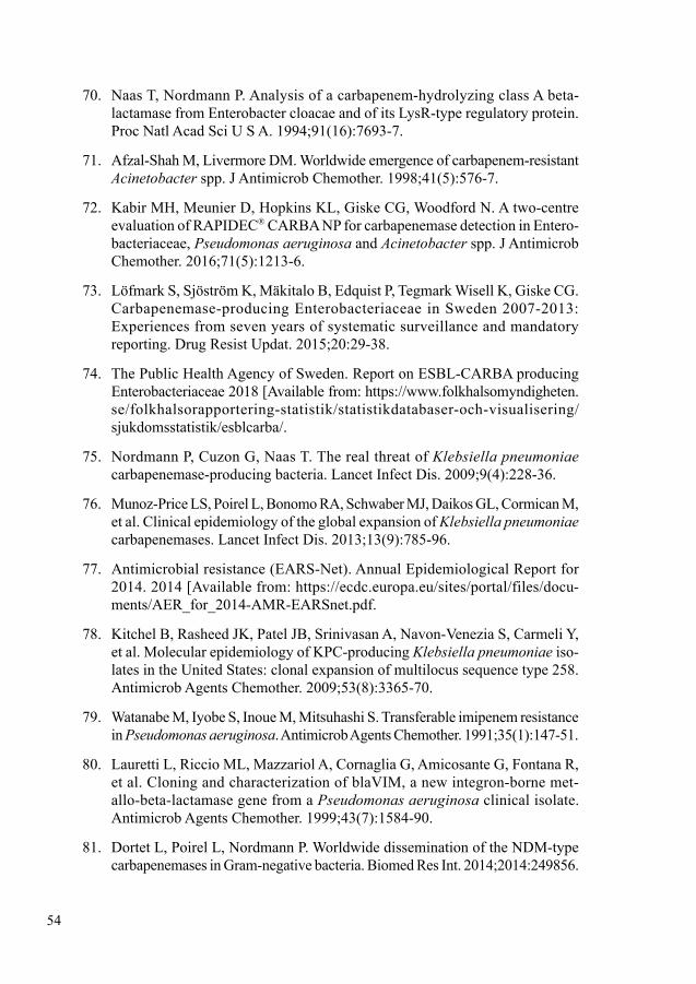
54
70. Naas T, Nordmann P. Analysis of a carbapenem-hydrolyzing class A beta-lactamase from Enterobacter cloacae and of its LysR-type regulatory protein. Proc Natl Acad Sci U S A. 1994;91(16):7693-7.
71. Afzal-Shah M, Livermore DM. Worldwide emergence of carbapenem-resistant Acinetobacter spp. J Antimicrob Chemother. 1998;41(5):576-7.
72. Kabir MH, Meunier D, Hopkins KL, Giske CG, Woodford N. A two- centre evaluation of RAPIDEC® CARBA NP for carbapenemase detection in Entero-bacteriaceae, Pseudomonas aeruginosa and Acinetobacter spp. J Antimicrob Chemother. 2016;71(5):1213-6.
73. Löfmark S, Sjöström K, Mäkitalo B, Edquist P, Tegmark Wisell K, Giske CG. Carbapenemase-producing Enterobacteriaceae in Sweden 2007-2013: Experiences from seven years of systematic surveillance and mandatory reporting. Drug Resist Updat. 2015;20:29-38.
74. The Public Health Agency of Sweden. Report on ESBL-CARBA producing Enterobacteriaceae 2018 [Available from: https://www.folkhalsomyndigheten.se/folkhalsorapportering-statistik/statistikdatabaser-och-visualisering/sjukdomsstatistik/esblcarba/.
75. Nordmann P, Cuzon G, Naas T. The real threat of Klebsiella pneumoniae carbapenemase-producing bacteria. Lancet Infect Dis. 2009;9(4):228-36.
76. Munoz-Price LS, Poirel L, Bonomo RA, Schwaber MJ, Daikos GL, Cormican M, et al. Clinical epidemiology of the global expansion of Klebsiella pneumoniae carbapenemases. Lancet Infect Dis. 2013;13(9):785-96.
77. Antimicrobial resistance (EARS-Net). Annual Epidemiological Report for 2014. 2014 [Available from: https://ecdc.europa.eu/sites/portal/files/docu-ments/AER_for_2014-AMR-EARSnet.pdf.
78. Kitchel B, Rasheed JK, Patel JB, Srinivasan A, Navon-Venezia S, Carmeli Y, et al. Molecular epidemiology of KPC-producing Klebsiella pneumoniae iso-lates in the United States: clonal expansion of multilocus sequence type 258. Antimicrob Agents Chemother. 2009;53(8):3365-70.
79. Watanabe M, Iyobe S, Inoue M, Mitsuhashi S. Transferable imipenem resistance in Pseudomonas aeruginosa. Antimicrob Agents Chemother. 1991;35(1):147-51.
80. Lauretti L, Riccio ML, Mazzariol A, Cornaglia G, Amicosante G, Fontana R, et al. Cloning and characterization of blaVIM, a new integron-borne met-allo-beta-lactamase gene from a Pseudomonas aeruginosa clinical isolate. Antimicrob Agents Chemother. 1999;43(7):1584-90.
81. Dortet L, Poirel L, Nordmann P. Worldwide dissemination of the NDM-type carbapenemases in Gram-negative bacteria. Biomed Res Int. 2014;2014:249856.
55
82. Cantón R, Akóva M, Carmeli Y, Giske CG, Glupczynski Y, Gniadkowski M, et al. Rapid evolution and spread of carbapenemases among Entero-bacteriaceae in Europe. Clin Microbiol Infect. 2012;18(5):413-31.
83. Poirel L, Héritier C, Tolün V, Nordmann P. Emergence of oxacillinase- mediated resistance to imipenem in Klebsiella pneumoniae. Antimicrob Agents Chemother. 2004;48(1):15-22.
84. Poirel L, Potron A, Nordmann P. OXA-48-like carbapenemases: the phantom menace. J Antimicrob Chemother. 2012;67(7):1597-606.
85. Pitout JD, Laupland KB. Extended-spectrum beta-lactamase-producing Enterobacteriaceae: an emerging public-health concern. Lancet Infect Dis. 2008;8(3):159-66.
86. Bennett PM. Plasmid encoded antibiotic resistance: acquisition and transfer of antibiotic resistance genes in bacteria. Br J Pharmacol. 2008;153 Suppl 1:S347-57.
87. Nordström K. Plasmid R1--replication and its control. Plasmid. 2006;55(1):1-26.
88. Carattoli A. Plasmids and the spread of resistance. Int J Med Microbiol. 2013;303(6-7):298-304.
89. Ramirez MS, Tolmasky ME. Aminoglycoside modifying enzymes. Drug Resist Updat. 2010;13(6):151-71.
90. Wei DD, Wan LG, Yu Y, Xu QF, Deng Q, Cao XW, et al. Characterization of extended-spectrum beta-lactamase, carbapenemase, and plasmid quinolone determinants in Klebsiella pneumoniae isolates carrying distinct types of 16S rRNA methylase genes, and their association with mobile genetic elements. Microb Drug Resist. 2015;21(2):186-93.
91. Martínez-Martínez L, Pascual A, Jacoby GA. Quinolone resistance from a transferable plasmid. Lancet. 1998;351(9105):797-9.
92. Strahilevitz J, Jacoby GA, Hooper DC, Robicsek A. Plasmid-mediated qui-nolone resistance: a multifaceted threat. Clin Microbiol Rev. 2009;22(4):664-89.
93. Liu YY, Wang Y, Walsh TR, Yi LX, Zhang R, Spencer J, et al. Emergence of plasmid-mediated colistin resistance mechanism MCR-1 in animals and human beings in China: a microbiological and molecular biological study. Lancet Infect Dis. 2016;16(2):161-8.
94. Hasman H, Hammerum AM, Hansen F, Hendriksen RS, Olesen B, Agersø Y, et al. Detection of mcr-1 encoding plasmid-mediated colistin-resistant Escherichia coli isolates from human bloodstream infection and imported chicken meat, Denmark 2015. Euro Surveill. 2015;20(49).

56
95. The Public Health Agency of Sweden. Report on Plasmid-mediated colistin resistance. 2017 [Available from: https://www.folkhalsomyndigheten.se/contentassets/9ecbe90539af46428de9305435db522b/plasmidmedierad-kolistin-resistens.pdf.
96. Di Pilato V, Arena F, Tascini C, Cannatelli A, Henrici De Angelis L, Fortunato S, et al. mcr-1.2, a New mcr Variant Carried on a Transferable Plasmid from a Colistin-Resistant KPC Carbapenemase-Producing Klebsiella pneumoniae Strain of Sequence Type 512. Antimicrob Agents Chemother. 2016;60(9):5612-5.
97. Falgenhauer L, Waezsada SE, Yao Y, Imirzalioglu C, Käsbohrer A, Roesler U, et al. Colistin resistance gene mcr-1 in extended-spectrum β-lactamase-producing and carbapenemase-producing Gram-negative bacteria in Germany. Lancet Infect Dis. 2016;16(3):282-3.
98. Mendes AC, Novais Â, Campos J, Rodrigues C, Santos C, Antunes P, et al. mcr-1 in Carbapenemase-Producing Klebsiella pneumoniae with Hospitalized Patients, Portugal, 2016-2017. Emerg Infect Dis. 2018;24(4):762-6.
99. Xavier BB, Lammens C, Ruhal R, Kumar-Singh S, Butaye P, Goossens H, et al. Identification of a novel plasmid-mediated colistin-resistance gene, mcr-2, in Escherichia coli, Belgium, June 2016. Euro Surveill. 2016;21(27).
100. Tanise V. D DdLM, Afonso L. B. Plasmid-mediated Colistin Resistance: What Do We Know? Journal of Infectiology. 2018;1(2):16-22.
101. European Committee on Antimicrobial Susceptibility Testing. Breakpoint tables for interpretation of MICs and zone diameters. EUCAST; Version 3.0 2013.
102. Clinical and Laboratory Standards Institute. Performance Standards for Antimicrobial Susceptibility Testing: Twenty--- first Informational Supplement M 100--- S21. Wayne, PA, USA:: CLSI; 2011.
103. Hope R, Potz NA, Warner M, Fagan EJ, Arnold E, Livermore DM. Efficacy of practised screening methods for detection of cephalosporin-resistant Entero-bacteriaceae. J Antimicrob Chemother. 2007;59(1):110-3.
104. Oliver A, Weigel LM, Rasheed JK, McGowan JE, Raney P, Tenover FC. Mechanisms of decreased susceptibility to cefpodoxime in Escherichia coli. Antimicrob Agents Chemother. 2002;46(12):3829-36.
105. Drieux L, Brossier F, Sougakoff W, Jarlier V. Phenotypic detection of extended-spectrum beta-lactamase production in Enterobacteriaceae: review and bench guide. Clin Microbiol Infect. 2008;14 Suppl 1:90-103.
106. Paterson DL, Bonomo RA. Extended-spectrum beta-lactamases: a clinical update. Clin Microbiol Rev. 2005;18(4):657-86.
57
107. M’Zali FH, Chanawong A, Kerr KG, Birkenhead D, Hawkey PM. Detection of extended-spectrum beta-lactamases in members of the family entero-bacteriaceae: comparison of the MAST DD test, the double disc and the Etest ESBL. J Antimicrob Chemother. 2000;45(6):881-5.
108. Nordmann P, Dortet L, Poirel L. Rapid detection of extended-spectrum-β-lactamase-producing Enterobacteriaceae. J Clin Microbiol. 2012;50(9):3016-22.
109. Dortet L, Poirel L, Nordmann P. Rapid detection of ESBL-producing Entero-bacteriaceae in blood cultures. Emerg Infect Dis. 2015;21(3):504-7.
110. Renvoisé A, Decré D, Amarsy-Guerle R, Huang TD, Jost C, Podglajen I, et al. Evaluation of the βLacta test, a rapid test detecting resistance to third- generation cephalosporins in clinical strains of Enterobacteriaceae. J Clin Microbiol. 2013;51(12):4012-7.
111. Gazin M, Paasch F, Goossens H, Malhotra-Kumar S, Teams MWaSWS. Current trends in culture-based and molecular detection of extended-spectrum-β-lactamase-harboring and carbapenem-resistant Enterobacteriaceae. J Clin Microbiol. 2012;50(4):1140-6.
112. Kruse EB, Aurbach U, Wisplinghoff H. Carbapenem-Resistant Entero-bacteriaceae: Laboratory Detection and Infection Control Practices. Curr Infect Dis Rep. 2013.
113. Nordmann P, Girlich D, Poirel L. Detection of carbapenemase producers in Enterobacteriaceae by use of a novel screening medium. J Clin Microbiol. 2012;50(8):2761-6.
114. Girlich D, Anglade C, Zambardi G, Nordmann P. Comparative evaluation of a novel chromogenic medium (chromID OXA-48) for detection of OXA-48 pro-ducing Enterobacteriaceae. Diagn Microbiol Infect Dis. 2013;77(4):296-300.
115. Kotsakis SD, Petinaki E, Scopes E, Siatravani E, Miriagou V, Tzelepi E. Laboratory evaluation of Brilliance™ CRE Agar for screening carbapenem-resistant Enterobacteriaceae: Performance on a collection of characterised clinical isolates from Greece. J Glob Antimicrob Resist. 2013;1(2):85-90.
116. Vasoo S, Lolans K, Li H, Prabaker K, Hayden MK. Comparison of the CHROMagar™ KPC, Remel Spectra™ CRE, and a direct ertapenem disk method for the detection of KPC-producing Enterobacteriaceae from peri-rectal swabs. Diagn Microbiol Infect Dis. 2014;78(4):356-9.
117. European Committee on Antimicrobial Susceptibility Testing. EUCAST guidelines for detection of resistance mechanisms and specific resistance of clinical and/or epidemiological importance; Version 2.0 2017.
58
118. Vading M, Samuelsen Ø, Haldorsen B, Sundsfjord AS, Giske CG. Comparison of disk diffusion, Etest and VITEK2 for detection of carbapenemase-producing Klebsiella pneumoniae with the EUCAST and CLSI breakpoint systems. Clin Microbiol Infect. 2011;17(5):668-74.
119. Giske CG, Gezelius L, Samuelsen Ø, Warner M, Sundsfjord A, Woodford N. A sensitive and specific phenotypic assay for detection of metallo-β-lactamases and KPC in Klebsiella pneumoniae with the use of meropenem disks supple-mented with aminophenylboronic acid, dipicolinic acid and cloxacillin. Clin Microbiol Infect. 2011;17(4):552-6.
120. Doyle D, Peirano G, Lascols C, Lloyd T, Church DL, Pitout JD. Laboratory detection of Enterobacteriaceae that produce carbapenemases. J Clin Microbiol. 2012;50(12):3877-80.
121. Porres-Osante N, de Champs C, Dupont H, Torres C, Mammeri H. Use of avi-bactam to detect Ambler class A carbapenemases and OXA-48 β-lactamases in Enterobacteriaceae. Diagn Microbiol Infect Dis. 2014;79(3):399-400.
122. Dortet L, Poirel L, Nordmann P. Rapid detection of carbapenemase-producing Pseudomonas spp. J Clin Microbiol. 2012;50(11):3773-6.
123. Nordmann P, Poirel L, Dortet L. Rapid detection of carbapenemase-producing Enterobacteriaceae. Emerg Infect Dis. 2012;18(9):1503-7.
124. Yusuf E, Van Der Meeren S, Schallier A, Piérard D. Comparison of the Carba NP test with the Rapid CARB Screen Kit for the detection of carbapenemase-producing Enterobacteriaceae and Pseudomonas aeruginosa. Eur J Clin Microbiol Infect Dis. 2014;33(12):2237-40.
125. Poirel L, Nordmann P. Rapidec Carba NP Test for Rapid Detection of Carba-penemase Producers. J Clin Microbiol. 2015;53(9):3003-8.
126. Huang TD, Berhin C, Bogaerts P, Glupczynski Y. Comparative evaluation of two chromogenic tests for rapid detection of carbapenemase in Enterobacteriaceae and in Pseudomonas aeruginosa isolates. J Clin Microbiol. 2014;52(8):3060-3.
127. Noël A, Huang TD, Berhin C, Hoebeke M, Bouchahrouf W, Yunus S, et al. Com-parative Evaluation of Four Phenotypic Tests for Detection of Carbapenemase-Producing Gram-Negative Bacteria. J Clin Microbiol. 2017;55(2):510-8.
128. Pasteran F, Veliz O, Ceriana P, Lucero C, Rapoport M, Albornoz E, et al. Evaluation of the Blue-Carba test for rapid detection of carbapenemases in gram-negative bacilli. J Clin Microbiol. 2015;53(6):1996-8.
129. Bernabeu S, Poirel L, Nordmann P. Spectrophotometry-based detection of carbapenemase producers among Enterobacteriaceae. Diagn Microbiol Infect Dis. 2012;74(1):88-90.
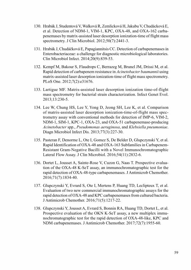
59
130. Hrabák J, Studentová V, Walková R, Zemlicková H, Jakubu V, Chudácková E, et al. Detection of NDM-1, VIM-1, KPC, OXA-48, and OXA-162 carba-penemases by matrix-assisted laser desorption ionization-time of flight mass spectrometry. J Clin Microbiol. 2012;50(7):2441-3.
131. Hrabák J, Chudáčková E, Papagiannitsis CC. Detection of carbapenemases in Enterobacteriaceae: a challenge for diagnostic microbiological laboratories. Clin Microbiol Infect. 2014;20(9):839-53.
132. Kempf M, Bakour S, Flaudrops C, Berrazeg M, Brunel JM, Drissi M, et al. Rapid detection of carbapenem resistance in Acinetobacter baumannii using matrix-assisted laser desorption ionization-time of flight mass spectrometry. PLoS One. 2012;7(2):e31676.
133. Lartigue MF. Matrix-assisted laser desorption ionization time-of-flight mass spectrometry for bacterial strain characterization. Infect Genet Evol. 2013;13:230-5.
134. Lee W, Chung HS, Lee Y, Yong D, Jeong SH, Lee K, et al. Comparison of matrix-assisted laser desorption ionization-time-of-flight mass spec-trometry assay with conventional methods for detection of IMP-6, VIM-2, NDM-1, SIM-1, KPC-1, OXA-23, and OXA-51 carbapenemase-producing Acinetobacter spp., Pseudomonas aeruginosa, and Klebsiella pneumoniae. Diagn Microbiol Infect Dis. 2013;77(3):227-30.
135. Pasteran F, Denorme L, Ote I, Gomez S, De Belder D, Glupczynski Y, et al. Rapid Identification of OXA-48 and OXA-163 Subfamilies in Carbapenem-Resistant Gram-Negative Bacilli with a Novel Immunochromatographic Lateral Flow Assay. J Clin Microbiol. 2016;54(11):2832-6.
136. Dortet L, Jousset A, Sainte-Rose V, Cuzon G, Naas T. Prospective evalua-tion of the OXA-48 K-SeT assay, an immunochromatographic test for the rapid detection of OXA-48-type carbapenemases. J Antimicrob Chemother. 2016;71(7):1834-40.
137. Glupczynski Y, Evrard S, Ote I, Mertens P, Huang TD, Leclipteux T, et al. Evaluation of two new commercial immunochromatographic assays for the rapid detection of OXA-48 and KPC carbapenemases from cultured bacteria. J Antimicrob Chemother. 2016;71(5):1217-22.
138. Glupczynski Y, Jousset A, Evrard S, Bonnin RA, Huang TD, Dortet L, et al. Prospective evaluation of the OKN K-SeT assay, a new multiplex immu-nochromatographic test for the rapid detection of OXA-48-like, KPC and NDM carbapenemases. J Antimicrob Chemother. 2017;72(7):1955-60.
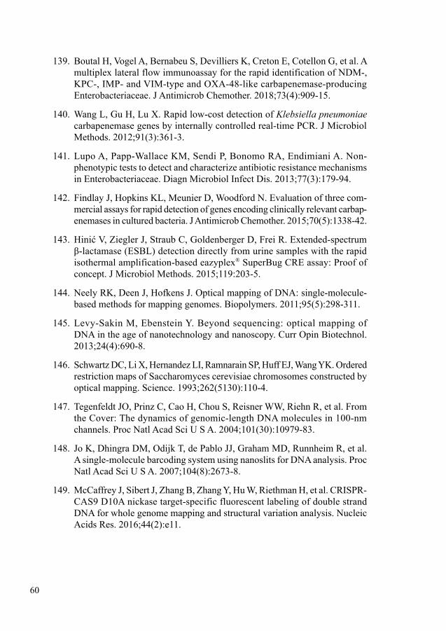
60
139. Boutal H, Vogel A, Bernabeu S, Devilliers K, Creton E, Cotellon G, et al. A multiplex lateral flow immunoassay for the rapid identification of NDM-, KPC-, IMP- and VIM-type and OXA-48-like carbapenemase-producing Enterobacteriaceae. J Antimicrob Chemother. 2018;73(4):909-15.
140. Wang L, Gu H, Lu X. Rapid low-cost detection of Klebsiella pneumoniae carbapenemase genes by internally controlled real-time PCR. J Microbiol Methods. 2012;91(3):361-3.
141. Lupo A, Papp-Wallace KM, Sendi P, Bonomo RA, Endimiani A. Non-phenotypic tests to detect and characterize antibiotic resistance mechanisms in Enterobacteriaceae. Diagn Microbiol Infect Dis. 2013;77(3):179-94.
142. Findlay J, Hopkins KL, Meunier D, Woodford N. Evaluation of three com-mercial assays for rapid detection of genes encoding clinically relevant carbap-enemases in cultured bacteria. J Antimicrob Chemother. 2015;70(5):1338-42.
143. Hinić V, Ziegler J, Straub C, Goldenberger D, Frei R. Extended-spectrum β-lactamase (ESBL) detection directly from urine samples with the rapid isothermal amplification-based eazyplex® SuperBug CRE assay: Proof of concept. J Microbiol Methods. 2015;119:203-5.
144. Neely RK, Deen J, Hofkens J. Optical mapping of DNA: single-molecule-based methods for mapping genomes. Biopolymers. 2011;95(5):298-311.
145. Levy-Sakin M, Ebenstein Y. Beyond sequencing: optical mapping of DNA in the age of nanotechnology and nanoscopy. Curr Opin Biotechnol. 2013;24(4):690-8.
146. Schwartz DC, Li X, Hernandez LI, Ramnarain SP, Huff EJ, Wang YK. Ordered restriction maps of Saccharomyces cerevisiae chromosomes constructed by optical mapping. Science. 1993;262(5130):110-4.
147. Tegenfeldt JO, Prinz C, Cao H, Chou S, Reisner WW, Riehn R, et al. From the Cover: The dynamics of genomic-length DNA molecules in 100-nm channels. Proc Natl Acad Sci U S A. 2004;101(30):10979-83.
148. Jo K, Dhingra DM, Odijk T, de Pablo JJ, Graham MD, Runnheim R, et al. A single-molecule barcoding system using nanoslits for DNA analysis. Proc Natl Acad Sci U S A. 2007;104(8):2673-8.
149. McCaffrey J, Sibert J, Zhang B, Zhang Y, Hu W, Riethman H, et al. CRISPR-CAS9 D10A nickase target-specific fluorescent labeling of double strand DNA for whole genome mapping and structural variation analysis. Nucleic Acids Res. 2016;44(2):e11.
61
150. Nyberg LK, Persson F, Berg J, Bergström J, Fransson E, Olsson L, et al. A single-step competitive binding assay for mapping of single DNA molecules. Biochem Biophys Res Commun. 2012;417(1):404-8.
151. Nilsson AN, Emilsson G, Nyberg LK, Noble C, Stadler LS, Fritzsche J, et al. Competitive binding-based optical DNA mapping for fast identification of bacteria--multi-ligand transfer matrix theory and experimental applications on Escherichia coli. Nucleic Acids Res. 2014;42(15):e118.
152. Nordberg V, Jonsson K, Giske CG, Iversen A, Aspevall O, Jonsson B, et al. Neonatal intestinal colonization with extended-spectrum β-lactamase-producing Enterobacteriaceae-a 5-year follow-up study. Clin Microbiol Infect. 2018;24(9):1004-9.
153. Olsson-Liljequist B, Larsson P, Walder M, Miörner H. Antimicrobial suscep-tibility testing in Sweden. III. Methodology for susceptibility testing. Scand J Infect Dis Suppl. 1997;105:13-23.
154. Yigit H, Queenan AM, Anderson GJ, Domenech-Sanchez A, Biddle JW, Steward CD, et al. Novel carbapenem-hydrolyzing beta-lactamase, KPC-1, from a carbapenem-resistant strain of Klebsiella pneumoniae. Antimicrob Agents Chemother. 2001;45(4):1151-61.
155. Poirel L, Heritier C, Tolun V, Nordmann P. Emergence of oxacillinase-medi-ated resistance to imipenem in Klebsiella pneumoniae. Antimicrob Agents Chemother. 2004;48(1):15-22.
156. Mushtaq S, Irfan S, Sarma JB, Doumith M, Pike R, Pitout J, et al. Phylogenetic diversity of Escherichia coli strains producing NDM-type carbapenemases. The Journal of antimicrobial chemotherapy. 2011;66(9):2002-5.
157. Ellington MJ, Kistler J, Livermore DM, Woodford N. Multiplex PCR for rapid detection of genes encoding acquired metallo-beta-lactamases. The Journal of antimicrobial chemotherapy. 2007;59(2):321-2.
158. Queenan AM, Torres-Viera C, Gold HS, Carmeli Y, Eliopoulos GM, Moellering RC, Jr., et al. SME-type carbapenem-hydrolyzing class A beta-lactamases from geographically diverse Serratia marcescens strains. Antimicrob Agents Chemother. 2000;44(11):3035-9.
159. Poirel L, Le Thomas I, Naas T, Karim A, Nordmann P. Biochemical sequence analyses of GES-1, a novel class A extended-spectrum beta-lactamase, and the class 1 integron In52 from Klebsiella pneumoniae. Antimicrob Agents Chemother. 2000;44(3):622-32.
160. Rasmussen BA, Bush K, Keeney D, Yang Y, Hare R, O’Gara C, et al. Character-ization of IMI-1 beta-lactamase, a class A carbapenem-hydrolyzing enzyme from Enterobacter cloacae. Antimicrob Agents Chemother. 1996;40(9):2080-6.
62
161. Woodford N, Ellington MJ, Coelho JM, Turton JF, Ward ME, Brown S, et al. Multiplex PCR for genes encoding prevalent OXA carbapenemases in Acinetobacter spp. Int J Antimicrob Agents. 2006;27(4):351-3.
162. Pasanen T, Koskela S, Mero S, Tarkka E, Tissari P, Vaara M, et al. Rapid molecular characterization of Acinetobacter baumannii clones with rep-PCR and evaluation of carbapenemase genes by new multiplex PCR in Hospital District of Helsinki and Uusimaa. PloS one. 2014;9(1):e85854.
163. Doumith M, Day M, Ciesielczuk H, Hope R, Underwood A, Reynolds R, et al. Rapid identification of major Escherichia coli sequence types causing urinary tract and bloodstream infections. J Clin Microbiol. 2015;53(1):160-6.
164. Turton JF, Perry C, Elgohari S, Hampton CV. PCR characterization and typing of Klebsiella pneumoniae using capsular type-specific, variable number tandem repeat and virulence gene targets. J Med Microbiol. 2010;59(Pt 5):541-7.
165. Brisse S, Issenhuth-Jeanjean S, Grimont PA. Molecular serotyping of Klebsiella species isolates by restriction of the amplified capsular antigen gene cluster. J Clin Microbiol. 2004;42(8):3388-98.
166. Brisse S, Fevre C, Passet V, Issenhuth-Jeanjean S, Tournebize R, Diancourt L, et al. Virulent clones of Klebsiella pneumoniae: identification and evolution-ary scenario based on genomic and phenotypic characterization. PLoS One. 2009;4(3):e4982.
167. Giske CG, Fröding I, Hasan CM, Turlej-Rogacka A, Toleman M, Livermore D, et al. Diverse sequence types of Klebsiella pneumoniae contribute to the dissemination of blaNDM-1 in India, Sweden, and the United Kingdom. Antimicrob Agents Chemother. 2012;56(5):2735-8.
168. Habeeb MA, Haque A, Iversen A, Giske CG. Occurrence of virulence genes, 16S rRNA methylases, and plasmid-mediated quinolone resistance genes in CTX-M-producing Escherichia coli from Pakistan. Eur J Clin Microbiol Infect Dis. 2014;33(3):399-409.
169. Johnson JR, Stell AL. Extended virulence genotypes of Escherichia coli strains from patients with urosepsis in relation to phylogeny and host compromise. J Infect Dis. 2000;181(1):261-72.
170. Zankari E, Hasman H, Cosentino S, Vestergaard M, Rasmussen S, Lund O, et al. Identification of acquired antimicrobial resistance genes. J Antimicrob Chemother. 2012;67(11):2640-4.
171. Larsen MV, Cosentino S, Rasmussen S, Friis C, Hasman H, Marvig RL, et al. Multilocus sequence typing of total-genome-sequenced bacteria. J Clin Microbiol. 2012;50(4):1355-61.
63
172. Carattoli A, Zankari E, García-Fernández A, Voldby Larsen M, Lund O, Villa L, et al. In silico detection and typing of plasmids using PlasmidFinder and plasmid multilocus sequence typing. Antimicrob Agents Chemother. 2014;58(7):3895-903.
173. Diancourt L, Passet V, Verhoef J, Grimont PA, Brisse S. Multilocus sequence typing of Klebsiella pneumoniae nosocomial isolates. J Clin Microbiol. 2005;43(8):4178-82.
174. Tamura K, Peterson D, Peterson N, Stecher G, Nei M, Kumar S. MEGA5: molecular evolutionary genetics analysis using maximum likelihood, evo-lutionary distance, and maximum parsimony methods. Mol Biol Evol. 2011;28(10):2731-9.
175. Brisse S, Verhoef J. Phylogenetic diversity of Klebsiella pneumoniae and Klebsiella oxytoca clinical isolates revealed by randomly amplified poly-morphic DNA, gyrA and parC genes sequencing and automated ribotyping. Int J Syst Evol Microbiol. 2001;51(Pt 3):915-24.
176. Bryant D, Moulton V. Neighbor-net: an agglomerative method for the con-struction of phylogenetic networks. Mol Biol Evol. 2004;21(2):255-65.
177. Librado P, Rozas J. DnaSP v5: a software for comprehensive analysis of DNA polymorphism data. Bioinformatics. 2009;25(11):1451-2.
178. Maatallah M, Cheriaa J, Backhrouf A, Iversen A, Grundmann H, Do T, et al. Population structure of Pseudomonas aeruginosa from five Mediterranean countries: evidence for frequent recombination and epidemic occurrence of CC235. PLoS One. 2011;6(10):e25617.
179. Feil EJ, Li BC, Aanensen DM, Hanage WP, Spratt BG. eBURST: inferring patterns of evolutionary descent among clusters of related bacterial genotypes from multilocus sequence typing data. J Bacteriol. 2004;186(5):1518-30.
180. Charlson ME, Pompei P, Ales KL, MacKenzie CR. A new method of clas-sifying prognostic comorbidity in longitudinal studies: development and validation. J Chronic Dis. 1987;40(5):373-83.
181. Müller V, Rajer F, Frykholm K, Nyberg LK, Quaderi S, Fritzsche J, et al. Direct identification of antibiotic resistance genes on single plasmid mol-ecules using CRISPR/Cas9 in combination with optical DNA mapping. Sci Rep. 2016;6:37938.
182. Nyberg L, Persson F, Akerman B, Westerlund F. Heterogeneous staining: a tool for studies of how fluorescent dyes affect the physical properties of DNA. Nucleic Acids Res. 2013;41(19):e184.
64
183. Persson F, Tegenfeldt JO. DNA in nanochannels--directly visualizing genomic information. Chem Soc Rev. 2010;39(3):985-99.
184. Alizadehheidari M. Nanoconfined Circular and Linear DNA: Equilibrium Conformations and Unfolding Kinetics. Macromolecules. 2015;48:871-8.
185. Nyberg LK, Quaderi S, Emilsson G, Karami N, Lagerstedt E, Müller V, et al. Rapid identification of intact bacterial resistance plasmids via optical map-ping of single DNA molecules. Sci Rep. 2016;6:30410.
186. Müller V, Karami N, Nyberg LK, Pichler C, Torche Pedreschi PC, Quaderi S, et al. Rapid Tracing of Resistance Plasmids in a Nosocomial Outbreak Using Optical DNA Mapping. ACS Infect Dis. 2016;2(5):322-8.
187. Joensen KG, Scheutz F, Lund O, Hasman H, Kaas RS, Nielsen EM, et al. Real-time whole-genome sequencing for routine typing, surveillance, and outbreak detection of verotoxigenic Escherichia coli. J Clin Microbiol. 2014;52(5):1501-10.
188. Blin C, Passet V, Touchon M, Rocha EPC, Brisse S. Metabolic diversity of the emerging pathogenic lineages of Klebsiella pneumoniae. Environ Microbiol. 2017;19(5):1881-98.
189. Rodrigues C, Passet V, Rakotondrasoa A, Diallo TA, Criscuolo A, Brisse S. Description of Klebsiella africanensis sp. nov., Klebsiella variicola subsp. tropicalensis subsp. nov. and Klebsiella variicola subsp. variicola subsp. nov. Res Microbiol. 2019.
190. Brisse S, van Himbergen T, Kusters K, Verhoef J. Development of a rapid identification method for Klebsiella pneumoniae phylogenetic groups and analysis of 420 clinical isolates. Clin Microbiol Infect. 2004;10(10):942-5.
191. Bowers JR, Lemmer D, Sahl JW, Pearson T, Driebe EM, Wojack B, et al. KlebSeq, a Diagnostic Tool for Surveillance, Detection, and Monitoring of Klebsiella pneumoniae. J Clin Microbiol. 2016;54(10):2582-96.
192. Alves MS, Dias RC, de Castro AC, Riley LW, Moreira BM. Identification of clinical isolates of indole-positive and indole-negative Klebsiella spp. J Clin Microbiol. 2006;44(10):3640-6.
193. Fontana L, Bonura E, Lyski Z, Messer W. The Brief Case: J Clin Microbiol. 2019;57(1).
194. Long SW, Linson SE, Ojeda Saavedra M, Cantu C, Davis JJ, Brettin T, et al. Whole-Genome Sequencing of Human Clinical. mSphere. 2017;2(4).
65
195. Garza-Ramos U, Silva-Sánchez J, Martínez-Romero E, Tinoco P, Pina-Gonzales M, Barrios H, et al. Development of a multiplex-PCR probe sys-tem for the proper identification of Klebsiella variicola. BMC Microbiol. 2015;15:64.
196. Rosenblueth M, Martínez L, Silva J, Martínez-Romero E. Klebsiella variicola, a novel species with clinical and plant-associated isolates. Syst Appl Microbiol. 2004;27(1):27-35.
197. Berry GJ, Loeffelholz MJ, Williams-Bouyer N. An Investigation into Laboratory Misidentification of a Bloodstream Klebsiella variicola Infection. J Clin Microbiol. 2015;53(8):2793-4.
198. de Melo ME, Cabral AB, Maciel MA, da Silveira VM, de Souza Lopes AC. Phylogenetic groups among Klebsiella pneumoniae isolates from Brazil: relationship with antimicrobial resistance and origin. Curr Microbiol. 2011;62(5):1596-601.
199. Seki M, Gotoh K, Nakamura S, Akeda Y, Yoshii T, Miyaguchi S, et al. Fatal sepsis caused by an unusual Klebsiella species that was misidentified by an automated identification system. J Med Microbiol. 2013;62(Pt 5):801-3.
200. Meatherall BL, Gregson D, Ross T, Pitout JD, Laupland KB. Incidence, risk factors, and outcomes of Klebsiella pneumoniae bacteremia. Am J Med. 2009;122(9):866-73.
201. Thom KA, Schweizer ML, Osih RB, McGregor JC, Furuno JP, Perencevich EN, et al. Impact of empiric antimicrobial therapy on outcomes in patients with Escherichia coli and Klebsiella pneumoniae bacteremia: a cohort study. BMC Infect Dis. 2008;8:116.
202. Tsay RW, Siu LK, Fung CP, Chang FY. Characteristics of bacteremia between community-acquired and nosocomial Klebsiella pneumoniae infection: risk factor for mortality and the impact of capsular serotypes as a herald for community-acquired infection. Arch Intern Med. 2002;162(9):1021-7.
203. Li J, Ren J, Wang W, Wang G, Gu G, Wu X, et al. Risk factors and clini-cal outcomes of hypervirulent Klebsiella pneumoniae induced bloodstream infections. Eur J Clin Microbiol Infect Dis. 2018;37(4):679-89.
204. Farzana R, Jones LS, Rahman MA, Andrey DO, Sands K, Portal E, et al. Outbreak of Hypervirulent Multidrug-resistant Klebsiella variicola Causing High Mortality in Neonates in Bangladesh. Clin Infect Dis. 2019;68(7):1225-7.
205. Zhang Y, Zhao C, Wang Q, Wang X, Chen H, Li H, et al. High Prevalence of Hypervirulent Klebsiella pneumoniae Infection in China: Geographic Distribution, Clinical Characteristics, and Antimicrobial Resistance. Antimicrob Agents Chemother. 2016;60(10):6115-20.
66
206. Tsai SS, Huang JC, Chen ST, Sun JH, Wang CC, Lin SF, et al. Characteristics of Klebsiella pneumoniae bacteremia in community-acquired and nosocomial infections in diabetic patients. Chang Gung Med J. 2010;33(5):532-9.
207. Lee KH, Hui KP, Tan WC, Lim TK. Klebsiella bacteraemia: a report of 101 cases from National University Hospital, Singapore. J Hosp Infect. 1994;27(4):299-305.
208. Valverde A, Coque TM, García-San Miguel L, Baquero F, Cantón R. Complex molecular epidemiology of extended-spectrum beta-lactamases in Klebsiella pneumoniae: a long-term perspective from a single institution in Madrid. J Antimicrob Chemother. 2008;61(1):64-72.
209. Selander RK, Caugant, D.A., and Whittam, T.S. Genetic structure and vari-ation in natural populations of Escherichia coli. In Escherichia coli and Salmonella typhimurium C ellular and molecular biology. American Society for Microbiology. 1987:1625-48.
210. Desjardins P, Picard B, Kaltenböck B, Elion J, Denamur E. Sex in Escherichia coli does not disrupt the clonal structure of the population: evidence from random amplified polymorphic DNA and restriction-fragment-length poly-morphism. J Mol Evol. 1995;41(4):440-8.
211. Escobar-Páramo P, Clermont O, Blanc-Potard AB, Bui H, Le Bouguénec C, Denamur E. A specific genetic background is required for acquisition and expression of virulence factors in Escherichia coli. Mol Biol Evol. 2004;21(6):1085-94.
212. Touchon M, Hoede C, Tenaillon O, Barbe V, Baeriswyl S, Bidet P, et al. Organised genome dynamics in the Escherichia coli species results in highly diverse adaptive paths. PLoS Genet. 2009;5(1):e1000344.
213. Beghain J, Bridier-Nahmias A, Le Nagard H, Denamur E, Clermont O. ClermonTyping: an easy-to-use and accurate in silico method for Escherichia genus strain phylotyping. Microb Genom. 2018;4(7).
214. Bevan ER, McNally A, Thomas CM, Piddock LJV, Hawkey PM. Acquisition and Loss of CTX-M-Producing and Non-Producing Escherichia coli in the Fecal Microbiome of Travelers to South Asia. MBio. 2018;9(6).
215. Titelman E, Hasan CM, Iversen A, Nauclér P, Kais M, Kalin M, et al. Faecal carriage of extended-spectrum β-lactamase-producing Enterobacteriaceae is common 12 months after infection and is related to strain factors. Clin Microbiol Infect. 2014;20(8):O508-15.
67
216. Rottier WC, Bamberg YR, Dorigo-Zetsma JW, van der Linden PD, Ammerlaan HS, Bonten MJ. Predictive value of prior colonization and antibiotic use for third-generation cephalosporin-resistant enterobacteriaceae bacteremia in patients with sepsis. Clin Infect Dis. 2015;60(11):1622-30.
217. Arcilla MS, van Hattem JM, Haverkate MR, Bootsma MCJ, van Genderen PJJ, Goorhuis A, et al. Import and spread of extended-spectrum β-lactamase-producing Enterobacteriaceae by international travellers (COMBAT study): a prospective, multicentre cohort study. Lancet Infect Dis. 2017;17(1):78-85.
218. Tijet N, Boyd D, Patel SN, Mulvey MR, Melano RG. Evaluation of the Carba NP test for rapid detection of carbapenemase-producing Enterobacteriaceae and Pseudomonas aeruginosa. Antimicrob Agents Chemother. 2013;57(9):4578-80.
219. Hombach M, von Gunten B, Castelberg C, Bloemberg GV. Evaluation of the Rapidec Carba NP Test for Detection of Carbapenemases in Enterobacteriaceae. J Clin Microbiol. 2015;53(12):3828-33.
220. Longo G, Alonso-Sarduy L, Rio LM, Bizzini A, Trampuz A, Notz J, et al. Rapid detection of bacterial resistance to antibiotics using AFM cantilevers as nanomechanical sensors. Nat Nanotechnol. 2013;8(7):522-6.
221. Choi J, Jung YG, Kim J, Kim S, Jung Y, Na H, et al. Rapid antibiotic sus-ceptibility testing by tracking single cell growth in a microfluidic agarose channel system. Lab Chip. 2013;13(2):280-7.
222. Von Ah U, Wirz D, Daniels AU. Isothermal micro calorimetry--a new method for MIC determinations: results for 12 antibiotics and reference strains of E. coli and S. aureus. BMC Microbiol. 2009;9:106.
223. Schoepp NG, Schlappi TS, Curtis MS, Butkovich SS, Miller S, Humphries RM, et al. Rapid pathogen-specific phenotypic antibiotic susceptibility testing using digital LAMP quantification in clinical samples. Sci Transl Med. 2017;9(410).