Nora Al-Kubaisi [email protected]. Gram’s +ve Cocci Irregular Clusters Tetrads Chains or Pairs...
40
MEDICAL BACTERIOLOGY THIRD LAP Nora Al-Kubaisi nalkubaisi@ksu. edu.sa
-
Upload
delphia-baker -
Category
Documents
-
view
223 -
download
0
Transcript of Nora Al-Kubaisi [email protected]. Gram’s +ve Cocci Irregular Clusters Tetrads Chains or Pairs...
MEDICAL BACTERIOLOGY
THIRD LAP
Nora Al-Kubaisinalkubaisi@ksu.
edu.sa
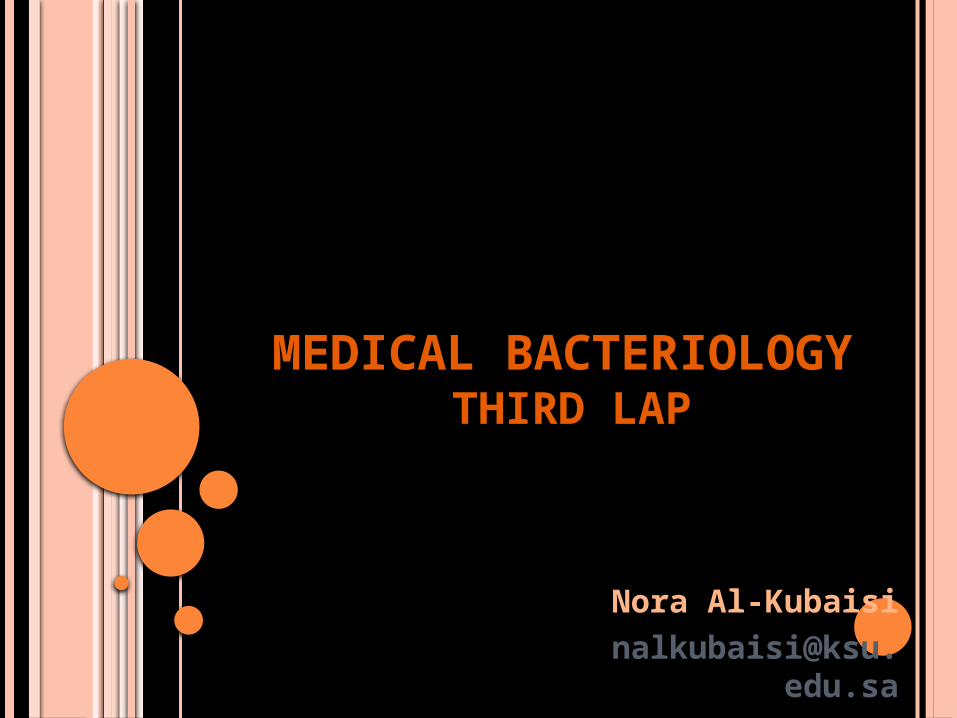
STREPTOCOCCUS
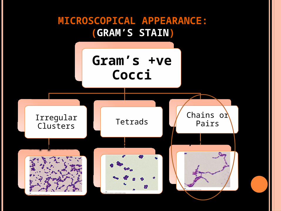
MICROSCOPICAL APPEARANCE:(GRAM’S STAIN)
Gram’s +ve Cocci
Irregular Clusters Tetrads
Chains or Pairs
Staphylococci
Micrococci Streptococci
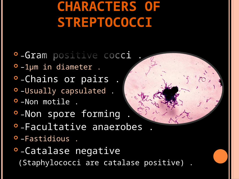
CHARACTERS OF STREPTOCOCCI
–Gram positive cocci . –1μm in diameter . –Chains or pairs . –Usually capsulated . –Non motile . –Non spore forming . –Facultative anaerobes . –Fastidious . –Catalase negative (Staphylococci are catalase positive) .
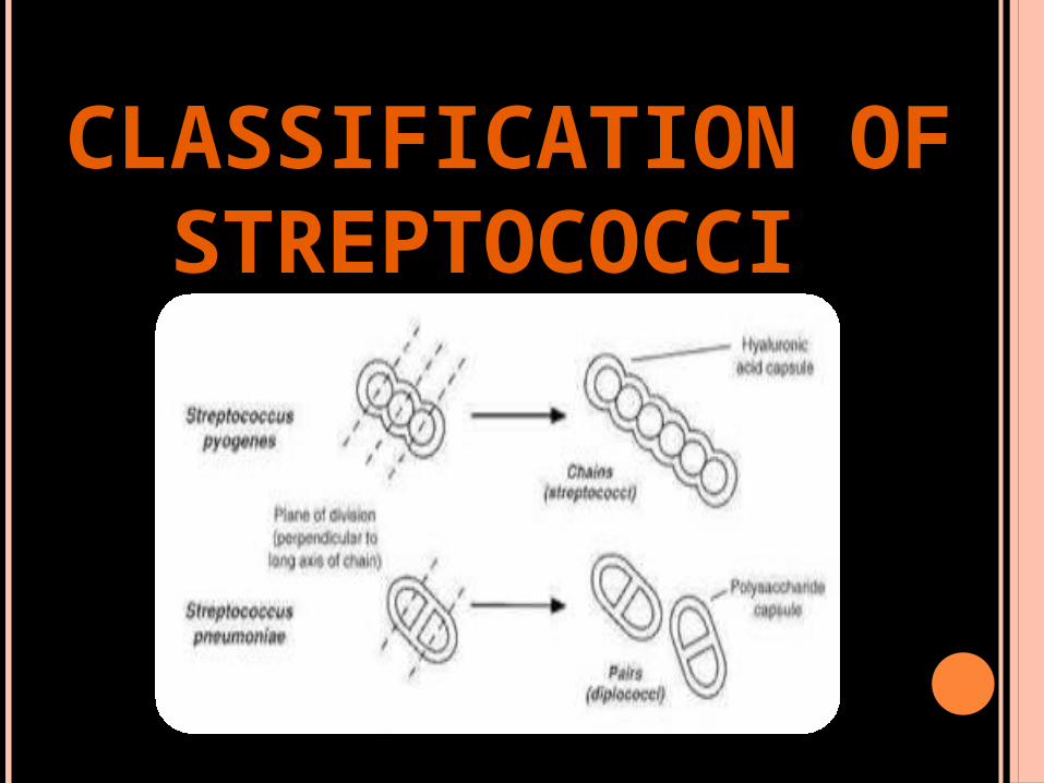
CLASSIFICATION OF STREPTOCOCCI
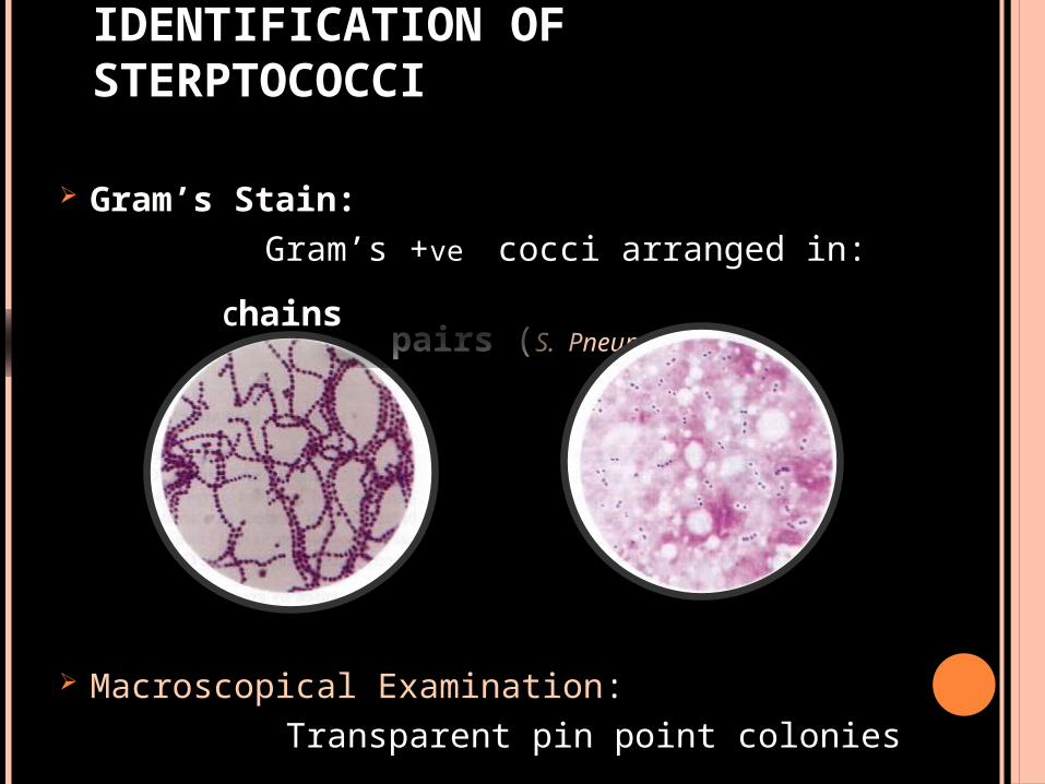
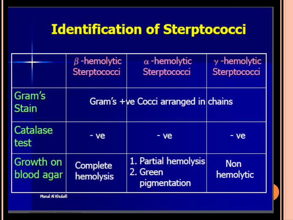
IDENTIFICATION OF STERPTOCOCCI
Gram’s Stain: Gram’s +ve cocci arranged in: pairs (S. Pneumonia)
Macroscopical Examination: Transparent pin point colonies
Chains
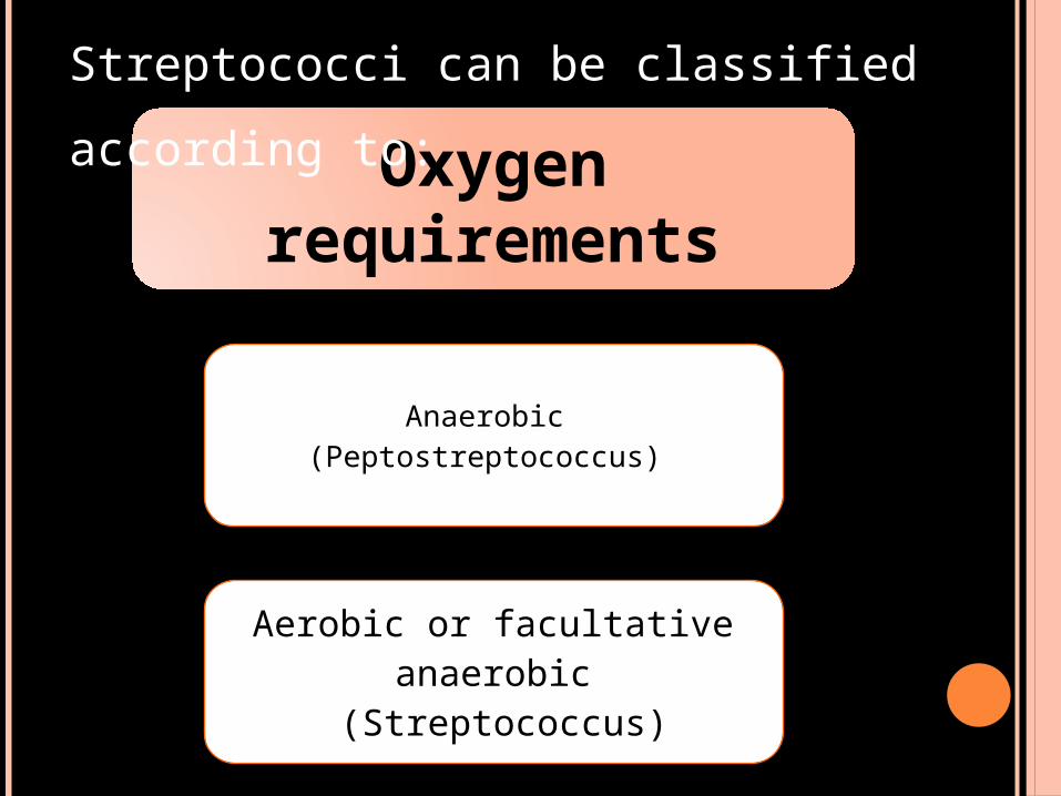
Oxygen requirements
Anaerobic (Peptostreptococcus)
Aerobic or facultative anaerobic
(Streptococcus)
Streptococci can be classified
according to:
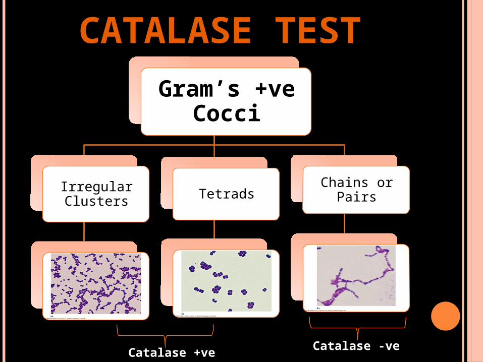
CATALASE TEST
Gram’s +ve Cocci
Irregular Clusters Tetrads
Chains or Pairs
Catalase +ve Catalase -ve
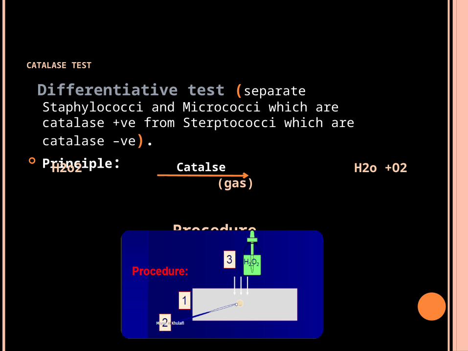
CATALASE TEST
Differentiative test (separate Staphylococci and Micrococci which are catalase +ve from Sterptococci which are catalase –ve).
Principle:
Procedure
H2o2 H2o +O2 (gas)Catalse Air bubbles Air bubbles
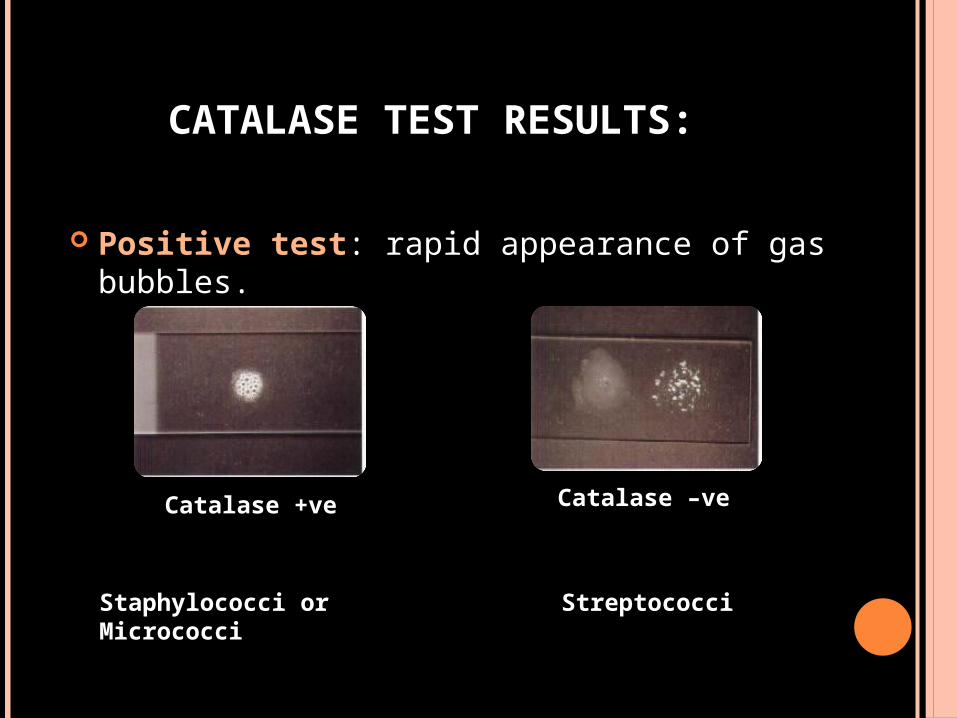
CATALASE TEST RESULTS:
Positive test: rapid appearance of gas bubbles.
Catalase +ve Catalase –ve
Staphylococci or Micrococci Streptococci
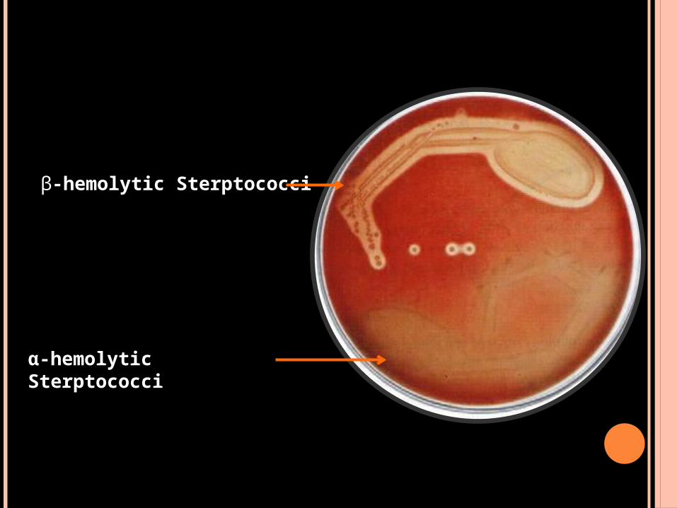
GROWTH ON BLOOD AGAR
Sterptococci are divided into three main groups accorging to its action on erythrocytes:
1.β-hemolytic Sterptococci. 2.α-hemolytic Sterptococci. 3.γ-hemolytic Sterptococci.
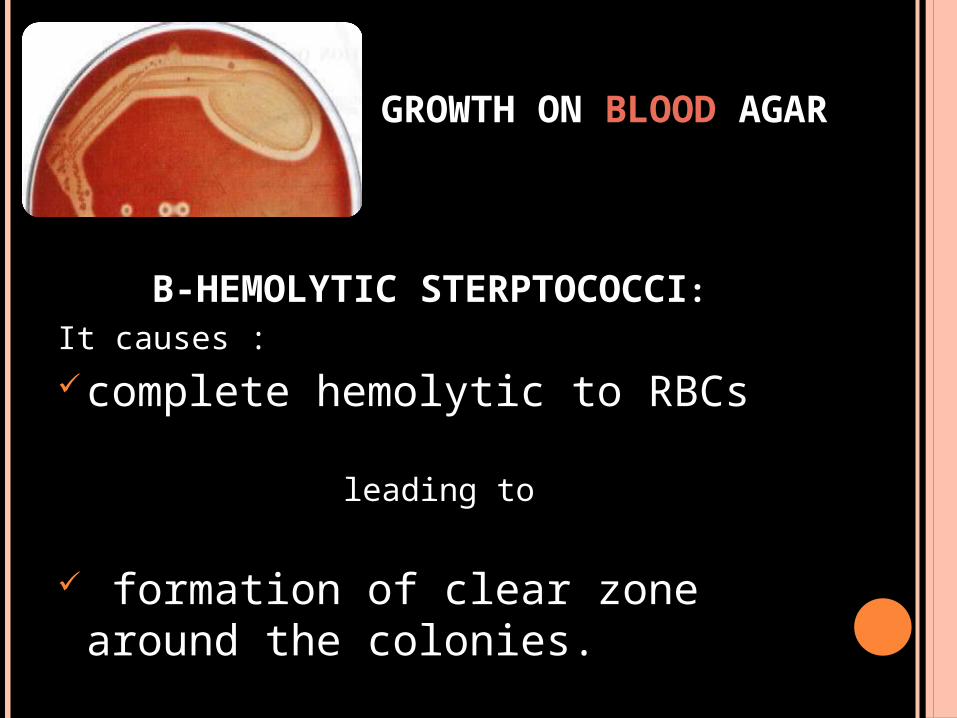
GROWTH ON BLOOD AGAR
Β-HEMOLYTIC STERPTOCOCCI: It causes :
complete hemolytic to RBCs
leading to
formation of clear zone around the colonies.
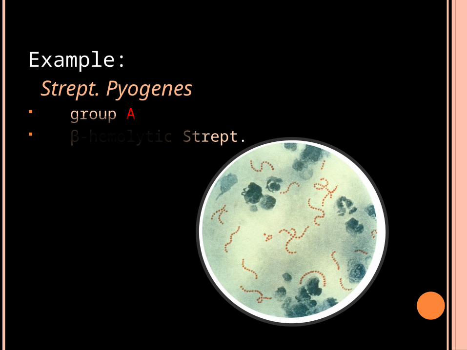
Example: Strept. Pyogenes group A β-hemolytic Strept.
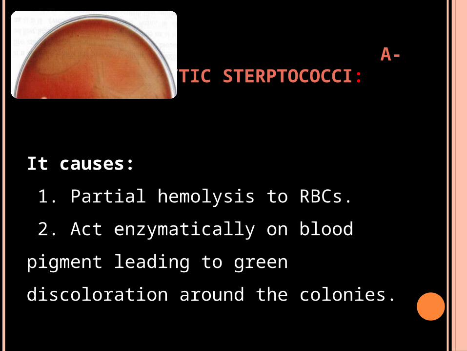
Α-HEMOLYTIC STERPTOCOCCI:
It causes:
1. Partial hemolysis to RBCs.
2. Act enzymatically on blood pigment
leading to green discoloration around
the colonies.
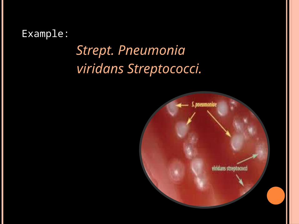
Example:
Strept. Pneumonia viridans Streptococci.
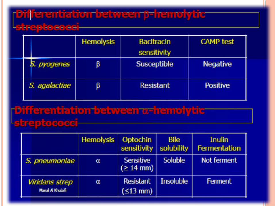
β-hemolytic Sterptococci
α-hemolytic Sterptococci:
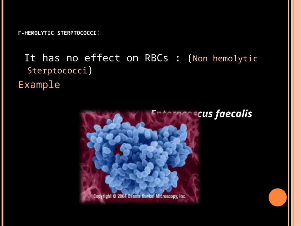
Γ-HEMOLYTIC STERPTOCOCCI:
It has no effect on RBCs : (Non hemolytic Sterptococci)
Example
Enterococcus faecalis
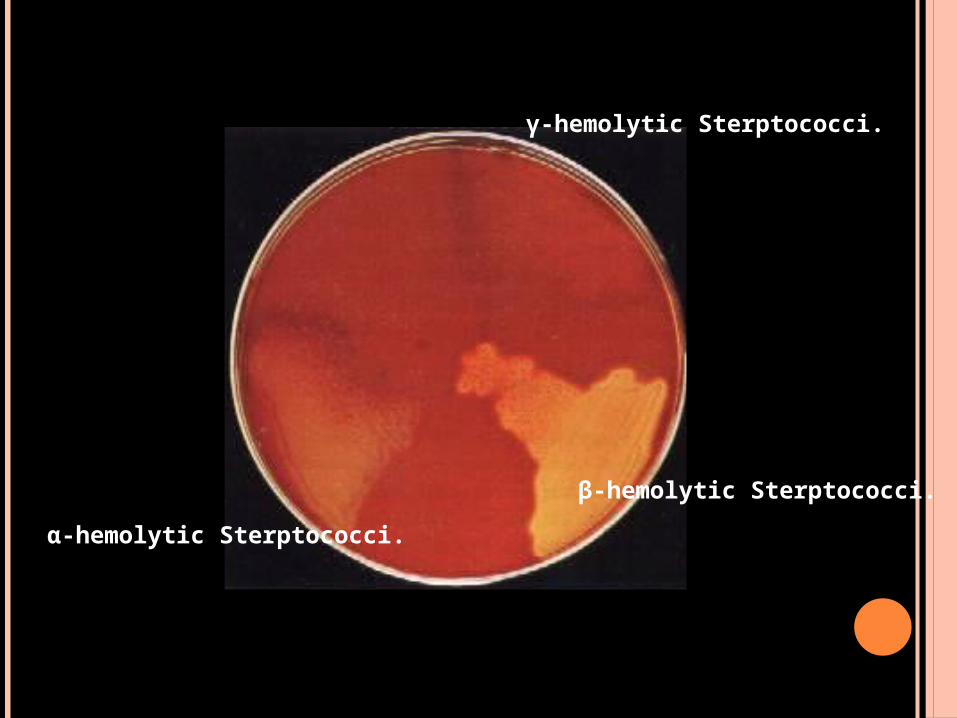
γ-hemolytic Sterptococci.
α-hemolytic Sterptococci.
β-hemolytic Sterptococci.
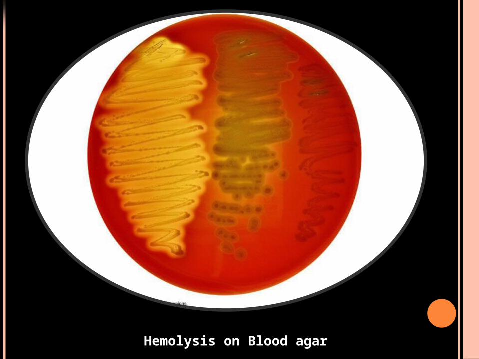
Hemolysis on Blood agar
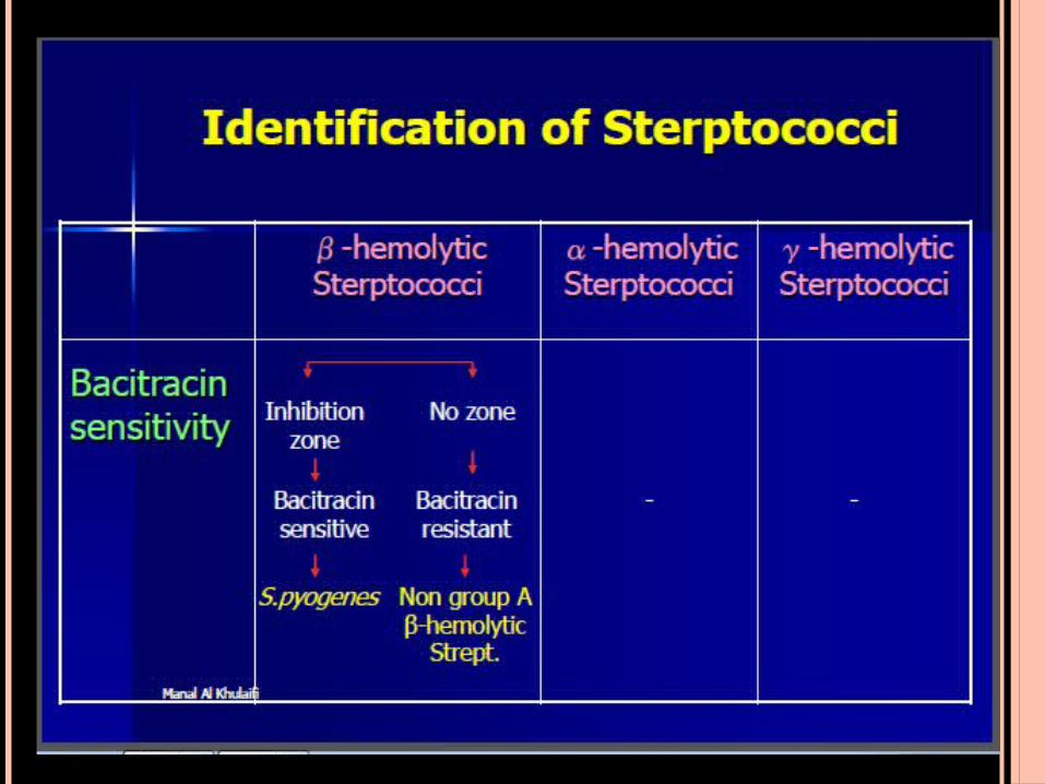
β-hemolytic Sterptococci
Definitive test to differentiate between ;
S.Pyogenes & Non group A β-hemolytic
Streptococci
Principle:
(Bacitracin Sensitivity Test)
A low conc. of Bacitracin (0.04 units) will selectively inhibit the growth of S.pyogenes giving a zone of inhibition around the disc .
Procedure: 1. Inoculate blood agar plate with the test organism.
2. Aseptically apply Bacitracin disc onto the center of the streaked area.
3. Incubate the plate at 35oC for 18 hrs.
Results: Positive test: any zone of inhibition around the disc.
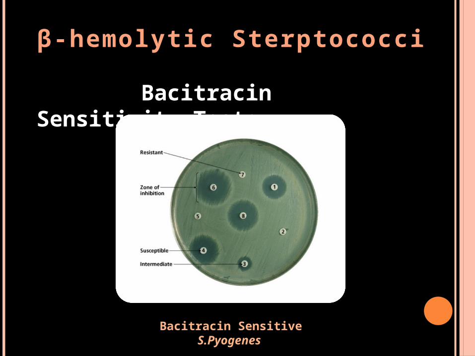
β-hemolytic Sterptococci
Bacitracin Sensitivity Test:
Bacitracin Sensitive S.Pyogenes
Group A streptococci
Pathogenesis and Virulence Factors; Structural components: M protein M Lipoteichoic acid & F protein Hyaluronic acid capsule, which acts to camouflage the bacteria Enzymes Streptokinases Deoxynucleases C5a peptidase Pyrogenic toxins Streptolysins Streptolysin O lyse red blood cells, white blood cells, and platelets Streptolysin S
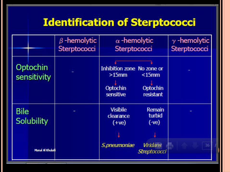
Α-HEMOLYTIC STREPTOCOCCI
Definitive test to differentiate between S.Pneumoniae & Viridans Streptococci
1. Optochin Sensitivity Test:
S.Pneumoniae is inhibited by less than 5
μg/ml Optochin reagent giving a zone of
inhibition more than 15 mm in diameter.
α-hemolytic Sterptococci 1.Optochin Sensitivity Test:
Procedure: 1. Inoculate blood agar plate with the
test organism. 2. Aseptically apply Optochin disc
onto the center of the streaked area. 3. Incubate the plate at 35oC for 18
hrs. 4. Accurately measure the diameter
of the inhibition zone around the disc.
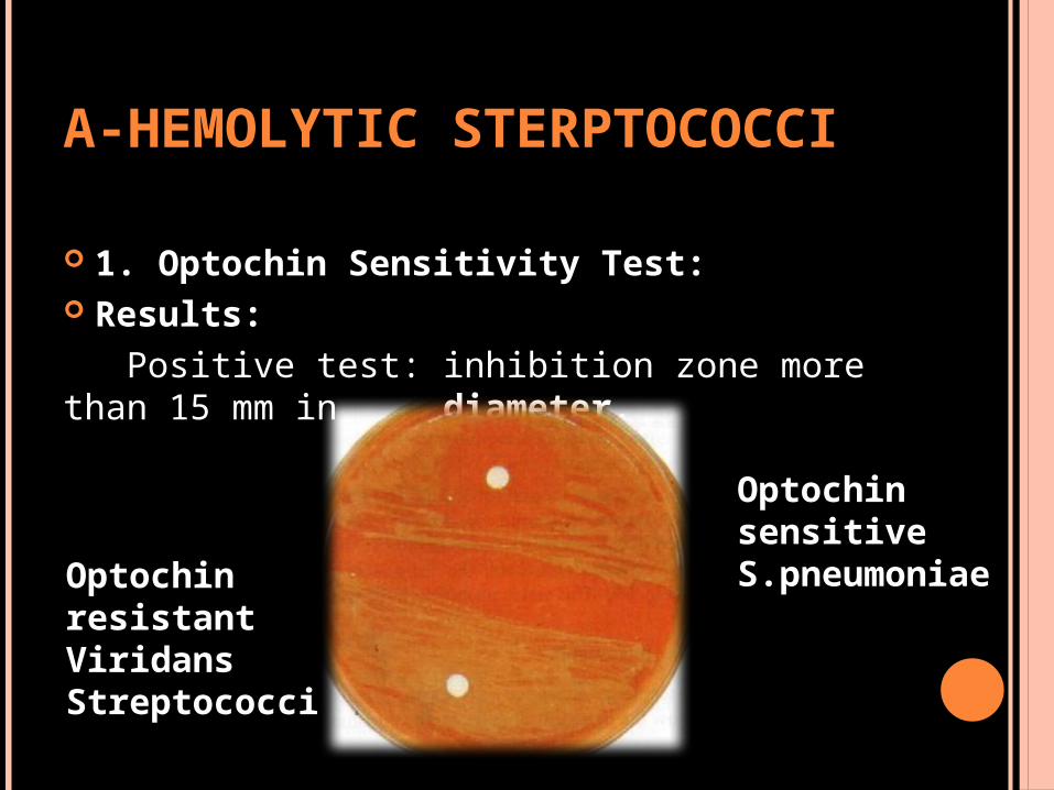
Α-HEMOLYTIC STERPTOCOCCI
1. Optochin Sensitivity Test: Results: Positive test: inhibition zone more than 15 mm in diameter.
Optochin sensitive S.pneumoniae
Optochin resistant Viridans Streptococci
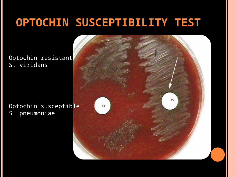
OPTOCHIN SUSCEPTIBILITY TEST
Optochin resistant S. viridans
Optochin susceptible S. pneumoniae
Α-HEMOLYTIC STREPTOCOCCI
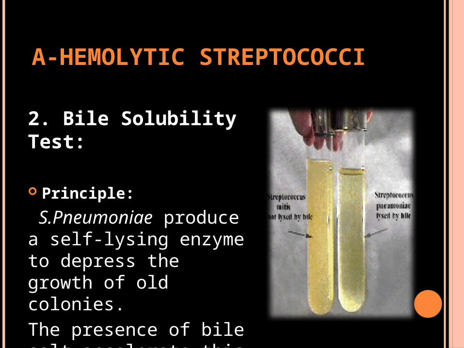
2. Bile Solubility Test:
Principle:
S.Pneumoniae produce a self-lysing enzyme to depress the growth of old colonies. The presence of bile salt accelerate this process.
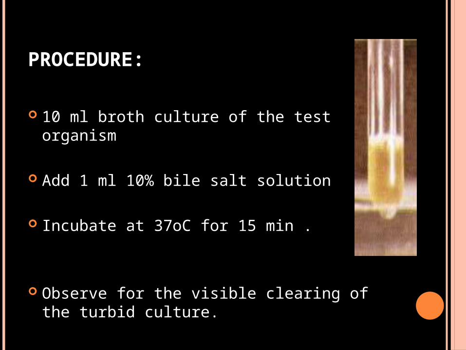
PROCEDURE:
10 ml broth culture of the test organism
Add 1 ml 10% bile salt solution
Incubate at 37oC for 15 min .
Observe for the visible clearing of the turbid culture.
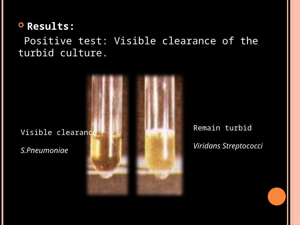
Results: Positive test: Visible clearance of the turbid culture.
Remain turbid
Viridans Streptococci
Visible clearance S.Pneumoniae
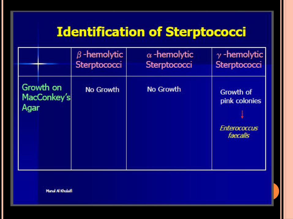
Γ-HEMOLYTIC STERPTOCOCCI
Definitive test for Enterococcus faecalis Growth on MacConkey’s agar.
Principle: MacConkey’s agar is a selective medium for Gram’s
–ve bacteria. It contains bile salt and crystal violet to inhibit the
growth of Gram’s +ve bacteria. Enterococcus faecalis is the only Streptococcus
species which can grow on MacConkey’s agar giving pink colonies.
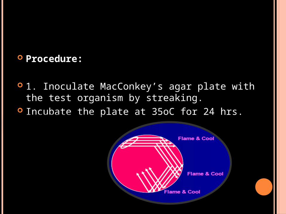
Procedure:
1. Inoculate MacConkey’s agar plate with the test organism by streaking.
Incubate the plate at 35oC for 24 hrs.
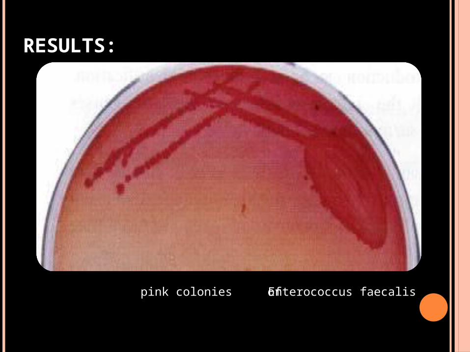
RESULTS:
pink colonies of Enterococcus faecalis
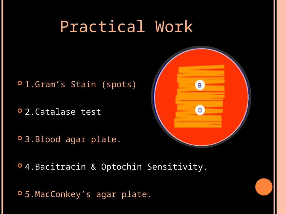
Practical Work
1.Gram’s Stain (spots)
2.Catalase test
3.Blood agar plate.
4.Bacitracin & Optochin Sensitivity.
5.MacConkey’s agar plate.
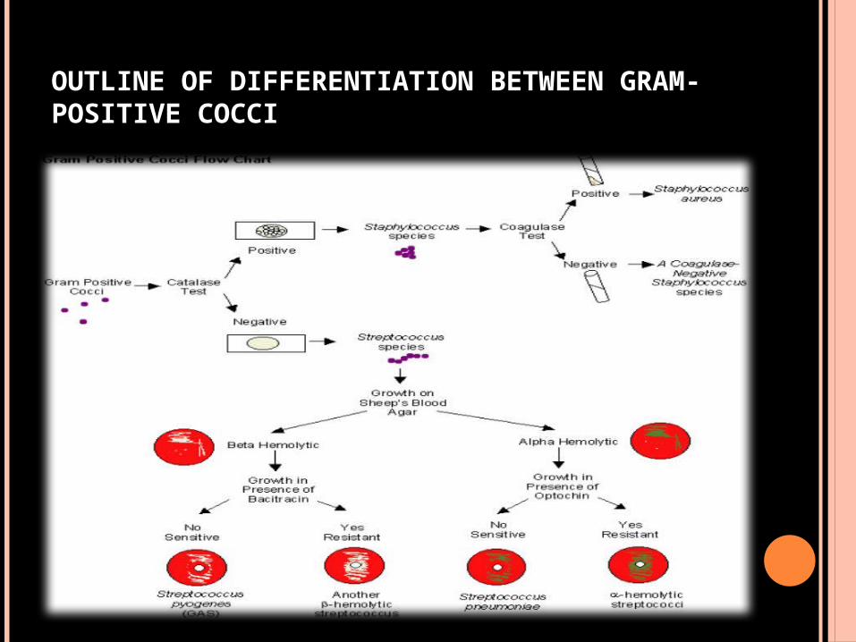
OUTLINE OF DIFFERENTIATION BETWEEN GRAM-POSITIVE COCCI