Εnhancing contrast agents and radiotracers performance through … · 2017-08-16 · potential...
3
1,8 Mario Ganau MD, PhD, FACS, 2 Nikolaos Ch. Syrmos MD, ΜSc, PhD, 3 Felice D'Arco MD, 4 Laura Ganau 5 Salvatore Chibbaro MD, PhD, 6 Lara Prisco MD, Msc, 7 Gianfranco K.I. Ligarotti MD, 1,4 Rossano Ambu MD, 8 Andrea Soddu PhD 1. Department of Surgical Science, University of Cagliari, Italy 2. Department of Physical Education and Sport Science, Physical Activity and Quality of Life, Aristotle University of Thessaloniki, Macedonia, Greece 3. Department of Neuroradiology, Great Ormond Street Hospital for Children, UK 4. School of Medicine, University of Cagliari, Italy 5. Department of Neurosurgery, Hautepierre University Hospital, Strasbourg, France 6. Neurosciences Intensive Care Unit, John Radcliffe Hospital and Nuffield Department of Clinical Neuroscience, University of Oxford, UK 7. Aerospace Medical Institute, Milan, Italy 8. Department of Physics and Astrophysics, University of Western Ontario, London, Canada Keywords: Contrast Agents - Radiotracers -Biomedical Engineering - Nanotechnology -Hyaluronic Acid Corresponding author: Mario Ganau MD, PhD, FACS Address: Department of Surgical Science, University of Cagliari, Italy [email protected] Receved: 6 March 2017 Accepted revised: 17 May 2017 Ε nhancing contrast agents and radiotracers performance through hyaluronic acid-coating in neuroradiology and nuclear medicine Abstract The use of hyaluronic acid nanoshells has been proposed to encapsulate prodrugs and exploit the mec- hanisms of interactions between living cells, like endocytes or cancer cells and hyaluronic acid, which is a natural component of the extracellular matrix. In this review we describe the potential and the limits of this promising research trend and discuss the theoretical advantages of such an engineering approach. Is it a possible scalability to increase the efficacy and biodegradability of molecules like contrast media and radio- tracers especially for neuroradiology and nuclear medicine studies. Hell J Nucl Med 2017; 20(2): 166-168 Epub ahead of print: 12 July 2017 Published online: 8 August 2017 Introduction O ne of the rapidly growing research elds referring to conventional and high-Tes- la magnetic resonance imaging (MRI) and nuclear medicine studies is the study of new contrast agents and radiotracers capable to early detect microaggre- gates of cancer cells and cancer recurrence. Creation and development of solid tumors, of circulating tumor cells (CTC) and of early metastases may thus be a reality in the future Interaction between hyaluronic acid nanoparticles and living cells Since hyaluronic acid (HA) has immunoneutrality, others [2, 4] and we [3, 5], proposed it as a biocompatible and biodegradable material for tissue engineering and for the deve- lopment of delivery of various drug systems. Recently, formulations of several drugs or prodrugs conjugated to polymeric coated HA nanoparticles of poly(ε-coprolactone), polylactide, poly(lactic-co-glycolic acid), polyethylene-glycol, polycarylates and chito- san were found effective as smart delivery systems both in vitro and in vivo [5-9]. Hyaluronic acid is a natural linear polysaccharide constituted by repeating units of N- acetyl-D-glucosamine and D-glucuronic acid with monosaccharides and linked together by alternating β-1,3 and β-1,4 glycosidic bonds. The carboxyl groups of HA are predominantly ionized at pH 7.4 and therefore in physiological conditions, HA appears as a polyanion, known as hyaluronan [10]. Hyaluronic acid is found in a wide range of molecular weights ranging from 20kDa of the HA oligomers (o-HA), to the high-mole- cular weight (HMW) of bulk HA (~1.5MDa). In solution, the chains of HA adopt a random coil conformation, and its high hydrophilic nature leads to multiple hydrogen bonds with H O, explaining the viscous and elastic characteristics of the connective tissues in 2 which this polysaccharide is abundant. Besides chemical conjugation, it has been proved that HA can also be linked to other prodrugs or to proper delivery systems by weak interactions such as those involved in the formation of ion pairs [11, 12] expanding the number of possible candidates for conjugation with or encapsulation within those nanocarriers. Hyaluronic acid conjugates could leverage on their propensity to overcome the blood brain barrier (BBB), so that rst to produce biologic effects on the central nervous system (CNS) and secondly to provide a specic tumor targeting activity, which takes advan- tage of the peculiar interaction between HA receptors on the bilipidic membrane of glioma cells and the enzymes for HA degradation contained in the extracellular matrix (ECM). The rst, is mainly based on receptor-mediated endocytosis of HA nanoparticles at the level of brain capillary endothelial cells [13]. The second, represents the basis for Short Review Article Hellenic Journal of Nuclear Medicine May-August 2017 • www.nuclmed.gr 166
Transcript of Εnhancing contrast agents and radiotracers performance through … · 2017-08-16 · potential...
1,8Mario Ganau MD, PhD, FACS,2Nikolaos Ch. Syrmos MD, ΜSc,
PhD,3Felice D'Arco MD,4Laura Ganau
5Salvatore Chibbaro MD, PhD,6Lara Prisco MD, Msc,
7Gianfranco K.I. Ligarotti MD,1,4Rossano Ambu MD,
8Andrea Soddu PhD
1. Department of Surgical Science, University of Cagliari, Italy 2. Department of Physical Education and Sport Science, Physical Activity and Quality of Life, Aristotle University of Thessaloniki, Macedonia, Greece3. Department of Neuroradiology, Great Ormond Street Hospital for Children, UK4. School of Medicine, University of Cagliari, Italy5. Department of Neurosurgery, Hautepierre University Hospital, Strasbourg, France6. Neurosciences Intensive Care Unit, John Radcliffe Hospital and Nuffield Department of Clinical Neuroscience, University of Oxford, UK7. Aerospace Medical Institute, Milan, Italy8. Department of Physics and Astrophysics, University of Western Ontario, London, Canada
Keywords: Contrast Agents
- Radiotracers -Biomedical
Engineering - Nanotechnology
-Hyaluronic Acid
Corresponding author: Mario Ganau MD, PhD, FACSAddress: Department of Surgical Science, University of Cagliari, Italy [email protected]
Rece�ved: 6 March 2017 Accepted revised: 17 May 2017
Εnhancing contrast agents and radiotracers performance
through hyaluronic acid-coating in neuroradiology and
nuclear medicine
AbstractThe use of hyaluronic acid nanoshells has been proposed to encapsulate prodrugs and exploit the mec-hanisms of interactions between living cells, like endocytes or cancer cells and hyaluronic acid, which is a natural component of the extracellular matrix. In this review we describe the potential and the limits of this promising research trend and discuss the theoretical advantages of such an engineering approach. Is it a possible scalability to increase the efficacy and biodegradability of molecules like contrast media and radio-tracers especially for neuroradiology and nuclear medicine studies.
Hell J Nucl Med 2017; 20(2): 166-168 Epub ahead of print: 12 July 2017 Published online: 8 August 2017
Introduction
One of the rapidly growing research �elds referring to conventional and high-Tes-la magnetic resonance imaging (MRI) and nuclear medicine studies is the study of new contrast agents and radiotracers capable to early detect microaggre-
gates of cancer cells and cancer recurrence. Creation and development of solid tumors, of circulating tumor cells (CTC) and of early metastases may thus be a reality in the future
Interaction between hyaluronic acid nanoparticles and living cellsSince hyaluronic acid (HA) has immunoneutrality, others [2, 4] and we [3, 5], proposed it as a biocompatible and biodegradable material for tissue engineering and for the deve-lopment of delivery of various drug systems. Recently, formulations of several drugs or prodrugs conjugated to polymeric coated HA nanoparticles of poly(ε-coprolactone), polylactide, poly(lactic-co-glycolic acid), polyethylene-glycol, polycarylates and chito-san were found effective as smart delivery systems both in vitro and in vivo [5-9].
Hyaluronic acid is a natural linear polysaccharide constituted by repeating units of N-acetyl-D-glucosamine and D-glucuronic acid with monosaccharides and linked together by alternating β-1,3 and β-1,4 glycosidic bonds. The carboxyl groups of HA are predominantly ionized at pH 7.4 and therefore in physiological conditions, HA appears as a polyanion, known as hyaluronan [10]. Hyaluronic acid is found in a wide range of molecular weights ranging from 20kDa of the HA oligomers (o-HA), to the high-mole-cular weight (HMW) of bulk HA (~1.5MDa). In solution, the chains of HA adopt a random coil conformation, and its high hydrophilic nature leads to multiple hydrogen bonds with H O, explaining the viscous and elastic characteristics of the connective tissues in 2
which this polysaccharide is abundant. Besides chemical conjugation, it has been proved that HA can also be linked to other prodrugs or to proper delivery systems by weak interactions such as those involved in the formation of ion pairs [11, 12] expanding the number of possible candidates for conjugation with or encapsulation within those nanocarriers.
Hyaluronic acid conjugates could leverage on their propensity to overcome the blood brain barrier (BBB), so that �rst to produce biologic effects on the central nervous system (CNS) and secondly to provide a speci�c tumor targeting activity, which takes advan-tage of the peculiar interaction between HA receptors on the bilipidic membrane of glioma cells and the enzymes for HA degradation contained in the extracellular matrix (ECM). The �rst, is mainly based on receptor-mediated endocytosis of HA nanoparticles at the level of brain capillary endothelial cells [13]. The second, represents the basis for
Short Review Article
93Hellenic Journal of Nuclear Medicine May-August 2017• www.nuclmed.gr 166
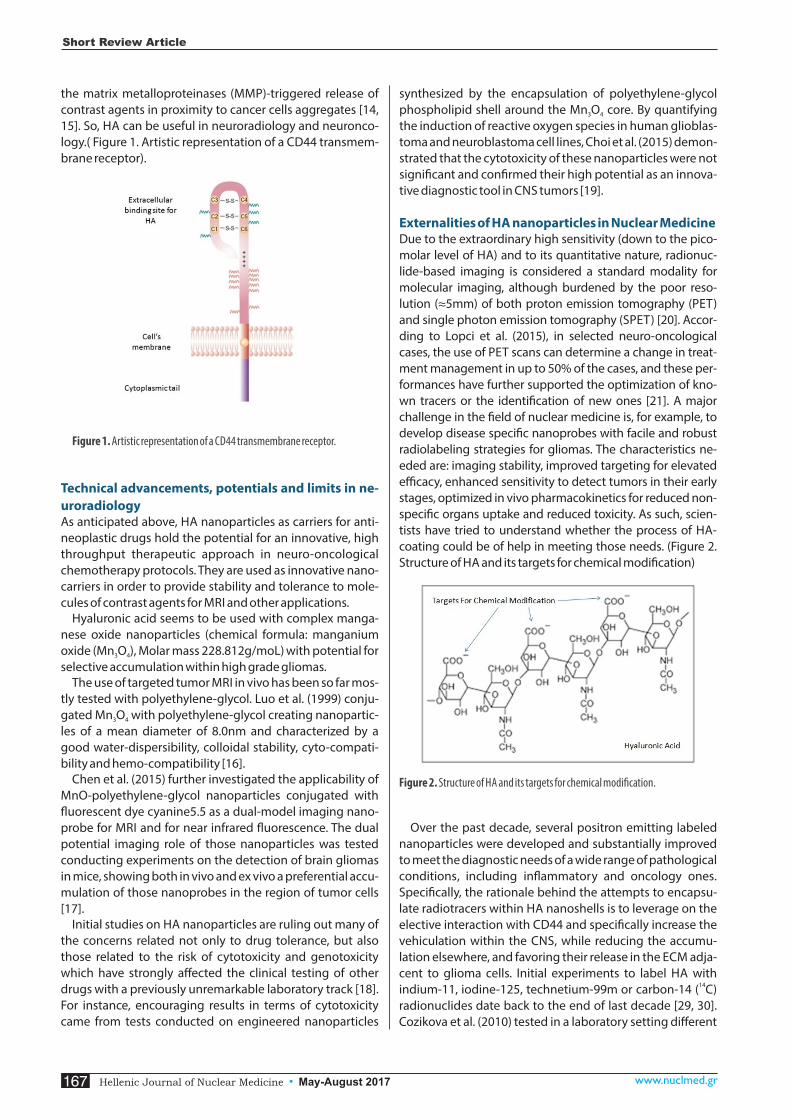
the matrix metalloproteinases (MMP)-triggered release of contrast agents in proximity to cancer cells aggregates [14, 15]. So, HA can be useful in neuroradiology and neuronco-logy.( Figure 1. Artistic representation of a CD44 transmem-brane receptor).
Figure 1. Artistic representation of a CD44 transmembrane receptor.
Technical advancements, potentials and limits in ne-uroradiologyAs anticipated above, HA nanoparticles as carriers for anti-neoplastic drugs hold the potential for an innovative, high throughput therapeutic approach in neuro-oncological chemotherapy protocols. They are used as innovative nano-carriers in order to provide stability and tolerance to mole-cules of contrast agents for MRI and other applications.
Hyaluronic acid seems to be used with complex manga-nese oxide nanoparticles (chemical formula: manganium oxide (Mn O ), Molar mass 228.812g/moL) with potential for 3 4
selective accumulation within high grade gliomas. The use of targeted tumor MRI in vivo has been so far mos-
tly tested with polyethylene-glycol. Luo et al. (1999) conju-gated Mn O with polyethylene-glycol creating nanopartic-3 4
les of a mean diameter of 8.0nm and characterized by a good water-dispersibility, colloidal stability, cyto-compati-bility and hemo-compatibility [16].
Chen et al. (2015) further investigated the applicability of MnO-polyethylene-glycol nanoparticles conjugated with �uorescent dye cyanine5.5 as a dual-model imaging nano-probe for MRI and for near infrared �uorescence. The dual potential imaging role of those nanoparticles was tested conducting experiments on the detection of brain gliomas in mice, showing both in vivo and ex vivo a preferential accu-mulation of those nanoprobes in the region of tumor cells [17].
Initial studies on HA nanoparticles are ruling out many of the concerns related not only to drug tolerance, but also those related to the risk of cytotoxicity and genotoxicity which have strongly affected the clinical testing of other drugs with a previously unremarkable laboratory track [18]. For instance, encouraging results in terms of cytotoxicity came from tests conducted on engineered nanoparticles
synthesized by the encapsulation of polyethylene-glycol phospholipid shell around the Mn O core. By quantifying 3 4
the induction of reactive oxygen species in human glioblas-toma and neuroblastoma cell lines, Choi et al. (2015) demon-strated that the cytotoxicity of these nanoparticles were not signi�cant and con�rmed their high potential as an innova-tive diagnostic tool in CNS tumors [19].
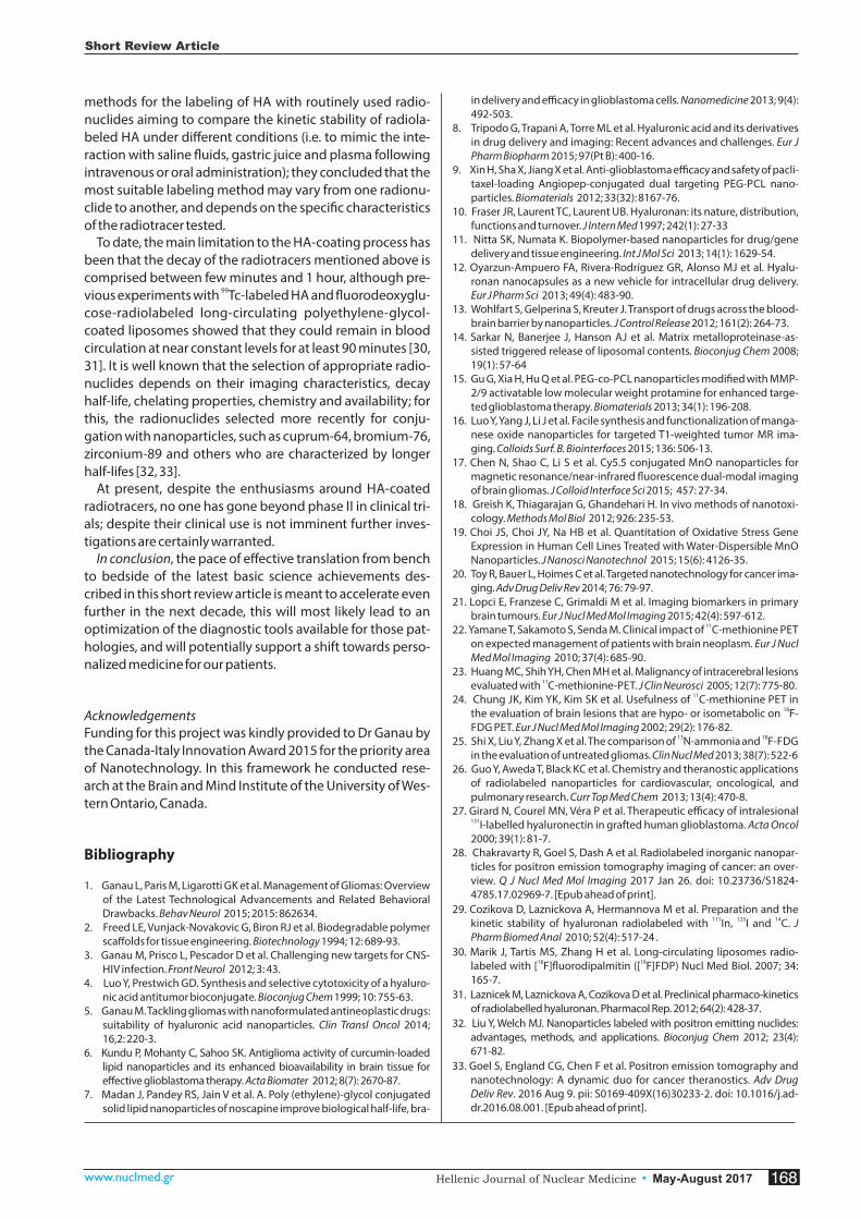
Externalities of HA nanoparticles in Nuclear Medicine Due to the extraordinary high sensitivity (down to the pico-molar level of HA) and to its quantitative nature, radionuc-lide-based imaging is considered a standard modality for molecular imaging, although burdened by the poor reso-lution (≈5mm) of both proton emission tomography (PET) and single photon emission tomography (SPET) [20]. Accor-ding to Lopci et al. (2015), in selected neuro-oncological cases, the use of PET scans can determine a change in treat-ment management in up to 50% of the cases, and these per-formances have further supported the optimization of kno-wn tracers or the identi�cation of new ones [21]. A major challenge in the �eld of nuclear medicine is, for example, to develop disease speci�c nanoprobes with facile and robust radiolabeling strategies for gliomas. The characteristics ne-eded are: imaging stability, improved targeting for elevated efficacy, enhanced sensitivity to detect tumors in their early stages, optimized in vivo pharmacokinetics for reduced non-speci�c organs uptake and reduced toxicity. As such, scien-tists have tried to understand whether the process of HA-coating could be of help in meeting those needs. (Figure 2. Structure of HA and its targets for chemical modi�cation)
Figure 2. Structure of HA and its targets for chemical modi�cation.
Over the past decade, several positron emitting labeled nanoparticles were developed and substantially improved to meet the diagnostic needs of a wide range of pathological conditions, including in�ammatory and oncology ones. Speci�cally, the rationale behind the attempts to encapsu-late radiotracers within HA nanoshells is to leverage on the elective interaction with CD44 and speci�cally increase the vehiculation within the CNS, while reducing the accumu-lation elsewhere, and favoring their release in the ECM adja-cent to glioma cells. Initial experiments to label HA with
14indium-11, iodine-125, technetium-99m or carbon-14 ( C) radionuclides date back to the end of last decade [29, 30]. Cozikova et al. (2010) tested in a laboratory setting different
Short Review Article
993 Hellenic Journal of Nuclear Medicine May-August 2017• www.nuclmed.gr167
methods for the labeling of HA with routinely used radio-nuclides aiming to compare the kinetic stability of radiola-beled HA under different conditions (i.e. to mimic the inte-raction with saline �uids, gastric juice and plasma following intravenous or oral administration); they concluded that the most suitable labeling method may vary from one radionu-clide to another, and depends on the speci�c characteristics of the radiotracer tested.
To date, the main limitation to the HA-coating process has been that the decay of the radiotracers mentioned above is comprised between few minutes and 1 hour, although pre-
99vious experiments with Tc-labeled HA and �uorodeoxyglu-cose-radiolabeled long-circulating polyethylene-glycol-coated liposomes showed that they could remain in blood circulation at near constant levels for at least 90 minutes [30, 31]. It is well known that the selection of appropriate radio-nuclides depends on their imaging characteristics, decay half-life, chelating properties, chemistry and availability; for this, the radionuclides selected more recently for conju-gation with nanoparticles, such as cuprum-64, bromium-76, zirconium-89 and others who are characterized by longer half-lifes [32, 33].
At present, despite the enthusiasms around HA-coated radiotracers, no one has gone beyond phase II in clinical tri-als; despite their clinical use is not imminent further inves-tigations are certainly warranted.
In conclusion, the pace of effective translation from bench to bedside of the latest basic science achievements des-cribed in this short review article is meant to accelerate even further in the next decade, this will most likely lead to an optimization of the diagnostic tools available for those pat-hologies, and will potentially support a shift towards perso-nalized medicine for our patients.
Acknowledgements Funding for this project was kindly provided to Dr Ganau by the Canada-Italy Innovation Award 2015 for the priority area of Nanotechnology. In this framework he conducted rese-arch at the Brain and Mind Institute of the University of Wes-tern Ontario, Canada.
Bibliography
1. Ganau L, Paris M, Ligarotti GK et al. Management of Gliomas: Overview of the Latest Technological Advancements and Related Behavioral Drawbacks. Behav Neurol 2015; 2015: 862634.
2. Freed LE, Vunjack-Novakovic G, Biron RJ et al. Biodegradable polymer scaffolds for tissue engineering. Biotechnology 1994; 12: 689-93.
3. Ganau M, Prisco L, Pescador D et al. Challenging new targets for CNS-HIV infection. Front Neurol 2012; 3: 43.
4. Luo Y, Prestwich GD. Synthesis and selective cytotoxicity of a hyaluro-nic acid antitumor bioconjugate. Bioconjug Chem 1999; 10: 755-63.
5. Ganau M. Tackling gliomas with nanoformulated antineoplastic drugs: suitability of hyaluronic acid nanoparticles. Clin Transl Oncol 2014; 16,2: 220-3.
6. Kundu P, Mohanty C, Sahoo SK. Antiglioma activity of curcumin-loaded lipid nanoparticles and its enhanced bioavailability in brain tissue for effective glioblastoma therapy. Acta Biomater 2012; 8(7): 2670-87.
7. Madan J, Pandey RS, Jain V et al. A. Poly (ethylene)-glycol conjugated solid lipid nanoparticles of noscapine improve biological half-life, bra-
in delivery and efficacy in glioblastoma cells. Nanomedicine 2013; 9(4): 492-503.
8. Tripodo G, Trapani A, Torre ML et al. Hyaluronic acid and its derivatives in drug delivery and imaging: Recent advances and challenges. Eur J Pharm Biopharm 2015; 97(Pt B): 400-16.
9. Xin H, Sha X, Jiang X et al. Anti-glioblastoma efficacy and safety of pacli-taxel-loading Angiopep-conjugated dual targeting PEG-PCL nano-particles. Biomaterials 2012; 33(32): 8167-76.
10. Fraser JR, Laurent TC, Laurent UB. Hyaluronan: its nature, distribution, functions and turnover. J Intern Med 1997; 242(1): 27-33
11. Nitta SK, Numata K. Biopolymer-based nanoparticles for drug/gene delivery and tissue engineering. Int J Mol Sci 2013; 14(1): 1629-54.
12. Oyarzun-Ampuero FA, Rivera-Rodríguez GR, Alonso MJ et al. Hyalu-ronan nanocapsules as a new vehicle for intracellular drug delivery. Eur J Pharm Sci 2013; 49(4): 483-90.
13. Wohlfart S, Gelperina S, Kreuter J. Transport of drugs across the blood-brain barrier by nanoparticles. J Control Release 2012; 161(2): 264-73.
14. Sarkar N, Banerjee J, Hanson AJ et al. Matrix metalloproteinase-as-sisted triggered release of liposomal contents. Bioconjug Chem 2008; 19(1): 57-64
15. Gu G, Xia H, Hu Q et al. PEG-co-PCL nanoparticles modi�ed with MMP-2/9 activatable low molecular weight protamine for enhanced targe-ted glioblastoma therapy. Biomaterials 2013; 34(1): 196-208.
16. Luo Y, Yang J, Li J et al. Facile synthesis and functionalization of manga-nese oxide nanoparticles for targeted T1-weighted tumor MR ima-ging. Colloids Surf. B. Biointerfaces 2015; 136: 506-13.
17. Chen N, Shao C, Li S et al. Cy5.5 conjugated MnO nanoparticles for magnetic resonance/near-infrared �uorescence dual-modal imaging of brain gliomas. J Colloid Interface Sci 2015; 457: 27-34.
18. Greish K, Thiagarajan G, Ghandehari H. In vivo methods of nanotoxi-cology. Methods Mol Biol 2012; 926: 235-53.
19. Choi JS, Choi JY, Na HB et al. Quantitation of Oxidative Stress Gene Expression in Human Cell Lines Treated with Water-Dispersible MnO Nanoparticles. J Nanosci Nanotechnol 2015; 15(6): 4126-35.
20. Toy R, Bauer L, Hoimes C et al. Targeted nanotechnology for cancer ima-ging. Adv Drug Deliv Rev 2014; 76: 79-97.
21. Lopci E, Franzese C, Grimaldi M et al. Imaging biomarkers in primary brain tumours. Eur J Nucl Med Mol Imaging 2015; 42(4): 597-612.
1122. Yamane T, Sakamoto S, Senda M. Clinical impact of C-methionine PET on expected management of patients with brain neoplasm. Eur J Nucl Med Mol Imaging 2010; 37(4): 685-90.
23. Huang MC, Shih YH, Chen MH et al. Malignancy of intracerebral lesions 11evaluated with C-methionine-PET. J Clin Neurosci 2005; 12(7): 775-80.
1124. Chung JK, Kim YK, Kim SK et al. Usefulness of C-methionine PET in 18the evaluation of brain lesions that are hypo- or isometabolic on F-
FDG PET. Eur J Nucl Med Mol Imaging 2002; 29(2): 176-82.13 1825. Shi X, Liu Y, Zhang X et al. The comparison of N-ammonia and F-FDG
in the evaluation of untreated gliomas. Clin Nucl Med 2013; 38(7): 522-626. Guo Y, Aweda T, Black KC et al. Chemistry and theranostic applications
of radiolabeled nanoparticles for cardiovascular, oncological, and pulmonary research. Curr Top Med Chem 2013; 13(4): 470-8.
27. Girard N, Courel MN, Véra P et al. Therapeutic efficacy of intralesional 131I-labelled hyaluronectin in grafted human glioblastoma. Acta Oncol 2000; 39(1): 81-7.
28. Chakravarty R, Goel S, Dash A et al. Radiolabeled inorganic nanopar-ticles for positron emission tomography imaging of cancer: an over-view. Q J Nucl Med Mol Imaging 2017 Jan 26. doi: 10.23736/S1824-4785.17.02969-7. [Epub ahead of print].
29. Cozikova D, Laznickova A, Hermannova M et al. Preparation and the 111 125 14kinetic stability of hyaluronan radiolabeled with In, I and C. J
Pharm Biomed Anal 2010; 52(4): 517-24 .30. Marik J, Tartis MS, Zhang H et al. Long-circulating liposomes radio-
18 18labeled with [ F]�uorodipalmitin ([ F]FDP) Nucl Med Biol. 2007; 34: 165-7.
31. Laznicek M, Laznickova A, Cozikova D et al. Preclinical pharmaco-kinetics of radiolabelled hyaluronan. Pharmacol Rep. 2012; 64(2): 428-37.
32. Liu Y, Welch MJ. Nanoparticles labeled with positron emitting nuclides: advantages, methods, and applications. Bioconjug Chem 2012; 23(4): 671-82.
33. Goel S, England CG, Chen F et al. Positron emission tomography and nanotechnology: A dynamic duo for cancer theranostics. Adv Drug Deliv Rev. 2016 Aug 9. pii: S0169-409X(16)30233-2. doi: 10.1016/j.ad-dr.2016.08.001. [Epub ahead of print].
93Hellenic Journal of Nuclear Medicine May-August 2017• www.nuclmed.gr 168
Short Review Article