Neurosteroids Enhance Spontaneous Glutamate Release in … · 2002-05-31 · aminobutyric...
36
Neurosteroids Enhance Spontaneous Glutamate Release in Hippocampal Neurons: Possible Role of Metabotropic σ 1 ↑like Receptors Douglas A. Meyer, Mario Carta, L. Donald Partridge, Douglas F. Covey 1 and C. Fernando Valenzuela Department of Neurosciences University of New Mexico Health Sciences Center Albuquerque, NM 87131 and 1 Department of Molecular Biology and Pharmacology Washington University School of Medicine St. Louis, MO 63110 Running Title : Neurosteroids and glutamate release Supported by : NIH grants AA12684 and MH63126 (to C.F.V. and L.D.P.). D.F.C. is supported by NIH grant GM 47969. Send Correspondence to: C. Fernando Valenzuela, M.D., Ph.D. Department of Neurosciences BMSB 145 University of New Mexico HSC Albuquerque, NM 87131-5223 (505) 272-3128 Fax (505) 272-8082 [email protected] 1 Copyright 2002 by The American Society for Biochemistry and Molecular Biology, Inc. JBC Papers in Press. Published on May 31, 2002 as Manuscript M202592200 by guest on January 30, 2020 http://www.jbc.org/ Downloaded from
Transcript of Neurosteroids Enhance Spontaneous Glutamate Release in … · 2002-05-31 · aminobutyric...
Neurosteroids Enhance Spontaneous Glutamate Release in Hippocampal Neurons: Possible Role of Metabotropic σ 1↑like Receptors
Douglas A. Meyer, Mario Carta, L. Donald Partridge,Douglas F. Covey1 and C. Fernando Valenzuela
Department of NeurosciencesUniversity of New Mexico Health Sciences Center
Albuquerque, NM 87131
and
1Department of Molecular Biology and Pharmacology
Washington University School of MedicineSt. Louis, MO 63110
Running Title: Neurosteroids and glutamate release
Supported by: NIH grants AA12684 and MH63126 (to C.F.V. and L.D.P.). D.F.C. is supported by NIH grant GM 47969.
Send Correspondence to:
C. Fernando Valenzuela, M.D., Ph.D.Department of NeurosciencesBMSB 145University of New Mexico HSCAlbuquerque, NM 87131-5223(505) 272-3128Fax (505) [email protected]
1
Copyright 2002 by The American Society for Biochemistry and Molecular Biology, Inc.
JBC Papers in Press. Published on May 31, 2002 as Manuscript M202592200 by guest on January 30, 2020
http://ww
w.jbc.org/
Dow
nloaded from
Summary:
Pregnenolone sulfate (PREGS), one of the most abundantly produced neurosteroids
in the mammalian brain, improves cognitive performance in rodents. The mechanism of this
effect has been attributed to its allosteric modulatory actions on glutamate- and γ-
aminobutyric acid-gated ion channels. Here we report a novel effect of PREGS that could
also mediate some of its actions in the nervous system. We found that PREGS induces a
robust potentiation of the frequency, but not the amplitude of miniature excitatory
postsynaptic currents (mEPSCs) mediated by α-amino-3-hydroxy-5-methylisoxazole-4-
propionate (AMPA) receptors in cultured hippocampal neurons. PREGS also decreased
paired-pulse facilitation of autaptic EPSCs evoked by depolarization, indicating that it
modulates glutamate release probability presynaptically. PREGS potentiation of mEPSCs
was mimicked by dehydroepiandrosterone sulfate and (+)-pentazocine, but not by (-)-
pentazocine, the synthetic (-) enantiomer of PREGS or the inactive steroid
isopregnanolone. The σ receptor antagonists, haloperidol and BD-1063, blocked the effect
of PREGS on mEPSCs, as did pertussis toxin and the membrane-permeable Ca2+ chelator
BAPTA-AM. These results suggest that PREGS increases spontaneous glutamate release
via activation of a presynaptic Gi/o-coupled σ receptor and an elevation in intracellular
Ca2+ levels. We postulate that presynaptic actions of neurosteroids have a role in the
maturation and/or maintenance of synaptic networks and the processing of information in
the central nervous system.
2
by guest on January 30, 2020http://w
ww
.jbc.org/D
ownloaded from
Introduction
In the 1980s, Baulieu and collaborators made the important discovery that certain
steroids are synthesized in the central and peripheral nervous systems (1). These
compounds, known as neurosteroids, are produced locally in glial and neuronal cells and
can exert important modulatory actions in the nervous system. A particularly abundant
neurosteroid in the central nervous system is pregnenolone sulfate (PREGS) (1,2).
Although the neurophysiological role of endogenous PREGS has yet to be conclusively
established, experiments involving exogenous administration of this compound suggest that
it has a promnesic effect (3-7). For example, post-training injection of PREGS into the
hippocampus and amygdala of mice improves retention for foot-shock active avoidance
training (4). In addition, low levels of PREGS in the hippocampus were found to be
correlated with a deficiency in cognitive performance in aged rats, which could be
ameliorated by intra-hippocampal injections of this neurosteroid (5). More recently, it was
demonstrated that PREGS attenuates amyloid peptide-induced amnesia in mice (8). Thus,
PREGS or its analogs could potentially be used for the treatment of Alzheimer’s disease
and other neuropsychiatric disorders.
Although the mechanisms by which PREGS produces cognitive effects are not fully
understood, numerous studies suggest that this agent modulates several neuronal ion
channels (9). For instance, PREGS has been shown to inhibit γ-aminobutyric acid-type A
(GABAA) receptors (10), to potentiate N-methyl-D-aspartate (NMDA) receptors (11-14)
and to inhibit voltage-gated Ca2+ channels (15). In addition to ion channels, PREGS has
3
by guest on January 30, 2020http://w
ww
.jbc.org/D
ownloaded from
also been shown to target metabotropic receptors, such as σ receptors. These receptors
were initially thought to belong to the opioid family of receptors, but are now categorized
separately (16). Two classes of pharmacologically defined σ receptors are widely accepted
and are denoted as the σ1 and σ2 subtypes (16). σ1 receptors bind (+)-benzomorphans
and haloperidol with high affinity. In contrast, σ2 receptors bind haloperidol and (+)-
benzomorphans with low affinity, and they also bind benzomorphans without
enantioselectivity.
σ ligand binding sites can be detected both intracellularly on the endoplasmic reticulum (ER)
and extracellularly on the plasma membrane (17). A σ ligand-binding protein has recently
been cloned from a number of tissues, including brain (18). This binding protein exhibits the
pharmacological profile of σ1 receptors and is expressed in the ER (19-23). Although its
sequence has no known homology to other mammalian proteins, it shows similarity to the
yeast sterol C8-C7 isomerase involved in ergosterol biosynthesis (24). Plasma membrane σ
receptors can be activated by PREGS and other neurosteroids but, in contrast to σ ligand
binding proteins expressed in the ER, appear to be directly coupled to pertussis-sensitive G
proteins (25-28). Both the cDNA sequence and physiological role of these G protein-
coupled metabotropic σ receptors have yet to be fully characterized.
In this paper, we report a novel effect of PREGS on glutamate release that depends on
activation of plasma membrane σ receptors. Specifically, we measured the effects of this
neurosteroid on miniature excitatory postsynaptic currents (mEPSCs) mediated by the α-
4
by guest on January 30, 2020http://w
ww
.jbc.org/D
ownloaded from
amino-3-hydroxy-5-methylisoxazole-4-propionate (AMPA)-subtype of ionotropic
glutamate receptors. Miniature synaptic currents are the most elementary forms of synaptic
transmission, representing the postsynaptic responses to action potential-independent
spontaneous release of single presynaptic vesicles. It is well established that when a
modulator affects presynaptic neurotransmitter release, it produces a change in the
frequency, but not in the amplitude of miniature synaptic events (for instance, see (29)). We
found that PREGS selectively induces a robust increase in the frequency of mEPSCs,
indicating that it enhances the probability of glutamate release from presynaptic terminals.
Moreover, we show that this effect depends on an elevation in intracellular Ca2+ levels
triggered by activation of presynaptic Gi/o protein-coupled σ1↑like receptors.
5
by guest on January 30, 2020http://w
ww
.jbc.org/D
ownloaded from
Experimental Procedures
Cell Culture. Animal procedures were approved by the Institutional Animal Care and Use Committee of the University
of New Mexico Health Sciences Center and conform to National Institutes of Health guidelines. Neuronal cultures were
prepared in all cases from postnatal day 3-4 Sprague-Dawley rats. These experiments utilized either mixed hippocampal
cell cultures (prepared as described previously (30)) or autaptic neuronal cultures grown on glial cells attached to
collagen-on-agarose microislands (prepared as described elsewhere (31)). Neurons grown on microislands were used for
the studies of paired-pulse facilitation (PPF). Neurons were used for electrophysiological experiments 8-14 days after
culture.
Electrophysiology. Whole-cell patch-clamp experiments were performed using instrumentation and software previously
described (30), with the exception that mEPSCs were first recorded on digital audiotape and then digitized by using a
Digidata 1200 interface and pClamp 7 software (Axon Laboratories, Foster City, CA). Miniature EPSCs were analyzed
using the Mini Analysis Program from Synaptosoft (Decatur, GA). We recorded from pyramidal-like neurons that had
large somas and well-defined dendritic processes. Neurons were clamped at –70 mV for most experiments and, when
indicated, at –90 mV. The external solution contained (in mM): NaCl 130, KCl 5, CaCl2 2, MgCl2 1, Hepes 10, glucose
11, and bicuculine methiodide 0.02 (pH 7.4, ~320 mOsm). For mEPSC recordings, this solution also contained 600 nM
tetrodotoxin. For some recordings of autaptic currents, the concentration of Mg2+ was increased to 2 mM to favor PPF
(32). For recordings of mEPSCs, the internal solution contained (in mM): CsCl 5, CsCH3SO3 140, EGTA 10, Hepes 10,
pH 7.4, ~300 mOsm. To record autaptic currents, the composition of the internal solution was (in mM): NaCl 4, CaCl2
0.5, EGTA 5, Hepes 10, potassium gluconate 140, pH 7.25, ~280 mOsm. Patch pipette electrodes had resistances ranging
from 3-7 MΩ. Autaptic EPSCs were generated by a 1.5 or 2 msec depolarizing pulse (from
70 mV to +20 mV); for studies of PPF, two pulses separated by 50 or 60 msec were
delivered at a frequency of 0.05 Hz. Compounds were dissolved in DMSO before dilution
into external solution, and equal volumes of DMSO were added to control external solutions.
6
by guest on January 30, 2020http://w
ww
.jbc.org/D
ownloaded from
DMSO concentrations never exceeded 0.05%.
Chemicals: Tetrodotoxin and 6-cyano-7-nitroquinoxaline-2,3-dione (CNQX) were from
Alexis Biochemicals (San Diego, CA); 1,2-bis(2-aminophenoxy)ethane-N,N,N’,N’-
tetraacetic acid (acetoxymethyl) ester (BAPTA-AM) and pertussis toxin were from
Calbiochem (La Jolla, CA); PREGS was from Steraloids (Newport, RI); BD-1063 and DL-
2-amino-5-phosphonovalerate (AP5) were from Tocris (Ellisville, MO). Synthesis of the (-)
enantiomer of PREGS (ent-PREGS) has been described elsewhere (33). (+)-Pentazocine
succinate was generously provided by Mr. Kevin Gormley (National Institute on Drug
Abuse). All other chemicals were from Sigma-Aldrich or Fluka (St. Louis, MO).
Statistical Analysis. The effects of all compounds were quantified with respect to the
average of control and washout responses. The Kolmogorov-Smirnov test was used
initially to test for significant differences between treatments in individual cells and to
determine if data followed a Gaussian distribution. Statistical comparison of pooled data was
performed by one-way ANOVA followed by Bonferroni’s post hoc test or by student’s t test.
In all cases, a P<0.05 was considered to indicate statistical significance. Statistical analyses
were performed with Mini Analysis Program or Prism (GraphPad, San Diego, CA). Data are
presented as mean ± S.E.M. in all cases.
7
by guest on January 30, 2020http://w
ww
.jbc.org/D
ownloaded from
Results
We first measured the effect of PREGS on mEPSCs to determine whether it
modulated spontaneous glutamate release. To isolate AMPAR-mediated events, we
recorded mEPSCs at 70 mV in 1 mM Mg2+-containing external solution. Under these
conditions, CNQX (20 µM) reduced the frequency of events by 96 ± 3% (n = 7) with respect
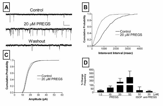
to control (data not shown). As illustrated in Fig 1, PREGS caused a robust, concentration-
dependent, increase in mEPSC frequency. The effect of PREGS was significant at
concentrations ≥ 10 µM (Fig. 1D). The onset of this effect occurred within ~1 min after bath
application of the neurosteroid and was fully reversible within ~2-4 min after washout. The
increase in mEPSC frequency was observed at both 70 and 90 mV. At these membrane
potentials, PREGS (50 µM) increased mEPSC frequency by 494 ± 184% (n=6) and 488 ±
192% (n =7), respectively (data not shown). This finding indicates that the effect of PREGS
is not due simply to an increase in the detection of subthreshold mEPSCs. The effect of
PREGS was also not due to NMDAR activation, because recording in Mg2+-free external
solution containing the NMDAR antagonist AP5 (50 µM) had no effect on the action of this
neurosteroid. PREGS (50 µM) increased mEPSC frequency by 448 ± 151% (n = 4) or 485
± 182% (n = 9) with respect to control in the presence of AP5 or Mg2+-containing external
solution, respectively (data not shown). We did not detect any effect of PREGS on mEPSC
amplitude at any of the concentrations examined; mEPSC amplitudes were changed by 1.6
± 2.8%, 0.55 ± 3.6%, -1.6 ± 4.4% and -1.1 ± 3.5% with respect to control in the presence of
1, 10, 20 and 50 µM PREGS, respectively (see Fig 1C for an illustration of a lack of an effect
of 20 µM PREGS). To eliminate the possibility of a nonspecific action and to determine the
8
by guest on January 30, 2020http://w
ww
.jbc.org/D
ownloaded from
enantioselectivity of the PREGS effect, we tested the effect of the inactive neurosteroid,
isopregnanolone, and the (-) enantiomer of PREGS, ent-PREGS, respectively. As shown
(Fig. 1D), these steroids did not induce a change in the frequency of mEPSCs. Since ent-
PREGS has been shown to exert more potent U-shaped effects than PREGS under some
experimental conditions (6), we also tested its effect at a 1 µM concentration and found that
it does not affect mEPSC frequency (2.5 ± 0.9% change with respect to control; n=3).
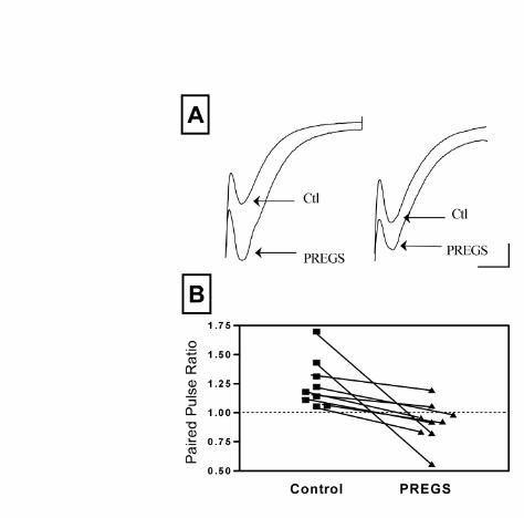
To confirm that PREGS increases the probability of glutamate release, we measured its
effect on PPF of autaptic AMPAR-mediated EPSCs evoked by depolarizing pulses (Fig 2).
It is well established that manipulations that enhance the basal probability of release
increase the impact of the first action potential (i.e. deplete synaptic vesicles), resulting in a
reduction in PPF (For an example, see (34)). Accordingly, we found that treatment with
PREGS (50 µM) increases the amplitude of the first EPSC by 43 ± 11% (n =9; p<0.01 by
one sample t test vs. a theoretical mean of zero; Fig 2A) and also reduces PPF (Fig 2B),
confirming that PREGS increases the probability of presynaptic glutamate release.
PREGS has been shown to inhibit NMDA receptor-dependent [3H]-norepinephrine release
in hippocampal slices by a mechanism that involves σ1 receptors (28). We therefore
examined the role of these receptors on the effects of PREGS on glutamate release. As
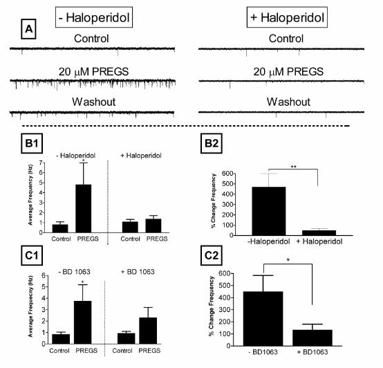
shown in Fig 3, the σ1 receptor antagonists, haloperidol and BD-1063 (preincubation for
30-45 minutes at 37 0C) blocked the PREGS-induced increase in mEPSC frequency.
Moreover, pretreatment with haloperidol or BD1063 did not affect the average basal
9
by guest on January 30, 2020http://w
ww
.jbc.org/D
ownloaded from
frequency of mEPSCs (Fig. B1, C1). This finding indicates that the decrease in PREGS
efficacy in the presence of these two compounds is not due to an overall decline in the
spontaneous release probability. We next determined whether the effect of PREGS could be
mimicked by other σ receptor agonists. We tested the effect of DHEAS, another
neurosteroid that activates σ1-like receptors in the brain (25), and of (+)-pentazocine, the
prototypical σ1 receptor agonist. As shown in Fig 4, these compounds produced a similar
increase in mEPSC frequency to that produced by PREGS (Fig 1D). Conversely, (-)-
pentazocine did not increase mEPSC frequency (Fig 1D). None of these compounds
significantly affected mEPSC amplitude (data not shown).
Plasma membrane σ receptors have been shown to be coupled to Gi/o proteins (25-28).
Therefore, we tested the effect of pertussis toxin treatment on the PREGS-induced increase
in mEPSC frequency. As illustrated in Fig 5, incubation of neurons for 36-48 hr at 37 oC
with 50 ng/ml of pertussis toxin significantly reduced the effect of PREGS on mEPSC
frequency. This result indicates that the effect of PREGS requires activation of the Gi/o
subtype of G proteins. As also shown, pretreatment with pertussis toxin did not affect basal
frequency of mEPSCs (Fig. 5B). This suggests that the decrease in PREGS efficacy in the
presence of pertussis toxin is not due to an overall decline in the spontaneous release
probability and is most likely due to a direct effect of pertussis toxin on the PREGS-
mediated second messenger cascades that result in increased spontaneous glutamate
release.
10
by guest on January 30, 2020http://w
ww
.jbc.org/D
ownloaded from
σ receptors have been shown to regulate intracellular calcium (16,35) and, therefore, we
examined its role in the mechanism of action of PREGS. The results of these experiments
are shown in Fig 6. As a control, neurons were first exposed to 50 µM PREGS, which
produced the expected increase in mEPSC frequency. After washout, the same neurons
were incubated for 15-20 min with the membrane permeable Ca2+ chelator, BAPTA-AM
(10 or 20 µM). A subsequent exposure to 50 µM PREGS failed to induce an elevation in
mEPSC frequency (Fig 6A,B). This result indicates that an elevation in intracellular Ca2+
levels is required for the presynaptic actions of PREGS. To eliminate the possibility that the
effect of BAPTA-AM was an artifact due to run down of the effect of PREGS, we applied
PREGS twice under control conditions (Fig 6C). As shown, the effect of a second
application of PREGS closely reproduced that of the first application (same result seen in 3
additional neurons).
11
by guest on January 30, 2020http://w
ww
.jbc.org/D
ownloaded from
Discussion
PREGS Increases Glutamate Release Probability:
In this paper, we report that PREGS induces a robust increase in the frequency, but
not the amplitude of AMPAR-mediated mEPSCs in hippocampal neurons cultured from
neonatal rats. Moreover, we found that PREGS reduces PPF of AMPAR-mediated synaptic
responses, indicating that this neurosteroid increases the probability of glutamate release at
the presynaptic level. To the best of our knowledge, this is the first report of a modulatory
effect of PREGS on the basal probability of glutamate release in CNS neurons. It is
noteworthy, however, that we previously found that PREGS exerts presynaptic modulatory
actions on glutamatergic terminals in the rat hippocampus. Specifically, we demonstrated
that PREGS enhances PPF of NMDAR- and AMPAR-mediated EPSPs in CA1 pyramidal
neurons in hippocampal slices from adult rats (36). Importantly, we did not find evidence
indicating that PREGS affects the basal probability of glutamate release in these slices (36).
Thus, PREGS appears to exert distinct effects on glutamate release depending on either the
neuronal developmental stage or the type of neuronal preparation being used; i.e., it
increases the probability of spontaneous glutamate release in hippocampal neurons
cultured from neonatal rats and increases facilitation of evoked glutamate release in CA1
pyramidal neurons in hippocampal slices from adult rats.
Our finding that PREGS affects glutamate release contributes to the growing evidence that this neurosteroid has important
regulatory actions on the release of a number of neurotransmitters. In agreement with the results of our study, in vivo
microdialysis experiments have demonstrated that PREGS increases basal acetylcholine release in the hippocampus and
cortex of rats (37-39) and that it also increases basal dopamine release in the rat nucleus accumbens (40). Not all studies,
however, have shown a potentiating effect of PREGS on basal neurotransmitter release. Teschemacher et al (41) found
12
by guest on January 30, 2020http://w
ww
.jbc.org/D
ownloaded from
that PREGS (1-50 µM) reduces the probability of GABA release in cultured hippocampal
neurons. Taken together with our finding that PREGS increases glutamate release in the
same type of neurons, the results of Teschemacher et al (41) indicate that this neurosteroid
differentially regulates basal neurotransmitter release from GABAergic versus glutamatergic
axonal terminals. It should also be emphasized that another study found that PREGS (10
nM-3 µM) does not affect basal [3H]-norepinephrine release in hippocampal slices from
adult rats (28). Thus, the effects of PREGS appear to depend on the neurotransmitter
specificity of a particular presynaptic terminal.
Role of σ1↑like Receptors in the Mechanism of Action of PREGS:
Monnet et al. (28) determined that PREGS decreases NMDA-evoked overflow of
[3H]-norepinephrine in hippocampal slices of adult rats and that σ receptor antagonists block
this effect. This finding prompted us to evaluate the role of σ receptors in the mechanism of
the PREGS-induced increase in spontaneous quantal glutamate release. We found that the
σ receptor antagonists, haloperidol and BD-1063, block the effect of PREGS on the
probability of glutamate release. In addition, the effect of PREGS was mimicked by DHEAS,
another neurosteroid that binds to σ receptors. Importantly, (+)-pentazocine, the
prototypical σ1 receptor agonist, also mimicked the effects of PREGS, albeit at higher
concentrations (5-50 µM) than expected; lower concentrations of (+)-pentazocine (0.1-10
µM) have been shown to activate metabotropic σ1↑like receptors in brain membranes (25).
However, it is possible that a relatively higher level of σ receptor occupancy may be
required to induce an increase in spontaneous glutamate release in presynaptic terminals of
13
by guest on January 30, 2020http://w
ww
.jbc.org/D
ownloaded from
developing neurons. In spite of this uncertainty, our finding that (-)-pentazocine did not
affect mEPSC frequency clearly argues for an involvement of a σ1-like receptor in this
process. Thus, our results and those reported by Monnet et al (28) indicate that σ receptors
are key players in the mechanism of the presynaptic actions of PREGS in the CNS. Studies
by Ueda and collaborators (25,27) have recently shown that PREGS, and other σ receptor
agonists, stimulate binding of [35S]-GTPγS to synaptic membranes from the mouse brain
in a pertussis toxin-sensitive manner. Reconstitution experiments performed by the same
group of investigators showed that Gi couples to brain metabotropic σ receptors (25,27).
Consequently, our finding that the effect of PREGS on quantal glutamate release is blocked
by pertussis toxin suggests that these Gi-coupled metabotropic σ receptors mediate the
actions of PREGS on glutamate release in cultured hippocampal neurons.
Our finding that PREGS increases mEPSC frequency at concentrations ≥10 µM is in
agreement with the estimated binding affinity (~3 µM) of this neurosteroid for brain σ
receptors (42), although there is an apparent contradiction between our findings and the
results of Ueda et al (25). These investigators reported that PREGS increases [35S]-GTPγS
binding in brain homogenates in the 10 nM-10 µM range, which indicates that this
neurosteroid has a more potent interaction with metabotropic σ receptors. However, the
prefrontal cortex and amygdala of adult mice were used to prepare the homogenates for
those studies and it is possible that higher concentrations of PREGS are required to activate
metabotropic σ receptors in axonal terminals of developing hippocampal neurons. In these
neurons, presynaptic metabotropic σ receptors could be associated with different proteins or
14
by guest on January 30, 2020http://w
ww
.jbc.org/D
ownloaded from
could be regulated by posttranslational mechanisms that are only active during
development.
It seems unlikely that the recently cloned σ binding protein is directly involved, at least initially, in the
mechanism of the presynaptic actions of PREGS for several reasons (19-23). First, the σ
binding protein sequence contains an ER retention signal (18) and, therefore, PREGS would
not be expected to be able to reach this intracellular protein in intact neurons because of its
limited lipid solubility. Second, cloning of the σ binding protein revealed that it only has a
single transmembrane domain, and also that it lacks a G protein binding domain (18).
Therefore, our finding that the effect of PREGS is blocked by pertussis toxin treatment
suggests that the σ binding protein is not involved in this process. Finally, a direct
interaction of neurosteroids with cloned σ binding proteins has not been conclusively
demonstrated. For instance, DHEAS, which mimicked the effects of PREGS on glutamate
release probability, did not significantly displace [3H](+)-pentazocine binding from
recombinant σ binding proteins expressed in Sf21 cell membranes (25). However, Maurice
et al (43) found that intracerebroventricular infusion of a 16-mer oligodeoxynucleotide
antisense to the ER σ binding protein blocked the anti-amnesic effect of DHEAS. Thus, this
neurosteroid may interact with the σ binding protein in vivo but not in vitro. Interestingly, the
anti-amnesic effect of PREGS was not affected by antisense treatment, which argues
against an interaction, at least in vivo, of PREGS with the ER σ binding protein.
Our studies do not exclude the participation of σ binding proteins at later stages of the signal
transduction cascade mediating the PREGS-induced increase in glutamate release
15
by guest on January 30, 2020http://w
ww
.jbc.org/D
ownloaded from
probability. Morin-Surun et al (44) found that treatment of an adult guinea pig brain stem
preparation with σ receptor ligands resulted in translocation of σ binding proteins from the
cytoplasm to the plasma membrane. This translocation was correlated with inhibition of
hypoglossal spontaneous motor rhythmic activity, which was prevented by incubation with a
phospholipase C (PLC) inhibitor. The authors of this study postulate that the translocation
of the single transmembrane domain σ binding protein regulates the activity of pertussis
toxin-sensitive G proteins by an unconventional mechanism, which in turn, activate PLC
(44). Interestingly, it has been recently demonstrated that σ binding proteins form a trimeric
complex with ankyrin and inositol 1,4,5-trisphosphate receptors (IP3Rs) in the ER, and that
σ ligands cause translocation of ankyrin-IP3R complexes to other organelles, including the
plasma membrane (35,45,46). Importantly, dissociation of ankyrin from IP3Rs enhances
efflux of Ca2+ from the ER via these receptors, which would be consistent with our finding
that a Ca2+ chelator blocks the actions of PREGS (see below for a more detailed
discussion). Therefore, it is possible that PREGS interacts with a membrane-bound
receptor that initiates a signal transduction cascade leading to translocation of σ binding
proteins and activation of pertussis-sensitive G proteins.
Role of Ca2+ in the Mechanism of Action of PREGS:
We found that the membrane-permeable Ca2+ chelator BAPTA-AM blocks the
PREGS-induced increase in glutamate release. This finding demonstrates that the
mechanism of action of this neurosteroid involves an elevation in [Ca2+]i. This finding is
16
by guest on January 30, 2020http://w
ww
.jbc.org/D
ownloaded from
consistent with several reports demonstrating that σ receptors regulate [Ca2+]i (35,45,47-
49). It is also in agreement with recent reports that a significant percent of miniature
synaptic currents in hippocampal neurons are regulated by changes in [Ca2+]i in axonal
terminals (50,51). Enhancement of mEPSC frequency in cultured hippocampal neurons by
brain-derived neurotrophic factor was shown to require an elevation in [Ca2+]i mediated by
activation of a signal transduction cascade involving PLC-γ and IP3Rs (29). Miniature
postsynaptic currents in retinal ganglion cells were also shown to depend on Ca2+ released
from internal stores via the PLC-β/IP3-R pathway (52). Although Gq proteins have been
linked to activation of this pathway, recent evidence suggests that Gi/o can also activate
PLC↑β in neurons (53). Thus, it is possible that the mechanism by which activation of a
Gi/o-coupled σ receptor triggers an elevation in [Ca2+]i involves activation of PLC- β and we
are currently investigating this possibility as well as the contribution of different Ca2+
sources to the mechanism of action of PREGS.
It was recently demonstrated that σ2, rather than σ1 receptors, modulate [Ca2+]i in a
neuroblastoma cell line that expresses both types of receptor (49). However, we do not
think that these receptors play a significant role in the mechanism of action of PREGS for
several reasons. First, the effect of PREGS was blocked by BD-1063, which has been
demonstrated to have preferential affinity for σ1 sites (54). Second, the prototypical σ1
ligand, (+)-pentazocine, mimicked the actions of PREGS. Third, haloperidol has been
17
by guest on January 30, 2020http://w
ww
.jbc.org/D
ownloaded from
shown to act as an agonist of σ2 receptors (55). For instance, it has been shown to elevate
[Ca2+]i, which would have been expected to increase basal mEPSC frequency and/or to
potentiate the effect of PREGS in cultured hippocampal neurons (49). Since haloperidol did
not have any of these effects under our experimental conditions, it is unlikely that σ2
receptors mediate the effects of PREGS on glutamate release. Finally, binding of PREGS,
DHEAS and other neurosteroids to σ2 receptors has not been demonstrated (55). Thus, we
conclude that a metabotropic receptor with the pharmacological profile of the σ1 subtype
mediates the actions of PREGS on the probability of glutamate release.
An important finding of our study is that ent-PREGS, the synthetic (-) enantiomer of PREGS, did not affect mEPSC
frequency. This result indicates that the interaction between presynaptic σ1-like receptors and PREGS is
enantioselective. This finding is not surprising given that the action of pentazocine and
other benzomorphans on σ1 receptors is also enantioselective. Interestingly, the interaction
of PREGS with NMDA and GABAA receptors is not enantioselective in cultured neurons
(7,33). Since ent-PREGS has been shown to exert either more potent (6) or less potent (7)
effects on memory in rodents than PREGS, it would be important to determine if the lack of
interaction of ent-PREGS with presynaptic metabotropic σ1↑like receptors contributes to its
differential effects in vivo.
Significance:
18
by guest on January 30, 2020http://w
ww
.jbc.org/D
ownloaded from
PREGS modulated glutamate release probability at concentrations ≥10 µM, which
appears to be a higher concentration than expected for a physiologically relevant action.
Recent papers reported concentrations of ~8 ng/g of rat brain tissue (2) and ~13 ng/g of rat
hippocampal tissue (5). However, these concentrations of PREGS were determined in
homogenates of brain tissue from adult rats and the actual synaptic concentrations of this
neurosteroid are unknown. It is entirely possible that PREGS reaches µM levels in synaptic
regions under certain conditions. Moreover, PREGS levels could be higher in the immature
nervous system where the NMDAR activity is higher than in the mature nervous system.
Indeed, Ca2+ influx through NMDARs has been shown to stimulate PREGS synthesis in
cultured hippocampal neurons (56) and in isolated rat retinas (57). Thus, we believe that
our findings have significant physiological implications. It should also be emphasized that
we detected a clear effect of DHEAS at concentrations as low as 0.1 and 1 µM, which also
lends support to the physiological relevance of our findings.
In conclusion, we postulate that the effects of PREGS and DHEAS on quantal glutamatergic
synaptic transmission may have an impact on synaptic development in the CNS. Recent
studies with knockout mice lacking proteins involved in regulation of neurotransmitter vesicle
exocytosis have demonstrated that quantal transmitter release is not essential for neuronal
differentiation and axonal pathfinding but for the persistence of synaptic neuronal networks
(58-60). Thus, it is possible that the effects of neurosteroids on spontaneous glutamate
release in developing synapses could contribute to the maturation and/or maintenance of
these specialized structures.
19
by guest on January 30, 2020http://w
ww
.jbc.org/D
ownloaded from

Acknowledgements
We thank Alicia Barnes, Todd Dettmer and Dr. Dan Savage for assistance and Dr.
Yvette Akwa for excellent suggestions to improve our manuscript.
20
by guest on January 30, 2020http://w
ww
.jbc.org/D
ownloaded from
21
References
1. Baulieu, E. E. (1998) Psychoneuroendocrinology 23, 963-987
2. Liere, P., Akwa, Y., Weill-Engerer, S., Eychenne, B., Pianos, A., Robel, P., Sjovall, J.,
Schumacher, M., and Baulieu, E. E. (2000) J Chromatogr B Biomed Sci Appl 739,
301-312
3. Flood, J. F., Morley, J. E., and Roberts, E. (1992) Proc Natl Acad Sci U S A 89, 1567-
1571.
4. Flood, J. F., Morley, J. E., and Roberts, E. (1995) Proc. Natl. Acad. Sci. USA 92,
10806-10810
5. Vallee, M., Mayo, W., Darnaudery, M., Corpechot, C., Young, J., Koehl, M., Le Moal,
M., Baulieu, E. E., Robel, P., and Simon, H. (1997) Proc Natl Acad Sci U S A 94,
14865-14870
6. Akwa, Y., Ladurelle, N., Covey, D. F., and Baulieu, E. E. (2001) Proc Natl Acad Sci U
S A 98, 14033-14037
7. Vallee, M., Shen, W., Heinrichs, S. C., Zorumski, C. F., Covey, D. F., Koob, G. F.,
and Purdy, R. H. (2001) Eur J Neurosci 14, 2003-2010
8. Maurice, T., Su, T. P., and Privat, A. (1998) Neuroscience 83, 413-428
9. Rupprecht, R., and Holsboer, F. (1999) Trends Neurosci 22, 410-416
10. Lambert, J. J., Belelli, D., Hill-Venning, C., Callachan, H., and Peters, J. A. (1996)
Cell Mol Neurobiol 16, 155-174
11. Irwin, R. P., Maragakis, N. J., Rogawski, M. A., Purdy, R. H., Farb, D. H., and Paul,
S. M. (1992) Neurosci Lett 141, 30-34
by guest on January 30, 2020http://w
ww
.jbc.org/D
ownloaded from
22
12. Wu, F. S., Gibbs, T. T., and Farb, D. H. (1991) Mol Pharmacol 40, 333-336
13. Park-Chung, M., Wu, F. S., Purdy, R. H., Malayev, A. A., Gibbs, T. T., and Farb, D.
H. (1997) Mol Pharmacol 52, 1113-1123
14. Bowlby, M. R. (1993) Mol Pharmacol 43, 813-819
15. ffrench-Mullen, J. M., Danks, P., and Spence, K. T. (1994) J Neurosci 14, 1963-1977
16. Maurice, T., Phan, V. L., Urani, A., Kamei, H., Noda, Y., and Nabeshima, T. (1999)
Jpn J Pharmacol 81, 125-155
17. McCann, D. J., and Su, T. P. (1990) Eur J Pharmacol 188, 211-218
18. Moebius, F. F., Striessnig, J., and Glossmann, H. (1997) Trends Pharmacol Sci 18,
67-70
19. Kekuda, R., Prasad, P. D., Fei, Y. J., Leibach, F. H., and Ganapathy, V. (1996)
Biochem Biophys Res Commun 229, 553-558
20. Seth, P., Leibach, F. H., and Ganapathy, V. (1997) Biochem Biophys Res Commun
241, 535-540
21. Seth, P., Fei, Y. J., Li, H. W., Huang, W., Leibach, F. H., and Ganapathy, V. (1998) J
Neurochem 70, 922-931
22. Hanner, M., Moebius, F. F., Flandorfer, A., Knaus, H. G., Striessnig, J., Kempner, E.,
and Glossmann, H. (1996) Proc Natl Acad Sci U S A 93, 8072-8077
23. Mei, J., and Pasternak, G. W. (2001) Biochem Pharmacol 62, 349-355
24. Moebius, F. F., Bermoser, K., Reiter, R. J., Hanner, M., and Glossmann, H. (1996)
Biochemistry 35, 16871-16878
by guest on January 30, 2020http://w
ww
.jbc.org/D
ownloaded from
23
25. Ueda, H., Yoshida, A., Tokuyama, S., Mizuno, K., Maruo, J., Matsuno, K., and Mita,
S. (2001) Neurosci Res 41, 33-40
26. Tokuyama, S., Hirata, K., Ide, A., and Ueda, H. (1997) Neurosci Lett 233, 141-144
27. Tokuyama, S., Hirata, K., Yoshida, A., Maruo, J., Matsuno, K., Mita, S., and Ueda, H.
(1999) Neurosci Lett 268, 85-88
28. Monnet, F. P., Mahe, V., Robel, P., and Baulieu, E. E. (1995) Proc Natl Acad Sci U S
A 92, 3774-3778
29. Li, Y. X., Zhang, Y., Lester, H. A., Schuman, E. M., and Davidson, N. (1998) J
Neurosci 18, 10231-10240
30. Costa, E. T., Olivera, D. S., Meyer, D. A., Ferreira, V. M., Soto, E. E., Frausto, S.,
Savage, D. D., Browning, M. D., and Valenzuela, C. F. (2000) J Biol Chem 275,
38268-38274.
31. Segal, M. M., and Furshpan, E. J. (1990) J Neurophysiol 64, 1390-1399
32. Mennerick, S., and Zorumski, C. F. (1995) J Physiol 488, 85-101.
33. Nilsson, K. R., Zorumski, C. F., and Covey, D. F. (1998) J Med Chem 41, 2604-2613
34. Sullivan, J. M. (1999) J Neurophysiol 82, 1286-1294
35. Hayashi, T., Maurice, T., and Su, T. P. (2000) J Pharmacol Exp Ther 293, 788-798.
36. Partridge, L. D., and Valenzuela, C. F. (2001) Neurosci Lett 301, 103-106.
37. Rhodes, M. E., Li, P. K., Flood, J. F., and Johnson, D. A. (1996) Brain Res 733, 284-
286
38. Darnaudery, M., Koehl, M., Pallares, M., Le Moal, M., and Mayo, W. (1998) J
Neurochem 71, 2018-2022
by guest on January 30, 2020http://w
ww
.jbc.org/D
ownloaded from
24
39. Darnaudery, M., Koehl, M., Piazza, P. V., Le Moal, M., and Mayo, W. (2000) Brain
Res 852, 173-179
40. Barrot, M., Vallee, M., Gingras, M. A., Le Moal, M., Mayo, W., and Piazza, P. V.
(1999) Eur J Neurosci 11, 3757-3760
41. Teschemacher, A., Kasparov, S., Kravitz, E. A., and Rahamimoff, R. (1997) Brain
Res 772, 226-232
42. Su, T. P., London, E. D., and Jaffe, J. H. (1988) Science 240, 219-221
43. Maurice, T., Phan, V. L., Urani, A., and Guillemain, I. (2001) Br J Pharmacol 134,
1731-1741
44. Morin-Surun, M. P., Collin, T., Denavit-Saubie, M., Baulieu, E. E., and Monnet, F. P.
(1999) Proc Natl Acad Sci U S A 96, 8196-8199
45. Su, T. P., and Hayashi, T. (2001) Trends Pharmacol Sci 22, 456-458
46. Hayashi, T., and Su, T. P. (2001) Proc Natl Acad Sci U S A 98, 491-496
47. Brent, P. J., Saunders, H., and Dunkley, P. R. (1996) Neurosci Lett 211, 138-142
48. Brent, P. J., Herd, L., Saunders, H., Sim, A. T., and Dunkley, P. R. (1997) J
Neurochem 68, 2201-2211
49. Vilner, B. J., and Bowen, W. D. (2000) J Pharmacol Exp Ther 292, 900-911
50. Savic, N., and Sciancalepore, M. (1998) Eur J Neurosci 10, 3379-3386
51. Emptage, N. J., Reid, C. A., and Fine, A. (2001) Neuron 29, 197-208.
52. Han, M. H., Kawasaki, A., Wei, J. Y., and Barnstable, C. J. (2001) Neuroreport 12,
2203-2207
by guest on January 30, 2020http://w
ww
.jbc.org/D
ownloaded from
25
53. Perroy, J., Prezeau, L., De Waard, M., Shigemoto, R., Bockaert, J., and Fagni, L.
(2000) J Neurosci 20, 7896-7904
54. Matsumoto, R. R., Bowen, W. D., Tom, M. A., Vo, V. N., Truong, D. D., and De
Costa, B. R. (1995) Eur J Pharmacol 280, 301-310
55. Maurice, T., Urani, A., Phan, V. L., and Romieu, P. (2001) Brain Res Brain Res Rev
37, 116-132
56. Kimoto, T., Tsurugizawa, T., Ohta, Y., Makino, J., Tamura, H., Hojo, Y., Takata, N.,
and Kawato, S. (2001) Endocrinology 142, 3578-3589
57. Guarneri, P., Russo, D., Cascio, C., De Leo, G., Piccoli, F., and Guarneri, R. (1998)
Eur J Neurosci 10, 1752-1763
58. Verhage, M., Maia, A. S., Plomp, J. J., Brussaard, A. B., Heeroma, J. H., Vermeer,
H., Toonen, R. F., Hammer, R. E., van den Berg, T. K., Missler, M., Geuze, H. J., and
Sudhof, T. C. (2000) Science 287, 864-869
59. Schoch, S., Deak, F., Konigstorfer, A., Mozhayeva, M., Sara, Y., Sudhof, T. C., and
Kavalali, E. T. (2001) Science 294, 1117-1122
60. Washbourne, P., Thompson, P. M., Carta, M., Costa, E. T., Mathews, J. R., Lopez-
Benditó, G., Molnár, Z., Becher, M. W., Valenzuela, C. F., Partridge, L. D., and
Wilson, M. C. (2002) Nat Neurosci 5, 19-26
by guest on January 30, 2020http://w
ww
.jbc.org/D
ownloaded from
Figure 1. Modulation of glutamate release by PREGS. (A)
Sample traces of mEPSC recordings obtained before
(Control), during administration of PREGS (20 µM), and after
washout from a single representative neuron (scale bars: 41 pA and 256 msec). (B)
Average cumulative probability histograms of control and PREGS (20 µM) interevent
intervals from three neurons (error bars have been removed for clarity). Control plots were
obtained by averaging baseline and washout data for each of the three neurons. Interevent
intervals were significantly reduced by application of PREGS (p<0.001; Kolmogorov-
Smirnov two sample test). (C) Average cumulative probability histogram for the amplitude
of the same neurons described in panel B. Amplitude distribution was not significantly
affected by PREGS treatment (p>0.05; Kolmogorov-Smirnov two sample test). Event
detection threshold was set at 10 pA and error bars have also been removed for clarity. (D)
PREGS enhancement of mEPSC frequency is dose dependent at concentrations between 1
and 50 µM (n = 5-10 neurons per group; *, p<0.05 by unpaired t test vs theoretical mean of
zero; one-way ANOVA yielded a p<0.03; see text for mEPSC amplitude data). Also
illustrated in this panel is the lack of an effect of the (-) enantiomer of PREGS (ent-PREGS;
n = 4) and the inactive steroid isopregnanolone (ISOP; n = 8) on mEPSC frequency.
Figure 2. PREGS reduces paired-pulse facilitation of autaptically-induced EPSCs. (A)
22
by guest on January 30, 2020http://w
ww
.jbc.org/D
ownloaded from
Sample traces of paired EPSCs obtained before (CTL) and after application of PREGS (50
µM). The stimulus artifacts and sodium spikes are truncated. The interpulse interval was 60
msec (scale bars: 100 pA and 12 msec). Note that PREGS induces a large increase in
amplitude of the first of the paired EPSCs and reduces the paired pulse ratio. (B) Summary
of the effects of PREGS (50 µM) on the paired pulse ratio in 9 neurons. The average ±
S.E.M. of the paired-pulse ratios were 1.25 ± 0.07and 0.9 ± 0.06 in the absence and
presence of 50 µM PREGS, respectively (n=9; p<0.02 by paired t test).
Figure 3. PREGS enhancement of mEPSC frequency is blocked by σ1-receptor antagonists. (A) Sample
traces obtained from untreated (-haloperidol) and haloperidol (300 nM) pre-treated
(+haloperidol) neurons obtained in the absence, presence of 20 µM PREGS, and after
washout (scale bars: 27 pA and 2.6 sec). (B, C) Summary of the effect of pretreatment with
300 nM haloperidol and 1 µM BD-1063. Neurons were incubated with these inhibitors for
30-45 minutes at 37 0C. The concentration of PREGS was 20 µM in all cases. (B1)
Combined plots of the average frequencies of mEPSCs under control and PREGS
treatment conditions for untreated (n = 8) and haloperidol treated (n = 9) neurons. Each bar
represents the mean ± S.E.M (*, p<0.05 for Control vs PREGS in the minus haloperidol
group by paired t test). (B2) Average percent change in mEPSC frequency for neurons
shown in panel B1. The PREGS-induced percent change in mEPSC frequency for each
individual neuron was calculated with respect to the average of control and washout
responses (**, p<0.01 by unpaired t test). (C1) Combined plots of the average frequencies
of mEPSCs under control and PREGS treatment for untreated (-BD1063; n = 6) and
23
by guest on January 30, 2020http://w
ww
.jbc.org/D
ownloaded from
BD1063 treated (+BD1063; n = 8) neurons. Each bar represents the mean ± S.E.M
(*p<0.05 vs for Control vs PREGS in the minus BD1063 group by paired t test). (C2) Average
percent change in mEPSC frequency for the neurons shown in panel C1. The PREGS-
induced percent change in mEPSC frequency for each individual neuron was calculated
with respect to the average of control and washout responses (*p<0.05 by paired t test). For
all the experiments shown in this figure, treated neurons were from sister cultures prepared
in parallel and used for experiments on the same day.
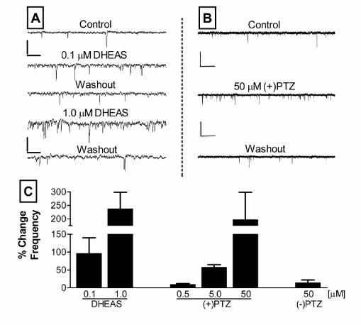
Figure 4. σ 1-receptor agonists DHEAS and (+)-pentazocine (PTZ) mimic the PREGS
dependent enhancement of mEPSC frequency. (A) Sample traces of mEPSC recordings
obtained before (Control), during administration of DHEAS (0.1 and 1.0 µM), and after
washout of both concentrations from a single representative neuron (scale bars: 41 pA and
256 msec). (B) Sample traces of mEPSC recordings obtained before (Control), during
administration of (+)-pentazocine (50 µM), and after washout from a single representative
neuron (scale bars: 41 pA and 1.3 sec). (C) Summary graph of average percent change in
mEPSC frequency obtained with 0.1 (n = 6) and 1.0 µM (n = 6) DHEAS. Also shown are the
effects of 0.5 (n = 2), 5 (n = 6) and 50 µM (+)-pentazocine (n = 5), and 50 µM (-)-
pentazocine (n = 5). Each bar represents the average ± S.E.M. (*p<0.05 by one sample t
test vs a theoretical mean of zero)
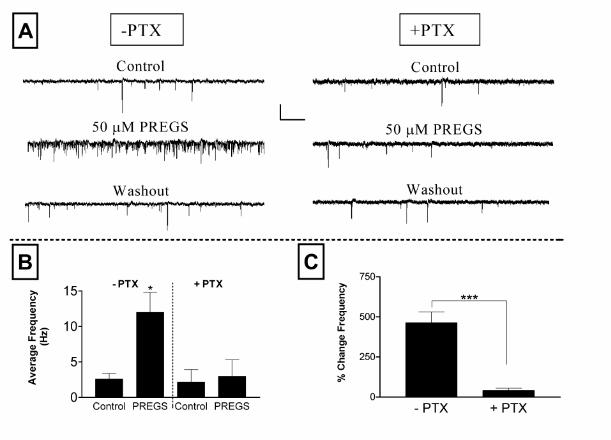
Figure 5. PREGS enhancement of mEPSC frequency is blocked by pertussis toxin. ( A)
Sample traces obtained from untreated (-PTX) and pertussis toxin pre-treated (+PTX, 50
24
by guest on January 30, 2020http://w
ww
.jbc.org/D
ownloaded from
ng/ml for 36-48 hr at 37 0C) neurons (scale bars: 16 pA and 1.3 sec). (B, C) Summary of
the effect of pretreatment with pertussis toxin. (B) Average frequencies of mEPSCs under
Control and 50 µM PREGS treatment conditions for untreated (n = 5) and PTX treated (n =
7) neurons. Each bar represents the mean ± S.E.M. (*, p<0.05 for Control (-PTX) vs
PREGS (-PTX) by paired t test). (C) Average percent change in frequency for the neurons
shown in panel B. The PREGS-induced percent change for each individual neuron was
calculated with respect to the average of control and washout responses (***, p<0.001 by
unpaired student’s t test).
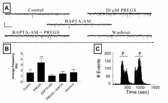
Figure 6. Treatment with the membrane permeable calcium chelator BAPTA-AM blocks the
effect of PREGS on mEPSC frequency. (A) Sample traces obtained from a neuron under
control conditions followed by the sequential addition of PREGS (50 µM), BAPTA-AM (20
µM for 15-20 min), PREGS plus BAPTA-AM, and then control external solution (scale bars: 16
pA and 1.3 sec). (B) Summary graph of the result of these experiments. Each bar
represents the mean ± S.E.M. of 5 neurons (***, p<0.001: PREGS vs all other treatments by
repeated measures ANOVA followed by Bonferronis post-hoc test). (C) Time frequency
histogram illustrating a control experiment in which the sequential application of PREGS (P;
35 µM) causes a reproducible increase in mEPSC frequency (see text for more details).
25
by guest on January 30, 2020http://w
ww
.jbc.org/D
ownloaded from
ValenzuelaDouglas A. Meyer, Mario Carta, L. Donald Partridge, Douglas F. Covey and C. Fernando
ALT="sigma ">1-like receptorspossible role of metabotropic <IMG SRC="/math/sigma.gif" ALIGN="BASELINE"
Neurosteroids enhance spontaneous glutamate release in hippocampal neurons:
published online May 31, 2002J. Biol. Chem.
10.1074/jbc.M202592200Access the most updated version of this article at doi:
Alerts:
When a correction for this article is posted•
When this article is cited•
to choose from all of JBC's e-mail alertsClick here
by guest on January 30, 2020http://w
ww
.jbc.org/D
ownloaded from


![γ-aminobutyric acid (GABA) on insomnia, …treatment of climacteric syndrome and senile mental disorders in humans. [Introduction] γ-Aminobutyric acid (GABA), an amino acid widely](https://static.fdocument.org/doc/165x107/5fde3ef21cfe28254446893f/-aminobutyric-acid-gaba-on-insomnia-treatment-of-climacteric-syndrome-and-senile.jpg)