Neuroscience Microscopy Service (NMS)2P summary ! pulsed infrared laser " less scattering " deeper...
28
Neuroscience Microscopy Service (NMS) Andrew Olson web: nisms.stanford.edu email: [email protected] Lokey Stem Cell basement: G0901
Transcript of Neuroscience Microscopy Service (NMS)2P summary ! pulsed infrared laser " less scattering " deeper...
Neuroscience Microscopy Service (NMS)
Andrew Olson web: nisms.stanford.edu email: [email protected] Lokey Stem Cell basement: G0901
Three imaging techniques:
n Laser scanning confocal microscopy (LSCM) n Two-photon microscopy (2P) n Structured Illumination (SI) microscopy
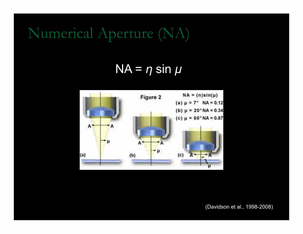
Numerical Aperture (NA)
NA = η sin µ
(Davidson et al., 1998-2008)
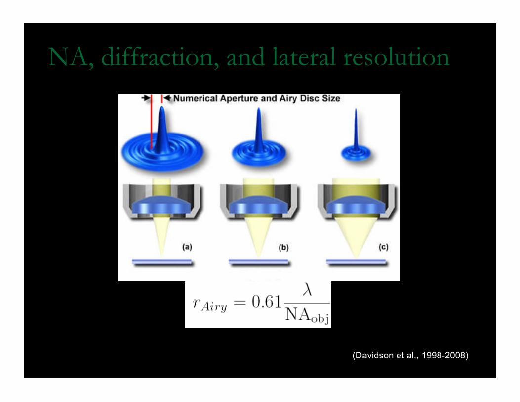
NA, diffraction, and lateral resolution
(Davidson et al., 1998-2008)
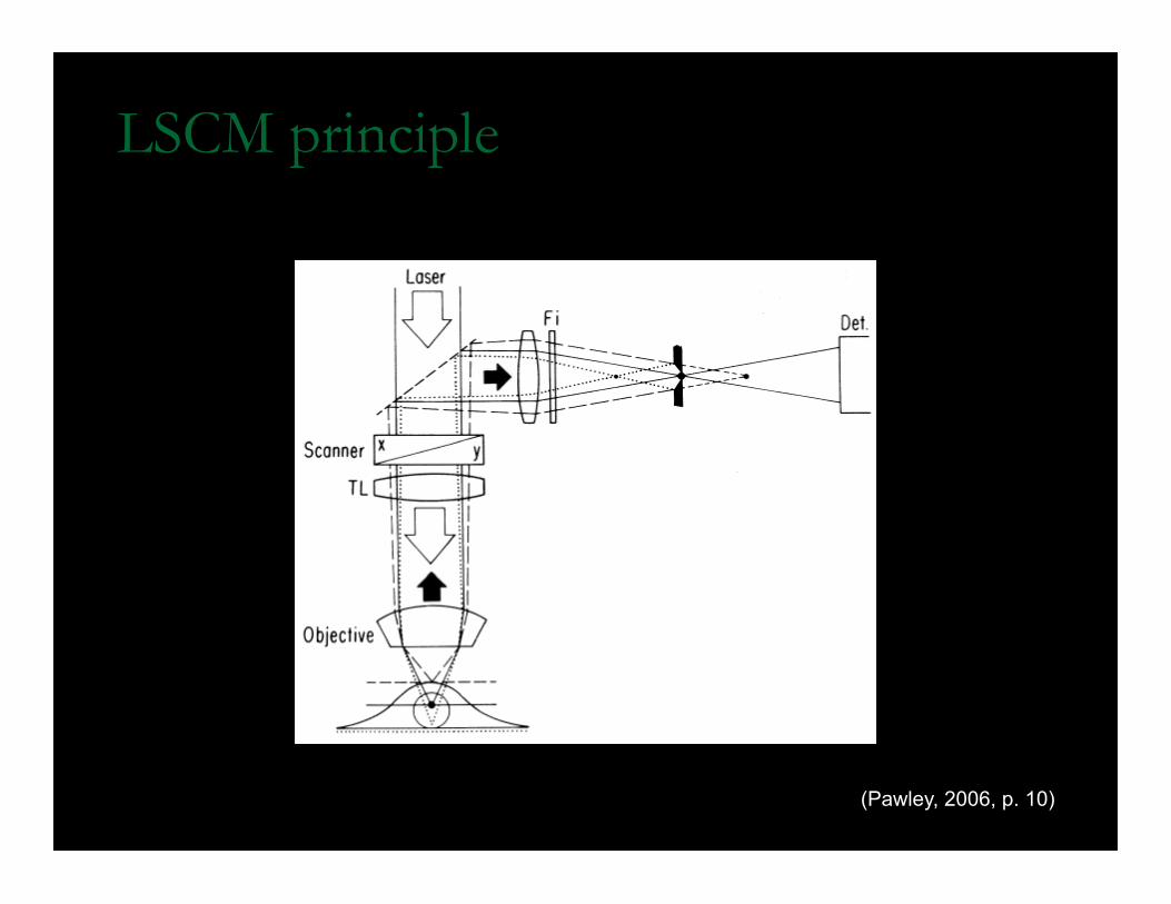
LSCM principle
(Pawley, 2006, p. 10)
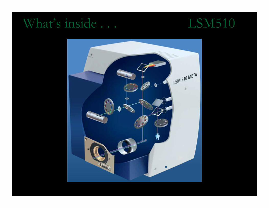
What’s inside . . . LSM510
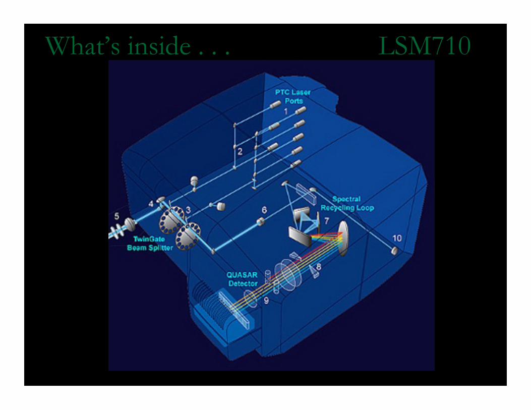
What’s inside . . . LSM710
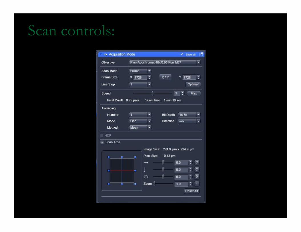
Scan controls:
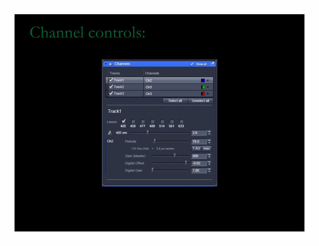
Channel controls:
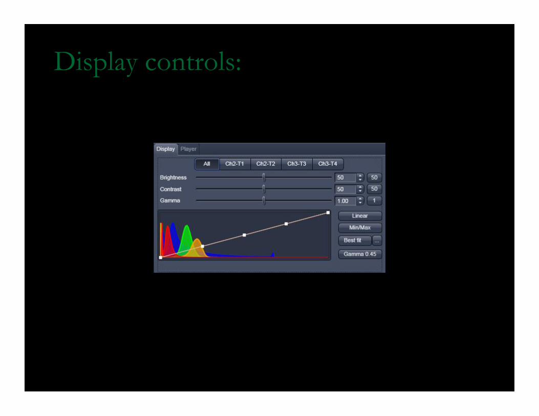
Display controls:
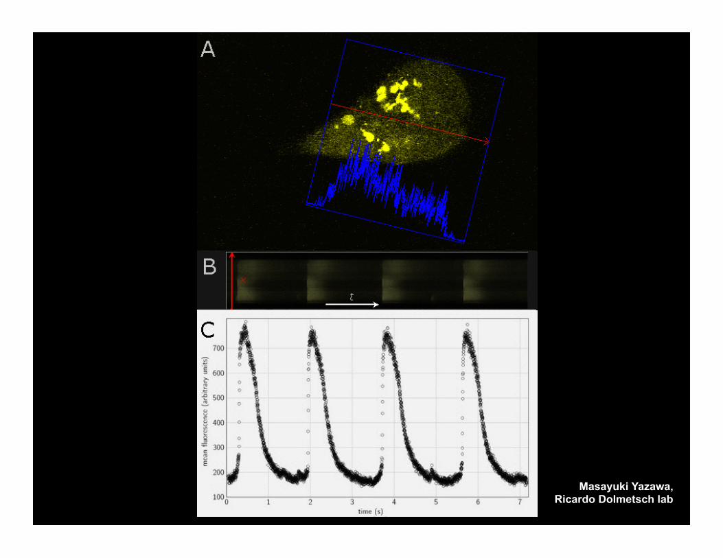
Masayuki Yazawa, Ricardo Dolmetsch lab
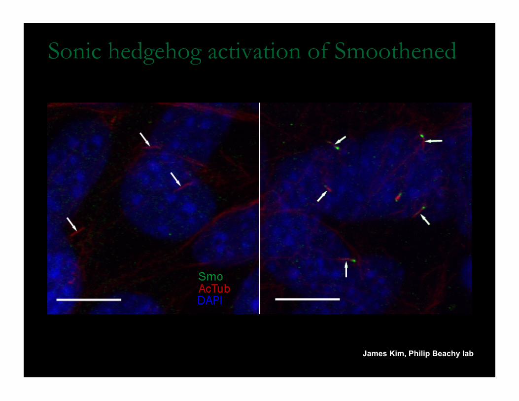
Sonic hedgehog activation of Smoothened
James Kim, Philip Beachy lab
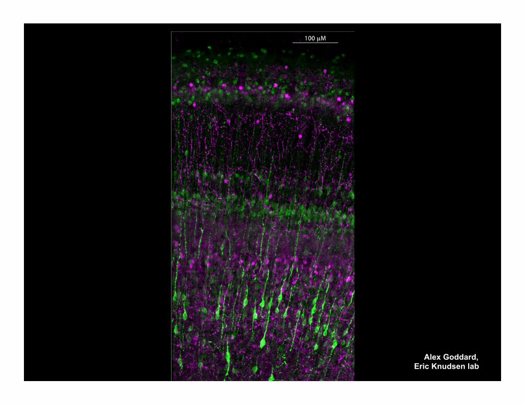
Alex Goddard, Eric Knudsen lab
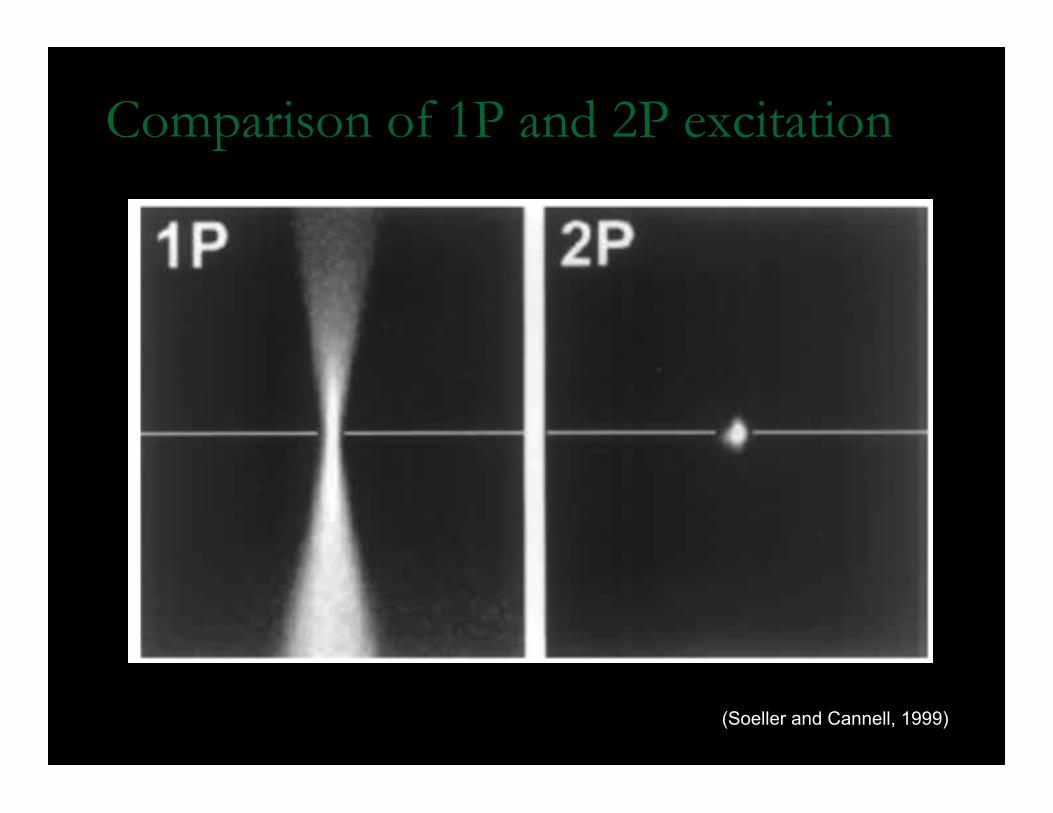
Comparison of 1P and 2P excitation
(Soeller and Cannell, 1999)
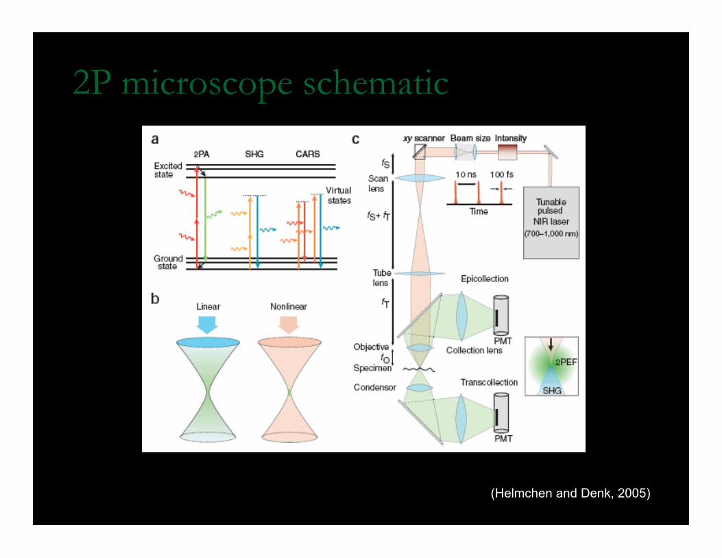
2P microscope schematic
(Helmchen and Denk, 2005)
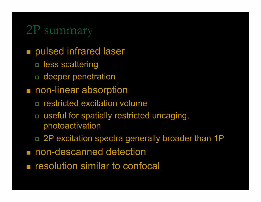
2P summary
n pulsed infrared laser q less scattering q deeper penetration
n non-linear absorption q restricted excitation volume q useful for spatially restricted uncaging,
photoactivation q 2P excitation spectra generally broader than 1P
n non-descanned detection n resolution similar to confocal
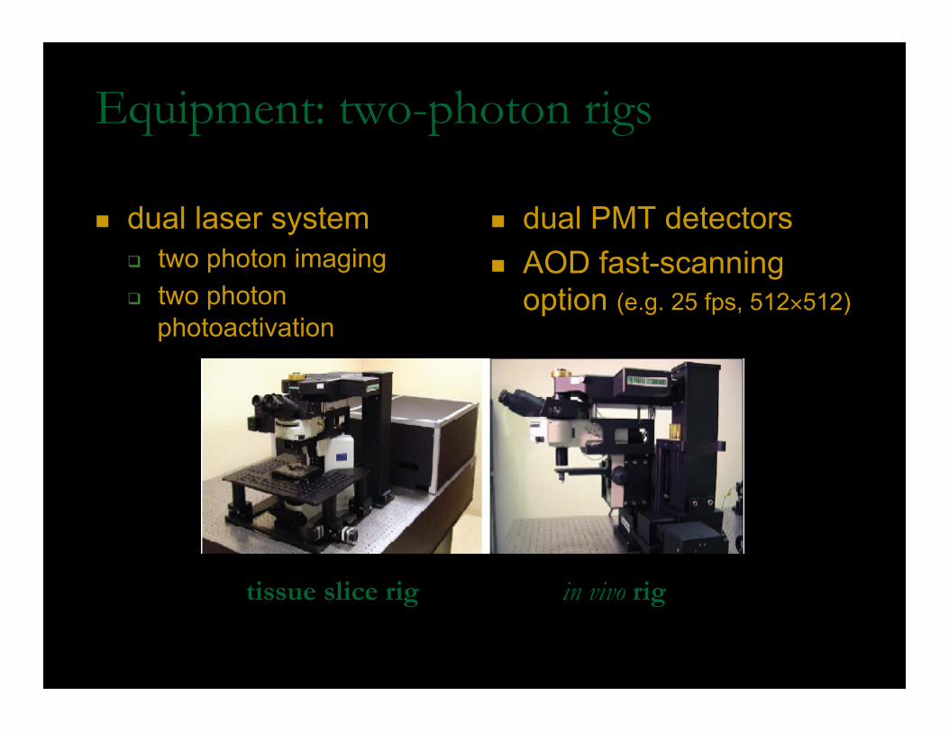
Equipment: two-photon rigs
n dual laser system q two photon imaging q two photon
photoactivation
n dual PMT detectors n AOD fast-scanning
option (e.g. 25 fps, 512×512)
tissue slice rig in vivo rig
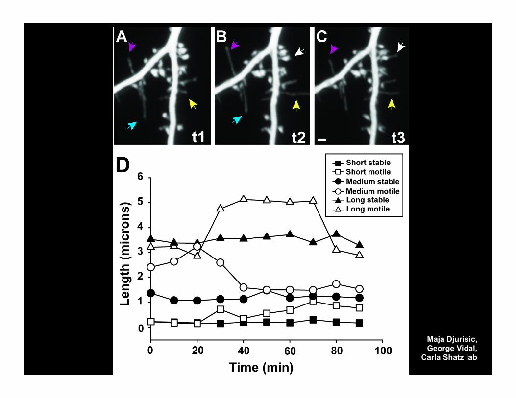
Maja Djurisic, George Vidal,
Carla Shatz lab
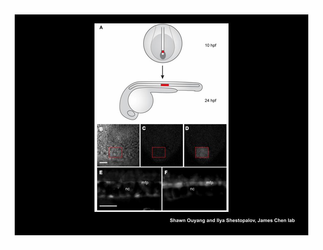
Shawn Ouyang and Ilya Shestopalov, James Chen lab
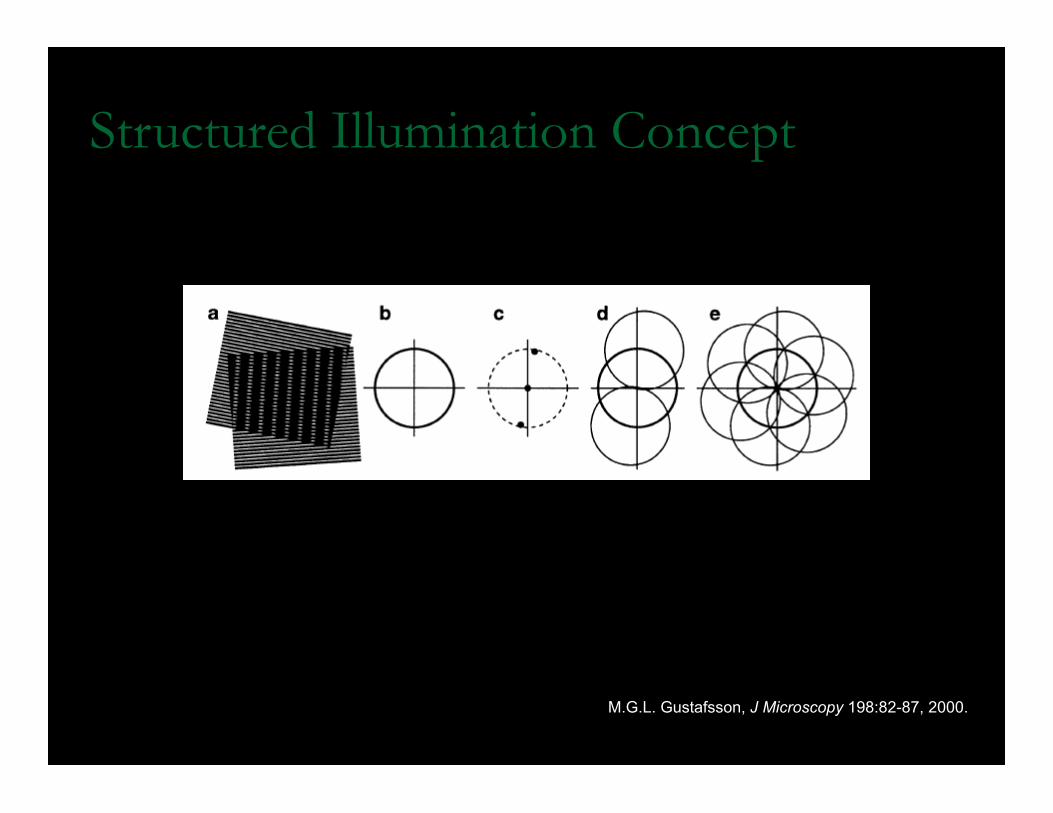
Structured Illumination Concept
M.G.L. Gustafsson, J Microscopy 198:82-87, 2000.
Structured illumination Microscopy (SIM)
n 3D- sinusoidally patterned illumination via laser diffraction
n Interference between patterned illumination (known) and the fluorescent sample (unknown) is used to computationally reconstruct a super-resolution image
n 5 phase shifts × 3 rotations = 15 images per z-plane
Structured illumination Microscopy (SIM)
n 2-fold increase in x, y and z resolution vs. confocal
n Sample must be near cover slip (10-20 µm)
n Uses standard immunofluorescent probes (blue, green, red)
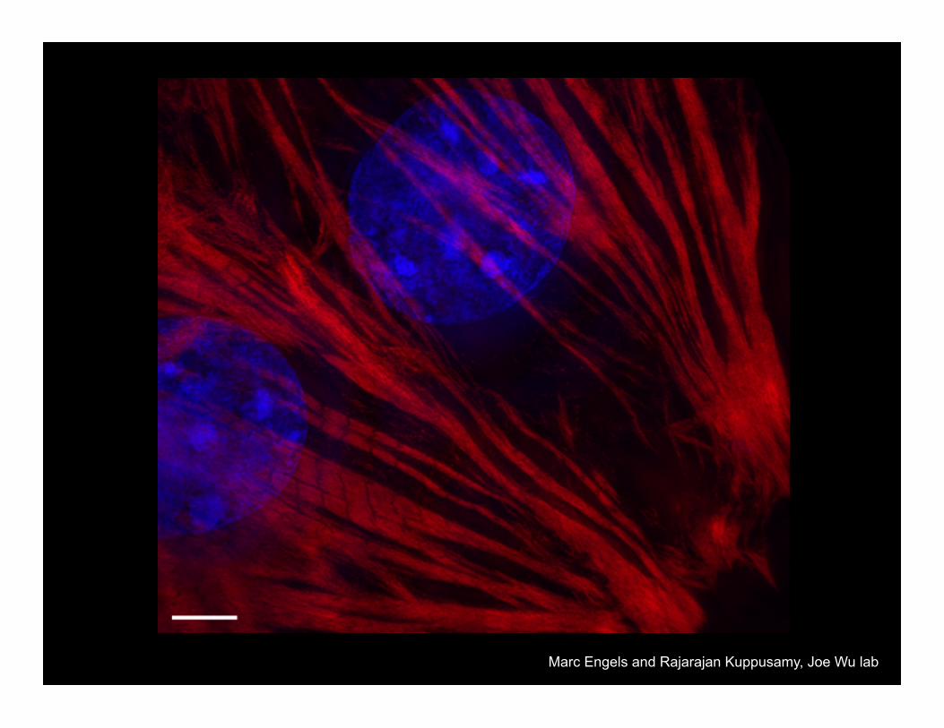
Marc Engels and Rajarajan Kuppusamy, Joe Wu lab
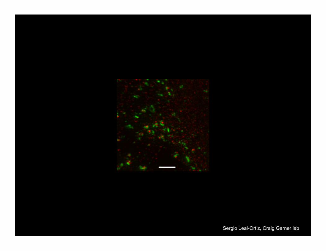
Sergio Leal-Ortiz, Craig Garner lab
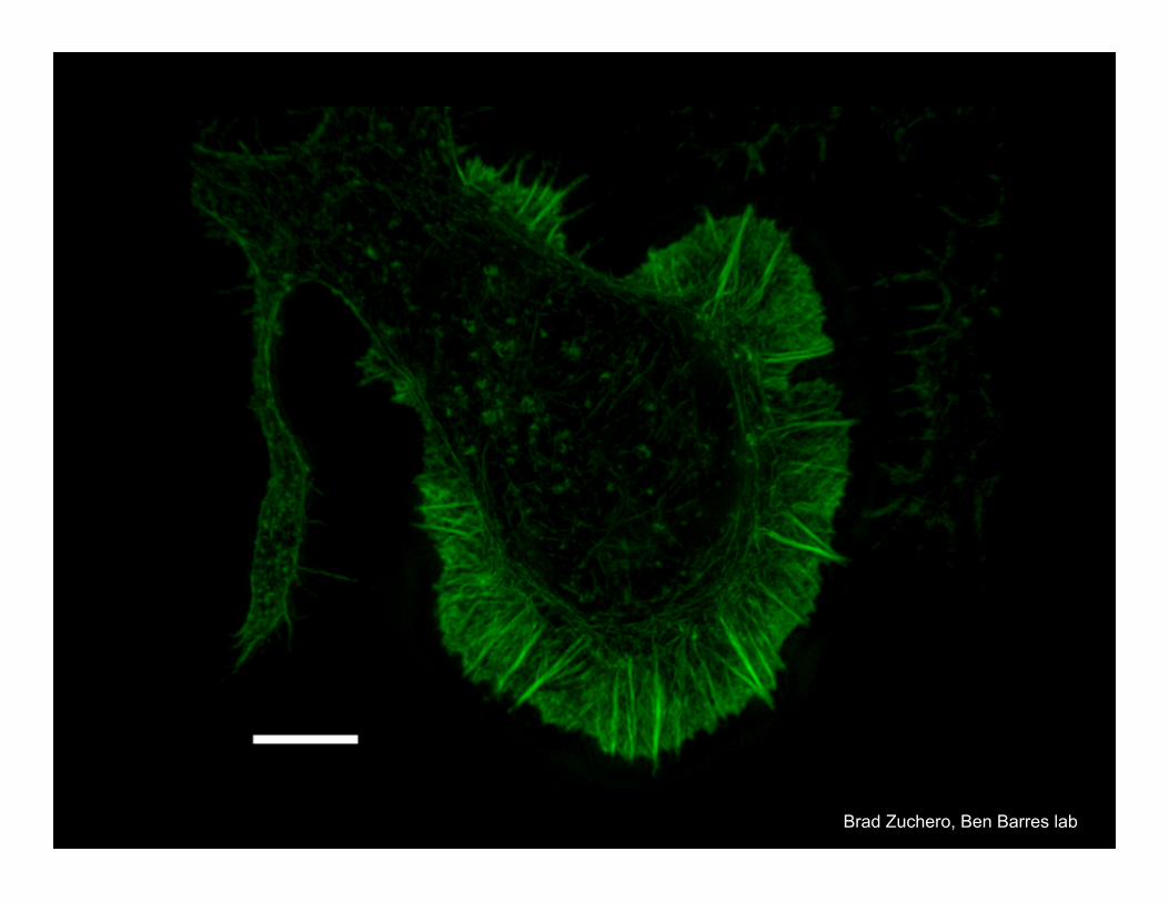
Brad Zuchero, Ben Barres lab

Questions?
References
n Davidson, et al., 1998-2008. Molecular Expressions Optical Microscopy Primer. http://micro.magnet.fsu.edu/primer/anatomy/kohler.html
n ibid. http://micro.magnet.fsu.edu/primer/anatomy/numaperture.html n Invitrogen, 2007. The Handbook — A Guide to Fluorescent Probes
and Labeling Technologies, Tenth Edition. http://probes.invitrogen.com/handbook/
n Pawley, JB, ed., 2006. Handbook of Biological Confocal Microscopy, 3rd Edition, Springer:New York.
n Spring and Davidson, 2000-2008. Nikon MicroscopyU: Introduction to Fluorescence Microscopy. http://www.microscopyu.com/articles/fluorescence/fluorescenceintro.html
References
n Gustafsson, M.G.L. Surpassing the lateral resolution limit by a factor of two using structured illumination microscopy J Microscopy 2000; 198:82-87.
n Helmchen, F. and Denk, W. Deep tissue two-photon microscopy Nature Methods 2005; 2:932–940.
n Rubart, M. Two-Photon Microscopy of Cells and Tissue http://www.biology-online.org/articles/two-photon_microscopy_cells_tissue/abstract.html
n Soeller C, Cannell MB. Two-photon microscopy: imaging in scattering samples and three-dimensionally resolved flash photolysis. Microsc Res Tech. 1999;47:182–195.