Neonatal/Pediatric Cardiopulmonary Care
56
Neonatal/Pediatric Cardiopulmonary Care Assessment
-
Upload
brandee-canute -
Category
Documents
-
view
63 -
download
0
description
Neonatal/Pediatric Cardiopulmonary Care. Assessment. Anatomic and Physiologic Differences. Cardiopulmonary System Metabolic System Other. Cardiopulmonary Differences. Tongue proportionally larger Large amt. lymphoid tissue in pharynx . Cardiopulmonary Differences. Epiglottis - PowerPoint PPT Presentation
Transcript of Neonatal/Pediatric Cardiopulmonary Care
Neonatal/PediatricCardiopulmonary Care
Assessment

2
Anatomic and Physiologic Differences
• Cardiopulmonary System
• Metabolic System
• Other
3
Cardiopulmonary Differences• Tongue proportionally larger
• Large amt. lymphoid tissue in pharynx
4
Cardiopulmonary Differences• Epiglottis
– Proportionally larger– Less flexible– Omega-shaped ( Ω )– Lies more horizontal
5
Cardiopulmonary Differences• Larynx
– Lies higher in relation to cervical spine– = narrowest segment of infant airway
(cricoid ring)
6
Cardiopulmonary Differences• Diameter of trachea at carina =
• Length of trachea =
7
Cardiopulmonary Differences
All differences (so far) combined
•
•
8
Cardiopulmonary Differences
• Less rigid
in neg. pressure effort (to ventilation) just chest size since thorax is less rigid
Result
Ribs & sternum
9
• Ribs more horizontal
Infant can’t increase A-P diameter
Result
Cardiopulmonary DifferencesRibs & sternum
10
• Any attempted increase in ventilation is accomplished by increasing -
• Increasing respiratory rate increases -
Cardiopulmonary DifferencesRibs & sternum
11
• Heart– Larger in proportion to thorax size
(imposes on lungs)
• Abdominal content– Larger in proportion to thorax size (push
up on diaphragm)
• Alveoli– Infant - – Adult -
Cardiopulmonary Differences
12
Cardiopulmonary DifferencesRibs, sternal, heart, abdominal & alveolar
differences

13
Cardiopulmonary Differences• Obligate nose-breathers
– Breathe through nose under most conditions
– Any in nasopharynx diameter increases airway resistance and WOB
14
Metabolic Differences
• Caloric requirement:– Neonates =
– Adults =
• Neonate has higher oxygen need in proportion to body size (VO2)– Infant -
– Adult -
15
Metabolic Differences
• Do not respond to medication therapy in any predictable manner– Similar infants may have dramatically
different reactions to same meds– No definitive dosages or frequencies of
administration established– Each time a drug is given, dosage must be
adjusted for each patient
16
Other Differences
• Large amount of skin surface area weight– Adult male: – Term neonate: – 28 wk. Premie:
17
Other Differences
• 80% of body weight = water– Found in extracellular spaces
18
• Transition from uterine life to survival outside is critical time
• Responsibility of HCG to determine how well infant is adapting
• Vital to know– Obstetric history– Pregnancy history– L & D history
19
Gestational Age Assessment• Until 1960’s gestational age was based mostly
on birth weight
– <2500 g. -
– >4000 g. -
• Assumed all fetuses grow at same rate
• Important to determine age to anticipate potential problems to treat or avoid
20
Dubowitz Scale
• Assesses gestational age with physical (11) & neurological (10) exam
• Scored 0-5 for each sign• Physical signs more accurate• When both evaluated = more accurate
than either used alone• Accurate to within 2 weeks• Is a slow method, so …. … .. .
21
Ballard Scale
• 6 neuro signs & 6 physical signs (scored 0-5)• Comparable to Dubowitz in accuracy• Requires less time• Assess:
– Sole creases – Posture– Skin maturity – Wrist angle– Lanugo – Arm recoil– Ear recoil – Hip angle– Breast tissue – Scarf sign– Genitalia – Heel to ear
22
Classification of Neonate
• Gestational age + weight
– SGA (small for gestational age)– AGA (appropriate for gestational age)– LGA (large for gestational age)
23
Physical Assessment
• Purposes– Discover physical defects– Successful transition?– Effect of L & D, anesthetics, analgesics– Assess gestational age– Signs of infection or metabolic disorder– Baseline for further comparison
24
Physical Assessment
• Done when infant is stabilized (keep warm)
• 2 parts to exam– Quiet observation– Hands-on
25
Quiet Observation
• Observe color– Light-skinned -- skin color– Dark-skinned -- mucous membranes– Should be pink– Blue or pale = hypoxemia– Blue feet, hands OK for 1st few hours– Yellow hue to skin or eyes = jaundice– Dark green = meconium (asphyxia may
have been present in utero)
26
Quiet Observation
• Look for presence of lanugo• Skin maturity• Activity
– Symmetry of movement– Good muscle tone– Normal movement of all extremities
• Overall appearance of patient– Malformations– Head size-to-body size– Cysts, tumors
27
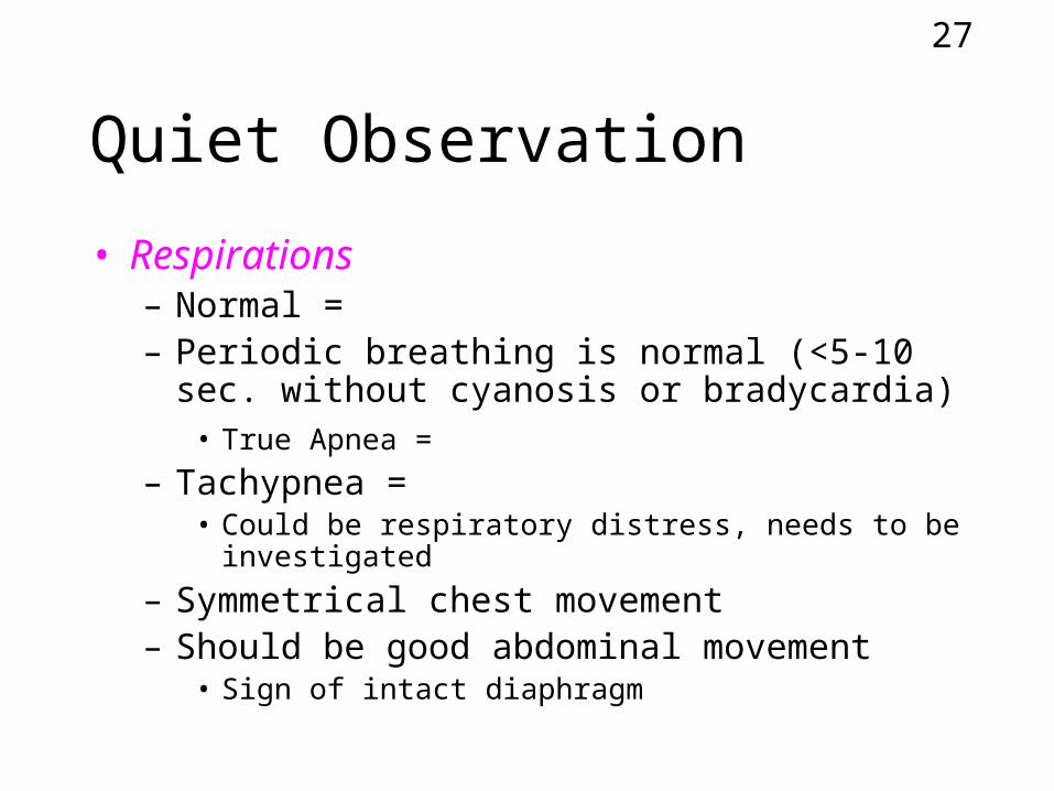
Quiet Observation
• Respirations– Normal = – Periodic breathing is normal (<5-10 sec.
without cyanosis or bradycardia)• True Apnea =
– Tachypnea = • Could be respiratory distress, needs to be
investigated
– Symmetrical chest movement– Should be good abdominal movement
• Sign of intact diaphragm
28
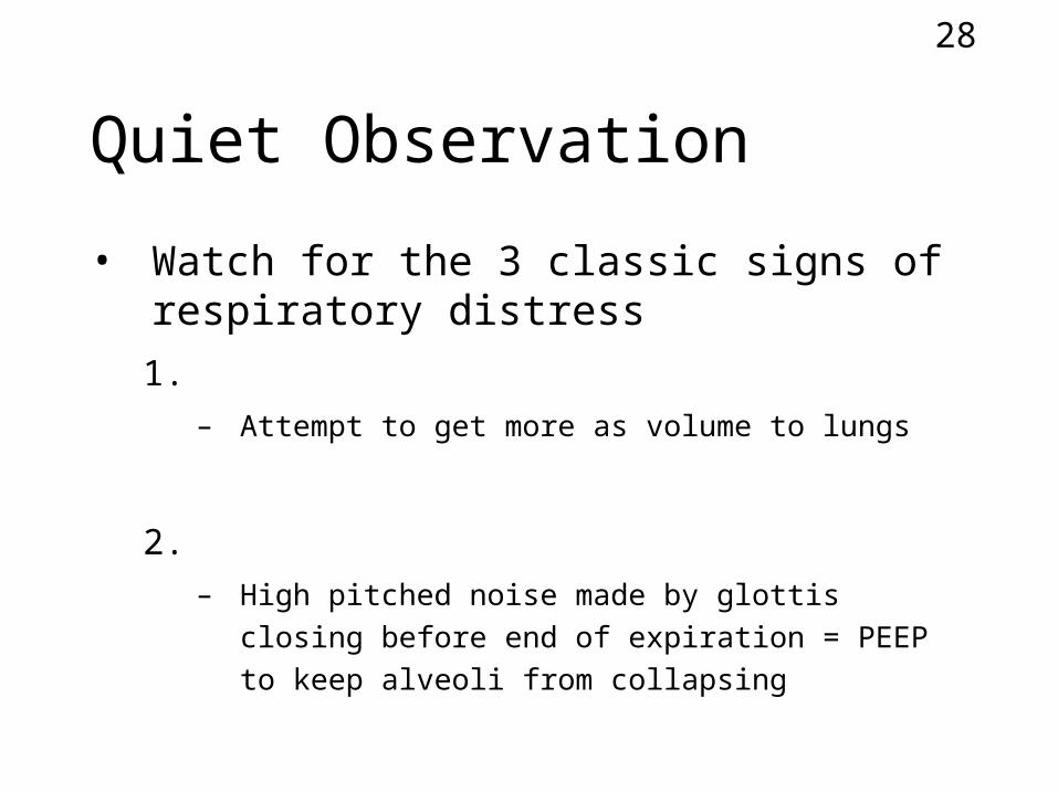
Quiet Observation
• Watch for the 3 classic signs of respiratory distress
1. – Attempt to get more as volume to lungs
2. – High pitched noise made by glottis closing
before end of expiration = PEEP to keep alveoli from collapsing
29
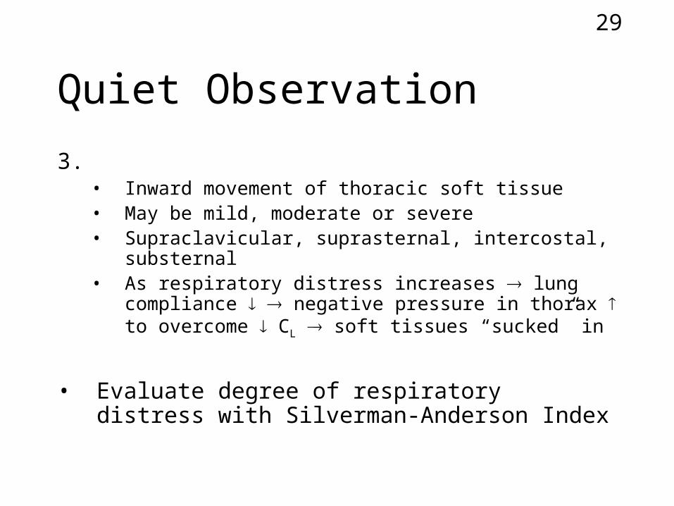
Quiet Observation
3. • Inward movement of thoracic soft tissue• May be mild, moderate or severe• Supraclavicular, suprasternal, intercostal,
substernal• As respiratory distress increases lung compliance
negative pressure in thorax to overcome CL soft tissues “sucked” in
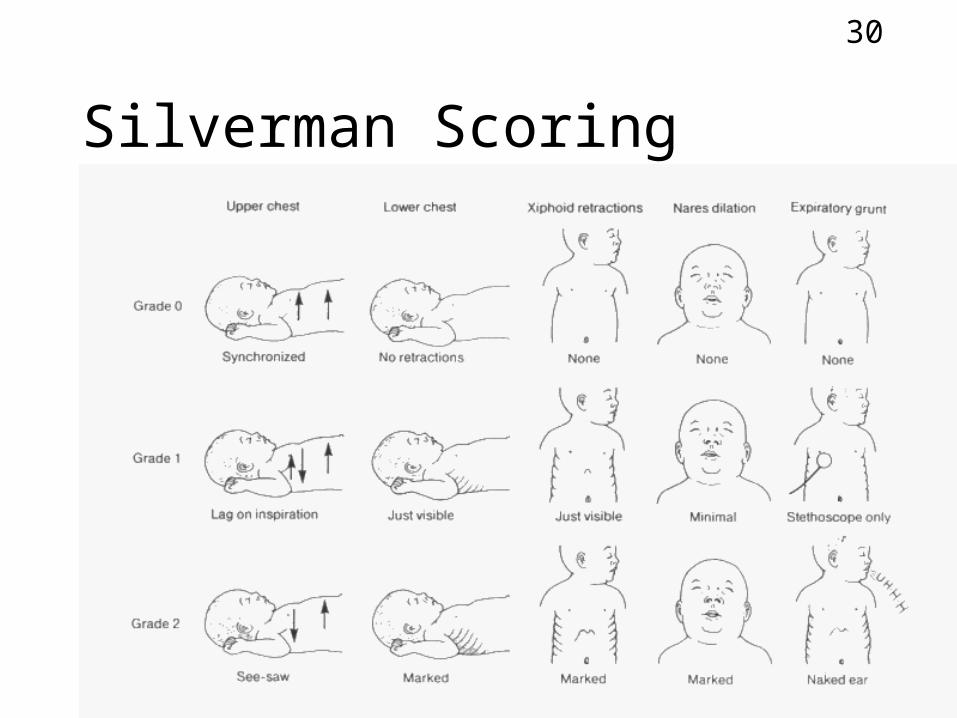
• Evaluate degree of respiratory distress with Silverman-Anderson Index
30
Silverman Scoring
31
Hands-On Exam
• Warm hands, warm stethoscope• Start at head and work down
• Head– Inspected for cuts, bruises, edema– Fontanelles (soft spots; anterior & posterior)
• Should be firm but soft, not bulging ( ICP) or depressed (dehydrated)
32
Hands-On Exam
• Mouth (clefts)
• Ears (age)
• Neck (cysts, tumors)
• Breast tissue (age)
33
Hands-On Exam
• Heart– Auscultated– HR
• Normal -
• <100 =
• <80 -
• >160 =
34
Hands-On Exam
• Heart– Apical pulse
• Point on chest where heart sounds heard loudest• = point of maximal intensity (PMI)• Normal is at left 5th intercostal space, mid-
clavicular• If moves later
–
–
35
Hands-On Exam
• Heart– Normally 2 distinct heart sounds– 1st sound louder– Murmurs
• turbulent flow in heart• Valve defects, septal defects, PDA, aortic stenosis• Not all murmurs are bad
36
Hands-On Exam
• Lungs– Well-aerated, no adventitious sounds
• Pulses– Brachial pulses compared to femoral– Should be of equal intensity & symmetrical
in rhythm
– Both weak = hypotension, QT, peripheral vasoconstriction
– Femoral weak, brachial normal = coarctation of aorta, PDA
37
Hands-On Exam
• Blood pressure– Normally varies with gestational age,
weight, cuff size, state of alertness– Taken with Doppler or electronic (cuff
around thigh), UAC– Diastolic may be difficult to assess– Normal =
38
Hands-On Exam
• Abdomen– Palpated for cysts, tumors– Liver palpated & measured in cm– Normally abdomen protrudes– If scaphoid (sunken) = diaphragmatic hernia– Check umbilical stump for 3 vessels– Bowel sounds documented
39
Hands-On Exam
• Genitalia - age
• Feet - age
• Temperature– Rectally or axillary or ear– 36.2°C - 37.3°C (97.2°F - 99.1°F)
40
Neurological Exam
• Much of neuro exam can be done during physical exam– Movement– Crying– Response to touch– Body tone
41
Neurological Exam
• Reflex exams– Rooting reflex
• Gently stroke corner of mouth• Infant should turn head towards side stroked
– Suck reflex• Place pacifier or clean finger into mouth• Infant should begin to suck
42
Neurological Exam
• Reflex exams– Grasp reflex
• Place index finger into infant’s palm• Grasp finger & place your thumb over fingers• Gently pull infant to sitting position• Assess degree of head control• Healthy infant can keep head upright
43
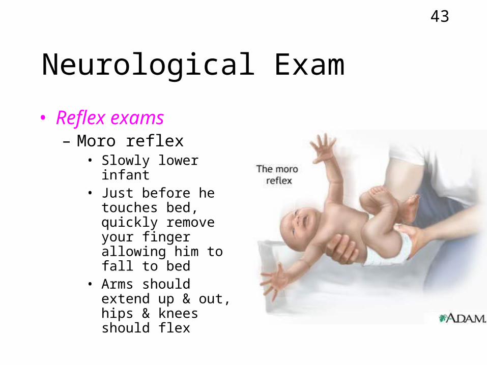
Neurological Exam
• Reflex exams– Moro reflex
• Slowly lower infant• Just before he
touches bed, quickly remove your finger allowing him to fall to bed
• Arms should extend up & out, hips & knees should flex
44
Neurological Exam
• Dubowitz or Ballard Scale scoring– Aloan, Respiratory Care of the Newborn and
Child, pg. 45
45
Chest Radiography
• Cannot be used for diagnosis of NB lung disease– Dx made from physical exam, lab data,
clinical signs– Erroneous interpretation common
• Artifact• Improper technique• Patient movement
• Used to -• Can also be used to differentiate
between diseases with -
46
Anatomic Considerations (on CXR)
• Can cause confusion if not understood
• Position of carina– Higher than adult
• NB -
• adult -
47
Anatomic Considerations (on CXR)
• Thymus gland– Extends in mediastinum from lower edge of
thyroid gland to near 4th rib– Less dense than heart, more dense than
lung tissue– Often confused with heart border– Can appear as an upper lobe atelectasis or
pneumonia– Often delta ()-shaped - called
48
CXR Interpretation
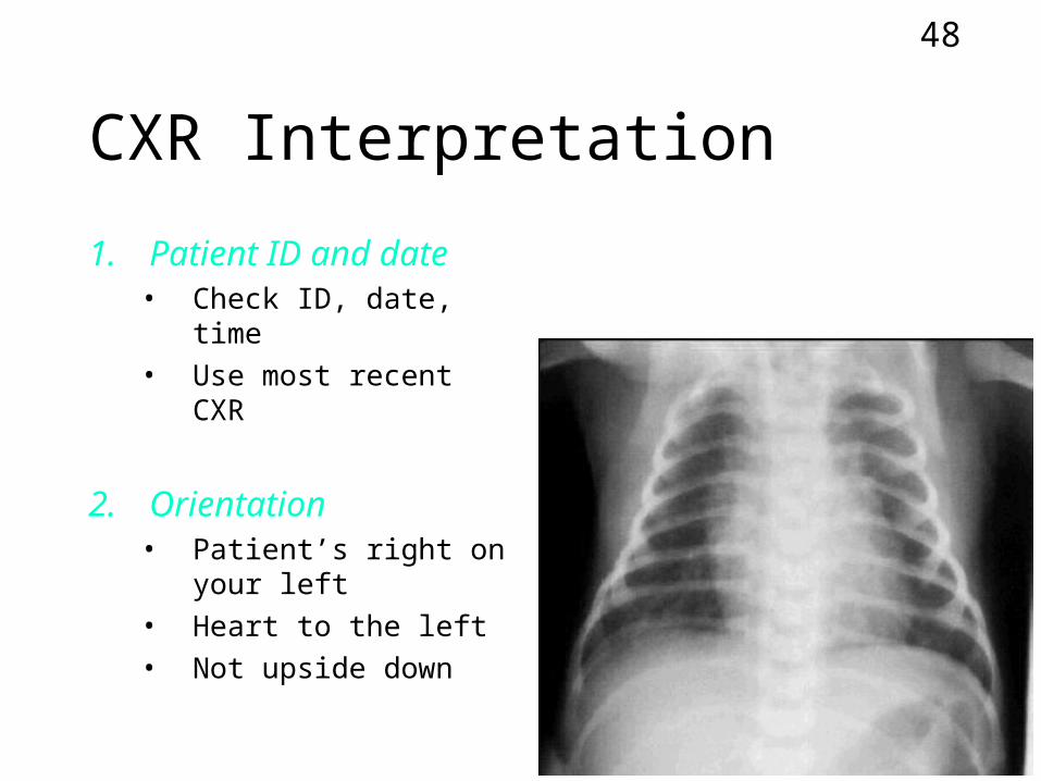
1. Patient ID and date• Check ID, date, time• Use most recent CXR
2. Orientation• Patient’s right on your
left• Heart to the left• Not upside down
49
CXR Interpretation
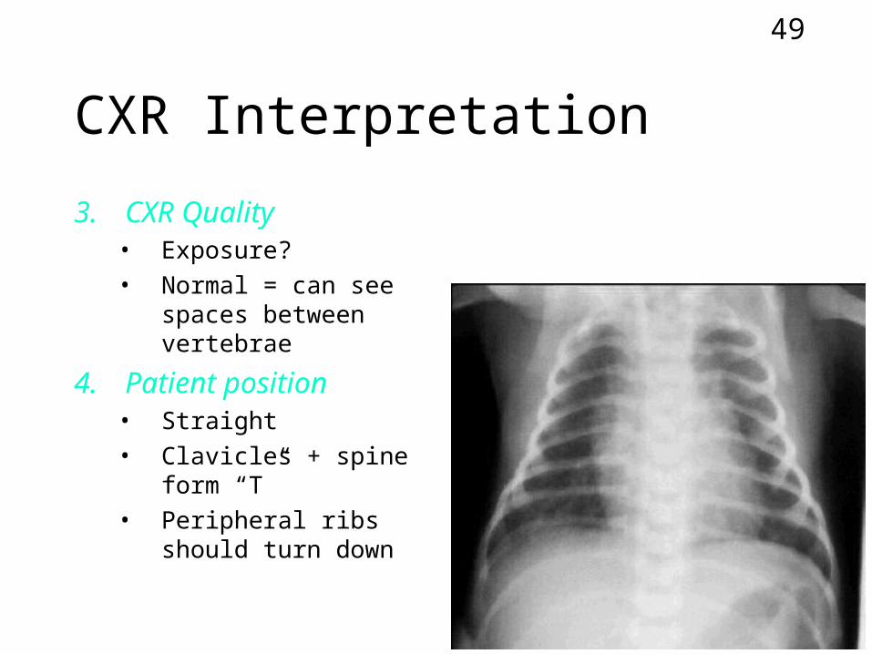
3. CXR Quality• Exposure?• Normal = can see
spaces between vertebrae
4. Patient position• Straight• Clavicles + spine
form “T”• Peripheral ribs
should turn down
50
CXR Interpretation
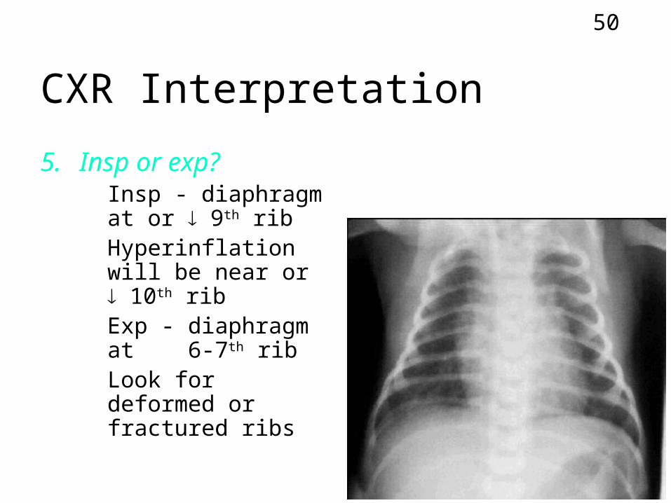
5. Insp or exp?• Insp - diaphragm at
or 9th rib• Hyperinflation will
be near or 10th rib• Exp - diaphragm at
6-7th rib• Look for deformed
or fractured ribs
51
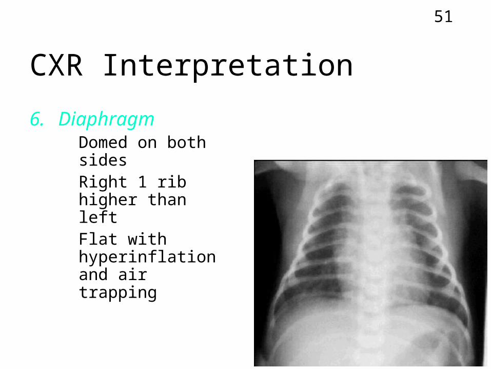
CXR Interpretation
6. Diaphragm• Domed on both
sides• Right 1 rib higher
than left• Flat with
hyperinflation and air trapping
52
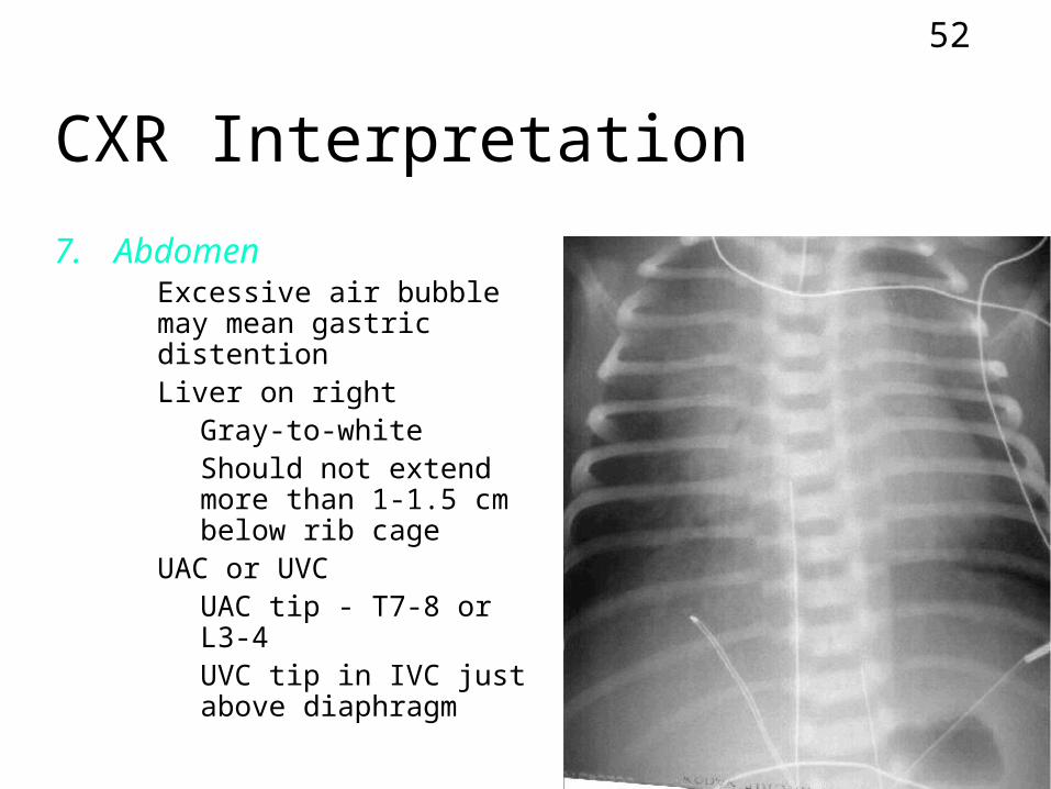
CXR Interpretation
7. Abdomen• Excessive air bubble
may mean gastric distention
• Liver on right• Gray-to-white• Should not extend
more than 1-1.5 cm below rib cage
• UAC or UVC• UAC tip - T7-8 or L3-4• UVC tip in IVC just
above diaphragm
53
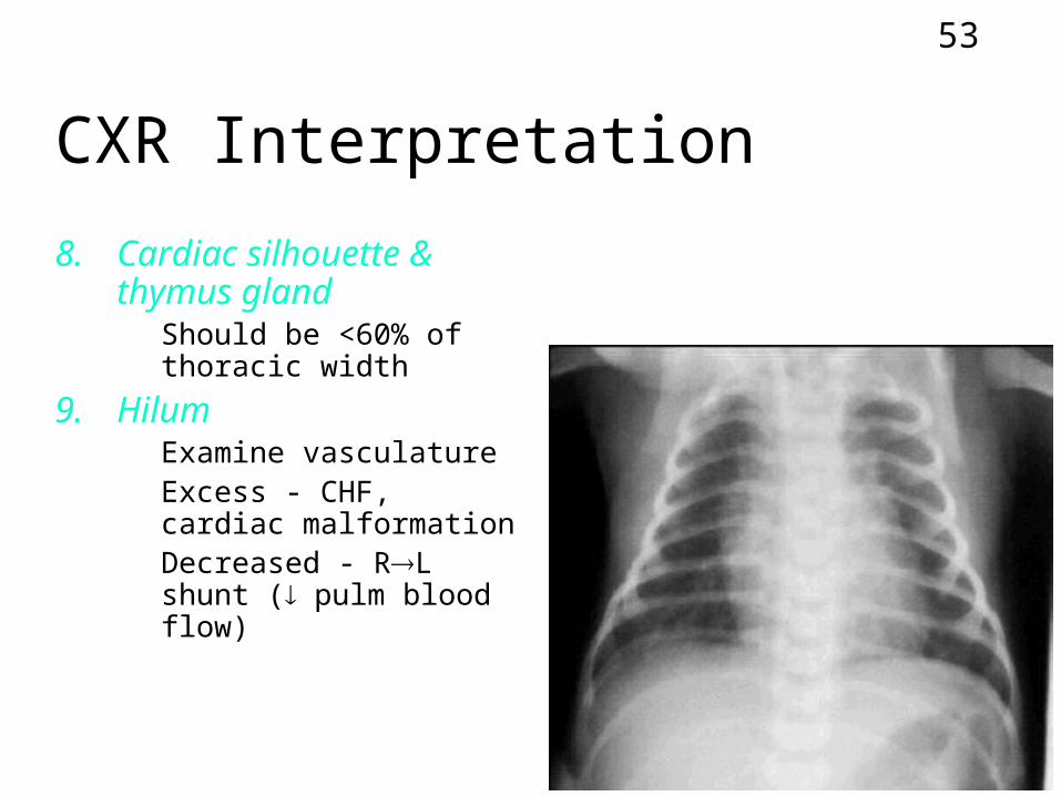
CXR Interpretation
8. Cardiac silhouette & thymus gland• Should be <60% of
thoracic width
9. Hilum• Examine vasculature• Excess - CHF, cardiac
malformation• Decreased - RL shunt
( pulm blood flow)
54
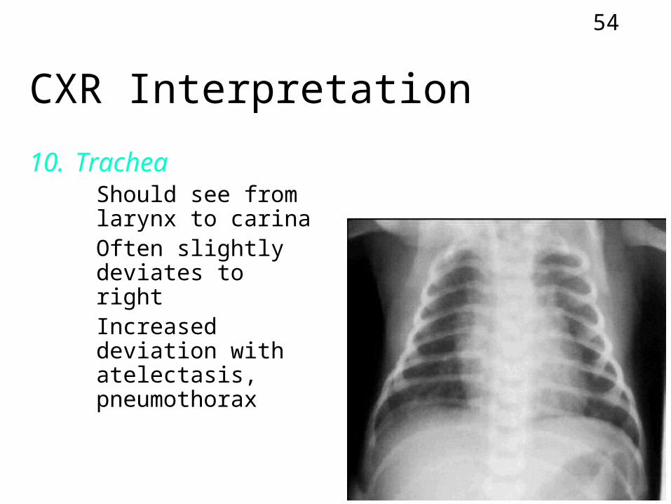
CXR Interpretation
10. Trachea• Should see from
larynx to carina• Often slightly
deviates to right• Increased deviation
with atelectasis, pneumothorax
55
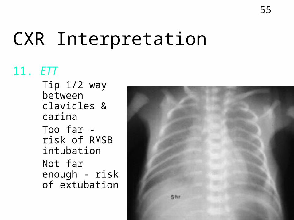
CXR Interpretation
11. ETT• Tip 1/2 way
between clavicles & carina
• Too far - risk of RMSB intubation
• Not far enough - risk of extubation
56
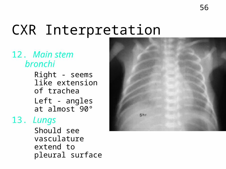
CXR Interpretation
12. Main stem bronchi• Right - seems like
extension of trachea
• Left - angles at almost 90°
13. Lungs• Should see
vasculature extend to pleural surface