Nectandrin B activates eNOS phosphorylation in endothelial cells:...
56
MOL #73502 1 Nectandrin B activates eNOS phosphorylation in endothelial cells: Role of the AMP-activated protein kinase/estrogen receptor α/ phosphatidylinositol 3-kinase/Akt pathway Tran Thi Hien, Won Keun Oh, Nguyen Phi Hung, Seok Jeong Oh, Moo Yeol Lee, Keon Wook Kang BK21 Project Team, College of Pharmacy, Chosun University, Gwangju 501-759 (T.T.H, W.K.O., N.P.H.); College of Pharmacy, Dongguk University, Goyang, Gyeonggi-do 410-820 (S.J.O., M.Y.L.); College of Pharmacy and Research Institute of Pharmaceutical Sciences, Seoul National University, Seoul 151-742 (T.T.H., K.W.K.), Republic of Korea. Molecular Pharmacology Fast Forward. Published on September 22, 2011 as doi:10.1124/mol.111.073502 Copyright 2011 by the American Society for Pharmacology and Experimental Therapeutics. This article has not been copyedited and formatted. The final version may differ from this version. Molecular Pharmacology Fast Forward. Published on September 22, 2011 as DOI: 10.1124/mol.111.073502 at ASPET Journals on April 20, 2020 molpharm.aspetjournals.org Downloaded from
Transcript of Nectandrin B activates eNOS phosphorylation in endothelial cells:...
MOL #73502
1
Nectandrin B activates eNOS phosphorylation in endothelial cells:
Role of the AMP-activated protein kinase/estrogen receptor α/
phosphatidylinositol 3-kinase/Akt pathway
Tran Thi Hien, Won Keun Oh, Nguyen Phi Hung, Seok Jeong Oh, Moo Yeol Lee, Keon Wook Kang
BK21 Project Team, College of Pharmacy, Chosun University, Gwangju 501-759 (T.T.H,
W.K.O., N.P.H.); College of Pharmacy, Dongguk University, Goyang, Gyeonggi-do
410-820 (S.J.O., M.Y.L.); College of Pharmacy and Research Institute of
Pharmaceutical Sciences, Seoul National University, Seoul 151-742 (T.T.H., K.W.K.),
Republic of Korea.
Molecular Pharmacology Fast Forward. Published on September 22, 2011 as doi:10.1124/mol.111.073502
Copyright 2011 by the American Society for Pharmacology and Experimental Therapeutics.
This article has not been copyedited and formatted. The final version may differ from this version.Molecular Pharmacology Fast Forward. Published on September 22, 2011 as DOI: 10.1124/mol.111.073502
at ASPE
T Journals on A
pril 20, 2020m
olpharm.aspetjournals.org
Dow
nloaded from
MOL #73502
2
Running title: Effect of nectandrin B on eNOS phosphorylation
Corresponding author: Keon Wook Kang, Ph.D. College of Pharmacy and Research
Institute of Pharmaceutical Sciences, Seoul National University, Seoul 151-742,
Republic of Korea. Phone: 82-2-880-7851; E-mail: [email protected]
Document statistics
Number of text pages: 43
Number of tables: 0
Number of figures: 10
Number of references: 57
Number of words in the abstract: 188
Number of words in the introduction: 463
Number of words in the discussion: 1009
Abbreviations: ACC, acetyl-CoA carboxylase; ACh, acetylcholine; AICAR, 5-
aminoimidazole-4-carboxamide-1-β-D-ribofuranoside; AMPK, adenosine 5’-
monophosphate-activated protein kinase; CA-AMPK, constitutive active AMPK;
CaMK II, calmodulin-dependent protein kinase II; DAF-2, DA (4,5-diaminofluorescein
diacetate); DN-AMPK, dominant negative mutant of AMPK; DPN, Diarylpropionitrile;
eNOS, endothelial nitric oxide synthase; ER, estrogen receptor; GR, glucocorticoid
receptor; HUVECs, human umbilical vein endothelial cells; MPP, Methyl-piperidino-
pyrazole; L-NAME, NG-nitro-L-arginine methyl ester; NO, nitric oxide; NOS, nitric
oxide synthase; PI3-kinase, phosphatidylinositol 3-kinase; PPT, 4,4',4''-(4-Propyl-[1H]-
pyrazole-1,3,5-triyl)trisphenol; THC, tetrahydrochrysene. (THC)
This article has not been copyedited and formatted. The final version may differ from this version.Molecular Pharmacology Fast Forward. Published on September 22, 2011 as DOI: 10.1124/mol.111.073502
at ASPE
T Journals on A
pril 20, 2020m
olpharm.aspetjournals.org
Dow
nloaded from
MOL #73502
3
ABSTRACT
We previously revealed that nectandrin B isolated from Myristica fragrans (nutmeg,
Myristicaceae) functions as a potent AMP-activated protein kinase (AMPK) activator
and showed its anti-obesity effect. In this study, we investigated whether nectandrin B
affects phosphorylation of endothelial nitric oxide synthase (eNOS) in human
endothelial cells. Nectandrin B increased the phosphorylation of eNOS and nitric oxide
(NO) production in a concentration dependent manner and maximal effect was found at
10 μg/ml. Nectandrin B activates AMP-activated protein kinase (AMPK) presumably
via CaM kinase II activation and nectandrin B-stimulated eNOS phosphorylation was
reversed by AMPK inhibition. Both the enzyme activity of phosphatidylinositol 3-
kinase (PI3K) and the estrogen receptor (ER)-dependent reporter gene transcription
were enhanced by nectandrin B. ERα inhibition by specific antagonist or siRNA
suppressed nectandrin B-mediated eNOS phosphorylation. Moreover, AMPK inhibition
significantly reversed the activation of ER-dependent transcription and PI3K activation
in response to nectandrin B. Nectandrin B evoked endothelium-dependent relaxation in
rat aortic rings and this was blocked by inhibition of AMPK, ER or PI3-kinase. These
results suggest that potent AMPK activator, nectandrin B enhances NO production via
eNOS phosphorylation in endothelial cells and ERα-dependent PI3-kinase activity is
required.
This article has not been copyedited and formatted. The final version may differ from this version.Molecular Pharmacology Fast Forward. Published on September 22, 2011 as DOI: 10.1124/mol.111.073502
at ASPE
T Journals on A
pril 20, 2020m
olpharm.aspetjournals.org
Dow
nloaded from
MOL #73502
4
Introduction
Endothelial function is frequently impaired during atherosclerosis,
hyperlipidemia and diabetes, and this has been well-correlated with a high risk of
cardiovascular events (Widlansky et al., 2003; Gokce et al., 2003). In particular,
endothelial dysfunction is a key event in both clinical and experimental type II diabetes
(Morcos et al., 2001; Schalkwijk and Stehouwer, 2005). Impaired endothelium-
dependent vasodilatation and increased adhesion of monocytes and platelets are
frequently found in diabetes (Fatehi-Hassanabad et al., 2010).
The endothelial nitric-oxide synthase (eNOS) has important basal regulatory
functions in the vasculature. Constitutively expressed eNOS in endothelial cells
oxidizes L-arginine to generate L-citrulline and nitric oxide (NO) in response to diverse
stimuli such as shear stress (Moncada and Higgs, 1993; Li et al., 2000). Because
endothelial NO production evokes a decrease in vascular tone and inhibits oxidation of
low density lipoprotein (Howes et al., 1997), safe compounds that activate eNOS may
be beneficial for patients with chronic cardiovascular diseases.
A series of recent studies have revealed that activation of AMP-activated
protein kinase (AMPK) has beneficial effects on endothelial dysfunction. AMPK
activation inhibits oxidized low density lipoprotein-triggered endoplasmic reticulum
This article has not been copyedited and formatted. The final version may differ from this version.Molecular Pharmacology Fast Forward. Published on September 22, 2011 as DOI: 10.1124/mol.111.073502
at ASPE
T Journals on A
pril 20, 2020m
olpharm.aspetjournals.org
Dow
nloaded from
MOL #73502
5
stress in endothelial cells (Dong et al., 2010) and metformin, a clinical AMPK activator,
normalizes endothelial function by suppressing vasoconstrictor prostanoids in arteries
from a type II diabetes rat model (Matsumoto et al., 2008). Moreover, AMPK activity is
involved in eNOS activation. An experimental AMPK activator, 5-aminoimidazole-4-
carboxamide-1-β-D-ribofuranoside (AICAR), increased eNOS activity via Ser 1177
phosphorylation; infusion of AICAR markedly increased muscle microvascular blood
volume (Bradley et al., 2010). Conversely, silencing AMPKa1 in human umbilical vein
endothelial cells (HUVECs) reduced eNOS content (Colombo and Moncada, 2009).
Plant-derived compounds are becoming of increasing interest as potential anti-
atherosclerosis therapeutics. It has been reported that some natural compounds in fruits,
vegetables, oil seeds, and herbs have lipid lowering effects and reduce atherosclerotic
lesions (Katsuda et al., 2009; Afrose et al., 2009; Magnone et al., 2009). Myristica
fragrans (nutmeg) has been used as a food and cosmetic as well as a traditional oriental
medicine against dysentery, diarrhea and pain (Grover et al., 2002). Furthermore, its
seed extracts possess anti-hyperlipidemic and anti-atherosclerotic activities in vivo
(Sharma et al., 1995). However, the mechanisms underlying these activities have not
been clarified. To address this gap in our knowledge, we isolated seven 2,5-bis-aryl-3,4-
dimethyltetrahydrofuran lignans from total extracts of Myristica fragrans and
This article has not been copyedited and formatted. The final version may differ from this version.Molecular Pharmacology Fast Forward. Published on September 22, 2011 as DOI: 10.1124/mol.111.073502
at ASPE
T Journals on A
pril 20, 2020m
olpharm.aspetjournals.org
Dow
nloaded from
MOL #73502
6
nectandrin B had a strong AMPK stimulation effect at a concentration of 5 µM in
differentiated C2C12 cells (Nguyen et al., 2010 ).
In the present study, we found that nectandrin B potently activates AMPK in
both ECV 304 (a human endothelial cell line) and HUVECs in primary cultures. We
determined whether nectandrin B affects phosphorylation and expression of eNOS and
tried to identify cellular signaling pathways for the phosphorylation and expression of
eNOS in response to nectandrin B.
Materials and methods
Nectandrin B isolation. The dried semens of Myristica fragrans (nutmeg)
were purchased at a folk medicine market in Gwangju city, Republic of Korea. The
sample was identified by Professor YH Moon at Chosun University, and its specimen
(No. 0010) was deposited at the Department of Pharmacy, Chosun University. The
EtOH extract of M. fragrans was subjected to an HP-20 column (10 × 60 cm), eluted
with a gradient of EtOH in H2O (60, 80, 90, and 100 %, each 3 L), and finally washed
by acetone (2 L) to give five fractions. Bioassay of the five fractions on the AMPK
activity revealed that the 80% ethanol-eluted fraction was most active as AMPK
activator. This fraction was further chromatographed over silica gel (6 × 60 cm; 63–200
This article has not been copyedited and formatted. The final version may differ from this version.Molecular Pharmacology Fast Forward. Published on September 22, 2011 as DOI: 10.1124/mol.111.073502
at ASPE
T Journals on A
pril 20, 2020m
olpharm.aspetjournals.org
Dow
nloaded from
MOL #73502
7
μm particle size) using a gradient of n-hexane/acetone (from 6:1 to 0:1), to yield five
fractions (F.1 – F.5) according to their profiles. Nectandrin B was purified from a part of
fraction 2 by chromatography on a reversed phase ODS-A column (5.0 × 60 cm, 150
μm particle size) eluted with MeOH/H2O (1.5:1, to 2:1, each 3 L) and purity was
confirmed by high performance liquid chromatography.
Materials. Antibodies against eNOS, phospho-eNOS, phospho-Akt, Akt,
phospho-MAP kinase, p38, phospho-extracellular signal regulated kinase (ERK), ERK,
phospho-c-Jun N-terminal kinase (JNK), JNK, ERα, phospho-AMP-activated protein
kinase (AMPK), AMPK, phospho-acetyl CoA carboxylase (ACC), ACC, phospho-
calmodulin-dependent protein kinase II (CaMK II), horseradish peroxidase-conjugated
anti-mouse and anti-rabbit IgG antibodies were purchased from Cell Signaling
Technology (Beverly, MA). LY294002, PD98059, SB203580, SP600125, ICI-182780,
A3281, compound C and NG -nitro-L-arginine methyl ester (L-NAME)(NOS inhibitor)
were purchased from Calbiochem (La Jolla, CA). 4,4',4''-(4-Propyl-[1H]-pyrazole-1,3,5-
triyl)trisphenol (PPT), Diarylpropionitrile (DPN), Methyl-piperidino-pyrazole (MPP)
and tetrahydrochrysene (THC) were obtained from Tocris Biosciences (Ellisville, MI,
USA). ERβ antibody was obtained from abcam (Cambridge, MA, USA). siRNAs
targeting for ERα and ERβ were purchased from Santa Cruz Biotechnology (Santa Cruz,
This article has not been copyedited and formatted. The final version may differ from this version.Molecular Pharmacology Fast Forward. Published on September 22, 2011 as DOI: 10.1124/mol.111.073502
at ASPE
T Journals on A
pril 20, 2020m
olpharm.aspetjournals.org
Dow
nloaded from
MOL #73502
8
CA). Actin antibody and other reagents used for molecular studies were obtained from
Sigma (St. Louis, MO). Dominant negative mutant (DN-AMPK) or constitutive active
forms (CA-AMPK) of AMPK overexpression plasmids were kindly donated by Dr. Ha
JH of Kyunghee University (Seoul, Korea).
Cell culture. ECV 304 cells were obtained from the American type culture
collection (Bethesda, MD). The cells were maintained at 37 °C in an incubator with a
humidified atmosphere of 5% CO2 and cultured in DMEM containing 10% fetal bovine
serum. Primary cultured HUVEC cells were purchased from Innopharmascreen (Asan,
Chungnam, Korea) and cultured in M199 medium containing 10 units/ml heparin, 20%
fetal bovine serum and 20 ng/ml fibroblast growth factor. Nectandrin B was dissolved
in dimethylsulfoxide (DMSO) and the stock solutions were added directly to the culture
media. Control cells were treated with DMSO only. The final concentration of solvent
was always <0.1%.
Cytotoxicity of nectandrin B in ECV 304 cells. Viable adherent cells were
stained with MTT [3-(4,5-dimethylthiazol-2-yl)-2,5-diphenyl-tetrazolium bromide] (2
mg/ml) for 4 h. Media were then removed and the formazan crystals produced were
dissolved by adding 200 μl of dimethylsulfoxide. Absorbance was assayed at 570 nm
using a microplate reader (LB941, Berthold Technologies) and cell viabilities were
This article has not been copyedited and formatted. The final version may differ from this version.Molecular Pharmacology Fast Forward. Published on September 22, 2011 as DOI: 10.1124/mol.111.073502
at ASPE
T Journals on A
pril 20, 2020m
olpharm.aspetjournals.org
Dow
nloaded from
MOL #73502
9
expressed as ratios versus untreated control cells.
Reporter gene analysis. A dual-luciferase reporter assay system (Promega,
Madison, WI) was used to determine gene promoter activity. Briefly, cells were plated
in 12-well plates and transiently transfected with the reporter and phRL-SV plasmids
(hRenilla luciferase expression for normalization; Promega) using Hillymax reagent
(Dojindo Molecular Technologies). The cells were then incubated in culture medium
without serum for 18 h. Firefly and hRenilla luciferase activities in the cell lysates were
measured using a luminometer (LB941, Berthold Technologies). The relative luciferase
activity was calculated by normalizing the promoter-driven firefly luciferase activity to
the hRenilla luciferase activity.
Western blot analysis. After treatment, cells were collected and washed with
cold phosphate-buffered saline (PBS). The harvested cells were then lysed on ice for
30 min in 100 μl lysis buffer [120 mM NaCl, 40 mM Tris (pH 8), 0.1% NP40 (Nonidet
P-40)] and centrifuged at 12,000 rpm for 30 min. Supernatants were collected from the
lysates and protein concentrations were determined using the BCA protein assay kit
(Pierce, Rockford, IL). Aliquots of the lysates (30 μg of protein) were boiled for 5 min
and electrophoresed on 10% SDS-polyacrylamide gels. Proteins in the gels were
transferred onto nitrocellulose membranes, which were then incubated with primary
This article has not been copyedited and formatted. The final version may differ from this version.Molecular Pharmacology Fast Forward. Published on September 22, 2011 as DOI: 10.1124/mol.111.073502
at ASPE
T Journals on A
pril 20, 2020m
olpharm.aspetjournals.org
Dow
nloaded from
MOL #73502
10
antibodies or mouse monoclonal β-actin antibodies. The membranes were further
incubated with secondary anti-mouse or anti-rabbit antibodies. Finally, protein bands
were detected using an enhanced chemiluminescence western blotting detection kit
(Pierce Biotechnology, Rockford, IL).
Measurement of NO production. Production of NO was assessed using the
NO-specific fluorescent dye 4,5-diaminofluorescein diacetate (DAF-2 DA; Cayman
Chemical, Ann Arbor, MI) as described previously (Formoso et al.,2006). Briefly, ECV
304 cells were grown to 95% confluence in chamber slides (Lab-Tek, Rochester, NY)
and serum-starved overnight. Cells were then loaded with DAF-2 DA (final
concentration, 2 μM) for 30 min at 37 °C, rinsed 3 times with DMEM media and kept
in the dark. Cells were then treated without or with nectandrin B as indicated in the
figure legends. The cells were fixed in 5% paraformaldehyde for 5 min at 4 °C. Fixed
cells were visualized using a fluorescence microscope (Axiovert 200M; Carl Zeiss,
Germany) and an inverted epifluorescence microscope with an attached charge-coupled
device camera using appropriate filters with a peak excitation wavelength of 480 nm
and a peak emission wavelength of 510 nm.
The amount of NO in the culture media was also determined using the Griess
reagent (Assay Designs, Ann Arbor, MI). Culture media obtained 24 h after treatment
This article has not been copyedited and formatted. The final version may differ from this version.Molecular Pharmacology Fast Forward. Published on September 22, 2011 as DOI: 10.1124/mol.111.073502
at ASPE
T Journals on A
pril 20, 2020m
olpharm.aspetjournals.org
Dow
nloaded from
MOL #73502
11
with various concentrations of nectandrin B were incubated with nitrate reductase for 1
h at 37°C to reduce nitrate to nitrite. An equal volume of Griess reagent was then added
and the optical density of the samples measured at a wavelength of 540 nm. Data were
expressed as fold change over untreated controls.
Recombinant ERα binding assay. Vehicle or test chemicals were incubated
with 1 nM tritiated estradiol (3H-E2; Perkinelmer, Boston, MA) and 0.6 nM recombinant
human ERα (MyBioSource, San Diego, CA) in TE buffer (10 mM Tris, 1 mM EDTA,
PH 7.5) at 4 °C overnight. Hydroxylapatite (60% in TE buffer) was added, mixed well,
and incubated for 15 min at room temperature. The resulting slurry was washed three
times by centrifugation with TE buffer. Bound ligand was extracted by incubation of the
slurry with absolute ethanol at 30 °C for 10 min. Tritium (3H) decay (counts per minute)
was measured by liquid scintillation in β-counter (Wallac, Gaithersburg, MD).
Organ chamber study. Male Sprague–Dawley rats (270–330 g) were
sacrificed and thoracic aortas were carefully removed and placed in a modified Krebs–
Ringer-bicarbonate solution containing (in mM) NaCl, 118.3; KCl, 4.7; MgSO4, 1.2;
KH2PO4, 1.2; CaCl2, 2.5; NaHCO3, 25.0; Ca2+EDTA, 0.016 and glucose, 11.1 (control
solution). The aortas were cleaned of loose connective tissue and then cut into rings (2
mm wide), The aortic rings were suspended horizontally between two stainless steel
This article has not been copyedited and formatted. The final version may differ from this version.Molecular Pharmacology Fast Forward. Published on September 22, 2011 as DOI: 10.1124/mol.111.073502
at ASPE
T Journals on A
pril 20, 2020m
olpharm.aspetjournals.org
Dow
nloaded from

MOL #73502
12
stirrups in the organ chambers filled with 5 ml of control solution (37 °C, pH 7.4) and
bubbled with 95% O2 and 5% CO2. The change in tension was measured isometrically
with Grass FT03 force transducers (Grass Instrument Co., Quincy, MA), and data were
acquired and analyzed with a PowerLab 8/30 Data Acquisition System and LabChart
pro software (AD Instruments, Colorado Springs, CO). The rings were stretched
progressively to the optimal tension (2 g) before the addition of 90 mM KCl. Once the
plateau of the contraction elicited by KCl was obtained, the aortic rings were rinsed
three times with warm control solution and 1 μM acetylcholine-mediated relaxation was
tested in the precontracted rings by 1 μM phenylephrine to check endothelium-
dependent relaxation responsiveness. In some experiments, rings were incubated for 30
min with LY294002 (10 μM), ICI-182,780 (100 nM) or compound C (20 μM). After
30-min incubation, 10 μg/ml nectandrin B was added and the relaxation response was
monitored in the precontracted aortic rings after addition of 1 μM phenylephrine.
Statistical analysis. Unpaired Student’s t-test was used to determine the
significance of differences between treatment groups. Statistical significance was
accepted for p values of <0.05.
This article has not been copyedited and formatted. The final version may differ from this version.Molecular Pharmacology Fast Forward. Published on September 22, 2011 as DOI: 10.1124/mol.111.073502
at ASPE
T Journals on A
pril 20, 2020m
olpharm.aspetjournals.org
Dow
nloaded from
MOL #73502
13
Results
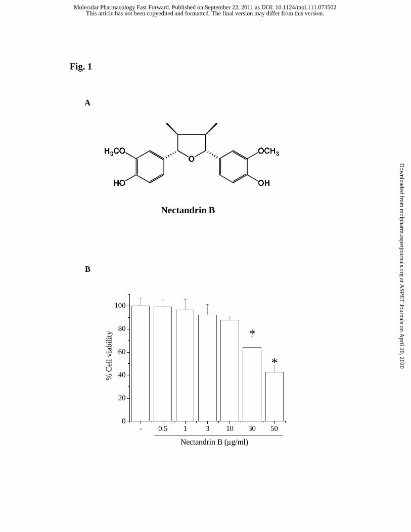
Cytotoxicity of nectandrin B in ECV 304 cells. Initially, we determined the
cytotoxicity of nectandrin B to ECV 304 cells by MTT assay. Fig. 1B shows that
nectandrin B at the tested concentrations did not cause cytotoxicity except at
concentrations above 30 μg/ml. Thus, we treated cells with nectandrin B in the
concentration range 1-10 μg/ml during subsequent experiments.
Nectandrin B increases phosphorylation of eNOS and production of NO in
endothelial cells. NO production from eNOS activation plays a protective physiological
role in the vasculature (Li and Förstermann, 2000). We did western blot analyses to
detect changes in eNOS phosphorylation. When cells were treated with 1-10 μg/ml
nectandrin B for 1 h and 10 μg/ml nectandrin B at the indicated time points (5 min to 60
min), the levels of phosphorylated eNOS were increased in a concentration- and time-
dependent manner (Fig. 2A and 2B). However, nectandrin B incubation for 3 to 24 h
did not affect the expression level of eNOS in ECV 304 cells (Fig. 2C). To examine
whether eNOS phosphorylation by nectandrin B stimulates NO production, ECV 304
cells were loaded with DAF-2 DA, a dye that upon binding to an oxidized species of
NO results in fluorescence. As shown in Fig. 2D and 2E, green fluorescence (indicative
This article has not been copyedited and formatted. The final version may differ from this version.Molecular Pharmacology Fast Forward. Published on September 22, 2011 as DOI: 10.1124/mol.111.073502
at ASPE
T Journals on A
pril 20, 2020m
olpharm.aspetjournals.org
Dow
nloaded from
MOL #73502
14
of NO production) was increased by nectandrin B in a concentration- and time-
dependent manner. We further determined nitrite levels in culture medium. Nitrite
production was significantly enhanced by 3 or 10 μg/ml nectandrin B (Fig. 3A). To
confirm these results, HUVECs were treated with 1-10 μg/ml nectandrin B and eNOS
phosphorylation and NO production were detected. As expected, phosphorylated eNOS
level and DAF-2 DA fluorescence were enhanced by nectandrin B treatment in
HUVECs (Fig. 3B and 3C).
Nectandrin B increases eNOS phosphorylation via AMP-activated protein
kinase through calmodulin-dependent protein kinase II. It is known phosphorylation
of Ser-1177 in eNOS, which plays an important role in the regulation of eNOS activity,
is induced by AMPK and results in NO production in endothelial cells (Chen et al.,
1999; Morrow et al., 2003). In our previous study, we reported that nectandrin B
potently activates AMPK in differentiated skeletal muscle cells (Nguyen et al., 2010).
Hence, we examined the effect of nectandrin B on AMPK activity in human endothelial
cells. Representative AMPK activation markers, phosphorylation of AMPK and ACC,
were increased by nectandrin B in concentration- and time-dependent manners in ECV
304 cells (Fig. 4A and 4B). The same results were also found in HUVECs (Fig. 4C).
This article has not been copyedited and formatted. The final version may differ from this version.Molecular Pharmacology Fast Forward. Published on September 22, 2011 as DOI: 10.1124/mol.111.073502
at ASPE
T Journals on A
pril 20, 2020m
olpharm.aspetjournals.org
Dow
nloaded from

MOL #73502
15
LKB1 (also called STK11) is a mammalian kinase that activates AMPK by
phosphorylating Thr172 on its catalytic (α) subunit (Carling et al., 2008). Recent studies
have reported evidence that Ca2+/calmodulin-dependent protein kinase II (CaMKs) can
act upstream of AMPK, at least in some cell types (Hurley et al., 2005; Woods et al.,
2005). ECV 304 cells were treated with nectandrin B for the indicated times and we
determined the active phosphorylated form of CaMK II and LKB1. Fig. 4B shows that
the levels of phospho-CaMK II were increased by 10 μg/ml nectandrin B, but this
treatment did not affect LKB1 phosphorylation (data not shown), which suggest that
nectandrin B-induced AMPK activation may be mediated through CaMK II activation.
We then tested whether AMPK is necessary for eNOS phosphorylation induced by
nectandrin B. AMPK activation by 1 mM AICAR treatment increased NO production in
ECV 304 cells (Fig. 5A). ECV 304 cells were pretreated with compound C, an AMPK
inhibitor and we examined nectandrin B-dependent eNOS phosphorylation. Nectandrin
B-induced phosphorylation of ACC was impaired in compound C-pretreated ECV 304
cells, and eNOS phosphorylation was attenuated (Fig. 5B), which suggests that AMPK
is required for eNOS activation. To confirm these results, we used overexpression
vector for DN-AMPK or CA-AMPK of AMPK. Transfection of DN-AMPK prior to
nectandrin B treatment reduced nectandrin B-stimulated ACC and eNOS
This article has not been copyedited and formatted. The final version may differ from this version.Molecular Pharmacology Fast Forward. Published on September 22, 2011 as DOI: 10.1124/mol.111.073502
at ASPE
T Journals on A
pril 20, 2020m
olpharm.aspetjournals.org
Dow
nloaded from
MOL #73502
16
phosphorylation (Fig. 5C). Conversely, CA-AMPK transfection markedly increased
phosphorylation levels of ACC and eNOS (Fig. 5D). To investigate whether the CaMK
II pathway is involved in the process by which nectandrin B causes AMPK and eNOS
phosphorylation, ECV 304 cells were pretreated with a CaMK II inhibitor, A3281 (10
μM), and exposed to nectandrin B. As shown in Fig. 5E, A3281 blocked nectandrin B-
induced phosphorylation of eNOS, ACC and AMPK. These results suggest that
nectandrin B-mediated eNOS phosphorylation is dependent on AMPK signaling
through CaMK II.
Role of the PI3-kinase/Akt pathway in eNOS phosphorylation by
nectandrin B. Diverse kinases such as PI3K/Akt, p38 kinase, ERK and JNK have been
shown to be involved in cellular signaling for vascular relaxation and NO production
(Merla et al., 2007; Grossini et al., 2008). To further elucidate the upstream signaling
pathways involved in nectandrin B-mediated eNOS phosphorylation and subsequent
NO production, we examined the activity of PI3K, ERK, p38 kinase and JNK in
nectandrin B-treated ECV 304 cells. Western blot analyses using phospho-specific
antibodies showed that nectandrin B incubation caused a sustained phosphorylation of
Akt or JNK, but not ERK and p38 (Fig. 6A). To address the role of PI3K or JNK
This article has not been copyedited and formatted. The final version may differ from this version.Molecular Pharmacology Fast Forward. Published on September 22, 2011 as DOI: 10.1124/mol.111.073502
at ASPE
T Journals on A
pril 20, 2020m
olpharm.aspetjournals.org
Dow
nloaded from

MOL #73502
17
activation in eNOS phosphorylation by nectandrin B, the effects of specific kinase
inhibitors were investigated. LY294002, a PI3K inhibitor significantly reduced
nectandrin B-induced eNOS phosphorylation (Fig. 6B). However, an inhibitor of the
JNK pathway (SP600125: JNK inhibitor) had no effect (Fig. 6C).
Involvement of the ERα-dependent PI3K/Akt pathway in phosphorylation
of eNOS by nectandrin B. A recent study suggests that AMPK activation restores
estrogen responsiveness in human endothelial cells (Chakrabarti and Davidge, 2009). It
has also been shown that a lignan, nordihydroguaiaretic acid, has estrogenic activity
(Fujimoto et al., 2004). Until now, no evidence has been reported of a correlation
between estrogen receptor (ER) and eNOS phosphorylation induced by nectandrin B.
When we determined ER-dependent transcription by using an ERE reporter, nectandrin
B significantly increased the reporter activities of ERE in a concentration-dependent
manner, but the increase was marginal compared to the effects of a full ER agonist, 17-
β-estradiol (Fig. 7A). It has been shown that ERα directly interacts with PI3K and
modulates its activity in human vascular endothelial cells (Simoncini et al., 2000). To
further test the possible role of ER activation in PI3K-dependent eNOS phosphorylation
in nectandrin B-exposed endothelial cells, we examined whether ICI-182780, an ER
This article has not been copyedited and formatted. The final version may differ from this version.Molecular Pharmacology Fast Forward. Published on September 22, 2011 as DOI: 10.1124/mol.111.073502
at ASPE
T Journals on A
pril 20, 2020m
olpharm.aspetjournals.org
Dow
nloaded from
MOL #73502
18
specific inhibitor, affects the eNOS phosphorylation that occurs in response to
nectandrin B. ICI-182780 potently suppressed eNOS phosphorylation as well as Akt
phosphorylation (Fig. 7B). These results indicate that nectandrin B-stimulated eNOS
phosphorylation is linked with ER-dependent PI3-kinase/Akt pathways. We then
confirmed the effects of an ER antagonist and a PI3-kinase inhibitor on NO production
induced by nectandrin B. As shown in Fig. 7C, nectandrin B-mediated NO production
(DAF-2 fluorescence) was suppressed by ICI-182780 or LY294002 pretreatment. These
data suggest that ER activation is critical for PI3-kinase/Akt-mediated eNOS
phosphorylation by nectandrin B.
It has been shown that both ERα and ERβ are involved in eNOS
phosphorylation in endothelial cells (Mineo and Shaul, 2006). Immunoblot analyses
showed that both the receptor types were expressed in ECV 304 cells (Fig. 8A, left).
Either DPN (ERα selective agonist) or PPT (ERβ selective agonist) increased eNOS
phosphorylation in ECV 304 cells, demonstrating both the ER subtypes are coupled
with eNOS phosphorylation process in this cell type (Fig. 8A, right). We further
determined the effects of specific antagonists targeting ERα and ERβ on eNOS
phosphorylation by nectandrin B. Methyl-piperidino-pyrazole (MPP, a selective ERα
antagonist) potently suppressed nectandrin B-mediated eNOS phosphorylation, while
This article has not been copyedited and formatted. The final version may differ from this version.Molecular Pharmacology Fast Forward. Published on September 22, 2011 as DOI: 10.1124/mol.111.073502
at ASPE
T Journals on A
pril 20, 2020m
olpharm.aspetjournals.org
Dow
nloaded from

MOL #73502
19
tetrahydrochrysene (THC, a selective ERβ antagonist) did not affect (Fig. 8B).
Moreover, ERα siRNA potently suppressed nectandrin B-mediated eNOS
phosphorylation, but ERβ siRNA marginally affected eNOS phosphorylation in
response to nectandrin B (Fig. 8C). In order to assess whether nectandrin B directly
bind to ERα, we further performed ERα ligand binding assay using human recombinant
ERα. 3 and 10 μg/ml nectandrin B significantly inhibited tritiated estradiol binding to
human ERα, but the inhibition intensity is lower than 10 nM 17-β-estradiol (Fig. 8D).
These data suggest that nectandrin B acts as a relatively selective agonist on ERα.
ER-dependent eNOS phosphorylation is coupled with diverse signaling
molecules. It has been shown that Src kinase mediates PI3K/Akt-dependent rapid eNOS
activation in endothelial cells (Haynes et al., 2003). We found that Src specific inhibor,
4-amino-5-(4-chlorophenyl)-7-(t-butyl)pyrazolo[3,4-d]pyrimidine (PP2) suppressed
eNOS phosphorylation by nectandrin B (Fig. 8E). Coimmunoprecipitation studies of
plasma membranes from COS-7 cells transfected with ERα and Gα proteins
demonstrated estrogen-stimulated selective interaction between ERα and Gαi.
Moreover, Gαi inhibitor pertussis toxin blocked estrogen-dependent eNOS activation
(Mineo and Shaul, 2006). However, pertussis toxin did not abrogate nectandrin B-
stimulated eNOS phosphorylation in ECV 304 cells (Fig. 8E). Hence, Src tyrosine
This article has not been copyedited and formatted. The final version may differ from this version.Molecular Pharmacology Fast Forward. Published on September 22, 2011 as DOI: 10.1124/mol.111.073502
at ASPE
T Journals on A
pril 20, 2020m
olpharm.aspetjournals.org
Dow
nloaded from
MOL #73502
20
kinase, but not G proteins, may be involved in the ERα and subsequent eNOS activation
process by nectandrin B.
Both PI3K/Akt and AMPK pathways are required for eNOS phosphorylation
by nectandrin B. Hence, we studied possible cross-talk between the two kinase
pathways. PI3K inhibition by LY294002 pretreatment failed to inhibit the
phosphorylation of ACC in response to nectandrin B (Fig. 9A). However, AMPK
inhibition by compound C impaired nectandrin B-induced Akt phosphorylation (Fig.
9B). In addition, compound C attenuated ER-dependent reporter activity induced by
nectandrin B (Fig. 9C). These data support the conclusion that AMPK/ERα/PI3-
kinase/Akt regulates eNOS phosphorylation in response to nectandrin B.
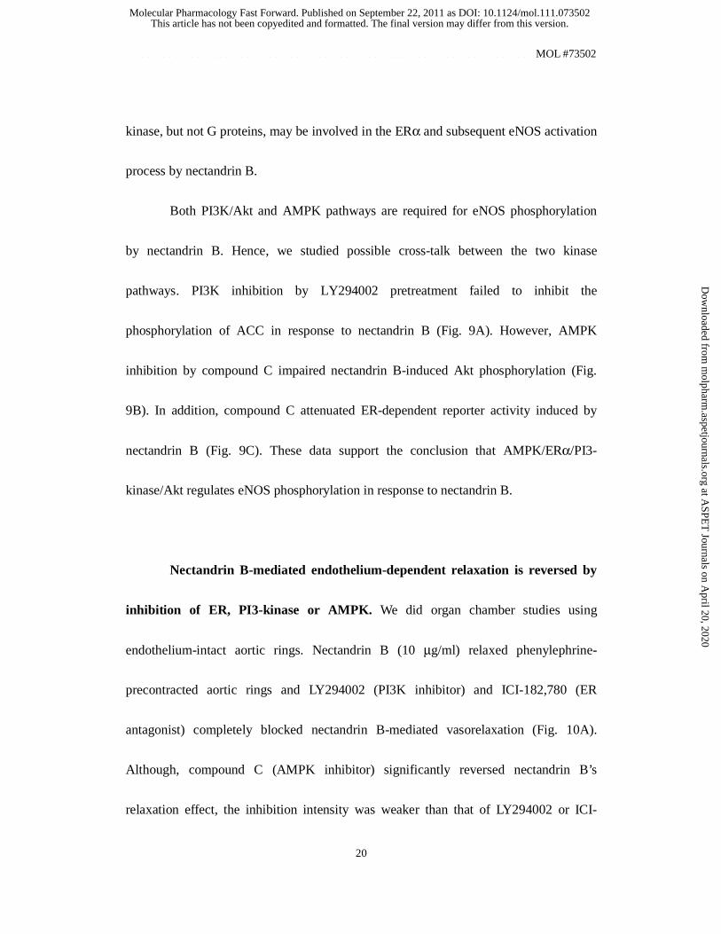
Nectandrin B-mediated endothelium-dependent relaxation is reversed by
inhibition of ER, PI3-kinase or AMPK. We did organ chamber studies using
endothelium-intact aortic rings. Nectandrin B (10 μg/ml) relaxed phenylephrine-
precontracted aortic rings and LY294002 (PI3K inhibitor) and ICI-182,780 (ER
antagonist) completely blocked nectandrin B-mediated vasorelaxation (Fig. 10A).
Although, compound C (AMPK inhibitor) significantly reversed nectandrin B’s
relaxation effect, the inhibition intensity was weaker than that of LY294002 or ICI-
This article has not been copyedited and formatted. The final version may differ from this version.Molecular Pharmacology Fast Forward. Published on September 22, 2011 as DOI: 10.1124/mol.111.073502
at ASPE
T Journals on A
pril 20, 2020m
olpharm.aspetjournals.org
Dow
nloaded from
MOL #73502
21
182,780 (Fig. 10A). Immunoblot analysis using the homogenates of aortic rings showed
that nectandrin B increased the level of phosphorylated eNOS at 15-30 min after 10
μg/ml nectandrin B exposure to aortic rings (Fig. 10B), which suggest that eNOS
phosphorylation and subsequent NO production by nectandrin B is directly coupled
with vascular relaxation.
Discussion
The species M. fragrans has been used traditionally for spices and various medicinal
purposes – as a stomachic, carminative, tonic, aphrodisiac, and nervous system
stimulant (Nguyen et al., 2010). Several studies have shown that lignan compounds
from M. fragrans have beneficial anti-inflammatory, anti-diabetes and anti-oxidant
effects (Han et al., 2008; Anggakusuma et al., 2009; Ma et al., 2009; Kwon et al., 2008).
AMPK is activated either by an increase in the AMP/ATP ratio during metabolic stress
or by activation of upstream kinases such as LKB1 and CaMK (Hardie, 2007). Because
AMPK activation suppresses ATP-consuming anabolic pathways and conversely
activates ATP-generating catabolic pathways, a selective AMPK activator could
function as an anti-diabetes and anti-obesity agent (Misra, 2008). In this sense,
nectandrin B is an attractive natural compound, since the lignan is a potent AMPK
This article has not been copyedited and formatted. The final version may differ from this version.Molecular Pharmacology Fast Forward. Published on September 22, 2011 as DOI: 10.1124/mol.111.073502
at ASPE
T Journals on A
pril 20, 2020m
olpharm.aspetjournals.org
Dow
nloaded from
MOL #73502
22
activator at relatively low concentrations compared to other phytochemicals.
Here, we demonstrated that nectandrin B stimulates eNOS phosphorylation in human
endothelial cells. Although it has been reported that eNOS levels are reduced in AMPK
silencing cells (Colombo and Moncada, 2009), protein expression of eNOS was not
affected by nectandrin B. The PI3K/Akt pathway was reported to be essential for Ser
1177 or Ser1179 eNOS phosphorylation (Dimmeler et al., 1999; Fulton et al., 1999) and
therefore functions as an essential kinase in regulating eNOS activity and NO
production in various circumstances (Thomas et al., 2002; Cai et al., 2003). The current
experiments show that nectandrin B strongly activates PI3K, and that eNOS
phosphorylation and NO production in response to nectandrin B are attenuated by PI3K
inhibition. A recent study showed that ERα directly interacts with PI3K and modulates
its activity in human vascular endothelial cells (Simoncini et al., 2000). Furthermore,
PI3-kinase is included in ER-dependent signaling (Campbell et al., 2001). In our study,
we found that nectandrin B has weak agonistic activities on the ERα, whereas
nectandrin B-induced eNOS and Akt phosphorylations are suppressed by ER
antagonists. Furthermore, nectandrin B-stimulated eNOS phosphorylation was inhibited
by ERα antagonist or ERα siRNA, but not by ERβ blocking. These data suggest that
ERα activation is critical for PI3K/Akt-mediated eNOS phosphorylation by nectandrin
This article has not been copyedited and formatted. The final version may differ from this version.Molecular Pharmacology Fast Forward. Published on September 22, 2011 as DOI: 10.1124/mol.111.073502
at ASPE
T Journals on A
pril 20, 2020m
olpharm.aspetjournals.org
Dow
nloaded from
MOL #73502
23
B.
In addition to the PI3K/Akt pathway, MAP kinase pathways have been reported to be
involved in eNOS regulation. Both the activities of ERK and p38 kinase were related
with eNOS activation by several vasodilators (Grossini et al., 2009; Kan et al., 2008)
and the JNK pathway is a downstream target kinase after eNOS activation (Go et al.,
2001). Here, we found that nectandrin B activates JNK but neither ERK nor p38, and
were confirmed that these pathways are not involved in nectandrin B-induced eNOS
phosphorylation.
Although it is obvious that PI3K/Akt is a key kinase in regulating eNOS
phosphorylation (Shiojima et al., 2002), regulation of eNOS phosphorylation can be
under the control of other kinases as well, including AMPK (Chen et al., 1999; Morrow
et al., 2003), protein kinase A (Namkoong et al., 2009) and protein kinase C (Michell et
al., 2001). AMPK is a serine/threonine protein kinase that is a critical mediator of
energy metabolism (Hardie and Hawley, 2001; Carling, 2004). We showed that
overexpression of the constitutively active form of AMPK alone was enough to increase
eNOS phosphorylation in endothelial cells, indicating that AMPK also functions as an
eNOS activator in our system. Moreover, nectandrin B potently activated AMPK, and
this activation is closely associated with eNOS phosphorylation, which was shown
This article has not been copyedited and formatted. The final version may differ from this version.Molecular Pharmacology Fast Forward. Published on September 22, 2011 as DOI: 10.1124/mol.111.073502
at ASPE
T Journals on A
pril 20, 2020m
olpharm.aspetjournals.org
Dow
nloaded from
MOL #73502
24
using chemical inhibitors or a dominant negative mutant of AMPK. Previous studies
demonstrated that CaMKs and LKB1 act upstream of the AMPK pathway (Woods et al.,
2005) and regulate eNOS phosphorylation (Mount et al., 2008; Bair et al., 2009). In our
study, CaMK II phosphorylation was increased by nectandrin B, and, furthermore,
CaMK II inhibitors significantly inhibit nectandrin B-induced phosphorylation of eNOS
as well as AMPK. These results indicate that nectandrin B stimulates eNOS
phosphorylation via the AMPK pathway, presumably through CaMK II activation.
Levine et al. showed that AMPK lies upstream of Akt in the pathway leading from
receptor activation to eNOS stimulation (Dimmeler et al., 1999). Because inhibition of
either PI3-kinase or AMPK simultaneously blocks nectandrin B-stimulated eNOS
phosphorylation, we further tested whether cross-talk between PI3-kinase/Akt and
AMPK is associated with eNOS phosphorylation by nectandrin B. We found that AKT-
phosphorylation induced by nectandrin B was attenuated by compound C, but that
LY294002 did not affect nectandrin B-mediated AMPK phosphorylation. In addition,
compound C decreased the ER reporter activity stimulated by nectandrin B. Thus, we
can conclude that nectandrin B first activates AMPK, and that this may be responsible
for further serial activation of the ERα/PI3K/Akt pathway and subsequent eNOS
phosphorylation.
This article has not been copyedited and formatted. The final version may differ from this version.Molecular Pharmacology Fast Forward. Published on September 22, 2011 as DOI: 10.1124/mol.111.073502
at ASPE
T Journals on A
pril 20, 2020m
olpharm.aspetjournals.org
Dow
nloaded from
MOL #73502
25
In an organ chamber study, we found that 10 μg/ml nectandrin B potently
relaxed rat aortic rings, and inhibitors against ER or PI3K completely suppressed
nectandrin B-stimulated vascular relaxation. Although compound C also results in
significant inhibition, the inhibition intensity was marginal compared to LY294002 and
ICI-182,780. The discrepancy between the effectiveness of compound C in endothelial
cell culture and that in organ chamber system may be due to the incomplete tissue
uptake of compound C in the rat aortic rings.
In a previous study, 200 mg/kg tetrahydrofuran mixture of Myristica fragrans,
which contains seven lignan compounds including nectandrin B, was daily administered
in C57/BL6 mice for 6 weeks. The mixture showed anti-obesty effect against high fat-
diet feeding, but it did not cause any toxic response in mice (Nguyen et al., 2010). We
also found that oral administration of 20 mg/kg nectandrin B in C57/BL6 mice for 6
weeks did not make any significant change in the organs of mice (data not shown).
Considering relatively long treatment schedules in these studies, we believe 1-10 μg/ml
nectandrin B seems to be a clinically relevant concentration range in vivo. In summary,
the present study shows that nectandrin B activates eNOS via eNOS phosphorylation,
and that the ER/PI3K/Akt pathway under the control of AMPK plays a critical role in
nectandrin B-mediated eNOS phosphorylation. Nectandrin B would be applicable to
This article has not been copyedited and formatted. The final version may differ from this version.Molecular Pharmacology Fast Forward. Published on September 22, 2011 as DOI: 10.1124/mol.111.073502
at ASPE
T Journals on A
pril 20, 2020m
olpharm.aspetjournals.org
Dow
nloaded from
MOL #73502
26
prevent cardiovascular diseases and our observations have important implications for
the elucidation of the pharmacological mechanism of nectandrin B.
Authorship Contribution
Participated in research design: Kang, and Lee
Conducted experiments: Hien, Hung, and Oh, S.J.
Contributed new reagents or analytic tools: Oh, W.K.
Performed data analysis: Hien, and Lee
Wrote or contributed to the writing of the manuscript: Hien, and Kang
This article has not been copyedited and formatted. The final version may differ from this version.Molecular Pharmacology Fast Forward. Published on September 22, 2011 as DOI: 10.1124/mol.111.073502
at ASPE
T Journals on A
pril 20, 2020m
olpharm.aspetjournals.org
Dow
nloaded from
MOL #73502
27
References
Afrose S, Hossain MS, Maki T and Tsujii H (2009) Karaya root saponin exerts a
hypocholesterolemic response in rats fed a high-cholesterol diet. Nutr Res 29: 350-
354.
Anggakusuma, Yanti and Hwang JK (2010) Effects of macelignan isolated from
Myristica fragrans Houtt. on UVB-induced matrix metalloproteinase-9 and
cyclooxygenase-2 in HaCaT cells. J Dermatol Sci 57: 114-122.
Bair AM, Thippegowda PB, Freichel M, Cheng N, Ye RD, Vogel SM, Yu Y, Flockerzi V,
Malik AB and Tiruppathi C (2009) Ca2+ Entry via TRPC Channels Is Necessary
for Thrombin-induced NF-kB Activation in Endothelial Cells through AMP-
activated Protein Kinase and Protein Kinase Cδ. J Biol Chem 284: 563-574.
Bradley EA, Eringa EC, Stehouwer CD, Korstjens I, van Nieuw Amerongen GP,
Musters R Sipkema P, Clark MG and Rattigan S. (2010) Activation of AMP-activated
protein kinase by 5-aminoimidazole-4-carboxamide-1-beta-D-ribofuranoside in the
muscle microcirculation increases nitric oxide synthesis and microvascular
perfusion. Arterioscler Thromb Vasc Biol 30: 1137-1142.
Cai H, Li Z, Davis ME, Kanner W, Harrison DG and Dudley SCJ (2003) Akt-dependent
This article has not been copyedited and formatted. The final version may differ from this version.Molecular Pharmacology Fast Forward. Published on September 22, 2011 as DOI: 10.1124/mol.111.073502
at ASPE
T Journals on A
pril 20, 2020m
olpharm.aspetjournals.org
Dow
nloaded from
MOL #73502
28
phosphorylation of serine 1179 and mitogen-activated protein kinase
kinase/extracellular signal-regulated kinase 1/2 cooperatively mediate activation of
the endothelial nitric-oxide synthase by hydrogen peroxide. Mol Pharmacol 63:
325–331.
Campbell RA, Bhat-Nakshatri P, Patel NM, Constantinidou D, Ali S and Nakshatri H
(2001) Phosphatidylinositol 3-kinase/AKT-mediated activation of estrogen receptor
alpha: a new model for anti-estrogen resistance. J Biol Chem 276: 9817–9824.
Carling D (2004) The AMP-activated protein kinase cascade-a unifying system for
energy control. Trends Biochem Sci 29: 18–24.
Carling D, Sanders MJ and Woods A (2008) The regulation of AMP-activated protein
kinase by upstream kinases. Int J Obes 4: S55-S59.
Chakrabarti S and Davidge ST (2009) High glucose-induced oxidative stress alters
estrogen effects on ERalpha and ERbeta in human endothelial cells: reversal by
AMPK activator. J Steroid Biochem Mol Biol 117: 99-106.
Chen ZP, Mitchelhill KI, Michell BJ, Stapleton D, Rodriguez-Crespo I, Witters LA,
Power DA, Ortiz de Montellano PR and Kemp BE (1999) AMP-activated protein
kinase phosphorylation of endothelial NO synthase. FEBS Lett 443: 285–289.
Dimmeler S, Fleming I, Fisslthaler B, Hermann C, Busse R and Zeiher AM (1999)
This article has not been copyedited and formatted. The final version may differ from this version.Molecular Pharmacology Fast Forward. Published on September 22, 2011 as DOI: 10.1124/mol.111.073502
at ASPE
T Journals on A
pril 20, 2020m
olpharm.aspetjournals.org
Dow
nloaded from
MOL #73502
29
Activation of nitric oxide synthase in endothelial cells by Akt-dependent
phosphorylation. Nature 399: 601-605.
Dong Y, Zhang M, Wang S, Liang B, Zhao Z, Liu C, Wu M, Choi HC, Lyons TJ and
Zou MH (2010) Activation of AMP-activated protein kinase inhibits oxidized LDL-
triggered endoplasmic reticulum stress in vivo. Diabetes 59: 1386-1396.
Fatehi-Hassanabad Z, Chan CB and Furman BL (2010) Reactive oxygen species and
endothelial function in diabetes. Eur J Pharmacol 636: 8-17.
Formoso G, Chen H, Kim JA, Montagnani M, Consoli A and Quon MJ (2006)
Dehydroepiandrosterone mimics acute actions of insulin to stimulate production of
both nitric oxide and endothelin 1 via distinct phosphatidylinositol 3-kinase- and
mitogen-activated protein kinase-dependent pathways in vascular endothelium. Mol
Endocrinol 20: 1153–1163.
Fujimoto N, Kohta R and Kitamura S (2004) Estrogenic activity of an antioxidant,
nordihydroguaiaretic acid (NDGA). Life Sci 74: 1417-1425.
Fulton D, Gratton JP, McCabe TJ, Fontana J, Fujio Y, Walsh K Franke TF,
Papapetropoulos A and Sessa WC (1999) Regulation of endothelium-derived nitric
oxide production by the protein kinase Akt. Nature 399: 597-601.
Glass CK and Witztum JL (2001) Atherosclerosis: The road ahead. Cell 104:503–516.
This article has not been copyedited and formatted. The final version may differ from this version.Molecular Pharmacology Fast Forward. Published on September 22, 2011 as DOI: 10.1124/mol.111.073502
at ASPE
T Journals on A
pril 20, 2020m
olpharm.aspetjournals.org
Dow
nloaded from
MOL #73502
30
Go YM, Levonen AL, Moellering D, Ramachandran A, Patel RP, Jo H and Darley-
Usmar VM. (2001) Endothelial NOS-dependent activation of c-Jun NH(2)-
terminal kinase by oxidized low-density lipoprotein. Am J Physiol Heart Circ
Physiol 281: H2705-H2713.
Gokce N, Keaney JJ, Hunter LM, Watkins MT, Nedeljkovic ZS, Menzoian JO and Vita
JA (2003) Predictive value of noninvasively determined endothelial dysfunction for
long-term cardiovascular events inpatients with peripheral vascular disease. J Am
Coll Cardiol 41: 1769–1775.
Grossini E, Molinari C, Caimmi PP, Uberti F and Vacca G (2009) Levosimendan
induces NO production through p38 MAPK, ERK and Akt in porcine coronary
endothelial cells: role for mitochondrial K (ATP) channel. Br J Pharmacol 156:
250-261.
Grossini E, Molinari C, Mary DA, Uberti F, Caimmi PP, Surico N and Vacca G. (2008)
Intracoronary genistein acutely increases coronary blood flow in anesthetized pigs
through beta-adrenergic mediated nitric oxide release and estrogenic receptors.
Endocrinology 149: 2678-2687.
Grover JK, Khandkar S, Vats V, Dhunnoo Y and Das D (2002) Pharmacological studies
on Myristica fragrans--antidiarrheal, hypnotic, analgesic and hemodynamic (blood
This article has not been copyedited and formatted. The final version may differ from this version.Molecular Pharmacology Fast Forward. Published on September 22, 2011 as DOI: 10.1124/mol.111.073502
at ASPE
T Journals on A
pril 20, 2020m
olpharm.aspetjournals.org
Dow
nloaded from
MOL #73502
31
pressure) parameters. Methods Find Exp Clin Pharmacol 24: 675-680.
Han CY, Cho KB, Choi HS, Han HK and Kang KW (2008) Role of FoxO1 activation in
MDR1 expression in adriamycin-resistant breast cancer cells. Carcinogenesis
29:1837-1844.
Han KL, Choi JS, Lee JY, Song J, Joe MK, Jung MH and Hwang JK (2008) Therapeutic
potential of peroxisome proliferators--activated receptor-alpha/gamma dual agonist
with alleviation of endoplasmic reticulum stress for the treatment of diabetes.
Diabetes 57: 737-745.
Hardie DG (2007) AMP-activated protein kinase as a drug target. Annu Rev Pharmacol
Toxicol 47: 185-210.
Hardie DG and Hawley SA (2001) AMP-activated protein kinase: the energy charge
hypothesis revisited. Bioessays 23: 1112–1119.
Howes LG, Abbott D and Straznicky NE (1997) Lipoproteins and cardiovascular
reactivity. Br J Clin Pharmacol 44: 319-324.
Hurley RL, Anderson KA, Franzone JM, Kemp BE, Means AR and Witters LA (2005)
The Ca2+/calmodulin-dependent protein kinase kinases are AMP-activated protein
kinase kinases. J Biol Chem 280: 29060–29066.
Kan WH, Hsu JT, Ba ZF, Schwacha MG, Chen J, Choudhry MA, Bland KI and Chaudry
IH (2008) p38 MAPK-dependent eNOS upregulation is critical for 17beta-
This article has not been copyedited and formatted. The final version may differ from this version.Molecular Pharmacology Fast Forward. Published on September 22, 2011 as DOI: 10.1124/mol.111.073502
at ASPE
T Journals on A
pril 20, 2020m
olpharm.aspetjournals.org
Dow
nloaded from
MOL #73502
32
estradiol-mediated cardioprotection following trauma-hemorrhage. Am J Physiol
Heart Circ Physiol 294: H2627-H2636.
Katsuda S, Suzuki K, Koyama N, Takahashi M, Miyake M, Hazama A and Takazawa K
(2009) Safflower seed polyphenols (N-(p-coumaroyl)serotonin and N-
feruloylserotonin) ameliorate atherosclerosis and distensibility of the aortic wall in
Kurosawa and Kusanagi-hypercholesterolemic (KHC) rabbits. Hypertens Res 32:
944-949.
Kosaka T, Miyata A, Ihara H, Hara S, Sugimoto T, Takeda O, Takahashi E and Tanabe T
(1994) Characterization of the human gene (PTGS2) encoding prostaglandin-
endoperoxide synthase 2. Eur J Biochem 221: 889-897.
Kwon HS, Kim MJ, Jeong HJ, Yang MS, Park KH, Jeong TS and Lee WS (2008) Low-
density lipoprotein (LDL)-antioxidant lignans from Myristica fragrans seeds.
Bioorg Med Chem Lett 18: 194-198.
Ma J, Hwang YK, Cho WH, Han SH, Hwang JK and Han JS (2009) Macelignan
attenuates activations of mitogen-activated protein kinases and nuclear factor kappa
B induced by lipopolysaccharide in microglial cells. Biol Pharm Bull 32: 1085-
1090.
Magnone M, Bruzzone S, Guida L, Damonte G, Millo E, Scarfì S, Usai C, Sturla L,
This article has not been copyedited and formatted. The final version may differ from this version.Molecular Pharmacology Fast Forward. Published on September 22, 2011 as DOI: 10.1124/mol.111.073502
at ASPE
T Journals on A
pril 20, 2020m
olpharm.aspetjournals.org
Dow
nloaded from
MOL #73502
33
Palombo D, De Flora A and Zocchi E. (2009) Abscisic acid released by human
monocytes activates monocytes and vascular smooth muscle cell responses
involved in atherogenesis. J Biol Chem 284: 17808-17818.
Matsumoto T, Noguchi E, Ishida K, Kobayashi T, Yamada N and Kamata K (2008)
Metformin normalizes endothelial function by suppressing vasoconstrictor
prostanoids in mesenteric arteries from OLETF rats, a model of type 2 diabetes. Am
J Physiol Heart Circ Physiol 295: H1165-H1176.
Merla R, Ye Y, Lin Y, Manickavasagam S, Huang MH, Perez-Polo RJ, Uretsky BF and
Birnbaum Y (2007) The central role of adenosine in statin-induced ERK1/2, Akt,
and eNOS phosphorylation. Am J Physiol Heart Circ Physiol 293: H1918-H1928.
Michell BJ, Chen ZP, Tiganis T, Stapleton D, Katsis F, Power DA, Sim AT and Kemp
BE (2001) Coordinated control of endothelial nitric-oxide synthase phosphorylation
by protein kinase C and the cAMP-dependent protein kinase. J Biol Chem 276:
17625-17628.
Mineo C, Shaul PW (2006) Circulating cardiovascular disease risk factors and signaling
in endothelial cell caveolae. Cardiovasc Res 70:31-41.
Misra P (2008) AMP activated protein kinase: a next generation target for total
metabolic control. Expert Opin Ther Targets 12: 91-100.
This article has not been copyedited and formatted. The final version may differ from this version.Molecular Pharmacology Fast Forward. Published on September 22, 2011 as DOI: 10.1124/mol.111.073502
at ASPE
T Journals on A
pril 20, 2020m
olpharm.aspetjournals.org
Dow
nloaded from
MOL #73502
34
Moncada S and Higgs A (1993) The L-arginine-nitric oxide pathway. N Engl J Med 329:
2002–2012.
Morcos M, Borcea V, Isermann B, Gehrke S, Ehret T, Henkels M, Schiekofer S,
Hofmann M, Amiral J, Tritschler H, Ziegler R, Wahl P and Nawroth PP (2001)
Effect of alpha-lipoic acid on the progression of endothelial cell damage and
albuminuria in patients with diabetes mellitus: an exploratory study. Diabetes Res
Clin Pract 52: 175–183.
Morrow VA, Foufelle F, Connell JM, Petrie JR, Gould GW and Salt IP (2003) Direct
activation of AMP-activated protein kinase stimulates nitric-oxide synthesis in
human aortic endothelial cells. J Biol Chem 278: 31629–31639.
Mount PF, Lane N, Venkatesan S, Steinberg GR, Fraser SA, Kemp BE and Power DA
(2008) Bradykinin stimulates endothelial cell fatty acid oxidation by CaMKK-
dependent activation of AMPK. Atherosclerosis 200: 28-36.
Namkoong S, Kim CK, Cho YL, Kim JH, Lee H, Ha KS, Choe J, Kim PH, Won MH,
Kwon YG, Shim EB and Kim YM (2009) Forskolin increases angiogenesis through
the coordinated cross-talk of PKA-dependent VEGF expression and Epac-mediated
PI3K/Akt/eNOS signaling. Cell Signal 21: 906-915.
Nathan C and Xie QW (1994) Nitric oxide synthases: roles, tolls, and controls. Cell 78:
This article has not been copyedited and formatted. The final version may differ from this version.Molecular Pharmacology Fast Forward. Published on September 22, 2011 as DOI: 10.1124/mol.111.073502
at ASPE
T Journals on A
pril 20, 2020m
olpharm.aspetjournals.org
Dow
nloaded from
MOL #73502
35
915–918.
Nguyen PH, Le TV, Kang HW, Chae J, Kim SK, Kwon KI, Seo DB, Lee SJ and Oh WK
(2010) AMP-activated protein kinase (AMPK) activators from Myristica fragrans
(nutmeg) and their anti-obesity effect. Bioorg Med Chem Lett 20: 4128-4131.
Price DT and Loscalzo J (1999) Cellular adhesion molecules and atherogenesis. Am J
Med 107: 85–97.
Schalkwijk CG and Stehouwer CD (2005) Vascular complications in diabetes mellitus:
the role of endothelial dysfunction. Clin Sci (Lond) 109: 143–159.
Sharma A, Mathur R and Dixit VP (1995) Prevention of hypercholesterolemia and
atherosclerosis in rabbits after supplementation of Myristica fragrans seed extract.
Indian J Physiol Pharmacol 39: 407-410.
Shiojima I and Walsh K (2002) Role of Akt signaling in vascular homeostasis and
angiogenesis. Circ Res 90: 1243-1250.
Simoncini T, Hafezi-Moghadam A, Brazil DP, Ley K, Chin WW and Liao JK (2000)
Interaction of oestrogen receptor with the regulatory subunit of
phosphatidylinositol-3-OH kinase. Nature 407: 538–541.
Suzuki K, Uchida K, Nakanishi N and Hattori Y (2008) Cilostazol activates AMP-
activated protein kinase and restores endothelial function in diabetes. Am J
This article has not been copyedited and formatted. The final version may differ from this version.Molecular Pharmacology Fast Forward. Published on September 22, 2011 as DOI: 10.1124/mol.111.073502
at ASPE
T Journals on A
pril 20, 2020m
olpharm.aspetjournals.org
Dow
nloaded from
MOL #73502
36
Hypertens 21: 451-457.
Thomas SR, Chen K and Keaney JFJ (2002) Hydrogen peroxide activates endothelial
nitric-oxide synthase through coordinated phosphorylation and dephosphorylation
via a phosphoinositide 3-kinase-dependent signaling pathway. J Biol Chem 277:
6017–6024.
Vivanco I and Sawyers CL (2002) The phosphatidylinositol 3- kinase AKT pathway in
human cancer. Nature 2: 489–501.
Widlansky ME, Gokce N, Keaney JJ and Vita JA (2003) The clinical implications of
endothelial dysfunction. J Am Coll Cardiol 42: 1149–1160.
Woods A, Dickerson K, Heath R, Hong SP, Momcilovic M, Johnstone SR, Carlson M
and Carling D (2005) Ca2+/calmodulin-dependent protein kinase kinase-b acts
upstream of AMP-activated protein kinase in mammalian cells. Cell Metab 2: 21–
33.
Zeiher AM (1996) Endothelia vasodilator dysfunction: pathogenetic link to myocardial
ischaemia or epiphenomenon?. Lancet 348: s10-s12.
This article has not been copyedited and formatted. The final version may differ from this version.Molecular Pharmacology Fast Forward. Published on September 22, 2011 as DOI: 10.1124/mol.111.073502
at ASPE
T Journals on A
pril 20, 2020m
olpharm.aspetjournals.org
Dow
nloaded from
MOL #73502
37
FOOTNOTES
This work was supported by National Research Foundation of Korea (NRF) grant
funded by the Korea government [Grant 2010-0001707]; and the Post 21st Frontier
Research Program grant funded by the Ministry of Education Science and Technology
of the Korea government. [Grant 2010-0026355].
This article has not been copyedited and formatted. The final version may differ from this version.Molecular Pharmacology Fast Forward. Published on September 22, 2011 as DOI: 10.1124/mol.111.073502
at ASPE
T Journals on A
pril 20, 2020m
olpharm.aspetjournals.org
Dow
nloaded from
MOL #73502
38
Figure legends
Fig. 1. (A) Structure of nectandrin B. (B) Cytotoxicity of nectandrin B in ECV 304 cells.
Cells were seed in a 48-well plate and various concentrations of nectandrin B were
incubated for 24 h. Cell viability were estimated by MTT assay. Each bar represents the
mean ± SD calculated from eight different samples (significant as compared to control,
*p<0.05).
Fig. 2. Effect of nectandrin B on eNOS phosphorylation and expression. (A)
Concentration-dependent eNOS phosphorylation by nectandrin B. ECV 304 cells were
treated with nectandrin B (1 - 10 μg/ml) for 1 h and the total cell lysates were subjected
to immunoblottings with antibody against Ser-1177 phosphorylated eNOS or total
eNOS. Relative changes in the eNOS phosphorylation were assessed by scanning
densitometry. Data represent the means±SD of 3 separate experiments (significant as
compared to control, *p<0.05; control level = 1). (B) Time course of eNOS
phosphorylation by nectandrin B. ECV 304 cells were treated with nectandrin B (10
μg/ml) for the indicated time (5 min to 60 min). Under identical condition,
phosphorylated eNOS and total eNOS levels were detected by Western blot analyses.
Relative changes in the eNOS phosphorylation were assessed by scanning densitometry.
Data represent the means±SD of 3 separate experiments (significant as compared to
control, *p<0.05; control level = 1). (C) No change of eNOS expression by nectandrin B.
ECV 304 cells were treated with nectandrin B (10 μg/ml) for 3 to 24 h and the total cell
lysates were subjected to immunoblottings with eNOS and β-actin antibodies. Relative
change in the eNOS protein expression was assessed by scanning densitometry. Data
This article has not been copyedited and formatted. The final version may differ from this version.Molecular Pharmacology Fast Forward. Published on September 22, 2011 as DOI: 10.1124/mol.111.073502
at ASPE
T Journals on A
pril 20, 2020m
olpharm.aspetjournals.org
Dow
nloaded from

MOL #73502
39
represent the means±SD of 3 separate experiments (control level = 1). Concentration-
(D) and time-dependent (E) NO production by nectandrin B. ECV 304 cells were
serum-starved overnight and loaded with DAF2-DA as described under “Materials and
Methods”. Cells were then stimulated with nectandrin B (0.5 - 10 μg/ml) for 60 min or
nectandrin B (10 μg/ml) for the indicated time. After nectandrin B treatments, cells
were fixed in 5% paraformaldehyde and visualized with an epifluorescent microscope.
Emission of green fluorescence is indicative of NO production.
Fig. 3. Effect of nectandrin B on eNOS phosphorylation and NO production in HUVEC
cells. (A) Nitrite amounts in culture medium. ECV 304 cells were serum-starved
overnight, incubated with nectandrin B (1 - 10 μg/ml) for additional 24 h and culture
media were collected for nitrite determination. Data represent the means±SD of 3
separate experiments (significant as compared to control, *p<0.05; control level = 1).
(B) Concentration-dependent eNOS phosphorylation in HUVEC cells. HUVEC cells
were treated with nectandrin B (1 - 10 μg/ml) for 1 h and the total cell lysates were
subjected to immunoblottings with antibodies against Ser-1177 phosphorylated eNOS,
total eNOS and β-actin. (C) NO production by nectandrin B in HUVEC cells. HUVEC
cells were serum-starved overnight and loaded with DAF2-DA as described under
“Materials and Methods.”
Fig. 4. AMPK activation by nectandrin B. (A) Concentration-dependent
phosphorylation of AMPK or ACC by nectandrin B in ECV 304 cells. The cells were
incubated with nectandrin B (1 - 10 μg/ml) for 1 h. 1 mM AICAR was used as a
representative AMPK activator. (B) Time course phosphorylation of AMPK, ACC or
This article has not been copyedited and formatted. The final version may differ from this version.Molecular Pharmacology Fast Forward. Published on September 22, 2011 as DOI: 10.1124/mol.111.073502
at ASPE
T Journals on A
pril 20, 2020m
olpharm.aspetjournals.org
Dow
nloaded from
MOL #73502
40
CaMK II by nectandrin B in ECV 304 cells. 10 μg/ml nectandrin B was treated for the
indicated time. (C) Concentration-dependent phosphorylation of AMPK or ACC by
nectandrin B in HUVEC cells. The cells were incubated with nectandrin B (1 - 10
μg/ml) for 1 h.
Fig. 5. Involvement of AMPK in nectandrin B-induced eNOS phosphorylation. (A) NO
production by AICAR. 1 mM AICAR was treated for 1 h in ECV 304 cells. (B) Effect
of compound C on nectandrin B-induced eNOS phosphorylation. ECV cells were
pretreated with 10 μM compound C (AMPK inhibitor) for 30 min and then incubated
with 10 μg/ml nectandrin B for additional 60 min. Phosphorylated eNOS and ACC were
detected by Western blot analyses. Relative changes in the eNOS phosphorylation were
assessed by scanning densitometry. Data represent the means±SD of 3 separate
experiments (significant as compared to control, *p<0.05; control level = 1; significant
as compared to nectandrin B-treated group, #p<0.05). (C) Effect of DN-AMPK
transfection on nectandrin B-induced eNOS phosphorylation. ECV 304 cells were
transfected with DN-AMPK and pcDNA control for 24 h, the cells were then treated
with 10 μg/ml nectandrin B for 60 min. Phosphorylated eNOS and ACC were detected
by Western blot analyses. Relative changes in the eNOS phosphorylation were assessed
by scanning densitometry. Data represent the means±SD of 3 separate experiments
(significant as compared to control, *p<0.05; control level = 1; significant as compared
to nectandrin B-treated group, #p<0.05). (D) Effect of CA-AMPK on the
phosphorylation of eNOS and ACC. ECV 304 cells were transfected with CA-AMPK
and pcDNA control for 24 h, the cells were then treated with or without 10 μg/ml
nectandrin B for 60 min. Phosphorylated eNOS and ACC were detected by Western blot
This article has not been copyedited and formatted. The final version may differ from this version.Molecular Pharmacology Fast Forward. Published on September 22, 2011 as DOI: 10.1124/mol.111.073502
at ASPE
T Journals on A
pril 20, 2020m
olpharm.aspetjournals.org
Dow
nloaded from
MOL #73502
41
analyses. Relative changes in the eNOS phosphorylation were assessed by scanning
densitometry. Data represent the means±SD of 3 separate experiments (significant as
compared to control, *p<0.05; control level = 1). (E) Role of CaMK II in nectandrin B-
mediated AMPK activation and eNOS phosphorylation. ECV 304 cells were pretretaed
with 10 μM A3281 (Calmodulin inhibitor) for 30 min and then incubated with 10 μg/ml
nectandrin B for additional 60 min. Phosphorylation intensity of eNOS, AMPK or ACC
were detected by Western blot analyses. Relative changes in the eNOS phosphorylation
were assessed by scanning densitometry. Data represent the means±SD of 3 separate
experiments (significant as compared to control, *p<0.05; control level = 1; significant
as compared to nectandrin B-treated group, #p<0.05).
Fig. 6. Role of PI3K/Akt pathway in nectandrin B-stimulated eNOS phosphorylation.
(A) Effect of nectandrin B on the activities of PI3-kinase, ERK, JNK and p38 kinase.
ECV 304 Cells were treated with 10 μg/ml nectandrin B for the indicated times and then
immunoblotted with phosphorylation-specific antibodies that recognize phospho-Akt
(p-Akt), phospho-ERK (p-ERK), phospho-p38 kinase (p-p38) and phospho-JNK (p-
JNK). Parallel immunoblots were analyzed for total kinase levels with anti-Akt, ERK,
p38, and JNK antibodies. (B) Effect of PI3-kinase inhibitor on nectandrin B-stimulated
eNOS phosphorylation. ECV 304 cells were preincubated with 10 μM LY 294002 and
then cells were incubated with 10 μg/ml nectandrin B for 60 min. Cell lysates were
subjected to Western blotting analyses with antibodies against phosphortlated eNOS,
eNOS, phosphorylated Akt or Akt. (C) Effects of JNK inhibitor on nectandrin B-
stimulated eNOS phosphorylation. ECV 304 cells were preincubated with 10 μM
SP600125 (SP) and then cells were incubated with 10 μg/ml nectandrin B for 60 min.
This article has not been copyedited and formatted. The final version may differ from this version.Molecular Pharmacology Fast Forward. Published on September 22, 2011 as DOI: 10.1124/mol.111.073502
at ASPE
T Journals on A
pril 20, 2020m
olpharm.aspetjournals.org
Dow
nloaded from
MOL #73502
42
Fig. 7. Role of estrogen receptor signaling in Akt-dependent eNOS phosphorylation.
(A) Nectandrin B-induced ERE reporter activation. ECV 304 cells were transiently
transfected with ERE-Luc plasmid. Following transfection, cells were treated with
nectandrin B (1 - 10 μg/ml) or 17-β-estradiol (E2, 100 nM) for 24 h prior to lysis and
measurement of ERE reporter activity. Data represent the means±SD of 4 separate
samples (significant as compared to control, *p<0.05). (B) Effect of ER antagonist on
nectandrin B-induced phosphorylation of Akt and eNOS. ECV 304 cells were
preincubated with 100 nM ICI-182780 (ER antagonist). The cells were then incubated
with 10 μg/ml nectandrin B for 60 min. Cell lysates were subjected to Western blotting
analyses using antibodies against phosphorylated eNOS, eNOS, phosphorylated Akt or
Akt. (C) Effects of PI3K inhibitor or ER antagonist on nectandrin B-mediated NO
production. Under identical condition, the cells were serum-starved overnight and
loaded with DAF2-DA as described under “Materials and Methods.” Cells were
preincubated with inhibitors for 30 min and then incubated with with 10 μg/ml
nectandrin B. L-NAME was used as a NOS inhibitor.
Fig. 8. Crucial role of ERα/Src in nectandrin B-mediated eNOS phosphorylation. (A)
eNOS activation effects of ERα and ERβ agonists in ECV 304 cells. Left, ERα and
ERβ expression was detected by immunoblottings using specific antibodies. Right,
DPN (ERα agonist, 100 nM) and PPT (ERβ agonist, 100 nM) were exposed to ECV
304 cells for 60 min and eNOS phosphorylation was determined. (B) Effects of ERα
(MPP) and ERβ (THC) antagonists on eNOS phosphorylation induced by nectandrin B.
ECV 304 cells were preincubated with 10 μM MPP or 10 μM THC and then cells were
This article has not been copyedited and formatted. The final version may differ from this version.Molecular Pharmacology Fast Forward. Published on September 22, 2011 as DOI: 10.1124/mol.111.073502
at ASPE
T Journals on A
pril 20, 2020m
olpharm.aspetjournals.org
Dow
nloaded from
MOL #73502
43
incubated with 10 μg/ml nectandrin B for 60 min. (C) Effects of siRNAs for ERα (left)
and ERβ (right) on eNOS phosphorylation induced by nectandrin B. ECV 304 cells
were preincubated with 60 pmole control siRNA, ERα siRNA or ERβ siRNA and then
cells were incubated with 10 μg/ml nectandrin B for 60 min. (D) ERα binding activity
of nectandrin B. 10 nM 17-β-estradiol (E2) or 1-10 μg/ml nectandrin B were incubated
with 1 nM tritiated estradiol and 0.6 nM recombinant human ERα protein. Data
represent the means±SD of 3 separate samples (significant as compared to control,
*p<0.05; **p<0.01). (E) Effects of Gαi inhibitor (pertussis toxin, PTX, 10 nM) and Src
inhibitor (PP2, 10 μM) on eNOS phosphorylation induced by nectandrin B. ECV 304
cells were preincubated with 10 nM PTX or 10 μM PP2 and then cells were incubated
with 10 μg/ml nectandrin B for 60 min.
Fig. 9. AMPK is required for the activation of ER/Akt pathway by nectandrin B. (A)
Effect of PI3K inhibitor on nectandrin B-stimulated AMPK activation. ECV cells were
pretreated with 10 μM LY294002 (LY, PI3K inhibitor) for 30 min and then incubated
with 10 μg/ml nectandrin B for additional 60 min. Phosphorylated AMPK was detected
by Western blot analysis. (B) Effect of AMPK inhibitor on nectandrin B-stimulated Akt
activation. ECV cells were pretreated with 10 μM compound C (AMPK inhibitor) for
30 min and then incubated with 10 μg/ml nectandrin B for additional 60 min.
Phosphorylated Akt was detected by Western blot analysis. (C) Effect of compound C
on nectandrin B-induced increase in ERE activity. ECV 304 Cells were transiently
transfected with ERE-Luc plasmid. Following transfection, cells were incubated with
compound C (10 μM) and 10 μg/ml nectandrin B for 24 h prior to lysis and
measurement of ERE reporter activity. Data represent the means±SD of 4 separate
This article has not been copyedited and formatted. The final version may differ from this version.Molecular Pharmacology Fast Forward. Published on September 22, 2011 as DOI: 10.1124/mol.111.073502
at ASPE
T Journals on A
pril 20, 2020m
olpharm.aspetjournals.org
Dow
nloaded from
MOL #73502
44
samples (significant as compared to control, *p<0.05; significant as compared to
nectandrin B-treated group, #p<0.05).
Fig. 10. Role of ER/PI3-kinase and AMPK pathways in nectandrin B-mediated
vasorelaxation. (A) Effects of inhibitors targeting ER, PI3-kinase and AMPK on
nectandrin B-mediated vasorelaxation. Endothelium-intact aortic rings were anchored to
the organ chamber and preincubated with or without LY294002 (LY, 10 μM), ICI-
182780 (ICI, 100 nM), or compound C (20 μM) for 30 min and 10 μg/ml nectandrin B-
mediated vascular relaxation was monitored in the precontracted aortic rings by 1 μM
phenylephrine. Data are expressed as relative relaxation percent to acetylcholine (ACh,
1 μM)-mediated relaxation and represent the means±SD of 4 separate experiments
(significant as compared to nectandrin B-treated sample, *p<0.05; control level=1). (B)
Effect of nectandrin B on eNOS phosphorylation in aortic rings. Rat aortic rings were
incubated with 10 μM nectandrin B for the indicated time (5 min to 90 min) and the
homogenates of aortic rings were subjected to phosphorylated eNOS immunoblotting.
This article has not been copyedited and formatted. The final version may differ from this version.Molecular Pharmacology Fast Forward. Published on September 22, 2011 as DOI: 10.1124/mol.111.073502
at ASPE
T Journals on A
pril 20, 2020m
olpharm.aspetjournals.org
Dow
nloaded from
- 0.5 1 3 10 30 500
20
40
60
80
100
*
*
% C
ell v
iabi
lity
Nectandrin B (μg/ml)
Fig. 1
A
B
Nectandrin B
This article has not been copyedited and formatted. The final version may differ from this version.Molecular Pharmacology Fast Forward. Published on September 22, 2011 as DOI: 10.1124/mol.111.073502
at ASPE
T Journals on A
pril 20, 2020m
olpharm.aspetjournals.org
Dow
nloaded from
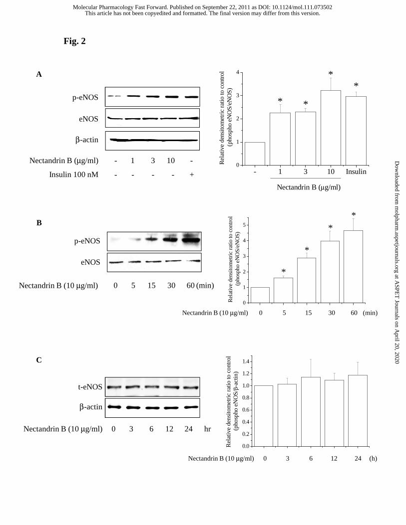
Fig. 2
A
p-eNOS
eNOS
β-actin
-- 1 3 10Nectandrin B (μg/ml)
+- - - -Insulin 100 nM - 1 3 10 Insulin0
1
2
3
4
**
**
Rel
ativ
e de
nsito
met
ric
ratio
to c
ontr
ol
(pho
spho
eN
OS/
eNO
S)
Nectandrin B (μg/ml)
p-eNOS
eNOS
600 5 15 30Nectandrin B (10 μg/ml) (min)
B
C
0
1
2
3
4
5
**
*
*
Rel
ativ
e de
nsito
met
ric
ratio
to c
ontr
ol
(pho
spho
eN
OS/
eNO
S)
600 5 15 30Nectandrin B (10 μg/ml) (min)
0.0
0.2
0.4
0.6
0.8
1.0
1.2
1.4
Rel
ativ
e de
nsito
met
ric
ratio
to c
ontr
ol
(pho
spho
eN
OS/
β-ac
tin)
240 3 6 12Nectandrin B (10 μg/ml) (h)
240 3 6 12Nectandrin B (10 μg/ml) hr
t-eNOS
β-actin
This article has not been copyedited and formatted. The final version may differ from this version.Molecular Pharmacology Fast Forward. Published on September 22, 2011 as DOI: 10.1124/mol.111.073502
at ASPE
T Journals on A
pril 20, 2020m
olpharm.aspetjournals.org
Dow
nloaded from
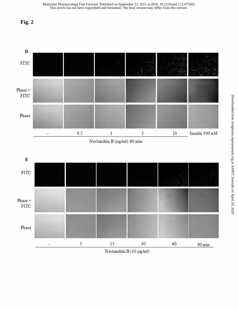
Fig. 2
This article has not been copyedited and formatted. The final version may differ from this version.Molecular Pharmacology Fast Forward. Published on September 22, 2011 as DOI: 10.1124/mol.111.073502
at ASPE
T Journals on A
pril 20, 2020m
olpharm.aspetjournals.org
Dow
nloaded from
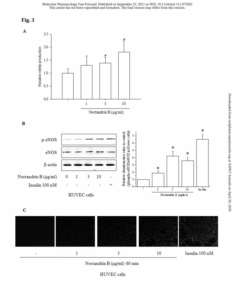
Fig. 3
This article has not been copyedited and formatted. The final version may differ from this version.Molecular Pharmacology Fast Forward. Published on September 22, 2011 as DOI: 10.1124/mol.111.073502
at ASPE
T Journals on A
pril 20, 2020m
olpharm.aspetjournals.org
Dow
nloaded from
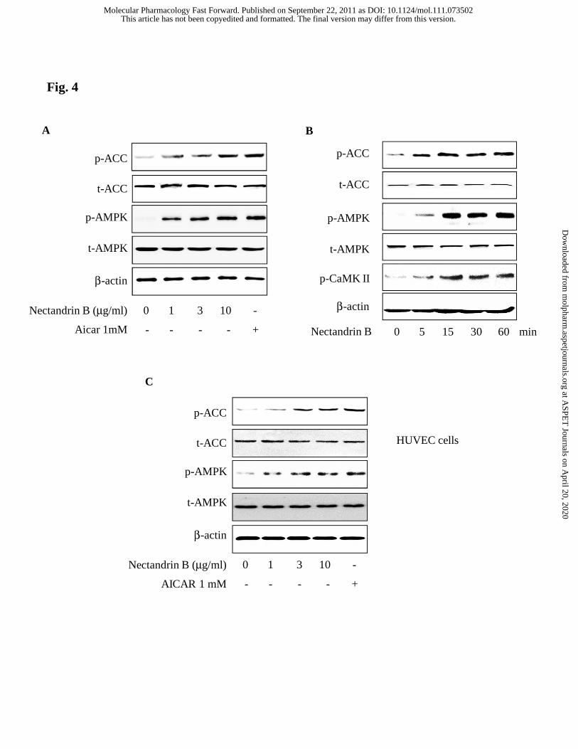
p-AMPK
t-AMPK
p-ACC
t-ACC
600 5 15 30Nectandrin B min
p-CaMK II
β-actin
B
Fig. 4
A
p-AMPK
t-AMPK
p-ACC
t-ACC
β-actin
-0 1 3 10Nectandrin B (μg/ml)
+- - - -Aicar 1mM
p-AMPK
t-AMPK
p-ACC
t-ACC
β-actin
-0 1 3 10Nectandrin B (μg/ml)
+- - - -AICAR 1 mM
HUVEC cells
C
This article has not been copyedited and formatted. The final version may differ from this version.Molecular Pharmacology Fast Forward. Published on September 22, 2011 as DOI: 10.1124/mol.111.073502
at ASPE
T Journals on A
pril 20, 2020m
olpharm.aspetjournals.org
Dow
nloaded from
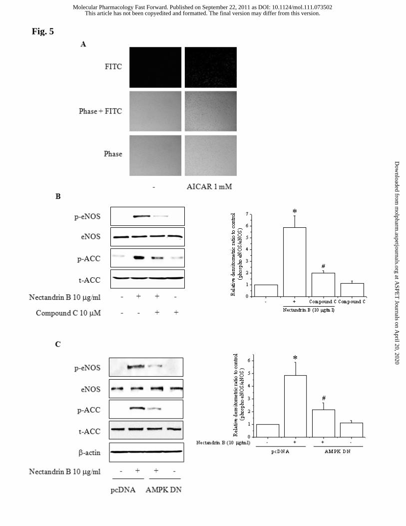
Fig. 5
This article has not been copyedited and formatted. The final version may differ from this version.Molecular Pharmacology Fast Forward. Published on September 22, 2011 as DOI: 10.1124/mol.111.073502
at ASPE
T Journals on A
pril 20, 2020m
olpharm.aspetjournals.org
Dow
nloaded from
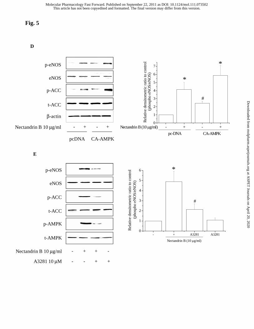
p-eNOS
eNOS
p-AMPK
t-AMPK
p-ACC
t-ACC
A3281 10 μM
- + -+
- + +-
Nectandrin B 10 μg/ml
Nectandrin B 10 μg/ml - - ++
pcDNA CA-AMPK
p-eNOS
eNOS
p-ACC
t-ACC
β-actin
Fig. 5
D
E
- + - +0
1
2
3
4
5
6
7
*
CA-AMPKpc-DNA
#
*
Rel
ativ
e de
nsito
met
ric
ratio
to c
ontr
ol
(pho
spho
eN
OS/
eNO
S)
Nectandrin B (10 μg/ml)
- + A3281 A32810
1
2
3
4
5
6
#
*
Rel
ativ
e de
nsito
met
ric
ratio
to c
ontr
ol
(pho
spho
eN
OS/
eNO
S)
Nectandrin B (10 μg/ml)
This article has not been copyedited and formatted. The final version may differ from this version.Molecular Pharmacology Fast Forward. Published on September 22, 2011 as DOI: 10.1124/mol.111.073502
at ASPE
T Journals on A
pril 20, 2020m
olpharm.aspetjournals.org
Dow
nloaded from
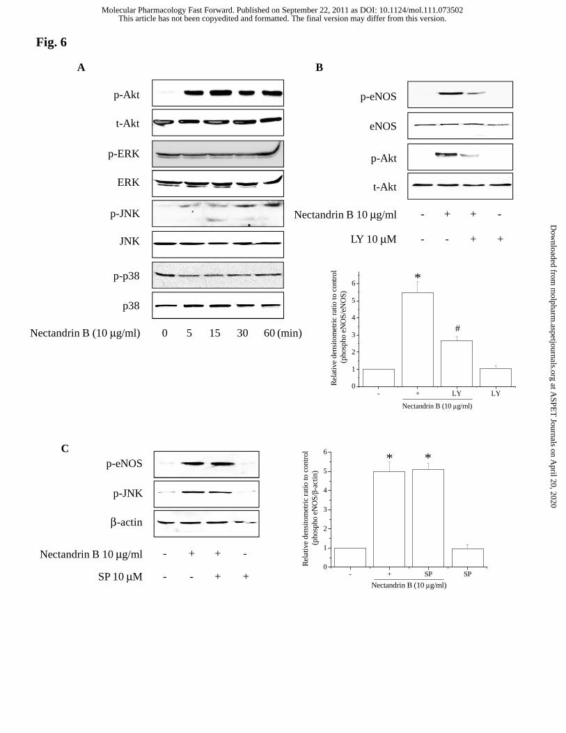
Fig. 6
p-Akt
t-Akt
600 5 15 30Nectandrin B (10 μg/ml) (min)
p-JNK
p-ERK
ERK
JNK
p38
p-p38
A
LY 10 μM
Nectandrin B 10 μg/ml - + -+
- + +-
p-Akt
t-Akt
p-eNOS
eNOS
B
p-eNOS
p-JNK
β-actin
SP 10 μM
Nectandrin B 10 μg/ml - + -+
- + +-
C
- + LY LY0
1
2
3
4
5
6
#
*
Rel
ativ
e de
nsito
met
ric
ratio
to c
ontr
ol
(pho
spho
eN
OS/
eNO
S)
Nectandrin B (10 μg/ml)
- + SP SP0
1
2
3
4
5
6**
Rel
ativ
e de
nsito
met
ric
ratio
to c
ontr
ol
(pho
spho
eN
OS/
β-ac
tin)
Nectandrin B (10 μg/ml)
This article has not been copyedited and formatted. The final version may differ from this version.Molecular Pharmacology Fast Forward. Published on September 22, 2011 as DOI: 10.1124/mol.111.073502
at ASPE
T Journals on A
pril 20, 2020m
olpharm.aspetjournals.org
Dow
nloaded from
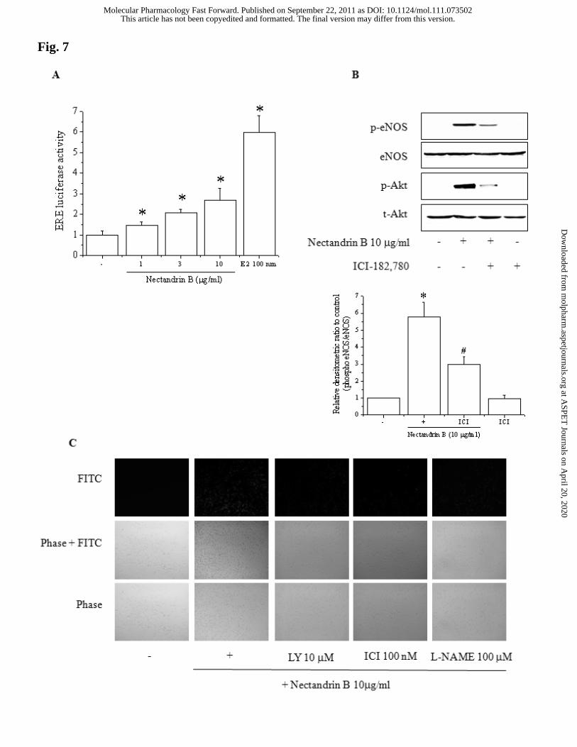
Fig. 7
This article has not been copyedited and formatted. The final version may differ from this version.Molecular Pharmacology Fast Forward. Published on September 22, 2011 as DOI: 10.1124/mol.111.073502
at ASPE
T Journals on A
pril 20, 2020m
olpharm.aspetjournals.org
Dow
nloaded from
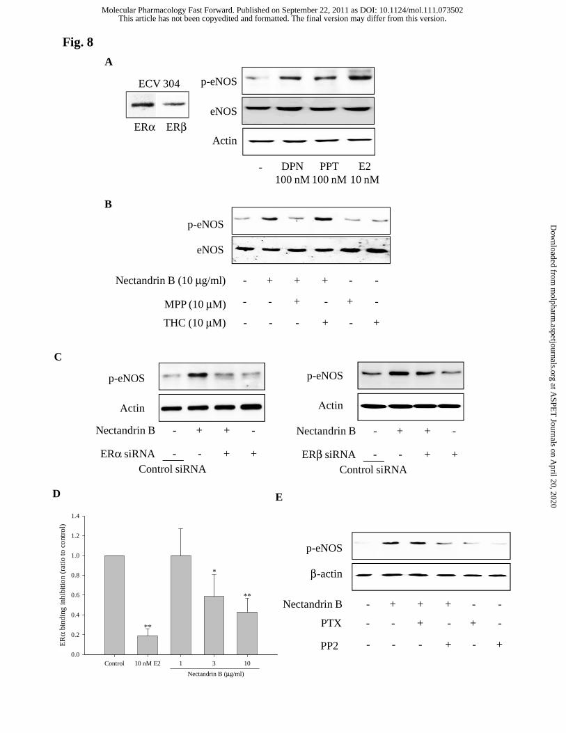
Control 10 nM E2 1 3 10
ER
α bi
ndin
g in
hibi
tion
(rat
io to
con
trol
)
0.0
0.2
0.4
0.6
0.8
1.0
1.2
1.4
**
**
*
Nectandrin B (μg/ml)
ERα ERβ
ECV 304
A
eNOS
p-eNOS
- DPN100 nM
PPT100 nM
E210 nM
Actin
Fig. 8
-- + + +Nectandrin B (10 μg/ml) -
p-eNOS
eNOS
+- - + -MPP (10 μM) -
-- - - +THC (10 μM) +
B
C
-- + + +Nectandrin B -
p-eNOS
β-actin
+- - + -PTX -
-- - - +PP2 +
-- + +Nectandrin B
+- - +ERα siRNAControl siRNA
p-eNOS
Αctin
p-eNOS
Αctin
-- + +Nectandrin B
+- - +ERβ siRNAControl siRNA
D E
This article has not been copyedited and formatted. The final version may differ from this version.Molecular Pharmacology Fast Forward. Published on September 22, 2011 as DOI: 10.1124/mol.111.073502
at ASPE
T Journals on A
pril 20, 2020m
olpharm.aspetjournals.org
Dow
nloaded from
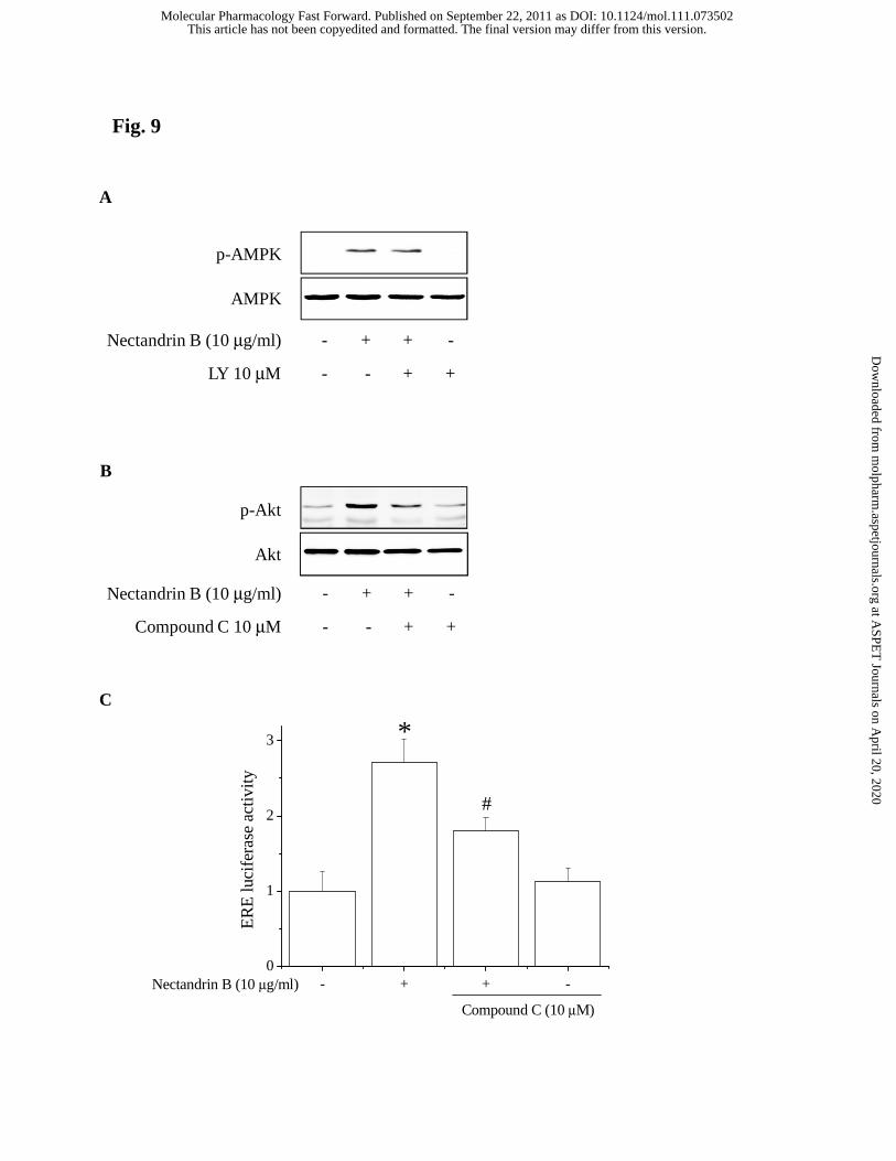
Nectandrin B (10 μg/ml)
LY 10 μM
- +
- -
+
+
-
+
AMPK
p-AMPK
Nectandrin B (10 μg/ml)
Compound C 10 μM
- +
- -
+
+
-
+
Akt
p-Akt
A
Fig. 9
B
- + + -0
1
2
3
#
*
Compound C (10 μM)
ER
E lu
cife
rase
act
ivity
Nectandrin B (10 μg/ml)
C
This article has not been copyedited and formatted. The final version may differ from this version.Molecular Pharmacology Fast Forward. Published on September 22, 2011 as DOI: 10.1124/mol.111.073502
at ASPE
T Journals on A
pril 20, 2020m
olpharm.aspetjournals.org
Dow
nloaded from
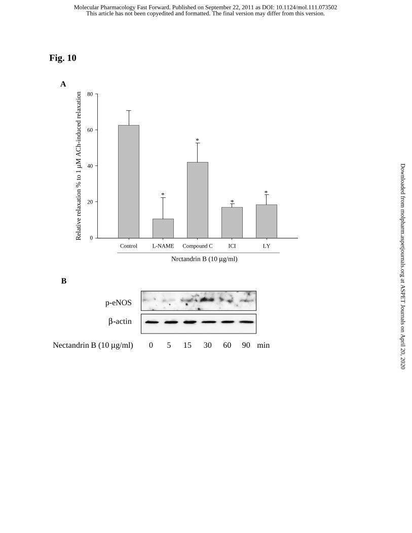
Fig. 10
Control L-NAME Compound C ICI LY
Rel
ativ
e re
laxa
tion
% to
1 μ
M A
Ch-
indu
ced
rela
xatio
n
0
20
40
60
80
**
*
*
Nrctandrin B (10 μg/ml)
600 5 15 30Nectandrin B (10 μg/ml) min90
p-eNOS
β-actin
A
B
This article has not been copyedited and formatted. The final version may differ from this version.Molecular Pharmacology Fast Forward. Published on September 22, 2011 as DOI: 10.1124/mol.111.073502
at ASPE
T Journals on A
pril 20, 2020m
olpharm.aspetjournals.org
Dow
nloaded from