030215 - REQUEST TO REOPEN EEOC CHARGE - 1st Heritage Credit - Greek
1
The Role of Liver X Receptor-α (LXRα) in the Fatty Acid
Regulation of Hepatic Gene Expression.*
Anjali Pawar§, Daniela Botolin§, David J. Mangelsdorf¶, and Donald B. Jump§¦
From the
§Departments of Physiology, Biochemistry and Molecular Biology Michigan State University East Lansing, MI 48824
¶Department of Pharmacology and Howard Hughes Medical Institute
University of Texas Southwestern Medical Center
Dallas, TX 75390-9050 *This research was supported by National Institutes of Health Grant DK43220, US Department of Agriculture Grant 98-35200-6064 and the Michigan Agriculture Experiment Station. ¦To whom correspondence should be addressed. Corresponding Author: Donald B. Jump, Ph.D. Department of Physiology 3165 Biomedical and Physical Science Bldg Michigan State University East Lansing, MI 48824 Email: [email protected] Phone: 517-355-6475, ext 1133 FAX: 517-355-5125
Copyright 2003 by The American Society for Biochemistry and Molecular Biology, Inc.
JBC Papers in Press. Published on August 13, 2003 as Manuscript M307973200 by guest on N
ovember 15, 2018
http://ww
w.jbc.org/
Dow
nloaded from
2
1The abbreviations used are: LXR, liver X receptor; LXRE, LXR regulatory element; PPAR, peroxisome proliferator activated receptor; RXR, retinoid X receptor; LBD, ligand binding domain; SREBP, sterol regulatory element binding protein; NEFA, non-esterified fatty acids; HEK, human embryonic kidney; PUFA, polyunsaturated fatty acids; ABC, ATP-binding cassette; GPAT, glycerophosphate acyl transferase; ACS-1, acyl CoA synthetase-1; CYP4A, cytochrome P450 4A; mtHMGCoAsyn, mitochondrial HMG-CoA synthase; FAS, fatty acid synthase, S14, S14 protein; L-PK, L-type pyruvate kinase; HNF-4, hepatic nuclear factor-4; CYP7A, 7α-hydroxylase; GlRR, glucose regulatory region; RLA, relative luciferase activity. 2Unpublished observation. Running title: Fatty Acid Regulation of LXRα
by guest on Novem
ber 15, 2018http://w
ww
.jbc.org/D
ownloaded from
3
Abstract: Liver X receptors (LXR, α and β) play an important role in regulating the expression
of genes involved in hepatic bile and fatty acid synthesis, glucose metabolism as well as sterol
efflux. Studies with human embryonic kidney 293 cells indicate that unsaturated fatty acids
interfere with oxysterols binding to LXR and antagonize oxysterol-induced LXRα activity. In this
report, we evaluated the effects of unsaturated fatty acids on LXR-regulated hepatic gene
expression. The LXR agonist, T1317, induced mRNAs encoding sterol regulatory element
binding protein-1c (SREBP-1c) and two SREBP-1c-regulated lipogenic genes, e.g., fatty acid
synthase (FAS) and the S14 protein, in primary hepatocytes. Treatment of hepatocytes with
eicosapentaenoic acid (20:5,n3) suppressed these mRNAs in the absence and presence of T1317.
The cis-regulatory elements targeted by T1317 were not required for fatty acid suppression of
FAS or S14 promoter activity. In contrast to SREBP-1-regulated lipogenic genes, 20:5,n3 had no
effect on the T1317 induction of ABCG5 or ABCG8 in the rat hepatoma cell line, FTO-2B. These
two genes require LXR, but not SREBP-1c, for their expression. Feeding rats a diet supplemented
with fish oil suppressed hepatic SREBP-1c regulated genes and induced PPARα-regulated genes,
but had no effect on the LXR-regulated transcripts, CYP7A1, ABCG5 or ABCG8. Transfection
studies, using either full length hLXRα or a chimera containing only the LXRα ligand binding
domain, indicate that a wide array of unsaturated fatty acids had little effect on LXRα activity in
primary hepatocytes or FTO-2B. These studies suggest that LXRα is not a target for unsaturated
fatty acid regulation in primary rat hepatocytes or in liver. Thus, oxysterol/LXR-mediated
regulation of transcripts involved in bile acid synthesis or sterol efflux appear insensitive to
dietary unsaturated fatty acids. The unsaturated fatty acid suppression of SREBP-1 and its
targeted lipogenic genes is independent of LXRα.
by guest on Novem
ber 15, 2018http://w
ww
.jbc.org/D
ownloaded from
4
Introduction.
Liver X receptors (LXR, α and β) are ligand regulated nuclear receptors that play an
important role in hepatic bile acid and fatty acid synthesis, glucose metabolism and sterol efflux
(1-3). Oxysterols, like 22[R]-hydroxycholesterol and 24,25-epoxycholesterol, bind to and activate
liver X receptors. Together with RXR, LXR bind DNA regulatory elements, i.e., DR4, and induce
the transcription of multiple genes involved in bile acid synthesis (CYP7A), sterol efflux
(ABCA1, ABCG5 and ABCG8), glucose metabolism and de novo lipogenesis. Recent studies
with human embryonic kidney 293 (HEK293) cells suggest that unsaturated fatty acids bind to
LXRα (Kd~1-4 µM) and antagonize oxysterol-activation of the LXRα, but not LXRβ (4,5).
Unsaturated fatty acids have also been reported to interfere with LXR/RXR binding to DNA
regulatory elements (6). Fatty acid interference with oxysterol regulation of LXR has important
physiological implications because of the potential to affect the expression of genes involved in
bile acid, fatty acid, cholesterol and glucose metabolism.
The effect of LXR on lipogenesis involves both direct and indirect mechanisms.
LXR/RXR heterodimers bind lipogenic gene promoters, e.g., fatty acid synthase (FAS), or
regulate lipogenic gene expression by controlling levels of SREBP-1c (3,7). SREBP-1c is a basic
helix-loop-helix-leucine zipper transcription factor that is translated as a ~125 kd precursor
(pSREBP-1c) attached to the endoplasmic reticulum (8,9). After proteolytic processing in the
Golgi, the active form, nSREBP-1c (~65 kd), accumulates in the nucleus where it binds sterol
regulatory elements (SRE) in promoters of many genes involved in fatty acid and triglyceride
synthesis. Transcription of the SREBP-1c gene is induced by insulin (10,11) and oxysterols
through LXR (12). Insulin induction of LXRα gene transcription might also account for some of
this control (13). Much of insulin action on lipogenic gene transcription has been ascribed to the
insulin-mediated induction of SREBP-1c (9,14). Unsaturated fatty acid suppression of nuclear
SREBP-1c levels is complex. In established cell lines, unsaturated fatty acids inhibit transcription
by guest on Novem
ber 15, 2018http://w
ww
.jbc.org/D
ownloaded from
5
of the SREBP-1 gene (15), enhance mRNASREBP-1 turnover (16) and interfere with proteolytic
processing of SREBPs (17). In primary hepatocytes, unsaturated fatty acids have little impact on
SREBP-1c gene transcription (18,19), but enhance mRNASREBP-1c turnover (16). Over expression
of nSREBP-1c in primary hepatocytes eliminates the polyunsaturated fatty acid (PUFA) effects
on several lipogenic genes indicating that SREBP-1c is a key target for PUFA action on de novo
lipogenesis (19,20).
Because LXRα is a target for fatty acid inhibition in HEK293 cells, we were interested in
evaluating the role LXRα played in the fatty acid regulation of hepatic gene expression. Our
studies will show that under conditions sufficient to suppress SREBP-1c and lipogenic gene
expression, certain LXR-regulated transcripts are resistant to PUFA regulation.
Materials and Methods.
Animals: Male Sprague Dawley rats (Charles River, Kalamazoo, MI) were maintained on a Tek-
Lad chow diet, ad lib. For feeding studies, rats were meal-fed a high carbohydrate diet
supplemented with olive oil at 10% w/w for 10 days. The high carbohydrate-fat free diet
(glucose replaces sucrose) was obtained from ICN Biomedicals, Inc (Aurora, OH). Meal feeding
involved allowing the rats to eat between 8 AM to noon daily. Animals were either maintained on
this diet or switched to a high carbohydrate diet supplemented with fish oil (10%, w/w) (Dyets,
Bethlehem, PA). After 5 days on the olive oil or fish oil diets, animals were euthanized ~2 hrs
after completing the meal and livers were removed for RNA analysis (19).
Primary Hepatocytes and Transfections. Male Sprague-Dawley rats maintained on a Tek-Lad
chow diet, ad lib were used for primary hepatocyte preparation (19). For RNA analysis, cells
were plated onto 100 mm type I collagen-coated plates or Primaria plastic (BD Bioscience,
Bedford, MA) dishes at 107 cells/plate in Williams E with 10 mM lactate, 10 nM dexamethasone,
by guest on Novem
ber 15, 2018http://w
ww
.jbc.org/D
ownloaded from
6
100 nM insulin and 10 % fetal calf serum. For transfection studies, cells were plated in the same
media onto 6 well type I-collagen coated plates or Primaria plastic dishes at 106 cells/well.
Transfection conditions have been described previously (19). The ratio of culture medium to cell
number was maintained constant for the different plating conditions. After a 4-6 hr attachment
period, media was changed to a serum-free medium, Williams E with 10 mM lactate, 10 nM
dexamethasone, 100 nM insulin. Cells were transfected in this serum-free media using lipofectin
or lipofectamine 2000 (Invitrogen, Carlsbad, CA) as described (5). The media was changed the
next morning to Williams E with 25 mM glucose, 10 nM dexamethasone, 100 nM insulin and
250 µM fatty acid (NuChek Prep, Elysian, MN), 50 µM bovine serum albumin-very low
endotoxin and fatty acid free (Serological Proteins, Inc, Kankakee Ill) or drug treatment (TO-
901317 (T1317), Cayman Chemicals, Ann Arbor, MI).
FTO-2B cells were obtained from L. Reid (University of North Carolina, Chapel Hill,
NC) (21) and maintained in DMEM/F12 (Invitrogen) plus 7.5% fetal bovine serum on plastic
culture dishes. HEK293 cells were described previously(5). Cells were grown in 6 well plastic
culture plates for transfection and 100 mm plastic culture plates for RNA studies. At confluence,
cells were transfected in serum free media as described above. For RNA studies, cells were
incubated overnight in serum-free medium. At the time of treatment, FTO-2B cells received 100
nM insulin and 10 nM dexamethasone along with fatty acids and/or the LXR-agonist, T1317.
Cells were transfected using lipofectamine 2000 or used for RNA extraction as described above.
RNA Isolation and Northern Analysis. RNA was extracted from rat liver, primary hepatocytes or
FTO-2B cells using Triazol (Invitrogen, Carlsbad, CA.(5). RNA was separated
electrophoretically in denaturating agarose gels, transferred to nitrocellulose and probed with 32P-
cDNAs. Levels of hybridization were quantified using a Molecular Dynamics Phosphoimager
820 (Molecular Dynamics, Sunnyvale, CA).
by guest on Novem
ber 15, 2018http://w
ww
.jbc.org/D
ownloaded from
7
Plasmids: cDNAs for SREBP-1c, fatty acid synthase and cytochrome P450 4A, (Cyp4A) were
previously described (19,22,23). A cDNA for 7α-hydroxylase, CYP7A1 (24) was obtained from
David Russell, University of Texas-Southwestern Medical Center, Dallas, TX. The plasmids
containing ABC transporters G5 and G8 were obtained from Helen Hobbs, University of Texas-
Southwestern Medical Center, Dallas TX (25). Plasmids containing GPAT and ACS1 were
obtained from Roselind Coleman, University of North Carolina, Chapel Hill, NC (26,27). Inserts
from plasmids were 32P-labeled and used to measure levels of specific mRNAs by northern
analysis. CMX-Gal4-hLXRα, CMX-hLXRα, and TK-MH100X4-Luc were previously described
(5). phRG-Luc was obtained from Promega (Madison, WS) and served as an internal control for
transfection efficiency. The S14CAT and LPKCAT reporter genes were described previously
(19,23,28,29). The FASCAT plasmids with its 5’ end point at -2369 was obtained from Steven D.
Clarke, Pennington, Baton Rouge, LA.
Cell Extracts and Western Blotting: Extracts of primary hepatocytes for analysis of protein levels
(western blotting) were prepared by homogenizing cells in Buffer A (0.25 M sucrose, 10 mM
Tris-Cl, pH 7.5, 3 mM MgCl2 plus protease inhibitors ( phenylmethylsulfonyl fluoride, 1 mM,
pefabloc, 0.1 mM, pepstatin, 5 µg/ml, leupeptin, 5 µg/ml and aprotinin, 2 µg/ml)(30). The
homogenate was centrifuged (1500 X g, 5 mins, 2oC.). The supernatant was centrifuged (100,000
x g for 1 hr, 4oC) to obtain microsomes. The pellet from the first centrifugation was resuspended
in Buffer A, adjusted to 1% NP40 and homogenized. The homogenate was centrifuged (300 X g,
5 mins, 2oC.). The supernatant was retained for analysis. The nuclear pellet was resuspended in
Buffer B (50 mM HEPES, pH 7.4, 0.1 M KCl, 3 mM MgCl2, 1 mM EDTA, 10% glycerol, plus
protease inhibitors (phenylmethylsulfonyl fluoride, 1 mM; pefabloc, 0.1 mM; pepstatin, 5 µg/ml;
leupeptin; 5 µg/ml and aprotinin, 2 µg/ml) adjusted to 0.4 M ammonium sulfate and centrifuged
at 25,000 x g for 15 mins. The supernatant was used for analysis of nuclear proteins.
by guest on Novem
ber 15, 2018http://w
ww
.jbc.org/D
ownloaded from
8
Proteins (50-100 µg) were separated electrophoretically by SDS-polyacrylamide gel
electrophoresis (NuPage 4-10% polyacrylamide-Bis-tris, Invitrogen, Carlsbad, CA) and
transferred to nitrocellulose membranes. Membranes were incubated with antibodies for SREBP-
1c (IgG-2A4) obtained from the supernatant of the hybridoma cell line CRL 2121 (American
Type Culture Collection, Manassas, VA). The anti-mouse secondary antibody was obtained from
Bio-Rad (Hercules, CA). The detection system employed the SuperSignal West Pico
Chemiluminescence kit (Pierce, Rockford, Ill).
Results.
Effects of eicosapentaenoic acid (20:5,n3) and T1317 on SREBP-1c expression in primary
hepatocytes. The LXR agonist, TO901317 (T1317) was used in all studies with primary
hepatocytes and FTO-2B cells. The native oxysterols, 22[R]-hydroxycholesterol and 24,25-
epoxycholesterol, have no effect on LXR activity or LXR-regulated transcripts in these cells,
probably because of their rapid metabolism. T1317 at 5 µM was selected as the minimal dose to
achieve reliable induction of LXR activity in primary hepatocytes and FTO-2B cells. In HEK293
cells, T1317-induced LXRα activity is attenuated by both 20:4,n6 and 20:5,n3, while PPARα
activity was induced by these fatty acids. These effects on LXRα and PPARα activity correlated
with significant changes in intracellular non-esterified unsaturated fatty acids (5). In the studies
reported here, 20:5,n3 was used as the fatty acid for most studies with primary hepatocytes and
FTO-2B cells. 20:5,n3 is a very low abundance unsaturated fatty acid in primary hepatocytes and
FTO-2B cells. Its addition at 250 µM leads to a >10-fold increase in mass of 20:5,n3 in the
intracellular non-esterified fatty acid pool of these cells. Changes in 20:5,n3 in the NEFA pool
correlate with the suppression of SREBP-1c mRNA, as well as the activation of PPARα and
PPARα-regulated genes (31).
by guest on Novem
ber 15, 2018http://w
ww
.jbc.org/D
ownloaded from
9
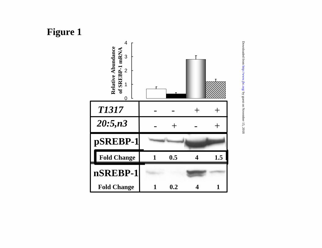
Treating primary hepatocytes with the LXR agonist, T1317 induced mRNASREBP-1c, as
well as the precursor (pSREBP-1) and nuclear (nSREBP-1) forms by 3 to 4-fold (Fig. 1). Treating
cells with 20:5,n3 in the presence or absence of T1317 suppressed mRNASREBP-1c, pSREBP-1 and
nSREBP-1 by 50-80%. This same pattern of control is seen for the 20:5,n3 and T1317 regulation
of mRNAs encoding fatty acid synthase (FAS) and the S14 protein (S14) as well as reporter
genes driven by the FAS and S14 promoters (Fig. 2A and B). While L-pyruvate kinase (LPK)
mRNA and LPKCAT reporter gene were suppressed by 20:5,n3 treatment, T1317 did not induce
this gene.
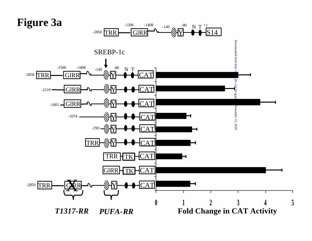
SREBP-1 binds the S14 promoter at the SRE located at -139/-129 bp; it is near a Y-box (-
104/-99 bp) that binds NF-Y (19,28,32). Together, these elements play an obligatory role in S14
gene transcription. Mutation of either element essentially abrogates S14 gene transcription
(28,32). The SRE/NF-Y elements are in the S14 PUFA-RR, a region previously identified as the
principal target for PUFA suppression of S14 gene transcription (33). The FAS promoter has
distinct regulatory elements for both SREBP and LXR (3). Our goal is to determine if the T1317
effect on S14 promoter activity is simply due to T1317/LXR-mediated induction of SREBP-1c.
Accordingly, a detail promoter analysis was preformed to locate the cis-regulatory target for
T1317.
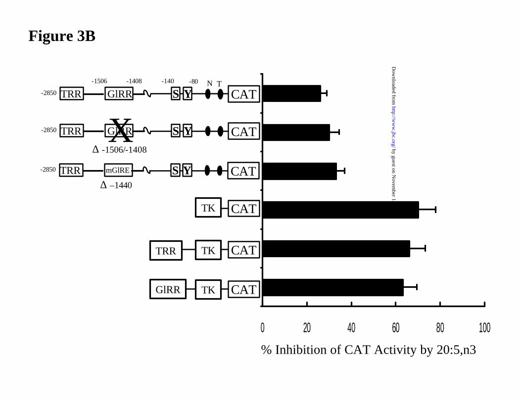
Deletion and mutation analyses show that the T1317 regulatory region (T1317-RR) is
located in a region previously identified as a glucose regulatory region (GlRR, -1.6/-1.4 kb) (Fig.
3A)(34). The E-box in the GlRR binds glucose-regulated binding proteins as well as SREBP-1c
(35,36). Deletion of the GlRR (-1.6/-1.4 kb) or mutation of the E-box within (at -1440 bp) has no
effect on PUFA suppression of S14CAT activity (Fig. 3B). In the context of the thymidine kinase
promoter, neither the GlRR or the TRR are sensitive to 20:5,n3 suppression. Preliminary studies
suggest that at least 2 T1317 elements are located within the GlRR. Studies are underway to
define these elements. A similar analysis using FASCAT reporter genes indicted that the
FASCAT activity is suppressed by 50% by 20:5,n3 treatment. Like the S14 promoter the presence
by guest on Novem
ber 15, 2018http://w
ww
.jbc.org/D
ownloaded from
10
and absence of the LXRE at -669 bp (3) did not influence promoter sensitivity to 20:5,n32. These
studies show that while S14 and FAS have distinct cis-regulatory elements for T1317/LXR and
SREBP-1c, the T1317/LXR-cis regulatory elements are not required for PUFA control of these
transcripts in rat primary hepatocytes.
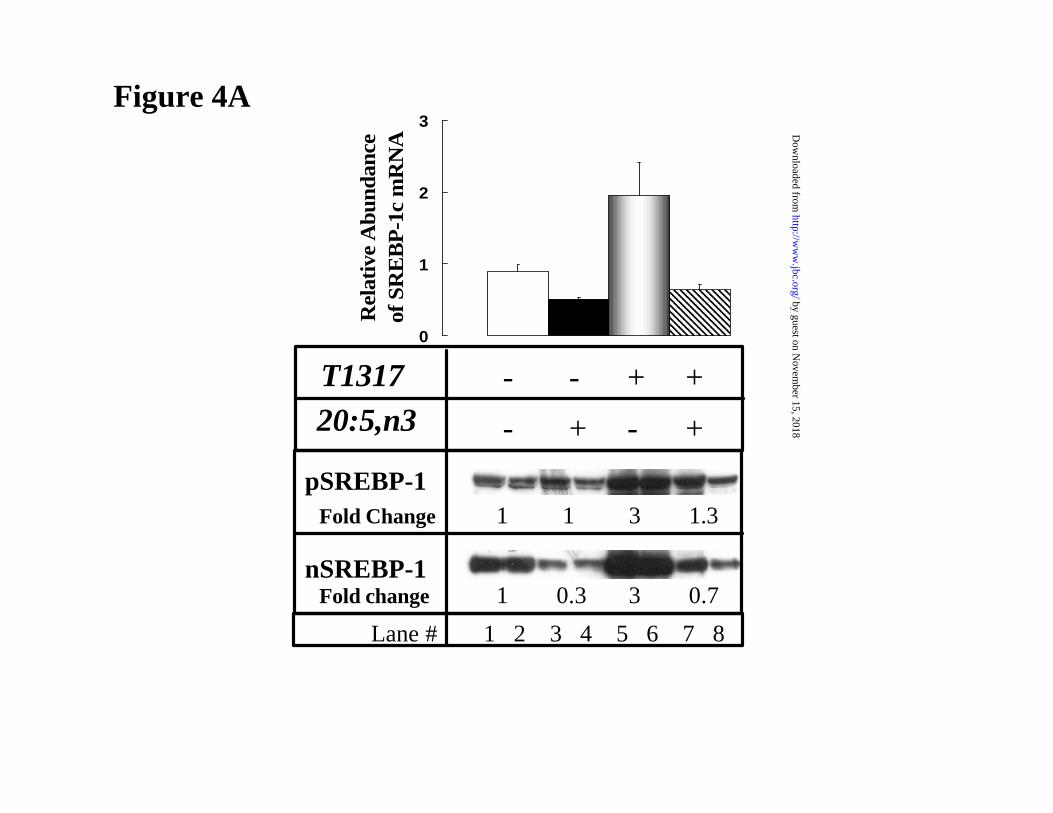
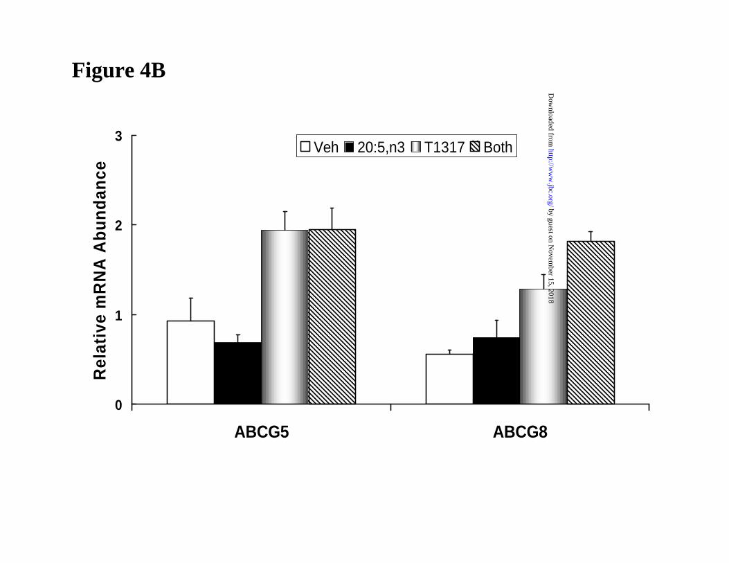
20:5,n3 effects on LXR-regulated transcripts in FTO-2B hepatoma cells. While the
expression of SREBP-1c and various lipogenic genes are easily examined in primary rat
hepatocytes, certain LXR-regulated transcripts, e.g., CYP7A, ABCG5 and ABCG8, decrease to
nearly undetectable levels when compared to their expression in liver. The reason for this decline
is likely due to the loss of key transcription factors controlling expression of these transcripts.
Fortunately, ABCG5 and ABCG8 are well-expressed and regulated by T1317 in FTO-2B cells
(25). Because the T1317 regulation of these transcripts is independent of SREBP-1c, we
examined the effect of 20:5,n3 on the T1317-mediated induction of SREBP-1c, ABCG5 and
ABCG8 in FTO-2B cells (Fig. 4).
Previous studies indicated that the predominant SREBP-1 subtype in FTO-2B cells is
SREBP-1c (4). RNA protection studies indicate the ratio of SREBP-1c to -1a is >4-fold2. T1317
induced mRNASREBP-1c as well as the precursor and nuclear forms of SREBP-1c 2 to 3-fold in
FTO-2B cells (Fig. 4A). 20:5,n3 suppressed mRNASREBP-1c and the nuclear form of SREBP-1c by
50-70% in the absence and presence of T1317. The 20:5,n3 and T1317 regulation of SREBP-1c
in FTO-2B cells is similar to that seen in primary hepatocytes (Fig. 1). While T1317 induced
mRNAABCG5 and mRNAABCG8 ~2-fold, 20:5,n3 had no effect on the level of these transcripts (Fig.
4B). This finding suggests that LXR, per se, is not a target for unsaturated fatty acid antagonism
in FTO-2B cells.
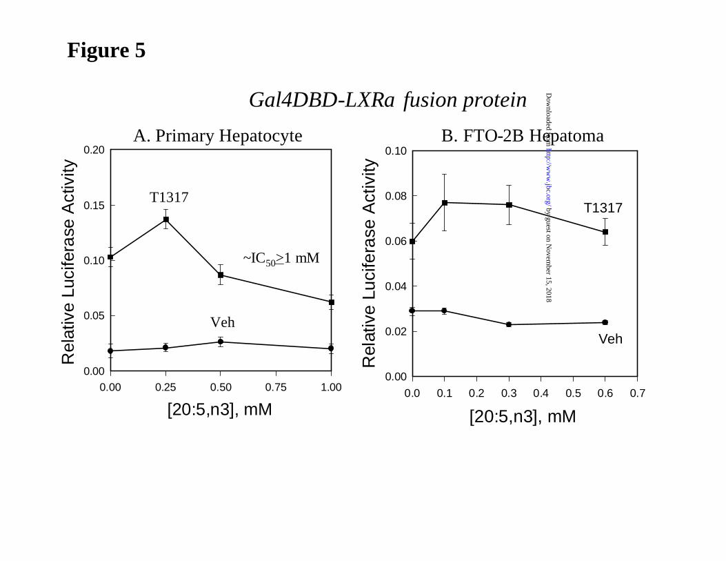
20:5,n3 regulation of LXRα activity in primary hepatocytes and FTO-2B hepatoma cells. In
an effort to examine the effect of 20:5,n3 on LXRα activity directly, we used a transfection
by guest on Novem
ber 15, 2018http://w
ww
.jbc.org/D
ownloaded from
11
approach. Because our previous studies established that only LXRα, and not LXRβ, was affected
by unsaturated fatty acid treatment (5), our studies focused on LXRα. Accordingly, primary
hepatocytes were transfected with an expression vector containing the ligand-binding domain of
LXRα fused to the Gal-4 DNA binding domain (LXRα-LBD) and the MH-TK-LUC reporter
containing 4 Gal 4-regulatory elements (Fig. 5). The use of this chimeric receptor allows for an
evaluation of fatty acid effects on LXR activity without the requirement for RXR
heterodimerization. Following treatment of primary hepatocytes with T1317, LXR activity was
induced 4-fold. No significant fatty acid-mediated antagonism of LXRα activity was detected
until 20:5,n3 levels reached 1 mM. At lower levels (0.25 mM), levels typically used to examine
PUFA regulation of lipogenic gene transcription, 20:5,n3 augments LXRα activity by 30%. A
similar study with FTO-2B cells revealed no evidence of 20:5,n3 interference with T1317-
mediated induction of LXRα activity. In each case, fatty acid treatment had no effect on basal
LUC activity in the absence of T1317. For comparison, a recent dose response analysis of
20:4,n6 effects on LXR activity in HEK293 cells showed that the IC50 for the antagonism of
20:4,n6 was 22 µM2. Clearly, LXRα is considerably more sensitive to PUFA inhibition in
HEK293 cells than in primary hepatocytes. More importantly, the inhibitory effect of 20:5,n3 on
LXRα activity is only seen when very high non-physiological levels of the fatty acid are added.
Such levels are likely never reached in vivo.
To determine if other unsaturated fatty acids affect LXRα activity in primary
hepatocytes, transfected cells were treated with various mono- and poly-unsaturated fatty acids
(at 250 µM) in the absence and presence T1317 (Fig. 6A). In no case did any fatty acid tested
affect LXRα activity. Of the fatty acids tested, all but 18:1,n9, suppress SREBP-1c mRNA levels
in primary hepatocytes (19).
Others reported that, in addition to fatty acid effects on oxysterol binding, unsaturated
fatty acids interfered with LXR-RXR heterodimerization and DNA binding (6). To determine if
by guest on Novem
ber 15, 2018http://w
ww
.jbc.org/D
ownloaded from
12
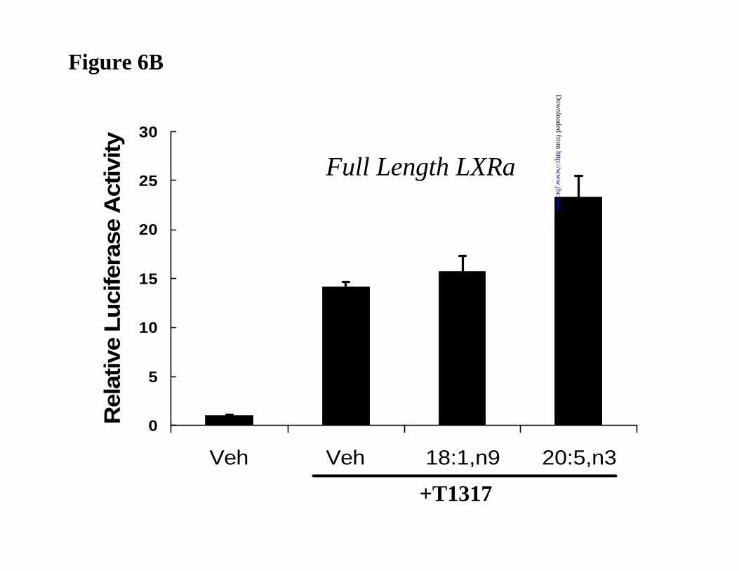
unsaturated fatty acids interfered with the activity of the full length LXRα, primary hepatocytes
were transfected with a full length LXRα expression vector and the LXRE-TK-Luc reporter
vector (Fig. 6B). For the reporter plasmid to respond to T1317, endogenous RXR must be
recruited to the promoter by LXRα. Accordingly, treatment of primary hepatocytes with T1317
induced LUC activity 15-fold. Treatment of cells with 18:1,n9 or 20:5,n3 in the absence of T1317
had no effect on basal LUC activity (not shown). Co-treatment with T1317 and either 18:1,n9 and
20:5,n3 did not inhibit LUC activity. Thus, the capacity of the full length LXRα to activated
promoters containing LXREs is not impeded by the addition of unsaturated fatty acids to primary
hepatocytes.
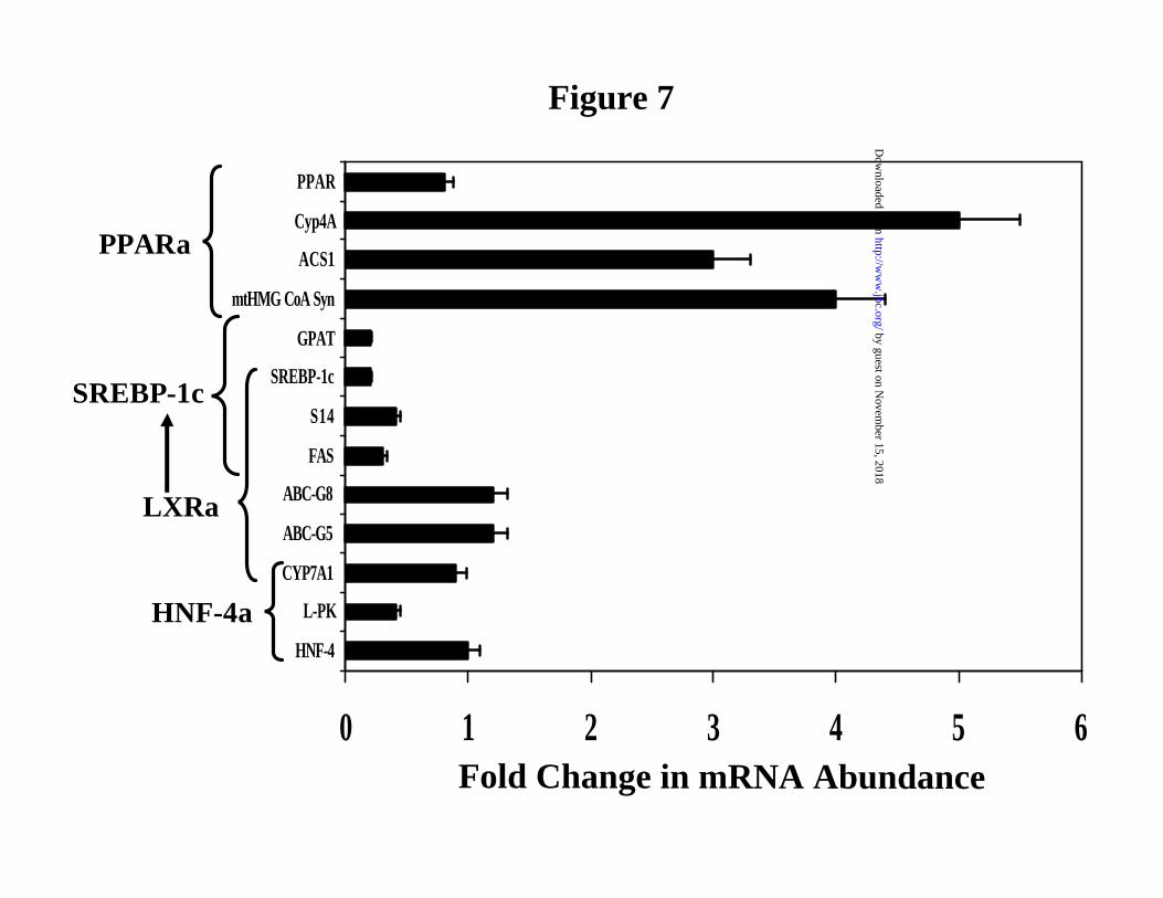
Effects of fish oil on LXR-regulated gene expression in rat liver. In an effort to assess the in
vivo effects of fatty acids on LXR-regulated hepatic gene expression we fed rats fish oil. Fish oil
is enriched in 20:5,n3 and 22:6,n3. Addition of fish oil to diets has well-established effects on
PPARα and SREBP-1c-regulated gene expression in rat liver (19,23) . In this study, rats were
meal-fed a high carbohydrate diet containing either olive or fish oil for 5 days (Fig. 7). Fish oil
feeding induced hepatic mRNA levels for several PPARα target genes, i.e., cytochrome P450-4A
(CYP4A), mitochondrial HMG-CoA synthase (mtHMG-CoA syn), and acyl CoA synthetase-1
(ACS1) >3-fold (Fig. 7), as well as acyl CoA oxidase (AOX)2. The mRNAs encoding SREBP-1c,
FAS, S14, glycerophosphate acyl transferase (GPAT) and L-PK were suppressed by 50-80%.
SREBP-1c regulates the transcription of FAS, S14 and GPAT, but not L-PK (9,19). Fish oil
treatment had no effect on transcripts encoding PPARα and HNF4α. More importantly, fish oil
feeding did not affect hepatic levels of the LXR-regulated transcripts, CYP7A1, ABC-G5 or
ABC-G8. RT-PCR analysis for ABC-A1 also shows this transcript is insensitive to fish oil
treatment2. This in vivo analysis agrees favorably with our previous analysis of these transcripts
in primary hepatocytes (Fig. 1 and 2) and FTO-2B hepatoma cells (Figure 4). In vivo, n3-PUFA
induced PPARα-regulated transcripts and suppressed SREBP-1c-regulated transcripts, but had no
by guest on Novem
ber 15, 2018http://w
ww
.jbc.org/D
ownloaded from
13
effect on those LXR regulated transcripts that do not require SREBP-1c. The in vivo studies
corroborate the cell culture studies with primary hepatocytes and FTO-2B hepatoma cells.
Discussion.
Liver X receptors (LXR α and β) play a key role in regulating the transcription of
multiple genes involved in bile acid and fatty acid synthesis, glucose metabolism and sterol efflux
(1,3,7,12,25,37,38). As such these nuclear receptors influence a broad array of hepatic metabolic
events that influence whole body glucose, lipid and cholesterol metabolism. Moreover, the liver is
a major target for fatty acid regulated gene expression (39). Finding that unsaturated fatty acids
antagonized LXRα, but not LXRβ, activity suggested that sphere of influence of unsaturated fatty
acids may potentially be extended beyond their well known effects on the expression of genes
involved in fatty acid synthesis and oxidation (39). In this report, we used several approaches to
evaluate the fatty acid regulation of LXR in liver. The new information reported here includes: 1)
the T1317-mediated induction of hepatic mRNA SREBP-1c, as well as the precursor and nuclear
forms of SREBP-1, is suppressed by 20:5,n3 treatment (Fig. 1); 2) the T1317 regulation of
SREBP-1c nuclear content parallels its effects on FAS and S14 gene expression and promoter
activity (Fig. 2); 3) the T1317/LXR regulated cis-acting elements in the S14 and FAS genes are
not required for fatty acid regulation of these promoters (Fig. 3); 4) the T1317 induction of LXR-
regulated transcripts, i.e., CYP7A, ABCG5, ABCG8 and ABCA1, is not affected by unsaturated
fatty acids in FTO-2B cells and in rat liver (Fig. 4 and 7); 5) hLXRα activity is antagonized by
PUFA only at high, non-physiological levels, levels >20-fold of that found for the PUFA
antagonism of hLXRα in HEK293 cells (Fig. 5). Based on these findings, we conclude that
LXRα is not a target for fatty acid antagonism in rat liver, rat primary hepatocytes or rat FTO-2B
hepatoma cells. Thus, unsaturated fatty acids activate PPARα regulatory networks and suppress
by guest on Novem
ber 15, 2018http://w
ww
.jbc.org/D
ownloaded from
14
SREBP-1c regulatory networks, but do not impede hepatic LXR regulatory networks in primary
hepatocytes or in vivo.
Because 20:5,n3 is a minor unsaturated fatty acid in the total lipid fraction and NEFA
pool of hepatocytes, its addition to primary hepatocytes leads to >50- and >10-fold increase in
mass of this fatty acid in the total lipid and intracellular NEFA fractions, respectively, within 90
minutes (31). The level of 20:5,n3 in the NEFA pool is sustained for ~6 hrs, after which it
declines to levels ~3-fold above basal values by 24 hrs. The change in cellular 20:5,n3 in the
NEFA fraction correlates well with the activation of PPARα and the induction of PPARα-
regulated transcripts (31) and the suppression of SREBP-1c mRNA2. The 250 µM dose of
20:5,n3 used here is sufficient to suppress mRNAs for SREBP-1c and lipogenic genes and to
induce PPARα and PPARα-regulated transcripts, but is insufficient to affect LXRα-activity.
Only very high, non-physiologic levels of 20:5,n3 inhibited LXRα activity (Fig. 5). Clearly,
LXRα activity in primary hepatocytes is far less sensitive to PUFA than either PPARα or
SREBP-1c or the PUFA regulation of LXRα in HEK293 cells (ED50<50 µM) (5). Finding that
full length LXRα was insensitive to PUFA treatment (Fig. 6B) argues against effects of PUFA on
LXR/RXR heterodimerization and DNA binding. Interestingly, in HEK293 cells, LXRα and
PPARα are equally sensitive to PUFA action suggesting that both receptors might serve as
sensors to intracellular NEFA levels (5). While PPARα can be considered as monitor of
intracellular NEFA (31), the studies reported here indicate that LXRα is not responsive to major
changes in intracellular NEFA in liver or primary hepatocytes.
The fact that we found no evidence for unsaturated fatty acid regulation of LXR activity
in liver does not exclude LXRα from being sensitive to fatty acid control in another cell context.
Clearly, fatty acids affect LXRα activity in HEK293 cells(4,5). While the mechanism for this
control has been ascribed to competitive binding, in vivo mechanisms may be more complicated
involving receptor-coactivator interaction or the regulation of other signaling mechanism that do
by guest on Novem
ber 15, 2018http://w
ww
.jbc.org/D
ownloaded from
15
not exist in liver or primary hepatocytes, e.g., cyclooxygenase and lipoxygenase (29). Previous
efforts to examine fatty acid effects on targets of LXR action, i.e., CYP7A, did not reveal specific
effects of unsaturated fatty acids (40-42). In fact, effects of fatty acids on CYP7A have been
attributed to PPARα (42), not LXR. A recent report on the LXR-regulated gene, ABCA1, in
macrophage suggests that fatty acid treatment inhibited cholesterol efflux by increasing ABCA1
protein degradation, without effects on ABCA1 mRNA (43). These observations have clinical
significance because if fish oil interfered with bile acid synthesis or sterol efflux, then serum
cholesterol levels would rise significantly increasing the risk for atherosclerosis and coronary
disease. If anything, fish oil or EPA/DHA treatment of rats and humans is cardio-protective
having pronounced hypolipemic effects (44,45).
Finally, we have identified a prospective cis-regulatory region for LXR action in the S14
promoter. This region, tentatively identified as a T1317-regulatory region (T1317-RR) is located
in the glucose-regulatory region of the S14 promoter located between -1.4 and -1.6 kb upstream
from the transcription start site. This region contains an E-box that is known to bind glucose-
regulated binding proteins as well as SREBP-1c (35,46). Additional studies will be required to
identify the specific minimal cis-regulatory targets for T1317 action and whether LXR/RXR
binds these elements. Despite this limitation, deletion of the entire GlRR eliminates T1317
regulation of S14 promoter activity, but fails to impact PUFA control of S14 promoter activity
(Fig. 3). Previous studies established that the PUFA-regulatory region, binding both SREBP-1c
and NF-Y, is indispensable for S14 gene transcription. Moreover, this region is the principal
target for PUFA control of the S14 promoter activity (28,32,33). These findings indicate that the
key elements involved in PUFA control of S14 are distinct from the GlRR containing T1317-
regulated factors. The fatty acid synthase promoter contains an LXRE at -669 bp, that is distinct
from two SREs (-150 and -65 bp) (3). Analysis of FASCAT reporter genes indicates that the
LXRE is not required for PUFA suppression of FASCAT activity2. Based on these studies,
by guest on Novem
ber 15, 2018http://w
ww
.jbc.org/D
ownloaded from
16
T1317/LXR-regulatory elements in the FAS and S14 promoters are dispensable for PUFA control
of these promoters.
In summary, we have examined the role LXRα plays in PUFA regulation of hepatic gene
expression. While LXRα is clearly a target for PUFA antagonism in HEK293 cells (4,5), it is not
a target for fatty acid regulation in rat liver, primary hepatocytes or FTO-2B hepatoma cells. This
conclusion is based on the lack of 20:5,n3 antagonism on LXRα activity or the LXR-regulated
transcripts, ABCG5 and G8 at doses sufficient to repress SREBP-1c mRNA or to induce PPARα
activity. The fact remains that LXRα activity is well antagonized by PUFA in HEK293 cells.
This antagonism might be important for LXR action in some tissues. However, the absence of
any effect on LXR in liver suggests that fatty acid effects on bile acid and fatty acid synthesis,
sterol efflux or glucose metabolism cannot be explain by abrogated LXR action.
Acknowledgements: The authors would like to thank Drs. Rosalind Coleman, Helen Hobbs and
David Russell for the generous gifts of plasmids used in this study. The authors would also like to
thank the efforts of Barbara Christian for excellent technical assistance and Drs. Julia Busik and
L. Karl Olson for critical review of the manuscript.
by guest on Novem
ber 15, 2018http://w
ww
.jbc.org/D
ownloaded from
17
Figure Legends.
Figure 1: Eicosapentaenoic acid (20:5,n3) and T1317 regulation of SREBP-1 in rat
primary hepatocytes. Primary hepatocytes were treated with 250 µM 20:5,n3 (black bars); 5 µM
TO-901317 [T1317] (shaded bars); or both compounds (stripped bars) for 24 hrs. Vehicle treated
cells (white bars) received 50 µM BSA. [Upper panel]: SREBP-1 northern analysis, RNA was
extracted and separated by electrophoresis for northern analysis. Blots were probed with 32P-
cDNA for SREBP-1c. Levels of hybridization were quantified by phosphoimager analysis.
Results are expressed as Relative mRNA Abundance; the mRNA levels in the treated cells are
normalized to the level of the corresponding RNA in a rat liver standard from a chow-fed male
rat. These results are the mean + SD, N=9 of three separate studies. [Lower Panel]: SREBP-1
Immunoblot. Cells were treated as above and extracted for microsomal and nuclear proteins
(Materials and methods) for immunoblotting. The precursor form of SREBP-1, i.e., pSREBP-1 is
found in the microsomal fraction, while the nuclear form, i.e., nSREBP-1, is recovered in the
nuclear fraction. The figure is representative of at least 3 separate studies. Analysis of the nuclear
extracts for HNF-4α revealed robust levels of HNF-4, with no consistent treatment effect (not
shown).
Figure 2: Eicosapentaenoic acid (20:5,n3) and T1317 regulation of fatty acid synthase,
S14 protein and L-pyruvate kinase expression in rat primary hepatocytes. Primary
hepatocytes were treated with 250 µM 20:5,n3 (black bars); 5 µM T1317 (shaded bars); or both
compounds (stripped bars) for 24 hrs. Vehicle treated cells (white bars) received 50 µM BSA.
[A]: RNA was extracted and separated by electrophoresis for northern analysis. Blots were
probed with 32P-cDNAs for FAS, S14 or LPK. Levels of hybridization were quantified by
phosphoimager analysis. Results are expressed as Relative mRNA Abundance; the mRNA levels
by guest on Novem
ber 15, 2018http://w
ww
.jbc.org/D
ownloaded from
18
in the treated cells are normalized to the level of the corresponding RNA in a rat liver standard
from a chow-fed male rat. These results are the mean + SD, N=9 of three separate studies. [B]:
Primary hepatocytes were transfected with fatty acid synthase, S14 and L-pyruvate kinase
reporter genes containing promoter elements fused to the chloramphenicol acetyltransferase
(CAT) reporter gene. FASCAT contains -2363 to + 16 bp of the FAS promoter; S14CAT
contains -2800 to + 19 bp of the S14 promoter and LPKCAT contains (-4300 to + 14 bp) of the
L-pyruvate kinase promoter. After transfection, hepatocytes were treated with 20:5,n3 and T1317
as described above. After a 24 hrs treatment, cells were harvested for CAT and protein assays.
Results are reported as Fold Change in CAT activity. The results are the mean of 3 independent
studies involving triplicate samples.
Figure 3. Role of the T1317 regulatory region in the PUFA control of S14 promoter
activity. [A] Mapping the T1317 regulatory element in the S14 promoter. Primary hepatocytes
were transfected with CAT reporter genes containing various elements from the S14 promoter. A
schematic of the S14 promoter is illustrated at the top of the figure and the composition of each
promoter-reporter gene construct is illustrated in the figure. The thyroid hormone regulatory
region (TRR) and the glucose regulatory region (GlRR) were excised from the S14 promoter and
fused to the thymidine kinase (TK) promoter to yield TRR-TK-CAT and GlRR-TK-CAT,
respectively. The reporter gene at the bottom of the figure has the GlRR (-1506 to -1408 bp)
removed and replaced by a Nsi I restriction site. After transfection, hepatocytes were treated with
5 µM T1317 as described above. After a 24 hrs treatment, cells were harvested for CAT and
protein assays. Results are reported as Fold Change in CAT activity. The results are the mean of 3
independent studies involving triplicate samples. T1317-RR, T1317 regulatory region; PUFA-
RR, PUFA-regulatory region. [B] The S14 T1317-regulatory region is not involved in PUFA
suppression of S14CAT activity. Primary hepatocytes were transfected with CAT reporter genes
containing various elements from the S14 promoter or S14 promoter elements fused to the
by guest on Novem
ber 15, 2018http://w
ww
.jbc.org/D
ownloaded from
19
thymidine kinase (TK)-promoter. The composition of the promoter elements is illustrated in the
figure. The S14 reporter constructs lacking the entire GlRR or the E-box in the GlRR are replaced
by the Nsi I restriction site. After transfection, hepatocytes were treated with 250 µM 20:5,n3 as
described above. After a 24 hrs treatment, cells were harvested for CAT and protein assays.
Results are reported as % Inhibition of CAT Activity by 20:5,n3. The results are the mean + SD
of 2 independent studies involving triplicate samples.
Figure 4: The effect of T1317 and 20:5,n3 on LXRα-regulated transcripts in rat FTO-
2B hepatoma cells cells. Confluent FTO-2B cells were treated with insulin (100 nM) and
dexamethasone (10 nM) for 48 hrs prior to the study. Cells then received T1317 (5 µM) and/or
20:5,n3 (300 µM) for 24 hrs. Cells were harvested for RNA extraction or microsomal and nuclear
proteins as described in Figure 1. [A]: (Upper panel) Measurement of mRNASREBP-1 by northern
analysis; (Lower panel) Measurement of pSREBP-1 and nSREBP-1 protein by western analysis.
The lower panel displays duplicate samples for each treatment. As in Figure 1, Fold Change of
SREBP-1 is included in the figure. Lane # is identified at the bottom of the figure. [B]:
Measurement of mRNAABCG5 and mRNAABCG8 by northern analysis. Results are representative of
2 separate studies with duplicate samples; mean + SD.
Figure 5: Fatty acid regulation of LXRα activity in primary hepatocytes and FTO-2B
hepatoma cells. Primary hepatocytes [A] and FTO-2B hepatoma cells [B] were transfected with
an expression vector containing LXRα-LBD fused to the Gal4 DNA binding domain (CMX-
Gal4-hLXRα) and the reporter plasmid, TK-MH100X4-Luc. Cells also received phRG-Luc as an
internal control for transfection efficiency. After an overnight transfection, cells were treated
without or with an LXR agonist T1317 (5 µM), without and with 20:5,n3 ranging from 0.25 to 1
mM. The 20:5,n3:BSA ratio was kept constant at 5:1. Vehicle treated cells received the same
by guest on Novem
ber 15, 2018http://w
ww
.jbc.org/D
ownloaded from
20
level of BSA. After 24 hr of treatment, cells were lysed and assayed for luciferase activity and
protein. Relative luciferase activity is the ratio of firefly luciferase activity to renilla luciferase
activity (internal control). Results are expressed as Relative Luciferase Activity and are
representative of >2 studies. Mean + SD; N = 6.
Figure 6: Effect of unsaturated fatty acids on LXRα activity in primary hepatocytes.
Primary hepatocytes were transfected with [A] CMX-Gal4-hLXRα and the TK-MH100X4-Luc
reporter or [B] CMX-hLXRα and the LXREx3-Luc reporter plasmid. After an overnight
transfection, cells were treated without or with the LXR agonist 5 µM T1317 and without and
with various unsaturated fatty acids at 250 µM for 24 hrs. BSA was included at a 5:1 ratio of fatty
acid to BSA. After the 24 hr treatment period, cells were lysed and assayed for luciferase activity.
Results are expressed as Relative Luciferase Activity and are representative of >2 separate
studies. Mean + SD; N = 6.
Figure 7: Dietary fat regulation of hepatic gene expression. Male Sprague-Dawley rats
were meal-fed high carbohydrate diets supplemented with 10% olive or 10% fish oil (w/w) for 5
days. RNA was extracted and assayed for various transcripts (Materials and Methods). Results
are expressed as fold change in mRNA. The expression of the various transcripts was normalized
to olive oil-fed rats. Mean + SD, N=3.The results are representative of several studies.
by guest on Novem
ber 15, 2018http://w
ww
.jbc.org/D
ownloaded from
21
References:
1. Lu, T. T., Repa, J. J., and Mangelsdorf, D. J. (2001) J Biol Chem 276, 37735-37738
2. Laffitte, B. A., Chao, L.C., Li, J., Walczak, R., Hummasti, S., Joseph, S.B., Castrillo, A., Wilpitz, D.C., Mangelsdorf, D.J., Collins, J.S. Saez, E., and Tontonoz, P. (2003) Proc Natl Acad Sci, USA 100, 5419-5424
3. Joseph, S. B., Laffitte, B. A., Patel, P. H., Watson, M. A., Matsukuma, K. E., Walczak, R., Collins, J. L., Osborne, T. F., and Tontonoz, P. (2002) J Biol Chem 277, 11019-11025
4. Ou, J., Tu, H., Shan, B., Luk, A., DeBose-Boyd, R. A., Bashmakov, Y., Goldstein, J. L., and Brown, M. S. (2001) Proc Natl Acad Sci U S A 98, 6027-6032
5. Pawar, A., Xu, J., Jerks, E., Mangelsdorf, D. J., and Jump, D. B. (2002) J Biol Chem 277, 39243-39250
6. Yoshikawa, T., Shimano, H., Yahagi, N., Ide, T., Amemiya-Kudo, M., Matsusuka, T., Kakakuki, M., Tomita, S., Okazaki, H., Tamura, Y., Iizuka, Y., Ohashi, K., Takahashi, A., Sone, H., Osuga, J-I., Gotoda, T., Ishibashi, S., and Yamada, N.,. (2002) J Biol Chem 277, 1705-1711
7. Laffitte, B. A., Chao, L. C., Li, J., Walczak, R., Hummasti, S., Joseph, S. B., Castrillo, A., Wilpitz, D. C., Mangelsdorf, D. J., Collins, J. L., Saez, E., and Tontonoz, P. (2003) Proc Natl Acad Sci U S A
8. Brown, M. S., and Goldstein, J. L. (1997) Cell 89, 331-340 9. Horton, J. D., Goldstein, J. L., and Brown, M. S. (2002) J Clin Invest 109, 1125-
1131 10. Azzout-Marniche, D., Becard, D., Guichard, C., Foretz, M., Ferre, P. and
Foufelle, F. (2000) Biochem J 350, 389-393 11. Foretz, M., Guichard, C., Ferre, P., and Foufelle, F. (1999) ProcNatl Acad Sci
USA 96, 12737-12742 12. Schultz, J. R., Tu, H., Luk, A., Repa, J. J., Medina, J. C., Li, L., Schwendner, S.,
Wang, S., Thoolen, M., Mangelsdorf, D. J., Lustig, K. D., and Shan, B. (2000) Genes Dev 14, 2831-2838
13. Tobin, K. A., Ulven, S. M., Schuster, G. U., Steineger, H. H., Andresen, S. M., Gustafsson, J. A., and Nebb, H. I. (2002) J Biol Chem 277, 10691-10697
14. Liang, G., Yang, J., Horton, J. D., Hammer, R. E., Goldstein, J. L., and Brown, M. S. (2002) J Biol Chem 277, 9520-9528
15. Hannah, V. C., Ou, J., Luong, A., Goldstein, J. L., and Brown, M. S. (2001) J Biol Chem 276, 4365-4372
16. Xu, J., Teran-Garcia, M., Park, J. H., Nakamura, M. T., and Clarke, S. D. (2001) J Biol Chem 276, 9800-9807
17. Worgall, T. S., Johnson, R.A., Seo, T., Gierens, H., and Deckelbaum, R.J. (2002) J Biol Chem 277, 3878-3885
18. Xu, J., Nakamura, M. T., Cho, H. P., and Clarke, S. D. (1999) J Biol Chem 274, 23577-23583
by guest on Novem
ber 15, 2018http://w
ww
.jbc.org/D
ownloaded from
22
19. Mater, M. K., Thelen, A. P., Pan, D. A., and Jump, D. B. (1999) J Biol Chem 274, 32725-32732
20. Yahagi, N., Shimano, H. Hasty, A.H., Amemiya-Kudo, M., Okazaki, H., Tamura, Y., Iizuka, Y., Shionoiri, F., Ohashi, K., Osuga, J., Harada, K., Gotoda, T., Nagai, R., Ishibashi, S., and Yamada, N. (1999) J Biol Chem 274, 35840-35844
21. Zvibel, I., Fiorino, A.S., Brill, S., and Reid, L.M. (1998) Differentiation 63, 215-223
22. Ren, B., Thelen, A. P., Peters, J. M., Gonzalez, F. J., and Jump, D. B. (1997) J Biol Chem 272, 26827-26832
23. Pan, D. A., Mater, M. K., Thelen, A. P., Peters, J. M., Gonzalez, F. J., and Jump, D. B. (2000) J Lipid Res 41, 742-751
24. Jelinek, D. F., Andersson, S., Slaughter, C.A. and Russell, D.W. (1990) J Biol Chem 265, 8190-8197
25. Repa, J. J., Berge, K. E., Pomajzl, C., Richardson, J. A., Hobbs, H., and Mangelsdorf, D. J. (2002) J Biol Chem 277, 18793-18800
26. Lewin, T. M., Granger, D. A., Kim, J. H., and Coleman, R. A. (2001) Arch Biochem Biophys 396, 119-127
27. Kim, J. H., Lewin, T. M., and Coleman, R. A. (2001) J Biol Chem 276, 24667-24673
28. Jump, D. B., Thelen, A. P., and Mater, M. K. (2001) J Biol Chem 276, 34419-34427
29. Mater, M. K., Thelen, A. P., and Jump, D. B. (1999) J Lipid Res 40, 1045-1052 30. Botolin, D., and Jump, D. B. (2003) J Biol Chem 278, 6959-6962 31. Pawar, A. and Jump, D.B. (2003) J Biol Chem 278, In Press 32. Jump, D. B., Badin, M. V., and Thelen, A. (1997) J Biol Chem 272, 27778-27786 33. Jump, D. B., Clarke, S. D., MacDougald, O., and Thelen, A. (1993) Proc Natl
Acad Sci U S A 90, 8454-8458 34. Shih, H. M., and Towle, H. C. (1992) J Biol Chem 267, 13222-13228 35. Kim, J. B., Spotts, G. D., Halvorsen, Y. D., Shih, H. M., Ellenberger, T., Towle,
H. C., and Spiegelman, B. M. (1995) Mol Cell Biol 15, 2582-2588 36. Koo, S. H., Dutcher, A. K., and Towle, H. C. (2001) J Biol Chem 276, 9437-9445 37. Zhang, Y., Repa, J. J., Gauthier, K., and Mangelsdorf, D. J. (2001) J Biol Chem
276, 43018-43024 38. Laffitte, B. A., Joseph, S. B., Chen, M., Castrillo, A., Repa, J., Wilpitz, D.,
Mangelsdorf, D.J., and Tontonoz, P. (2003) Mol Cell Biol 23, 2182-2191 39. Jump, D. B. (2002) Curr Opin Lipidol 13, 155-164 40. Cheema, S. K., Cikaluk, D. and Agellon, L.B. (1997) Journal of Lipid Research
38, 315-323 41. Cheema, S. K. and Agellon, L.B. (1999) J Nutr 129, 1718-1724 42. Cheema, S. K. and Agellon, L.B. (2000) J Biol Chem 275, 12530-12536 43. Wang, Y. and Oram, J.F. (2002) J Biol Chem 277, 5692-5697 44. Nordoy, A., Hansen, J.B., Brox, J. and Svensson, B. (2001) Nutr Metab
Cardiovasc Dis 11, 7-16 45. Chan, D. C., Watts, G.F., Mori, T.A., Barrett, P.H., Beilin, L.J. and Redgrave,
T.G. (2002) Eur. J. Clin. Invest. 32, 429-436 46. Koo, S. H., and Towle, H. C. (2000) J Biol Chem 275, 5200-5207
by guest on Novem
ber 15, 2018http://w
ww
.jbc.org/D
ownloaded from
pSREBP-1
nSREBP-1
T131720:5,n3 - + - +
- - + +
Fold Change 1 0.2 4 1
Fold Change 1 0.5 4 1.5
0
1
2
3
4
Rel
ativ
e A
bund
ance
of
SR
EB
P-1
mR
NA
Figure 1
by guest on Novem
ber 15, 2018http://w
ww
.jbc.org/D
ownloaded from
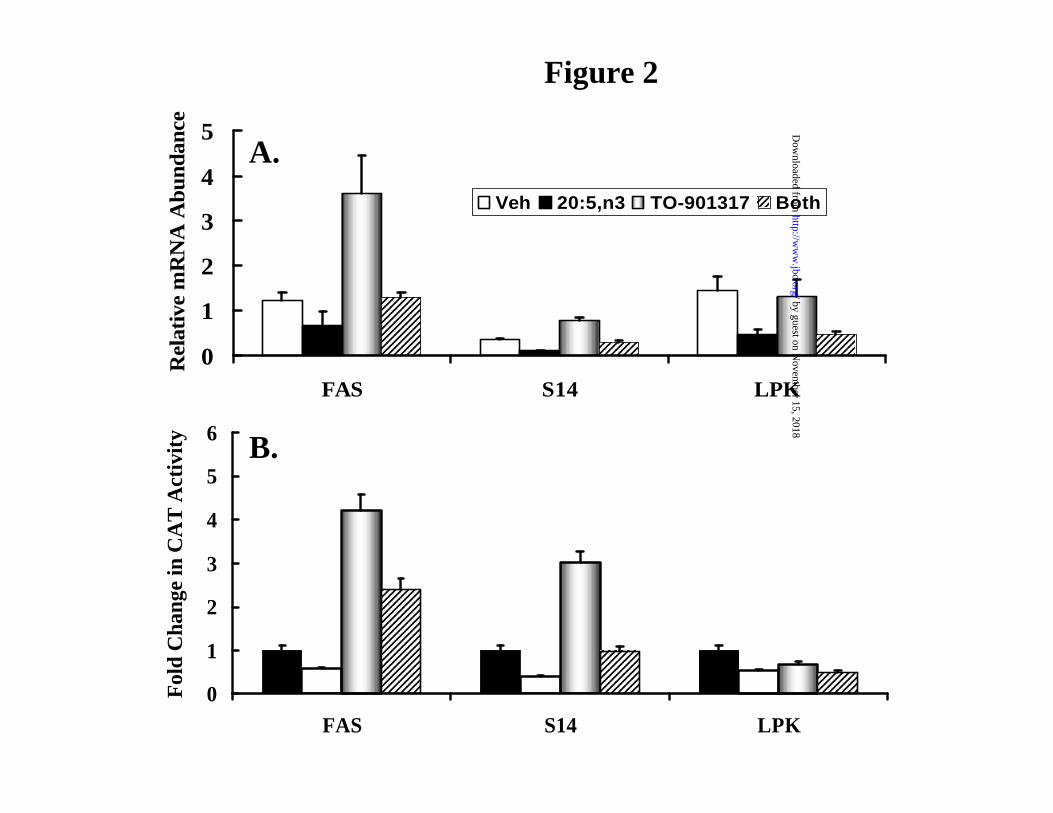
Figure 2
0
1
2
3
4
5
6
FAS S14 LPK
Fold
Cha
nge
in C
AT
Act
ivity
A.
B.
0
1
2
3
4
5
FAS S14 LPK
Rel
ativ
e m
RN
A A
bund
ance
Veh 20:5,n3 TO-901317 Both
by guest on Novem
ber 15, 2018http://w
ww
.jbc.org/D
ownloaded from
0 1 2 3 4 5Fold Change in CAT Activity
Figure 3a
CAT
GlRR
Y
CATGlRR
-1074
Y
CATTRR TK
CATY
CATGlRR-2850 YTRR
-2110
TRR CATY
-1601
CATTK
GlRR
-290 CATY
S14GlRR+1-140
-2850
-1408 TNYTRR
X
SREBP-1c
-1506
PUFA-RRT1317-RR
CATGlRR-80-140
-2850
-1408 TNYTRR
-1506
-80
by guest on Novem
ber 15, 2018http://w
ww
.jbc.org/D
ownloaded from
0 20 40 60 80 100
1
2
3
4
5
6
% Inhibition of CAT Activity by 20:5,n3
CATGlRR-2850 S YTRR
CATGlRR
X∆ -1506/-1408
CATmGlRE-2850 S YTRR∆ –1440
CAT
CAT
TRR
CATGlRR-80-140
-2850
-1506 TNS YTRR
-1408
TK
TK
TK
Figure 3B
by guest on Novem
ber 15, 2018http://w
ww
.jbc.org/D
ownloaded from
pSREBP-1
nSREBP-1
0
1
2
3
1
Rel
ativ
e A
bund
ance
of
SR
EB
P-1
c m
RN
AT131720:5,n3 - + - +
- - + +
Fold Change 1 1 3 1.3
Fold change 1 0.3 3 0.7
Figure 4A
1 2 3 4 5 6 7 8Lane #
by guest on Novem
ber 15, 2018http://w
ww
.jbc.org/D
ownloaded from
0
1
2
3
ABCG5 ABCG8
Rel
ativ
e m
RN
A A
bund
ance
Veh 20:5,n3 T1317 Both
Figure 4B
by guest on Novem
ber 15, 2018http://w
ww
.jbc.org/D
ownloaded from
[20:5,n3], mM0.00 0.25 0.50 0.75 1.00
Rel
ativ
e Lu
cife
rase
Act
ivity
0.00
0.05
0.10
0.15
0.20
Veh
T1317
~IC50>1 mM
Figure 5
[20:5,n3], mM0.0 0.1 0.2 0.3 0.4 0.5 0.6 0.7
Rel
ativ
e Lu
cife
rase
Act
ivity0.00
0.02
0.04
0.06
0.08
0.10
T1317
Veh
A. Primary Hepatocyte B. FTO-2B Hepatoma
Gal4DBD-LXRα fusion protein
by guest on Novem
ber 15, 2018http://w
ww
.jbc.org/D
ownloaded from
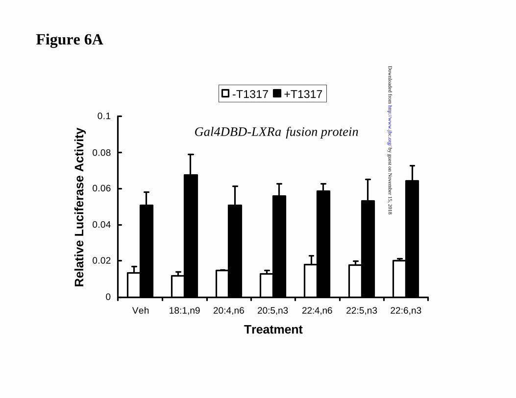
Figure 6A
0
0.02
0.04
0.06
0.08
0.1
Veh 18:1,n9 20:4,n6 20:5,n3 22:4,n6 22:5,n3 22:6,n3
Treatment
Rel
ativ
e L
uci
fera
se A
ctiv
ity
-T1317 +T1317
Gal4DBD-LXRα fusion protein by guest on N
ovember 15, 2018
http://ww
w.jbc.org/
Dow
nloaded from
0
5
10
15
20
25
30
Veh Veh 18:1,n9 20:5,n3
Rel
ativ
e L
uci
fera
se A
ctiv
ity
+T1317
Figure 6B
Full Length LXRα by guest on N
ovember 15, 2018
http://ww
w.jbc.org/
Dow
nloaded from
0 1 2 3 4 5 6
HNF-4
L-PK
CYP7A1
ABC-G5
ABC-G8
FAS
S14
SREBP-1c
GPAT
mtHMG CoA Syn
ACS1
Cyp4A
PPAR
Fold Change in mRNA Abundance
HNF-4α
SREBP-1c
LXRα
PPARα
Figure 7
by guest on Novem
ber 15, 2018http://w
ww
.jbc.org/D
ownloaded from
Anjali Pawar, Daniela Botolin, David J. Mangelsdorf and Donald B. Jumpgene expression
The role of liver X receptor-alpha (LXR-alpha) in the fatty acid regulation of hepatic
published online August 13, 2003J. Biol. Chem.
10.1074/jbc.M307973200Access the most updated version of this article at doi:
Alerts:
When a correction for this article is posted•
When this article is cited•
to choose from all of JBC's e-mail alertsClick here
by guest on Novem
ber 15, 2018http://w
ww
.jbc.org/D
ownloaded from