
Molecular Ultraviolet and Visible Spectroscopypostonp/ch313/PDF/Chapter 6 Solutions.pdf · The...
16
Chapter 6: Molecular Ultraviolet and Visible Spectroscopy Problem 6.1: What is the frequency and energy of a photon with a wavelength of the following: (a) 650 nm (b) 50 nm (c) 1,700 m (d) 0.65 m (e) 195 nm (f) 700 nm (g) 0.5 m (h) 500 nm a. 650 nm: c = λν = c/ (2.998 10 8 ⁄) 650 10 −9 = 4.612 x 10 14 Hz E = hc/λ = (6.626 10 −34 ⁄ )(2.998 10 8 ⁄) (650 10 −9 ) = 3.056 x 10 -19 J c. 1700 μm: c = λν = c/ (2.998 10 8 ⁄) 1,700 10 −6 = 1.763 x 10 11 Hz E = hc/λ = (6.626 10 −34 ⁄ )(2.998 10 8 ⁄) (1,700 10 −6 ) = 1.169 x 10 -22 J e. 500 nm: c = λν = c/ (2.998 10 8 ⁄) 500 10 −9 ν = 5.976 x 10 14 Hz E = hc/λ = (6.626 10 −34 ⁄ )(2.998 10 8 ⁄) (500 10 −9 ) = 3.973 x 10 -19 J g. 0.5 m: c = λν = c/ 2.998 10 8 ⁄ 0.5 10 −6 ν = 5.976 x 10 14 Hz E = hc/λ = (6.626 10 −34 ⁄ )(2.998 10 8 ⁄) (500 10 −9 ) = 3.973 x 10 -19 J
Transcript of Molecular Ultraviolet and Visible Spectroscopypostonp/ch313/PDF/Chapter 6 Solutions.pdf · The...
Chapter 6:
Molecular Ultraviolet and Visible Spectroscopy Problem 6.1: What is the frequency and energy of a photon with a wavelength of the following:
(a) 650 nm
(b) 50 nm
(c) 1,700 m
(d) 0.65 m
(e) 195 nm
(f) 700 nm
(g) 0.5 m
(h) 500 nm
a. 650 nm: c = λν = c/ (2.998 𝑋 108𝑚
𝑠⁄ )
650 𝑋 10−9𝑚 = 4.612 x 10¹⁴ Hz
E = hc/λ = (6.626 𝑋 10−34𝐽
𝑠⁄ )(2.998 𝑋 108𝑚𝑠⁄ )
(650 𝑋 10−9𝑚) = 3.056 x 10⁻¹⁹ J
c. 1700 µm: c = λν = c/ (2.998 𝑋 108𝑚
𝑠⁄ )
1,700 𝑋 10−6𝑚 = 1.763 x 10¹¹ Hz
E = hc/λ = (6.626 𝑋 10−34𝐽
𝑠⁄ )(2.998 𝑋 108𝑚𝑠⁄ )
(1,700 𝑋 10−6𝑚) = 1.169 x 10⁻²² J
e. 500 nm: c = λν = c/ (2.998 𝑋 108𝑚
𝑠⁄ )
500 𝑋 10−9𝑚 ν = 5.976 x 10¹⁴ Hz
E = hc/λ = (6.626 𝑋 10−34𝐽
𝑠⁄ )(2.998 𝑋 108𝑚𝑠⁄ )
(500 𝑋 10−9𝑚) = 3.973 x 10⁻¹⁹ J
g. 0.5 m: c = λν = c/ 2.998 𝑋 108 𝑚 𝑠⁄
0.5 𝑋 10−6𝑚 ν = 5.976 x 10¹⁴ Hz
E = hc/λ = (6.626 𝑋 10−34𝐽
𝑠⁄ )(2.998 𝑋 108𝑚𝑠⁄ )
(500 𝑋 10−9𝑚) = 3.973 x 10⁻¹⁹ J
Problem 6.2: Refer to Figure 6.2. To what part of the EMS do photons with the following
frequencies belong:
(a) 2.99 107 Hz
(b) 10.00 Hz
(c) 3.21 1020 Hz
(d) 1.54 1015Hz
(e) 6.31 1018 Hz
(f) 7.50 1014 Hz
(g) 4.29 1014 Hz
(h) 3.33 1014 Hz
Figure 6.2 shows wavelength ranges in units of nanometers.
a. 2.99 X 10⁷ Hz: = c/= 2.998 𝑋 108 𝑚 𝑠⁄
2.99 𝑋 107 𝑠−1 = 10.03 meters 1.00 X 10¹⁰ nm (Radio Waves)
c. 3.21 X 10²⁰ Hz: = c/= 2.998 𝑋 108 𝑚 𝑠⁄
3.21 𝑋 1020 𝑠−1 = 9.34 X 10⁻¹³ meters 9.34 X 10⁻⁴ nm (Gamma Waves)
e. 6.31 X 10¹⁸ Hz: = c/= 2.998 𝑋 108 𝑚 𝑠⁄
6.31𝑋 1018 𝑠−1 = 4.75 X 10⁻¹¹ meters 4.75X 10⁻² nm (Hard X-Rays)
g. 4.25 X 10¹⁴ Hz: = c/= 2.998 𝑋 108 𝑚 𝑠⁄
4.25 𝑋 1014 𝑠−1 = 7.05 X 10⁻⁷ meters 705 nm (Visible - Red)
Problem 6.3: We have stated that the typical range of a UV-vis spectrophotometer is 195 nm to 900 nm. What
is this range in Hertz?
= c/, Note! Remember to convert wavelength to meters!
when = 195 nm = 3.0 𝑋 108𝑚
2⁄
1.95 𝑋 10−7𝑚 = 1.53 X 10¹⁵ s-1
= 900 nm = 3.0 𝑋 108𝑚
2⁄
9.00 𝑋 10−7𝑚 = 3.33 X 10¹⁴ s-1
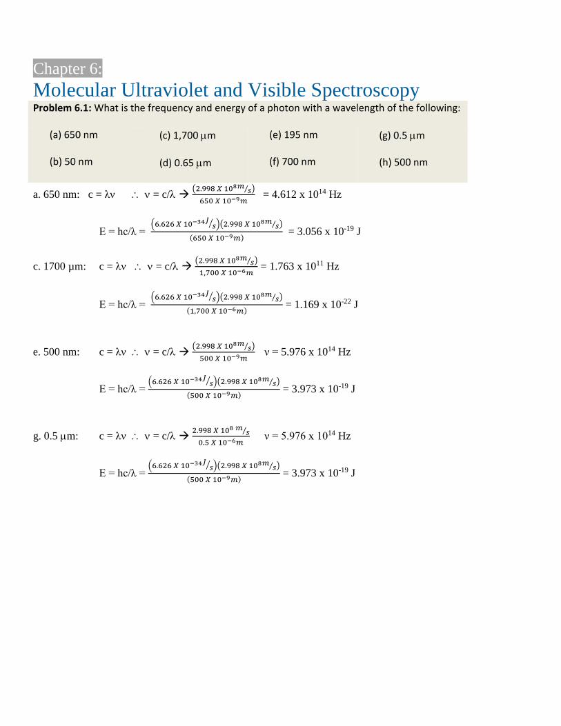
Problem 6.4: Using Figure 6.3 as a guide, construct an M.O. diagram for ethylene (ethene). How is the M.O. diagram similar to the M.O. diagram of formaldehyde? How is the M.O. diagram different from the M.O. diagram of formaldehyde? Using your M.O. diagram, predict the UV-vis absorption properties of ethylene.
Figure 6.3 is presented here and we see
that the LUMO is *. The HOMO-1
results from the non-bonding lone pair
orbitals on oxygen and the HOMO-2
results from the -bond between carbon
and oxygen. We should expect to see a
low energy n * transition and a higher
energy * transition in
formaldehyde. Due to the poor spatial
overlap between the n-orbitals and the *
orbitals, we would expect the intensity of
the n * transition to be less than the
intensity of the * transition.
The M.O. of ethylene is similar to
formaldehyde in that it has a *
resulting from the bond. We should
expect to see this transition at a similar
energy (wavelength) in both molecules.
However since oxygen is lower in energy
than is carbon, we should expect the
* in ethylene to be at a slightly longer
wavelength than the same transition seen
in formaldehyde. Unlike formaldehyde,
there are no lone pair electrons capable of
producing a low energy n* transition
in ethylene. Therefore we would not
expect to see this transition in ethylene.
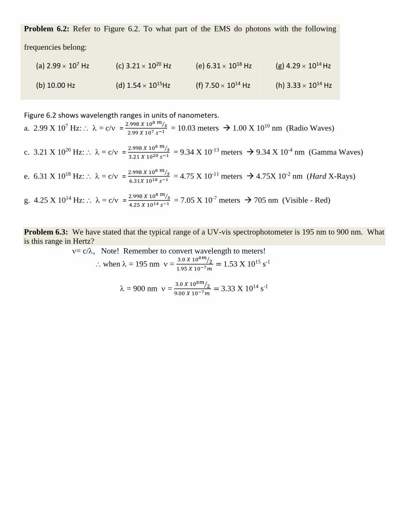
Problem 6.5: Benzophenone has a structure that is very similar to di-2-pyridyl ketone (dpk). Using our case
study of dpk as a guide, discuss in general terms, how the UV-vis spectrum of benzophenone will be similar to
that dpk and how will it be different. Then sketch your prediction of the UV-vis spectra of benzophenone.
According to the case study of dpk, the very small
peak at around 350 nm was due to the n-π* transition
of the nitrogen in the pyridyl ring. Therefore, we
would expect the spectrum to be essentially the same
but with the absence of that peak at 350.
Problem 6.6: There are three peaks in the UV-vis spectrum of DPK (Figure 6.7) with max values of 238 nm,
270 nm and 354 nm. Use Equation 6.2 and
a) calculate the energy gap (in Joules) associated with each of these transitions b) calculate the frequency (in Hz) of the photon absorbed for each of these transitions
(a) 238 nm: E = hc/λ = (6.626x10⁻³⁴Js)(2.998x10⁸m/s)/(238 x 10-9 m) = 8.345 x 10⁻¹⁹ J
270 nm: E = hc/λ = (6.626x10⁻³⁴Js)(2.998x10⁸m/s)/(270 x 10-9 m) = 7.357 x 10⁻¹⁹ J
354 nm: E = hc/λ = (6.626x10⁻³⁴Js)(2.998x10⁸m/s)/(354 x 10-9 m) = 5.612 x 10⁻¹⁹ J
(b) 238 nm: ν = c/λ = (2.998x10⁸m/s)/(238 x 10-9 m) = 1.260 x 10¹⁵ J
270 nm: ν = c/λ = (2.998x10⁸m/s)/(270 x 10-9 m) = 1.110 x 10¹⁵ J
354 nm: ν = c/λ = (2.998x10⁸m/s)/(354 x 10-9 m) = 8.469 x 10¹⁵ J
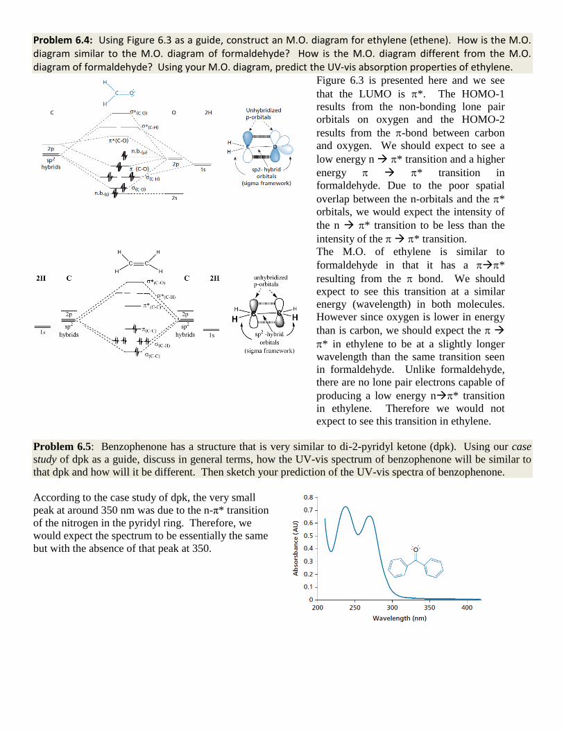
Problem 6.7: Refer back to Figure 6.4. Determine the total spin multiplicity of the ground and excited states
for panels A & B. State if the transition is spin allowed or spin forbidden.
For panel (A) and panel (B) the ground state and excited state spin
multiplicities are, M = 2s+1 = 2(0) + 1 = 1.
The ground and excited states for both panels are singlet states and
therefore both transitions are spin allowed.
Problem 6.8: Discuss the two transitions seen in Figure 6.4 in terms of the LaPorte selection rule. Make sure you discuss how the LaPorte selection rule would affect peak intensity.
The LaPorte rule states that for a centrosymmetric molecule, transitions between orbitals of the same symmetry
are forbidden. Although the * transitions are between orbitals with the same symmetry, formaldehyde is
not a centrosymmetric molecule and therefore we do not expect the LaPorte rule to play a significant role in the
spectroscopy.
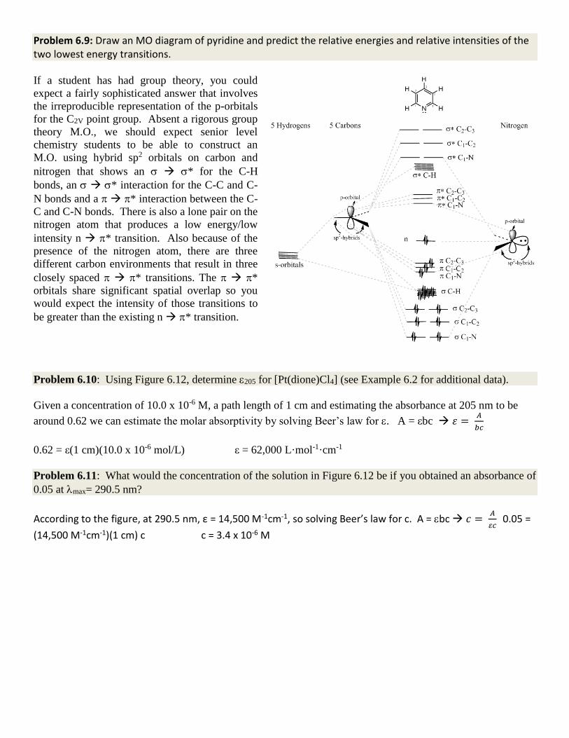
Problem 6.9: Draw an MO diagram of pyridine and predict the relative energies and relative intensities of the two lowest energy transitions.
If a student has had group theory, you could
expect a fairly sophisticated answer that involves
the irreproducible representation of the p-orbitals
for the C2V point group. Absent a rigorous group
theory M.O., we should expect senior level
chemistry students to be able to construct an
M.O. using hybrid sp2 orbitals on carbon and
nitrogen that shows an * for the C-H
bonds, an * interaction for the C-C and C-
N bonds and a * interaction between the C-
C and C-N bonds. There is also a lone pair on the
nitrogen atom that produces a low energy/low
intensity n * transition. Also because of the
presence of the nitrogen atom, there are three
different carbon environments that result in three
closely spaced * transitions. The *
orbitals share significant spatial overlap so you
would expect the intensity of those transitions to
be greater than the existing n * transition.
Problem 6.10: Using Figure 6.12, determine 205 for [Pt(dione)Cl4] (see Example 6.2 for additional data).
Given a concentration of 10.0 x 10⁻⁶ M, a path length of 1 cm and estimating the absorbance at 205 nm to be
around 0.62 we can estimate the molar absorptivity by solving Beer’s law for . A = εbc 𝜀 = 𝐴
𝑏𝑐
0.62 = ε(1 cm)(10.0 x 10⁻⁶ mol/L) ε = 62,000 L·mol-1·cm-1
Problem 6.11: What would the concentration of the solution in Figure 6.12 be if you obtained an absorbance of
0.05 at max= 290.5 nm?
According to the figure, at 290.5 nm, ε = 14,500 M-1cm-1, so solving Beer’s law for c. A = bc 𝑐 = 𝐴
𝜀𝑐 0.05 =
(14,500 M-1cm-1)(1 cm) c c = 3.4 x 10⁻⁶ M
Problem 6.12: Explain why it is considered a “best practice” to keep the total absorbance below 1 when
conducting quantitative work.
In practice the slope of A vs c is usually linear if the total absorbance is kept below 1. When the absorbance
exceeds 1, the slope of the line often curves downward and flattens out. Remember that absorbance is -log(T)
so at an absorbance of 1, only 10% of the radiant power is reaching the detector. For this reason, it is
considered a best practice to keep absorbance values below 1.
Problem 6.13: Explain why it is important that the ionic strength of your blank be similar to the ionic strength
of your samples, even if the salt does not absorb light at the frequency of interest.
If your analyte has strong intermolecular forces of attraction for itself (for example it is capable of hydrogen
bonding), or if there are other chemical species in solution that can polarize the orbitals of your solute (such as a
salt) then it is possible that will vary with concentration. Ionic strength can also affect the density of the
solution and therefore influence the refractive index of the solution.
Problem 6.14: What are the two most common sources used in a UV-vis spectrophotometer?
A deuterium discharge lamp and a tungsten-halogen bulb.
Problem 6.15: Why is it a common design feature to see two different sources used in a UV-vis spectrophotometer? In the most common design, the UV portion of the source radiation is provided by a deuterium discharge lamp
and the visible and near infrared radiation is provided by the tungsten-halogen bulb.
Problem 6.16: Why is iodine sometimes added to tungsten lamps? The addition of a small amount of iodine to the lamp extends the life span of the lamp and allows for the
operation of the lamp at higher voltages. With iodine present, the sublimed tungsten atoms react to form a
gaseous W-I compound; when the W-I compound strikes the hot filament, it decomposes and the tungsten
atoms are deposited back onto the filament. Problem 6.17 For each of the types of noise described so far discuss how the presence of noise affects the output data, give a physical explanation for what caused the noise, include equations (if applicable). Also discuss methods or ways to minimize the noise. Stray Light
The effect of stray light hitting the detector is an apparent loss of absorption.
𝐴𝑜𝑏𝑠 = −𝑙𝑜𝑔(𝑃+𝑃𝑠)
(𝑃𝑜+ 𝑃𝑠) Eq. 6.10
Aobs = the observed absorbance Ps = the radiant power of the stray light
Minimizing the amount of stray light reaching the detector is an issue of design. Careful screening of source radiation or external radiation is therefore crucial.
Detector Noise (Dark Current) Most detector types have a dark current which becomes a limiting factor at the lower detection limits of the instrument.
𝐴𝑜𝑏𝑠 = −𝑙𝑜𝑔(𝑃+𝑃𝐷)
(𝑃𝑜+ 𝑃𝐷) Eq. 6.11
Aobs = the observed absorbance PD = power output from dark current
Minimizing the effects of dark current is accomplished by increasing the intensity of the source radiation. This in turn reduces the impact of the PD term in equation 6.11.
Detector Noise (Shot Noise) Shot noise is described by
𝑖𝑟𝑚𝑠 = √2𝐼𝑒∆𝑓 Eq. 6.12
𝑖𝑟𝑚𝑠 = the root mean square of the current fluctuation 𝐼 = the average output current 𝑒 = the charge on an electron ∆𝑓 = the bandwidth of the signal.
You can decrease shot noise by decreasing the bandwidth of the detector. However that will be accompanied by a slower detector response.
Source Noise
The most common type of noise from the source is thermal noise and is generated by the random motion of electrons found in electric conductors. This motion is present even in the absence of an applied voltage. Thermal noise is considered to be white noise and as such can be reduced through signal averaging. The equation describing thermal noise is
𝑖𝑟𝑚𝑠2 =
4𝑘𝐵𝑇∆𝑓
𝑅 Eq. 6.13
kb = Boltzmann Constant T = Kelvin Temperature
f = bandwidth of the instrument R = resistance of the device in Ohms.
Thermal noise can also be reduced by reducing the temperature or the bandwidth. The trade-off of lowering the bandwidth to reduce white noise is an accompanying reduction in response time.
Exercise 6.1: What is the wavelength range of a typical UV-vis spectrophotometer? Briefly discuss the physical constraints that limit a typical UV-vis spectrometer to this wavelength range.
For spectroscopic purposes, the ultraviolet spectrum begins at about 195 nm and ends at 400 nm. Although the
region extends further, below 195 nm, the air (oxygen, nitrogen, and moisture) begins to absorb the radiant light
and therefore background interference becomes a limiting factor for a typical UV-vis spectrometer.
Exercise 6.2: To what does the term n-electrons refer? What is the spectroscopic significance of n-electrons? A spectroscopist will refer to lone pair electrons as n-electrons. The “n” stands for non-bonding. If a lone pair
of electrons is conjugated with a -system, the existence of the n-electrons produces a low energy n *
transition. Exercise 6.3: Why are the peaks in molecular UV-vis spectroscopy broad relative to atomic UV-vis spectroscopy? The electronic transitions in molecular UV-vis spectroscopy have superimposed vibrational transitions (vibronic) whereas the transitions in atomic UV-vis spectroscopy are purely electronic in nature.
Exercise 6.4: Discuss the significance of max when reporting molecular UV-vis absorption data. Why do
spectroscopist report max for a transition and not simply ? Molecular UV-vis spectra are typically broad and insufficient bandwidth resolution of the monochromator can
lead to deviations in Beer’s Law. See Equations 6.7 and 6.8. At the point of maximum absorption, the peak is
relatively flat and any deviations in the monochromator’s ability to select a specific wavelength will still result
in a very similar absorption reading. Therefore, it is a best practice to report wavelength at the point of
maximum absorption. Exercise 6.5: What is the percent transmittance if the absorbance is
a. 0.5 b. 1.0 c. 2.5 d. 0.05 e. 0.001 f. 0.8
A = -log(T) T = 10-A a. T = 10-0.5 T = 0.3162 %T = 31.6% (30 for correct SF) b. T = 10-1 T = 0.1 %T = 10% c. T = 10-2.5 T = 0.00316 %T = 0.316 (0.3 for correct SF)
Exercise 6.6: What is the absorbance if the percent transmittance is (a) 3% (c) 30% (e) 90% (b) 0.5% (d) 10% (f) 50%
A = -log(T)
a. A = -log(0.03) = 1.523 (1.5 for correct SF)
b. A = -log(0.005) = 2.30
c. A = -log(0.3) = 0.523 (0.5 for correct SF)
Exercise 6.7: If the molar absorptivity for a compound is 3500 M-1cm-1, calculate the concentration of each solution in Exercise 6.5.
A = -log(T) = bc 𝑐 = 𝐴
𝜀𝑏
a. 𝑐 = 0.5
(3500 𝑀−1𝑐𝑚−1)(1 𝑐𝑚)= 1.43 𝑋 10−4𝑀
b. 𝑐 = 1
(3500 𝑀−1𝑐𝑚−1)(1 𝑐𝑚)= 2.86 𝑋 10−4𝑀
c. 𝑐 = 2.5
(3500 𝑀−1𝑐𝑚−1)(1 𝑐𝑚)= 7.14 𝑋 10−4𝑀 This result is unreliable since you are operating above 1 Abs.
Exercise 6.8: If the dark current from a spectrophotometer’s PMT was 0.03 mA and a blank solution produced a current at the PMT of 4.45 mA, what is the absorbance of a sample if the reading at the PMT was 3.75 mA?
A = -log T = -log 𝑃
𝑃𝑜 = bc Eq. 6.5
The corrected power reading of the blank is determined by subtracting the dark current reading from the blank current reading and the power reading of the sample is
𝐴 = −𝑙𝑜𝑔(3.75 𝑋 10−3𝑚𝐴−0.03 𝑋 10−3𝑚𝐴)
(4.45 𝑋 10−3𝑚𝐴−0.03 𝑋 10−3𝑚𝐴) = 0.075
Exercise 6.9: Research the photoelectric effect and describe its importance in modern spectrophotometer design. In your discussion explain why PMT detectors are not used for infrared spectroscopy. There are many possible answers for this problem however the student should relate the basic principles of a PMT to the photoelectric effect and the student should talk about the fact that infrared photons lack sufficient energy to overcome the work function of the PMT’s dynodes. Exercise 6.10: The 3s 3p transition in sodiuim is observe at about 5893 Å. What is this wavelength in nanometers? What is the frequency of the absorbed photon? What is the energy of the absorbed photon?
λ = 589.3 nm
ν = c/λ = 5.0874 x 10¹⁴ Hz
E = hc/ λ =3.3709 x 10⁻¹⁹ J
Exercise 6.11: The max for the complex [Fe(phen)3]2+ is 510 nm. If the molar absorptivity is 1.89 X 10⁴ M-1cm-1, what is the concentration of a [Fe(phen)3]2+ solution if it produced an absorbance of 0.03 in a 1 cm cell?
A = εbc 0.03 = (1.89 x 10⁴ M-1cm-1)(1 cm) c c = 1.59 x 10⁻⁶ M (2 x 10⁻⁶ M for SF)
Exercise 6.12: Compare and contrast the optical components of a single-beam and a dual-beam spectrophotometer. Compare: Both spectrometer types utilize similar sources, optical components and detector types. Both types of spectrometers use a single source. Contrast:
A single-beam spectrometer collimates the source radiation, and passes that light through a single optical path. The operator must take two spectra, one of a blank (Po) and one of the actual analyte (P).
The transmittance is then determined as the ratio of 𝑃
𝑃𝑜.
A dual-beam spectrometer uses a beam splitter to send approximately half of the source radiation down two separate optical paths. One path contains a blank (Po) and the other contains the analyte
(P). The transmittance is the ratio of 𝑃
𝑃𝑜 at the two detectors.
Exercise 6.14: Compare and contrast the optical components of a single-beam and a diode array spectrophotometer. Compare: Both spectrometer types utilize similar sources and optics with the exception of the detector. Contrast:
The single-beam spectrometer typically use a PMT as the detector type. PMT detectors can achieve current gains of up to 108. A single beam spectrometer must scan through a range of wavelengths and obtain the transmittance at each wavelength. The analysis time of a single-beam spectrometer is relatively slow compared to a diode array spectrometer. The diode array spectrometer uses an array of light sensitive diodes on a small chip as the detector. The diode array spectrometer does not scan but rather disperses the source radiation as a function of wavelength onto the array. Each diode on the detector array measures the transmittance of a discreet bandwidth of light. The typical diode array is capable of current gains in the range of 102. Although the sensitivity of a diode array spectrometer is less than a single-beam spectrometer, the analysis time is much shorter.
Exercise 6.14: What were the market forces that have allowed array spectrophotometers to dominate the UV-vis spectrometer market?
When the cost of the array detector chips became comparable to that of a PMT, the smaller foot print and the
faster analysis time of the diode array spectrometer relative to the scanning spectrometers have allowed the
diode array spectrometer to dominate the market.
Exercise 6.15: Rank each of the following molecules from highest to lowest max. for the * transition.
This exercise draws from the quantum mechanics principle of the particle in a box.
Eq. 6.1
where “a” in the denominator is the length of the box.
As the conjugated -system increases in length, the size of the “box” for the excited state * increases and the
energy of the * state decreases. Since the size of the conjugated -system increases from benzene to
phenalene, the energy of the transition between * for the three molecules decreases from benzene to
phenalene. Since wavelength is inversely proportional to wavelength, we would expect to see max increase from benzene to phenalene.
Benzene < naphthalene < phenalene
Exercise 6.16: During the course of this chapter, we used the term “best practice” three times. List each of these best practices and describe the objective of each of these best practices.
1. It is considered a best practice to keep absorbance values below 1 when conducting quantitative analysis.
At ABS = 1 only 10% of the source radiation is reaching the detector. Also, deviations from a linear Beer’s Law dependency are often see above ABS = 1.
2. It is considered a best practice to keep analyte concentrations below 0.01M when conducting quantitative analysis.
For molecules with the ability to interact strongly (i.e. hydrogen bond), deviations in can be seen with concentration as the solute molecules perturb the orbital electron density of neighboring molecules.
3. It is considered a best practice to take absorption readings at the max value when conducting quantitative measurements.
The peaks in UV-vis spectroscopy tend to be broad and the slope near the top of the peak is relatively flat.
Taking measurements at max minimizes any error due to poor spectral resolution. Exercise 6.17: Why is the regulation of the radiant source less critical in a dual-beam spectrophotometer than it is in a single-beam spectrophotometer? In a single beam spectrophotometer, the blank scan is taken and then stored in a computer then the analyte
scan is taken, stored and the ration of 𝑃
𝑃𝑜 is taken to get the transmittance. Any fluctuation in the radiant
source’s output will result in an error in the absorption reading. In a dual beam spectrophotometer, the blank scan is taken concurrently with the analyte scan and any fluctuations in the radiant source’s output will affect the value of P and Po identically.
2
28n
nE
ma

Exercise 6.18: We discussed two basic classes of monochromators: prism and grating. Discuss the advantages and disadvantages of choosing one type of monochromator over the other. Either a prism or a grating can be used to disperse light as a function of wavelength. Prism:
Relative Advantages – Prisms are relatively less expensive than gratings however this advantage has lessened with the development of laser assisted etching techniques. Additionally, prisms do not produced higher order dispersions as does a diffraction grating. Relative Disadvantages – The dispersion of light using a prism varies with wavelength. Monochromators that use a prism as the dispersion element will have a spectral resolution that similarly varies with wavelength. A second disadvantage of a prism monochromator is the fact that the source radiation must pass through the prism. Some absorption of source power occurs.
Grating:
Relative Advantages – The dispersion of light using a grating does not vary with wavelength. Monochromators that use a grating as the dispersion element have a constant spectral resolution over the range of the monochromator. Additionally, the source radiation does not pass through the grating and therefore is not partially absorbed by the grating resulting in higher power readings at the detector. Relative Disadvantages - Gratings are relatively more expensive than prisms however this disadvantage has lessened with the development of laser assisted etching techniques. Additionally, gratings can produced higher order diffractions which can result in false power readings at the detector. Additional care in the design of the spectrometer is required to ameliorate the effects of higher order diffractions.
Exercise 6.19: It has been stated that it is considered a best practice to take absorption readings at max. What were the two reasons given to justify this best practice?
1. The peaks in UV-vis spectroscopy tend to be broad and the slope near the top of the peak is relatively
flat. Taking measurements at max minimizes any error due to poor spectral resolution of the monochromator.
2. If the instrumental technique has a small systematic error, it will be amplified by taking the absorption
reading in a region of the peak where the values are changing rapidly.
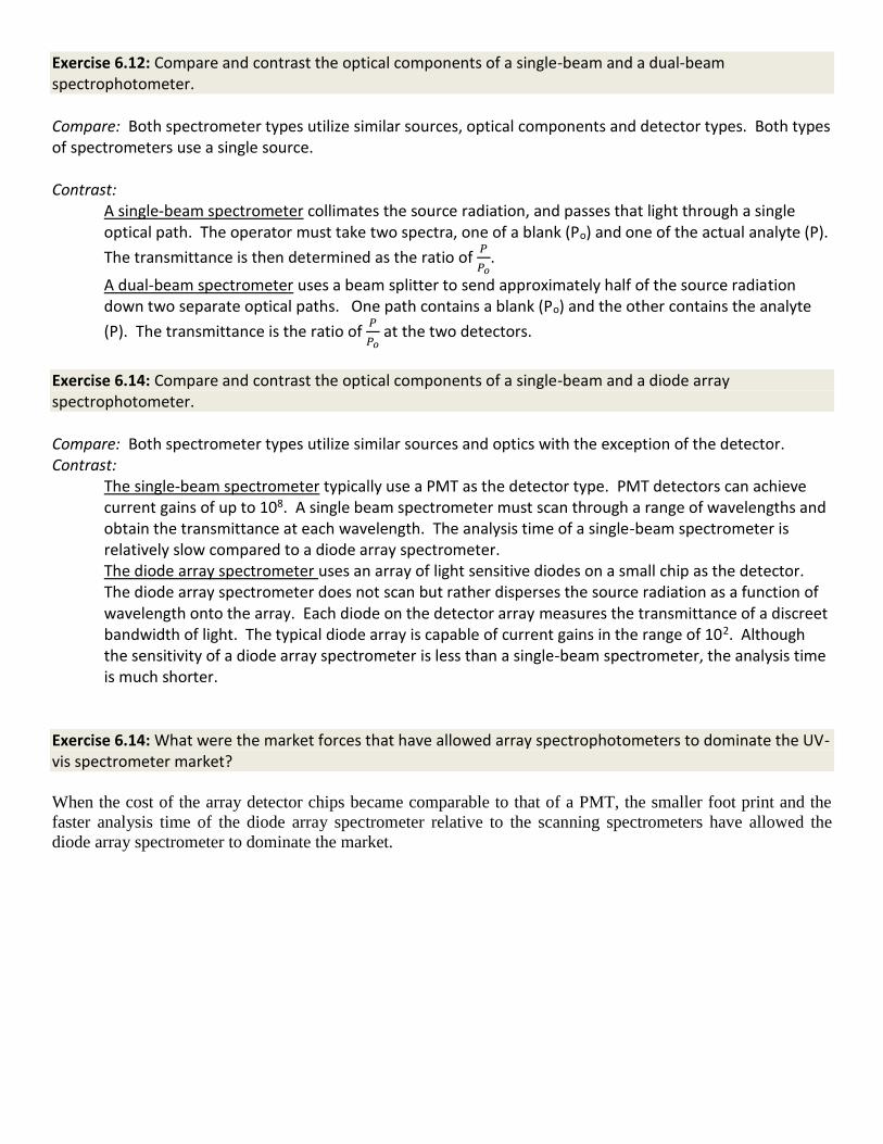
Exercise 6.20: A plant extract was analyzed by UV-vis spectroscopy and the following data was obtained, What is the molar absorptivity of the chromophore?
Concentration (M) Absorbance
5 X 10⁻⁵ 0.051
10 X 10-5 0.098
15 X 10-5 0.157
Since A = bc, a plot of A vs. c will give a slope of b. Since b =
1, the slope simply equals .
A plot of A vs. c gives us a slope of = 1025.7 M-1 cm-1.
You could also obtain this value by solving Beer’s law for for each data point and averaging the three values.
Exercise 6.21: Diamond has a refractive index of 2.42. The frequency of light does not change when it passes between from a material of one refractive index (η) to one with a different η, but both wavelength and
velocity do change. Given that 𝜂 = c
v (v is the velocity of the light in the medium of higher refractive index)
what is the velocity and wavelength of a photon with a fundamental wavelength (λ0) of 622 nm as it passes through the diamond? The velocity of the light in a vacuum (or air) is c = 2.998 x 10⁸ m/s. We need to calculate v, the velocity in diamond.
𝜂 = c
v so v =
c
𝜂=
2.998 𝑥 108𝑚
𝑠
2.42 = 1.239 x 108 = 1.24 x 10⁸ m/s
We know that v = λf (we will use f for frequency here rather than nu, ν, to avoid confusion with velocity, v), and that c = λ0f.
f = v
𝜆=
𝑐
𝜆0 so 𝜆 =
v 𝜆0
𝑐=
(1.239 𝑥 108𝑚
𝑠)(622 𝑛𝑚)
(2.998 𝑥 108𝑚
𝑠)
= 257 nm
Exercise 6.22: The 340 nm peak in Benzophenone is seen to shift to longer wavelengths when placed in a more polar solvent. What predictions can you make about the type of transition responsible for this peak? A shift to longer wavelengths indicates a decrease in energy for the transition. The benzophenone molecule
has both n * and * transitions. A polar solvent can lower the energy of a lone pair orbital (n-orbital)
or the energy of a * orbital. A lowering of the n-orbital would increase the energy of the transition and a
lowering of the * orbital would decrease the energy of the transition. Therefore we can conclude that the
peak at 340 nm in benzophenone is the result of a * transition. Exercise 6.23: What are the physical characteristics of a grating monochromator responsible for determining the spectral resolution. There are three main design features of a grating monochromator that affect spectral resolution.
1. The spacing of the grooves affects spectral resolution according to the grating equation.
(sin sin )s m d Eq. 3.9
The closer the groves the easier it is to resolve two closely spaced frequencies of light.
2. The pathlength within the monochromator prior to passing through the exit slits. If two dispersed and closely spaced frequencies of light can travel a sufficient distance before passing through the exit slits, the two frequencies can be resolved.
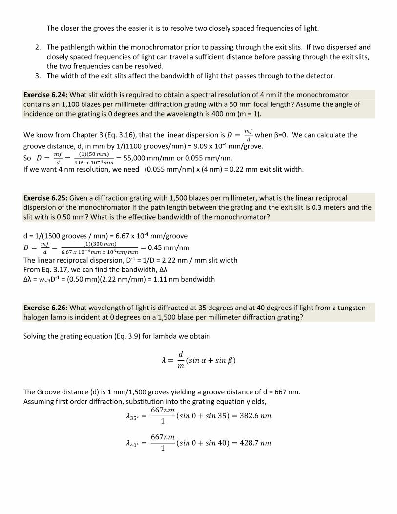
3. The width of the exit slits affect the bandwidth of light that passes through to the detector. Exercise 6.24: What slit width is required to obtain a spectral resolution of 4 nm if the monochromator contains an 1,100 blazes per millimeter diffraction grating with a 50 mm focal length? Assume the angle of incidence on the grating is 0 degrees and the wavelength is 400 nm (m = 1).
We know from Chapter 3 (Eq. 3.16), that the linear dispersion is 𝐷 = 𝑚𝑓
𝑑 when β=0. We can calculate the
groove distance, d, in mm by 1/(1100 grooves/mm) = 9.09 x 10⁻⁴ mm/grove.
So 𝐷 = 𝑚𝑓
𝑑=
(1)(50 𝑚𝑚)
9.09 𝑥 10−4𝑚𝑚= 55,000 mm/mm or 0.055 mm/nm.
If we want 4 nm resolution, we need (0.055 mm/nm) x (4 nm) = 0.22 mm exit slit width. Exercise 6.25: Given a diffraction grating with 1,500 blazes per millimeter, what is the linear reciprocal dispersion of the monochromator if the path length between the grating and the exit slit is 0.3 meters and the slit with is 0.50 mm? What is the effective bandwidth of the monochromator? d = 1/(1500 grooves / mm) = 6.67 x 10⁻⁴ mm/groove
𝐷 = 𝑚𝑓
𝑑=
(1)(300 𝑚𝑚)
6.67 𝑥 10−4𝑚𝑚 𝑥 106𝑛𝑚/𝑚𝑚= 0.45 mm/nm
The linear reciprocal dispersion, D-1 = 1/D = 2.22 nm / mm slit width From Eq. 3.17, we can find the bandwidth, Δλ Δλ = wslitD-1 = (0.50 mm)(2.22 nm/mm) = 1.11 nm bandwidth Exercise 6.26: What wavelength of light is diffracted at 35 degrees and at 40 degrees if light from a tungsten–halogen lamp is incident at 0 degrees on a 1,500 blaze per millimeter diffraction grating? Solving the grating equation (Eq. 3.9) for lambda we obtain
𝜆 = 𝑑
𝑚(𝑠𝑖𝑛 𝛼 + 𝑠𝑖𝑛 𝛽)
The Groove distance (d) is 1 mm/1,500 groves yielding a groove distance of d = 667 nm. Assuming first order diffraction, substitution into the grating equation yields,
𝜆35° = 667𝑛𝑚
1(𝑠𝑖𝑛 0 + 𝑠𝑖𝑛 35) = 382.6 𝑛𝑚
𝜆40° = 667𝑛𝑚
1(𝑠𝑖𝑛 0 + 𝑠𝑖𝑛 40) = 428.7 𝑛𝑚
Exercise 6.27: The 𝜀𝜆238 𝑖𝑠 8760 for DPK in acetonitrile. What would be the predicted absorption if a 1-ml
aliquot of 2 M DPK were diluted to 1 liter? The new concentration, after dilution, can be found using the equation for standard dilutions.
C1V1 = C2V2
C2 = C1V1 ÷ V2. = (2M)(0.001L) ÷ 1L = 0.002M
The absorption of the diluted solution can be found using Beer’s law
A = bc = (8760 M-1cm-1)( 1 cm)(0.002M) = 17.52 This absorption is well above 1 ABS indicating that the user would need to dilute the solution further. Perhaps by another factor of 1:1000. Exercise 6.28: What is the physical basis of the Nyquist theory ( Johnson noise) Nyquist-Johnson noise, is a temperature dependent voltage fluctuation caused by random motion of electrons within the electronic components of the instrument. Exercise 6.29: Imagine you have been hired as an outside consultant to a local paint and dye company wishing to purchase a UV-vis spectrophotometer. The spectrophotometer will be used in the quality control laboratory to test colors between different batches of dyes from the supplier. Outline the pros and cons of the various sources and detectors one might choose from, and defend your recommendation.
The PMT detector is the gold standard for low level detection however they are also more expensive and less robust than solid-state light-sensitive diodes. Since you would not require low level detection a solid-state detector would suffice and also give a more robust, longer service-life instrument. Also since you are not measuring kinetic data, a dual beam spectrometer would not be required and would simply add more cost and complexity. Because time is money in an industrial setting, a scanning spectrometer would be too slow. A diode array spectrometer capable of interfacing with either an auto-sampler or a flow cell would be ideal.
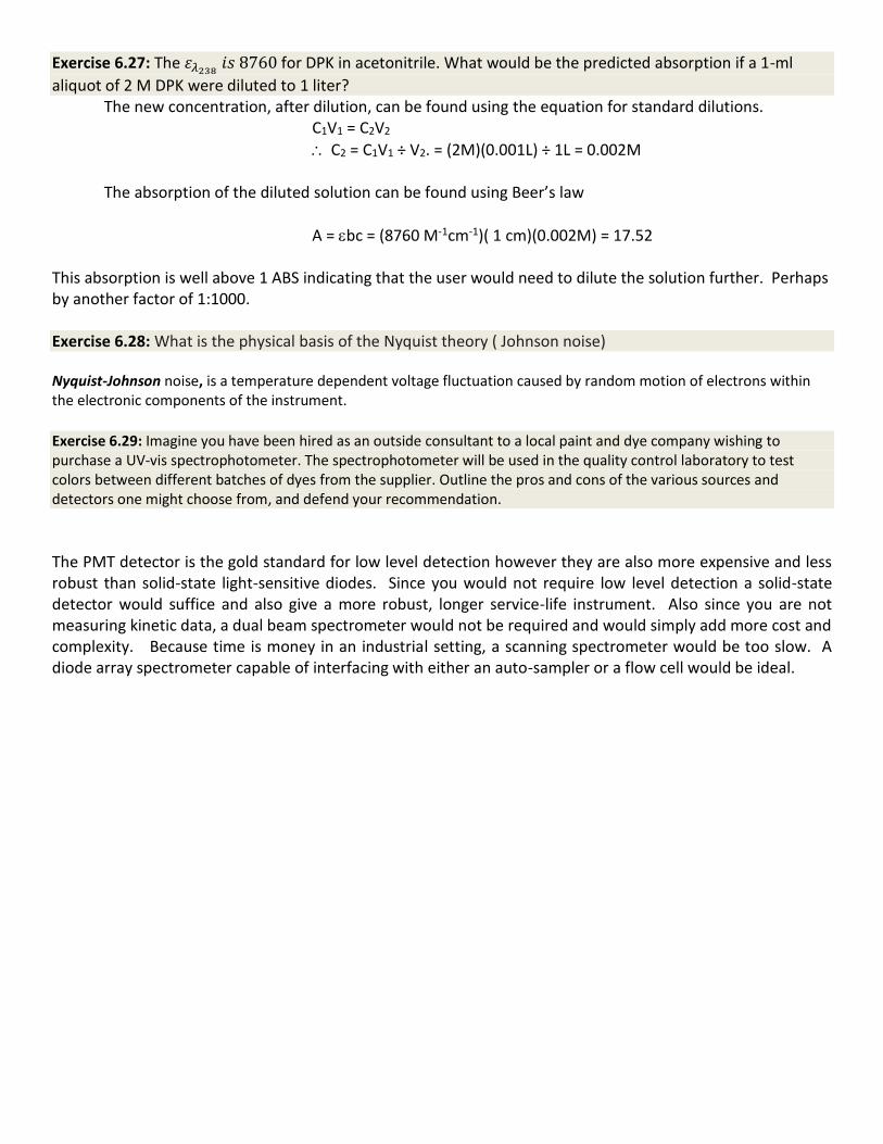
Exercise 6.30 : The UV-vis spectrum of 1,10- phenanthroline-5,6-dione (see Figure 6.12) has three distinct peaks.
Absorption Maxima for free 1,10-phenanthroline-5,6-dione in various solventsa Solvent Polarity
Indexb max (nm) max (nm) max (nm)
Cyclohexane 0.2 254 282(sh) --- Ethanol 5.0 --- 300 358(sh)
Use the data in the table to assign
the peaks as nox*, *
(carbonyl), nN*, or
*(aromatic)
Acetonitrile 5.8 --- 300 370 Dimethyl sulfoxide (DMSO) 7.2 262 294(sh) --- Water 9.0 --- 298 364(sh) a Data from; R. M.Granger, et al. JUCR 2005, 2, 47 b Polarity Index from The HPLC Solvent Guide Wiley Interscience, 2002.
The UV-vis spectrum shown here is for 1,10-phenanthroline-5,6-dione.
Use the data in the table to assign the peaks as nox π*, π π* (carbonyl), nN π*, or π π*(aromatic).
Students are encouraged to refer back to “DPK – A Case Study” for guidance on answering this question. For the molecule 1,10-phenanthroline-5,6-dione, the aromaticity of the three rings in the phenanthroline moiety is
broken by the presence of the two dione moieties. So the two outer rings in the phenanthroline skeleton can be modeled as pyridine rings for the purposes of analyzing the UV-vis spectrum. We should expect degenerate n * and * transitions for the two pyridine systems. Likewise we should expect degenerate n * and * transitions for the two dione moieties. Therefore there are a total of four possible transitions expected for this molecule. We would expect transitions for the conjugate -system in the pyridine rings to be lower in energy than the non-conjugated dione moiety (particle in a box argument) and the * transitions to be of higher intensity than the n * transitions (Franck Condon principle). The analysis of the UV-vis spectrum of 1,10-phenanthroline-5,6-dione is complicated by the fact that the four peaks are not well resolved. However we can make some well-informed judgements. Since the peak at max = 254 (cyclohexane) and max = 358 (Ethanol) both lost energy (shifted to longer wavelengths) when the analyte was placed in a more polar solvent, we can assign those two transitions to n *. Based on the difference in energy of these two transistions, we can assign max = 254 to the n p* transition in the dione moiety and the max = 358 transition to the n * transition in the pyridine rings. Lastly, the transition at lmax = 300 (ethanol) decreased in energy (shifted to shorter wavelengths) when placed in a more polar
solvent. Therefore we can assign that transition to *. Due to the relatively low energy, it is most likely resulting
from the conjugated pyridine rings. The last transition resulting from * of the dione moiety is most likely at a wavelength that is too short to see with this spectrometer.


![arXiv:0808.1664v1 [math.RT] 12 Aug 20081 → V∗ → ASp(V) → Sp(V) → 1. In addition, we construct a projective Weil representation eρ : ASp(V) → PGL(H), which extends as a](https://static.fdocument.org/doc/165x107/6025fdb181f0692e89671d81/arxiv08081664v1-mathrt-12-aug-2008-1-a-va-a-aspv-a-spv-a-1-in.jpg)

![Topic 5 Electricity 31.1.2018 [140 marks] · Topic 5 Electricity 31.1.2018 [140 marks] 1. The graph shows the variation of current with potential difference for a filament lamp. [1](https://static.fdocument.org/doc/165x107/5e625a0348969177d31d39ab/topic-5-electricity-3112018-140-marks-topic-5-electricity-3112018-140-marks.jpg)


![B-TOUCH intelligence passenger airbag deactivation warning lamp intelligence pre heating display reconfigurable intelligence radio command display ... passat [1997] (3b) edi (ΗΛΕΚΤΡΟΝΙΚΗ](https://static.fdocument.org/doc/165x107/6123f6172259f476611dad53/b-intelligence-passenger-airbag-deactivation-warning-lamp-intelligence-pre-heating.jpg)