Molecular Modeling 2021 -- lecture 2 -- fri Jan 29
37
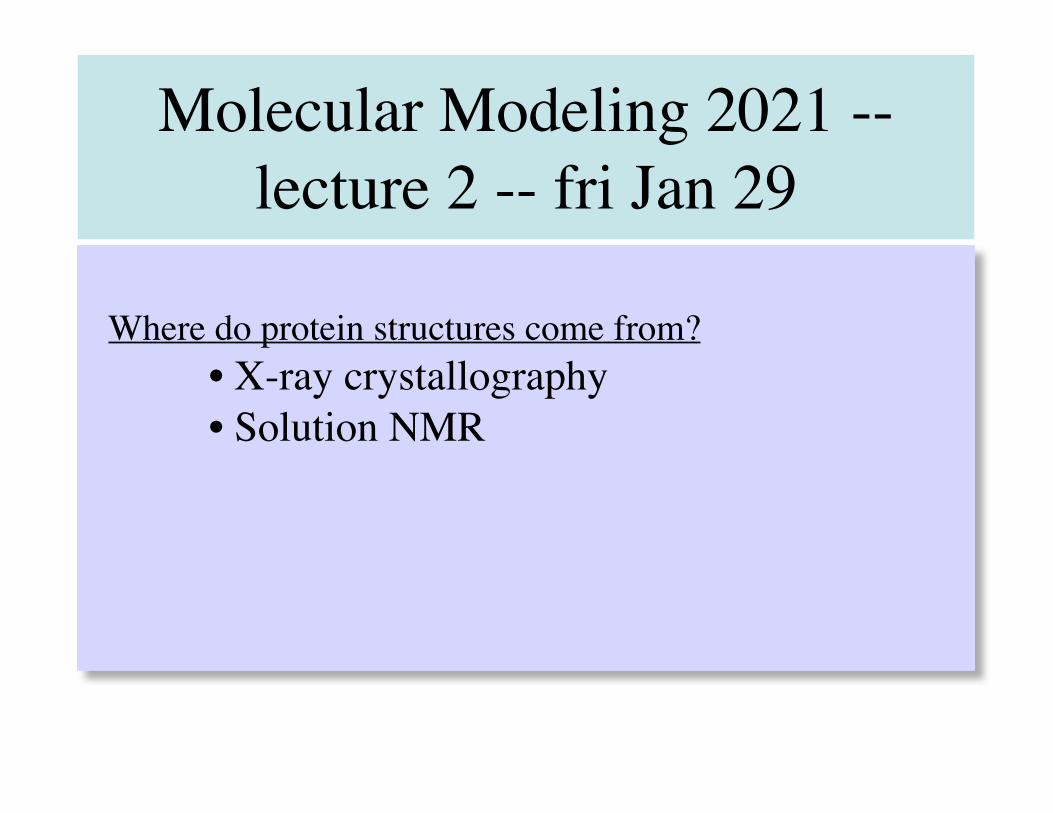
Molecular Modeling 2021 -- lecture 2 -- fri Jan 29 Where do protein structures come from? • X-ray crystallography • Solution NMR
Transcript of Molecular Modeling 2021 -- lecture 2 -- fri Jan 29
Molecular Modeling 2021 -- lecture 2 -- fri Jan 29
Where do protein structures come from?• X-ray crystallography• Solution NMR
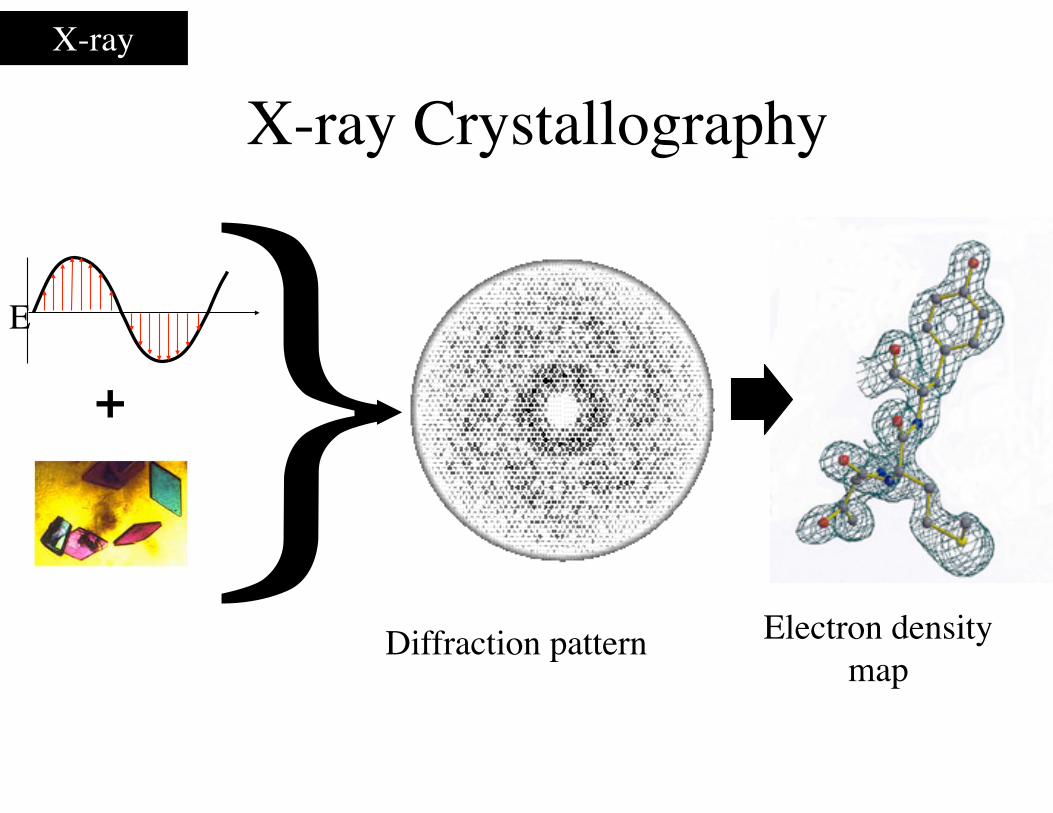
X-ray Crystallography
}+
Diffraction pattern Electron density map
E
X-ray
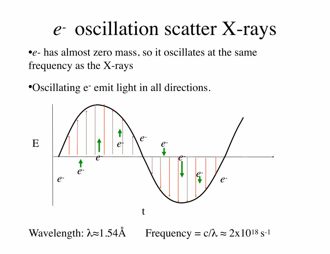
e- oscillation scatter X-rays•e- has almost zero mass, so it oscillates at the same frequency as the X-rays
•Oscillating e- emit light in all directions.
Ee- e-
e-e-e-
e-e-e-e-
t
Wavelength: λ≈1.54Å Frequency = c/λ ≈ 2x10¹⁸ s-1
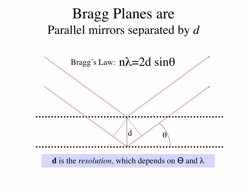
Bragg Planes are Parallel mirrors separated by d
d θ
nλ=2d sinθBragg’s Law:
d is the resolution, which depends on ϴ and λ
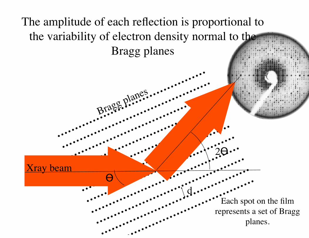
Each spot on the film represents a set of Bragg
planes.
The amplitude of each reflection is proportional to the variability of electron density normal to the
Bragg planes
d
2ϴXray beam
Bragg planes
ϴ
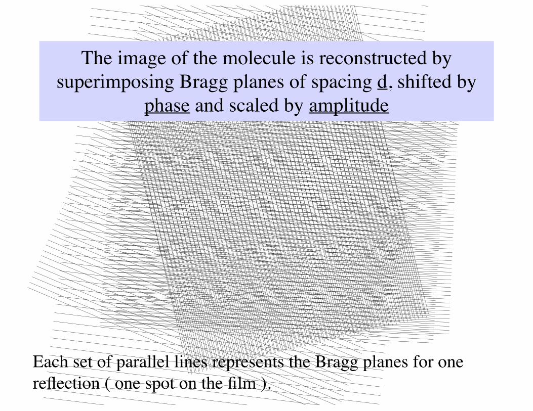
The image of the molecule is reconstructed by superimposing Bragg planes of spacing d, shifted by
phase and scaled by amplitude
Each set of parallel lines represents the Bragg planes for one reflection ( one spot on the film ).
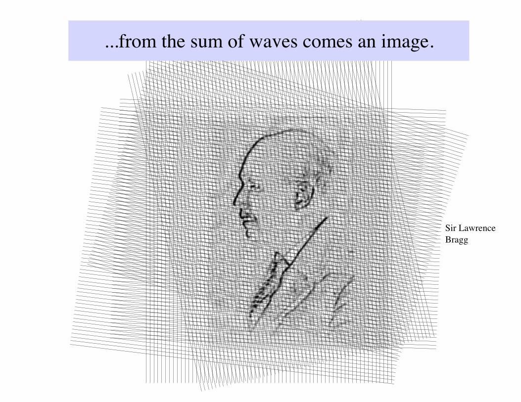
...from the sum of waves comes an image.
Sir Lawrence Bragg
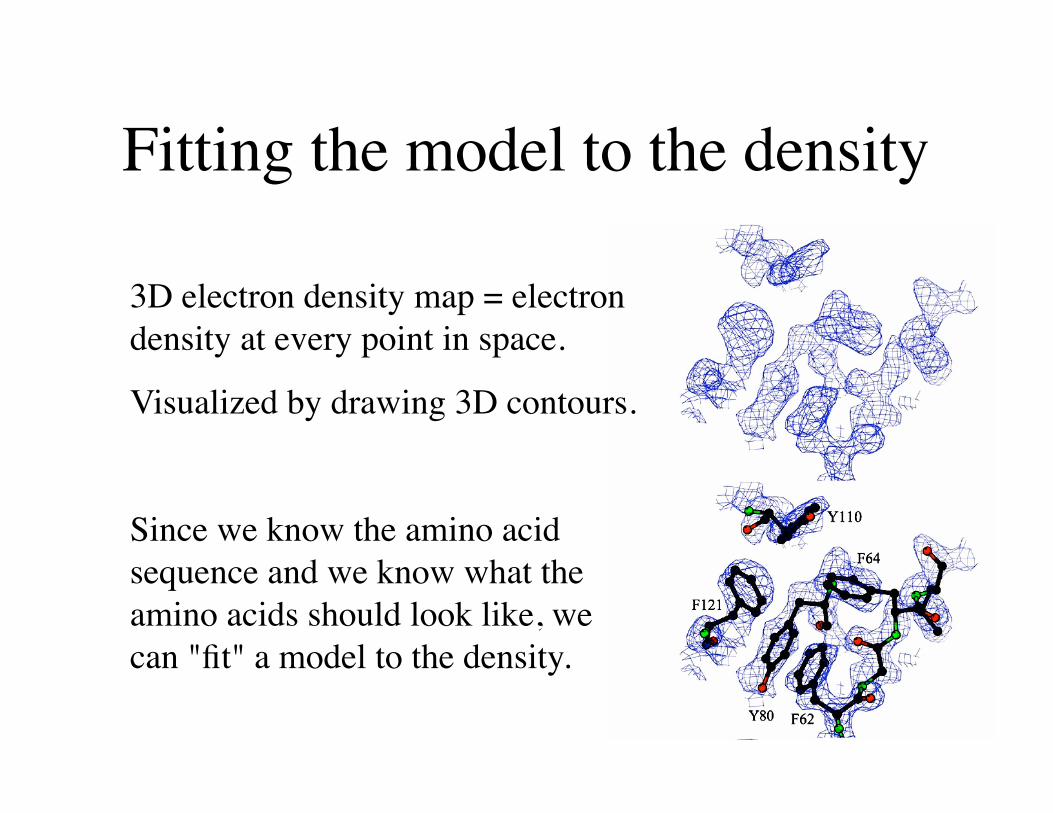
Fitting the model to the density
3D electron density map = electron density at every point in space.
Visualized by drawing 3D contours.
Since we know the amino acid sequence and we know what the amino acids should look like, we can "fit" a model to the density.

4 parameters are refined for each atom
Coordinate refinement
Each atom is moved in X,Y and Z until:
(1) good stereochemistry is achieved,
(2) there is a good match between the atoms and the density.
Each atom is assigned a B-factor or "temperature-factor", to better fit the density.
+ x
y z
B
+
high B density profile
low B density profile
the R-factor
Refined coordinates are deposited in the Protein Data Bank: www.rcsb.org
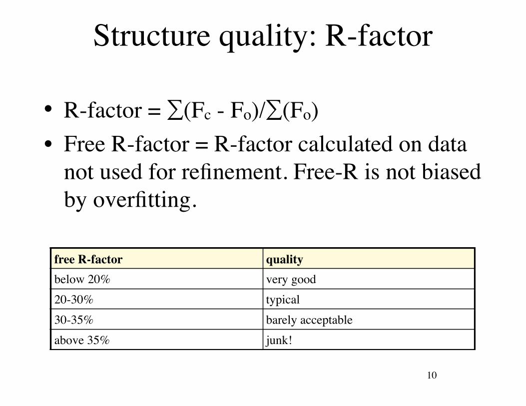
Structure quality: R-factor
• R-factor = ∑(Fc - Fo)/∑(Fo)• Free R-factor = R-factor calculated on data
not used for refinement. Free-R is not biased by overfitting.
10
free R-factor qualitybelow 20% very good20-30% typical30-35% barely acceptableabove 35% junk!
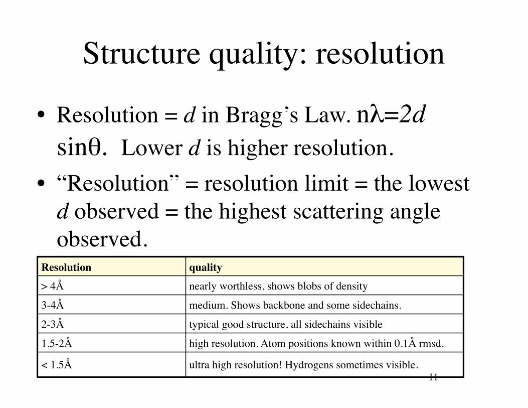
Structure quality: resolution
• Resolution = d in Bragg’s Law. nλ=2d sinθ. Lower d is higher resolution.
• “Resolution” = resolution limit = the lowest d observed = the highest scattering angle observed.
11
Resolution quality
> 4Å nearly worthless, shows blobs of density
3-4Å medium. Shows backbone and some sidechains.
2-3Å typical good structure, all sidechains visible
1.5-2Å high resolution. Atom positions known within 0.1Å rmsd.
< 1.5Å ultra high resolution! Hydrogens sometimes visible.
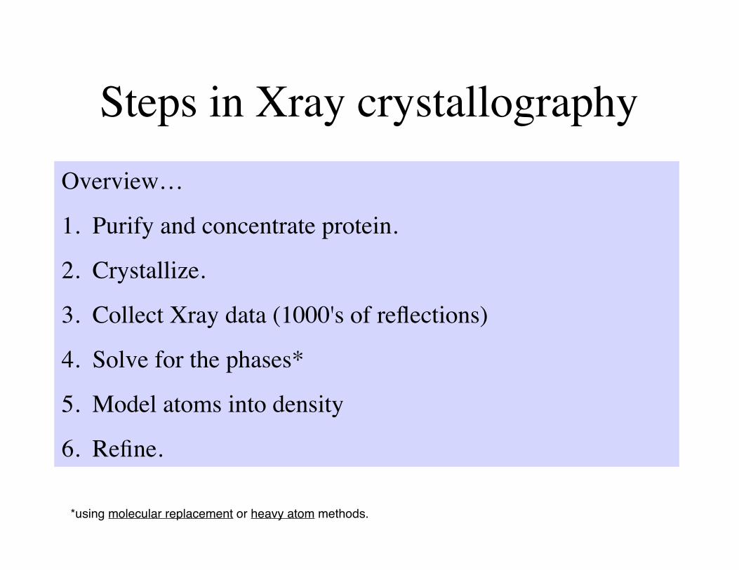
Steps in Xray crystallographyOverview…
1. Purify and concentrate protein.
2. Crystallize.
3. Collect Xray data (1000's of reflections)
4. Solve for the phases*
5. Model atoms into density
6. Refine.
*using molecular replacement or heavy atom methods.
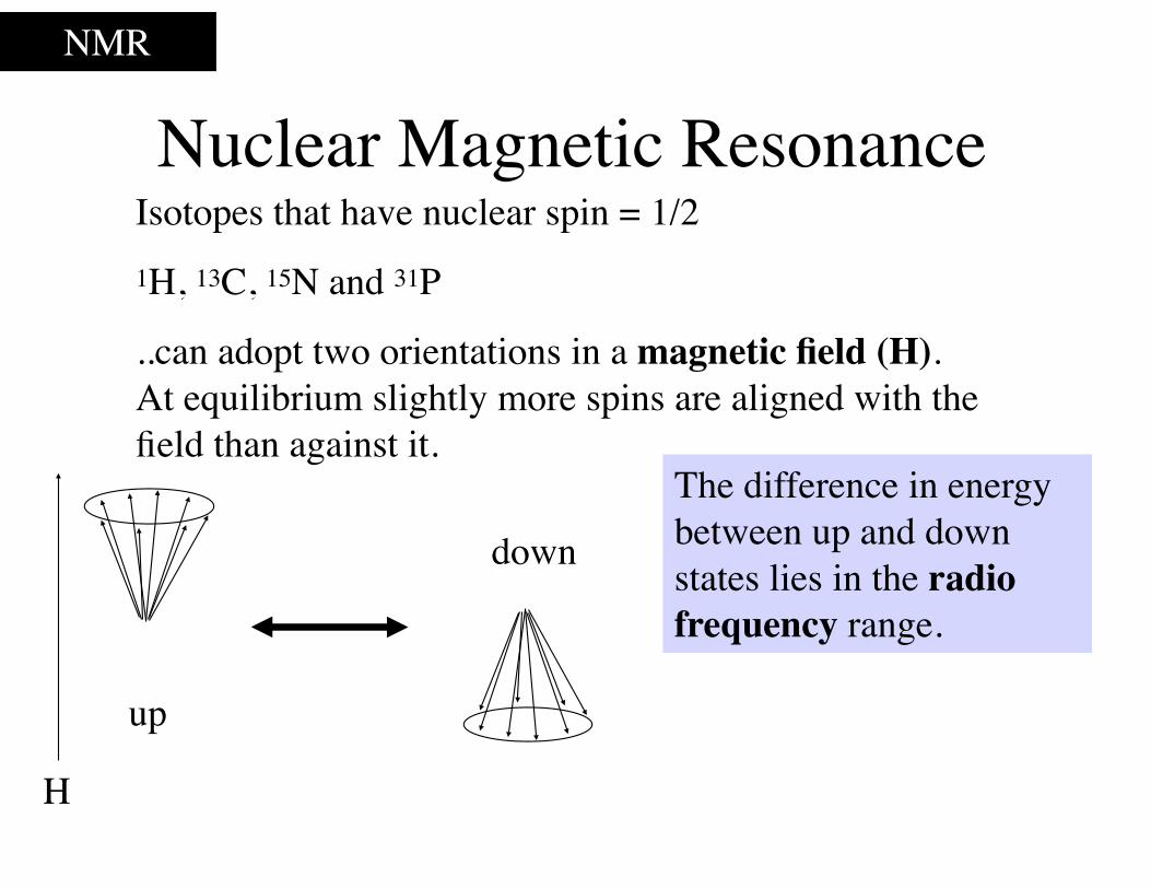
Nuclear Magnetic ResonanceNMR
Isotopes that have nuclear spin = 1/2 1H, 13C, 15N and 31P
..can adopt two orientations in a magnetic field (H). At equilibrium slightly more spins are aligned with the field than against it.
up
down
The difference in energy between up and down states lies in the radio frequency range.
H
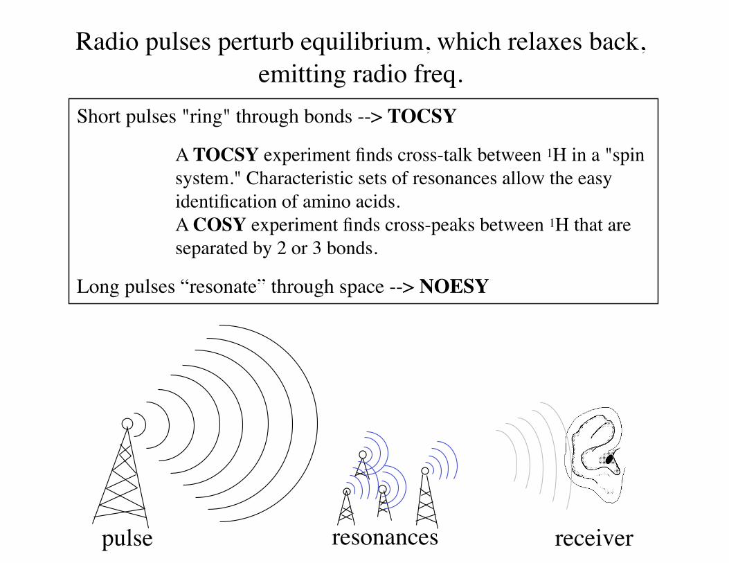
Radio pulses perturb equilibrium, which relaxes back, emitting radio freq.
pulse resonances receiver
Short pulses "ring" through bonds --> TOCSY
A TOCSY experiment finds cross-talk between 1H in a "spin system." Characteristic sets of resonances allow the easy identification of amino acids. A COSY experiment finds cross-peaks between 1H that are separated by 2 or 3 bonds.
Long pulses “resonate” through space --> NOESY
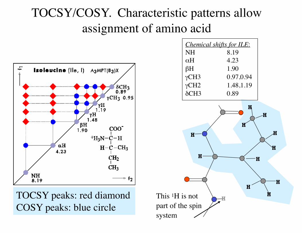
TOCSY/COSY. Characteristic patterns allow assignment of amino acid
TOCSY peaks: red diamond COSY peaks: blue circle
H
HH
H
H
H
HH
H
H
H
HThis 1H is not part of the spin system
Chemical shifts for ILE:NH 8.19αH 4.23 βH 1.90 γCH3 0.97,0.94 γCH2 1.48,1.19 δCH3 0.89
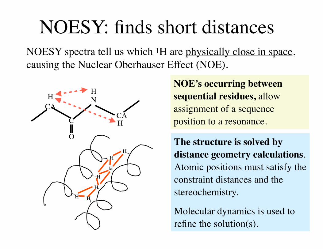
NOESY: finds short distancesNOESY spectra tell us which 1H are physically close in space, causing the Nuclear Oberhauser Effect (NOE).
HH
HH
H
H H
The structure is solved by distance geometry calculations. Atomic positions must satisfy the constraint distances and the stereochemistry.
Molecular dynamics is used to refine the solution(s).
CACAC
N
O
HH
H
NOE’s occurring between sequential residues, allow assignment of a sequence position to a resonance.
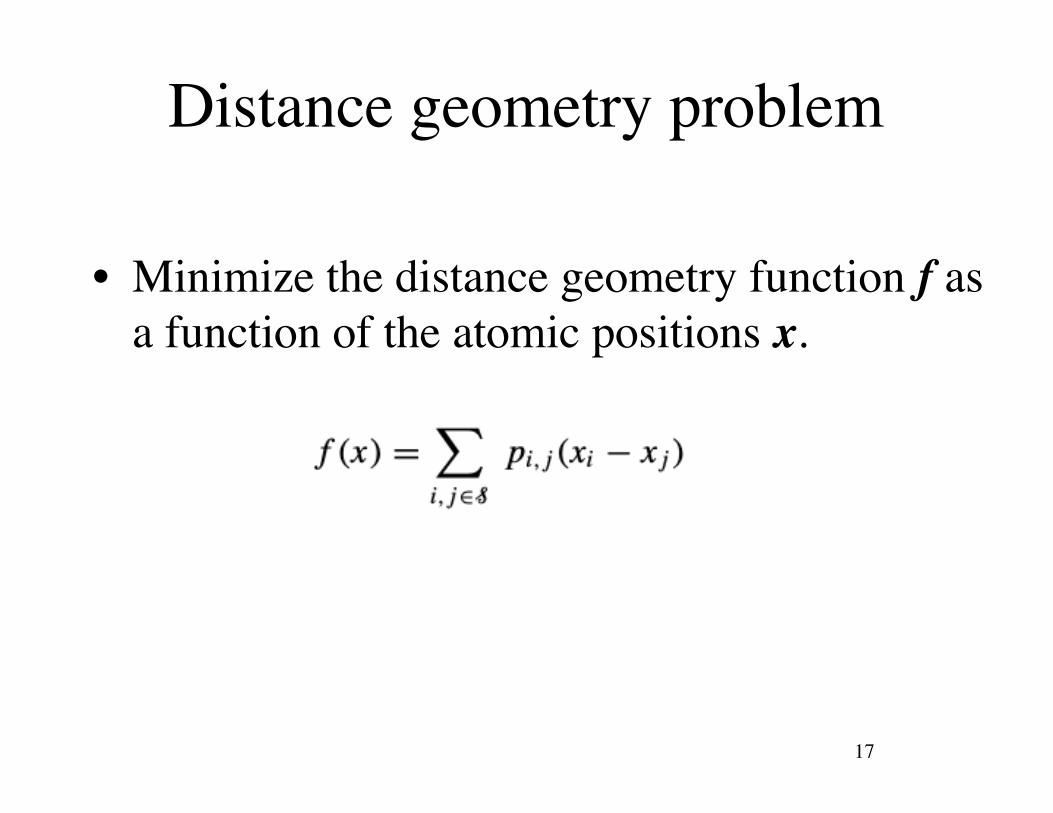
Distance geometry problem
• Minimize the distance geometry function f as a function of the atomic positions x.
17
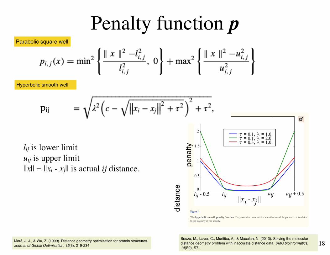
Penalty function p
18
lij is lower limituij is upper limit||x|| = ||xi - xj|| is actual ij distance.
Parabolic square well
Moré, J. J., & Wu, Z. (1999). Distance geometry optimization for protein structures. Journal of Global Optimization, 15(3), 219-234
Hyperbolic smooth well
Souza, M., Lavor, C., Muritiba, A., & Maculan, N. (2013). Solving the molecular distance geometry problem with inaccurate distance data. BMC bioinformatics, 14(S9), S7.
pij
pena
ltydi
stan
ce
Steps in Protein NMROverview…
1. Grow protein in 13C and/or 15N enriched media.
2. Purify and concentrate protein.
3. Collect NMR spectra (2,3 or 4-dimensions).
4. Assign the peaks (TOCSY/COSY).
5. Assign distance constraints (NOESY)
6. Solve the distance geometry problem.
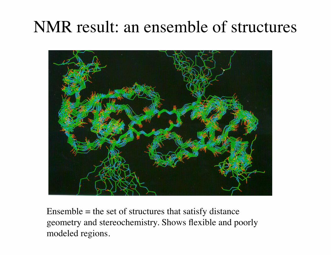
NMR result: an ensemble of structures
Ensemble = the set of structures that satisfy distance geometry and stereochemistry. Shows flexible and poorly modeled regions.
In class exercise 2.1• Go to www.rcsb.org• Search for DHFR• Select and download 2hqp and 3frd• Open the Xray file 3frd in MOE• Show all atoms. Make them spacefill. Hide solvent.• Color all atoms by B-factor.• How are the B-factors distributed?
21
In class exercise 2.1• Upload the NMR file 2hqp. Model limit: all• Select all 2hqp chains. Hide selected. Ribbon. • Are the uncertainties/flexibilities you see in 2hqp, in the same
place as the high B-factors in its homolog 3frd?
22
Other NMR experimentsAdditional information about the conformation may be gained by
• H/D-exchange Deuterium (2H) is invisible to NMR. Disappearing 1H's tell us which ones are exposed to solvent. Especially amide NH's.
• Temperature sensitivity of resonances.
Chemical shift oh 1H changes with T less if H-bonded.
• HSQC Direct coupling of 15N to 1H through a single bond.
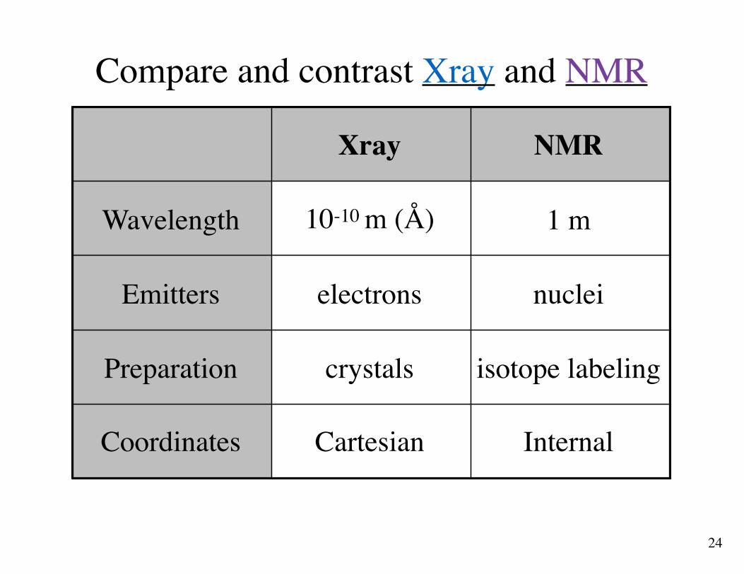
Compare and contrast Xray and NMR
24
Xray NMR
Wavelength 10-10 m (Å) 1 m
Emitters electrons nuclei
Preparation crystals isotope labeling
Coordinates Cartesian Internal
Review questions• What causes Xrays to scatter?
• What causes diffraction?
• What are the results of Xray crystallography?
• What is a temperature factor?
• What wavelength light is used in Xray crystallography?
• What does “resolution” mean in Xray crystallography?
• What is a crystal?
• What wavelength of light is used in NMR?
• What kind of atom resonates with light?
• What is an ensemble in NMR?
• What measure in NMR is the analog of resolution in Xray?
• What type of NMR experiement assigns resonances to amino acid types?
• What type of NMR experiment provides distances between different parts of the protein chain?
• Which method produces Cartesian coordinates? Internal coordinates?25

Supplementary slides
26
2.3 Coordinate systems
• Cartesian versus Internal
27
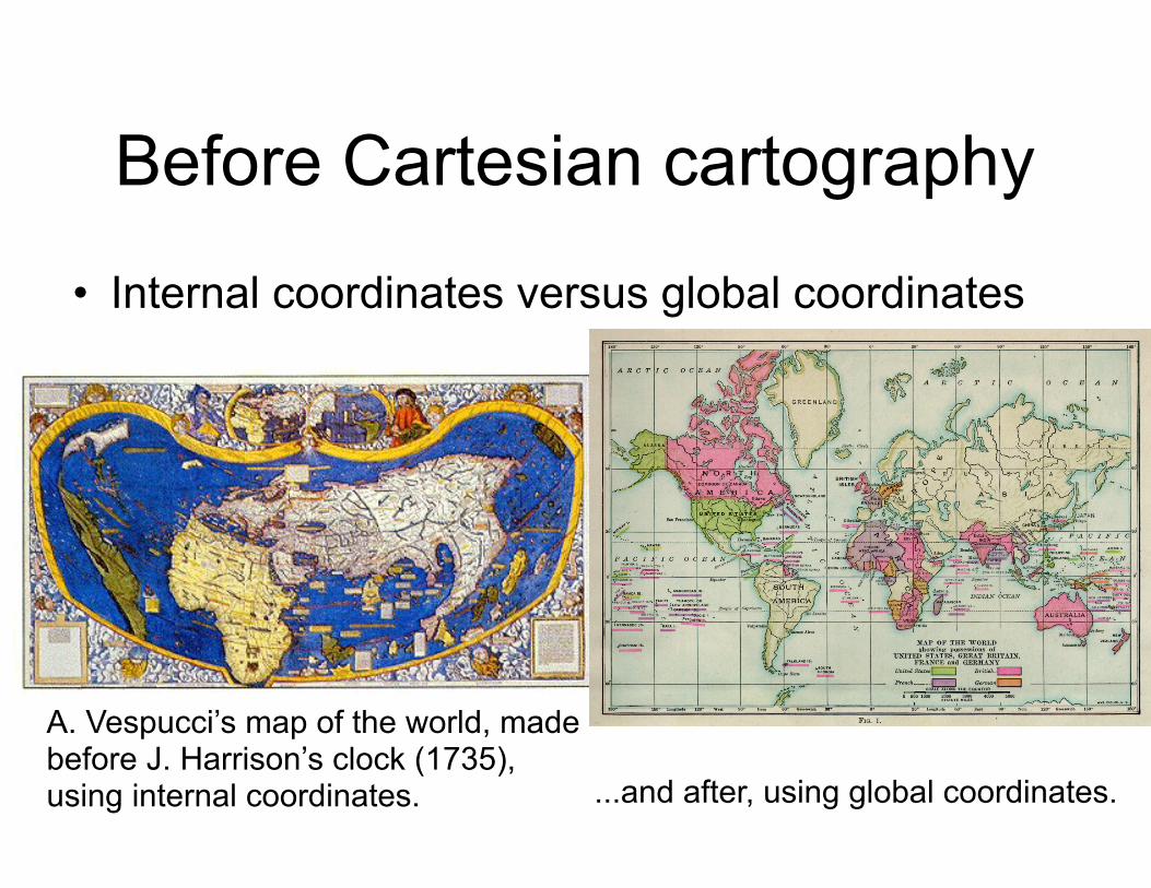
Before Cartesian cartography
• Internal coordinates versus global coordinates
A. Vespucci’s map of the world, made before J. Harrison’s clock (1735), using internal coordinates. ...and after, using global coordinates.
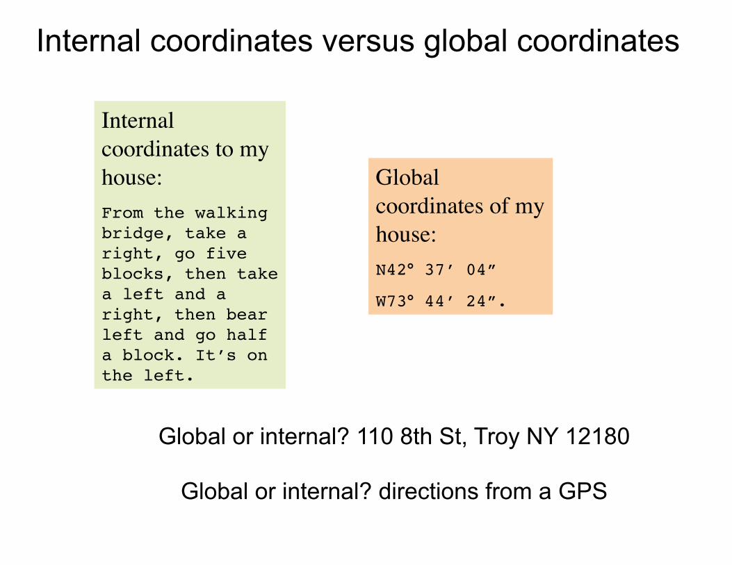
Internal coordinates to my house:From the walking bridge, take a right, go five blocks, then take a left and a right, then bear left and go half a block. It’s on the left.
Global coordinates of my house:N42° 37’ 04”
W73° 44’ 24”.
Internal coordinates versus global coordinates
Global or internal? 110 8th St, Troy NY 12180
Global or internal? directions from a GPS
Cartesian coordinates have a reference frame
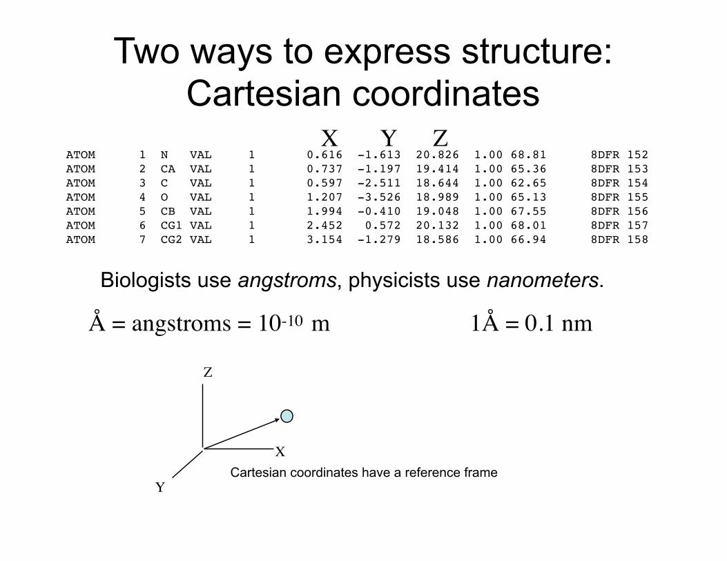
Two ways to express structure: Cartesian coordinates
ATOM 1 N VAL 1 0.616 -1.613 20.826 1.00 68.81 8DFR 152ATOM 2 CA VAL 1 0.737 -1.197 19.414 1.00 65.36 8DFR 153ATOM 3 C VAL 1 0.597 -2.511 18.644 1.00 62.65 8DFR 154ATOM 4 O VAL 1 1.207 -3.526 18.989 1.00 65.13 8DFR 155ATOM 5 CB VAL 1 1.994 -0.410 19.048 1.00 67.55 8DFR 156ATOM 6 CG1 VAL 1 2.452 0.572 20.132 1.00 68.01 8DFR 157ATOM 7 CG2 VAL 1 3.154 -1.279 18.586 1.00 66.94 8DFR 158
X Y Z
Å = angstroms = 10-10 m 1Å = 0.1 nm
X
Y
Z
Biologists use angstroms, physicists use nanometers.
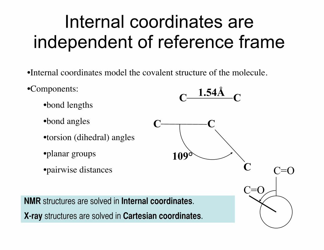
Internal coordinates are independent of reference frame
•Internal coordinates model the covalent structure of the molecule.
•Components:
•bond lengths
•bond angles
•torsion (dihedral) angles
•planar groups
•pairwise distances
NMR structures are solved in Internal coordinates.
X-ray structures are solved in Cartesian coordinates.
C C1.54Å
C C
C109°
C=O
C=O
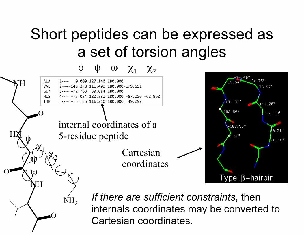
Short peptides can be expressed as a set of torsion angles
ALA 1~~~ 0.000 127.140 180.000VAL 2~~~-148.378 111.409 180.000-179.551GLY 3~~~ -72.763 39.684 180.000HIS 4~~~ -73.084 122.882 180.000 -87.256 -62.962THR 5~~~ -73.735 116.210 180.000 49.292
φ ψ ω χ1 χ2
internal coordinates of a 5-residue peptide
NH3
NH
HN
NH
O
O
O
ψ
φχ1 χ2
ω
If there are sufficient constraints, then internals coordinates may be converted to Cartesian coordinates.
Cartesian coordinates
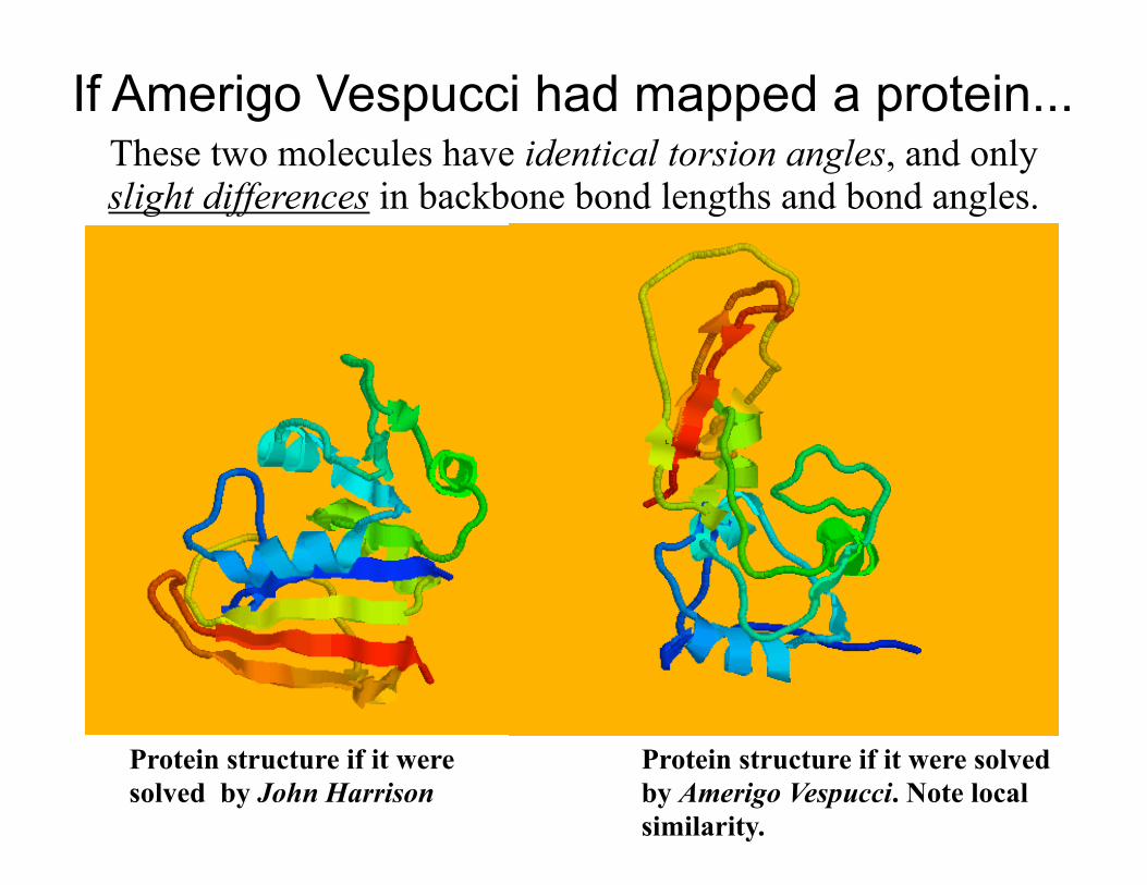
If Amerigo Vespucci had mapped a protein... These two molecules have identical torsion angles, and only slight differences in backbone bond lengths and bond angles.
Protein structure if it were solved by John Harrison
Protein structure if it were solved by Amerigo Vespucci. Note local similarity.
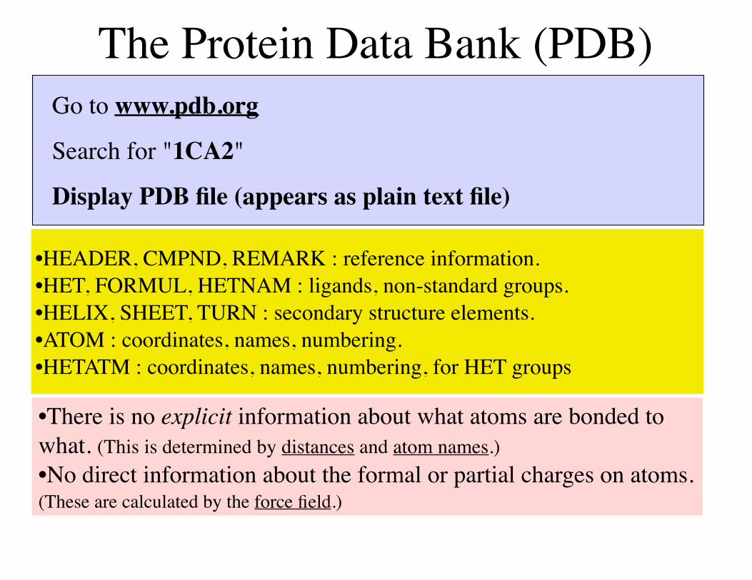
The Protein Data Bank (PDB)Go to www.pdb.org
Search for "1CA2"
Display PDB file (appears as plain text file)
•HEADER, CMPND, REMARK : reference information. •HET, FORMUL, HETNAM : ligands, non-standard groups.•HELIX, SHEET, TURN : secondary structure elements.•ATOM : coordinates, names, numbering.•HETATM : coordinates, names, numbering, for HET groups
•There is no explicit information about what atoms are bonded to what. (This is determined by distances and atom names.)•No direct information about the formal or partial charges on atoms. (These are calculated by the force field.)
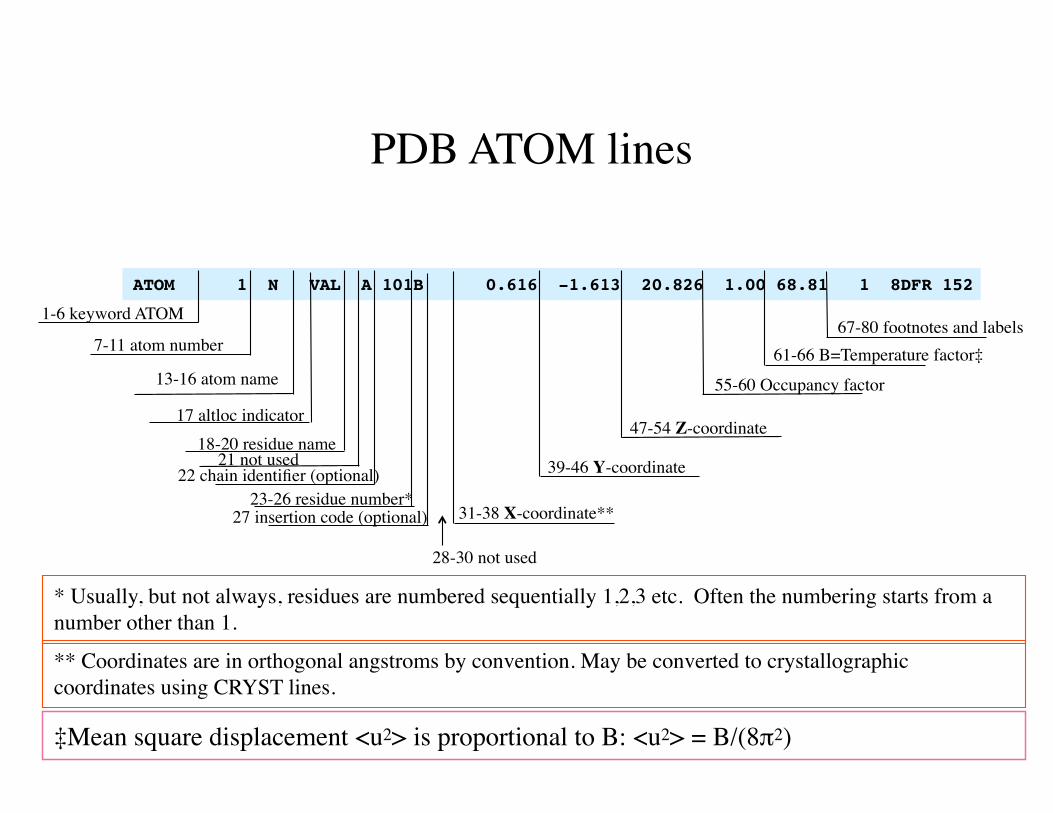
ATOM 1 N VAL A 101B 0.616 -1.613 20.826 1.00 68.81 1 8DFR 152
1-6 keyword ATOM
7-11 atom number
13-16 atom name
17 altloc indicator18-20 residue name
22 chain identifier (optional)23-26 residue number*
* Usually, but not always, residues are numbered sequentially 1,2,3 etc. Often the numbering starts from a number other than 1.
27 insertion code (optional) 31-38 X-coordinate**
39-46 Y-coordinate
47-54 Z-coordinate
55-60 Occupancy factor
61-66 B=Temperature factor‡67-80 footnotes and labels
28-30 not used
21 not used
** Coordinates are in orthogonal angstroms by convention. May be converted to crystallographic coordinates using CRYST lines.
PDB ATOM lines
‡Mean square displacement <u2> is proportional to B: <u2> = B/(8π2)
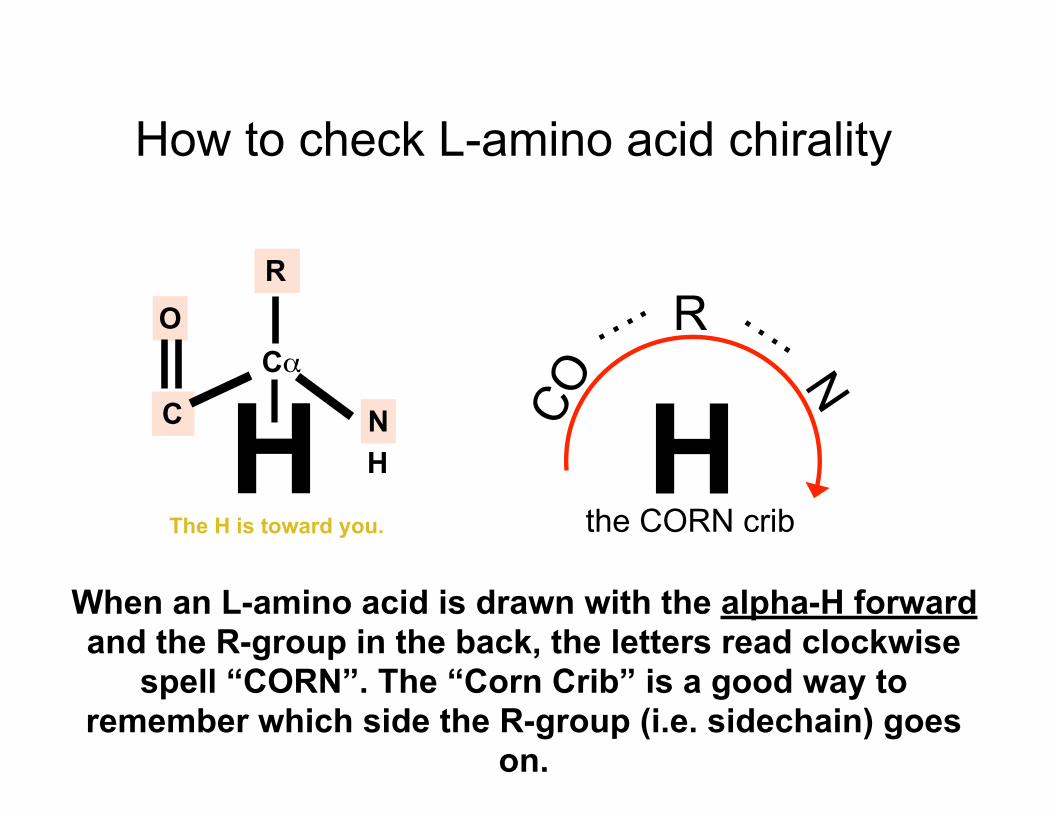
How to check L-amino acid chirality
HO
Cα
C N
R
H
When an L-amino acid is drawn with the alpha-H forward and the R-group in the back, the letters read clockwise
spell “CORN”. The “Corn Crib” is a good way to remember which side the R-group (i.e. sidechain) goes
on.
HR
CO N
….….
the CORN cribThe H is toward you.
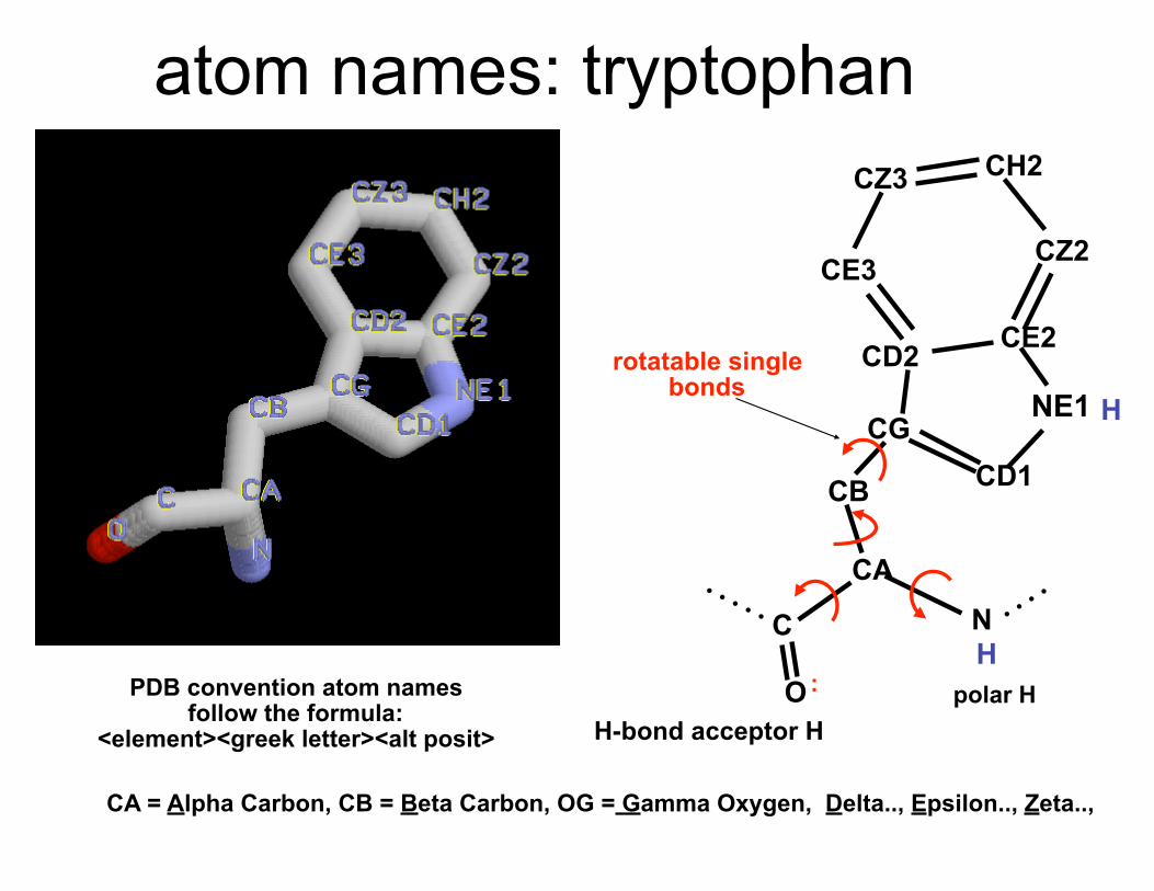
atom names: tryptophan
PDB convention atom names follow the formula:
<element><greek letter><alt posit>
C
CE3
CZ3 CH2
CZ2
CE2CD2
CGCD1
NE1
CB
CAN
OH
H
:
rotatable single bonds
polar HH-bond acceptor H
CA = Alpha Carbon, CB = Beta Carbon, OG = Gamma Oxygen, Delta.., Epsilon.., Zeta..,