Molecular imaging of human embryonic stem cells stably...
31
1 Molecular imaging of human embryonic stem cells stably expressing human PET reporter genes after zinc finger nucleases-mediated genome editing. Authors: Esther Wolfs 1 ‡*, Bryan Holvoet 1* , Laura Ordovas 2,3 , Natacha Breuls 1,4 , Nicky Helsen 2,3 , Matthias Schönberger 5 , Susanna Raitano 2,3 , Tom Struys 6 , Bert Vanbilloen 1 , Cindy Casteels 1 , Maurilio Sampaolesi 4 , Koen Van Laere 1 , Ivo Lambrichts 6 , Catherine M. Verfaillie 2,3 *, Christophe M. Deroose 1 * Affiliations: 1 Nuclear Medicine & Molecular Imaging and Molecular Small Animal Imaging Centre, Department of Imaging and Pathology, KU Leuven, Leuven, Belgium. 2 Stem Cell Institute, KU Leuven, Leuven, Belgium. 3 Department of Development and Regeneration, Stem Cell Biology and Embryology, KU Leuven, Leuven, Belgium. 4 Translational Cardiomyology Lab, Department of Development and Regeneration, KU Leuven, Leuven, Belgium. 5 Department of Pharmaceutical and Pharmacological Sciences, KU Leuven, Leuven, Belgium. 6 Biomedical Research Institute, Morphology Research Group, Lab of Histology, Universiteit Hasselt, Diepenbeek, Belgium. *Authors contributed equally to this manuscript. ‡Current affiliation: Biomedical Research Institute, Morphology Research Group, Lab of Histology, Universiteit Hasselt, Diepenbeek, Belgium Corresponding author: Prof. dr. Christophe Deroose UZ Leuven, Division of Nuclear Medicine Campus Gasthuisberg, Herestraat 49, B-3000 Leuven +3216343712 [email protected] First author: Dr. Esther Wolfs Biomedical Research Institute, UHasselt Agoralaan, Gebouw C, B-3590 Diepenbeek +3211269277 [email protected] Short title: ESC imaging after genome editing Journal of Nuclear Medicine, published on June 8, 2017 as doi:10.2967/jnumed.117.189779 by on June 6, 2018. For personal use only. jnm.snmjournals.org Downloaded from
Transcript of Molecular imaging of human embryonic stem cells stably...
1
Molecular imaging of human embryonic stem cells stably expressing human PET reporter genes after zinc finger nucleases-mediated genome editing.
Authors: Esther Wolfs1‡*, Bryan Holvoet1*, Laura Ordovas2,3, Natacha Breuls1,4, Nicky Helsen2,3, Matthias Schönberger5, Susanna Raitano2,3, Tom Struys6, Bert Vanbilloen1, Cindy Casteels1, Maurilio Sampaolesi4, Koen Van Laere1, Ivo Lambrichts6, Catherine M. Verfaillie2,3*, Christophe M. Deroose1*
Affiliations: 1Nuclear Medicine & Molecular Imaging and Molecular Small Animal Imaging Centre, Department of Imaging and Pathology, KU Leuven, Leuven, Belgium. 2Stem Cell Institute, KU Leuven, Leuven, Belgium. 3Department of Development and Regeneration, Stem Cell Biology and Embryology, KU Leuven, Leuven, Belgium. 4Translational Cardiomyology Lab, Department of Development and Regeneration, KU Leuven, Leuven, Belgium. 5Department of Pharmaceutical and Pharmacological Sciences, KU Leuven, Leuven, Belgium. 6Biomedical Research Institute, Morphology Research Group, Lab of Histology, Universiteit Hasselt, Diepenbeek, Belgium.
*Authors contributed equally to this manuscript.
‡Current affiliation: Biomedical Research Institute, Morphology Research Group, Lab of Histology, Universiteit Hasselt, Diepenbeek, Belgium
Corresponding author: Prof. dr. Christophe Deroose UZ Leuven, Division of Nuclear Medicine Campus Gasthuisberg, Herestraat 49, B-3000 Leuven +3216343712 [email protected] First author: Dr. Esther Wolfs Biomedical Research Institute, UHasselt Agoralaan, Gebouw C, B-3590 Diepenbeek +3211269277 [email protected]
Short title: ESC imaging after genome editing
Journal of Nuclear Medicine, published on June 8, 2017 as doi:10.2967/jnumed.117.189779by on June 6, 2018. For personal use only. jnm.snmjournals.org Downloaded from

2
ABSTRACT
Rationale. Molecular imaging is indispensable for determining the fate and persistence of
engrafted stem cells. Standard strategies for transgene induction involve the use of viral vectors
prone to silencing and insertional mutagenesis or the use of non-human genes.
Methods. We used zinc finger nucleases (ZFN) to induce stable expression of human imaging
reporter genes into the safe harbor locus adeno-associated virus integration site 1 (AAVS1) in
human embryonic stem cells (ESC). Plasmids were generated carrying reporter genes for
fluorescence, bioluminescence imaging (BLI), and human positron emission tomography (PET)
reporter genes.
Results. In vitro assays confirmed their functionality and ESC retained differentiation capacity.
Teratoma formation assays were performed and tumors were imaged over time with PET and
BLI.
Conclusions. This study demonstrates the application of genome editing for targeted integration
of human imaging reporter genes in human ESC for long-term molecular imaging.
Key Words: Genome Editing, Stem Cells, Reporter Genes, PET, Noninvasive Imaging
by on June 6, 2018. For personal use only. jnm.snmjournals.org Downloaded from

3
INTRODUCTION
For further development of stem cell-based therapies, long-term non-invasive imaging of
grafted cells is indispensable for determining their fate and viability. Therefore, non-invasive
imaging reporter genes are a suitable tool for long-term tracking of grafted stem cells (1).
Previous research has attempted to deliver exogenous genes into the genome of ESC and
induced pluripotent stem cells using various strategies (2). However, long-term follow-up of
grafted cells is difficult due to signal silencing (3,4). Furthermore, random and multiple
integrations in the host cell genome can perturb the biological properties and stability of cells
resulting in a non-isogenic cell population (5).
Genome editing approaches have recently been developed for an efficient targeting
approach without random or multiple integrations within the genome of the host cells (6). One
example is the use of ZFN (7-10) which contain specific recognition sites up to 36bp coupled to
a FokI endonuclease domain generating DNA double strand breaks upon dimerization. This is
repaired by either non-homologous end joining or homologous recombination if a donor plasmid
carrying the genes of interest is provided flanked by homologous sequences to the regions next
to the double strand break. This results in the introduction of the sequence of interest into the
host cell genome (5,7).
ZFN have been designed and used to target the AAVS1 locus, encoding the ubiquitously
expressed protein phosphatase 1 regulatory subunit 12C (PPP1R12C) gene located on
chromosome 19 (11,12). The AAVS1 locus is a “safe harbor” locus as the integration of target
genes in this locus does not evoke pathological responses (13) nor perturbs the proliferation,
by on June 6, 2018. For personal use only. jnm.snmjournals.org Downloaded from
4
karyotype or expression of pluripotency genes in ESC and induced pluripotent stem cells (7,14).
In addition, when ESC are differentiated the inserted gene is not silenced.
Here, the AAVS1 locus of ESC was used to introduce a construct carrying reporter genes
for non-invasive imaging. Enhanced green fluorescent protein (eGFP) was introduced for
histological validation. Firefly luciferase (Fluc) was included for BLI (15). Finally, a
radionuclide imaging reporter gene was included in the construct; either the human sodium
iodide symporter (hNIS) or the human somatostatin receptor subtype 2 (hSSTr2). As the insertion
site lies downstream of exon 1 of a transcribed gene, a promoterless puromycin resistance
cassette was included into the donor construct to maximize the selection efficiency of correctly
targeted cells (14).
The paradigm reporter gene for PET is the herpes simplex virus type 1 thymidine kinase
(HSV1-TK) (16-19). This reporter gene has allowed non-invasive documentation in animal
models of in vivo gene transfer, cell monitoring and protein-protein interactions (20), and has
also been used in clinical applications (17-19). Its gene product might be immunogenic;
furthermore, if treatment with antiviral drugs is necessary, the HSV1-TK-mediated conversion of
the prodrug to its cytotoxic form causes unwanted cell death. Finally, substrates for HSV1-TK
might require nucleoside transporters to incorporate the PET probe in the cells and alterations in
transporter status could influence the PET signal.
hNIS is commonly used for single photon emission computed tomography (SPECT), PET
and radionuclide therapy of the thyroid gland due to its ability to transport radioactive forms of
iodine and other negatively charged ions into thyroid cells (21,22). hNIS is principally present in
by on June 6, 2018. For personal use only. jnm.snmjournals.org Downloaded from

5
the thyroid. Other tissues such as the stomach mucosa, mammary glands or the salivary glands
also express hNIS at lower levels (22). This implies a low background signal for radionuclide
imaging and partly contributes to the interest in the application of hNIS as a PET reporter gene
(23).
The hSSTr2 has been imaged for years in the clinic for detection of neuroendocrine
tumors using 111In-pentetreotide or gallium-68 labeled peptides such as 68Ga-DOTA-octreotate
(68Ga-DOTATATE) (24). These molecules are commercially available or are in widespread use
for neuroendocrine tumor imaging (25). This receptor is predominantly expressed in the
gastrointestinal tract including the pancreas, the spleen as well as in the pituitary gland (24,26).
The goal of this study was to generate a stably expressing ESC line for non-invasive,
longitudinal multimodality imaging with human PET reporter genes through genome editing
with a ZFN-mediated approach.
MATERIALS AND METHODS
Cell culture
The human ESC line H9 (WA09) was purchased from WiCell Research Institute. ESC
were maintained feeder-free using mTeSR1 medium (StemCell Technologies, Vancouver,
Canada).
Generation of reporter cell lines
by on June 6, 2018. For personal use only. jnm.snmjournals.org Downloaded from
6
The pZ:puro-CAGGS-eGFP-P2A-Fluc-T2A-hNIS-plasmid and the pZ:puro-CAGGS-
eGFP-P2A-Fluc-T2A-hSSTr2-plasmid were generated using the pZ-donor AAVS1 vector
(CompoZr Targeted Integration Kit, Sigma Aldrich, MO, USA).
ESC were resuspended in nucleofection solution 2 (Amaxa, Lonza, Basel, Switzerland)
with 10μg donor plasmid and 3µg ZFN mRNA per 2x10⁶ cells. Cells were electroporated using
program F16. After a week of puromycin selection, individual clones were expanded resulting in
two reporter cell lines: hNIS+ and hSSTr2+ ESC.
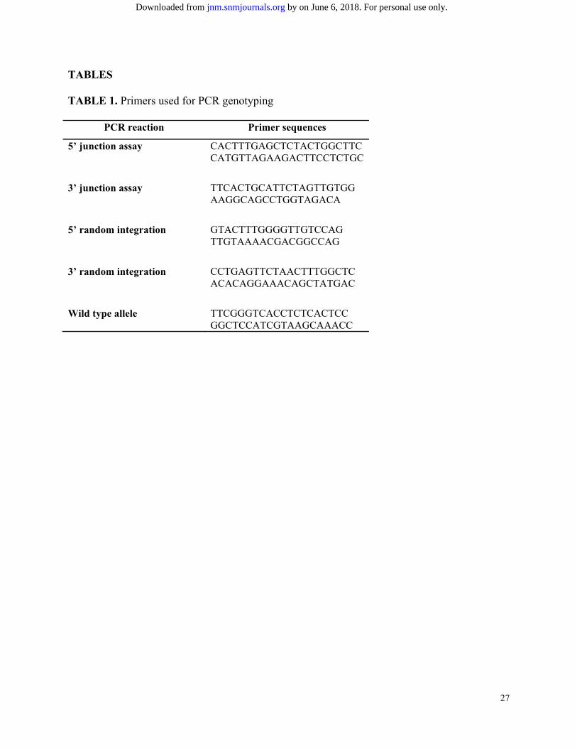
Polymerase chain reaction (PCR) genotyping
Genomic DNA was extracted using the QIAamp DNA mini kit (Qiagen, Venlo, The
Netherlands) following the manufacturers protocol.
To check for reporter gene incorporation, junction assays were performed at the 5’ and 3’
end of the target locus. Random integrations were checked using primers at 5’ and 3’ sites next
to the cassette. The wild-type allele was amplified with primers at the sites adjacent to the
insertion site to determine whether the integration was mono- or bi-allelic. Primer sequences can
be found in Table 1.
Southern blot
Genomic DNA was digested with EcoRI and loaded onto a 0.7% agarosegel. Fragments
were transferred to a nylon membrane (Zeta-probe; Biorad, California, US) which was UV-
cosslinked and prehybridization was performed. Digoxigenin (DIG; Roche, Basel, Switzerland)
labeling of the probe targeting the homology arm was done by PCR using the donor vector.
Hybridization of the membrane with the probe was performed using the DIG High Prime DNA
Labelling and Detection starter Kit II (Roche) according to manufacturer’s instructions.
by on June 6, 2018. For personal use only. jnm.snmjournals.org Downloaded from

7
Hepatic differentiation
Hepatic differentiation of ESC was performed as previously described (27).
In vitro validation of reporter gene expression
In vitro BLI was performed as previously described (28).
ESC were plated, washed and incubated with tracer solution (0.74MBq/mL) for 1h.
99mTcO4- and iodide (124I-, Perkin Elmer, Waltham, MA, USA) were used for hNIS+ ESC and
68Ga-DOTATATE for hSSTr2+ ESC. Cells were washed 3 times and tracer concentration in the
cell fraction was measured using a gamma counter (Wizard, Perkin Elmer). Values were
corrected for cell numbers. hNIS Blocking was performed with NaClO4 (0.74MBq/ml 99mTcO4-
in 10µM NaClO4) and hSSTr2 blocking with cold octreotide (0.74MBq/ml 68Ga-DOTATATE in
0.08, 0.25, 0.74, 2.2, 6.7, 20, 60nM octreotide). Efflux of 99mTcO4- was measured by incubating
cells with 99mTcO4- for 1h, followed by incubation with tracer-free medium for 5, 15, 30, 60 and
120 minutes. Saturation experiments were performed by incubation of hSSTr2+ ESC with
different solutions of 68Ga-DOTATATE (0.015MBq/mL; 0.022MBq/ml; 0.11MBq/ml;
0.37MBq/ml or 2.22MBq/ml). Each condition included a blocking with cold octreotide (20nM).
Cells were incubated for 10 minutes and activities in the cells were measured after 3 washing
steps. Based on activity inside the cell and specific activity the amount of 68Ga-DOTATATE
bound to the cells was calculated.
Real-time quantitative PCR
Gene expression analysis of pluripotency markers and ESC differentiation towards the
hepatic lineage was performed using RT-qPCR as previously described(27). Primer sequences
are listed in Table 2.
by on June 6, 2018. For personal use only. jnm.snmjournals.org Downloaded from

8
Immunostaining
Stainings were performed as described previously (28). Goat anti-eGFP (1:500, Abcam),
goat anti-Oct4 (1:200, Abcam, Cambridge, UK), rabbit anti-Nanog (1:200, Abcam), goat anti-
Sox2 (1:50, Santa Cruz Biotechnology, Dallas, USA) and mouse anti-TRA1-60 (1:50, Santa
Cruz) were used. Anti-rabbit, anti-goat and anti-mouse Alexa Fluor®594 (1:500 in 1% BSA,
Thermo Fisher) or anti-goat Alexa Fluor®488 (1:500, Thermo Fisher) secondary antibodies were
used.
Animal preparations
All animal protocols were approved by the Ethical Committee of the KU Leuven. 8-week
old female BALB/c Rag2-/-ɣc-/- mice were anesthetized with 2% isoflurane (Isoflurane ISP®,
Rothacher, Basel, Switzerland). 3-5x10⁶ ESC were resuspended 1:1 in PBS and Matrigel (BD
biosciences). hSSTr2+ ESC were injected on the left flank and hNIS+ ESC contralaterally (n=8).
Bioluminescence imaging
BLI was performed as previously described(28).
68Ga-DOTATATE preparation
See online supplemental files.
Small-animal PET
Imaging was performed on day 1, 22 and day 63, after intravenous injection of ~3.7MBq
of 68Ga-DOTATATE or 124I- per mouse (n≥5), with static acquisitions of 20min using a Focus
by on June 6, 2018. For personal use only. jnm.snmjournals.org Downloaded from

9
220 microPET system (Siemens Medical Solutions USA, Knoxville,TN). Time after injection
was 1h for 68Ga-DOTATATE and 2h for 124I-.
Images were reconstructed with a maximum a posteriori image reconstruction algorithm and
analyzed using PMOD 3.0 (PMOD technologies, Zürich, Switzerland). Images were converted to
standardized uptake value (SUV).
A manually delineated volume of interest was positioned around the teratomas. Ratios
were calculated comparing the signal to the contralateral tumor.
Histological processing
Masson’s trichrome staining was performed using standard protocols and analyzed to
identify the presence of derivatives of the three germ layers.
Statistical analysis
Data are presented as mean ± standard error of the mean. Analysis of variance analysis
was performed. P-values <0.05 were considered statistically significant. Data were processed
using GraphPad Prism version 5.00 for Windows (GraphPad Software, San Diego, California,
USA).
RESULTS
Generation of reporter cell lines
Two reporter gene constructs were generated, flanked by 800bp stretches of homology to
the ZFN target site AAVS1, downstream of exon 1 of the PPP1R12C gene on chromosome 19
(Fig. 1A). The reporter genes were eGFP, Fluc allowing BLI and either the hNIS or the hSSTr2
reporter gene for PET and/or SPECT.
by on June 6, 2018. For personal use only. jnm.snmjournals.org Downloaded from

10
PCR genotyping, Southern blot and expression of pluripotency markers
PCR genotyping was performed to assess proper integration of the construct and to
exclude random integrations (Fig. 1A). Both hSSTr2+ and hNIS+ colonies carried the expression
cassette as confirmed by the 5’ and 3’ junction assays. Single or bi-allelic integration of the
cassette was assessed. In both clones, bi-allelic integration into the AAVS1 locus was observed.
No random integrations had occurred.
Correct integration of the expression construct was confirmed with Southern blot. One
single band was present depicting bi-allelic integration (Fig. 1A). The expression of pluripotency
markers OCT4, NANOG, SOX2 and KLF4 was assessed and no difference in expression was
observed compared to WT ESC (Fig. 1B). Immunocytochemistry for OCT4, NANOG, SOX2 and
TRA1-60 showed a similar expression in all lines (Fig. 1C).
Hepatic differentiation
WT, hSSTr2+ and hNIS+ ESC showed a similar differentiation capacity towards the
hepatic lineage (Fig. 2). The expression of hepatocyte-specific genes HNF4α, AFP, Albumin and
A1AT increased in both cell lines. No significant difference was observed between the WT and
the genome-edited ESC. The definitive endoderm markers EOMES, MIXL1, CXCR4 and SOX17
were expressed similarly in all cell lines.
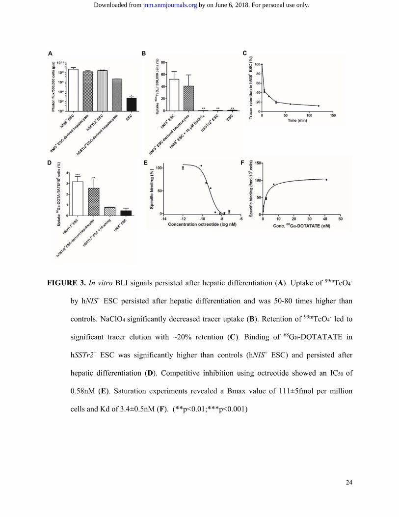
In vitro validation of reporter gene expression
Genome-edited cells, before and after differentiation, had significantly higher BLI signals
compared to WT ESC (Fig. 3A; p<0.05).
by on June 6, 2018. For personal use only. jnm.snmjournals.org Downloaded from

11
Functionality of hSSTr2 and hNIS was tested with radioligand uptake experiments. After
one hour, 52.1% of 99mTcO4- was taken up per 5x10⁵ hNIS+ ESC, 50-80 times more than controls
(p=0.001). NaClO4 reduced uptake levels significantly (0.77%). hNIS+ hepatocytes maintained an
uptake of 40.7%, not significantly different from undifferentiated cells (52.1%; p>0.05) (Fig.
3B). A decreased tracer retention was observed, although after 1h ~20% of the tracer remained
intracellularly (Fig. 3C).
hSSTr2+ ESC could bind 3.18% per million cells of the 68Ga-DOTATATE, 6.9 times
more than hNIS+ cells (p<0.001). hSSTr2+ hepatocytes maintained their 68Ga-DOTATATE
binding capacity. Octreotide administration caused a fourfold reduction of the 68Ga-DOTATATE
uptake (0.77%; p<0.001) (Fig. 3D). An IC50 value of 0.58nM was demonstrated (Fig. 3E). A
saturation experiment showed a Bmax value of 111±5fmol per million cells and Kd of
3.4±0.5nM (Fig. 3F).
In vivo noninvasive longitudinal imaging
BLI of hSSTr2+ and hNIS+ teratomas showed a robust signal that increased over 70 days
(Fig. 4). Small-animal PET was performed on day 1, 22 and 63 (Fig. 5). A focus of increased
68Ga-DOTATATE accumulation was observed at the hSSTr2+ teratoma. No increased 68Ga-
DOTATATE uptake was seen at the site of the hNIS+ teratoma. On day 1 the hSSTr2+ ESC
showed 3.07±0.84 times more signal than the hNIS+ ESC (p<0.01). This signal increased
significantly to 5.02±2.0 times more signal in the hSSTr2+ ESC on day 63 (Fig. 5A).
On the first day after engraftment of the hNIS+ ESC a focus of increased tracer uptake
was observed with 2.87±1.0 times more accumulation of 124I- than the hSSTr2+ teratomas
by on June 6, 2018. For personal use only. jnm.snmjournals.org Downloaded from

12
(p<0.001). On day 63 after engraftment the signal increased significantly to 4.66±0.7 times more
tracer uptake in hNIS+ teratomas compared to the controls (p<0.05) (Fig. 5B).
Teratoma assay
Histological examination confirmed teratoma formation and thus maintenance of
pluripotency after ZFN targeting. Teratomas from both lines contained cells derived from the
three different germ layers (Online supplemental Fig. 1). hSSTr2+ teratomas formed neural
rosettes, cartilage and intestinal lining epithelium. hNIS+ teratomas were less dense and
contained more fluid overall containing neural rosettes, cartilage and glandular tissue.
Immunohistochemistry confirmed expression of hSSTr2 and hNIS in hSSTr2+ and hNIS+
teratomas, respectively.
DISCUSSION
The delivery of genetic material into cells has mainly been done through the use of viral
vectors. Hence, insertional mutagenesis may occur resulting in the disruption of the host genetic
material and there is a potential risk for oncogenesis. Furthermore, non-isogenic cell lines are
generated. The primary transduced cell population is a heterogeneous mix of cells which are all
genomically different from each other. Furthermore, reporter gene silencing is dependent on the
integration site. These factors can result in signal loss and confound data interpretation (29,30).
In this work we used the innovative ZFN-mediated approach for genetic engineering of
ESC. Human PET imaging reporter genes were introduced into the AAVS1 locus which is known
to be a so-called safe harbor locus (7,13,14). AAVS1 has a continuously open chromatin structure
by on June 6, 2018. For personal use only. jnm.snmjournals.org Downloaded from
13
with insulator elements that prevents epigenetic silencing and thereby allows stable transgene
expression, at least when a transgene is expressed from a constitutive active promoter (7,14).
This technology is very translational as two major limitations of stem cell imaging are
tackled at once. ZFN provide safe and controlled introduction of genetic material. Second,
human PET reporter genes will not evoke immune responses. The tracers required for these
genes are available in routine clinical use and do not require an on-site cyclotron. We used two
reporter constructs for multimodal imaging enabling the monitoring of cells after injection using
BLI and PET or SPECT.
In this study we show the efficient introduction of the reporter cassette in both alleles of
the AAVS1 locus. Very high in vitro uptake ratios are reached (~2 orders of magnitude higher
than WT controls) and in vivo imaging is possible when the xenograft is not yet detectable by
clinical examination.
hNIS is a symporter protein and thus mediates an amplification of the imaging signal
(22,31). After their uptake through hNIS, no organification of tracer molecules occurs which
leads to a partial leakage (28). Nevertheless, hNIS has many advantages such as its human origin,
its low background expression and the availability of a number of tracer molecules that are
compatible. Receptors such as the hSSTr2 as imaging reporter proteins bind one single ligand in
a 1:1 ratio with high affinity (32). No signal amplification or tracer leakage will occur, but the
hSSTr2 is of human origin and is characterized by low endogenous expression outside some
specific endocrine organs.
Both imaging reporter genes have their disadvantages, but due to their evident
advantages, they are very good candidates to be used as imaging reporter genes. The choice of
by on June 6, 2018. For personal use only. jnm.snmjournals.org Downloaded from

14
the reporter gene will be mainly based on the anatomical location to be imaged, depending on
their endogenous expression.
BLI and small-animal PET was performed on hSSTr2+ and hNIS+ ESC injected subcutaneously.
BLI showed robust signal intensities which further increased specifically. PET signals increased
significantly over time and more predominantly in hSSTr2+ ESC teratomas. hNIS+ teratomas
contained more fluid-containing cysts not contributing to the signal. hSSTr2+ teratomas were
more dense causing a relatively higher PET signal.
With 68Ga-DOTATATE PET, mean SUV values of 0.07±0.03 in hSSTr2+ and 0.03±0.02
in hNIS+ teratomas were observed (Fig. 5A). 124I- PET led to mean SUV values of 0.21±0.08 and
0.06±0.01 in hNIS+ and SSTr2+ teratomas, respectively (Fig. 5B). Imaging of metastasis in a
mouse model using melanoma cells expressing HSV1-TK imaged with 18F-FHBG resulted in a
mean uptake of 3.3%ID/g (SUV~0.9, formula from (33)). Hepatoma and rat glioma cells
expressing HSV1-TK reporter led to ~1.6%ID/g and ~0.7%ID/g 1-2 weeks after engraftment with
14C-FIAU and 18F-FHBG,(34), implying SUV values of ~0.32 and ~0.14. Prostate cancer cells
expressing HSV1-TK resulted in an uptake of 0.2%ID/g, or a SUV of 0.04 (35). Our data suggest
slightly lower SUV values, however, others have used transfection or transduction to insert the
reporter genes. Hence, multiple copies are inserted leading to higher expression levels. In our
work, one single copy of the reporter gene is included into the genome, and a lower expression
level is inevitable. Also in hSSTr2+ teratomas, lower SUV values were obtained because of
similar reasons. Furthermore, lower receptor densities were observed in comparison with (36).
To circumvent this, antagonists can be used as they are able to bind more hSSTr2 sites (36,37).
by on June 6, 2018. For personal use only. jnm.snmjournals.org Downloaded from

15
Histological examination confirmed that ZFN targeting does not alter the pluripotency of
the ESC. Furthermore, in vitro hepatic differentiation of hSSTr2+ and hNIS+ ESC was equally
efficient compared to WT ESC.
Our results are in line with those of Wang et al. who introduced Fluc, monomeric red
fluorescent protein and HSV1-TK in the AAVS1 locus of ESC and induced pluripotent stem cells
(38). A high efficiency of the targeting procedure was shown with preservation of pluripotency,
differentiation capacity and long-term gene expression. The survival of the cells was monitored
with BLI but not with PET. Also, the gene product of HSV-TK is foreign to the human species
and might imply immunological consequences and unwanted cell death.
CONCLUSION
We demonstrate the introduction of human radionuclide reporter genes into the genome
of human ESC in a safe and controlled manner using the innovative ZFN-mediated strategy,
thereby surpassing possible immune reactions against the imaging reporter genes. Furthermore,
we long-term imaged ZFN-engineered ESC using reporter gene small-animal PET. Isogenic cell
lines were generated and these cells could be fully characterized for patient applications. Off-
target effects were described (39,40), but the search for novel strategies has led to the discovery
of the CRISPR/Cas (clustered regulatory interspaced short palindromic repeats) system (41). It is
clear that genetic engineering in the field of stem cell imaging can greatly accelerate the
transition of basic research to a clinical setting, and these innovative techniques can therefore be
used in order to explore other human PET reporter genes.
by on June 6, 2018. For personal use only. jnm.snmjournals.org Downloaded from

16
DISCLOSURE
CMD and KVL are senior clinical investigators of the Flemish Fund for Scientific
Research (FWO). CC is a post-doctoral fellow of the FWO. BH and NB are PhD students funded
by IWT. LO was funded by IWT/OZM/090838, IACS BPAMER3/08/04, and Government of
Aragon FMI048/08. Funding to CMV was from FWO G.0667.07 and G.0975.11; KU Leuven
(ETH-C1900-PF, EME-C2161-GOA/11/012), IWT-HEPSTEM and IWT-HILIM-3D, BELSPO-
IUAP-DEVREPAIR, FP7-HEMIBIO (266777).
ACKNOWLEDGEMENTS
We thank Manja Muijtjens, Pieter Berckmans, Jeanine Santermans and Ann Van
Santvoort for their help in data acquisition and processing. Radiopharmacy from Nuclear
Medicine UZ Leuven is acknowledged for 68Ga-DOTATATE preparations.
by on June 6, 2018. For personal use only. jnm.snmjournals.org Downloaded from

17
REFERENCES
1. Yaghoubi SS, Campbell DO, Radu CG, Czernin J. Positron emission tomography reporter genes and reporter probes: gene and cell therapy applications. Theranostics. 2012;2:374-391. 2. Giudice A, Trounson A. Genetic modification of human embryonic stem cells for derivation of target cells. Cell Stem Cell. 2008;2:422-433. 3. Herbst F, Ball CR, Tuorto F, et al. Extensive methylation of promoter sequences silences lentiviral transgene expression during stem cell differentiation in vivo. Mol Ther. 2012;20:1014-1021. 4. Krishnan M, Park JM, Cao F, et al. Effects of epigenetic modulation on reporter gene expression: implications for stem cell imaging. FASEB J. 2006;20:106-108. 5. Collin J, Lako M. Concise review: putting a finger on stem cell biology: zinc finger nuclease-driven targeted genetic editing in human pluripotent stem cells. Stem Cells. 2011;29:1021-1033. 6. Li M, Suzuki K, Kim NY, Liu GH, Izpisua Belmonte JC. A cut above the rest: targeted genome editing technologies in human pluripotent stem cells. J Biol Chem. 2014;289:4594-4599. 7. Hockemeyer D, Soldner F, Beard C, et al. Efficient targeting of expressed and silent genes in human ESCs and iPSCs using zinc-finger nucleases. Nat Biotechnol. 2009;27:851-857. 8. Lombardo A, Genovese P, Beausejour CM, et al. Gene editing in human stem cells using zinc finger nucleases and integrase-defective lentiviral vector delivery. Nat Biotechnol. 2007;25:1298-1306. 9. Zou J, Maeder ML, Mali P, et al. Gene targeting of a disease-related gene in human induced pluripotent stem and embryonic stem cells. Cell Stem Cell. 2009;5:97-110. 10. Raitano S, Ordovas L, De Muynck L, et al. Restoration of progranulin expression rescues cortical neuron generation in an induced pluripotent stem cell model of frontotemporal dementia. Stem Cell Reports. 2015;4:16-24. 11. Kotin RM, Linden RM, Berns KI. Characterization of a preferred site on human chromosome 19q for integration of adeno-associated virus DNA by non-homologous recombination. EMBO J. 1992;11:5071-5078. 12. Tan I, Ng CH, Lim L, Leung T. Phosphorylation of a novel myosin binding subunit of protein phosphatase 1 reveals a conserved mechanism in the regulation of actin cytoskeleton. J Biol Chem. 2001;276:21209-21216. 13. Smith JR, Maguire S, Davis LA, et al. Robust, persistent transgene expression in human embryonic stem cells is achieved with AAVS1-targeted integration. Stem Cells. 2008;26:496-504. 14. DeKelver RC, Choi VM, Moehle EA, et al. Functional genomics, proteomics, and regulatory DNA analysis in isogenic settings using zinc finger nuclease-driven transgenesis into a safe harbor locus in the human genome. Genome Res. 2010;20:1133-1142. 15. Keyaerts M, Caveliers V, Lahoutte T. Bioluminescence imaging: looking beyond the light. Trends Mol Med. 2012;18:164-172. 16. Gambhir SS, Bauer E, Black ME, et al. A mutant herpes simplex virus type 1 thymidine kinase reporter gene shows improved sensitivity for imaging reporter gene expression with positron emission tomography. Proc Natl Acad Sci U S A. 2000;97:2785-2790. 17. Jacobs A, Voges J, Reszka R, et al. Positron-emission tomography of vector-mediated gene expression in gene therapy for gliomas. Lancet. 2001;358:727-729.
by on June 6, 2018. For personal use only. jnm.snmjournals.org Downloaded from

18
18. Penuelas I, Mazzolini G, Boan JF, et al. Positron emission tomography imaging of adenoviral-mediated transgene expression in liver cancer patients. Gastroenterology. 2005;128:1787-1795. 19. Yaghoubi SS, Jensen MC, Satyamurthy N, et al. Noninvasive detection of therapeutic cytolytic T cells with 18F-FHBG PET in a patient with glioma. Nat Clin Pract Oncol. 2009;6:53-58. 20. Paulmurugan R, Massoud TF, Huang J, Gambhir SS. Molecular imaging of drug-modulated protein-protein interactions in living subjects. Cancer Res. 2004;64:2113-2119. 21. Van Sande J, Massart C, Beauwens R, et al. Anion selectivity by the sodium iodide symporter. Endocrinology. 2003;144:247-252. 22. Dohan O, De la Vieja A, Paroder V, et al. The sodium/iodide Symporter (NIS): characterization, regulation, and medical significance. Endocr Rev. 2003;24:48-77. 23. Penheiter AR, Russell SJ, Carlson SK. The sodium iodide symporter (NIS) as an imaging reporter for gene, viral, and cell-based therapies. Curr Gene Ther. 2012;12:33-47. 24. Deroose CM, Hindie E, Kebebew E, et al. Molecular imaging of gastroenteropancreatic neuroendocrine tumors: current status and future directions. J Nucl Med. 2016;57:1949-1956. 25. Serganova I, Ponomarev V, Blasberg R. Human reporter genes: potential use in clinical studies. Nucl Med Biol. 2007;34:791-807. 26. Reubi JC, Kvols L, Krenning E, Lamberts SW. Distribution of somatostatin receptors in normal and tumor tissue. Metabolism. 1990;39:78-81. 27. Roelandt P, Vanhove J, Verfaillie C. Directed differentiation of pluripotent stem cells to functional hepatocytes. Methods Mol Biol. 2013;997:141-147. 28. Wolfs E, Holvoet B, Gijsbers R, et al. Optimization of multimodal imaging of mesenchymal stem cells using the human sodium iodide symporter for PET and Cerenkov luminescence imaging. PLoS One. 2014;9:e94833. 29. Ellis J. Silencing and variegation of gammaretrovirus and lentivirus vectors. Hum Gene Ther. 2005;16:1241-1246. 30. Ramachandra CJ, Shahbazi M, Kwang TW, et al. Efficient recombinase-mediated cassette exchange at the AAVS1 locus in human embryonic stem cells using baculoviral vectors. Nucleic Acids Res. 2011;39:e107. 31. Ahn BC. Sodium iodide symporter for nuclear molecular imaging and gene therapy: from bedside to bench and back. Theranostics. 2012;2:392-402. 32. Cotugno G, Aurilio M, Annunziata P, et al. Noninvasive repetitive imaging of somatostatin receptor 2 gene transfer with positron emission tomography. Hum Gene Ther. 2011;22:189-196. 33. Phelps ME. PET: Molecular Imaging and Its Biological Applications: Springer New York; 2012. 34. Min JJ, Iyer M, Gambhir SS. Comparison of [18F]FHBG and [14C]FIAU for imaging of HSV1-tk reporter gene expression: adenoviral infection vs stable transfection. Eur J Nucl Med Mol Imaging. 2003;30:1547-1560. 35. Yang H, Berger F, Tran C, Gambhir SS, Sawyers CL. MicroPET imaging of prostate cancer in LNCAP-SR39TK-GFP mouse xenografts. Prostate. 2003;55:39-47. 36. Reubi JC, Schar JC, Waser B, et al. Affinity profiles for human somatostatin receptor subtypes SST1-SST5 of somatostatin radiotracers selected for scintigraphic and radiotherapeutic use. Eur J Nucl Med. 2000;27:273-282.
by on June 6, 2018. For personal use only. jnm.snmjournals.org Downloaded from

19
37. Reubi JC, Waser B, Macke H, Rivier J. Highly increased 125I-JR11 antagonist binding in vitro reveals novel indications for sst2 targeting in human cancers. J Nucl Med. 2017;58:300-306. 38. Wang Y, Zhang WY, Hu S, et al. Genome editing of human embryonic stem cells and induced pluripotent stem cells with zinc finger nucleases for cellular imaging. Circ Res. 2012;111:1494-1503. 39. Cathomen T, Joung JK. Zinc-finger nucleases: the next generation emerges. Mol Ther. 2008;16:1200-1207. 40. Pattanayak V, Ramirez CL, Joung JK, Liu DR. Revealing off-target cleavage specificities of zinc-finger nucleases by in vitro selection. Nat Methods. 2011;8:765-770. 41. Pennisi E. The CRISPR craze. Science. 2013;341:833-836.
by on June 6, 2018. For personal use only. jnm.snmjournals.org Downloaded from
20
ABBREVIATIONS LIST
A1AT Alpha-1 antitrypsin
AAVS1 adeno-associated virus integration site 1
AFP Alpha-Fetoprotein
BLI bioluminescence imaging
CXCR4 chemokine (C-X-C motif) receptor 4
eGFP enhanced green fluorescent protein
EOMES Eomesdermin
ESC embryonic stem cell
Fluc firefly luciferase 68Ga-DOTATATE 68Ga-DOTA-Tyr3-Thr8-octreotate
HNF4α Hepatocyte nuclear factor 4 alpha
hNIS human sodium iodide symporter
hSSTr2 human somatostatin receptor subtype 2
HSV1-TK herpes simplex virus type 1 thymidine kinase
iPSC induced pluripotent stem cell
MIXL1 MIX1 homeobox-like protein 1
NANOG nanog homeobox
OCT4 Octamer-binding transcription factor 4
PET positron emission tomography
PPP1R12C protein phosphatase 1, regulatory subunit 12C
SPECT single photon emission computed tomography
ZFN zinc finger nucleases
by on June 6, 2018. For personal use only. jnm.snmjournals.org Downloaded from

21
FIGURE 1. Correct integration of the transgenes. Primers used for the 5’/3’ junction assay, 5’/3’
random integration and WT allele are depicted in red, blue, orange, brown and green.
Recombinant lines incorporating transgenes showed correct integration. Southern blot
by on June 6, 2018. For personal use only. jnm.snmjournals.org Downloaded from

22
analysis confirmed correct integration of the construct (A). RT-qPCR showed no
significant difference in the expression of pluripotency markers OCT4, NANOG, SOX2
and KLF4 following genetic engineering (B). Immunocytochemistry for OCT4, NANOG,
SOX2 and TRA1-60 showed similar expression in all lines (C).
by on June 6, 2018. For personal use only. jnm.snmjournals.org Downloaded from

23
FIGURE 2. No significant difference in hepatic differentiation of hSSTr2+, hNIS+ and WT ESC.
RT-qPCR for hepatocyte-specific genes HNF4α, AFP, Albumin and A1AT and for
definitive endoderm markers EOMES, MIXL1, CXCR4 and SOX17.
by on June 6, 2018. For personal use only. jnm.snmjournals.org Downloaded from
24
FIGURE 3. In vitro BLI signals persisted after hepatic differentiation (A). Uptake of 99mTcO4-
by hNIS+ ESC persisted after hepatic differentiation and was 50-80 times higher than
controls. NaClO4 significantly decreased tracer uptake (B). Retention of 99mTcO4
- led to
significant tracer elution with ~20% retention (C). Binding of 68Ga-DOTATATE in
hSSTr2+ ESC was significantly higher than controls (hNIS+ ESC) and persisted after
hepatic differentiation (D). Competitive inhibition using octreotide showed an IC50 of
0.58nM (E). Saturation experiments revealed a Bmax value of 111±5fmol per million
cells and Kd of 3.4±0.5nM (F). (**p<0.01;***p<0.001)
by on June 6, 2018. For personal use only. jnm.snmjournals.org Downloaded from

25
FIGURE 4. BLI of the teratomas showed a clear signal coming from both hSSTr2+ and hNIS+
ESC persisting over 70 days.
by on June 6, 2018. For personal use only. jnm.snmjournals.org Downloaded from

26
FIGURE 5. PET confirmed tracer uptake at the correct locations. 68Ga-DOTATATE
accumulation observed at the hSSTr2+ ESC site persisted over time (orange arrow), no
signal was present at the hNIS+ ESC graft (green arrow) (A). Uptake of 124I- at the hNIS+
ESC site was clear and persisted over time, no signal at the contralateral hSSTr2+ ESC
graft (B).
by on June 6, 2018. For personal use only. jnm.snmjournals.org Downloaded from
27
TABLES
TABLE 1. Primers used for PCR genotyping
PCR reaction Primer sequences
5’ junction assay CACTTTGAGCTCTACTGGCTTC CATGTTAGAAGACTTCCTCTGC
3’ junction assay TTCACTGCATTCTAGTTGTGG AAGGCAGCCTGGTAGACA
5’ random integration GTACTTTGGGGTTGTCCAG TTGTAAAACGACGGCCAG
3’ random integration CCTGAGTTCTAACTTTGGCTC ACACAGGAAACAGCTATGAC
Wild type allele TTCGGGTCACCTCTCACTCC GGCTCCATCGTAAGCAAACC
by on June 6, 2018. For personal use only. jnm.snmjournals.org Downloaded from

28
TABLE 2. Primers used for RT-qPCR.
Gene Forward Reverse
GAPDH TCAAGAAGGTGGTGAAGCAGG ACCAGGAAATGAGCTTGACAAA
EOMES AACAACACCCAGATGATAGTC TCATAGTTGTCTCTGAAGCCT
CXCR4 CACCGCATCTGGAGAACCA GCCCATTTCCTCGGTGTAGTT
MIXL1 GGATCCAGGTATGGTTCCAG CATGAGTCCAGCTTTGAACC
SOX17 CGCTTTCATGGTGTGGGCTAAGGACG TAGTTGGGGTGGTCCTGCATGTGCTG
HNF4a ACTACGGTGCCTCGAGCTGT GGCACTGGTTCCTCTTGTCT
AFP TGAGCACTGTTGCAGAGGAG GTGGTCAGTTTGCAGCATTC
ALB ATGCTGAGGCAAAGGATGTC AGCAGCAGCACGACAGAGTA
A1AT AGGGCCTGAAGCTAGTGGAT TCCTCGGTGTCCTTGACTTC
by on June 6, 2018. For personal use only. jnm.snmjournals.org Downloaded from

Online supplemental files
MATERIALS AND METHODS 68Ga-DOTATATE preparation
68Ga-DOTATATE was prepared by heating gallium-68 chloride (400-800MBq) at pH4-4.4 with 30µg
DOTATATE (Bachem, Switzerland) (adapted from (1)). 68Ga-chloride was obtained by elution of a
germanium-68/gallium-68 generator with diluted HCl-solution followed by purification over a Dowex column
(Sigma-Aldrich/Fluka, St. Louis, Missouri). The reaction mixture was purified over a Sep-Pak C18 column
and formulated into a clinical-grade injectable solution.
FIGURES
Online supplemental Figure 1. Histological validation of teratoma formation. Teratomas derived from hSSTr2+ ESC (A). Differentiation towards neural rosettes, cartilage and intestinal lining epithelium. hNIS+ teratoma (B) differentiation towards neural rosettes, cartilage and glandular tissue. Immunohistochemistry for hSSTr2 was positive in hSSTr2+ teratomas (C). Immunohistochemistry for hNIS was positive in the hNIS+ teratoma (D). Scale bar overviews:1000µM. Others:50µM.
by on June 6, 2018. For personal use only. jnm.snmjournals.org Downloaded from

ONLINE SUPPLEMENTAL REFERENCES
1. Van Binnebeek S, Vanbilloen B, Baete K, et al. Comparison of diagnostic accuracy of (111)In-pentetreotide SPECT and (68)Ga-DOTATOC PET/CT: A lesion-by-lesion analysis in patients with metastatic neuroendocrine tumours. Eur Radiol. 2016;26:900-909.
by on June 6, 2018. For personal use only. jnm.snmjournals.org Downloaded from

Doi: 10.2967/jnumed.117.189779Published online: June 8, 2017.J Nucl Med. Verfaillie and Christophe M. DerooseTom Struys, Bert Vanbilloen, Cindy Casteels, Maurilio Sampaolesi, Koen Van Laere, Ivo Lambrichts, Catherine M. Esther Wolfs, Bryan Holvoet, Laura Ordovas, Natacha Breuls, Nicky Helsen, Matthias Schönberger, Susanna Raitano, reporter genes after zinc finger nucleases-mediated genome editingMolecular imaging of human embryonic stem cells stably expressing human PET
http://jnm.snmjournals.org/content/early/2017/06/07/jnumed.117.189779This article and updated information are available at:
http://jnm.snmjournals.org/site/subscriptions/online.xhtml
Information about subscriptions to JNM can be found at:
http://jnm.snmjournals.org/site/misc/permission.xhtmlInformation about reproducing figures, tables, or other portions of this article can be found online at:
and the final, published version.proofreading, and author review. This process may lead to differences between the accepted version of the manuscript
ahead of print area, they will be prepared for print and online publication, which includes copyediting, typesetting,JNMcopyedited, nor have they appeared in a print or online issue of the journal. Once the accepted manuscripts appear in the
. They have not beenJNM ahead of print articles have been peer reviewed and accepted for publication in JNM
(Print ISSN: 0161-5505, Online ISSN: 2159-662X)1850 Samuel Morse Drive, Reston, VA 20190.SNMMI | Society of Nuclear Medicine and Molecular Imaging
is published monthly.The Journal of Nuclear Medicine
© Copyright 2017 SNMMI; all rights reserved.
by on June 6, 2018. For personal use only. jnm.snmjournals.org Downloaded from