Molecular imaging agents for detection of β-amyloid plaques in ...
5
5 Molecular imaging agents for detection of β-amyloid plaques in Alzheimer’s disease Letícia Quental 1 , Goreti Ribeiro Morais 1 , Isabel Santos 1 , António Paulo 1-2 1. Grupo de Ciências Radiofarmacêuticas, Campus Tecnológico e Nuclear, Instituto Superior Técnico (CTN/IST) 2. Corresponding author: [email protected] ABSTRACT: The formation of amyloid structures is a neuropathological feature that characterizes several neurodegenerative disorders, such as Alzheimer´s and Parkinson´s disease. Up to now, the definitive diagnosis of these diseases can only be accomplished by immunostaining of post mortem brain tissues with dyes such Thioflavin T and congo red. Aiming at early in vivo diagnosis of Alzheimer´s disease (AD), several amyloid-avid radioprobes have been developed for b-amyloid imaging by positron emission tomogra- phy (PET) and single-photon emission computed tomography (SPECT). The aim of this paper is to present a perspective of the available amyloid imaging agents, special those that have been selected for clinical trials and are at the different stages of the US Food and Drugs Administration (FDA) approval. Keywords: Alzheimer´s disease, b-Amyloid aggregation, molecular imaging, molecular probes. Sondas moleculares para a deteção de placas β-amilóide na doença de Alzheimer RESUMO: A formação de estruturas amilóides é uma característica neuropatológica comum nas várias doenças neurodegenerativas, como a doença de Alzheimer e de Par- kinson. Até à data, o diagnóstico destas doenças apenas é conseguido post mortem por estudos histoquímicos com corantes, como a Tioflavina T e o vermelho do congo. Du- rante os últimos anos têm sido desenvolvidos vários compostos com afinidade para agregados de ß-amilóide para visualização dessas estruturas por tomografia de emissão de positrões (PET) e tomografia computadorizada de emissão de fotão único (SPECT). Neste trabalho pretendemos apresentar as principais sondas radioativas com potencial para imagiologia de estruturas amilóides, em especial aquelas que entraram em ensaios clínicos e se encontram em diferentes etapas de aprovação pela FDA. Palavras-chave: doença de Alzheimer, agregação da b-amilóide, imagiologia molecular, son- das moleculares. Introduction Alzheimer’s disease (AD) is a neurodegenerative disorder that affects millions of people worldwide 1 . The impact in the public health is considerable, with tendency to increase as the population gets older. The most common symptoms of AD are decline in the cognitive functions, irreversible memory loss, disorientation and language impairment. AD diagnosis is based mainly on the patient’s history and on neuropsychological tests. However, the overlapping of ear- ly AD symptoms with normal signs of aging difficults such diagnosis. Histopathologically, AD is characterized by the presence of senile plaques containing b-amyloid (Ab) plaques and neurofibrillary tangles (NFTs) containing highly phosphorylated tau protein. Currently, the accurate diag- nosis of AD is only possible post mortem after confirmation of extracellular Ab deposits and NFTs, through histopatho- logical studies using dyes such as thioflavin T (ThT) and congo red (CR) 2 . The molecular processes underlying the pathology are still unknown, however it is thought that the Aß deposits accumulate before the onset of the disease 3 . Ab is a soluble extracellular peptide composed by 40 (Ab 1-40 ) or 42 (Ab 1-42 ) aminoacids, which is formed from transmembrane amyloid-precursor protein (APP) by the action of b and g secretases 4 . Thus, in vivo imaging agents that can specifi- cally demonstrate the location and density of Ab deposits SAÚDE & TECNOLOGIA . NOVEMBRO | 2013 | #10 | P. 5-9 . ISSN: 1646-9704
Transcript of Molecular imaging agents for detection of β-amyloid plaques in ...
55
Molecular imaging agents for detection of β-amyloid plaques in Alzheimer’s diseaseLetícia Quental1, Goreti Ribeiro Morais1, Isabel Santos1, António Paulo1-2
1. Grupo de Ciências Radiofarmacêuticas, Campus Tecnológico e Nuclear, Instituto Superior Técnico (CTN/IST)2. Corresponding author: [email protected]
ABSTRACT: The formation of amyloid structures is a neuropathological feature that characterizes several neurodegenerative disorders, such as Alzheimer´s and Parkinson´s disease. Up to now, the definitive diagnosis of these diseases can only be accomplished by immunostaining of post mortem brain tissues with dyes such Thioflavin T and congo red. Aiming at early in vivo diagnosis of Alzheimer´s disease (AD), several amyloid-avid radioprobes have been developed for b-amyloid imaging by positron emission tomogra-phy (PET) and single-photon emission computed tomography (SPECT). The aim of this paper is to present a perspective of the available amyloid imaging agents, special those that have been selected for clinical trials and are at the different stages of the US Food and Drugs Administration (FDA) approval.
Keywords: Alzheimer´s disease, b-Amyloid aggregation, molecular imaging, molecular probes.
Sondas moleculares para a deteção de placas β-amilóide na doença de Alzheimer
RESUMO: A formação de estruturas amilóides é uma característica neuropatológica comum nas várias doenças neurodegenerativas, como a doença de Alzheimer e de Par-kinson. Até à data, o diagnóstico destas doenças apenas é conseguido post mortem por estudos histoquímicos com corantes, como a Tioflavina T e o vermelho do congo. Du-rante os últimos anos têm sido desenvolvidos vários compostos com afinidade para agregados de ß-amilóide para visualização dessas estruturas por tomografia de emissão de positrões (PET) e tomografia computadorizada de emissão de fotão único (SPECT). Neste trabalho pretendemos apresentar as principais sondas radioativas com potencial para imagiologia de estruturas amilóides, em especial aquelas que entraram em ensaios clínicos e se encontram em diferentes etapas de aprovação pela FDA.
Palavras-chave: doença de Alzheimer, agregação da b-amilóide, imagiologia molecular, son-das moleculares.
Introduction
Alzheimer’s disease (AD) is a neurodegenerative disorder that affects millions of people worldwide1. The impact in the public health is considerable, with tendency to increase as the population gets older. The most common symptoms of AD are decline in the cognitive functions, irreversible memory loss, disorientation and language impairment. AD diagnosis is based mainly on the patient’s history and on neuropsychological tests. However, the overlapping of ear-ly AD symptoms with normal signs of aging difficults such diagnosis. Histopathologically, AD is characterized by the presence of senile plaques containing b-amyloid (Ab) plaques and neurofibrillary tangles (NFTs) containing highly
phosphorylated tau protein. Currently, the accurate diag-nosis of AD is only possible post mortem after confirmation of extracellular Ab deposits and NFTs, through histopatho-logical studies using dyes such as thioflavin T (ThT) and congo red (CR)2.
The molecular processes underlying the pathology are still unknown, however it is thought that the Aß deposits accumulate before the onset of the disease3. Ab is a soluble extracellular peptide composed by 40 (Ab1-40) or 42 (Ab1-42) aminoacids, which is formed from transmembrane amyloid-precursor protein (APP) by the action of b and g secretases4. Thus, in vivo imaging agents that can specifi-cally demonstrate the location and density of Ab deposits
SAÚDE & TECNOLOGIA . NOVEMBRO | 2013 | #10 | P. 5-9 . ISSN: 1646-9704
66
Design of Aβ imaging agents
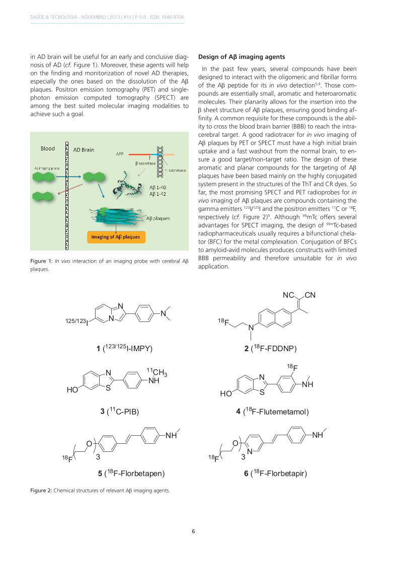
In the past few years, several compounds have been designed to interact with the oligomeric and fibrillar forms of the Ab peptide for its in vivo detection5-9. Those com-pounds are essentially small, aromatic and heteroaromatic molecules. Their planarity allows for the insertion into the b sheet structure of Ab plaques, ensuring good binding af-finity. A common requisite for these compounds is the abil-ity to cross the blood brain barrier (BBB) to reach the intra- cerebral target. A good radiotracer for in vivo imaging of Ab plaques by PET or SPECT must have a high initial brain uptake and a fast washout from the normal brain, to en-sure a good target/non-target ratio. The design of these aromatic and planar compounds for the targeting of Ab plaques have been based mainly on the highly conjugated system present in the structures of the ThT and CR dyes. So far, the most promising SPECT and PET radioprobes for in vivo imaging of Ab plaques are compounds containing the gamma emitters 123I/125I and the positron emitters 11C or 18F, respectively (cf. Figure 2)9. Although 99mTc offers several advantages for SPECT imaging, the design of 99mTc-based radiopharmaceuticals usually requires a bifunctional chela-tor (BFC) for the metal complexation. Conjugation of BFCs to amyloid-avid molecules produces constructs with limited BBB permeability and therefore unsuitable for in vivo application.
in AD brain will be useful for an early and conclusive diag-nosis of AD (cf. Figure 1). Moreover, these agents will help on the finding and monitorization of novel AD therapies, especially the ones based on the dissolution of the Ab plaques. Positron emission tomography (PET) and single-photon emission computed tomography (SPECT) are among the best suited molecular imaging modalities to achieve such a goal.
Figure 1: In vivo interaction of an imaging probe with cerebral Ab
plaques.
Figure 2: Chemical structures of relevant Ab imaging agents.
SAÚDE & TECNOLOGIA . NOVEMBRO | 2013 | #10 | P. 5-9 . ISSN: 1646-9704
7
Relevant Radiolabeled Aβ imaging probes
Although there are more SPECT than PET scanners, the same is not true with respect to agents for amyloid imag-ing. Among the SPECT amyloid imaging, the 123I-IMPY (cf. Figure 2[1]) has shown up as the most promising10, while more progress has been observed in the development of PET amyloid imaging radioprobes. 123I-IMPY displayed selective binding to Ab plaque ex vivo in autoradiographic experiments using mice AD model (PSAPP)11. However the signal-to-noise ratio for plaque labelling is not ideal, maybe due to the fast clearance from the brain and plas-ma observed in AD and normal subjects12.
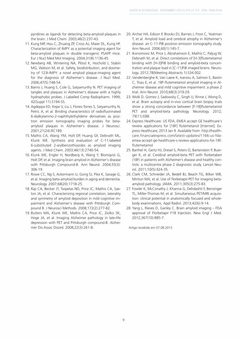
The compound 18F-FDDNP (cf. Figure 2[2]) was the first PET probe sucessfully developed for in vivo molecular imaging of Ab plaques13. However, PET imaging showed that 18F-FDDNP labels both Ab plaques and NFTs in the brain of AD, and thus is not selective for measuring Ab deposits load in the AD brain. Also, its excessive lipophili- city (log P = 3.92) contributed for high non-specific bind-ing in normal mice brain14. The “Pittsburgh compound B” (11C-PIB) (cf. Figure 2[3]) is one of the best characterized PET imaging agent for Ab plaques in the brain. It showed excellent initial brain uptake and a high binding affinity to Ab plaque (Ki = 0.87 ± 0.18 nM)15. In AD patients, 11C-PIB retention, which was increased in the cortical areas, cor-related inversely with cerebral glucose metabolism deter-mined with 18F-fluorodeoxyglucose (18F-FDG) (cf. Figure 3)16. Since then, other studies in thousands of AD patients have validated the usefulness of 11C-PIB as a PET Ab imaging probe17-20. However, the short half-life of 11C (t1/2 = 20 min) limits the clinical use of 11C-PIB to centers with an on-site cyclotron. Such limitation prompted seve- ral authors to search for alternative amyloid-binding radio- pharmaceuticals labelled with longer lived fluorine-18
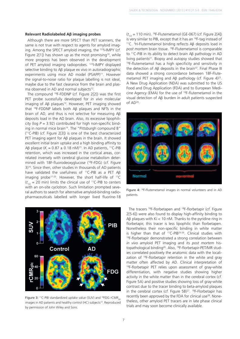
(t1/2 = 110 min). 18F-Flutemetamol (GE-067) (cf. Figure 2[4]) is very similar to PIB, except that it has an 18F-tag instead of 11C. 3H-Flutemetamol binding reflects Ab deposits load in post mortem brain tissue. 18F-Flutemetamol is comparable to 11C-PiB in its ability to detect brain Ab pathology in AD living patients21. Biopsy and autopsy studies showed that 18F-flutemetamol has a high specificity and sensitivity in the detection of Ab deposits in the brain22. Final Phase III data showed a strong concordance between 18F-Flute-metamol PET imaging and Ab pathology (cf. Figure 4)23. A New Drug Application (NDA) was submitted to the US Food and Drug Application (FDA) and to European Medi-cine Agency (EMA) for the use of 18F-flutemetamol in the visual detection of Ab burden in adult patients suspected of AD24.
Figure 4: 18F-Flutemetamol images in normal volunteers and in AD
patients.
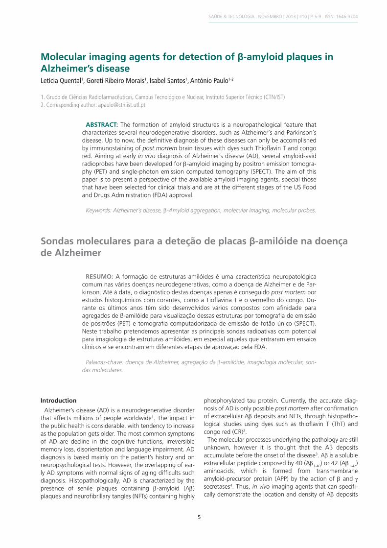
The tracers 18F-florbetapen and 18F-florbetapir (cf. Figure 2[5-6]) were also found to display high-affinity binding to Ab plaques with Ki < 10 nM. Thanks to the pyridine ring in florbetapir, this tracer is less lipophilic than florbetapen. Nonetheless their non-specific binding in white matter is higher than that of 11C-PIB25-26. Clinical studies with 18F-florbetapir demonstrated a strong correlation between in vivo amyloid PET imaging and its post mortem his-topathological binding26. Also, 18F-florbetapir-PET/MR stud-ies correlated positively the anatomic data with the locali-zation of 18F-florbetapir retention in the white and gray matter often affected by AD. Clinical interpretation of 18F-florbetapir PET relies upon assessment of gray-white differentiation, with negative studies showing higher activity in the white matter than in the cerebral cortex (cf. Figure 5A) and positive studies showing loss of gray-white contrast due to the tracer binding to beta-amyloid plaques in the cerebral cortex (cf. Figure 5B)27. 18F-Florbetapir has recently been approved by the FDA for clinical use28. None-theless, other amyloid PET tracers are in late phase clinical trials and may soon become clinically available.
Figure 3: 11C-PIB standardized uptake value (SUV) and 18FDG rCMRglc
images in AD patients and healthy control (HC) subjects13. Reproduced
by permission of John Wiley and Sons.
SAÚDE & TECNOLOGIA . NOVEMBRO | 2013 | #10 | P. 5-9 . ISSN: 1646-9704

8
Figure 5: Amyloid imaging with 18F-florbetapir. A) Normal control subject with no-to-sparse Ab plaques. B) Positive PET/MRI study, consistent
with moderate to frequent Ab plaques20.
3. Irvine GB, El-Agnaf OM, Shankar GM, Walsh DM. Protein aggregation in the brain: the molecular basis for Alzheimer’s and Parkinson’s diseases. Mol Med. 2008;14 (7-8): 451-64.
4. Vassar R, Bennett BD, Babu-Khan S, Kahn S, Mendiaz EA, Denis P, et al. Beta-secretase cleavage of Alzheimer’s amyloid precursor protein by the transmembrane aspartic protease BACE. Science. 1999;286(5440):735-41.
5. Ono M. Development of positron-emission tomography/ single-photon emission computed tomography imaging pro-bes for in vivo detection of beta-amyloid plaques in Alzheimer’s brains. Chem Pharm Bull. 2009;57(10):1029-39.
6. Mathis CA, Lopresti BJ, Klunk WE. Impact of amyloid imaging on drug development in Alzheimer’s disease. Nucl Med Biol. 2007;34(7):809-22.
7. Kung HF, Choi SR, Qu W, Zhang W, Skovronsky D. 18F stilbenes and styrylpyridines for PET imaging of A beta plaques in Alzheimer’s disease: a miniperspective. J Med Chem. 2010;53(3):933-41.
8. Ribeiro Morais G, Vicente Miranda H, Santos IC, Santos I, Outeiro TF, Paulo A. Synthesis and in vitro evaluation of fluorinated styryl benzazoles as amyloid-probes. Bioorg Med Chem. 2011;19(24):7698-710.
9. Ribeiro Morais G, Paulo A, Santos I. A synthetic overview of radiolabeled compounds forb-amyloid targeting. Eur J Org Chem. 2012;2012 (7):1279-93.
10. Zhuang ZP, Kung MP, Wilson A, Lee CW, Plössl K, Hou C, et al. Structure-activity relationship of imidazo[1,2-alpha]
Conclusions
The 11C-PIB, 18F-flutemetamol, 18F-florbetapen, and 18F-florbetapir have been well studied in humans as amy-loid imaging agents. The imaging performance of these four PET tracers is comparable with high retention in corti-cal regions, providing all of them good contrast with non-target regions. Despite being the best studied, 11C-PIB has not been yet approved by the FDA, while 18F-flute-metamol is pending FDA and EMA approval. So far, the only amyloid PET tracer authorized by the FDA is the 18F-florbetapir (Amyvid) for brain imaging of cognitively impaired adults undergoing evaluation of AD28.
Acknowledgments
We thank the Fundação para a Ciência e Tecnologia (FCT) – PTDC/QUI/102049/2008 and "Ciência 2008" program – for financial support.
References
1. Alzheimer Association. 2012 Alzheimer’s disease facts and figures. Alzheimers Dement. 2012;8(2):131-68.
2. Bacskai BJ, Hickey GA, Skoch J, Kajdasz ST, Wang Y, Huang GF, et al. Four-dimensional multiphoton imaging of brain entry, amyloid binding, and clearance of an amyloid-beta ligand in transgenic mice. Proc Natl Acad Sci U S A. 2003;100 (21):12462-7.
SAÚDE & TECNOLOGIA . NOVEMBRO | 2013 | #10 | P. 5-9 . ISSN: 1646-9704
9
pyridines as ligands for detecting beta-amyloid plaques in the brain. J Med Chem. 2003;46(2):237-43.
11. Kung MP, Hou C, Zhuang ZP, Cross AJ, Maier DL, Kung HF. Characterization of IMPY as a potential imaging agent for beta-amyloid plaques in double transgenic PSAPP mice. Eur J Nucl Med Mol Imaging. 2004;31(8):1136-45.
12. Newberg AB, Wintering NA, Plössl K, Hochold J, Stabin MG, Watson M, et al. Safety, biodistribution, and dosime-try of 123I-IMPY: a novel amyloid plaque-imaging agent for the diagnosis of Alzheimer's disease. J Nucl Med. 2006;47(5):748-54.
13. Barrio J, Huang S, Cole G, Satyamurthy N. PET imaging of tangles and plaques in Alzheimer's disease with a highly hydrophobic probes. J Labelled Comp Radiopharm. 1999; 42(Suppl 11):S194-S5.
14. Agdeppa ED, Kepe V, Liu J, Flores-Torres S, Satyamurthy N, Petric A, et al. Binding characteristics of radiofluorinated 6-dialkylamino-2-naphthylethylidene derivatives as posi-tron emission tomography imaging probes for beta-amyloid plaques in Alzheimer's disease. J Neurosci. 2001;21(24):RC189.
15. Mathis CA, Wang YM, Holt DP, Huang GF, Debnath ML, Klunk WE. Synthesis and evaluation of C-11-labeled 6-substituted 2-arylbenzothiazoles as amyloid imaging agents. J Med Chem. 2003;46(13):2740-54.
16. Klunk WE, Engler H, Nordberg A, Wang Y, Blomqvist G, Holt DP, et al. Imaging brain amyloid in Alzheimer's disease with Pittsburgh Compound-B. Ann Neurol. 2004;55(3): 306-19.
17. Rowe CC, Ng S, Ackermann U, Gong SJ, Pike K, Savage G, et al. Imaging beta-amyloid burden in aging and dementia. Neurology. 2007;68(20):1718-25.
18. Raji CA, Becker JT, Tsopelas ND, Price JC, Mathis CA, Sax-ton JA, et al. Characterizing regional correlation, laterality and symmetry of amyloid deposition in mild cognitive im-pairment and Alzheimer's disease with Pittsburgh Com-pound B. J Neurosci Methods. 2008;172(2):277-82.
19. Butters MA, Klunk WE, Mathis CA, Price JC, Ziolko SK, Hoge JA, et al. Imaging Alzheimer pathology in late-life depression with PET and Pittsburgh compound-B. Alzhei-mer Dis Assoc Disord. 2008;22(3):261-8.
20. Archer HA, Edison P, Brooks DJ, Barnes J, Frost C, Yeatman T, et al. Amyloid load and cerebral atrophy in Alzheimer's disease: an C-11-PIB positron emission tomography study. Ann Neurol. 2006;60(1):145-7.
21. Ikonomovic M, Price J, Abrahamson E, Mathis C, Paljug W, Debnath M, et al. Direct correlations of [H-3]flutemetamol binding with [H-3]PiB binding and amyloid-beta concen-tration and plaque load in [C-11]PiB imaged brains. Neuro-logy. 2012;78(Meeting Abstracts 1):S34.002.
22. Vandenberghe R, Van Laere K, Ivanoiu A, Salmon E, Bastin C, Triau E, et al. 18F-flutemetamol amyloid imaging in Al-zheimer disease and mild cognitive impairment: a phase 2 trial. Ann Neurol. 2010;68(3):319-29.
23. Wolk D, Gomez J, Sadowsky C, Singh U, Rinne J, Wong D, et al. Brain autopsy and in-vivo cortical brain biopsy trials show a strong concordance between [F-18]flutemetamol PET and amyloid-beta pathology. Neurology. 2012; 79(11):E88.
24. Express Healthcare. US FDA, EMEA accept GE healthcare's review applications for [18F] flutemetanol [Internet]. Ex-press Healthcare; 2013 Jan 9. Available from: http://health-care.financialexpress.com/latest-updates/1166-us-fda-emea-accept-ge-healthcare-s-review-applications-for-18f-flutemetamol
25. Barthel H, Gertz HJ, Dresel S, Peters O, Bartenstein P, Buer-ger K, et al. Cerebral amyloid-beta PET with florbetaben (18F) in patients with Alzheimer’s disease and healthy con-trols: a multicentre phase 2 diagnostic study. Lancet Neu-rol. 2011;10(5):424-35.
26. Clark CM, Schneider JA, Bedell BJ, Beach TG, Bilker WB, Mintun MA, et al. Use of florbetapir-PET for imaging beta-amyloid pathology. JAMA. 2011;305(3):275-83.
27. Fowler K, McConathy J, Khanna G, Dehdashti F, Benzinger TL, Miller-Thomas M, et al. Simultaneous PET/MRI acquisi-tion: clinical potential in anatomically focused and whole-body examinations. Appl Radiol. 2013;42(6):9-14.
28. Yang L, Rieves D, Ganley C. Brain amyloid imaging – FDA approval of Florbetapir F18 Injection. New Engl J Med. 2012;367(10):885-7.
Artigo recebido em 07.08.2013
SAÚDE & TECNOLOGIA . NOVEMBRO | 2013 | #10 | P. 5-9 . ISSN: 1646-9704