Molecular diversity of extended-spectrum β-lactamases and ...
13
REVIEW Open Access Molecular diversity of extended-spectrum β-lactamases and carbapenemases, and antimicrobial resistance Teiji Sawa 1* , Kunihiko Kooguchi 2 and Kiyoshi Moriyama 3 Abstract Along with the recent spread of multidrug-resistant bacteria, outbreaks of extended-spectrum β-lactamase (ESBL) and carbapenemase-producing bacteria present a serious challenge to clinicians. β-lactam antibiotics are the most frequently used antibacterial agents and ESBLs, and carbapenemases confer resistance not only to carbapenem antibiotics but also to penicillin and cephem antibiotics. The mechanism of β-lactam resistance involves an efflux pump, reduced permeability, altered transpeptidases, and inactivation by β-lactamases. Horizontal gene transfer is the most common mechanism associated with the spread of extended-spectrum β-lactam- and carbapenem resistance among pathogenic bacterial species. Along with the increase in antimicrobial resistance, many different types of ESBLs and carbapenemases have emerged with different enzymatic characteristics. For example, carbapenemases are represented across classes A to D of the Ambler classification system. Because bacteria harboring different types of ESBLs and carbapenemases require specific therapeutic strategies, it is essential for clinicians to understand the characteristics of infecting pathogens. In this review, we summarize the current knowledge on carbapenem resistance by ESBLs and carbapenemases, such as class A carbapenemases, class C extended-spectrum AmpC (ESAC), carbapenem-hydrolyzing class D β-lactamases (CHDLs), and class B metallo-β- lactamases, with the aim of aiding critical care clinicians in their therapeutic decision making. Keywords: β-Lactam, β-Lactamase, Carbapenemase, Classification, Multidrug resistance Background Among the recent spread of multidrug-resistant bacteria, outbreaks of extended-spectrum β-lactam- and carbapenem- resistant bacteria are a serious problem not only mak- ing treatment difficult but also worsening the prognosis of infected patients [1]. β-lactam antibiotics are the most frequently used antibacterial agents, and extended- spectrum β-lactams and carbapenems have been devel- oped as specific drugs to treat bacterial species resistant to penicillins and cephems [2]. Bacterial resistance to car- bapenems incorporates not only carbapenem antibiotics but also resistance to penicillin and cephem antibiotics. Therefore, resistance to carbapenems presents a signifi- cant threat to patients who are immunocompromised and are therefore susceptible to infections caused by multidrug-resistant bacteria all over the world [3]. The mechanism of resistance to extended-spectrum β- lactams and carbapenems involves an efflux pump, re- duced permeability, altered transpeptidases, and inacti- vation by β-lactamases. Horizontal gene transfer is the most common mechanism associated with the spread of antimicrobial resistance across pathogenic bacterial species, such as carbapenemase-producing Enterobacte- riaceae (CPE) [4, 5]. In various bacterial species, novel extended-spectrum β-lactamases (ESBLs) or carbapene- mases with different structures or characteristic features are reported each year. Various ESBLs and carbapenemases have been re- ported in the Enterobacteriaceae including Enterobacter, Klebsiella, Escherichia coli [6, 7], and other opportunistic species such as Serratia, Acinetobacter, and Pseudo- monas [8]. In addition, the genetic elements by which drug-resistant genes horizontally move across bacterial species have been studied among these bacterial species. © The Author(s). 2020 Open Access This article is distributed under the terms of the Creative Commons Attribution 4.0 International License (http://creativecommons.org/licenses/by/4.0/), which permits unrestricted use, distribution, and reproduction in any medium, provided you give appropriate credit to the original author(s) and the source, provide a link to the Creative Commons license, and indicate if changes were made. The Creative Commons Public Domain Dedication waiver (http://creativecommons.org/publicdomain/zero/1.0/) applies to the data made available in this article, unless otherwise stated. * Correspondence: [email protected] 1 Department of Anesthesiology, School of Medicine, Kyoto Prefectural University of Medicine, 465 Kajii-cho, Kamigyo, Kyoto 602-8566, Japan Full list of author information is available at the end of the article Sawa et al. Journal of Intensive Care (2020) 8:13 https://doi.org/10.1186/s40560-020-0429-6
Transcript of Molecular diversity of extended-spectrum β-lactamases and ...
REVIEW Open Access
Molecular diversity of extended-spectrumβ-lactamases and carbapenemases, andantimicrobial resistanceTeiji Sawa1* , Kunihiko Kooguchi2 and Kiyoshi Moriyama3
Abstract
Along with the recent spread of multidrug-resistant bacteria, outbreaks of extended-spectrum β-lactamase (ESBL)and carbapenemase-producing bacteria present a serious challenge to clinicians. β-lactam antibiotics are the mostfrequently used antibacterial agents and ESBLs, and carbapenemases confer resistance not only to carbapenemantibiotics but also to penicillin and cephem antibiotics. The mechanism of β-lactam resistance involves an effluxpump, reduced permeability, altered transpeptidases, and inactivation by β-lactamases. Horizontal gene transfer isthe most common mechanism associated with the spread of extended-spectrum β-lactam- and carbapenemresistance among pathogenic bacterial species. Along with the increase in antimicrobial resistance, many differenttypes of ESBLs and carbapenemases have emerged with different enzymatic characteristics. For example,carbapenemases are represented across classes A to D of the Ambler classification system. Because bacteriaharboring different types of ESBLs and carbapenemases require specific therapeutic strategies, it is essential forclinicians to understand the characteristics of infecting pathogens. In this review, we summarize the currentknowledge on carbapenem resistance by ESBLs and carbapenemases, such as class A carbapenemases, class Cextended-spectrum AmpC (ESAC), carbapenem-hydrolyzing class D β-lactamases (CHDLs), and class B metallo-β-lactamases, with the aim of aiding critical care clinicians in their therapeutic decision making.
Keywords: β-Lactam, β-Lactamase, Carbapenemase, Classification, Multidrug resistance
BackgroundAmong the recent spread of multidrug-resistant bacteria,outbreaks of extended-spectrum β-lactam- and carbapenem-resistant bacteria are a serious problem not only mak-ing treatment difficult but also worsening the prognosisof infected patients [1]. β-lactam antibiotics are themost frequently used antibacterial agents, and extended-spectrum β-lactams and carbapenems have been devel-oped as specific drugs to treat bacterial species resistant topenicillins and cephems [2]. Bacterial resistance to car-bapenems incorporates not only carbapenem antibioticsbut also resistance to penicillin and cephem antibiotics.Therefore, resistance to carbapenems presents a signifi-cant threat to patients who are immunocompromisedand are therefore susceptible to infections caused by
multidrug-resistant bacteria all over the world [3]. Themechanism of resistance to extended-spectrum β-lactams and carbapenems involves an efflux pump, re-duced permeability, altered transpeptidases, and inacti-vation by β-lactamases. Horizontal gene transfer is themost common mechanism associated with the spreadof antimicrobial resistance across pathogenic bacterialspecies, such as carbapenemase-producing Enterobacte-riaceae (CPE) [4, 5]. In various bacterial species, novelextended-spectrum β-lactamases (ESBLs) or carbapene-mases with different structures or characteristic featuresare reported each year.Various ESBLs and carbapenemases have been re-
ported in the Enterobacteriaceae including Enterobacter,Klebsiella, Escherichia coli [6, 7], and other opportunisticspecies such as Serratia, Acinetobacter, and Pseudo-monas [8]. In addition, the genetic elements by whichdrug-resistant genes horizontally move across bacterialspecies have been studied among these bacterial species.
© The Author(s). 2020 Open Access This article is distributed under the terms of the Creative Commons Attribution 4.0International License (http://creativecommons.org/licenses/by/4.0/), which permits unrestricted use, distribution, andreproduction in any medium, provided you give appropriate credit to the original author(s) and the source, provide a link tothe Creative Commons license, and indicate if changes were made. The Creative Commons Public Domain Dedication waiver(http://creativecommons.org/publicdomain/zero/1.0/) applies to the data made available in this article, unless otherwise stated.
* Correspondence: [email protected] of Anesthesiology, School of Medicine, Kyoto PrefecturalUniversity of Medicine, 465 Kajii-cho, Kamigyo, Kyoto 602-8566, JapanFull list of author information is available at the end of the article
Sawa et al. Journal of Intensive Care (2020) 8:13 https://doi.org/10.1186/s40560-020-0429-6
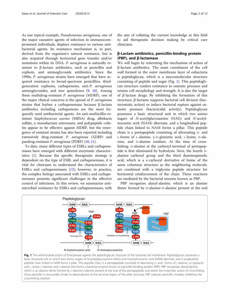
As one typical example, Pseudomonas aeruginosa, one ofthe major causative agents of infection in immunocom-promised individuals, displays resistance to various anti-bacterial agents. Its resistance mechanism is, in part,derived from the organism’s natural resistance, but isalso acquired through horizontal gene transfer and/ormutations within its DNA. P. aeruginosa is naturally re-sistant to β-lactam antibiotics, such as penicillin andcephem, and aminoglycoside antibiotics. Since the1990s, P. aeruginosa strains have emerged that have ac-quired resistance to broad-spectrum penicillins, third-generation cephems, carbapenems, anti-P. aeruginosaaminoglycosides, and new quinolones [9, 10]. Amongthese multidrug-resistant P. aeruginosa (MDRP), one ofthe major clinical concerns is the spread of P. aeruginosastrains that harbor a carbapenemase because β-lactamantibiotics including carbapenems are the most fre-quently used antibacterial agents. An anti-methicillin-re-sistant Staphylococcus aureus (MRSA) drug, albekacinsulfate, a monobactam aztreonam, and polypeptide colis-tin appear to be effective against MDRP, but the emer-gence of resistant strains has also been reported includingextensively drug-resistant P. aeruginosa (XDRP) andpandrug-resistant P. aeruginosa (PDRP) [10, 11].To date, many different types of ESBLs and carbapene-
mases have emerged with different enzymatic character-istics [1]. Because the specific therapeutic strategy isdependent on the type of ESBL and carbapenemase, it isvital for clinicians to understand the characteristics ofESBLs and carbapenemases [12]; however, in practice,the complex biology associated with ESBLs and carbape-nemases presents significant challenges in the effectivecontrol of infections. In this review, we summarize anti-microbial resistance by ESBLs and carbapenemases, with
the aim of collating the current knowledge in this fieldto aid therapeutic decision making by critical careclinicians.
β-Lactam antibiotics, penicillin-binding protein(PBP), and β-lactamaseWe will begin by reiterating the mechanism of action ofβ-lactam antibiotics. The main constituent of the cellwall formed in the outer membrane layer of eubacteriais peptidoglycan, which is a macromolecular structureconsisting of peptide and sugar (Fig. 1). This peptidogly-can structure confers resistance to osmotic pressure andretains cell morphology and strength. It is also the targetof β-lactam drugs. By inhibiting the formation of thisstructure, β-lactams suppress bacterial cell division (bac-teriostatic action) or induce bacterial rupture against os-motic pressure (bactericidal activity). Peptidoglycanpossesses a basic structural unit in which two aminosugars of N-acetylglucosamine (NAG) and N-acetyl-muramic acid (NAM) alternate, and a longitudinal pep-tide chain linked to NAM forms a pillar. This peptidechain is a pentapeptide consisting of alternating D- andL-forms of L-alanine, γ-D-glutamic acid, L-lysine, D-ala-nine, and D-alanine residues. At the time of cross-linking, D-alanine at the carboxyl-terminal of pentapep-tide is first eliminated by hydrolysis. Next, the fourth D-alanine carboxyl group and the third diaminopimelicacid, which is a ε-carboxyl derivative of lysine of thesame columnar structure as the neighboring molecule,are combined with a triglycine peptide structure forhorizontal reinforcement of the chain. These reactionsare mediated by the bacterial enzyme known as PBP.PBP recognizes alanyl-alanine, which is an alanine
dimer formed by D-alanine-D-alanine present at the end
Fig. 1 The antimicrobial action of β-lactamase against the peptidoglycan structure of the bacterial cell membrane. Peptidoglycan possesses abasic structural unit in which two amino sugars of N-acetylglucosamine (NAG) and N-acetylmuramic acid (NAM) alternate, and a longitudinalpeptide chain linked to NAM forms a pillar. This peptide chain is a pentapeptide consisted of alternating D- and L-forms of L-alanine, γ-D-glutamicacid, L-lysine, D-alanine, and D-alanine that forms a bacterial enzyme known as penicillin-binding protein (PBP). PBP recognizes alanyl-alanine,which is an alanine dimer formed by D-alanine-D-alanine present at the end of the pentapeptide, and exerts the enzymatic action of cross-linking.Since penicillin is structurally similar to alanyl-alanine at the terminal region of the pillar structure, PBP captures penicillin, thereby inhibiting thecross-linking reaction
Sawa et al. Journal of Intensive Care (2020) 8:13 Page 2 of 13
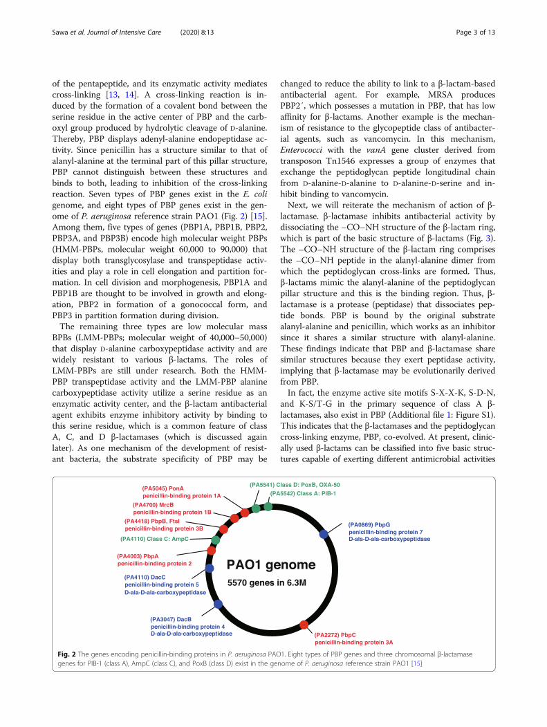
of the pentapeptide, and its enzymatic activity mediatescross-linking [13, 14]. A cross-linking reaction is in-duced by the formation of a covalent bond between theserine residue in the active center of PBP and the carb-oxyl group produced by hydrolytic cleavage of D-alanine.Thereby, PBP displays adenyl-alanine endopeptidase ac-tivity. Since penicillin has a structure similar to that ofalanyl-alanine at the terminal part of this pillar structure,PBP cannot distinguish between these structures andbinds to both, leading to inhibition of the cross-linkingreaction. Seven types of PBP genes exist in the E. coligenome, and eight types of PBP genes exist in the gen-ome of P. aeruginosa reference strain PAO1 (Fig. 2) [15].Among them, five types of genes (PBP1A, PBP1B, PBP2,PBP3A, and PBP3B) encode high molecular weight PBPs(HMM-PBPs, molecular weight 60,000 to 90,000) thatdisplay both transglycosylase and transpeptidase activ-ities and play a role in cell elongation and partition for-mation. In cell division and morphogenesis, PBP1A andPBP1B are thought to be involved in growth and elong-ation, PBP2 in formation of a gonococcal form, andPBP3 in partition formation during division.The remaining three types are low molecular mass
BPBs (LMM-PBPs; molecular weight of 40,000–50,000)that display D-alanine carboxypeptidase activity and arewidely resistant to various β-lactams. The roles ofLMM-PBPs are still under research. Both the HMM-PBP transpeptidase activity and the LMM-PBP alaninecarboxypeptidase activity utilize a serine residue as anenzymatic activity center, and the β-lactam antibacterialagent exhibits enzyme inhibitory activity by binding tothis serine residue, which is a common feature of classA, C, and D β-lactamases (which is discussed againlater). As one mechanism of the development of resist-ant bacteria, the substrate specificity of PBP may be
changed to reduce the ability to link to a β-lactam-basedantibacterial agent. For example, MRSA producesPBP2′, which possesses a mutation in PBP, that has lowaffinity for β-lactams. Another example is the mechan-ism of resistance to the glycopeptide class of antibacter-ial agents, such as vancomycin. In this mechanism,Enterococci with the vanA gene cluster derived fromtransposon Tn1546 expresses a group of enzymes thatexchange the peptidoglycan peptide longitudinal chainfrom D-alanine-D-alanine to D-alanine-D-serine and in-hibit binding to vancomycin.Next, we will reiterate the mechanism of action of β-
lactamase. β-lactamase inhibits antibacterial activity bydissociating the –CO–NH structure of the β-lactam ring,which is part of the basic structure of β-lactams (Fig. 3).The –CO–NH structure of the β-lactam ring comprisesthe –CO–NH peptide in the alanyl-alanine dimer fromwhich the peptidoglycan cross-links are formed. Thus,β-lactams mimic the alanyl-alanine of the peptidoglycanpillar structure and this is the binding region. Thus, β-lactamase is a protease (peptidase) that dissociates pep-tide bonds. PBP is bound by the original substratealanyl-alanine and penicillin, which works as an inhibitorsince it shares a similar structure with alanyl-alanine.These findings indicate that PBP and β-lactamase sharesimilar structures because they exert peptidase activity,implying that β-lactamase may be evolutionarily derivedfrom PBP.In fact, the enzyme active site motifs S-X-X-K, S-D-N,
and K-S/T-G in the primary sequence of class A β-lactamases, also exist in PBP (Additional file 1: Figure S1).This indicates that the β-lactamases and the peptidoglycancross-linking enzyme, PBP, co-evolved. At present, clinic-ally used β-lactams can be classified into five basic struc-tures capable of exerting different antimicrobial activities
Fig. 2 The genes encoding penicillin-binding proteins in P. aeruginosa PAO1. Eight types of PBP genes and three chromosomal β-lactamasegenes for PIB-1 (class A), AmpC (class C), and PoxB (class D) exist in the genome of P. aeruginosa reference strain PAO1 [15]
Sawa et al. Journal of Intensive Care (2020) 8:13 Page 3 of 13
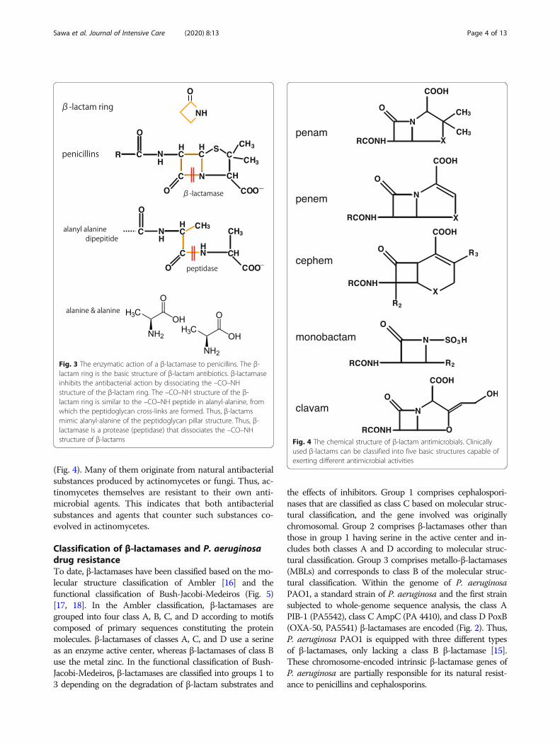
(Fig. 4). Many of them originate from natural antibacterialsubstances produced by actinomycetes or fungi. Thus, ac-tinomycetes themselves are resistant to their own anti-microbial agents. This indicates that both antibacterialsubstances and agents that counter such substances co-evolved in actinomycetes.
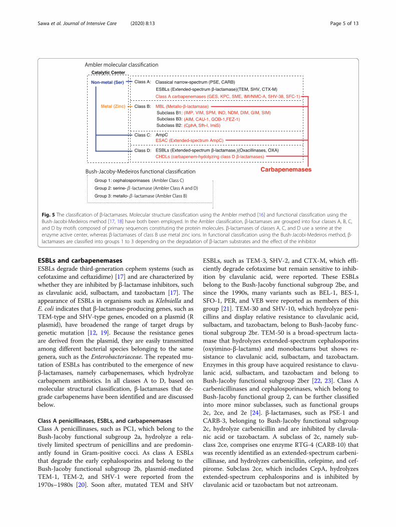
Classification of β-lactamases and P. aeruginosadrug resistanceTo date, β-lactamases have been classified based on the mo-lecular structure classification of Ambler [16] and thefunctional classification of Bush-Jacobi-Medeiros (Fig. 5)[17, 18]. In the Ambler classification, β-lactamases aregrouped into four class A, B, C, and D according to motifscomposed of primary sequences constituting the proteinmolecules. β-lactamases of classes A, C, and D use a serineas an enzyme active center, whereas β-lactamases of class Buse the metal zinc. In the functional classification of Bush-Jacobi-Medeiros, β-lactamases are classified into groups 1 to3 depending on the degradation of β-lactam substrates and
the effects of inhibitors. Group 1 comprises cephalospori-nases that are classified as class C based on molecular struc-tural classification, and the gene involved was originallychromosomal. Group 2 comprises β-lactamases other thanthose in group 1 having serine in the active center and in-cludes both classes A and D according to molecular struc-tural classification. Group 3 comprises metallo-β-lactamases(MBLs) and corresponds to class B of the molecular struc-tural classification. Within the genome of P. aeruginosaPAO1, a standard strain of P. aeruginosa and the first strainsubjected to whole-genome sequence analysis, the class APIB-1 (PA5542), class C AmpC (PA 4410), and class D PoxB(OXA-50, PA5541) β-lactamases are encoded (Fig. 2). Thus,P. aeruginosa PAO1 is equipped with three different typesof β-lactamases, only lacking a class B β-lactamase [15].These chromosome-encoded intrinsic β-lactamase genes ofP. aeruginosa are partially responsible for its natural resist-ance to penicillins and cephalosporins.
Fig. 3 The enzymatic action of a β-lactamase to penicillins. The β-lactam ring is the basic structure of β-lactam antibiotics. β-lactamaseinhibits the antibacterial action by dissociating the –CO–NHstructure of the β-lactam ring. The –CO–NH structure of the β-lactam ring is similar to the –CO–NH peptide in alanyl-alanine, fromwhich the peptidoglycan cross-links are formed. Thus, β-lactamsmimic alanyl-alanine of the peptidoglycan pillar structure. Thus, β-lactamase is a protease (peptidase) that dissociates the –CO–NHstructure of β-lactams Fig. 4 The chemical structure of β-lactam antimicrobials. Clinically
used β-lactams can be classified into five basic structures capable ofexerting different antimicrobial activities
Sawa et al. Journal of Intensive Care (2020) 8:13 Page 4 of 13
ESBLs and carbapenemasesESBLs degrade third-generation cephem systems (such ascefotaxime and ceftazidime) [17] and are characterized bywhether they are inhibited by β-lactamase inhibitors, suchas clavulanic acid, sulbactam, and tazobactam [17]. Theappearance of ESBLs in organisms such as Klebsiella andE. coli indicates that β-lactamase-producing genes, such asTEM-type and SHV-type genes, encoded on a plasmid (Rplasmid), have broadened the range of target drugs bygenetic mutation [12, 19]. Because the resistance genesare derived from the plasmid, they are easily transmittedamong different bacterial species belonging to the samegenera, such as the Enterobacteriaceae. The repeated mu-tation of ESBLs has contributed to the emergence of newβ-lactamases, namely carbapenemases, which hydrolyzecarbapenem antibiotics. In all classes A to D, based onmolecular structural classification, β-lactamases that de-grade carbapenems have been identified and are discussedbelow.
Class A penicillinases, ESBLs, and carbapenemasesClass A penicillinases, such as PC1, which belong to theBush-Jacoby functional subgroup 2a, hydrolyze a rela-tively limited spectrum of penicillins and are predomin-antly found in Gram-positive cocci. As class A ESBLsthat degrade the early cephalosporins and belong to theBush-Jacoby functional subgroup 2b, plasmid-mediatedTEM-1, TEM-2, and SHV-1 were reported from the1970s–1980s [20]. Soon after, mutated TEM and SHV
ESBLs, such as TEM-3, SHV-2, and CTX-M, which effi-ciently degrade cefotaxime but remain sensitive to inhib-ition by clavulanic acid, were reported. These ESBLsbelong to the Bush-Jacoby functional subgroup 2be, andsince the 1990s, many variants such as BEL-1, BES-1,SFO-1, PER, and VEB were reported as members of thisgroup [21]. TEM-30 and SHV-10, which hydrolyze peni-cillins and display relative resistance to clavulanic acid,sulbactam, and tazobactam, belong to Bush-Jacoby func-tional subgroup 2br. TEM-50 is a broad-spectrum lacta-mase that hydrolyzes extended-spectrum cephalosporins(oxyimino-β-lactams) and monobactams but shows re-sistance to clavulanic acid, sulbactam, and tazobactam.Enzymes in this group have acquired resistance to clavu-lanic acid, sulbactam, and tazobactam and belong toBush-Jacoby functional subgroup 2ber [22, 23]. Class Acarbenicillinases and cephalosporinases, which belong toBush-Jacoby functional group 2, can be further classifiedinto more minor subclasses, such as functional groups2c, 2ce, and 2e [24]. β-lactamases, such as PSE-1 andCARB-3, belonging to Bush-Jacoby functional subgroup2c, hydrolyze carbenicillin and are inhibited by clavula-nic acid or tazobactam. A subclass of 2c, namely sub-class 2ce, comprises one enzyme RTG-4 (CARB-10) thatwas recently identified as an extended-spectrum carbeni-cillinase, and hydrolyzes carbenicillin, cefepime, and cef-pirome. Subclass 2ce, which includes CepA, hydrolyzesextended-spectrum cephalosporins and is inhibited byclavulanic acid or tazobactam but not aztreonam.
Fig. 5 The classification of β-lactamases. Molecular structure classification using the Ambler method [16] and functional classification using theBush-Jacobi-Medeiros method [17, 18] have both been employed. In the Ambler classification, β-lactamases are grouped into four classes A, B, C,and D by motifs composed of primary sequences constituting the protein molecules. β-lactamases of classes A, C, and D use a serine at theenzyme active center, whereas β-lactamases of class B use metal zinc ions. In functional classification using the Bush-Jacobi-Medeiros method, β-lactamases are classified into groups 1 to 3 depending on the degradation of β-lactam substrates and the effect of the inhibitor
Sawa et al. Journal of Intensive Care (2020) 8:13 Page 5 of 13
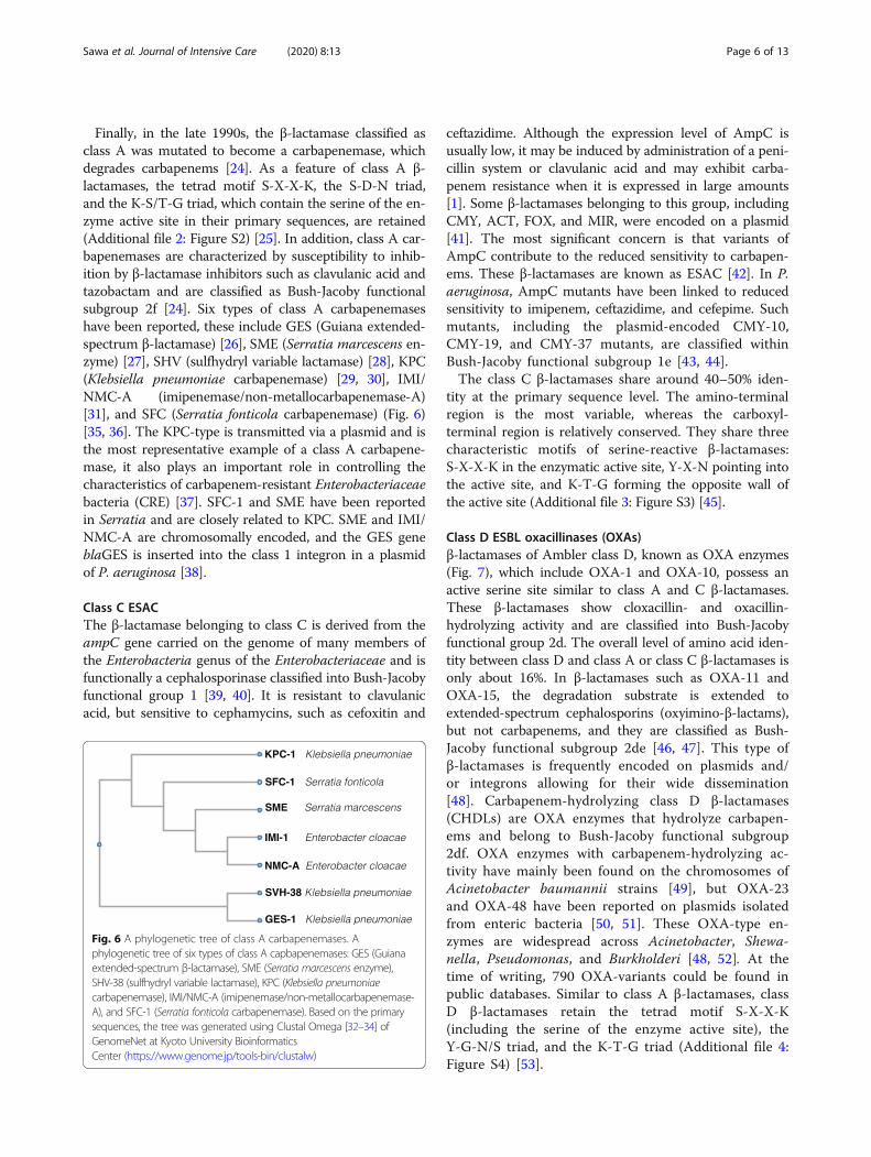
Finally, in the late 1990s, the β-lactamase classified asclass A was mutated to become a carbapenemase, whichdegrades carbapenems [24]. As a feature of class A β-lactamases, the tetrad motif S-X-X-K, the S-D-N triad,and the K-S/T-G triad, which contain the serine of the en-zyme active site in their primary sequences, are retained(Additional file 2: Figure S2) [25]. In addition, class A car-bapenemases are characterized by susceptibility to inhib-ition by β-lactamase inhibitors such as clavulanic acid andtazobactam and are classified as Bush-Jacoby functionalsubgroup 2f [24]. Six types of class A carbapenemaseshave been reported, these include GES (Guiana extended-spectrum β-lactamase) [26], SME (Serratia marcescens en-zyme) [27], SHV (sulfhydryl variable lactamase) [28], KPC(Klebsiella pneumoniae carbapenemase) [29, 30], IMI/NMC-A (imipenemase/non-metallocarbapenemase-A)[31], and SFC (Serratia fonticola carbapenemase) (Fig. 6)[35, 36]. The KPC-type is transmitted via a plasmid and isthe most representative example of a class A carbapene-mase, it also plays an important role in controlling thecharacteristics of carbapenem-resistant Enterobacteriaceaebacteria (CRE) [37]. SFC-1 and SME have been reportedin Serratia and are closely related to KPC. SME and IMI/NMC-A are chromosomally encoded, and the GES geneblaGES is inserted into the class 1 integron in a plasmidof P. aeruginosa [38].
Class C ESACThe β-lactamase belonging to class C is derived from theampC gene carried on the genome of many members ofthe Enterobacteria genus of the Enterobacteriaceae and isfunctionally a cephalosporinase classified into Bush-Jacobyfunctional group 1 [39, 40]. It is resistant to clavulanicacid, but sensitive to cephamycins, such as cefoxitin and
ceftazidime. Although the expression level of AmpC isusually low, it may be induced by administration of a peni-cillin system or clavulanic acid and may exhibit carba-penem resistance when it is expressed in large amounts[1]. Some β-lactamases belonging to this group, includingCMY, ACT, FOX, and MIR, were encoded on a plasmid[41]. The most significant concern is that variants ofAmpC contribute to the reduced sensitivity to carbapen-ems. These β-lactamases are known as ESAC [42]. In P.aeruginosa, AmpC mutants have been linked to reducedsensitivity to imipenem, ceftazidime, and cefepime. Suchmutants, including the plasmid-encoded CMY-10,CMY-19, and CMY-37 mutants, are classified withinBush-Jacoby functional subgroup 1e [43, 44].The class C β-lactamases share around 40–50% iden-
tity at the primary sequence level. The amino-terminalregion is the most variable, whereas the carboxyl-terminal region is relatively conserved. They share threecharacteristic motifs of serine-reactive β-lactamases:S-X-X-K in the enzymatic active site, Y-X-N pointing intothe active site, and K-T-G forming the opposite wall ofthe active site (Additional file 3: Figure S3) [45].
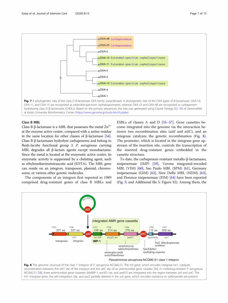
Class D ESBL oxacillinases (OXAs)β-lactamases of Ambler class D, known as OXA enzymes(Fig. 7), which include OXA-1 and OXA-10, possess anactive serine site similar to class A and C β-lactamases.These β-lactamases show cloxacillin- and oxacillin-hydrolyzing activity and are classified into Bush-Jacobyfunctional group 2d. The overall level of amino acid iden-tity between class D and class A or class C β-lactamases isonly about 16%. In β-lactamases such as OXA-11 andOXA-15, the degradation substrate is extended toextended-spectrum cephalosporins (oxyimino-β-lactams),but not carbapenems, and they are classified as Bush-Jacoby functional subgroup 2de [46, 47]. This type ofβ-lactamases is frequently encoded on plasmids and/or integrons allowing for their wide dissemination[48]. Carbapenem-hydrolyzing class D β-lactamases(CHDLs) are OXA enzymes that hydrolyze carbapen-ems and belong to Bush-Jacoby functional subgroup2df. OXA enzymes with carbapenem-hydrolyzing ac-tivity have mainly been found on the chromosomes ofAcinetobacter baumannii strains [49], but OXA-23and OXA-48 have been reported on plasmids isolatedfrom enteric bacteria [50, 51]. These OXA-type en-zymes are widespread across Acinetobacter, Shewa-nella, Pseudomonas, and Burkholderi [48, 52]. At thetime of writing, 790 OXA-variants could be found inpublic databases. Similar to class A β-lactamases, classD β-lactamases retain the tetrad motif S-X-X-K(including the serine of the enzyme active site), theY-G-N/S triad, and the K-T-G triad (Additional file 4:Figure S4) [53].
Fig. 6 A phylogenetic tree of class A carbapenemases. Aphylogenetic tree of six types of class A capbapenemases: GES (Guianaextended-spectrum β-lactamase), SME (Serratia marcescens enzyme),SHV-38 (sulfhydryl variable lactamase), KPC (Klebsiella pneumoniaecarbapenemase), IMI/NMC-A (imipenemase/non-metallocarbapenemase-A), and SFC-1 (Serratia fonticola carbapenemase). Based on the primarysequences, the tree was generated using Clustal Omega [32–34] ofGenomeNet at Kyoto University BioinformaticsCenter (https://www.genome.jp/tools-bin/clustalw)
Sawa et al. Journal of Intensive Care (2020) 8:13 Page 6 of 13
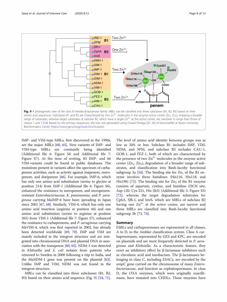
Class B MBLClass B β-lactamase is a MBL that possesses the metal Zn2+
at the enzyme active center, compared with a serine residuein the same location for other classes of β-lactamase [54].Class B β-lactamases hydrolyze carbapenems and belong toBush-Jacoby functional group 3. P. aeruginosa carryingMBL degrades all β-lactam agents except monobactams.Since the metal is located at the enzymatic active center, itsenzymatic activity is suppressed by a chelating agent, suchas ethylenediaminetetraacetic acid (EDTA). The MBL genecan reside on an integron, transposon, plasmid, chromo-some, or various other genetic molecules.The components of an integron first reported in 1989
comprised drug-resistant genes of class B MBLs and
ESBLs of classes A and D [55–57]. Gene cassettes be-come integrated into the genome via the interaction be-tween two recombination sites (attI and attC), and anintegrase catalyzes the genetic recombination (Fig. 8).The promoter, which is located in the integrase gene up-stream of the insertion site, controls the transcription ofthe inserted drug-resistant genes embedded in thecassette structure.To date, the carbapenem-resistant metallo-β-lactamases,
imipenemase (IMP) [59], Verona integrated-encodedMBL (VIM) [60], Sao Paulo MBL (SPM) [61], Germanyimipenemase (GIM) [62], New Delhi MBL (NDM) [63],and Florence imipenemase (FIM) [64] have been reported(Fig. 9, and Additional file 5: Figure S5). Among them, the
Fig. 7 A phylogenetic tree of the class D β-lactamase OXA family (oxiacillinase). A phylogenetic tree of the OXA types of β-lactamases. OXA-10,OXA-11, and OXA-15 are recognized as extended-spectrum cephalosporinases, whereas OXA-23 and OXA-48 are recognized as carbapenem-hydrolyzing class D β-lactamases (CHDLs). Based on the primary sequences, the tree was generated using Clustal Omega [32–34] of GenomeNetat Kyoto University Bioinformatics Center (https://www.genome.jp/tools-bin/clustalw)
Fig. 8 The genomic structure of the class 1 integron of P. aeruginosa NCGM2.S1. The intl gene, which encodes integrase Int1, catalyzesrecombination between the attl1 site of the integron and the attC site of an antimicrobial gene cassette [56]. In multidrug-resistant P. aeruginosaNCGM2.S1 [58], three antimicrobial gene cassettes (blaIMP-1, aac(6′)-1ae, and aadA1) are integrated into the region between attI and attC. TheInt1 integrase gene, the attI integration site, and qacE partially deleted in the sulI gene, which encodes resistance to sulfonamide are present
Sawa et al. Journal of Intensive Care (2020) 8:13 Page 7 of 13
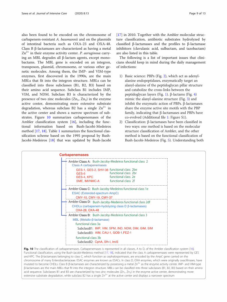
IMP- and VIM-type MBLs, first discovered in the 1990s,are the major MBLs [60, 65]. New variants of IMP- andVIM-type MBLs are constantly being identified(Additional file 6: Figure S6 and Additional file 7:Figure S7). At the time of writing, 83 IMP- and 66VIM-variants could be found in public databases. Themutations present in variants affect the spectrum of carba-penem activities, such as activity against imipenem, mero-penem, and doripenem [66]. For example, IMP-6, whichhas only one amino acid substitution (serine to glycine atposition 214) from IMP-1 (Additional file 6: Figure S6),enhanced the resistance to meropenem, and meropenem-resistant Enterobacteriaceae, K. pneumoniae, and P. aeru-ginosa carrying blaIMP-6 have been spreading in Japansince 2001 [67, 68]. Similarly, VIM-4, which has only oneamino acid insertion (arginine at position 44) and oneamino acid substitution (serine to arginine at position265) from VIM-1 (Additional file 7: Figure S7), enhancedthe resistance to carbapemems, and P. aeruginosa carryingblaVIM-4, which was first reported in 2002, has alreadybeen detected worldwide [69, 70]. IMP and VIM aremainly included in the integron structure and are inte-grated into chromosomal DNA and plasmid DNA in asso-ciation with the transposon [60, 65]. NDM-1 was detectedin Klebsiella and E. coli isolates from patients whoreturned to Sweden in 2008 following a trip to India, andthe blaNDM-1 gene was present on the plasmid [63].Unlike IMP and VIM, NDM was not found in theintegron structure.MBLs can be classified into three subclasses (B1, B2,
B3) based on their amino acid sequence (Fig. 9) [54, 71].
The level of amino acid identity between groups was aslow as 20% or less. Subclass B1 includes IMP, VIM,NDM, and SPM, and subclass B3 includes CAU-1,GOB-1, and FEZ-1, both of which are characterized bythe presence of two Zn2+ molecules in the enzyme activecenter (Zn1, Zn2), degradation of a broader range of sub-strates, and classification into Bush-Jacoby functionalsubgroup 3a [54]. The binding site for Zn1 of the B1 en-zyme involves three histidines (His116, His118, andHis196) [72]. The binding site for Zn2 of the B1 enzymeconsists of aspartate, cystine, and histidine (DCH site,Asp-120, Cys-221, His-263) (Additional file 5: Figure S5)[72], whereas the target degradation substrates forCphA, Sfh-I, and ImiS, which are MBLs of subclass B2having one Zn2+ at the active center, are narrow andthese MBLs are classified into Bush-Jacoby functionalsubgroup 3b [73, 74].
SummaryESBLs and carbapenemases are represented in all classes,A to D, in the Ambler classification system. Class A car-bapenemases, represented by GES and KPC, are encodedon plasmids and are most frequently detected in P. aeru-ginosa and Klebsiella. As a characteristic feature, theyexert an inhibitory effect by β-lactamase inhibitors suchas clavulanic acid and tazobactam. The β-lactamases be-longing to class C, including ESACs, are encoded by theampC gene carried on the chromosome of many Entero-bacteriaceae, and function as cephalosporinases. In classD, the OXA enzymes, which were originally oxacilli-nases, have mutated into CHDLs. These enzymes have
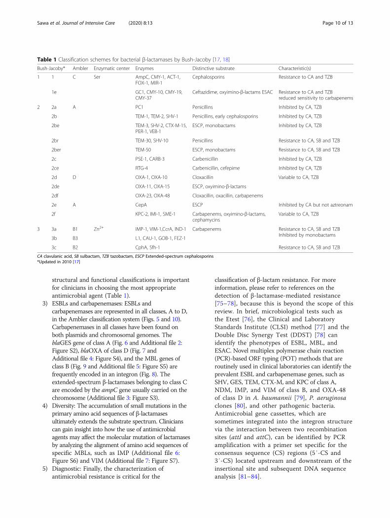
Fig. 9 A phylogenetic tree of the class B metallo-β-lactamase family. MBLs can be classified into three subclasses (B1, B2, B3) based on theiramino acid sequences. Subclasses B1 and B3 are characterized by two Zn2+ molecules in the enzyme active center (Zn1, Zn2), implying a broaderrange of substrates, whereas target substrates of subclass B2, which have a single Zn2+ at the active center, are narrower in range than those ofclasses 1 and 3 [54]. Based on the primary sequences, the tree was generated using Clustal Omega [32–34] of GenomeNet at Kyoto UniversityBioinformatics Center (https://www.genome.jp/tools-bin/clustalw)
Sawa et al. Journal of Intensive Care (2020) 8:13 Page 8 of 13
also been found to be encoded on the chromosome ofcarbapenem-resistant A. baumannii and on the plasmidsof intestinal bacteria such as OXA-23 and OXA-48.Class B β-lactamases are characterized as having a metalZn2+ in their enzyme activity center. P. aeruginosa carry-ing an MBL degrades all β-lactam agents, except mono-bactams. The MBL gene is encoded on an integron,transposon, plasmid, chromosome, or various other ge-netic molecules. Among them, the IMP- and VIM-typeenzymes, first discovered in the 1990s, are the mainMBLs that fit into the integron structure. MBLs can beclassified into three subclasses (B1, B2, B3) based ontheir amino acid sequence. Subclass B1 includes IMP,VIM, and NDM. Subclass B3 is characterized by thepresence of two zinc molecules (Zn1, Zn2) in the enzymeactive center, demonstrating more extensive substratedegradation, whereas subclass B2 has a single Zn2+ inthe active center and shows a narrow spectrum of sub-strates. Figure 10 summarizes carbapenemases of theAmbler classification system [16], including the func-tional information based on Bush-Jacobi-Medeirosmethod [17, 18]. Table 1 summarizes the functional clas-sification scheme based on the 1995 proposal by Bush-Jacobi-Medeiros [18] that was updated by Bush-Jacobi
[17] in 2010. Together with the Ambler molecular struc-ture classification, antibiotic substrates hydrolyzed byclassified β-lactamases and the profiles to β-lactamaseinhibitors (clavulanic acid, sulbactam, and tazobactam)are also listed in this table.The following is a list of important issues that clini-
cians should keep in mind during the daily managementof infections:
1) Basic science: PBPs (Fig. 2), which act as adenyl-alanine endopeptidases, enzymatically target analanyl-alanine of the peptidoglycan pillar structureand catabolize the cross-links between thepeptidoglycan layers (Fig. 1). β-lactams (Fig. 4)mimic the alanyl-alanine structure (Fig. 3) andinhibit the enzymatic action of PBPs. β-lactamasesshare the enzyme active site motifs with the PBPfamily, indicating that β-lactamases and PBPs haveco-evolved (Additional file 1: Figure S1).
2) Classification: β-lactamases have been classified intwo ways: one method is based on the molecularstructure classification of Ambler, and the othermethod is based on the functional classification ofBush-Jacobi-Medeiros (Fig. 5). Understanding both
Fig. 10 The classification of carbapenemases. Carbapenemases is represented in all classes, A to D, of the Ambler classification system [16].Functional classification, using the Bush-Jacobi-Medeiros method [17, 18], indicated that the class A carbapenemases were represented by GESand KPC. The β-lactamases belonging to class C, which function as cephalosporinases, are encoded by the AmpC gene carried on thechromosome of many Enterobacteriaceae. ESAC enzymes are known as ESACs. In class D, OXA enzymes, which were originally oxacillinases, havemutated to become CHDLs. Class B β-lactamases are characterized by possessing a metal Zn2+ as the enzyme activity center. IMP- and VIM-typeβ-lactamases are the main MBLs that fit into the integron structure. MBLs can be classified into three subclasses (B1, B2, B3) based on their aminoacid sequence. Subclasses B1 and B3 are characterized by two zinc molecules (Zn1, Zn2) in the enzyme active center, demonstrating moreextensive substrate degradation, while subclass B2 has a single Zn2+ at the active center and displays a narrower spectrum
Sawa et al. Journal of Intensive Care (2020) 8:13 Page 9 of 13
structural and functional classifications is importantfor clinicians in choosing the most appropriateantimicrobial agent (Table 1).
3) ESBLs and carbapenemases: ESBLs andcarbapenemases are represented in all classes, A to D,in the Ambler classification system (Figs. 5 and 10).Carbapenemases in all classes have been found onboth plasmids and chromosomal genomes. TheblaGES gene of class A (Fig. 6 and Additional file 2:Figure S2), blaOXA of class D (Fig. 7 andAdditional file 4: Figure S4), and the MBL genes ofclass B (Fig. 9 and Additional file 5: Figure S5) arefrequently encoded in an integron (Fig. 8). Theextended-spectrum β-lactamases belonging to class Care encoded by the ampC gene usually carried on thechromosome (Additional file 3: Figure S3).
4) Diversity: The accumulation of small mutations in theprimary amino acid sequences of β-lactamasesultimately extends the substrate spectrum. Clinicianscan gain insight into how the use of antimicrobialagents may affect the molecular mutation of lactamasesby analyzing the alignment of amino acid sequences ofspecific MBLs, such as IMP (Additional file 6:Figure S6) and VIM (Additional file 7: Figure S7).
5) Diagnostic: Finally, the characterization ofantimicrobial resistance is critical for the
classification of β-lactam resistance. For moreinformation, please refer to references on thedetection of β-lactamase-mediated resistance[75–78], because this is beyond the scope of thisreview. In brief, microbiological tests such asthe Etest [76], the Clinical and LaboratoryStandards Institute (CLSI) method [77] and theDouble Disc Synergy Test (DDST) [78] canidentify the phenotypes of ESBL, MBL, andESAC. Novel multiplex polymerase chain reaction(PCR)-based ORF typing (POT) methods that areroutinely used in clinical laboratories can identify theprevalent ESBL and carbapenemase genes, such asSHV, GES, TEM, CTX-M, and KPC of class A,NDM, IMP, and VIM of class B, and OXA-48of class D in A. baumannii [79], P. aeruginosaclones [80], and other pathogenic bacteria.Antimicrobial gene cassettes, which aresometimes integrated into the integron structurevia the interaction between two recombinationsites (attI and attC), can be identified by PCRamplification with a primer set specific for theconsensus sequence (CS) regions (5′-CS and3′-CS) located upstream and downstream of theinsertional site and subsequent DNA sequenceanalysis [81–84].
Table 1 Classification schemes for bacterial β-lactamases by Bush-Jacoby [17, 18]
Bush-Jacoby* Ambler Enzymatic center Enzymes Distinctive substrate Characteristic(s)
1 1 C Ser AmpC, CMY-1, ACT-1,FOX-1, MIR-1
Cephalosporins Resistance to CA and TZB
1e GC1, CMY-10, CMY-19,CMY-37
Ceftazidime, oxyimino-β-lactams ESAC Resistance to CA and TZBreduced sensitivity to carbapenems
2 2a A PC1 Penicillins Inhibited by CA, TZB
2b TEM-1, TEM-2, SHV-1 Penicillins, early cephalosporins Inhibited by CA, TZB
2be TEM-3, SHV-2, CTX-M-15,PER-1, VEB-1
ESCP, monobactams Inhibited by CA, TZB
2br TEM-30, SHV-10 Penicillins Resistance to CA, SB and TZB
2ber TEM-50 ESCP, monobactams Resistance to CA, SB and TZB
2c PSE-1, CARB-3 Carbenicillin Inhibited by CA, TZB
2ce RTG-4 Carbenicillin, cefepime Inhibited by CA, TZB
2d D OXA-1, OXA-10 Cloxacillin Variable to CA, TZB
2de OXA-11, OXA-15 ESCP, oxyimino-β-lactams
2df OXA-23, OXA-48 Cloxacillin, oxacillin, carbapenems
2e A CepA ESCP Inhibited by CA but not aztreonam
2f KPC-2, IMI-1, SME-1 Carbapenems, oxyimino-β-lactams,cephamycins
Variable to CA, TZB
3 3a B1 Zn2+ IMP-1, VIM-1,CcrA, IND-1 Carbapenems Resistance to CA, SB and TZBInhibited by monobactams
3b B3 L1, CAU-1, GOB-1, FEZ-1
3c B2 CphA, Sfh-1 Resistance to CA, SB and TZB
CA clavulanic acid, SB sulbactam, TZB tazobactam, ESCP Extended-spectrum cephalosporins*Updated in 2010 [17]
Sawa et al. Journal of Intensive Care (2020) 8:13 Page 10 of 13
ConclusionVarious ESBLs and carbapenemases belonging to one offour molecular classes have propagated and are beingdetected among bacteria worldwide, suggesting thatcareful detection and monitoring are vital when criticalcare clinicians are treating infections caused by ESBL-and carbapenem-resistant bacteria. For the classificationof β-lactamases, the Ambler method of molecularstructure classification [16] is simple and effective atorganizing the various ESBLs and carbapenemases, butfunctional classification using the Bush-Jacobi-Medeirosmethod [17, 18] is also important for clinicians facedwith treating patients in a critical condition due toESBL- and carbapenem-resistant bacterial infections.
Supplementary informationSupplementary information accompanies this paper at https://doi.org/10.1186/s40560-020-0429-6.
Additional file 1: Figure S1. The similarities in the structures of class Aβ-lactamases and penicillin-binding protein (PBP).
Additional file 2: Figure S2. The alignment of class A carbapenemasesbased on their primary sequences.
Additional file 3: Figure S3. The alignment of class C β-lactamases.
Additional file 4: Figure S4. Alignment of the class D β-lactamase OXAfamily (oxacillinase).
Additional file 5: Figure S5. Alignment of the class B metallo-β-lactamase family and their zinc enzymatic active center.
Additional file 6: Figure S6. The alignment of primary sequences ofthe class B metallo-β-lactamase IMP family.
Additional file 7: Figure S7. The alignment of primary sequences ofthe class B metallo-β-lactamase VIM family.
AbbreviationsCHDL: Carbapenem-hydrolyzing class D β-lactamases; CLSI: Clinical andLaboratory Standards Institute; CPE: Carbapenemase-producingEnterobacteriaceae; CS: Consensus sequence; DDST: Double disc synergy test;EDTA: Ethylenediaminetetraacetic acid; ESAC: Extended-spectrum AmpC;ESBL: Extended-spectrum β-lactamase; FIM: Florence imipenemase;GES: Guiana extended-spectrum β-lactamase; GIM: Germany imipenemase;HMW-PBP: High molecular weight penicillin-binding protein; IMI/NMC-A: Imipenemase/non-metallocarbapenemase-A; IMP: Imipenemase;KPC: Klebsiella pneumoniae carbapenemase; LMW-PBP: High molecularweight penicillin-binding protein; MBL: Metallo-β-lactamase;MDRP: Multidrug-resistant Pseudomonas aeruginosa; MRSA: Methicillin-resistant Staphylococcus aureus; NAG: N-acetylglucosamine; NAM: N-acetylmuramic acid; NDM: New Delhi metallo-β-lactamase; PBP: Penicillin-binding protein; PCR: Polymerase chain reaction; PDRP: Pandrug-resistantPseudomonas aeruginosa; POT: PCR-based ORF typing; SFC: Serratia fonticolacarbapenemase; SHV: Sulfhydryl variable lactamase; SME: Serratia marcescensenzyme; SPM: Sao Paulo metallo-β-lactamase; VIM: Verona integrated-encoded MBL; XDRP: Extensively drug-resistant Pseudomonas aeruginosa
AcknowledgementsWe thank Kate Fox, DPhil, from Edanz Group (www.edanzediting.com/ac) forediting a draft of this manuscript.
Authors’ contributionsTS wrote the manuscript, figure legends, and tables. KK and KM contributedto preparing and revising the manuscript. All authors have read andapproved the final manuscript.
Authors’ informationTS is a Professor of the Anesthesiology Department at Kyoto PrefecturalUniversity of Medicine, Japan. KK is a Chief Manager in the Division of CriticalCare, Kyoto City Hospital, and a Visiting Professor of the AnesthesiologyDepartment at Kyoto Prefectural University of Medicine, Japan. KM is anAssociate Professor in the Anesthesiology Department at Kyorin University,Japan.
FundingThis work was supported by the Japan Society for the Promotion of Science anda Grant-in-Aid for Scientific Research (KAKENHI) to Teiji Sawa (No. 24390403, No.15H05008, and No. 18H02905) from The Ministry of Education, Culture, Sports,Science and Technology, Japan.
Availability of data and materialsAll data generated or analyzed during this study are included in thispublished article and its supplementary information files.
Ethics approval and consent to participateNot applicable.
Consent for publicationNot applicable.
Competing interestsTS has a patent for immunization with PcrV from the Regent of theUniversity of California (Berkeley, CA, USA). All other authors declare that theyhave no competing interests.
Author details1Department of Anesthesiology, School of Medicine, Kyoto PrefecturalUniversity of Medicine, 465 Kajii-cho, Kamigyo, Kyoto 602-8566, Japan.2Department of Intensive Care, Kyoto City Hospital, 1-2 Higashitakada-cho,Mibu, Nakagyo, Kyoto 604-8845, Japan. 3Department of Anesthesiology,School of Medicine, Kyorin University, 6-20-2 Shinkawa, Mitaka, Tokyo181-8611, Japan.
Received: 16 December 2019 Accepted: 13 January 2020
References1. Codjoe FS, Donkor ES. Carbapenem resistance: a review. Med Sci (Basel).
2017;6. https://doi.org/10.3390/medsci6010001.2. Papp-Wallace KM, Endimiani A, Taracila MA, Bonomo RA. Carbapenems:
past, present, and future. Antimicrob Agents Chemother. 2011;55:4943–60.https://doi.org/10.1128/AAC.00296-11.
3. Bonomo RA, Burd EM, Conly J, Limbago BM, Poirel L, Segre JA, WestbladeLF. Carbapenemase-producing organisms: a global scourge. Clin Infect Dis.2018;66:1290–7. https://doi.org/10.1093/cid/cix893.
4. van Duin D, Doi Y. The global epidemiology of carbapenemase-producingEnterobacteriaceae. Virulence. 2017;8:460–9. https://doi.org/10.1080/21505594.2016.1222343.
5. Doi Y, Paterson DL. Carbapenemase-producing Enterobacteriaceae. SeminRespir Crit Care Med. 2015;36:74–84. https://doi.org/10.1055/s-0035-1544208.
6. van Loon K, Voor In ’t Holt AF, Vos MC. A systematic review and meta-analyses of the clinical epidemiology of carbapenem-resistantEnterobacteriaceae. Antimicrob Agents Chemother. 2017;62:e01730–17.https://doi.org/10.1128/AAC.01730-17.
7. Sheu CC, Chang YT, Lin SY, Chen YH, Hsueh PR. Infections caused bycarbapenem-resistant Enterobacteriaceae: an update on therapeutic options.Front Microbiol. 2019;10:80. https://doi.org/10.3389/fmicb.2019.00080.
8. Lee CS, Doi Y. Therapy of infections due to carbapenem-resistant gram-negative pathogens. Infect Chemother. 2014;46:149–64. https://doi.org/10.3947/ic.2014.46.3.149.
9. Aloush V, Navon-Venezia S, Seigman-Igra Y, Cabili S, Carmeli Y. Multidrug-resistant Pseudomonas aeruginosa: risk factors and clinical impact.Antimicrob Agents Chemother. 2006;50:43–8. https://doi.org/10.1128/AAC.50.1.43-48.2006.
10. Magiorakos AP, Srinivasan A, Carey RB, Carmeli Y, Falagas ME, Giske CG,Harbarth S, Hindler JF, Kahlmeter G, Olsson-Liljequist B, Paterson DL, Rice LB,Stelling J, Struelens MJ, Vatopoulos A, Weber JT, Monnet DL. Multidrug-
Sawa et al. Journal of Intensive Care (2020) 8:13 Page 11 of 13
resistant, extensively drug-resistant and pandrug-resistant bacteria: aninternational expert proposal for interim standard definitions for acquiredresistance. Clin Microbiol Infect. 2012;18:268–81. https://doi.org/10.1111/j.1469-0691.2011.03570.x.
11. Falagas ME, Koletsi PK, Bliziotis IA. The diversity of definitions of multidrug-resistant (MDR) and pandrug-resistant (PDR) Acinetobacter baumannii andPseudomonas aeruginosa. J Med Microbiol. 2006;55:1619–29. https://doi.org/10.1099/jmm.0.46747-0.
12. Bush K. Past and present perspectives on beta-lactamases. AntimicrobAgents Chemother. 2018;62:e01076–18. https://doi.org/10.1128/AAC.01076-18.
13. Ghuysen JM. Serine beta-lactamases and penicillin-binding proteins. AnnuRev Microbiol. 1991;45:37–67. https://doi.org/10.1146/annurev.mi.45.100191.000345.
14. Jamin M, Wilkin JM, Frère JM. Bacterial DD-transpeptidases and penicillin.Essays Biochem. 1995;29:1–24.
15. Stover CK, Pham XQ, Erwin AL, Mizoguchi SD, Warrener P, Hickey MJ,Brinkman FS, Hufnagle WO, Kowalik DJ, Lagrou M, Garber RL, Goltry L,Tolentino E, Westbrock-Wadman S, Yuan Y, Brody LL, Coulter SN, Folger KR,Kas A, Larbig K, Lim R, Smith K, Spencer D, Wong GK, Wu Z, Paulsen IT,Reizer J, Saier MH, Hancock RE, Lory S, Olson MV. Complete genomesequence of Pseudomonas aeruginosa PAO1, an opportunistic pathogen.Nature. 2000;406:959–64. https://doi.org/10.1038/35023079.
16. Ambler RP. The structure of beta-lactamases. Philos Trans R Soc Lond Ser BBiol Sci. 1980;289:321–31. https://doi.org/10.1098/rstb.1980.0049.
17. Bush K, Jacoby GA. Updated functional classification of beta-lactamases.Antimicrob Agents Chemother. 2010;54:969–76. https://doi.org/10.1128/AAC.01009-09.
18. Bush K, Jacoby GA, Medeiros AA. A functional classification scheme forbeta-lactamases and its correlation with molecular structure. AntimicrobAgents Chemother. 1995;39:1211–33. https://doi.org/10.1128/aac.39.6.1211.
19. Liakopoulos A, Mevius D, Ceccarelli D. A review of SHV extended-spectrumbeta-lactamases: neglected yet ubiquitous. Front Microbiol. 2016;7:1374.https://doi.org/10.3389/fmicb.2016.01374.
20. Sirot D. Extended-spectrum plasmid-mediated beta-lactamases. JAntimicrob Chemother. 1995;36(Suppl A):19–34. https://doi.org/10.1093/jac/36.suppl_a.19.
21. Bradford PA. Extended-spectrum beta-lactamases in the 21st century:characterization, epidemiology, and detection of this important resistancethreat. Clin Microbiol Rev. 2001;14:933–51. https://doi.org/10.1128/CMR.14.4.933-951.2001.
22. Malloy AM, Campos JM. Extended-spectrum beta-lactamases: a brief clinicalupdate. Pediatr Infect Dis J. 2011;30:1092–3. https://doi.org/10.1097/INF.0b013e31823c0e9d.
23. Paterson DL, Bonomo RA. Extended-spectrum beta-lactamases: a clinicalupdate. Clin Microbiol Rev. 2005;18:657–86. https://doi.org/10.1128/CMR.18.4.657-686.2005.
24. Walther-Rasmussen J, Hoiby N. Class A carbapenemases. J AntimicrobChemother. 2007;60:470–82. https://doi.org/10.1093/jac/dkm226.
25. Philippon A, Slama P, Deny P, Labia R. A structure-based classification ofclass A beta-lactamases, a broadly diverse family of enzymes. Clin MicrobiolRev. 2016;29:29–57. https://doi.org/10.1128/CMR.00019-15.
26. Poirel L, Le Thomas I, Naas T, Karim A, Nordmann P. Biochemical sequenceanalyses of GES-1, a novel class A extended-spectrum beta-lactamase, andthe class 1 integron In52 from Klebsiella pneumoniae. Antimicrob AgentsChemother. 2000;44:622–32. https://doi.org/10.1128/aac.44.3.622-632.2000.
27. Naas T, Vandel L, Sougakoff W, Livermore DM, Nordmann P. Cloning andsequence analysis of the gene for a carbapenem-hydrolyzing class A beta-lactamase, SME-1, from Serratia marcescens S6. Antimicrob AgentsChemother. 1994;38:1262–70. https://doi.org/10.1128/aac.38.6.1262.
28. Barthélémy M, Peduzzi J, Verchère-Beaur C, Ben Yaghlane H, Labia R.Purification and biochemical properties of Pitton’s type 2 beta-lactamase(SHV-1). Ann Inst Pasteur Microbiol (1985). 1986;137B:19–27. https://doi.org/10.1016/s0769-2609(86)80090-4.
29. Yigit H, Queenan AM, Anderson GJ, Domenech-Sanchez A, Biddle JW,Steward CD, Alberti S, Bush K, Tenover FC. Novel carbapenem-hydrolyzingbeta-lactamase, KPC-1, from a carbapenem-resistant strain of Klebsiellapneumoniae. Antimicrob Agents Chemother. 2001;45:1151–61. https://doi.org/10.1128/AAC.45.4.1151-1161.2001.
30. Arnold RS, Thom KA, Sharma S, Phillips M, Kristie Johnson J, Morgan DJ.Emergence of Klebsiella pneumoniae carbapenemase-producing bacteria.South Med J. 2011;104:40–5. https://doi.org/10.1097/SMJ.0b013e3181fd7d5a.
31. Nordmann P, Mariotte S, Naas T, Labia R, Nicolas MH. Biochemicalproperties of a carbapenem-hydrolyzing beta-lactamase from Enterobactercloacae and cloning of the gene into Escherichia coli. Antimicrob AgentsChemother. 1993;37:939–46. https://doi.org/10.1128/aac.37.5.939.
32. Sievers F, Higgins DG. Clustal Omega for making accurate alignments ofmany protein sequences. Protein Sci. 2018;27:135–45. https://doi.org/10.1002/pro.3290.
33. Sievers F, Higgins DG. Clustal Omega. Curr Protoc Bioinformatics. 2014;48:3.13.1–16. https://doi.org/10.1002/0471250953.bi0313s48.
34. Sievers F, Wilm A, Dineen D, Gibson TJ, Karplus K, Li W, Lopez R, McWilliamH, Remmert M, Soding J, Thompson JD, Higgins DG. Fast, scalablegeneration of high-quality protein multiple sequence alignments usingClustal Omega. Mol Syst Biol. 2011;7:539. https://doi.org/10.1038/msb.2011.75.
35. Henriques I, Moura A, Alves A, Saavedra MJ, Correia A. Molecularcharacterization of a carbapenem-hydrolyzing class A beta-lactamase, SFC-1,from Serratia fonticola UTAD54. Antimicrob Agents Chemother. 2004;48:2321–4. https://doi.org/10.1128/AAC.48.6.2321-2324.2004.
36. Naas T, Dortet L, Iorga BI. Structural and functional aspects of class Acarbapenemases. Curr Drug Targets. 2016;17:1006–28. https://doi.org/10.2174/1389450117666160310144501.
37. Logan LK, Weinstein RA. The epidemiology of carbapenem-resistantEnterobacteriaceae: the impact and evolution of a global menace. J InfectDis. 2017;215:S28–36. https://doi.org/10.1093/infdis/jiw282.
38. Poirel L, Weldhagen GF, Naas T, De Champs C, Dove MG, Nordmann P. GES-2, a class A beta-lactamase from Pseudomonas aeruginosa with increasedhydrolysis of imipenem. Antimicrob Agents Chemother. 2001;45:2598–603.
39. Jacoby GA. AmpC beta-lactamases. Clin Microbiol Rev. 2009;22:161–82.https://doi.org/10.1128/aac.45.9.2598-2603.2001.
40. Thomson KS. Extended-spectrum-beta-lactamase, AmpC, andcarbapenemase issues. J Clin Microbiol. 2010;48:1019–25. https://doi.org/10.1128/JCM.00219-10.
41. Philippon A, Arlet G, Jacoby GA. Plasmid-determined AmpC-type beta-lactamases. Antimicrob Agents Chemother. 2002;46:1–11. https://doi.org/10.1128/aac.46.1.1-11.2002.
42. Nordmann P, Mammeri H. Extended-spectrum cephalosporinases: structure,detection and epidemiology. Future Microbiol. 2007;2:297–307. https://doi.org/10.2217/17460913.2.3.297.
43. Ahmed AM, Shimamoto T. Emergence of a cefepime- and cefpirome-resistant Citrobacter freundii clinical isolate harbouring a novelchromosomally encoded AmpC beta-lactamase, CMY-37. Int J AntimicrobAgents. 2008;32:256–61. https://doi.org/10.1016/j.ijantimicag.2008.04.019.
44. Wachino J, Kurokawa H, Suzuki S, Yamane K, Shibata N, Kimura K, Ike Y,Arakawa Y. Horizontal transfer of blaCMY-bearing plasmids among clinicalEscherichia coli and Klebsiella pneumoniae isolates and emergence ofcefepime-hydrolyzing CMY-19. Antimicrob Agents Chemother. 2006;50:534–41. https://doi.org/10.1128/AAC.50.2.534-541.2006.
45. Michaux C, Massant J, Kerff F, Frere JM, Docquier JD, Vandenberghe I,Samyn B, Pierrard A, Feller G, Charlier P, Van Beeumen J, Wouters J. Crystalstructure of a cold-adapted class C beta-lactamase. FEBS J. 2008;275:1687–97. https://doi.org/10.1111/j.1742-4658.2008.06324.x.
46. Hall LM, Livermore DM, Gur D, Akova M, Akalin HE. OXA-11, an extended-spectrum variant of OXA-10 (PSE-2) beta-lactamase from Pseudomonasaeruginosa. Antimicrob Agents Chemother. 1993;37:1637–44. https://doi.org/10.1128/aac.37.8.1637.
47. Danel F, Hall LM, Gur D, Livermore DM. OXA-15, an extended-spectrumvariant of OXA-2 beta-lactamase, isolated from a Pseudomonas aeruginosastrain. Antimicrob Agents Chemother. 1997;41:785–90.
48. Antunes NT, Fisher JF. Acquired class D beta-lactamases. Antibiotics (Basel).2014;3:398–434. https://doi.org/10.3390/antibiotics3030398.
49. Zander E, Fernandez-Gonzalez A, Schleicher X, Dammhayn C, Kamolvit W,Seifert H, Higgins PG. Worldwide dissemination of acquired carbapenem-hydrolysing class D beta-lactamases in Acinetobacter spp. other thanAcinetobacter baumannii. Int J Antimicrob Agents. 2014;43:375–7. https://doi.org/10.1016/j.ijantimicag.2014.01.012.
50. Donald HM, Scaife W, Amyes SG, Young HK. Sequence analysis of ARI-1,a novel OXA beta-lactamase, responsible for imipenem resistance inAcinetobacter baumannii 6B92. Antimicrob Agents Chemother.2000;44:196–9.
51. Poirel L, Heritier C, Tolun V, Nordmann P. Emergence of oxacillinase-mediated resistance to imipenem in Klebsiella pneumoniae. Antimicrob
Sawa et al. Journal of Intensive Care (2020) 8:13 Page 12 of 13
Agents Chemother. 2004;48:15–22. https://doi.org/10.1128/aac.48.1.15-22.2004.
52. Walther-Rasmussen J, Hoiby N. OXA-type carbapenemases. J AntimicrobChemother. 2006;57:373–83. https://doi.org/10.1093/jac/dki482.
53. Paetzel M, Danel F, de Castro L, Mosimann SC, Page MG, Strynadka NC.Crystal structure of the class D beta-lactamase OXA-10. Nat Struct Biol. 2000;7:918–25. https://doi.org/10.1038/79688.
54. Palzkill T. Metallo-beta-lactamase structure and function. Ann N Y Acad Sci.2013;1277:91–104. https://doi.org/10.1111/j.1749-6632.2012.06796.x.
55. Stokes HW, Hall RM. A novel family of potentially mobile DNA elementsencoding site-specific gene-integration functions: integrons. Mol Microbiol.1989;3:1669–83. https://doi.org/10.1111/j.1365-2958.1989.tb00153.x.
56. Gillings MR. Integrons: past, present, and future. Microbiol Mol Biol Rev.2014;78:257–77. https://doi.org/10.1128/MMBR.00056-13.
57. Deng Y, Bao X, Ji L, Chen L, Liu J, Miao J, Chen D, Bian H, Li Y, Yu G.Resistance integrons: class 1, 2 and 3 integrons. Ann Clin MicrobiolAntimicrob. 2015;14:45. https://doi.org/10.1186/s12941-015-0100-6.
58. Miyoshi-Akiyama T, Kuwahara T, Tada T, Kitao T, Kirikae T. Complete genomesequence of highly multidrug-resistant Pseudomonas aeruginosa NCGM2.S1,a representative strain of a cluster endemic to Japan. J Bacteriol. 2011;193:7010. https://doi.org/10.1128/JB.06312-11.
59. Arakawa Y, Murakami M, Suzuki K, Ito H, Wacharotayankun R, Ohsuka S, KatoN, Ohta M. A novel integron-like element carrying the metallo-beta-lactamase gene blaIMP. Antimicrob Agents Chemother. 1995;39:1612–5.https://doi.org/10.1128/aac.39.7.1612.
60. Lauretti L, Riccio ML, Mazzariol A, Cornaglia G, Amicosante G, Fontana R,Rossolini GM. Cloning and characterization of blaVIM, a new integron-bornemetallo-beta-lactamase gene from a Pseudomonas aeruginosa clinicalisolate. Antimicrob Agents Chemother. 1999;43:1584–90.
61. Toleman MA, Simm AM, Murphy TA, Gales AC, Biedenbach DJ, Jones RN,Walsh TR. Molecular characterization of SPM-1, a novel metallo-beta-lactamase isolated in Latin America: report from the SENTRY antimicrobialsurveillance programme. J Antimicrob Chemother. 2002;50:673–9.https://doi.org/10.1093/jac/dkf210.
62. Castanheira M, Toleman MA, Jones RN, Schmidt FJ, Walsh TR. Molecularcharacterization of a beta-lactamase gene, blaGIM-1, encoding a newsubclass of metallo-beta-lactamase. Antimicrob Agents Chemother. 2004;48:4654–61. https://doi.org/10.1128/AAC.48.12.4654-4661.2004.
63. Yong D, Toleman MA, Giske CG, Cho HS, Sundman K, Lee K, Walsh TR.Characterization of a new metallo-beta-lactamase gene, bla(NDM-1), and anovel erythromycin esterase gene carried on a unique genetic structure inKlebsiella pneumoniae sequence type 14 from India. Antimicrob AgentsChemother. 2009;53:5046–54. https://doi.org/10.1128/AAC.00774-09.
64. Pollini S, Maradei S, Pecile P, Olivo G, Luzzaro F, Docquier JD, Rossolini GM.FIM-1, a new acquired metallo-beta-lactamase from a Pseudomonasaeruginosa clinical isolate from Italy. Antimicrob Agents Chemother. 2013;57:410–6. https://doi.org/10.1128/AAC.01953-12.
65. Laraki N, Galleni M, Thamm I, Riccio ML, Amicosante G, Frere JM, RossoliniGM. Structure of In31, a blaIMP-containing Pseudomonas aeruginosaintegron phyletically related to In5, which carries an unusual array of genecassettes. Antimicrob Agents Chemother. 1999;43:890–901.
66. Hong DJ, Bae IK, Jang IH, Jeong SH, Kang HK, Lee K. Epidemiology andcharacteristics of metallo-beta-lactamase-producing Pseudomonasaeruginosa. Infect Chemother. 2015;47:81–97. https://doi.org/10.3947/ic.2015.47.2.81.
67. Yano H, Kuga A, Okamoto R, Kitasato H, Kobayashi T, Inoue M. Plasmid-encoded metallo-beta-lactamase (IMP-6) conferring resistance tocarbapenems, especially meropenem. Antimicrob Agents Chemother. 2001;45:1343–8. https://doi.org/10.1128/AAC.45.5.1343-1348.2001.
68. Ohno Y, Nakamura A, Hashimoto E, Matsutani H, Abe N, Fukuda S, Hisashi K,Komatsu M, Nakamura F. Molecular epidemiology of carbapenemase-producing Enterobacteriaceae in a primary care hospital in Japan, 2010-2013. JInfect Chemother. 2017;23:224–9. https://doi.org/10.1016/j.jiac.2016.12.013.
69. Pournaras S, Tsakris A, Maniati M, Tzouvelekis LS, Maniatis AN. Novel variant(bla(VIM-4)) of the metallo-beta-lactamase gene bla(VIM-1) in a clinical strainof Pseudomonas aeruginosa. Antimicrob Agents Chemother. 2002;46:4026–8.https://doi.org/10.1128/aac.46.12.4026-4028.2002.
70. Karampatakis T, Antachopoulos C, Tsakris A, Roilides E. Molecularepidemiology of carbapenem-resistant Pseudomonas aeruginosa in anendemic area: comparison with global data. Eur J Clin Microbiol Infect Dis.2018;37:1211–20. https://doi.org/10.1007/s10096-018-3244-4.
71. Rasmussen BA, Bush K. Carbapenem-hydrolyzing beta-lactamases.Antimicrob Agents Chemother. 1997;41:223–32.
72. Cadag E, Vitalis E, Lennox KP, Zhou CL, Zemla AT. Computational analysis ofpathogen-borne metallo beta-lactamases reveals discriminating structuralfeatures between B1 types. BMC Res Notes. 2012;5:96. https://doi.org/10.1186/1756-0500-5-96.
73. Fonseca F, Bromley EH, Saavedra MJ, Correia A, Spencer J. Crystal structureof Serratia fonticola Sfh-I: activation of the nucleophile in mono-zincmetallo-beta-lactamases. J Mol Biol. 2011;411:951–9. https://doi.org/10.1016/j.jmb.2011.06.043.
74. Garau G, Bebrone C, Anne C, Galleni M, Frere JM, Dideberg O. A metallo-beta-lactamase enzyme in action: crystal structures of the monozinccarbapenemase CphA and its complex with biapenem. J Mol Biol. 2005;345:785–95. https://doi.org/10.1016/j.jmb.2004.10.070.
75. Livermore DM, Brown DF. Detection of beta-lactamase-mediated resistance.J Antimicrob Chemother. 2001;48(Suppl 1):59–64. https://doi.org/10.1093/jac/48.suppl_1.59.
76. Cormican MG, Marshall SA, Jones RN. Detection of extended-spectrum beta-lactamase (ESBL)-producing strains by the Etest ESBL screen. J ClinMicrobiol. 1996;34:1880–4.
77. CLSI: Performance Standards for Antimicrobial Susceptibility Testing. 27thed. CLSI supplement M100. Wayne: Clinical and Laboratory StandardsInstitute; 2017. p. 102–43.
78. Drieux L, Brossier F, Sougakoff W, Jarlier V. Phenotypic detection ofextended-spectrum beta-lactamase production in Enterobacteriaceae: reviewand bench guide. Clin Microbiol Infect. 2008;(Suppl 1):90–103. https://doi.org/10.1111/j.1469-0691.2007.01846.x.
79. Suzuki M, Hosoba E, Matsui M, Arakawa Y. New PCR-based open readingframe typing method for easy, rapid, and reliable identification ofAcinetobacter baumannii international epidemic clones without performingmultilocus sequence typing. J Clin Microbiol. 2014;52:2925–32. https://doi.org/10.1128/JCM.01064-14.
80. Suzuki M, Yamada K, Aoki M, Hosoba E, Matsumoto M, Baba H, Iinuma Y.Applying a PCR-based open-reading frame typing method for easygenotyping and molecular epidemiological analysis of Pseudomonasaeruginosa. J Appl Microbiol. 2016;120:487–97. https://doi.org/10.1111/jam.13016.
81. Shibata N, Doi Y, Yamane K, Yagi T, Kurokawa H, Shibayama K, Kato H, Kai K,Arakawa Y. PCR typing of genetic determinants for metallo-beta-lactamasesand integrases carried by gram-negative bacteria isolated in Japan, withfocus on the class 3 integron. J Clin Microbiol. 2003;41:5407–13.
82. Levesque C, Piche L, Larose C, Roy PH. PCR mapping of integrons revealsseveral novel combinations of resistance genes. Antimicrob AgentsChemother. 1995;39:185–91. https://doi.org/10.1128/jcm.41.12.5407-5413.2003.
83. Kainuma A, Momiyama K, Kimura T, Akiyama K, Inoue K, Naito Y, KinoshitaM, Shimizu M, Kato H, Shime N, Fujita N, Sawa T. An outbreak offluoroquinolone-resistant Pseudomonas aeruginosa ST357 harboring theexoU gene. J Infect Chemother. 2018;24:615–22. https://doi.org/10.1016/j.jiac.2018.03.008.
84. Mihara T, Kimura T, Momiyama K, Kainuma A, Akiyama K, Ohara J, Inoue K,Kinoshita M, Shimizu M, Moriyama K, Fujita N, Sawa T. Secondary in-hospitalepidemiological investigation after an outbreak of Pseudomonas aeruginosaST357. J Infect Chemother. 2019. https://doi.org/10.1016/j.jiac.2019.09.014.
Publisher’s NoteSpringer Nature remains neutral with regard to jurisdictional claims inpublished maps and institutional affiliations.
Sawa et al. Journal of Intensive Care (2020) 8:13 Page 13 of 13