Molecular Classification of HCC - Hepatology Society of...
40
3 rd APASL HCC Conference, Cebu, Philipines Molecular Classification of HCC Michiie Sakamoto MD, PhD Department of Pathology Keio University School of Medicine
Transcript of Molecular Classification of HCC - Hepatology Society of...
3rd APASL HCC Conference, Cebu, Philipines
Molecular Classification of HCC
Michiie Sakamoto MD, PhD
Department of Pathology
Keio University School of Medicine
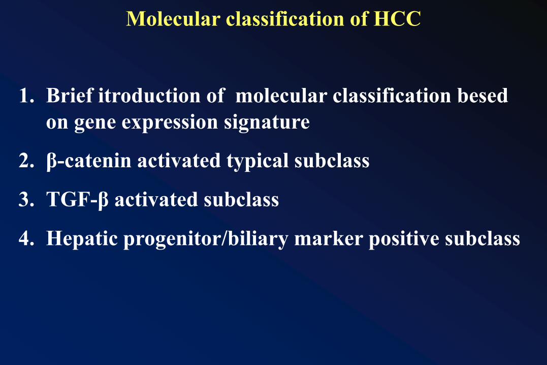
1. Brief itroduction of molecular classification besed
on gene expression signature
2. β-catenin activated typical subclass
3. TGF-β activated subclass
4. Hepatic progenitor/biliary marker positive subclass
Molecular classification of HCC
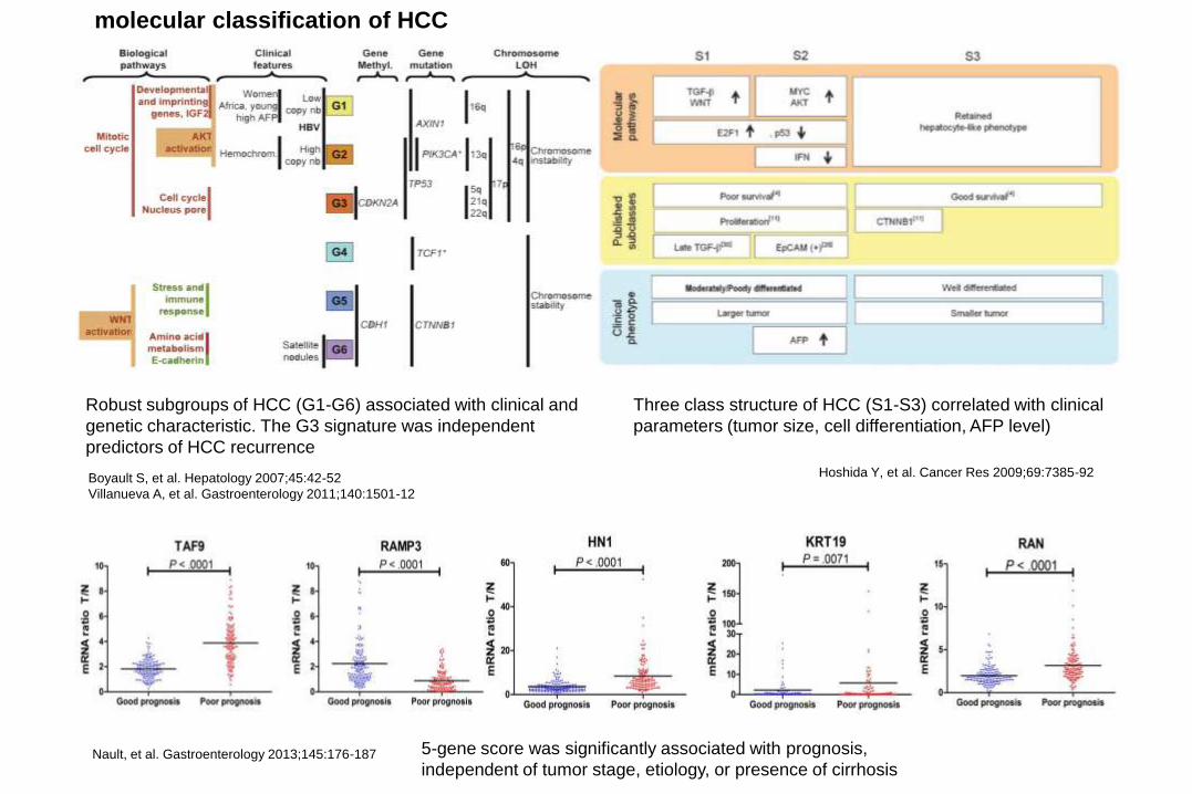
Nault, et al. Gastroenterology 2013;145:176-187 5-gene score was significantly associated with prognosis,
independent of tumor stage, etiology, or presence of cirrhosis
Hoshida Y, et al. Cancer Res 2009;69:7385-92
Three class structure of HCC (S1-S3) correlated with clinical
parameters (tumor size, cell differentiation, AFP level)
Boyault S, et al. Hepatology 2007;45:42-52
Villanueva A, et al. Gastroenterology 2011;140:1501-12
Robust subgroups of HCC (G1-G6) associated with clinical and
genetic characteristic. The G3 signature was independent
predictors of HCC recurrence
molecular classification of HCC
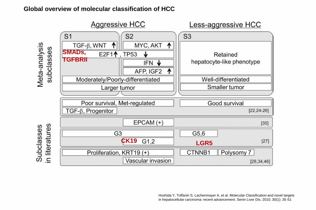
Hoshida Y, Toffanin S, Lachenmayer A, et al. Molecular Classification and novel targets
in hepatocellular carcinoma: recent advancement. Senin Liver Dis. 2010; 30(1): 35-51
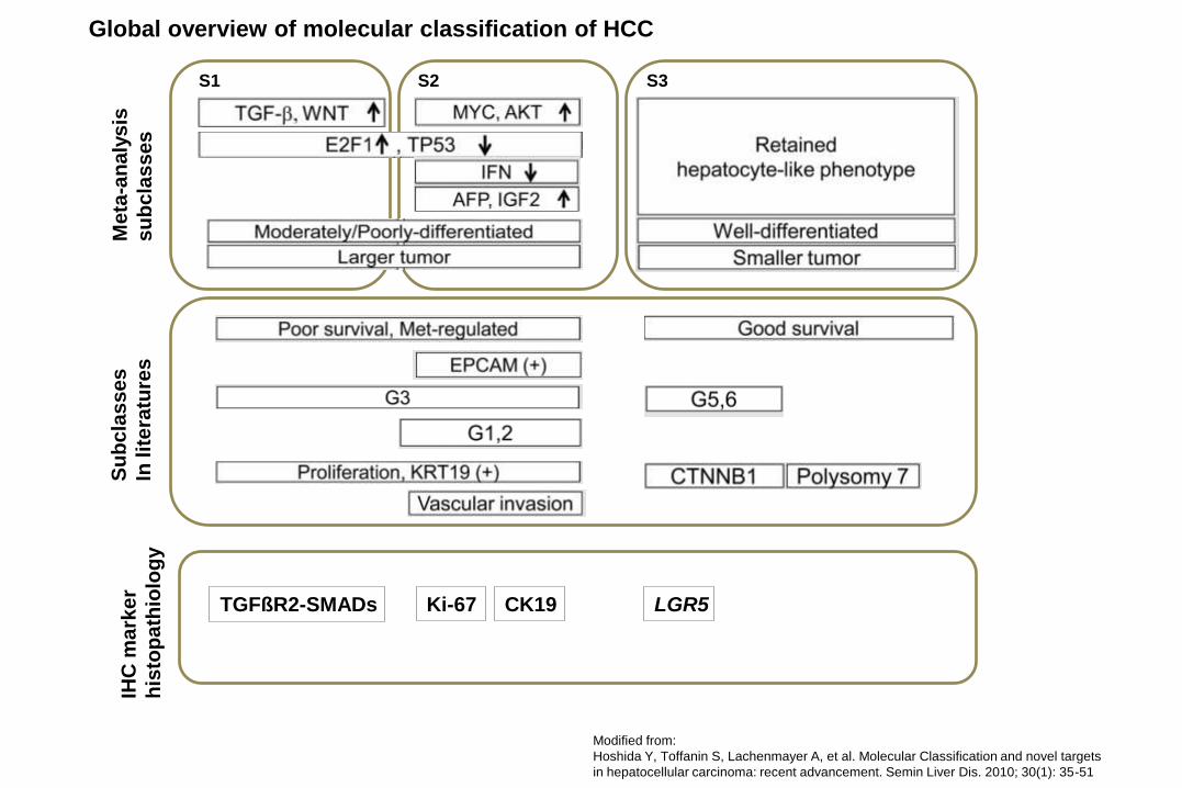
Global overview of molecular classification of HCC
LGR5
SMADs,
TGFBRII
CK19
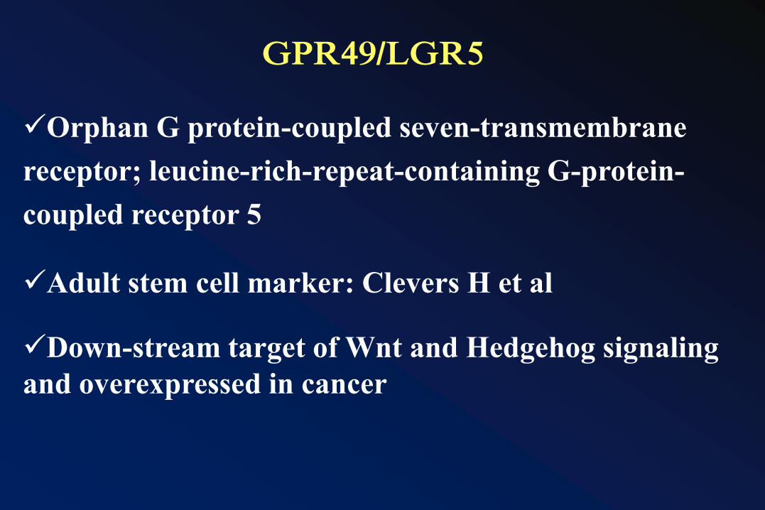
GPR49/LGR5
Orphan G protein-coupled seven-transmembrane
receptor; leucine-rich-repeat-containing G-protein-
coupled receptor 5
Adult stem cell marker: Clevers H et al
Down-stream target of Wnt and Hedgehog signaling
and overexpressed in cancer
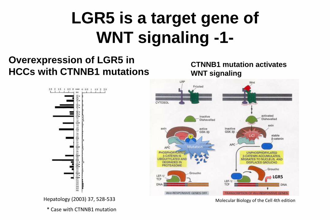
LGR5 is a target gene of
WNT signaling -1-
Overexpression of LGR5 in
HCCs with CTNNB1 mutationsCTNNB1 mutation activates
WNT signaling
Molecular Biology of the Cell 4th edition
Mutation
LiCl
LGR5
Hepatology (2003) 37, 528-533
* Case with CTNNB1 mutation
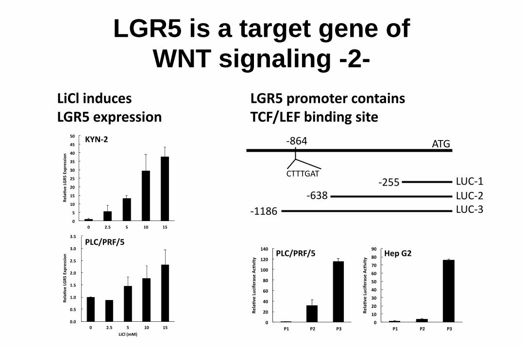
LGR5 is a target gene of
WNT signaling -2-
-864
CTTTGAT
ATG
LUC-3-1186
LUC-2-638LUC-1-255
0
20
40
60
80
100
120
140
P1 P2 P3
Rela
veLuciferaseAcvity PLC/PRF/5
0
10
20
30
40
50
60
70
80
90
P1 P2 P3
Rela
veLuciferaseAcvity Hep G2
LGR5 promoter containsTCF/LEF binding site
0
5
10
15
20
25
30
35
40
45
50
0 2.5 5 10 15
Rela
veLGR5Expression
LiCl(mM)
LiCl inducesLGR5 expression
0.0
0.5
1.0
1.5
2.0
2.5
3.0
3.5
0 2.5 5 10 15
Rela
veLGR5Expression
LiCl(mM)
PLC/PRF/5
KYN-2
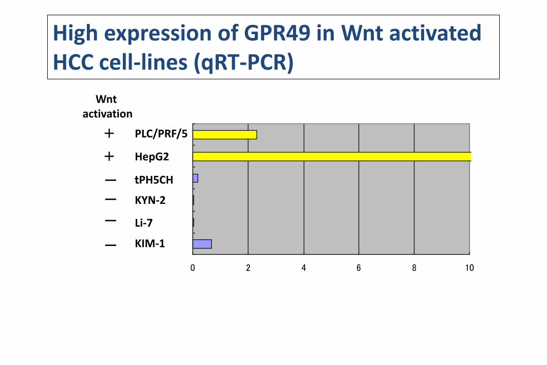
High expression of GPR49 in Wnt activated HCC cell-lines (qRT-PCR)
0 2 4 6 8 10
PLC/PRF/5
HepG2
tPH5CH
KYN-2
Li-7
KIM-1
+
+
ー
ー
ー
ー
Wnt activation
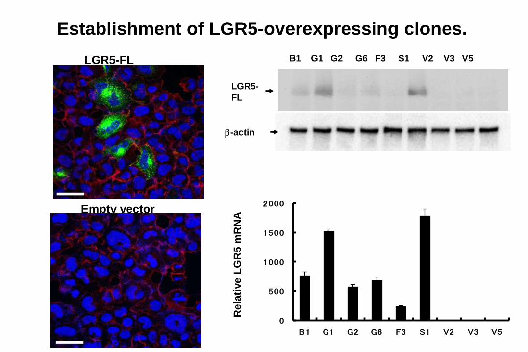
Establishment of LGR5-overexpressing clones.
LGR5-FL
Empty vector
LGR5-
FL
b-actin
B1 G1 G2 G6 F3 S1 V2 V3 V5
0
500
1000
1500
2000
B1 G1 G2 G6 F3 S1 V2 V3 V5
Re
lati
ve L
GR
5 m
RN
A
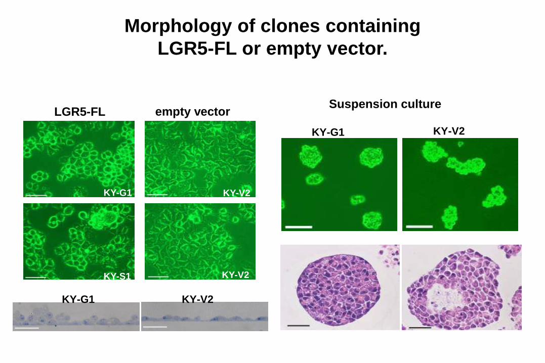
Morphology of clones containing
LGR5-FL or empty vector.
KY-G1
KY-S1
KY-V2
KY-V2
KY-G1 KY-V2
LGR5-FL empty vector
KY-G1 KY-V2
Suspension culture
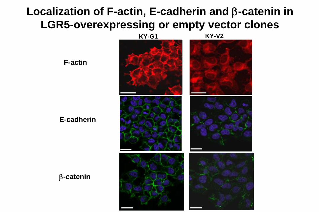
Localization of F-actin, E-cadherin and b-catenin in
LGR5-overexpressing or empty vector clones
F-actin
E-cadherin
b-catenin
KY-G1 KY-V2
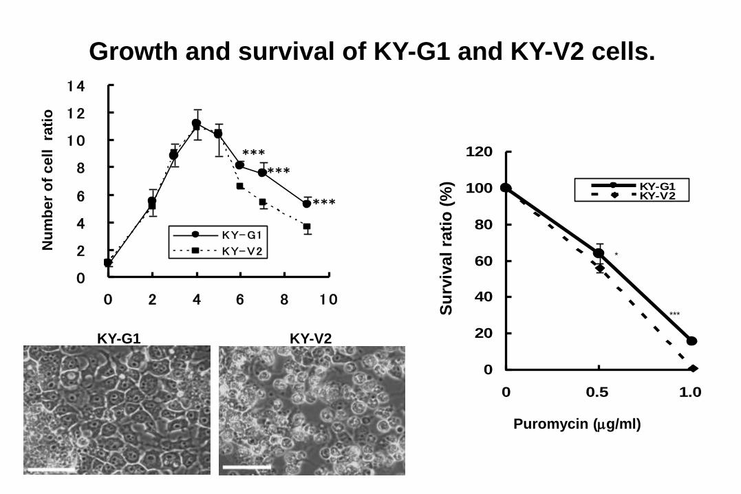
Growth and survival of KY-G1 and KY-V2 cells.
0
2
4
6
8
10
12
14
0 2 4 6 8 10
KY-G1
KY-V2
***
***
***
Nu
mb
er
of
cell
rati
o
0
20
40
60
80
100
120
0 0.5 1.0
KY-G1KY-V2
*
***Su
rviv
al
rati
o (
%)
Puromycin (mg/ml)
KY-G1 KY-V2
0
20
40
60
KY-G1 KY-V2
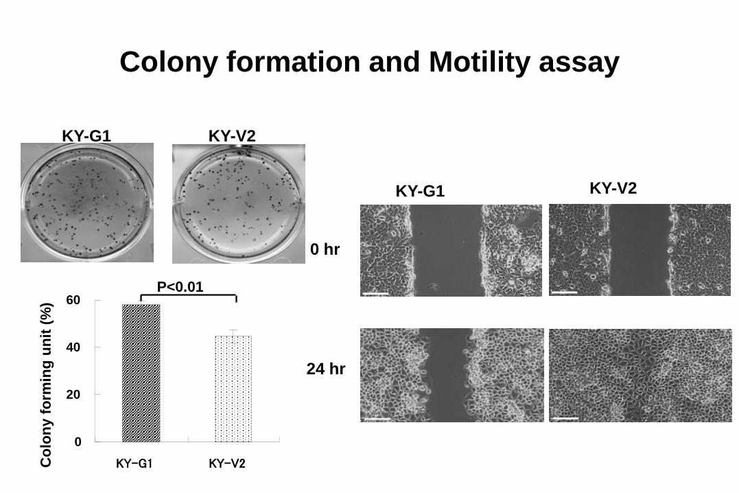
Colony formation and Motility assay
KY-G1 KY-V2
Co
lon
y f
orm
ing
un
it (
%)
P<0.01
0 hr
24 hr
KY-G1 KY-V2
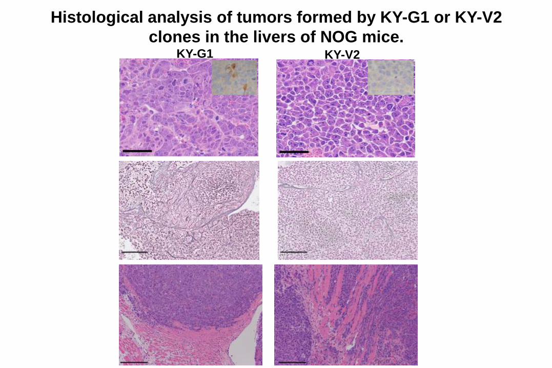
Histological analysis of tumors formed by KY-G1 or KY-V2
clones in the livers of NOG mice.KY-G1 KY-V2
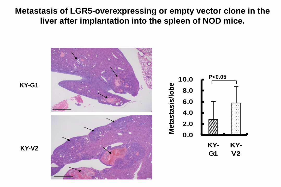
Metastasis of LGR5-overexpressing or empty vector clone in the
liver after implantation into the spleen of NOD mice.
0.0
2.0
4.0
6.0
8.0
10.0
KY-
G1
KY-
V2
Me
tas
tasis
/lo
be
P<0.05
KY-G1
KY-V2

Down-regulation of LGR5 in HCC cell lines by
treatment with siRNAs.
HepG2
PLC/PRF/5
siControl si585 si662
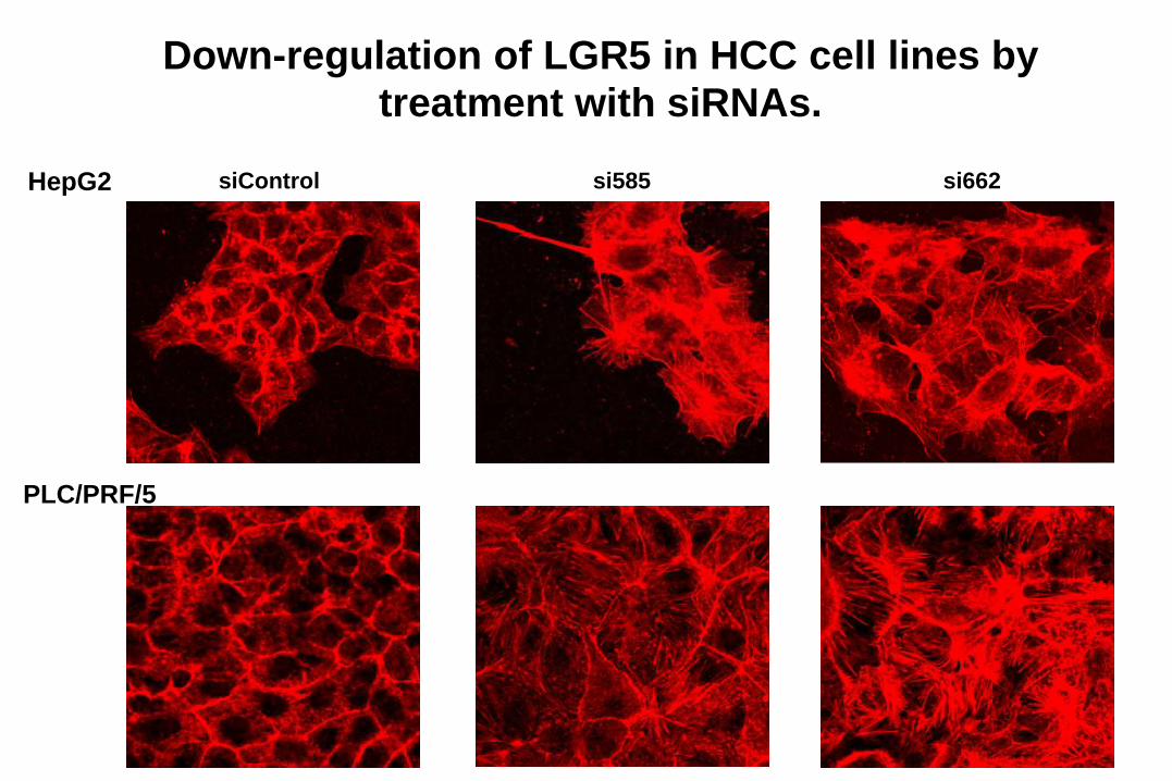
Down-regulation of LGR5 in HCC cell lines by treatment with siRNAs.
HepG2
PLC/PRF/5
siControl si585 si662
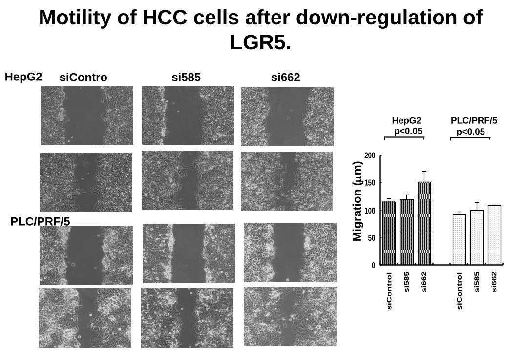
Motility of HCC cells after down-regulation of
LGR5.
0
50
100
150
200
siC
on
tro
l
si5
85
si6
62
siC
on
tro
l
si5
85
si6
62
0 hr
24 hr
24 hr
Mig
rati
on
(m
m)
HepG2 PLC/PRF/5p<0.05 p<0.05
siContro si585 si662HepG2
PLC/PRF/5
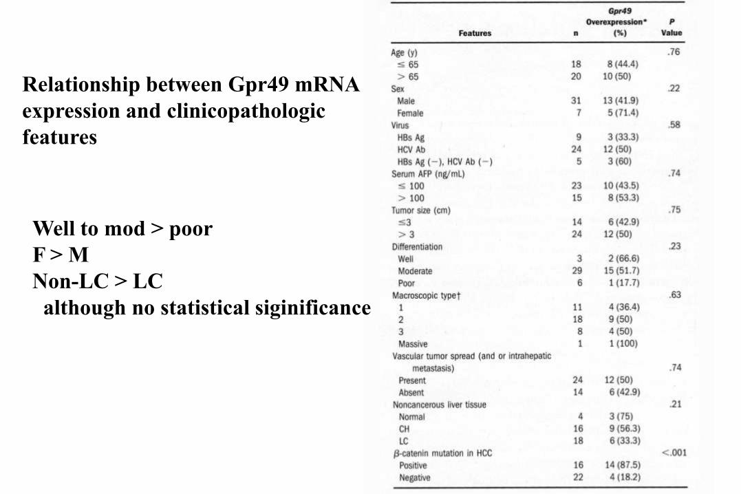
Relationship between Gpr49 mRNA
expression and clinicopathologic
features
Well to mod > poor
F > M
Non-LC > LC
although no statistical siginificance
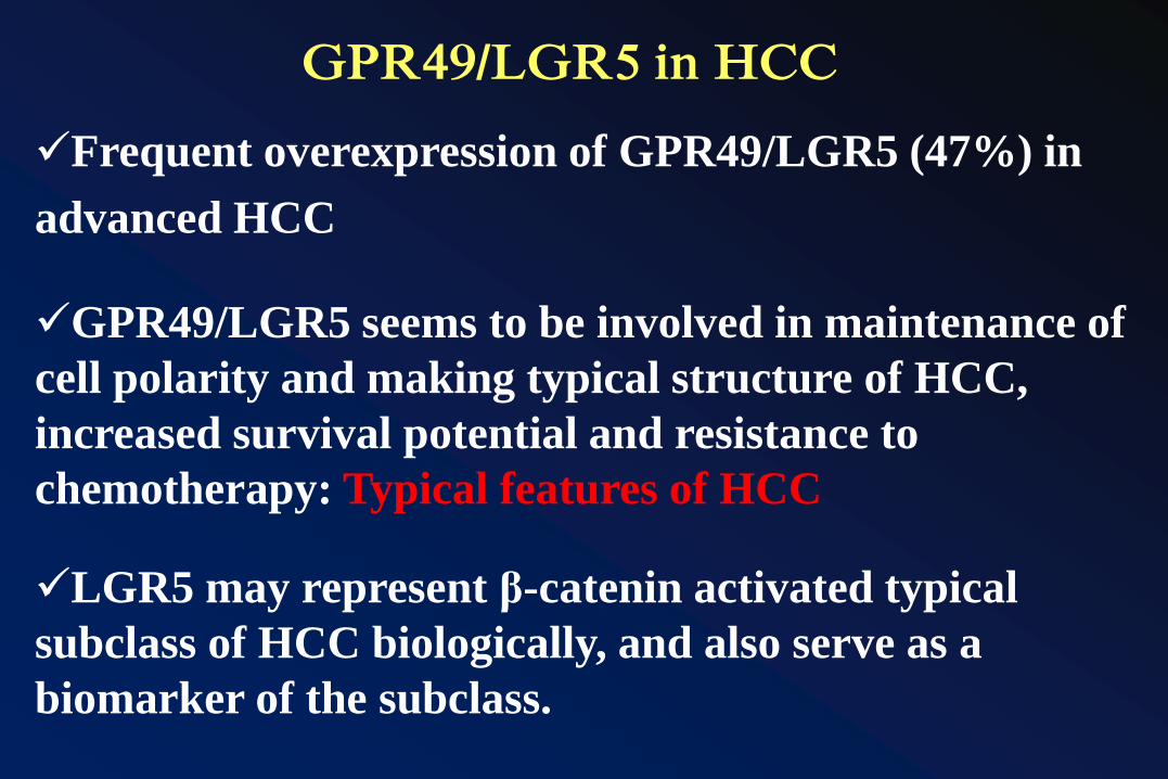
GPR49/LGR5 in HCC
Frequent overexpression of GPR49/LGR5 (47%) in
advanced HCC
GPR49/LGR5 seems to be involved in maintenance of
cell polarity and making typical structure of HCC,
increased survival potential and resistance to
chemotherapy: Typical features of HCC
LGR5 may represent β-catenin activated typical
subclass of HCC biologically, and also serve as a
biomarker of the subclass.
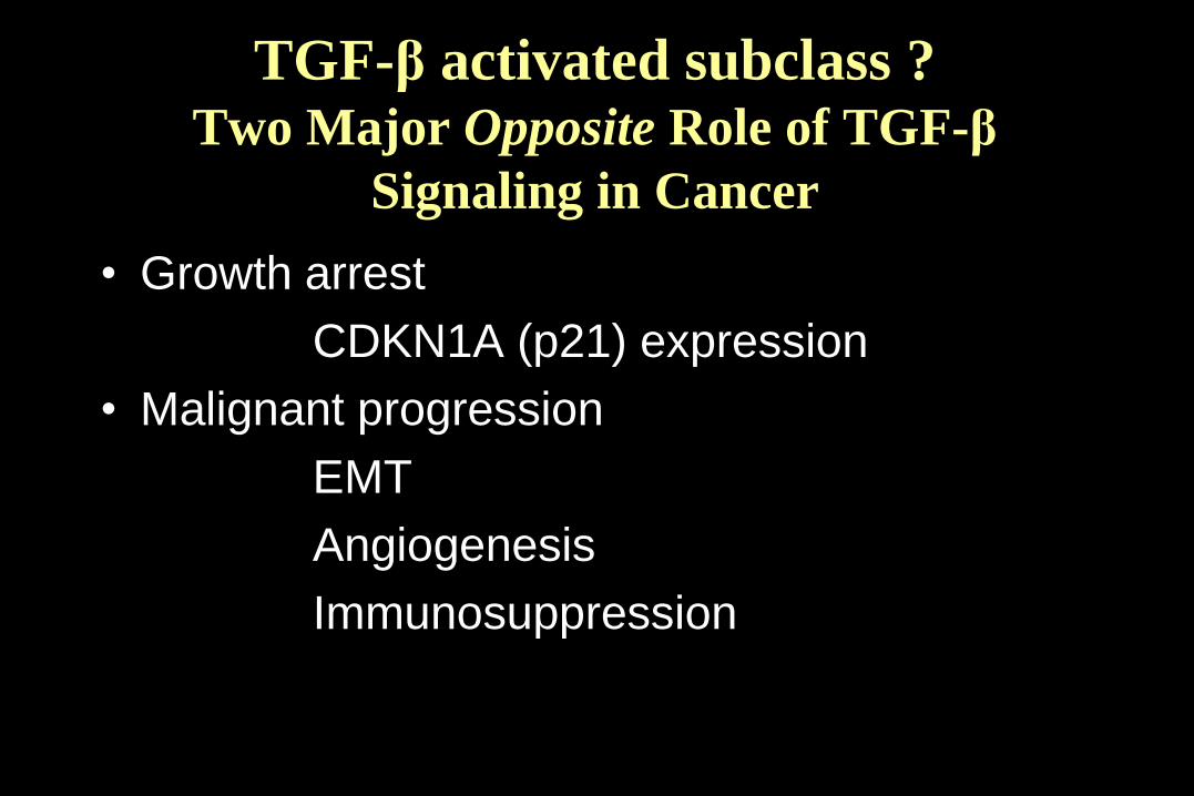
TGF-β activated subclass ? Two Major Opposite Role of TGF-β
Signaling in Cancer
• Growth arrest
CDKN1A (p21) expression
• Malignant progression
EMT
Angiogenesis
Immunosuppression
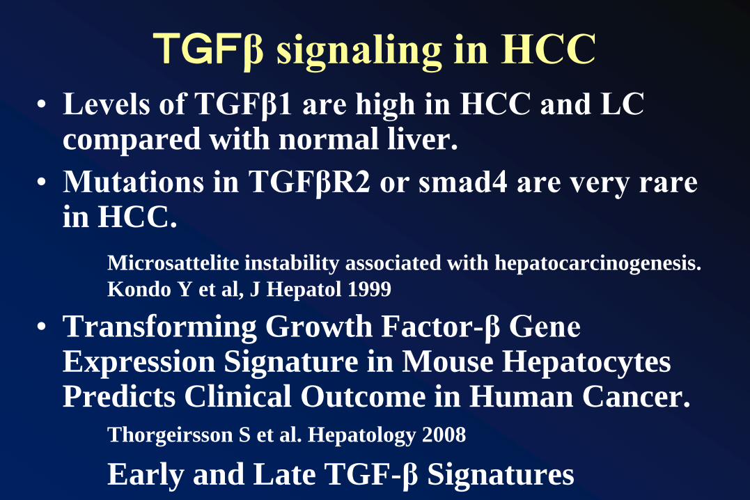
TGFβ signaling in HCC
• Levels of TGFβ1 are high in HCC and LC compared with normal liver.
• Mutations in TGFβR2 or smad4 are very rare in HCC.
Microsattelite instability associated with hepatocarcinogenesis.
Kondo Y et al, J Hepatol 1999
• Transforming Growth Factor-β Gene Expression Signature in Mouse HepatocytesPredicts Clinical Outcome in Human Cancer.
Thorgeirsson S et al. Hepatology 2008
Early and Late TGF-β Signatures
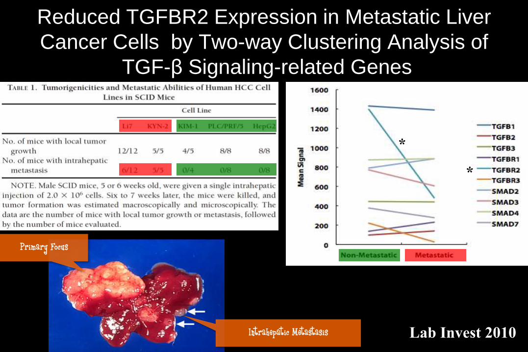
Reduced TGFBR2 Expression in Metastatic Liver
Cancer Cells by Two-way Clustering Analysis of
TGF-β Signaling-related Genes
Intrahepatic Metastasis
Primary Focus
*
*
Lab Invest 2010
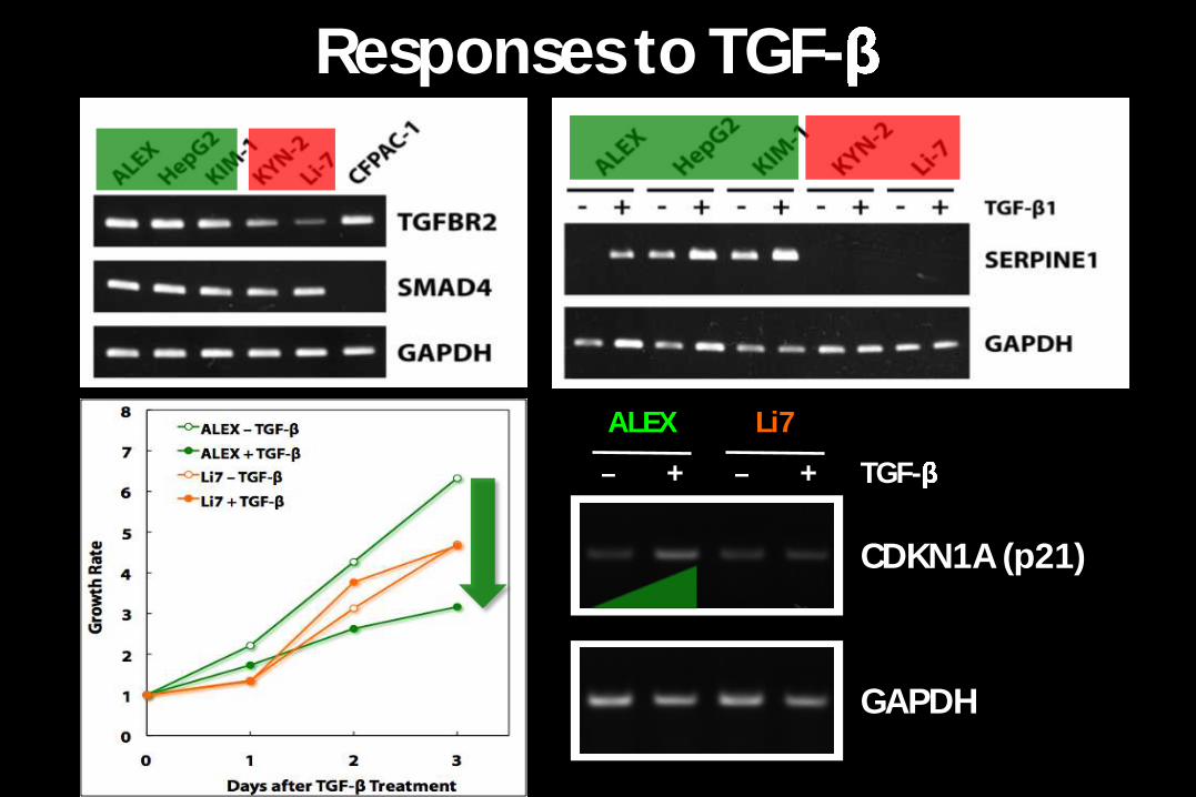
Responses to TGF-
TGF-
ALEX
+
Li7
+
CDKN1A (p21)
GAPDH
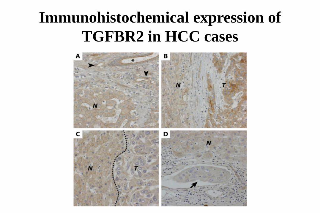
Immunohistochemical expression of
TGFBR2 in HCC cases
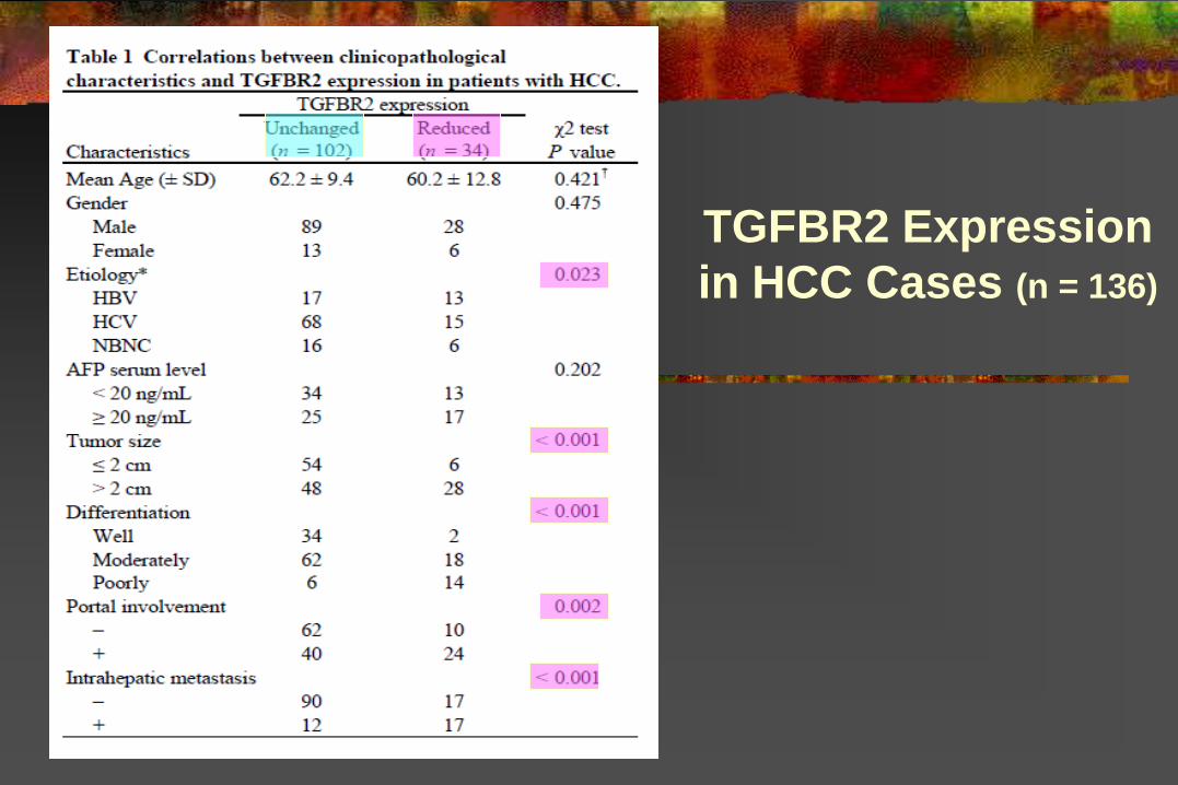
TGFBR2 Expression
in HCC Cases (n = 136)
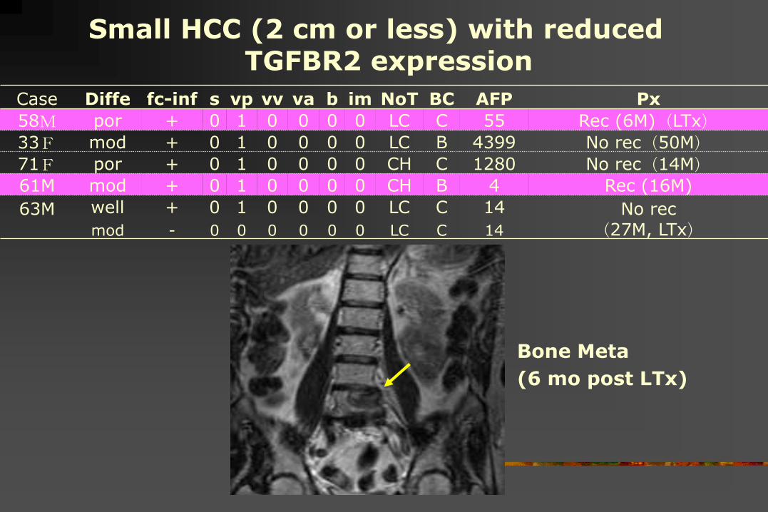
Case Diffe fc-inf s vp vv va b im NoT BC AFP Px
58M por + 0 1 0 0 0 0 LC C 55 Rec (6M)(LTx)33F mod + 0 1 0 0 0 0 LC B 4399 No rec(50M)71F por + 0 1 0 0 0 0 CH C 1280 No rec(14M)61M mod + 0 1 0 0 0 0 CH B 4 Rec (16M)
63M well + 0 1 0 0 0 0 LC C 14 No rec
(27M, LTx)mod - 0 0 0 0 0 0 LC C 14
Small HCC (2 cm or less) with reduced TGFBR2 expression
Bone Meta
(6 mo post LTx)
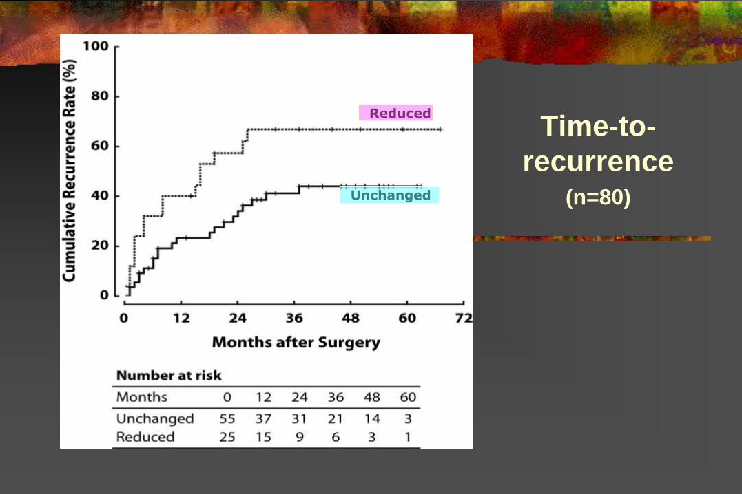
Time-to-
recurrence(n=80)Unchanged
Reduced
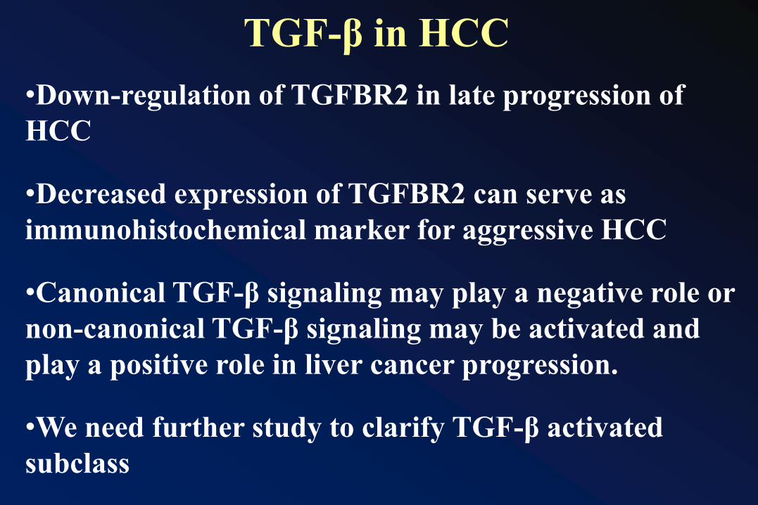
•Down-regulation of TGFBR2 in late progression of
HCC
•Decreased expression of TGFBR2 can serve as
immunohistochemical marker for aggressive HCC
•Canonical TGF-β signaling may play a negative role or
non-canonical TGF-β signaling may be activated and
play a positive role in liver cancer progression.
•We need further study to clarify TGF-β activated
subclass
TGF-β in HCC
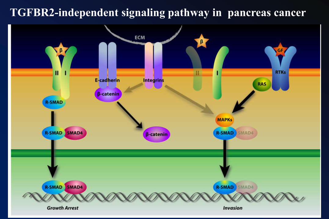
TGFBR2-independent signaling pathway in pancreas cancer
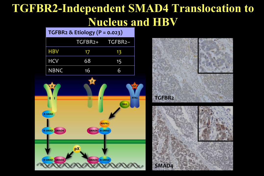
TGFBR2-Independent SMAD4 Translocation to
Nucleus and HBV
TGFBR2
SMAD4
TGFBR2 & Etiology (P = 0.023)
TGFBR2+ TGFBR2−
HBV 17 13
HCV 68 15
NBNC 16 6
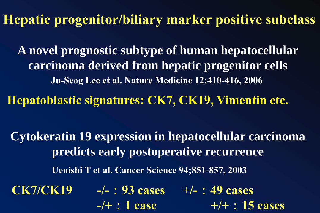
A novel prognostic subtype of human hepatocellular
carcinoma derived from hepatic progenitor cellsJu-Seog Lee et al. Nature Medicine 12;410-416, 2006
Hepatoblastic signatures: CK7, CK19, Vimentin etc.
Cytokeratin 19 expression in hepatocellular carcinoma
predicts early postoperative recurrence
Uenishi T et al. Cancer Science 94;851-857, 2003
CK7/CK19 -/-:93 cases +/-:49 cases
-/+:1 case +/+:15 cases
Hepatic progenitor/biliary marker positive subclass
0 20 40 60 80 100 120 140
高分化
中分化
低分化
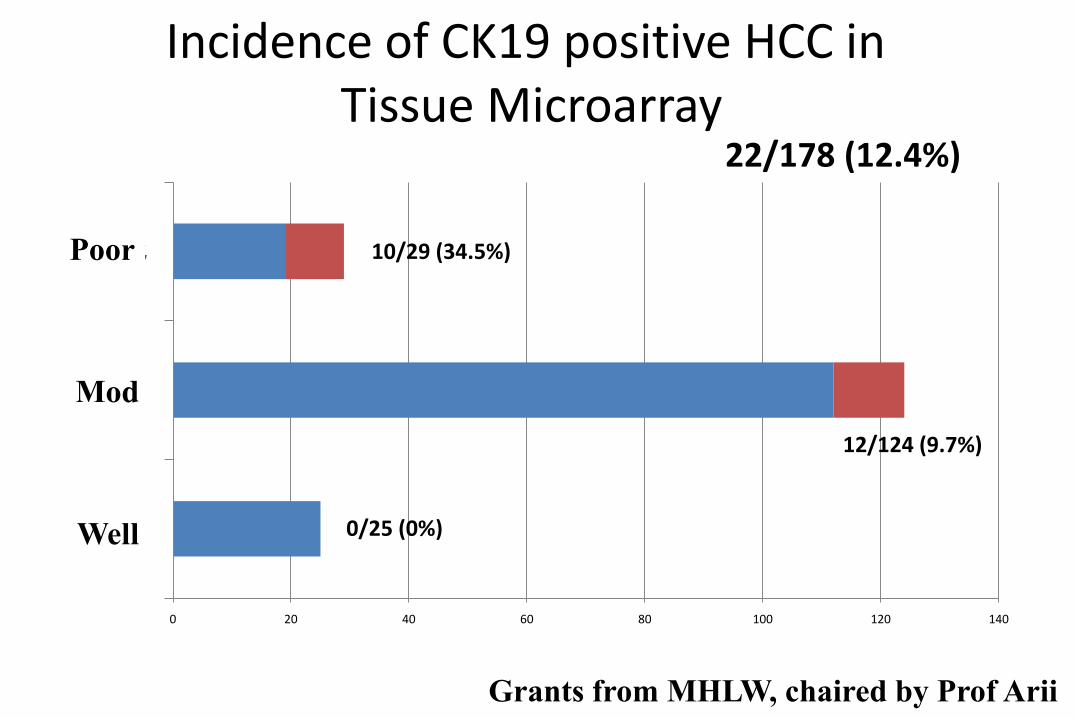
Incidence of CK19 positive HCC inTissue Microarray
10/29 (34.5%)
12/124 (9.7%)
0/25 (0%)
22/178 (12.4%)
Poor
Mod
Well
Grants from MHLW, chaired by Prof Arii
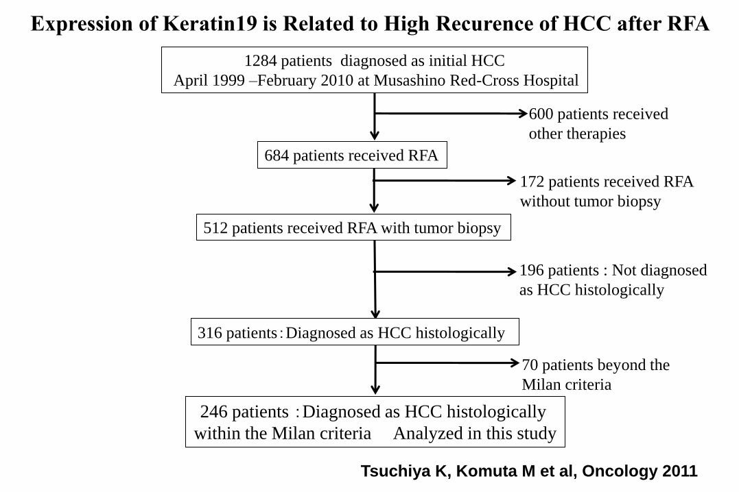
684 patients received RFA
512 patients received RFA with tumor biopsy
246 patients :Diagnosed as HCC histologically
within the Milan criteria Analyzed in this study
600 patients received
other therapies
172 patients received RFA
without tumor biopsy
1284 patients diagnosed as initial HCC
April 1999 –February 2010 at Musashino Red-Cross Hospital
316 patients:Diagnosed as HCC histologically
70 patients beyond the
Milan criteria
196 patients : Not diagnosed
as HCC histologically
Tsuchiya K, Komuta M et al, Oncology 2011
Expression of Keratin19 is Related to High Recurence of HCC after RFA
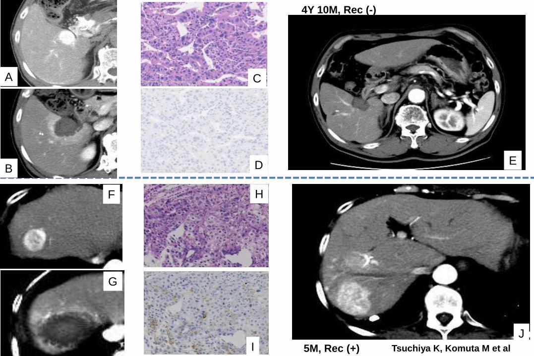
A
B
C
D E
F
G
H
IJ
4Y 10M, Rec (-)
5M, Rec (+) Tsuchiya K, Komuta M et al
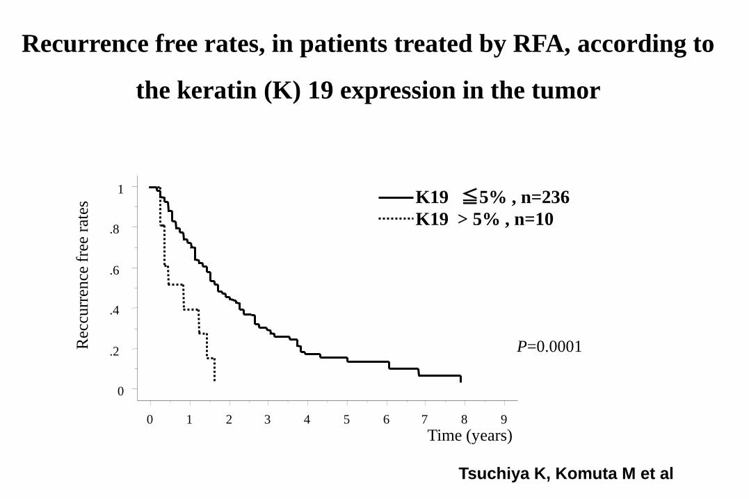
Recurrence free rates, in patients treated by RFA, according to
the keratin (K) 19 expression in the tumorR
eccu
rren
ce f
ree
rate
s
Time (years)
0
.2
.4
.6
.8
1
0 1 2 3 4 5 6 7 8 9
K19 ≦5% , n=236
K19 > 5% , n=10
P=0.0001
Tsuchiya K, Komuta M et al
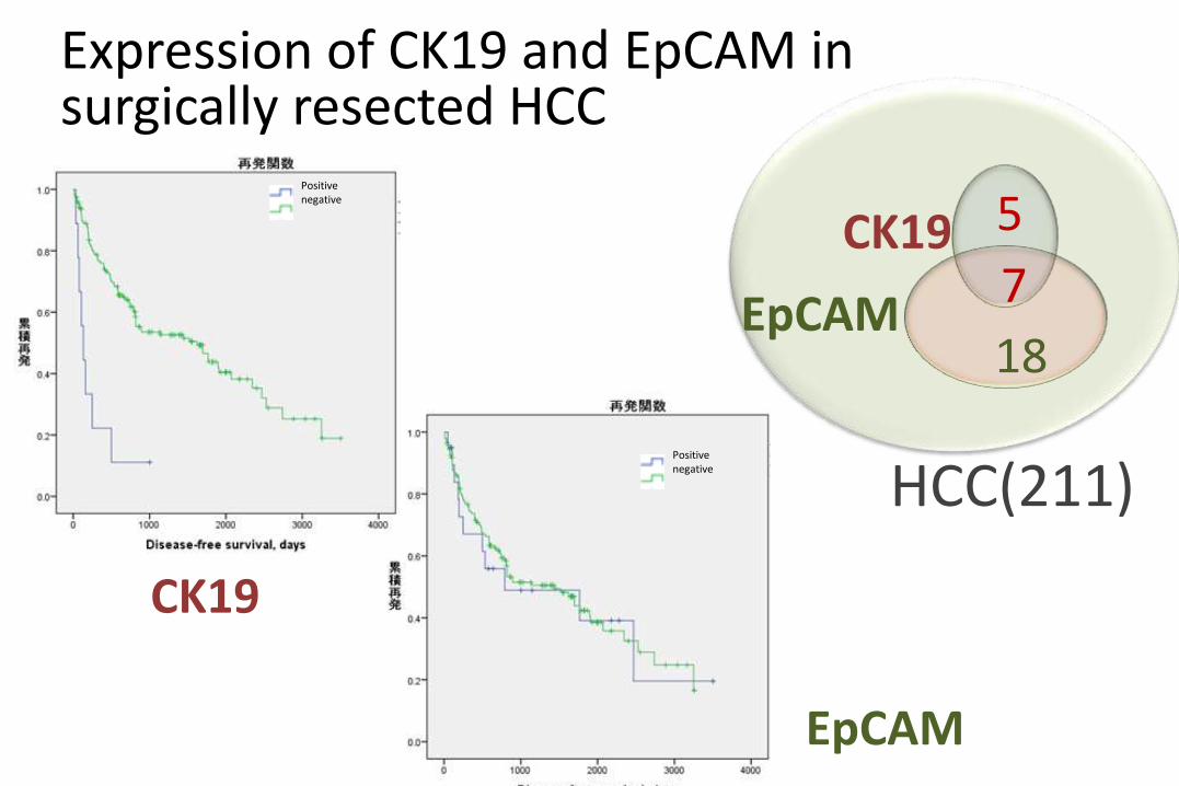
Expression of CK19 and EpCAM in surgically resected HCC
Positivenegative
HCC(211)
CK19
EpCAM
5
7
18
Positivenegative
CK19
EpCAM
S1 S2 S3
Me
ta-a
naly
sis
su
bc
las
ses
Su
bcla
sses
In lit
era
ture
sIH
C m
ark
er
his
top
ath
iolo
gy
Global overview of molecular classification of HCC
Modified from:
Hoshida Y, Toffanin S, Lachenmayer A, et al. Molecular Classification and novel targets
in hepatocellular carcinoma: recent advancement. Semin Liver Dis. 2010; 30(1): 35-51
TGFßR2-SMADs Ki-67 CK19 LGR5
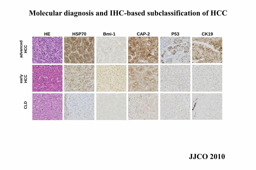
HE HSP70 Bmi-1 CAP-2 P53 CK19
CL
De
arl
y
HC
C
ad
va
nce
d
HC
C
JJCO 2010
Molecular diagnosis and IHC-based subclassification of HCC
Acknowledgement
Department of Pathology, Keio Univ
Taketo Yamada Kathryn Effendi
Akinori Hashiguchi Taizo Hibi
Mariko Fukuma Akihisa Ueno
Wenlin Du Junya Douguchi
Youhei Masugi Keiji Tanese
Yuichiro Hayashi Hiroshi Uchida
Ken Yamazaki Taisuke Mori
Tokiya Abe Mina Komuta
Hanako Tsujikawa
Department of Surgery, Internal Medicine and Radiology, Keio Univ
National Cancer Center Research Institute