Molecular Biology of Postsynaptic Structures. Biochemical Fractionation of the PSD Structure –...
45
Molecular Biology of Postsynaptic Structures
-
date post
21-Dec-2015 -
Category
Documents
-
view
243 -
download
1
Transcript of Molecular Biology of Postsynaptic Structures. Biochemical Fractionation of the PSD Structure –...
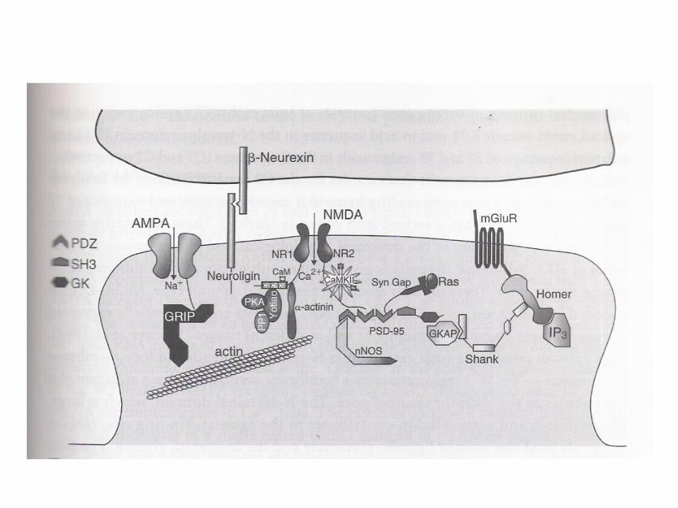
Molecular Biology of Postsynaptic Structures
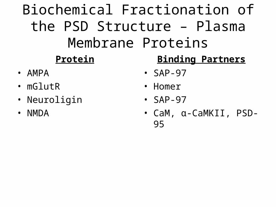
Biochemical Fractionation of the PSD Structure – Plasma Membrane Proteins
Protein• AMPA• mGlutR• Neuroligin• NMDA
Binding Partners• SAP-97• Homer• SAP-97• CaM, α-CaMKII, PSD-95
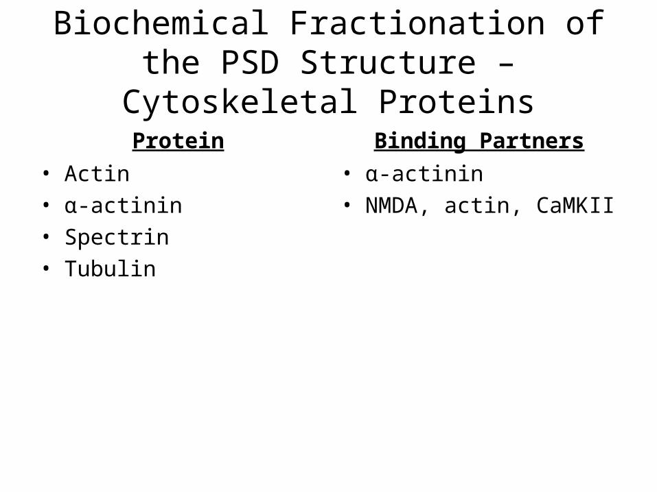
Biochemical Fractionation of the PSD Structure – Cytoskeletal Proteins
Protein• Actin• α-actinin• Spectrin• Tubulin
Binding Partners• α-actinin• NMDA, actin, CaMKII
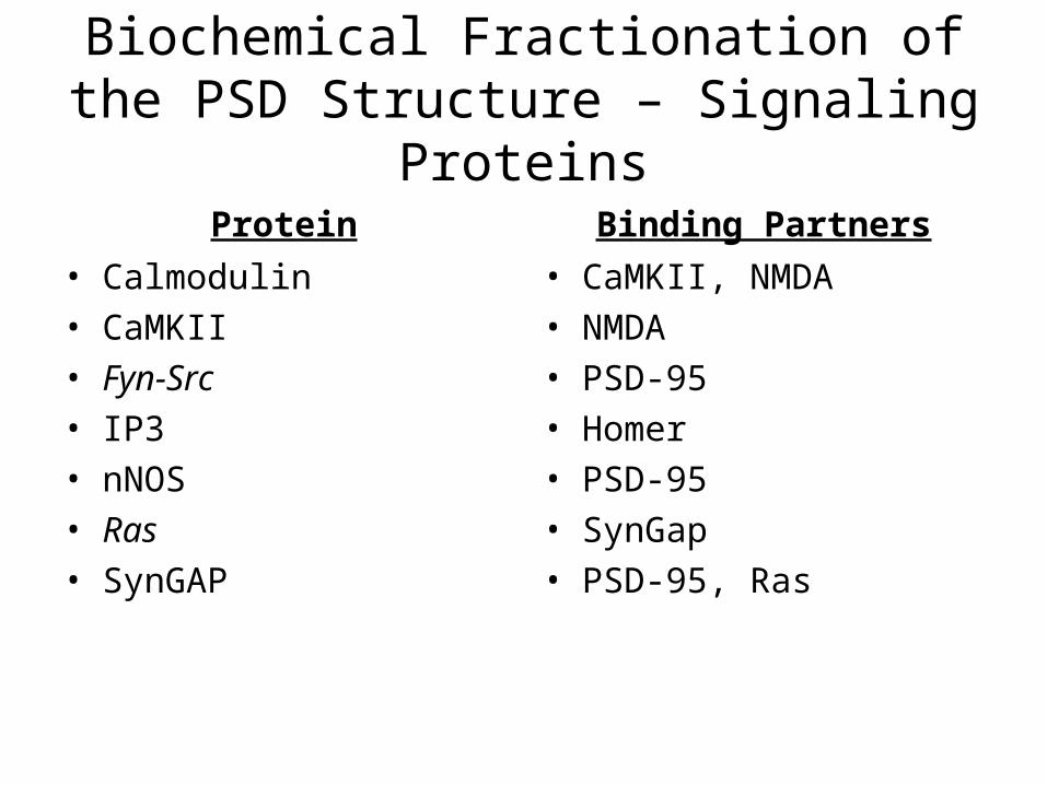
Biochemical Fractionation of the PSD Structure – Signaling Proteins
Protein• Calmodulin• CaMKII• Fyn-Src• IP3• nNOS• Ras• SynGAP
Binding Partners• CaMKII, NMDA• NMDA• PSD-95• Homer• PSD-95• SynGap• PSD-95, Ras
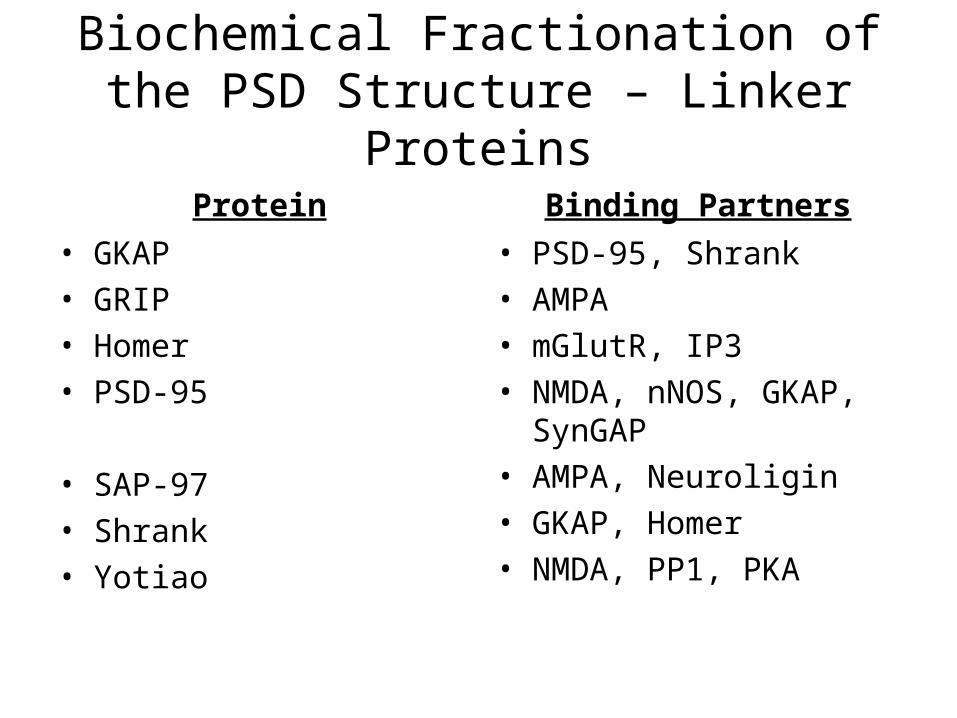
Biochemical Fractionation of the PSD Structure – Linker Proteins
Protein• GKAP• GRIP• Homer• PSD-95
• SAP-97• Shrank• Yotiao
Binding Partners• PSD-95, Shrank• AMPA• mGlutR, IP3• NMDA, nNOS, GKAP,
SynGAP• AMPA, Neuroligin• GKAP, Homer• NMDA, PP1, PKA
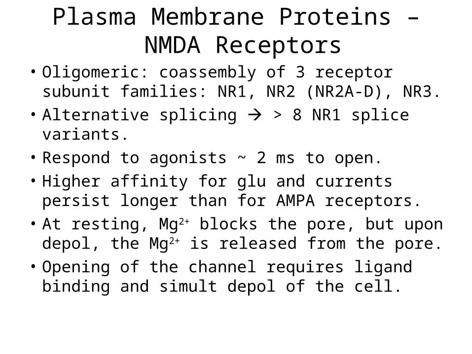
Plasma Membrane Proteins – NMDA Receptors
• Oligomeric: coassembly of 3 receptor subunit families: NR1, NR2 (NR2A-D), NR3.
• Alternative splicing > 8 NR1 splice variants.• Respond to agonists ~ 2 ms to open.• Higher affinity for glu and currents persist
longer than for AMPA receptors.• At resting, Mg2+ blocks the pore, but upon
depol, the Mg2+ is released from the pore.• Opening of the channel requires ligand
binding and simult depol of the cell.
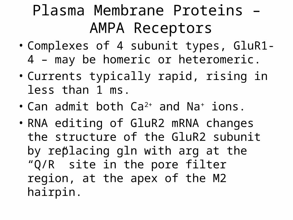
Plasma Membrane Proteins – AMPA Receptors
• Complexes of 4 subunit types, GluR1-4 – may be homeric or heteromeric.
• Currents typically rapid, rising in less than 1 ms.
• Can admit both Ca2+ and Na+ ions.• RNA editing of GluR2 mRNA changes the
structure of the GluR2 subunit by replacing gln with arg at the “Q/R” site in the pore filter region, at the apex of the M2 hairpin.
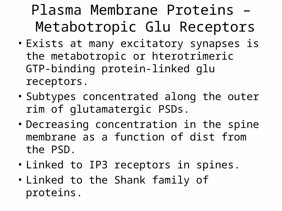
Plasma Membrane Proteins – Metabotropic Glu Receptors
• Exists at many excitatory synapses is the metabotropic or hterotrimeric GTP-binding protein-linked glu receptors.
• Subtypes concentrated along the outer rim of glutamatergic PSDs.
• Decreasing concentration in the spine membrane as a function of dist from the PSD.
• Linked to IP3 receptors in spines.• Linked to the Shank family of proteins.
Signaling Proteins
• Calmodulin• CaMKII• Fyn-Src – can activate Ras through the N-Shc
adaptor protein• IP3• nNOS• Ras• SynGAP – inhibited by the NMDA receptor
Cytoskeletal Proteins – Linker Proteins
• 4 closely related PSD-95 proteins (aka MAGUK proteins), each of which contains 5 protein-binding domains.
• 3 amino-terminal PDZ (PSD-95, Discs-large, ZO-1) domaines are followed by an SH3 domain, and a GK domain.
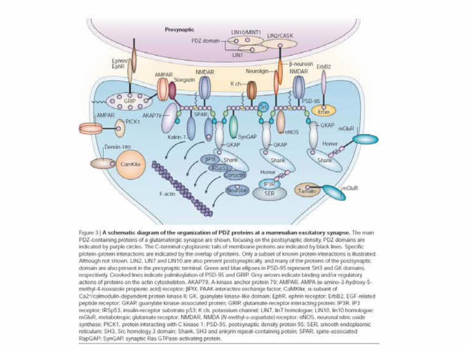
Functional Interactions Between PSD Components
• The 4 classes of proteins (above) all conspire to regulate complex functions (e.g., LTP, LTD).
• PSD-95 binds other PSD components (enzymes and modulators): PSD-95 binds nNOS via PDZ-PDZ interaction holding NOS in the appropriate position to sense Ca2+ influx through NMDA receptor channel opening. The 3rd PDZ domain of PSD-95 binds SynGAP providing a link between the NMDAR and the MAPK pathway.
• Ischemic challenge.• Intercellular communication.
Proposed Functions of the PSD
• PSD formed very early during synaptogenesis.• Synaptic activity affects PSD morphology.• Synaptic plasticity.
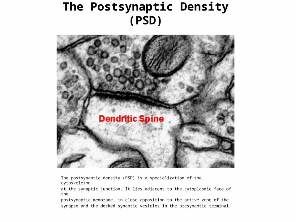
The Postsynaptic Density (PSD)
The postsynaptic density (PSD) is a specialization of the cytoskeletonat the synaptic junction. It lies adjacent to the cytoplasmic face of thepostsynaptic membrane, in close apposition to the active zone of thesynapse and the docked synaptic vesicles in the presynaptic terminal.
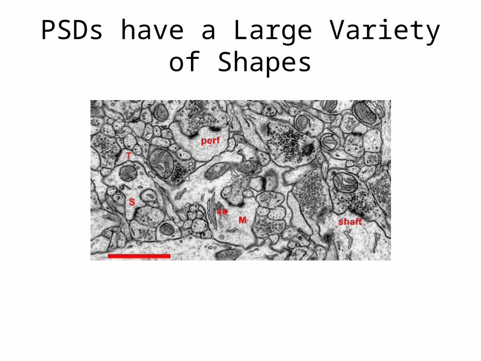
PSDs have a Large Variety of Shapes
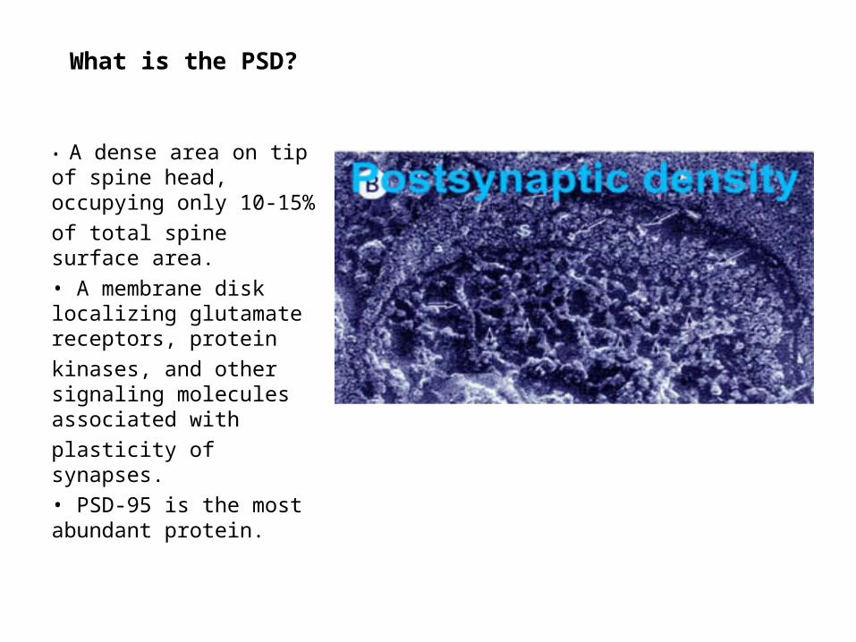
What is the PSD?
• A dense area on tip of spine head, occupying only 10-15%of total spine surface area.• A membrane disk localizing glutamate receptors, proteinkinases, and other signaling molecules associated withplasticity of synapses.• PSD-95 is the most abundant protein.
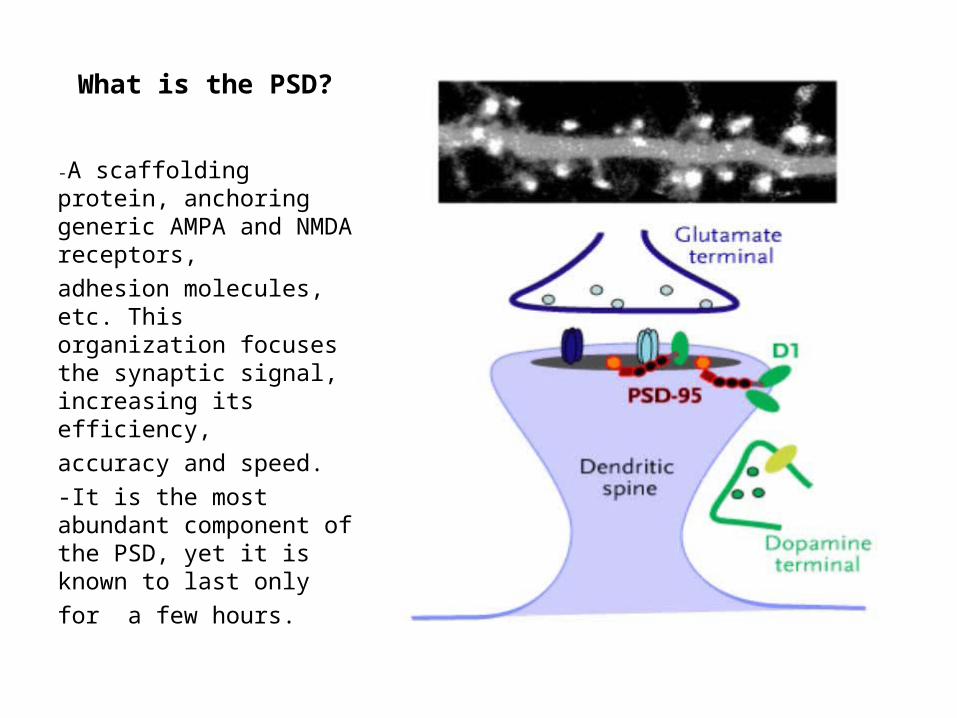
What is the PSD?
-A scaffolding protein, anchoring generic AMPA and NMDA receptors,adhesion molecules, etc. This organization focuses the synaptic signal, increasing its efficiency,accuracy and speed.-It is the most abundant component of the PSD, yet it is known to last onlyfor a few hours.

Organization of the Core Structure of the PSD
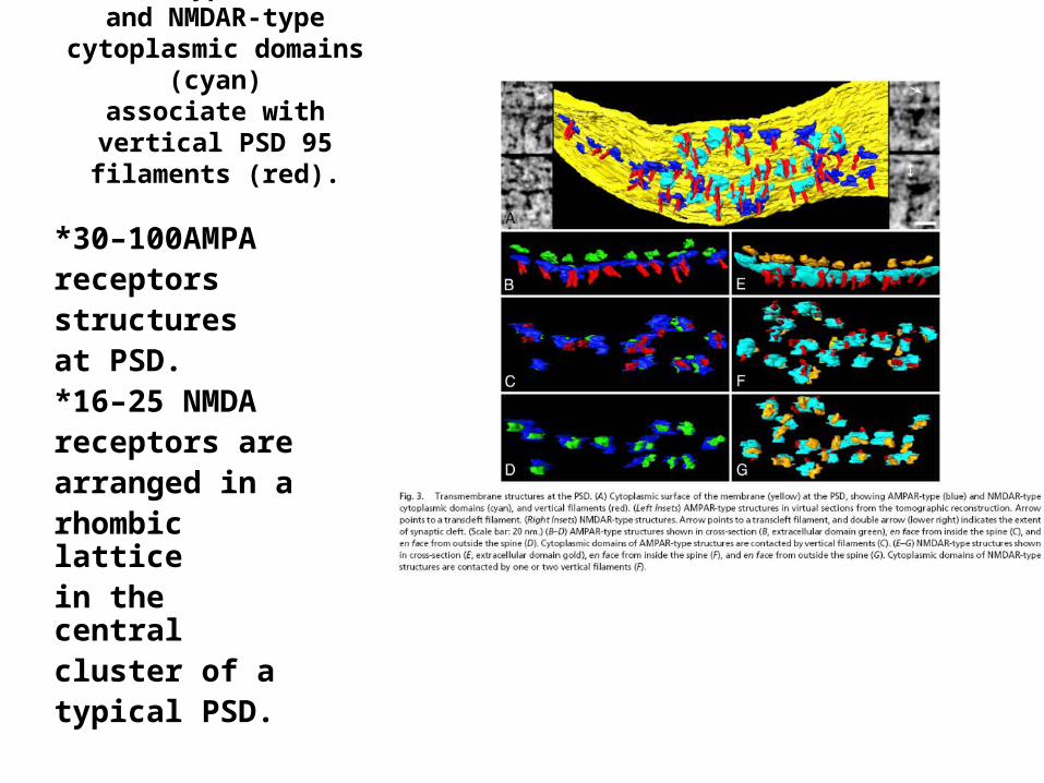
AMPAR-type (blue) and NMDAR-type cytoplasmic
domains (cyan)associate with vertical PSD 95
filaments (red).
*30–100AMPAreceptorsstructuresat PSD.*16–25 NMDAreceptors arearranged in arhombic latticein the centralcluster of atypical PSD.
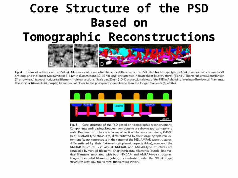
Core Structure of the PSD Based onTomographic Reconstructions
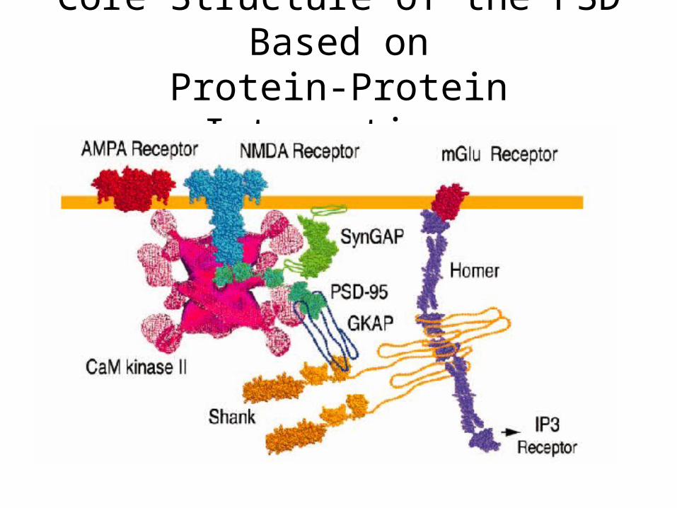
Core Structure of the PSD Based onProtein-Protein Interactions
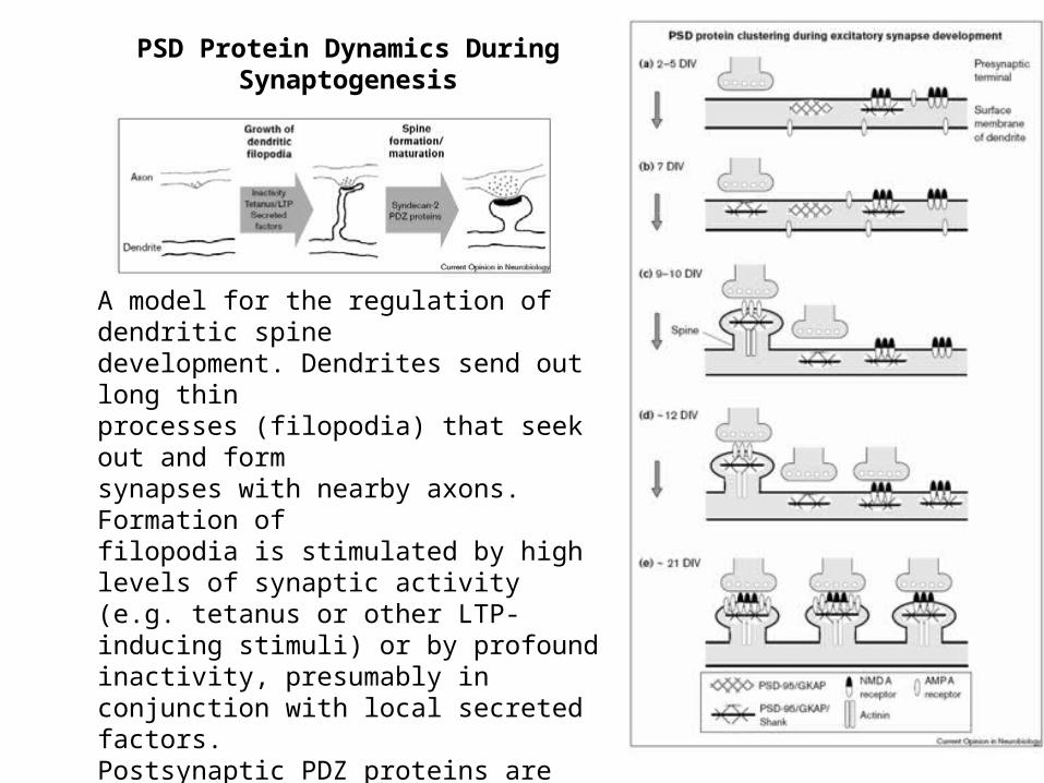
A model for the regulation of dendritic spinedevelopment. Dendrites send out long thinprocesses (filopodia) that seek out and formsynapses with nearby axons. Formation offilopodia is stimulated by high levels of synaptic activity (e.g. tetanus or other LTP-inducing stimuli) or by profound inactivity, presumably in conjunction with local secreted factors.Postsynaptic PDZ proteins are critical forthe development of filopodia into mature spines representative mature mushroom-shaped spine is shown).The maintenance of mature spines depends on low level stimulation of the AMPA receptor.
PSD Protein Dynamics DuringSynaptogenesis
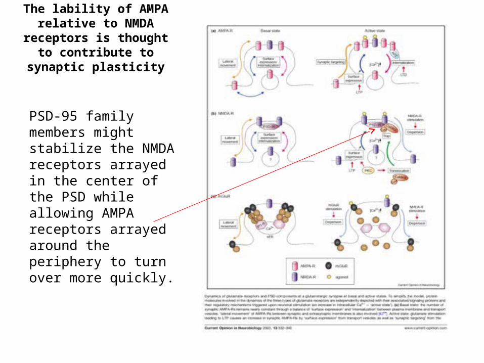
The lability of AMPA relative to NMDA receptors is thought to
contribute to synaptic plasticity
PSD-95 family members might stabilize the NMDA receptors arrayed in the center of the PSD while allowing AMPA receptors arrayed around the periphery to turn over more quickly.
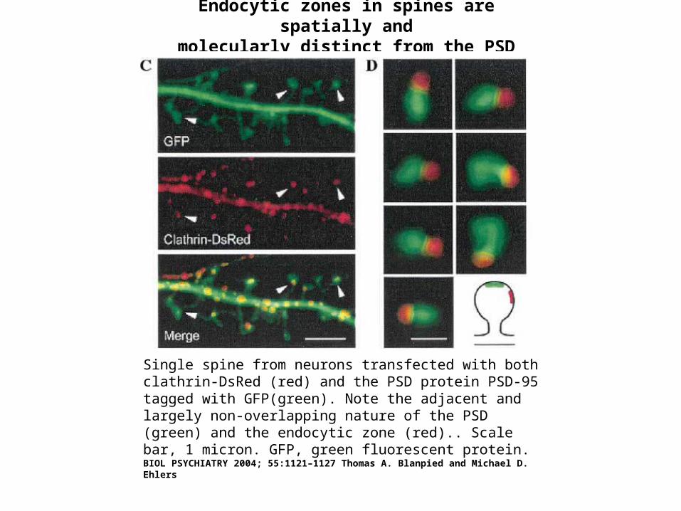
Endocytic zones in spines are spatially andmolecularly distinct from the PSD
Single spine from neurons transfected with both clathrin-DsRed (red) and the PSD protein PSD-95 tagged with GFP(green). Note the adjacent and largely non-overlapping nature of the PSD (green) and the endocytic zone (red).. Scale bar, 1 micron. GFP, green fluorescent protein. BIOL PSYCHIATRY 2004; 55:1121–1127 Thomas A. Blanpied and Michael D. Ehlers
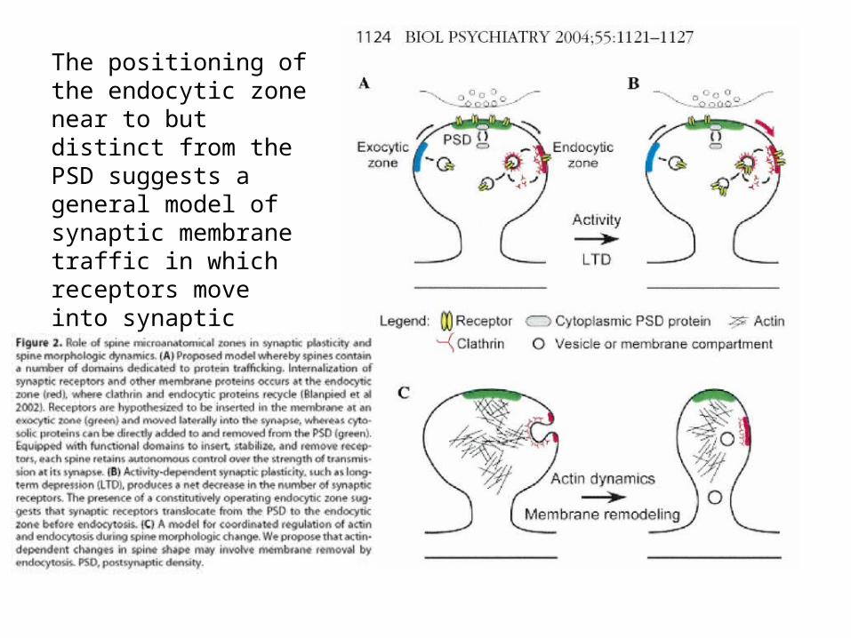
The positioning of the endocytic zone near to but distinct from the PSD suggests a general model of synaptic membrane traffic in which receptors move into synaptic membranes via perisynaptic regions.
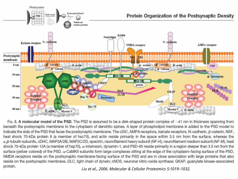
Liu et al., 2006. Molecular & Cellular Proteomics 5:1019–1032.
PSD-95 is Known to last for only a few Hrs
• How do synapses maintain their size and strength with unstable constituents?
• >>the investigators addressed this issue bystudying the movement of PSD-95.
(Rapid Redistribution of Synaptic PSD-95 in the Neocortex In VivoGray et. al. PLOS Biology 2006).
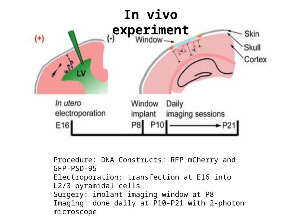
Procedure: DNA Constructs: RFP mCherry and GFP-PSD-95Electroporation: transfection at E16 into L2/3 pyramidal cellsSurgery: implant imaging window at P8Imaging: done daily at P10-P21 with 2-photon microscope
In vivo experiment
Methods
• During the second week of life, dendriticspines begin to stabilize.
• Photoactivatable-GFP (pa-GFP) was usedto monitor diffusion of one PSD-95 protein.
• pa-GFP is a type of GFP that is not normally fluorescent but can be ‘photoactivated’ by a specific wavelength of light (810nm)
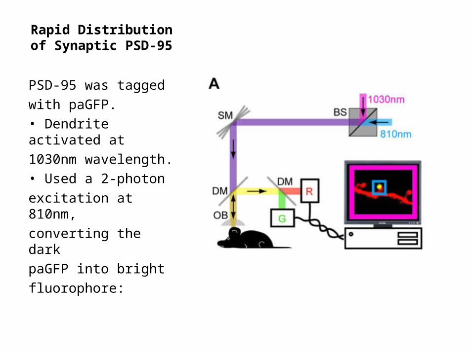
Rapid Distribution of Synaptic PSD-95
PSD-95 was taggedwith paGFP.• Dendrite activated at1030nm wavelength.• Used a 2-photonexcitation at 810nm,converting the darkpaGFP into brightfluorophore:
Key Points
• Spines seem to appear and disappear on dailybasis, which is consistent with high rates ofsynaptic formation and elimination duringdevelopmental stage in mice.
• Dendritic spines and their PSDs are quite stablearound second postnatal week.
• Stabilization also conserves the sizes of the spines.
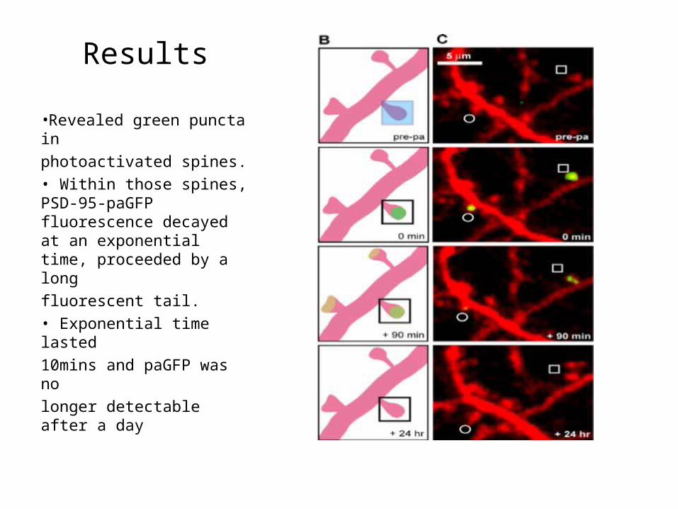
Results
•Revealed green puncta inphotoactivated spines.• Within those spines, PSD-95-paGFP fluorescence decayed at an exponential time, proceeded by a longfluorescent tail.• Exponential time lasted10mins and paGFP was nolonger detectable after a day
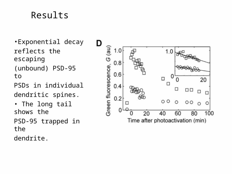
Results
•Exponential decayreflects the escaping(unbound) PSD-95 toPSDs in individualdendritic spines.• The long tail shows thePSD-95 trapped in thedendrite.
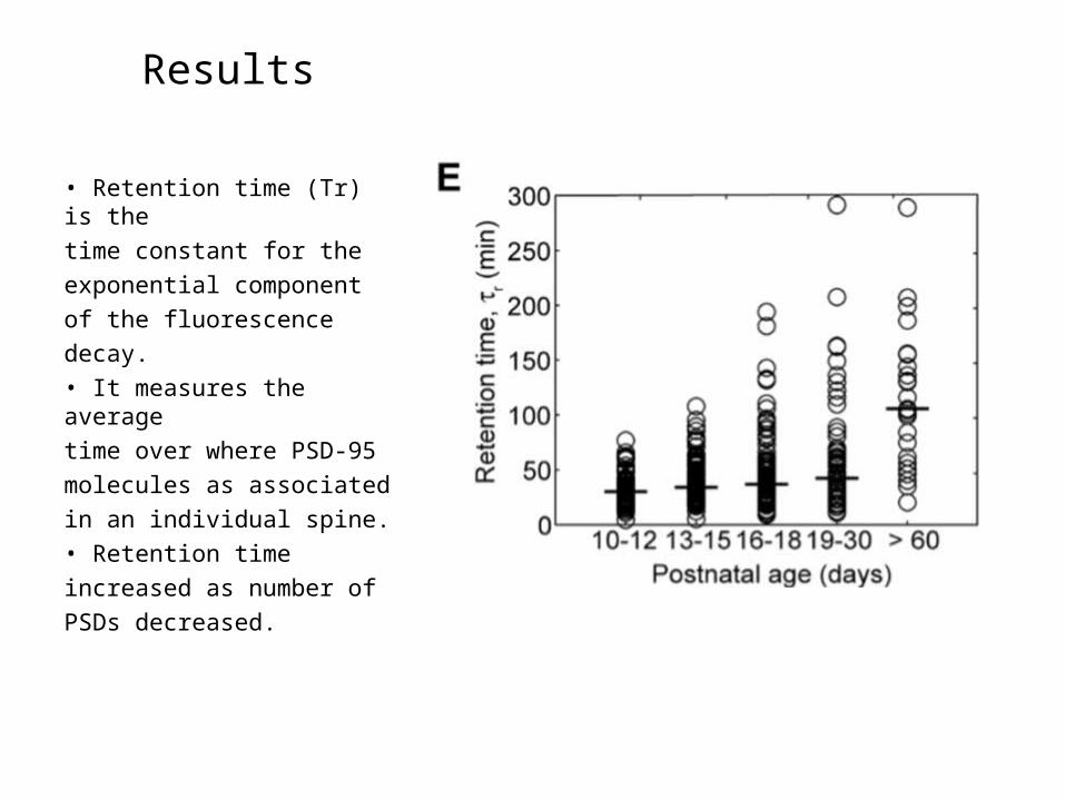
Results
• Retention time (Tr) is thetime constant for theexponential componentof the fluorescencedecay.• It measures the averagetime over where PSD-95molecules as associatedin an individual spine.• Retention timeincreased as number ofPSDs decreased.
Key Points
With increasing developmental age:• PSD-95 are bound to PSDs for approximately an
hour, significantly shorter than the lifetime of dendritic spines and their PSDs and the PSD-95.
• PSD-95 became less dynamic in the dendritic spines.
• PSDs decreased as development increased.• However, retention time in PSDs increased.
What mechanisms keep PSD-95-paGFP, thus determining the retention time?
2 possible reasons:• Retention time of PSD-95-paGFP could reflect
unbinding of PSD-95 from PSD.• PSD-95 could be trapped in the spine head of
diffusional compartmentalization by the narrow spine neck.
• Measured retention times for other proteinsnot known to be concentrated in the PSD:
• Cytoplasmic paGFP – determined by spinegeometry alone.paGFP-actin – depended on cycling of actin in dendritic spines.
• PSD-95-paGFP
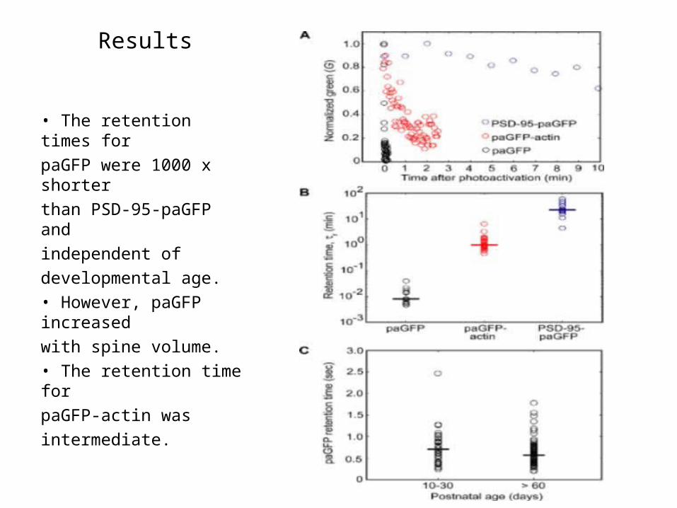
Results
• The retention times forpaGFP were 1000 x shorterthan PSD-95-paGFP andindependent ofdevelopmental age.• However, paGFP increasedwith spine volume.• The retention time forpaGFP-actin wasintermediate.
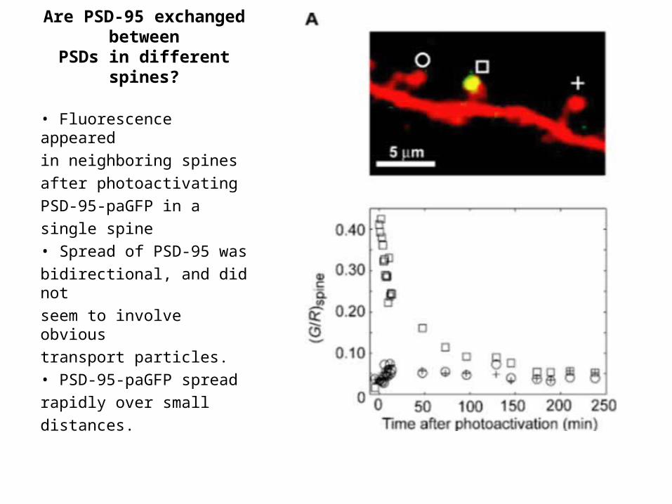
Are PSD-95 exchanged between
PSDs in different spines?
• Fluorescence appearedin neighboring spinesafter photoactivatingPSD-95-paGFP in asingle spine• Spread of PSD-95 wasbidirectional, and did notseem to involve obvioustransport particles.• PSD-95-paGFP spreadrapidly over smalldistances.
Key Points
• PSD-95 spreads from PSD to PSD by diffusion.• Hence, PSDs share a common pool of PSD-95.
Synapse-Specific Captureand Retention
• What governs the diffusional exchange of PSD-95 between spines?
• After PSD-95 unbinds from a PSD it rapidly diffuses along the dendritic shaft until captured by other PSDs.
• PSD-95 content of a PSD is roughly proportional to the PSD size(57,58)
• Stable sets of PSD-95 binding sites at individual PSDs could explain the stability of PSD-95 clusters
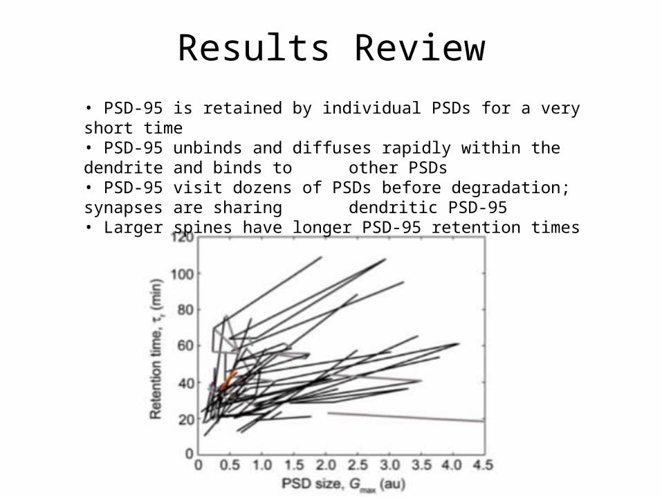
Results Review• PSD-95 is retained by individual PSDs for a very short time• PSD-95 unbinds and diffuses rapidly within the dendrite and binds to
other PSDs• PSD-95 visit dozens of PSDs before degradation; synapses are sharing
dendritic PSD-95• Larger spines have longer PSD-95 retention times
Results Review
• PSD-95 is retained by individual PSDs for a veryshort time in comparison
• PSD-95 unbinds and diffuses rapidly within thedendrite and binds to other PSDs
• PSD-95 visit dozens of PSDs before degradation;synapses are sharing dendritic PSD-95
• Larger spines have longer PSD-95 retention times• Spine stability increases with developmental age
Interpretation
• [PSD-95 binders] > [PSD-95] = competition• PSD-95 levels may determine synaptic
strength and combined with other PSDmolecules determine synaptic size.
• Redistribution of PSD-95 could play a role insynaptic plasticity (vs. gene alteration).
• Redistribution of PSD-95 (or other PSDmolecules) contributes to induction andmaintenance of LTP.