Molecular alterations in the TCR signaling pathway in ... · RESEARCH Open Access Molecular...
9
RESEARCH Open Access Molecular alterations in the TCR signaling pathway in patients with aplastic anemia Bo Li 1*† , Lixing Guo 1† , Yuping Zhang 2 , Yankai Xiao 3 , Mingjuan Wu 1,3 , Lingling Zhou 3 , Shaohua Chen 1 , Lijian Yang 1 , Xiang Lu 4 and Yangqiu Li 1,3,5* Abstract Background: A previous study has demonstrated a significantly increased CD3ζ gene expression level in aplastic anemia (AA). However, the mechanism underlying the upregulated CD3ζ mRNA expression level and that of T cell activation signaling molecules in AA patients remains unclear. Thus, we investigated the expression levels of the CD3ζ, CD28, CTLA-4, and Cbl-b genes, the SNP rs231775 in the CTLA-4 gene, and the distribution of the CD3ζ 3′- UTR splice variant in AA patients. Methods: CD3ζ 3′-UTR splice variants were identified in peripheral blood mononuclear cells (PBMCs) from 48 healthy individuals and 67 patients with AA [37 cases of severe aplastic anemia (SAA) and 30 cases of non-sever aplastic anemia (NSAA)] by RT-PCR. CD3ζ, CD28, CTLA-4, and Cbl-b gene expression was analyzed by real-time quantitative PCR. The SNP rs231775 in CTLA-4 gene was analyzed by PCR-RFLP. Results: CD3ζ and CD28 expression was significantly higher, while CTLA-4 and Cbl-b expression was significantly lower in AA patients compared with healthy individuals. Significantly higher CD3ζ expression was found in the NSAA subgroup compared with the SAA subgroup. 64 % of the AA samples had the same genotype (WT + AS + CD3ζ 3′-UTR); 22 % of the AA patients had a WT + AS - CD3ζ 3′-UTR genotype, and 14 % of the AA patients had a WT - AS + CD3ζ 3′-UTR genotype. The CD3ζ expression level of WT - AS + subgroup was the highest in the SAA patients. A significantly higher frequency of the GG genotype (mutant type, homozygous) of SNP rs231775 in CTLA-4 gene was found in the AA patients. Positive correlation between the CTLA-4 and Cbl-b gene expression levels was found in healthy individuals with the AA and AG genotypes, but not in the AA patients. Conclusions: This is the first study analyzing the expression characteristics of the CD28, CTLA-4, and Cbl-b genes in AA. Our results suggest that aberrant T cell activation may be related to the first and second signals of T cell activation in AA. The GG genotype of SNP rs231775 in CTLA-4 gene might be associated with AA risk in the Chinese population. The characteristics of CD3ζ 3′-UTR alternative splicing may be an index for evaluating the T cell activation status in AA patients, particularly in SAA patients. Keywords: CD3ζ, CD28, CTLA-4, Cbl-b, CD3ζ 3′-UTR splice variant, SNP, Gene expression level, AA * Correspondence: [email protected]; [email protected] † Equal contributors 1 Institute of Hematology, Medical College, Jinan University, Guangzhou, China Full list of author information is available at the end of the article © 2016 Li et al. Open Access This article is distributed under the terms of the Creative Commons Attribution 4.0 International License (http://creativecommons.org/licenses/by/4.0/), which permits unrestricted use, distribution, and reproduction in any medium, provided you give appropriate credit to the original author(s) and the source, provide a link to the Creative Commons license, and indicate if changes were made. The Creative Commons Public Domain Dedication waiver (http:// creativecommons.org/publicdomain/zero/1.0/) applies to the data made available in this article, unless otherwise stated. Li et al. Journal of Hematology & Oncology (2016) 9:32 DOI 10.1186/s13045-016-0261-6
Transcript of Molecular alterations in the TCR signaling pathway in ... · RESEARCH Open Access Molecular...
RESEARCH Open Access
Molecular alterations in the TCR signalingpathway in patients with aplastic anemiaBo Li1*†, Lixing Guo1†, Yuping Zhang2, Yankai Xiao3, Mingjuan Wu1,3, Lingling Zhou3, Shaohua Chen1, Lijian Yang1,Xiang Lu4 and Yangqiu Li1,3,5*
Abstract
Background: A previous study has demonstrated a significantly increased CD3ζ gene expression level in aplasticanemia (AA). However, the mechanism underlying the upregulated CD3ζ mRNA expression level and that of T cellactivation signaling molecules in AA patients remains unclear. Thus, we investigated the expression levels of theCD3ζ, CD28, CTLA-4, and Cbl-b genes, the SNP rs231775 in the CTLA-4 gene, and the distribution of the CD3ζ 3′-UTR splice variant in AA patients.
Methods: CD3ζ 3′-UTR splice variants were identified in peripheral blood mononuclear cells (PBMCs) from 48healthy individuals and 67 patients with AA [37 cases of severe aplastic anemia (SAA) and 30 cases of non-severaplastic anemia (NSAA)] by RT-PCR. CD3ζ, CD28, CTLA-4, and Cbl-b gene expression was analyzed by real-timequantitative PCR. The SNP rs231775 in CTLA-4 gene was analyzed by PCR-RFLP.
Results: CD3ζ and CD28 expression was significantly higher, while CTLA-4 and Cbl-b expression was significantlylower in AA patients compared with healthy individuals. Significantly higher CD3ζ expression was found in theNSAA subgroup compared with the SAA subgroup. 64 % of the AA samples had the same genotype (WT+AS+CD3ζ3′-UTR); 22 % of the AA patients had a WT+AS−CD3ζ 3′-UTR genotype, and 14 % of the AA patients had a WT−AS+CD3ζ 3′-UTR genotype. The CD3ζ expression level of WT−AS+ subgroup was the highest in the SAA patients. Asignificantly higher frequency of the GG genotype (mutant type, homozygous) of SNP rs231775 in CTLA-4 gene wasfound in the AA patients. Positive correlation between the CTLA-4 and Cbl-b gene expression levels was found inhealthy individuals with the AA and AG genotypes, but not in the AA patients.
Conclusions: This is the first study analyzing the expression characteristics of the CD28, CTLA-4, and Cbl-b genes inAA. Our results suggest that aberrant T cell activation may be related to the first and second signals of T cellactivation in AA. The GG genotype of SNP rs231775 in CTLA-4 gene might be associated with AA risk in theChinese population. The characteristics of CD3ζ 3′-UTR alternative splicing may be an index for evaluating the T cellactivation status in AA patients, particularly in SAA patients.
Keywords: CD3ζ, CD28, CTLA-4, Cbl-b, CD3ζ 3′-UTR splice variant, SNP, Gene expression level, AA
* Correspondence: [email protected]; [email protected]†Equal contributors1Institute of Hematology, Medical College, Jinan University, Guangzhou,ChinaFull list of author information is available at the end of the article
© 2016 Li et al. Open Access This article is distributed under the terms of the Creative Commons Attribution 4.0 InternationalLicense (http://creativecommons.org/licenses/by/4.0/), which permits unrestricted use, distribution, and reproduction in anymedium, provided you give appropriate credit to the original author(s) and the source, provide a link to the CreativeCommons license, and indicate if changes were made. The Creative Commons Public Domain Dedication waiver (http://creativecommons.org/publicdomain/zero/1.0/) applies to the data made available in this article, unless otherwise stated.
Li et al. Journal of Hematology & Oncology (2016) 9:32 DOI 10.1186/s13045-016-0261-6
BackgroundAplastic anemia (AA) is a serious hematological disordercharacterized by pancytopenia [1–3]. AA is an immune-mediated destruction of hematopoietic cells caused byabnormally activated T cells for most cases [4, 5]. Our pre-vious study has shown that, in addition to abnormal distri-bution and clonal expansion of the T cell receptor (TCR)repertoire, there is significantly higher CD3ζ expression inT cells in AA patients [6]. An abnormal CD3ζ gene expres-sion level may directly represent a characteristic of abnor-mal T cell activation.T cell recognition of antigenic peptide/major histocom-
patibility complexes plays a pivotal role in the initiationand regulation of the adaptive immune response [7–9].TCR activation plays a crucial role in T cell function. TheTCR itself does not possess signaling domains. Instead,the TCR is non-covalently coupled to a conserved multi-subunit signaling apparatus, i.e., the CD3 complex, whichcomprises the CD3εγ, CD3εδ, and CD3ζζ dimmers [10].However, the TCR/CD3 signaling complex alone is insuffi-cient for antigen-specific T cell responses and a secondpathway, co-stimulatory signaling, is required for T cellimmune responses. The co-stimulatory signaling moleculeCD28 that is found on T cells must bind B7-1 and B7-2,which are expressed on antigen presenting cells (APCs),to trigger T cell activation [11, 12]. Upon T cell activation,cytotoxic T-lymphocyte antigen-4 (CTLA-4) is inducedand outcompetes CD28 for B7-1 and B7-2 ligands, therebypreventing excessive T cell expansion [13, 14]. Thismechanism provides a key checkpoint in the regulationof T cell immunity [15, 16]. The SNP rs231775, A > Gtransition mutation which is located at exon 1 in theCTLA-4 gene, has been reported to potentially influ-ence the inhibitory function of CTLA-4 by reducing itscell surface expression [17, 18].Optimal T cell activation requires signaling through the
TCR and the CD28 co-stimulatory receptor. CD28 co-stimulation is believed to set the threshold for T cell acti-vation. Casitas B-lineage lymphoma proto-oncogene-b(Cbl-b), a RING finger E3 ubiquitin-protein ligase, is in-volved in CD28-dependent T cell activation [19]. Resultsfrom T cell activation assays in vitro have shown thatCD28 co-stimulation promotes TCR-induced Cbl-b deg-radation, whereas CTLA-4-B7 interaction potentiatesTCR-induced Cbl-b re-expression [20].Expression of the CD3ζ gene is regulated at the
transcriptional, posttranscriptional, and posttranslationallevels [21]. As previously described, there are two isoformsof the CD3ζ 3′-UTR: a wild type (WT) isoform (906 bp)and an alternatively spliced (AS) isoform (344 bp). Abnor-mal CD3ζ expression was found in T cell from SLE pa-tients, and this may be associated with decreased stabilityand translation of CD3ζ mRNAs that contain AS CD3ζ3′-UTRs [22]. However, the mechanism of upregulating
the CD3ζ mRNA expression level in AA patients isunclear.Thus, we investigated the expression level of the
CD28, CTLA-4, Cbl-b, and CD3ζ genes, the SNPrs231775 in CTLA-4 gene, CD3ζ-regulating factors, andthe distribution of the CD3ζ 3′-UTR splice variant. Weconcluded that analysis of these factors may facilitatethe comprehensive understanding of the abnormal Tcell immune characteristics of AA.
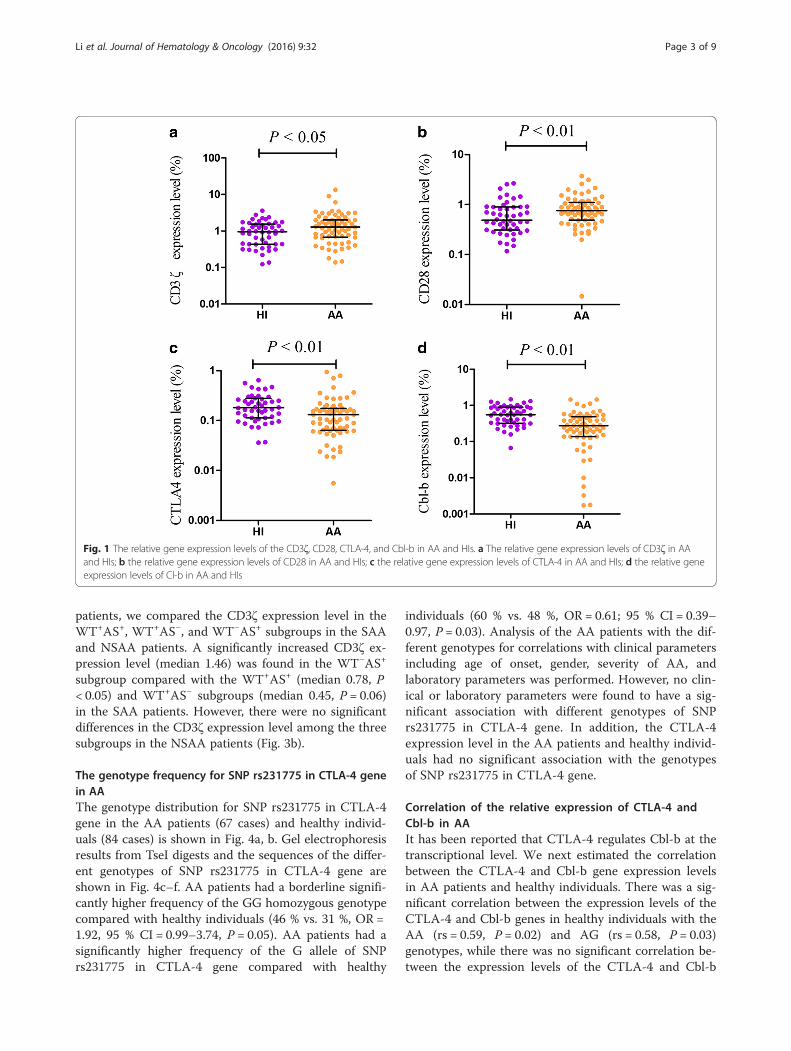
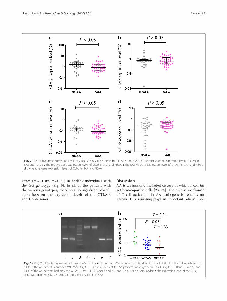
ResultsThe expression levels of CD3ζ, CD28, CTLA-4, and Cbl-b in AAThe expression levels of the CD3ζ, CD28, CTLA-4, andCbl-b genes in cDNA from PBMCs from 67 AA patientsbefore treatment and 48 healthy individuals were quanti-tatively assessed by real-time polymerase chain reaction(PCR) using the SYBR Green I technique. The resultsdemonstrated an increased expression level for CD28(median 0.75) and CD3ζ (median 1.27) in AA patientscompared with healthy individuals (CD28 0.49, P < 0.01;CD3ζ 0.95, P < 0.05; Fig. 1), while significantly decreasedCTLA-4 (median 0.13) and Cbl-b (median 0.27) expres-sion was found (CTLA-4 0.18, P < 0.01; Cbl-b 0.55, P <0.01; Fig. 1). Significantly increased CD3ζ expression (me-dian: 0.13) was found in the non-severe aplastic anemiapatients (NSAA) compared with sever aplastic anemiapatients (SAA, median: 0.18, P < 0.05). There were nosignificant differences in CD28, CTLA-4, and Cbl-bexpression between the SAA and NSAA groups (Fig. 2).
The expression characteristics of the CD3ζ gene withdifferent CD3ζ 3′-UTR isoforms in AATwo types of CD3ζ 3′-UTR splicing variants, WT and ASCD3ζ 3′-UTR, were detected in all of the healthy indi-viduals. However, a significantly lower frequency of theWT+AS+CD3ζ 3′-UTR genotype was detected in the AApatients (43 cases, P < 0.01), and a significantly higherfrequency of the WT+AS-CD3ζ 3′-UTR (15 cases,P < 0.01) and WT−AS+CD3ζ 3′-UTR (9 cases, P < 0.01)genotypes were detected in the AA patients (Fig. 3a).There were no significant differences in the frequencies ofthe three types of CD3ζ 3′-UTR between the SAA andNSAA patients although the frequency of the WT-AS+CD3ζ 3′-UTR was high in the SAA patients (19 %)compared with that of the NSAA patients (7 %), while theWT+AS+CD3ζ 3′-UTR frequency was low in the SAApatients (57 %) compared with that of the NSAA patients(73 %).Based on the CD3ζ 3′-UTR isoform expression, we
divided the 67 AA cases into three subgroups: WT+AS+,WT+AS− and WT−AS+. There were no significant differ-ences in the CD3ζ expression among the three sub-groups. To better comprehend the difference in theCD3ζ expression level between the SAA and NSAA
Li et al. Journal of Hematology & Oncology (2016) 9:32 Page 2 of 9
patients, we compared the CD3ζ expression level in theWT+AS+, WT+AS−, and WT−AS+ subgroups in the SAAand NSAA patients. A significantly increased CD3ζ ex-pression level (median 1.46) was found in the WT−AS+
subgroup compared with the WT+AS+ (median 0.78, P< 0.05) and WT+AS− subgroups (median 0.45, P = 0.06)in the SAA patients. However, there were no significantdifferences in the CD3ζ expression level among the threesubgroups in the NSAA patients (Fig. 3b).
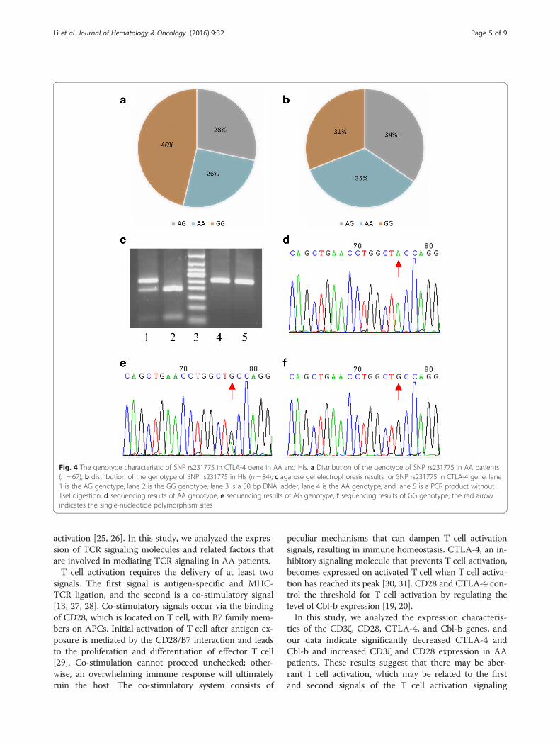
The genotype frequency for SNP rs231775 in CTLA-4 genein AAThe genotype distribution for SNP rs231775 in CTLA-4gene in the AA patients (67 cases) and healthy individ-uals (84 cases) is shown in Fig. 4a, b. Gel electrophoresisresults from TseI digests and the sequences of the differ-ent genotypes of SNP rs231775 in CTLA-4 gene areshown in Fig. 4c–f. AA patients had a borderline signifi-cantly higher frequency of the GG homozygous genotypecompared with healthy individuals (46 % vs. 31 %, OR =1.92, 95 % CI = 0.99–3.74, P = 0.05). AA patients had asignificantly higher frequency of the G allele of SNPrs231775 in CTLA-4 gene compared with healthy
individuals (60 % vs. 48 %, OR = 0.61; 95 % CI = 0.39–0.97, P = 0.03). Analysis of the AA patients with the dif-ferent genotypes for correlations with clinical parametersincluding age of onset, gender, severity of AA, andlaboratory parameters was performed. However, no clin-ical or laboratory parameters were found to have a sig-nificant association with different genotypes of SNPrs231775 in CTLA-4 gene. In addition, the CTLA-4expression level in the AA patients and healthy individ-uals had no significant association with the genotypesof SNP rs231775 in CTLA-4 gene.
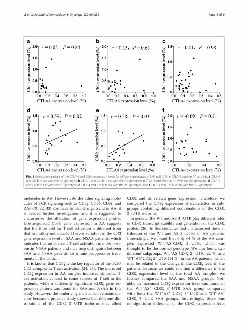
Correlation of the relative expression of CTLA-4 andCbl-b in AAIt has been reported that CTLA-4 regulates Cbl-b at thetranscriptional level. We next estimated the correlationbetween the CTLA-4 and Cbl-b gene expression levelsin AA patients and healthy individuals. There was a sig-nificant correlation between the expression levels of theCTLA-4 and Cbl-b genes in healthy individuals with theAA (rs = 0.59, P = 0.02) and AG (rs = 0.58, P = 0.03)genotypes, while there was no significant correlation be-tween the expression levels of the CTLA-4 and Cbl-b
Fig. 1 The relative gene expression levels of the CD3ζ, CD28, CTLA-4, and Cbl-b in AA and HIs. a The relative gene expression levels of CD3ζ in AAand HIs; b the relative gene expression levels of CD28 in AA and HIs; c the relative gene expression levels of CTLA-4 in AA and HIs; d the relative geneexpression levels of Cl-b in AA and HIs
Li et al. Journal of Hematology & Oncology (2016) 9:32 Page 3 of 9
genes (rs = −0.09, P = 0.71) in healthy individuals withthe GG genotype (Fig. 5). In all of the patients withthe various genotypes, there was no significant correl-ation between the expression levels of the CTLA-4and Cbl-b genes.
DiscussionAA is an immune-mediated disease in which T cell tar-get hematopoietic cells [23, 24]. The precise mechanismof T cell activation in AA pathogenesis remains un-known. TCR signaling plays an important role in T cell
Fig. 2 The relative gene expression levels of CD3ζ, CD28, CTLA-4, and Cbl-b in SAA and NSAA. a The relative gene expression levels of CD3ζ inSAA and NSAA; b the relative gene expression levels of CD28 in SAA and NSAA; c the relative gene expression levels of CTLA-4 in SAA and NSAA;d the relative gene expression levels of Cbl-b in SAA and NSAA
Fig. 3 CD3ζ 3′-UTR splicing variant isoforms in AA and HIs. a The WT and AS isoforms could be detected in all of the healthy individuals (lane 1),64 % of the AA patients contained WT+AS+CD3ζ 3′-UTR (lane 2), 22 % of the AA patients had only the WT+AS−CD3ζ 3′-UTR (lanes 4 and 5), and14 % of the AA patients had only the WT-AS+CD3ζ 3′-UTR (lanes 6 and 7). Lane 3 is a 100 bp DNA ladder; b the expression level of the CD3ζgene with different CD3ζ 3′-UTR splicing variant isoforms in SAA
Li et al. Journal of Hematology & Oncology (2016) 9:32 Page 4 of 9
activation [25, 26]. In this study, we analyzed the expres-sion of TCR signaling molecules and related factors thatare involved in mediating TCR signaling in AA patients.T cell activation requires the delivery of at least two
signals. The first signal is antigen-specific and MHC-TCR ligation, and the second is a co-stimulatory signal[13, 27, 28]. Co-stimulatory signals occur via the bindingof CD28, which is located on T cell, with B7 family mem-bers on APCs. Initial activation of T cell after antigen ex-posure is mediated by the CD28/B7 interaction and leadsto the proliferation and differentiation of effector T cell[29]. Co-stimulation cannot proceed unchecked; other-wise, an overwhelming immune response will ultimatelyruin the host. The co-stimulatory system consists of
peculiar mechanisms that can dampen T cell activationsignals, resulting in immune homeostasis. CTLA-4, an in-hibitory signaling molecule that prevents T cell activation,becomes expressed on activated T cell when T cell activa-tion has reached its peak [30, 31]. CD28 and CTLA-4 con-trol the threshold for T cell activation by regulating thelevel of Cbl-b expression [19, 20].In this study, we analyzed the expression characteris-
tics of the CD3ζ, CD28, CTLA-4, and Cbl-b genes, andour data indicate significantly decreased CTLA-4 andCbl-b and increased CD3ζ and CD28 expression in AApatients. These results suggest that there may be aber-rant T cell activation, which may be related to the firstand second signals of the T cell activation signaling
Fig. 4 The genotype characteristic of SNP rs231775 in CTLA-4 gene in AA and HIs. a Distribution of the genotype of SNP rs231775 in AA patients(n = 67); b distribution of the genotype of SNP rs231775 in HIs (n = 84); c agarose gel electrophoresis results for SNP rs231775 in CTLA-4 gene, lane1 is the AG genotype, lane 2 is the GG genotype, lane 3 is a 50 bp DNA ladder, lane 4 is the AA genotype, and lane 5 is a PCR product withoutTseI digestion; d sequencing results of AA genotype; e sequencing results of AG genotype; f sequencing results of GG genotype; the red arrowindicates the single-nucleotide polymorphism sites
Li et al. Journal of Hematology & Oncology (2016) 9:32 Page 5 of 9
molecules in AA. However, do the other signaling mole-cules of TCR signaling such as CD3γ, CD3δ, CD3ε, andZAP-70 [32, 33] also have similar change trend in AA, itis needed further investigation, and it is suggested tocharacterize the alteration of gene expression profile.Downregulated Cbl-b gene expression in AA suggeststhat the threshold for T cell activation is different fromthat in healthy individuals. There is variation in the CD3gene expression level in SAA and NSAA patients, whichindicates that an aberrant T cell activation is more obvi-ous in NSAA patients and may help distinguish betweenSAA and NSAA patients for immunosuppressive treat-ments in the clinic.It is known that CD3ζ is the key regulator of the TCR/
CD3 complex in T cell activation [34, 35]. The increasedCD3ζ expression in AA samples indicated abnormal Tcell activation at least in some subsets of T cell in thepatients, while a differently significant CD3ζ gene ex-pression pattern was found for SAA and NSAA in thisstudy. However, the underlying mechanism remains un-clear because a previous study showed that different dis-tributions of the CD3ζ 3′-UTR isoforms may affect
CD3ζ and its related gene expression. Therefore, wecompared the CD3ζ expression characteristics in sub-groups containing different combinations of the CD3ζ3′-UTR isoforms.In general, the WT and AS 3′-UTR play different roles
in CD3ζ transcript stability and generation of the CD3ζprotein [36]. In this study, we first characterized the dis-tribution of the WT and AS 3′-UTRs in AA patients.Interestingly, we found that only 64 % of the AA sam-ples expressed WT+AS+CD3ζ 3′-UTR, which wasthought to be the normal genotype. We also found twodifferent subgroups, WT+AS−CD3ζ 3′-UTR (22 %) andWT−AS+CD3ζ 3′-UTR (14 %), in the AA patients, whichmay be related to the change in the CD3ζ level in thepatients. Because we could not find a difference in theCD3ζ expression level in the total AA samples, wefurther compared the SAA and NSAA groups. Not-ably, an increased CD3ζ expression level was found inthe WT−AS+ CD3ζ 3′-UTR SAA group comparedwith both the WT+AS+ CD3ζ 3′-UTR and WT+AS−
CD3ζ 3′-UTR SAA groups. Interestingly, there wasno significant difference in the CD3ζ expression level
Fig. 5 Correlation analysis of the CTLA-4 and Cbl-b expression levels for different genotypes of SNP rs231775 in CTLA-4 gene in AA and HIs. a CTLA-4and Cbl-b in AA with the AA genotype, b CTLA-4 and Cbl-b in AA with the AG genotype, c CTLA-4 and Cbl-b in AA with the GG genotype, d CTLA-4and Cbl-b in HIs with the AA genotype, e CTLA-4 and Cbl-b in HIs with the AG genotype, and f CTLA-4 and Cbl-b in HIs with the GG genotype
Li et al. Journal of Hematology & Oncology (2016) 9:32 Page 6 of 9
between the WT+AS+, WT−AS+, and WT+AS− subgroupsfor the NSAA patients. Therefore, there may be a differ-ence in the regulation of the CD3ζ expression level and Tcell activation between SAA and NSAA. At a minimum, itis thought that the CD3ζ 3′-UTR isoforms may influencethe CD3ζ expression level in T cells from SAA, which maybe one reason for the abnormal T cell activation.It has been reported that aberrant CD3ζ expression
may be associated with the decreased stability and trans-lation of CD3ζ mRNAs with an AS CD3ζ 3′-UTR inSLE [22, 36]. In this study, the CD3ζ gene expressionlevel in some SAA patients was thought to be affectedby different splicing variants of the CD3ζ 3′-UTR. Asour results demonstrated, there was an increased CD3ζexpression level in SAA patients with the WT-AS+ CD3ζ3′-UTR genotype, and whether this is feedback regula-tion remains an open question. The T cell activationfunction in SAA patients will be examined in the futureto evaluate the influence of the CD3ζ 3′-UTR isoforms.To characterize the positive and negative regulatory fac-tors of the TCR signal pathway, it is necessary to analyzethe characteristics of the negative factors e.g., CTLA-4and Cbl-b. It has been reported that SNP rs231775 inCTLA-4 gene is associated with an increased frequencyof autoimmune diseases such as Graves’ disease, auto-immune hypothyroidism, type I diabetes, and multiplesclerosis [37–39]. ]s To determine whether this SNP alsoplays a role in AA, we analyzed its distribution in AAsamples. To our knowledge, this is the first study toanalyze this SNP in Chinese AA patients. Interestingly,we found that the homozygous GG mutant was signifi-cantly higher in AA patients, indicating that this CTLA-4 variant may have an association with susceptibility todeveloping AA. However, this result is in contrast with areport by Svahn J et al. who demonstrated that therewere no significant differences in the genotype or allelefrequency of SNP rs231775 in CTLA-4 gene betweenAA patients and healthy individuals in the Caucasianpopulation [40]. However, the CTLA-4 mRNA expres-sion level had no significant association with the geno-type of SNP rs231775 in CTLA-4 gene in this studybecause SNP rs231775 is associated with the proteinlevel [17, 18]. Further study is needed to analyze theCTLA-4 protein level in T cell from AA patients to con-firm this result.CTLA-4 regulates TCR signals via Cbl-b, and it plays
an important role in regulating it at the transcriptionallevel [20]. However, little is known about the mRNA ex-pression pattern of CTLA-4 and Cbl-b in AA patients.In this study, we found a significant positive correlationbetween CTLA-4 and Cbl-b in healthy individuals withthe AA and AG genotypes of SNP rs231775 in CTLA-4gene but not in those with the GG genotype of SNPrs231775 in CTLA-4 gene. However, there was no
significant correlation between CTLA-4 and Cbl-b inany of the patients. These results indicate that CTLA-4loses its proper regulatory role of Cbl-b expression atthe transcriptional level in AA patients.
ConclusionIn conclusion, this is the first study to analyze theexpression characteristics of the CD28, CTLA-4, andCbl-b genes, the distribution of the CD3ζ 3′-UTR splicevariant in AA patients, and the SNP rs231775 in CTLA-4 gene in Chinese AA patients. We found that aberrantT cell activation may be associated with either the firstor second T cell activation signaling molecules in AA.Different CD3ζ expression patterns in SAA and NSAApatients suggest different abnormal alternative CD3ζpathway patterns in SAA and NSAA patients. Themutant type homozygous GG and G allele for SNPrs231775 in CTLA-4 gene might be associated with AArisk for the Chinese population.
MethodsSamplesThe AA group consisted of 67 patients with newly diag-nosed AA (36 cases with SAA and 31 with NSAA, in-cluding 37 males and 30 females; median age 34 years,range 9–78 years). The information and clinical data ofthe patients are described in Table 1. Forty-eight healthyindividuals (21 males and 27 females; median age35.5 years, range 20–80 years) were used for detectingCD3ζ, CD28, CTLA-4, and Cbl-b genes expression levelsand CD3ζ 3′-UTR isoform, and 84 healthy individuals(40 males and 44 females; median age 33 years, range 6–88 years) were used for detecting for SNP rs231775 inCTLA-4 gene. AA diagnoses were established by bonemarrow biopsy and peripheral blood counts. All of the
Table 1 The demographic and clinical characteristics of AApatients
Patients characteristics (n = 67) No. (%)
Age (years) Median 34.0
Range 9.0–78.0
Gender
Male 37(55.2)
Female 30(44.8)
Severity of AA
AA 31 (46.3)
SAA 36 (53.7)
Clinical data
Hb (g/L) Median 65.0
ANC (109/L) Median 0.38
PLT (109/L) Median 11.5
Hb hemoglobin, ANC absolute neutrophil count, PLT platelet
Li et al. Journal of Hematology & Oncology (2016) 9:32 Page 7 of 9
procedures were performed according to the guidelinesof the Medical Ethics committee of the health bureau ofthe Guangdong Province of China. Peripheral bloodmononuclear cell (PBMC) isolation, RNA and DNA ex-traction, and cDNA synthesis were performed accordingto the manufacturer’s instructions.
RT-PCR for CD3ζ 3′-UTR isoformThe primer specificity for CD3ζ 3′-UTR amplificationwas as follows: forward 5′-CAGCCAGGGGATTTCAC-CACTCAAAG-3′ reverse 5′-CCCTAGTACATTGACGGGTTTTTCCTG-3′ [41]. The amplification was carriedout after initial denaturation at 94 °C for 4 min, 30 cyclesat 94 °C, 45 s; 67 °C, 1 min; 72 °C for 2 min; and a finalextension at 72 °C for 7 min. After completion of thePCR, 20 μl of the PCR products were electrophoresedon a 1.5 % agarose gel.
Real-time relative quantitative PCR for the CD3ζ, CD28,CTLA-4, and Cbl-b genesReal-time PCR using the SYBR Green I method [42, 43]was used to examine the CD3ζ, CD28, CTLA-4, andCbl-b genes with cDNA obtained from the PBMCs of AApatients and healthy individuals. The CD3ζ and β2Mprimer sequences and PCR conditions were previouslydescribed [26]. The other primer sequences were asfollows: CD28: 5′-GAAACACCTTTGTCCAA GTC-3′and 5′-GGGGAGTCATGTTCATGTAG-3′, CTLA-4: 5′-GTCAGCCTGCCGAAGC ACT-3′ and 5′-GTCAGCCTGCCGAAGCACT-3′, and Cbl-b: 5′-CCCTGGAATTGACCATTGGG-3′ and 5′-ACTTGCCCAACTCAGTGAGAA-3′. The real-time PCR reactions were performed ina total volume of 25 μl containing approximately 1 μlcDNA, 0.5 μM of each primer pair, and 11.25 μl 2.5×RealMasterMix (Tiangen, Beijing). After an initial de-naturation at 95 °C for 15 min, 40 cycles of the followingprocedure were performed using an MJ Research DNAEngine Opticon 2 PCR cycler (BIO-RAD, Hercules, CA,USA): 15 s at 95 °C and 30 s at 60 °C followed by 1 s at80 °C for plate reading. The 2(–ΔCT) method was used toanalyze the genes of interest relative to an internal controlgene.
Polymerase chain reaction and restriction fragmentlength polymorphism (RFLP)The SNP rs231775 in CTLA-4 gene exon 1 were ampli-fied using the following primers: 5′-CACATGTGTAA-TACATATCTGGG-3′ and 5′-TTGCAGAAGACAGGGATG AAGA-3′. The polymerase chain reaction (PCR)mixture contained 100 ng of genomic DNA, 0.1 mMeach of deoxynucleotide triphosphates, 12.5 pmol eachof the primers, and 1 U Taq polymerase (Applied Biosys-tems, Foster City, CA, USA) in a 25 μl final volume. Thesamples were heated at 95 °C for 2 min followed by
35 cycles of 94 °C for 60 s, 60 °C for 60 s, and 72 °C for60 s with a final extension at 72 °C for 5 min. The PCRproducts (50 μl) were digested overnight with the re-striction enzyme TseI (NEB, UK) according to the man-ufacturer’s protocol, and they were analyzed by 2.5 %agarose gel electrophoresis. The digestion of PCR prod-ucts in the presence of the restriction sites is indicativeof the presence of the G allele. The finding of a singleband of 250 bp is indicative of the AA genotype,whereas the GG genotype is indicated by the finding oftwo bands at 189 and 61 bp. On the other hand, theAG genotype is characterized by the appearance ofthree bands: one at 250 bp and the others at 189 and61 bp. The different genotypes were successfully con-firmed by DNA sequencing.
Statistical analysisThe Mann-Whitney test was performed to compareeach gene expression level between different groups.Spearman correlation analyses were used to estimatecorrelations between different gene expression levels.Fisher’s exact test was used to compare genotypes orallelic frequencies between different groups. A P < 0.05was considered to be statistically significant.
Competing interestsThe authors declare that they have no competing interests.
Authors’ contributionsBL and YQL contributed to concept development and study design. BL, LXG,YKX, MJW, and LLZ performed the laboratory studies. YPZ, XL, SHC, and LJYwere responsible for collection of clinical data. BL and YQL coordinated thestudy and helped draft the manuscript. All authors read and approved thefinal manuscript.
AcknowledgementsThis study was sponsored by grants from the National Natural ScienceFoundation of China (#81370605 and #81460026), a China Post-doctoral ScienceFoundation funded project (#20070410840), the Natural Science Foundation ofGuangdong Province (#S2012010008794 and #S2013020012863), the Technol-ogy Program of Guangdong Province (#2014A020212209), the Foundation forHigh-level Talents in Higher Education of Guangdong, China (#[2013]246-54), aScience and Information Technology of Guangzhou funded basic research forapplication project (#2011 J4100028), and Fundamental Research Funds for theCentral Universities (#21612425).
Author details1Institute of Hematology, Medical College, Jinan University, Guangzhou,China. 2Department of Hematology, Guangzhou First Municipal People’sHospital Affiliated to Guangzhou Medical College, Guangzhou, China. 3KeyLaboratory for Regenerative Medicine of Ministry of Education, JinanUniversity, Guangzhou, China. 4Department of Hematology, AffiliatedHospital of Youjiang Medical University for Nationalities, Baise, China.5Department of Hematology, First Affiliated Hospital, Jinan University,Guangzhou, China.
Received: 26 January 2016 Accepted: 18 March 2016
References1. Young NS, Scheinberg P, Calado RT. Aplastic anemia. Curr Opi Hematol.
2008;15(3):162–8.2. Young NS, Ogawa S. Somatic mutations and clonal hematopoiesis in aplastic
anemia. N Engl J Med. 2015;373(17):1675–6.
Li et al. Journal of Hematology & Oncology (2016) 9:32 Page 8 of 9
3. Killick SB, Bown N, Cavenagh J, Dokal I, Foukaneli T, Hill A, et al. Guidelinesfor the diagnosis and management of adult aplastic anaemia. Br JHaematol. 2016;172(2):187–207.
4. Vo PT, Pantin J, Ramos C, Cook L, Cho E, Kurlander R, et al. Conditioningwith rabbit versus horse ATG dramatically alters clinical outcomes inidentical twins with severe aplastic anemia transplanted with the sameallogeneic donor. J Hematol Oncol. 2015;8:78.
5. Hosokawa K, Muranski P, Feng X, Townsley DM, Liu B, Knickelbein J, et al.Memory stem T cells in autoimmune disease: high frequency of circulatingCD8+ memory stem cells in acquired aplastic anemia. J Immunol. 2016;196(4):1568–78.
6. Li B, Liu S, Niu Y, Fang S, Wu X, Yu Z, et al. Altered expression of the TCRsignaling related genes CD3 and FcεRIγ in patients with aplastic anemia.J Hematol Oncol. 2012;5:6.
7. Clemente-Casares X, Blanco J, Ambalavanan P, Yamanouchi J, Singha S,Fandos C, et al. Expanding antigen-specific regulatory networks to treatautoimmunity. Nature. 2016;17. doi: 10.1038/nature16962.
8. Ji X, Zhang L, Peng J, Hou M. T cell immune abnormalities in immunethrombocytopenia. J Hematol Oncol. 2014;7:72.
9. Wooldridge L, Individual MHC. I-restricted T-cell receptors are characterizedby a unique peptide recognition signature. Front Immunol. 2013;4:199.
10. Li Y, Chen S, Yang L, Chen S, Lin C, Wang L, et al. Change in expressionpattern of TCR-CD3 complex in patients with multiple myeloma.Hematology. 2011;16(3):143–50.
11. Ville S, Poirier N, Blancho G, Vanhove B. Co-stimulatory blockade of theCD28/CD80-86/CTLA-4 balance in transplantation: impact on memory Tcells? Front Immunol. 2015;6:411.
12. Goel G, Sun W. Advances in the management of gastrointestinalcancers—an upcoming role of immune checkpoint blockade. J HematolOncol. 2015;8:86.
13. Chen L, Flies DB. Molecular mechanisms of T cells co-stimulation andco-inhibition. Nat Rev Immunol. 2013;13(4):227–42.
14. Nurieva RI, Liu X, Dong C. Yin-Yang of costimulation: crucial controls ofimmune tolerance and function. Immunol Rev. 2009;229(1):88–100.
15. Aronin A, Amsili S, Prigozhina TB, Tzdaka K, Shen R, Grinmann L, et al. Highlyefficient, in-vivo Fas-mediated apoptosis of B-cell lymphoma by hexamericCTLA4-FasL. J Hematol Oncol. 2014;7:64.
16. Buchbinder E, Hodi FS. Cytotoxic T lymphocyte antigen-4 and immunecheckpoint blockade. J Clin Invest. 2015;125(9):3377–83.
17. Barton A, Jury F, Eyre S, Bowes J, Hinks A, Ward D, et al. Haplotype analysisin simplex families and novel analytic approaches in case-control cohortreveal no evidence of association of the CTLA-4 gene with rheumatoidarthritis. Arthritis Rheum. 2004;50(3):748–52.
18. Muñoz-Valle JF, Valle Y, Padilla-Gutiérrez JR, Parra-Rojas I, Rangel-VillalobosH, Vázquez del Mercado M. The +49A>G CTLA-4 polymorphism isassociated with rheumatoid arthritis in Mexican population. Clin Chim Acta.2010;411(9-10):725–8.
19. Krawczyk CM, Jones RG, Atfield A, Bachmaier K, Arya S, Odermatt B, et al.Differential control of CD28-regulated in vivo immunity by the E3 ligaseCbl-b. J Immunol. 2005;174(3):1472–8.
20. Li D, Gál I, Vermes C, Alegre ML, Chong AS, Chen L, et al. Cutting edge:Cbl-b: one of the key molecules tuning CD28 and CTLA-4-mediated T cellcostimulation. J Immunol. 2004;173(12):7135–9.
21. Clevers H, Alarcon B, Wileman T, Terhorst C. The T cell receptor/CD3complex: a dynamic protein ensemble. Annu Rev Immunol. 1988;6:629–62.
22. Nambiar MP, Enyedy EJ, Warke VG, Krishnan S, Dennis G, Kammer GM, et al.Polymorphisms/mutations of TCR-zeta-chain promoter and 3′untranslatedregion and selective expression of TCR zeta-chain with an alternativelyspliced 3′untranslated region in patients with systemic lupus erythematosus.J Autoimmun. 2001;16(2):133–42.
23. Hosokawa K, Muranski P, Feng X, Keyvanfar K, Townsley DM, Dumitriu B,et al. Identification of novel microRNA signatures linked to acquired aplasticanemia. Haematologica. 2015;100(12):1534–45.
24. Yan L, Fu R, Liu H, Wang H, Liu C, Wang T, et al. Abnormal quantity andfunction of regulatory T cells in peripheral blood of patients withsevereaplastic anemia. Cell Immunol. 2015;296(2):95–105.
25. Liu X, Berry CT, Ruthel G, Madara JJ, MacGillivray K, Gray CM, et al. T cellreceptor-induced NF-κB signaling and transcriptional activation areregulated by STIM1- and Orai1-mediated calcium entry. J Biol Chem. 2016.(In press).
26. Schnorfeil FM, Lichtenegger FS, Emmerig K, Schlueter M, Neitz JS, Draenert R,et al. T cells are functionally not impaired in AML: increased PD-1 expression isonly seen at time of relapse and correlates with a shift towards the memory Tcell compartment. J Hematol Oncol. 2015;8:93.
27. Xie A, Zheng X, Khattar M, Schroder P, Stepkowski S, Xia J, et al. TCRstimulation without co-stimulatory signals induces expression of"tolerogenic" genes in memory CD4 T cells but does not compromise cellproliferation. Mol Immunol. 2015;63(2):406–11.
28. Xu L, Zhang Y, Luo G, Li Y. The roles of stem cell memory T cells inhematological malignancies. J Hematol Oncol. 2015;8(1):113.
29. Gardner D, Jeffery LE, Sansom DM. Understanding the CD28/CTLA-4(CD152) pathway and its implications for costimulatory blockade. Am JTransplant. 2014;14(9):1985–91.
30. Tsai KK, Daud AI. Nivolumab plus ipilimumab in the treatment of advancedmelanoma. J Hematol Oncol. 2015;8(1):123.
31. Callahan MK, Postow MA, Wolchok JD. CTLA-4 and PD-1 pathway blockade:combinations in the clinic. Front Oncol. 2015;4:385.
32. Köhnke T, Krupka C, Tischer J, Knösel T, Subklewe M. Increase of PD-L1expressing B-precursor ALL cells in a patient resistant to the CD19/CD3-bispecificT cell engager antibody blinatumomab. J Hematol Oncol. 2015;8(1):111.
33. Liao Z, Zhou L, Wang C, He Z, Wang X, Luo X, et al. Characteristics of TCRζ,ZAP-70, and FcɛRIγ gene expression in patients with T- and NK/T-celllymphoma. DNA Cell Biol. 2015;34(3):201–7.
34. Chen S, Zha X, Shi L, Zhou L, Yang L, Li B, et al. Upregulated TCRζimproves cytokine secretion in T cell from patients with AML. J HematolOncol. 2015;8:72.
35. Niu Y, Yu W, Fang S, Liu S, Yang Z, Liu W, et al. Lead poisoning influencesTCR-related gene expression patterns in peripheral blood T-lymphocytes ofexposed workers. J Immunotoxicol. 2015;12(1):92–7.
36. Nambiar MP, Enyedy EJ, Warke VG, Krishnan S, Dennis G, Wong HK, et al. Tcell signaling abnormalities in systemic lupus erythematosus are associatedwith increased mutations/polymorphisms and splice variants of T cellreceptor zeta chain messenger RNA. Arthritis Rheum. 2001;44(6):1336–50.
37. Du P, Ma X, Wang C. Associations of CTLA4 gene polymorphisms withGraves’ ophthalmopathy: a meta-analysis. Int J Genomics. 2014;2014:537969.
38. Wang J, Liu L, Ma J, Sun F, Zhao Z, Gu M. Common variants on cytotoxic Tlymphocyte antigen-4 polymorphisms contributes to type 1 diabetessusceptibility: evidence based on 58 studies. PLoS One. 2014;9(1):e85982.
39. Banelli B, Morabito A, Laurent S, Piccioli P, Dozin B, et al. A novel multiplexpyrosequencing assay for genotyping functionally relevant CTLA-4polymorphisms: potential applications in autoimmunity and cancer. HumImmunol. 2014;75(8):730–9.
40. Svahn J, Capasso M, Lanciotti M, Marrone A, Haupt R, Bacigalupo A, et al.The polymorphisms -318C>T in the promoter and 49A>G in exon 1 ofCTLA4 and the risk of aplastic anemia in a Caucasian population. BoneMarrow Transplant. 2005;35 Suppl 1:S89–92.
41. Weissman AM, Hou D, Orloff DG, Modi WS, Seuanez H, O'Brien SJ, et al.Molecular cloning and chromosomal localization of human T-cell receptorchain: Distinction from molecular CD3 complex. Proc Natl Acad Sci U S A.1988;85:9709–13.
42. Zeng C, Yu X, Lai J, Yang L, Chen S, Li Y. Overexpression of the long non-coding RNA PVT1 is correlated with leukemic cell proliferation in acutepromyelocytic leukemia. J Hematol Oncol. 2015;8(1):126.
43. Skvarova Kramarzova K, Fiser K, Mejstrikova E, Rejlova K, Zaliova M, et al.Homeobox gene expression in acute myeloid leukemia is linked to typicalunderlying molecular aberrations. J Hematol Oncol. 2014;7:94.
• We accept pre-submission inquiries
• Our selector tool helps you to find the most relevant journal
• We provide round the clock customer support
• Convenient online submission
• Thorough peer review
• Inclusion in PubMed and all major indexing services
• Maximum visibility for your research
Submit your manuscript atwww.biomedcentral.com/submit
Submit your next manuscript to BioMed Central and we will help you at every step:
Li et al. Journal of Hematology & Oncology (2016) 9:32 Page 9 of 9