Mining Sequential Patterns and Tree Patterns to Detect Erroneous Sentences
Mobility and Core-Protein Binding Patterns of Disordered C‑TerminalTails in β‑Tubulin IsotypesYoann Laurin,† Joel Eyer,‡ Charles H. Robert,† Chantal Prevost,† and Sophie Sacquin-Mora*,†
†Laboratoire de Biochimie Theorique, UPR 9080 CNRS, Institut de Biologie Physico-Chimique, 13 rue Pierre et Marie Curie, 75005Paris, France‡Laboratoire de Neurobiologie & Transgenese, UPRES EA 3143, INSERM, Centre Hospitalier Universitaire, Angers, France
*S Supporting Information
ABSTRACT: Although they play a significant part in theregulation of microtubule structure, dynamics, and function, thedisordered C-terminal tails of tubulin remain invisible toexperimental structural methods and do not appear in thecrystallographic structures that are currently available in theProtein Data Bank. Interestingly, these tails concentrate most ofthe sequence variability between tubulin isotypes and are thesites of the principal post-translational modifications undergoneby this protein. Using homology modeling, we developed twocomplete models for the human αI/βI- and αI/βIII-tubulinisotypes that include their C-terminal tails. We then investigatedthe conformational variability of the two β-tails using long time-scale classical molecular dynamics simulations that revealedsimilar features, notably the unexpected presence of common anchoring regions on the surface of the tuulin dimer, but alsodistinctive mobility or interaction patterns, some of which could be related to the tail lengths and charge distributions. We alsoobserved in our simulations that the C-terminal tail from the βI isotype, but not the βIII isotype, formed contacts in the putativebinding site of a recently discovered peptide that disrupts microtubule formation in glioma cells. Hindering the binding site in theβI isotype would be consistent with this peptide’s preferential disruption of microtubule formation in glioma, whose cellsoverexpress βIII, compared to normal glial cells. While these observations need to be confirmed with more intensive sampling,our study opens new perspectives for the development of isotype-specific chemotherapy drugs.
Microtubules (MTs) are rigid tubular fibers composed ofα/β-tubulin heterodimers and are key components of
the cytoskeleton, controlling essential cellular functions such asmitosis, cell signaling, and intracellular transport.1 In manyorganisms, both α- and β-tubulins are encoded by multigenefamilies, comprising, for instance, six and seven isotypes,respectively, in the human genome.2 While specialized cellsmay express a single isotype, most tissues will express several,and as a result, microtubules are usually made of a mixture ofisotypes with their composition varying by cell type.3 Tubulinisotype βIII has been by far the most extensively studied inhuman cancer. Its expression in normal cells and tissues isusually neuronally associated, but it is also widely observed innumerous human malignancies4,5 and has been identified as astrong indicator of poor clinical outcomes.6
On the MT outer surface, the well-defined tubulin globulardomain is decorated by a highly charged carboxy-terminal tail(CTT) thought to play a critical role in regulating MTassembly.7−14 Interestingly, CTTs of α- and β-tubulin are themain sites of sequence variation among isotypes. For example,βI, the most frequently expressed isotype, and βIII isotypesdiffer by only 13 amino acids within their 1−429 fragment,while their CTTs are completely different.5 CTTs are also the
site of the majority of post-translational modifications (such astyrosination, glutamylation, or phosphorylation15,16). Despiteits important role in proper MT assembly, little is known aboutthe CTT conformation and its impact on tubulin’s interactionwith other proteins. Early experimental studies using nuclearmagnetic resonance (NMR) and circular dichroism have shownthat CTTs are disordered segments,17,18 and as a consequence,they do not appear in the tubulin crystal structures currentlyavailable in the Protein Data Bank (PDB).Because of the critical role they play in mitosis, MTs are
important cancer drug targets. Antimitotic compounds interferewith the polymerization or depolymerization of α,β-tubulindimers into MTs, thus inhibiting cancer cell division andproliferation.19,20 Widely used antitumor drugs include taxanes,vinca alkaloids or colchicin, which interact with tubulin viathree distinct and well-documented binding sites.19−21
Remarkably, none of these sites lie near the tubulin CTTs,and therefore, most molecular modeling studies of tubulin−ligand interactions did not include this disordered segment in
Received: September 28, 2016Revised: March 10, 2017Published: March 14, 2017
Article
pubs.acs.org/biochemistry
© XXXX American Chemical Society A DOI: 10.1021/acs.biochem.6b00988Biochemistry XXXX, XXX, XXX−XXX
their representation.21−28 More generally, few theoreticalstudies of tubulin take into account the CTTs, although, in aseminal work, Luchko et al. investigated the conformationalvariability of the isolated β-CTTs for nine isotypes using replicaexchange molecular dynamics simulations, but without takinginto account the main tubulin globular domain.29 Subsequently,Freedman et al. used molecular dynamics to predict theinteraction of the C-terminal tails with the tubulin heterodimerin the case of the human αIV-βI isotype.30 Two additionalstudies explicitly model the CTTs while investigating theinteraction of tubulin with proteins like γ-synuclein31 orkinesin,32 which bind the tubulin surface in the vicinity of theCTTs.The NFL-TBS.40−63 peptide is a recently discovered
tubulin binding agent derived from the light neurofilamentprotein, which has demonstrated a high capacity for theinhibition of microtubule formation in an in vitro polymer-ization assay.33 Further studies of this peptide have shown thatin vitro it is more efficiently internalized by glioma cells than bynormal cells, this difference not being species-specific.34
Moreover, once internalized by glioma cells, the peptidestrongly affected their microtubule network, attenuatedproliferation, and led to apoptosis. These in vitro propertiesof the NFL-TBS.40−63 peptide were also observed in vivo,when the peptide was injected into the brains of glioma-bearingrats, and make this peptide a promising candidate for malignantglioma treatment. Our own previous modeling study of theinteraction modes of the NFL-TBS.40−63 peptide on thetubulin surface35 showed that, while lacking a stable structure,the peptide may bind to a unique specific site located near theβ-tubulin CTT. This suggests that the NFL-TBS.40−63peptide binding to this site might hinder MT formation andits dynamic regulation in the cell by modifying the β-C-terminaltail mobility, thus preventing important intra- and interdimer
contacts within MTs, and interfering with MT-associatedproteins that usually bind on the MT’s outer side. As aconsequence, the proper modeling of the CTT dynamicsappears to be an essential issue, if we want to understandcorrectly on a molecular level the interaction between the NFL-TBS.40−63 peptide and tubulin.In this perspective, using homology modeling, we developed
two complete models for the human αI/βI- and αI/βIII-tubulinisotypes that include their C-terminal tails. We theninvestigated the mobilities of these two β-CTTs using classicalmolecular dynamics (MD) simulations and showed how eachappears to present specificities in its interaction pattern with thetubulin surface that may be related to its length and chargedistribution. As mentioned previously, tubulin isotype βIII isoverexpressed in cancer cells, and this overexpression isassociated with resistance to antimitotic drugs such astaxanes.4,36 Therefore, our work helps pave the way for abetter understanding of the molecular origins of this resistanceand opens new perspectives for the development of isotype-specific chemotherapy drugs.
■ MATERIALS AND METHODSComplete Models for the Human Tubulin Isotypes.
We used homology modeling to build the human αI/βI- andαI/βIII-tubulin isotypes. Each heterodimer is based on thesame template, i.e., a double tubulin heterodimer bound withstathmin (PDB entry 1SA0), which was chosen for its highdegree of similarity with the target sequences and which is themost complete of all available crystal structures. The 1SA0structure is already present in the template database ofModeller version 9.14 that we used, which also includes the1TUB structure and domains of FtsZ, a prokaryotic homologueof tubulin. The Modeller process is based on a classiccomparative modeling method consisting of four sequential
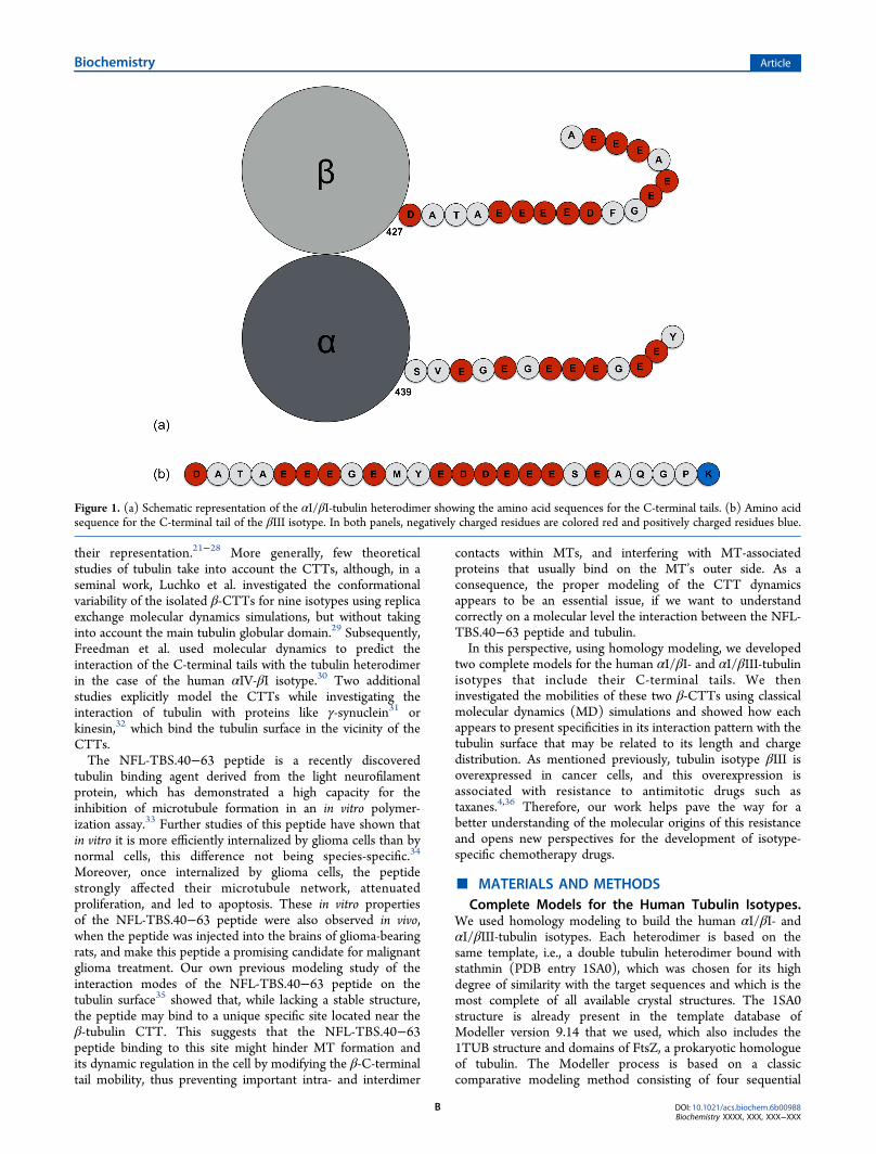
Figure 1. (a) Schematic representation of the αI/βI-tubulin heterodimer showing the amino acid sequences for the C-terminal tails. (b) Amino acidsequence for the C-terminal tail of the βIII isotype. In both panels, negatively charged residues are colored red and positively charged residues blue.
Biochemistry Article
DOI: 10.1021/acs.biochem.6b00988Biochemistry XXXX, XXX, XXX−XXX
B
steps: template selection, template−target alignment, modelbuilding, and a final model evaluation.37,38 The target sequencesfor human tubulin were retrieved from the UniProtKB Website39 as accession numbers Q71U36, P07437, and Q13509 forthe αI-, βI-, and βIII-subunits, respectively. Because of the highdegree of similarity between the target sequences and thestructural templates [>75% sequence identity (see Table SI-1)],there was no need for a manual rearrangement of thealignments. To model the tubulin CTTs, which are not presentin the template structures, Modeller performs an ab initioconformational search. For each β isotype, the five CTTstructures with the lowest Modeller score, based on energyscores [DOPE and MolPDF (see Table SI-2)], were selected.Note that for each tubulin isoform, the selected CTT structurespresent very similar energy scores with around 1 and 10%variations for the DOPE and MolPDF scores, respectively.The CTT for the αI-subunit starts with residue αSer439 and
contains 13 amino acids; in the case of the βI- and βIII-subunits, the CTT starts with residue βAsp427 and contains 18and 24 amino acids, respectively (see Figure 1). Theconventional protonation states at pH 7.0 were used for allresidues; as a consequence, the β-CTTs in both isotypes bearan important negative charge (−11e), because of their highnumber of glutamate and aspartate residues (see Table 1). Note
that the βIII-CTT ends with a lysine residue bearing a positivecharge. Subunits, modeled separately, were assembled to formstraight αI/βI and αI/βIII heterodimers using a spatialalignment on the highest-resolution structure available for thisconformation (PDB entry 1JFF, resolution of 3.5 Å). Finally,before performing MD simulations, we used SCWRL440 on thetubulin heterodimers to optimize the side-chain conformations.Molecular Dynamics Simulations. For both αI/βI and
αI/βIII heterodimers, we then performed MD simulations oneach of the five models that were produced with differentconformations of the β-CTTs. We used Gromacs,41 version5.0.4, with the OPLS-AA force field42 with periodic boundaryconditions. The first step of the simulation procedure is an invacuo minimization of the structure with the steepest descentalgorithm during 1000 steps without any constraints. We addeda triclinic water box of 2 nm around the tubulin, filled with aTIP3P molecule type,43 and the system was neutralized withthe addition of ions randomly placed in the box whilemaintaining the NaCl concentration at 150 mM. The totalsystem contains around 13700 atoms for the tubulinheterodimer, 136000 water molecules, and 320 ions (withapproximatively 180 Na+ and 140 Cl− ions, because of thehighly negatively charged CTTs). A second minimization stepwas performed with the same set of parameters as before during5000 steps to prevent possible water clashes with the tubulin.Molecular dynamics were performed using an integration timestep of 2 fs. Initial heating and pre-equilibration wereperformed by assigning random velocities from the Maxwell−Boltzmann distribution followed by 100 ps of dynamics under
NVT conditions and then 100 ps under NPT conditions.During this process and for all subsequent dynamics, thetemperature was fixed at 300 K using the velocity rescalemethod.44 All bonds were constrained with the LINCSalgorithm,45 and electrostatic interactions were computedusing the particle mesh Ewald method.46 For pressure couplingduring the NPT equilibration, we used the Parinello−Rahmanmethod47 at a value of 1 atm. Production phases were finallydone using the same set of parameters and algorithms during100 ns. Trajectories were saved every 10 ps.
Trajectory Analysis. Gromacs utilities were used for basicobservations along with VMD48 for visual inspection. Moreelaborate analyses used homemade scripts or programs. As theroot-mean-square deviation (RMSD) is not sufficient forinvestigating the flexibility of the β-terminal tails during theMD simulations, we performed additional analysis of the CTTposition with regard to the main tubulin heterodimer axis (axisz in Figure 2), radius of gyration, and pattern of contacts withthe main tubulin body.
■ RESULTS
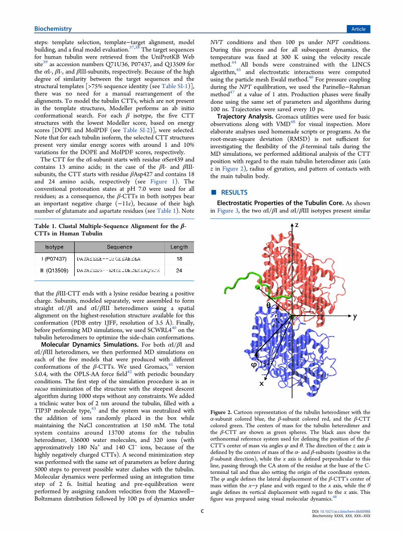
Electrostatic Properties of the Tubulin Core. As shownin Figure 3, the two αI/βI and αI/βIII isotypes present similar
Table 1. Clustal Multiple-Sequence Alignment for the β-CTTs in Human Tubulin
Figure 2. Cartoon representation of the tubulin heterodimer with theα-subunit colored blue, the β-subunit colored red, and the β-CTTcolored green. The centers of mass for the tubulin heterodimer andthe β-CTT are shown as green spheres. The black axes show theorthonormal reference system used for defining the position of the β-CTT’s center of mass via angles φ and θ. The direction of the z axis isdefined by the centers of mass of the α- and β-subunits (positive in theβ-subunit direction), while the x axis is defined perpendicular to thisline, passing through the CA atom of the residue at the base of the C-terminal tail and thus also setting the origin of the coordinate system.The φ angle defines the lateral displacement of the β-CTT’s center ofmass within the x−y plane and with regard to the x axis, while the θangle defines its vertical displacement with regard to the x axis. Thisfigure was prepared using visual molecular dynamics.48
Biochemistry Article
DOI: 10.1021/acs.biochem.6b00988Biochemistry XXXX, XXX, XXX−XXX
C
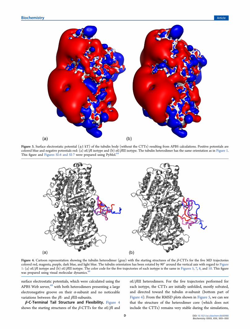
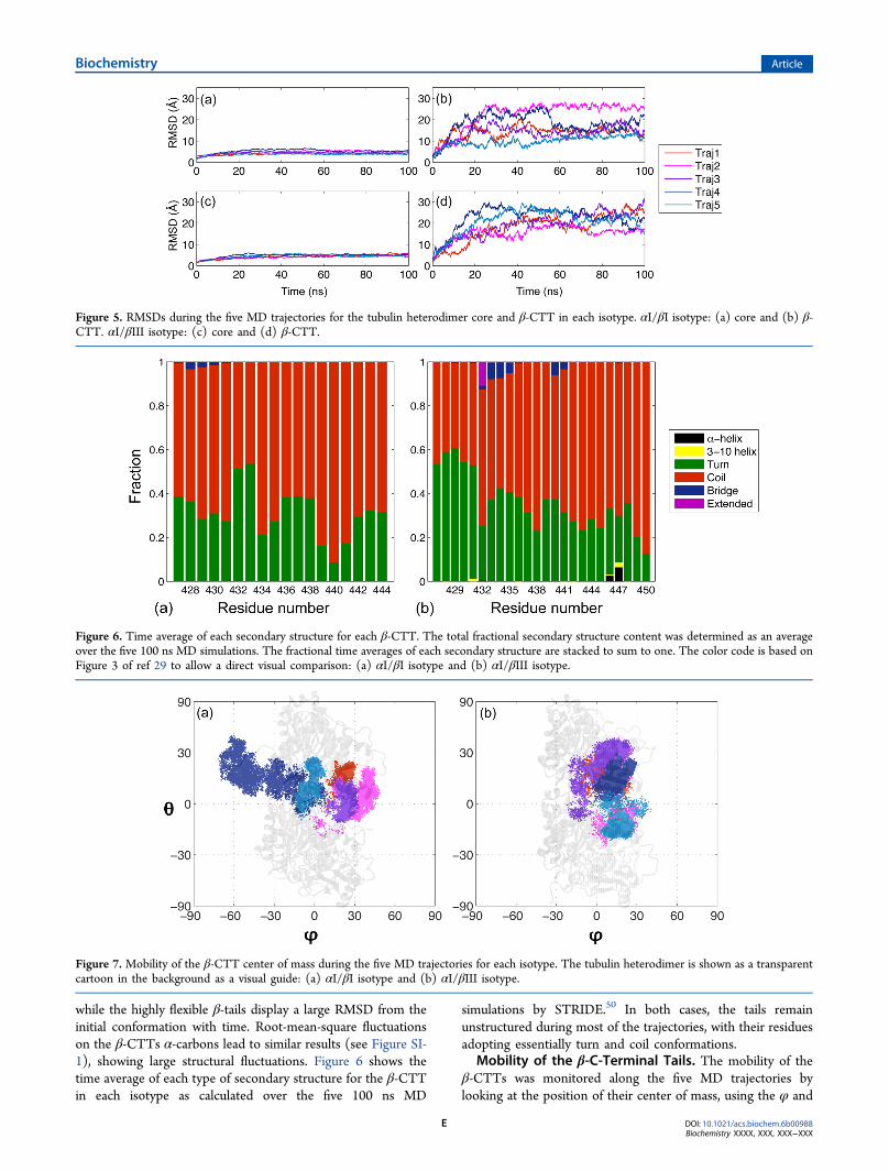
surface electrostatic potentials, which were calculated using theAPBS Web server,49 with both heterodimers presenting a largeelectronegative groove on their α-subunit and no noticeablevariations between the βI- and βIII-subunits.β-C-Terminal Tail Structure and Flexibility. Figure 4
shows the starting structures of the β-CTTs for the αI/βI and
αI/βIII heterodimers. For the five trajectories performed foreach isotype, the CTTs are initially unfolded, mostly solvated,and directed toward the tubulin α-subunit (bottom part ofFigure 4). From the RMSD plots shown in Figure 5, we can seethat the structure of the heterodimer core (which does notinclude the CTTs) remains very stable during the simulations,
Figure 3. Surface electrostatic potential (±1 kT) of the tubulin body (without the CTTs) resulting from APBS calculations. Positive potentials arecolored blue and negative potentials red: (a) αI/βI isotype and (b) αI/βIII isotype. The tubulin heterodimer has the same orientation as in Figure 1.This figure and Figures SI-6 and SI-7 were prepared using PyMol.64
Figure 4. Cartoon representation showing the tubulin heterodimer (gray) with the starting structures of the β-CTTs for the five MD trajectoriescolored red, magenta, purple, dark blue, and light blue. The tubulin orientation has been rotated by 90° around the vertical axis with regard to Figure1: (a) αI/βI isotype and (b) αI/βIII isotype. The color code for the five trajectories of each isotype is the same in Figures 5, 7, 8, and 10. This figurewas prepared using visual molecular dynamics.48
Biochemistry Article
DOI: 10.1021/acs.biochem.6b00988Biochemistry XXXX, XXX, XXX−XXX
D
while the highly flexible β-tails display a large RMSD from theinitial conformation with time. Root-mean-square fluctuationson the β-CTTs α-carbons lead to similar results (see Figure SI-1), showing large structural fluctuations. Figure 6 shows thetime average of each type of secondary structure for the β-CTTin each isotype as calculated over the five 100 ns MD
simulations by STRIDE.50 In both cases, the tails remainunstructured during most of the trajectories, with their residuesadopting essentially turn and coil conformations.
Mobility of the β-C-Terminal Tails. The mobility of theβ-CTTs was monitored along the five MD trajectories bylooking at the position of their center of mass, using the φ and
Figure 5. RMSDs during the five MD trajectories for the tubulin heterodimer core and β-CTT in each isotype. αI/βI isotype: (a) core and (b) β-CTT. αI/βIII isotype: (c) core and (d) β-CTT.
Figure 6. Time average of each secondary structure for each β-CTT. The total fractional secondary structure content was determined as an averageover the five 100 ns MD simulations. The fractional time averages of each secondary structure are stacked to sum to one. The color code is based onFigure 3 of ref 29 to allow a direct visual comparison: (a) αI/βI isotype and (b) αI/βIII isotype.
Figure 7. Mobility of the β-CTT center of mass during the five MD trajectories for each isotype. The tubulin heterodimer is shown as a transparentcartoon in the background as a visual guide: (a) αI/βI isotype and (b) αI/βIII isotype.
Biochemistry Article
DOI: 10.1021/acs.biochem.6b00988Biochemistry XXXX, XXX, XXX−XXX
E
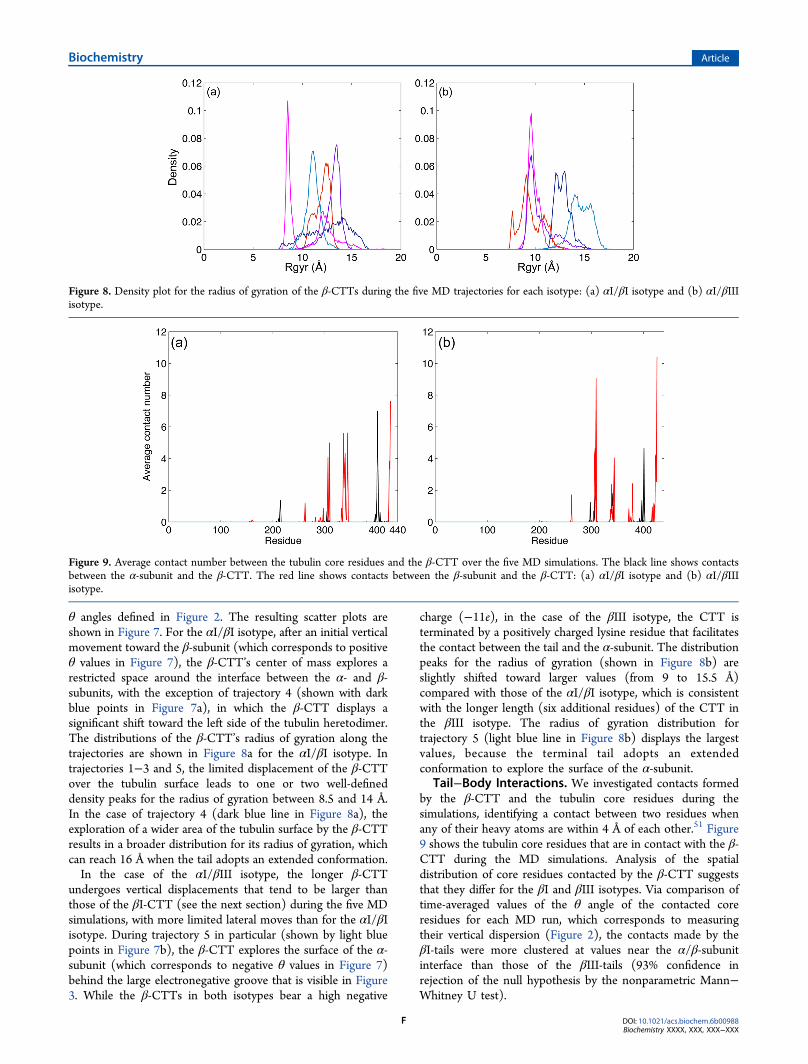
θ angles defined in Figure 2. The resulting scatter plots areshown in Figure 7. For the αI/βI isotype, after an initial verticalmovement toward the β-subunit (which corresponds to positiveθ values in Figure 7), the β-CTT’s center of mass explores arestricted space around the interface between the α- and β-subunits, with the exception of trajectory 4 (shown with darkblue points in Figure 7a), in which the β-CTT displays asignificant shift toward the left side of the tubulin heretodimer.The distributions of the β-CTT’s radius of gyration along thetrajectories are shown in Figure 8a for the αI/βI isotype. Intrajectories 1−3 and 5, the limited displacement of the β-CTTover the tubulin surface leads to one or two well-defineddensity peaks for the radius of gyration between 8.5 and 14 Å.In the case of trajectory 4 (dark blue line in Figure 8a), theexploration of a wider area of the tubulin surface by the β-CTTresults in a broader distribution for its radius of gyration, whichcan reach 16 Å when the tail adopts an extended conformation.In the case of the αI/βIII isotype, the longer β-CTT
undergoes vertical displacements that tend to be larger thanthose of the βI-CTT (see the next section) during the five MDsimulations, with more limited lateral moves than for the αI/βIisotype. During trajectory 5 in particular (shown by light bluepoints in Figure 7b), the β-CTT explores the surface of the α-subunit (which corresponds to negative θ values in Figure 7)behind the large electronegative groove that is visible in Figure3. While the β-CTTs in both isotypes bear a high negative
charge (−11e), in the case of the βIII isotype, the CTT isterminated by a positively charged lysine residue that facilitatesthe contact between the tail and the α-subunit. The distributionpeaks for the radius of gyration (shown in Figure 8b) areslightly shifted toward larger values (from 9 to 15.5 Å)compared with those of the αI/βI isotype, which is consistentwith the longer length (six additional residues) of the CTT inthe βIII isotype. The radius of gyration distribution fortrajectory 5 (light blue line in Figure 8b) displays the largestvalues, because the terminal tail adopts an extendedconformation to explore the surface of the α-subunit.
Tail−Body Interactions. We investigated contacts formedby the β-CTT and the tubulin core residues during thesimulations, identifying a contact between two residues whenany of their heavy atoms are within 4 Å of each other.51 Figure9 shows the tubulin core residues that are in contact with the β-CTT during the MD simulations. Analysis of the spatialdistribution of core residues contacted by the β-CTT suggeststhat they differ for the βI and βIII isotypes. Via comparison oftime-averaged values of the θ angle of the contacted coreresidues for each MD run, which corresponds to measuringtheir vertical dispersion (Figure 2), the contacts made by theβI-tails were more clustered at values near the α/β-subunitinterface than those of the βIII-tails (93% confidence inrejection of the null hypothesis by the nonparametric Mann−Whitney U test).
Figure 8. Density plot for the radius of gyration of the β-CTTs during the five MD trajectories for each isotype: (a) αI/βI isotype and (b) αI/βIIIisotype.
Figure 9. Average contact number between the tubulin core residues and the β-CTT over the five MD simulations. The black line shows contactsbetween the α-subunit and the β-CTT. The red line shows contacts between the β-subunit and the β-CTT: (a) αI/βI isotype and (b) αI/βIIIisotype.
Biochemistry Article
DOI: 10.1021/acs.biochem.6b00988Biochemistry XXXX, XXX, XXX−XXX
F
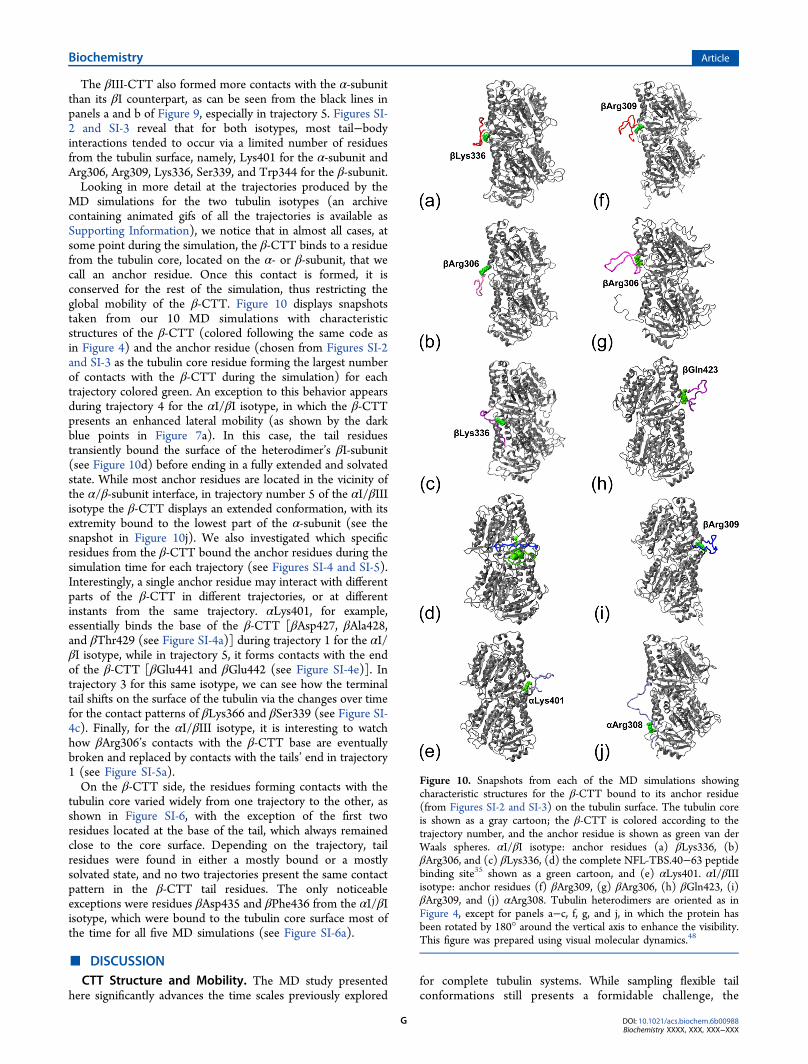
The βIII-CTT also formed more contacts with the α-subunitthan its βI counterpart, as can be seen from the black lines inpanels a and b of Figure 9, especially in trajectory 5. Figures SI-2 and SI-3 reveal that for both isotypes, most tail−bodyinteractions tended to occur via a limited number of residuesfrom the tubulin surface, namely, Lys401 for the α-subunit andArg306, Arg309, Lys336, Ser339, and Trp344 for the β-subunit.Looking in more detail at the trajectories produced by the
MD simulations for the two tubulin isotypes (an archivecontaining animated gifs of all the trajectories is available asSupporting Information), we notice that in almost all cases, atsome point during the simulation, the β-CTT binds to a residuefrom the tubulin core, located on the α- or β-subunit, that wecall an anchor residue. Once this contact is formed, it isconserved for the rest of the simulation, thus restricting theglobal mobility of the β-CTT. Figure 10 displays snapshotstaken from our 10 MD simulations with characteristicstructures of the β-CTT (colored following the same code asin Figure 4) and the anchor residue (chosen from Figures SI-2and SI-3 as the tubulin core residue forming the largest numberof contacts with the β-CTT during the simulation) for eachtrajectory colored green. An exception to this behavior appearsduring trajectory 4 for the αI/βI isotype, in which the β-CTTpresents an enhanced lateral mobility (as shown by the darkblue points in Figure 7a). In this case, the tail residuestransiently bound the surface of the heterodimer’s βI-subunit(see Figure 10d) before ending in a fully extended and solvatedstate. While most anchor residues are located in the vicinity ofthe α/β-subunit interface, in trajectory number 5 of the αI/βIIIisotype the β-CTT displays an extended conformation, with itsextremity bound to the lowest part of the α-subunit (see thesnapshot in Figure 10j). We also investigated which specificresidues from the β-CTT bound the anchor residues during thesimulation time for each trajectory (see Figures SI-4 and SI-5).Interestingly, a single anchor residue may interact with differentparts of the β-CTT in different trajectories, or at differentinstants from the same trajectory. αLys401, for example,essentially binds the base of the β-CTT [βAsp427, βAla428,and βThr429 (see Figure SI-4a)] during trajectory 1 for the αI/βI isotype, while in trajectory 5, it forms contacts with the endof the β-CTT [βGlu441 and βGlu442 (see Figure SI-4e)]. Intrajectory 3 for this same isotype, we can see how the terminaltail shifts on the surface of the tubulin via the changes over timefor the contact patterns of βLys366 and βSer339 (see Figure SI-4c). Finally, for the αI/βIII isotype, it is interesting to watchhow βArg306’s contacts with the β-CTT base are eventuallybroken and replaced by contacts with the tails’ end in trajectory1 (see Figure SI-5a).On the β-CTT side, the residues forming contacts with the
tubulin core varied widely from one trajectory to the other, asshown in Figure SI-6, with the exception of the first tworesidues located at the base of the tail, which always remainedclose to the core surface. Depending on the trajectory, tailresidues were found in either a mostly bound or a mostlysolvated state, and no two trajectories present the same contactpattern in the β-CTT tail residues. The only noticeableexceptions were residues βAsp435 and βPhe436 from the αI/βIisotype, which were bound to the tubulin core surface most ofthe time for all five MD simulations (see Figure SI-6a).
■ DISCUSSIONCTT Structure and Mobility. The MD study presented
here significantly advances the time scales previously exploredfor complete tubulin systems. While sampling flexible tailconformations still presents a formidable challenge, the
Figure 10. Snapshots from each of the MD simulations showingcharacteristic structures for the β-CTT bound to its anchor residue(from Figures SI-2 and SI-3) on the tubulin surface. The tubulin coreis shown as a gray cartoon; the β-CTT is colored according to thetrajectory number, and the anchor residue is shown as green van derWaals spheres. αI/βI isotype: anchor residues (a) βLys336, (b)βArg306, and (c) βLys336, (d) the complete NFL-TBS.40−63 peptidebinding site35 shown as a green cartoon, and (e) αLys401. αI/βIIIisotype: anchor residues (f) βArg309, (g) βArg306, (h) βGln423, (i)βArg309, and (j) αArg308. Tubulin heterodimers are oriented as inFigure 4, except for panels a−c, f, g, and j, in which the protein hasbeen rotated by 180° around the vertical axis to enhance the visibility.This figure was prepared using visual molecular dynamics.48
Biochemistry Article
DOI: 10.1021/acs.biochem.6b00988Biochemistry XXXX, XXX, XXX−XXX
G
improved sampling we have obtained provides a more realisticview of their conformational dynamics. For both β-CTTs, wecan directly compare the time average of each secondarystructure over the five 100 ns MD simulations shown in Figure6 with the results obtained by Luchko et al.29 in their early workon the CTTs of nine human β-tubulin isotypes, which includedthe βI- and βIII-tails. In their study, the β-CTTs were modeledas independent systems, without taking into account thetubulin globular domain, and they performed MD and replicaexchange MD simulations over the temperature range of 273−382 K. Figure 3 of ref 29 shows the resulting time average ofeach secondary structure over a 10 ns MD simulation at 311 K.While being mostly unstructured, the βI- and βIII-CTTs stillshowed a significant amount of transient helix, with residuesbeing either α- or 3−10-helical at least 40% of the time. In ourwork, this was not seen; the interactions between the tubulinsurface and the β-CTT may thus prevent the formation ofhelical secondary structures in the latter, favoring turn and coilconformations for the tail residues. Another point that has to betaken into account is the fact that the simulations performed byLuchko et al. used the AMBER99 force field, which has beenshown to increase the helical content in unstructured peptidescompared to the OPLS-AA force field used in our approach.52
However, one should note that choosing the most appropriateforce field for simulating folded tubulin core, the disorderedterminal tails, and the interaction between these two parts ofthe protein is a delicate matter because, for example, Amberappears to be better suited for modeling electrostaticinteractions between charged residues,53 while OPLS seemsto provide a better treatment of intrinsically disordered proteinsegments.52
While there are little experimental structural data availableregarding tubulin CTTs, the radius of gyration distributionscalculated from our MD simulations and shown in Figure 8 arecompatible with estimates obtained by Asakawa et al.54 fromatomic force microscopy, which show the C-terminal domainsas oval shapes on the tubulin surface with a size between 10 and30 Å. We can also evaluate the radius of gyration for the CTTsby using Flory’s equation,55 Rg = R0N
ν, where R0 and ν areconstant parameters and N is the number of residues in thepeptidic chain. Values of R0 and ν have been determined forintrinsically disordered proteins via SAXS experiments,56,57
leading to an R0 of 2.54 Å and a ν of 0.522, thus giving us radiiof gyration of 11.5 and 13.3 Å for the βI-CTT and the βIII-CTT, respectively. These values concur remarkably well withthose shown in Figure 8. If we compare the characteristic ratiosof the CTTs, that is, the ratio of the average radius of gyrationduring a single trajectory to the radius of gyration predicted byFlory’s law for a given isotype, we can see in Figure SI-8 howboth isotypes present similar values, with the βIII-CTT beingslightly more compact (i.e., presenting lower ratios) than its βIcounterpart (p = 0.07).One can note that any CTT conformation that might have
been unique to a specific tubulin isoform would have been lostin experimental structural studies by data averaging because ofthe high isoform heterogeneity in tubulin samples. However, ina very recent work, Vemu et al.58 managed to observe thestructure of the isotypically pure human unmodified αI/βIIIisotype by cryo-electron microscopy. Despite the chemicalhomogeneity of their sample, no density could be attributed tothe CTTs, indicating that they are intrinsically disordered,which is in agreement with the high RMSDs and variety of
structures that were observed during our five MS simulations ofthis isotype.
Tubulin Core−Tail Contacts. During all the MDtrajectories performed in this study, the β-CTT formedcontacts with a limited set of anchor residues located on thesurface of the tubulin core, and these tail−body contacts areconserved throughout the rest of the simulation, although notnecessarily with the same tail residues. As can be seen from thecontact patterns shown in Figures 10, SI-2, and SI-3, the anchorresidues for the αI/βI and αI/βIII isotypes are roughly thesame and are located above the electronegative patch thatpartially covers the surface of the α-subunit (see Figure SI-6),near the interface between the α- and β-subunits. Some of theseanchor residues, namely, αLys401 and βTrp344, were alsohighlighted as forming contacts with the β-CTT in a simulationwork by Freedman et al.30 on the αIV/βI isotype of humantubulin. Interestingly, this earlier study was conducted usingMD calculations and simulated annealing and employedanother force field, AMBER03, and an implicit solvent model(modified generalized Born59), while arriving at certain similarresults, thus showing the robustness of molecular simulationsfor modeling this system. These results are also in agreementwith those of a very recent NMR study by Wall et al.,60 inwhich tubulin CTTs were isotopically labeled and theirstructure was investigated both as isolated peptides and withincomplete tubulin heterodimers (for the αI/βI isotype fromTetrahymena thermophila). In both cases, the CTTs are fullydisordered, but they display significant chemical shift differ-ences between the isolated peptides and tails in the context ofthe full dimer, thus indicating the CTTs interact with theordered tubulin body. In addition, the CTTs show hetero-geneity in association with the tubulin body, which concurswith the variety of anchor residues found in our work.In our study, the core residues contacted by the βI-tails
tended to be more clustered in the α/β-interface region thanthe for the βIII-CTT. We also observed individual cases inwhich each of the two isotypes displayed a specific bindingpattern that was not observed for the other; this will of courserequire more extensive investigation. For example, in trajectory5, the βIII-CTT adopted an extended conformation and boundanchor residues located on the lowest part of the tubulin α-subunit (Figure 10j). This binding mode of the β-CTT behindthe electronegative groove covering the surface of the α-subunit(see Figure SI-8) is likely to be distinctive of the αI/βIIIisotype, which is six residues longer than the βI-tail and, inaddition to the numerous negatively charged glutamatescommonly found in C-terminal tails, is terminated by apositively charged lysine residue. On the other hand, the αI/βI-tubulin isotype presented a specific binding mode in trajectory4, where the βI-CTT extends to cover the preferential bindingsite for the NFL-TBS.40−63 peptide that was highlighted inour previous docking study30 (see Figure 10d). Quiteremarkably, this binding site coincides with a deep cleft withinthe electronegative patch covering the tubulin body surface (seeFigure SI-8a). More generally, mapping the contact patternsfrom Figure 9 on the tubulin body’s surface (see Figures SI-9and SI-10) allows us to see how the βI-CTT contacts are moreconcentrated in the α/β-interface region that borders theputative binding site, while the βIII-CTT movements sweepout a more vertical pattern (and present a broader distributionof its θ angles, as seen in Figure SI-10c). This is of particularinterest, because this peptide has been shown to specificallyinhibit microtubule formation in glioma cells,34 and the βIII-
Biochemistry Article
DOI: 10.1021/acs.biochem.6b00988Biochemistry XXXX, XXX, XXX−XXX
H
tubulin isotype is known to be overexpressed in several tumortypes.61 Therefore, our results allow us to formulate a firsthypothesis for the NFL-TBS.40−63 peptide’s specificity forglioma cells on a molecular level. The mobility patterns of theCTTS (shown in Figure SI-9) may suggest a higher affinitybetween the NFL-TBS.40−63 peptide and βIII-tubulin,compared to that for βI-tubulin, as the vertically distributedcontacts may encroach less on the peptide’s putative bindingsite. As glioma cells comprise a higher proportion of βIII-tubulin, they would thus be more affected by the NFL-TBS.40−63 peptide’s antimitotic action than normal cells.
■ CONCLUDING REMARKS
Using a combination of modeling approaches, we investigatedthe mobility and binding modes on the tubulin surface of the β-C-terminal tails for the human αI/βI and αI/βIII isotypes. Tothe best of our knowledge, this is the first time that completemodels (i.e., including the C-terminal tails) have been built fortwo human β-tubulin isotypes and jointly investigated usinglong time-scale (5 × 100 ns for each isotype) all-atommolecular dynamics simulations. While displaying similarcontacts with residues located next to the α/β-interface, theisotypes differ in that the βI-tails may be more likely to formcontacts in the interface and more particularly near the NFL-TBS.40−63 peptide binding site, thus potentially decreasing theaffinity between βI-tubulin and this antimitotic compound andproviding us with a first hypothesis regarding the peptide’senhanced activity in glioma cells (in which the βIII-tubulinisotype is known to be overexpressed). However, we cannot yetdraw any final conclusions, as the differences in the contact andmobility patterns for both isotypes could also be due toinsufficient sampling during our simulations. Additional work isneeded to confirm our hypothesis. In particular, we plan toperform new molecular docking studies of the NFL-TBS.40−63peptide on the αI/βI- and αI/βIII-tubulin isotypes that willinclude the tubulin C-terminal tails and take their flexibility intoaccount. Another important issue is the fact that CTTs can besubject to numerous post-translational modifications, whichimpacts their structure and dynamics, and are also likely to havean effect on the recognition process between tubulin and itsvarious ligands (such as other proteins, peptides, or smallerantimitotic compounds). One can also reflect on howembedding a tubulin heterodimer in a microtubule structurewill impact CTT mobility. Bocquet et al.33 showed that theNFL-TBS.40−63 peptide inhibits MT polymerization in vitroby binding unpolymerized tubulin, while having no effect onthe stability of assembled MTs. CTTs protruding from a MTsurface might present a specific mobility pattern, with anenhanced coverage of the NFL-TBS.40−63 peptide bindingsite, which would hinder the interaction between the peptideand the MT surface. Addressing this issue, however, will requirecostly calculations involving several interacting tubulinheterodimers.More generally, our work opens up new perspectives for the
understanding on a molecular level of the resistance to usualtubulin binding agents, such as paclitaxel, because it has beenrelated to the overexpression of the βIII isotype.22,29,62,63 Inaddition, our two structural models allow systematic drugbinding studies of both isotypes, thus permitting the develop-ment of drugs that will preferentially target cancer cells,potentially reducing the side effects that are usually associatedwith chemotherapies.
■ ASSOCIATED CONTENT*S Supporting InformationThe Supporting Information is available free of charge on theACS Publications website at DOI: 10.1021/acs.bio-chem.6b00988.
Additional data regarding the tubulin modeling, includingsequence identities, Modeller scores, root-mean-squarefluctuations on the β-CTTs, and detailed core−tailcontact patterns (PDF)Animated gifs of all 10 MD trajectories for both tubulinisotypes (ZIP)
■ AUTHOR INFORMATIONCorresponding Author*E-mail: [email protected]. Phone: +33 58 41 51 65.ORCIDSophie Sacquin-Mora: 0000-0002-2781-4333FundingY.L. thanks the “Initiative d’Excellence” program from theFrench State (Grant “DYNAMO”, ANR-11-LABX-011-01) fordoctoral funding.NotesThe authors declare no competing financial interest.
■ REFERENCES(1) Aylett, C. H., Lowe, J., and Amos, L. A. (2011) New insights intothe mechanisms of cytomotive actin and tubulin filaments. Int. Rev.Cell Mol. Biol. 292, 1−71.(2) Luduena, R. F. (1998) Multiple forms of tubulin: different geneproducts and covalent modifications. Int. Rev. Cytol. 178, 207−275.(3) Rezania, V., Azarenko, O., Jordan, M. A., Bolterauer, H., Luduena,R. F., Huzil, J. T., and Tuszynski, J. A. (2008) Microtubule assembly ofisotypically purified tubulin and its mixtures. Biophys. J. 95, 1993−2008.(4) Katsetos, C. D., Reginato, M. J., Baas, P. W., D’Agostino, L.,Legido, A., Tuszyn Ski, J. A., Draberova, E., and Draber, P. (2015)Emerging microtubule targets in glioma therapy. Seminars in pediatricneurology 22, 49−72.(5) Mariani, M., Karki, R., Spennato, M., Pandya, D., He, S., Andreoli,M., Fiedler, P., and Ferlini, C. (2015) Class III beta-tubulin in normaland cancer tissues. Gene 563, 109−114.(6) Kavallaris, M. (2010) Microtubules and resistance to tubulin-binding agents. Nat. Rev. Cancer 10, 194−204.(7) Serrano, L., de la Torre, J., Maccioni, R. B., and Avila, J. (1984)Involvement of the carboxyl-terminal domain of tubulin in theregulation of its assembly. Proc. Natl. Acad. Sci. U. S. A. 81, 5989−5993.(8) Sackett, D. L., Bhattacharyya, B., and Wolff, J. (1985) Tubulinsubunit carboxyl termini determine polymerization efficiency. J. Biol.Chem. 260, 43−45.(9) Serrano, L., Wandosell, F., de la Torre, J., and Avila, J. (1988)Effect of specific proteolytic cleavages on tubulin polymer formation.Biochem. J. 252, 683−691.(10) Wolff, J., Sackett, D. L., and Knipling, L. (1996) Cation selectivepromotion of tubulin polymerization by alkali metal chlorides. ProteinSci. 5, 2020−2028.(11) Joe, P. A., Banerjee, A., and Luduena, R. F. (2009) Roles of beta-tubulin residues Ala428 and Thr429 in microtubule formation in vivo.J. Biol. Chem. 284, 4283−4291.(12) Yadav, S., Verma, P. J., and Panda, D. (2014) C-terminal regionof MAP7 domain containing protein 3 (MAP7D3) promotesmicrotubule polymerization by binding at the C-terminal tail oftubulin. PLoS One 9, e99539.(13) Roll-Mecak, A. (2015) Intrinsically disordered tubulin tails:complex tuners of microtubule functions? Semin. Cell Dev. Biol. 37,11−19.
Biochemistry Article
DOI: 10.1021/acs.biochem.6b00988Biochemistry XXXX, XXX, XXX−XXX
I
(14) Bailey, M. E., Sackett, D. L., and Ross, J. L. (2015) KataninSevering and Binding Microtubules Are Inhibited by Tubulin CarboxyTails. Biophys. J. 109, 2546−2561.(15) Luduena, R. F. (2008) in The Role of Microtubules in Cell Biology,Neurobiology, and Oncology (Fojo, A. T., Ed.) pp 105−121, HumanaPress, Totowa, NJ.(16) Janke, C. (2014) The tubulin code: molecular components,readout mechanisms, and functions. J. Cell Biol. 206, 461−472.(17) Sugiura, M., Maccioni, R. B., Cann, J. R., York, E. J., Stewart, J.M., and Kotovych, G. (1987) A proton magnetic resonance and acircular dichroism study of the solvent dependent conformation of thesynthetic tubulin fragment Ac tubulin, alpha (430−441) amide and itsinteraction with substance-P. J. Biomol. Struct. Dyn. 4, 1105−1117.(18) Otter, A., and Kotovych, G. (1988) The solution conformationof the synthetic tubulin fragment AC-tubulin-alpha(430−441)-amidebased on two-dimensional ROESY experiments. Can. J. Chem. 66,1814−1820.(19) Amos, L. A. (2011) What tubulin drugs tell us aboutmicrotubule structure and dynamics. Semin. Cell Dev. Biol. 22, 916−926.(20) Dostal, V., and Libusova, L. (2014) Microtubule drugs: action,selectivity, and resistance across the kingdoms of life. Protoplasma 251,991−1005.(21) Massarotti, A., Coluccia, A., Silvestri, R., Sorba, G., and Brancale,A. (2012) The tubulin colchicine domain: a molecular modelingperspective. ChemMedChem 7, 33−42.(22) Freedman, H., Huzil, J. T., Luchko, T., Luduena, R. F., andTuszynski, J. A. (2009) Identification and characterization of anintermediate taxol binding site within microtubule nanopores and amechanism for tubulin isotype binding selectivity. J. Chem. Inf. Model.49, 424−436.(23) Rendine, S., Pieraccini, S., and Sironi, M. (2010) Vinblastineperturbation of tubulin protofilament structure: a computationalinsight. Phys. Chem. Chem. Phys. 12, 15530−15536.(24) Chakraborti, S., Chakravarty, D., Gupta, S., Chatterji, B. P.,Dhar, G., Poddar, A., Panda, D., Chakrabarti, P., Ghosh Dastidar, S.,and Bhattacharyya, B. (2012) Discrimination of ligands with differentflexibilities resulting from the plasticity of the binding site in tubulin.Biochemistry 51, 7138−7148.(25) Mane, J. Y., Semenchenko, V., Perez-Pineiro, R., Winter, P.,Wishart, D., and Tuszynski, J. A. (2013) Experimental andcomputational study of the interaction of novel colchicinoids with arecombinant human alphaI/betaI-tubulin heterodimer. Chem. Biol.Drug Des. 82, 60−70.(26) Liao, S. Y., Mo, G. Q., Chen, J. C., and Zheng, K. C. (2014)Exploration of the binding mode between (−)-zampanolide andtubulin using docking and molecular dynamics simulation. J. Mol.Model. 20, 2070.(27) Kumbhar, B. V., Borogaon, A., Panda, D., and Kunwar, A.(2016) Exploring the Origin of Differential Binding Affinities ofHuman Tubulin Isotypes alphabetaII, alphabetaIII and alphabetaIV forDAMA-Colchicine Using Homology Modelling, Molecular Dockingand Molecular Dynamics Simulations. PLoS One 11, e0156048.(28) Peng, L. X., Hsu, M. T., Bonomi, M., Agard, D. A., andJacobson, M. P. (2014) The free energy profile of tubulin straight-bentconformational changes, with implications for microtubule assemblyand drug discovery. PLoS Comput. Biol. 10, e1003464.(29) Luchko, T., Torin Huzil, J., Stepanova, M., and Tuszynski, J.(2008) Conformational analysis of the carboxy-terminal tails of humanbeta-tubulin isotypes. Biophys. J. 94, 1971−1982.(30) Freedman, H., Luchko, T., Luduena, R. F., and Tuszynski, J. A.(2011) Molecular dynamics modeling of tubulin C-terminal tailinteractions with the microtubule surface. Proteins: Struct., Funct.,Genet. 79, 2968−2982.(31) Panneerselvam, M., Muthu, K., Jayaraman, M., Sridharan, U.,Jenardhanan, P., and Ramadas, K. (2013) Molecular dynamicsimulations of the tubulin-human gamma synuclein complex:structural insight into the regulatory mechanism involved in inducingresistance against Taxol. Mol. BioSyst. 9, 1470−1488.
(32) Chakraborty, S., and Zheng, W. (2015) Decrypting thestructural, dynamic, and energetic basis of a monomeric kinesininteracting with a tubulin dimer in three ATPase states by all-atommolecular dynamics simulation. Biochemistry 54, 859−869.(33) Bocquet, A., Berges, R., Frank, R., Robert, P., Peterson, A. C.,and Eyer, J. (2009) Neurofilaments bind tubulin and modulate itspolymerization. J. Neurosci. 29, 11043−11054.(34) Berges, R., Balzeau, J., Peterson, A. C., and Eyer, J. (2012) Atubulin binding peptide targets glioma cells disrupting their micro-tubules, blocking migration, and inducing apoptosis. Mol. Ther. 20,1367−1377.(35) Laurin, Y., Savarin, P., Robert, C. H., Takahashi, M., Eyer, J.,Prevost, C., and Sacquin-Mora, S. (2015) Investigating the StructuralVariability and Binding Modes of the Glioma Targeting NFL-TBS.40−63 Peptide on Tubulin. Biochemistry 54, 3660−3669.(36) Chen, K., Huzil, J. T., Freedman, H., Ramachandran, P.,Antoniou, A., Tuszynski, J. A., and Kurgan, L. (2008) Identification oftubulin drug binding sites and prediction of relative differences inbinding affinities to tubulin isotypes using digital signal processing. J.Mol. Graphics Modell. 27, 497−505.(37) Fiser, A., Do, R. K., and Sali, A. (2000) Modeling of loops inprotein structures. Protein Sci. 9, 1753−1773.(38) Fiser, A., and Sali, A. (2003) Modeller: generation andrefinement of homology-based protein structure models. MethodsEnzymol. 374, 461−491.(39) UniProt Consortium (2015) UniProt: A hub for proteininformation. Nucleic Acids Res. 43, D204−D212.(40) Krivov, G. G., Shapovalov, M. V., and Dunbrack, R. L., Jr.(2009) Improved prediction of protein side-chain conformations withSCWRL4. Proteins: Struct., Funct., Genet. 77, 778−795.(41) Pronk, S., Pall, S., Schulz, R., Larsson, P., Bjelkmar, P.,Apostolov, R., Shirts, M. R., Smith, J. C., Kasson, P. M., van der Spoel,D., Hess, B., and Lindahl, E. (2013) GROMACS 4.5: a high-throughput and highly parallel open source molecular simulationtoolkit. Bioinformatics 29, 845−854.(42) Kaminski, G. A., Friesner, R. A., Tirado-Rives, J., and Jorgensen,W. L. (2001) Evaluation and reparametrization of the OPLS-AA forcefield for proteins via comparison with accurate quantum chemicalcalculations on peptides. J. Phys. Chem. B 105, 6474−6487.(43) Jorgensen, W. L., Chandrasekhar, J., Madura, J. D., Impey, R.W., and Klein, M. L. (1983) Comparison of Simple PotentialFunctions for Simulating Liquid Water. J. Chem. Phys. 79, 926−935.(44) Bussi, G., Donadio, D., and Parrinello, M. (2007) Canonicalsampling through velocity rescaling. J. Chem. Phys. 126, 014101.(45) Hess, B., Bekker, H., Berendsen, H. J. C., and Fraaije, J. (1997)LINCS: A linear constraint solver for molecular simulations. J. Comput.Chem. 18, 1463−1472.(46) Essmann, U., Perera, L., Berkowitz, M. L., Darden, T., Lee, H.,and Pedersen, L. G. (1995) A SMOOTH PARTICLE MESH EWALDMETHOD. J. Chem. Phys. 103, 8577−8593.(47) Parrinello, M., and Rahman, A. (1981) Polymorphic transitionsin single crystals: A new molecular dynamics method. J. Appl. Phys. 52,7182−7190.(48) Humphrey, W., Dalke, A., and Schulten, K. (1996) VMD: visualmolecular dynamics. J. Mol. Graphics 14, 27−38.(49) Baker, N. A., Sept, D., Joseph, S., Holst, M. J., and McCammon,J. A. (2001) Electrostatics of nanosystems: application to microtubulesand the ribosome. Proc. Natl. Acad. Sci. U. S. A. 98, 10037−10041.(50) Frishman, D., and Argos, P. (1995) Knowledge-based proteinsecondary structure assignment. Proteins: Struct., Funct., Genet. 23,566−579.(51) Janin, J. (2005) Assessing predictions of protein-proteininteraction: the CAPRI experiment. Protein Sci. 14, 278−283.(52) Smith, M. D., Rao, J. S., Segelken, E., and Cruz, L. (2015) Force-Field Induced Bias in the Structure of Abeta21−30: A Comparison ofOPLS, AMBER, CHARMM, and GROMOS Force Fields. J. Chem. Inf.Model. 55, 2587−2595.
Biochemistry Article
DOI: 10.1021/acs.biochem.6b00988Biochemistry XXXX, XXX, XXX−XXX
J
(53) Debiec, K. T., Gronenborn, A. M., and Chong, L. T. (2014)Evaluating the strength of salt bridges: a comparison of currentbiomolecular force fields. J. Phys. Chem. B 118, 6561−6569.(54) Asakawa, H., Ikegami, K., Setou, M., Watanabe, N., Tsukada, M.,and Fukuma, T. (2011) Submolecular-scale imaging of alpha-helicesand C-terminal domains of tubulins by frequency modulation atomicforce microscopy in liquid. Biophys. J. 101, 1270−1276.(55) Flory, P. J. (1969) Statistical mechanics of chain molecules,Interscience-Wiley Publishers, New York.(56) Bernado, P., and Svergun, D. I. (2012) Structural analysis ofintrinsically disordered proteins by small-angle X-ray scattering. Mol.BioSyst. 8, 151−167.(57) Kikhney, A. G., and Svergun, D. I. (2015) A practical guide tosmall angle X-ray scattering (SAXS) of flexible and intrinsicallydisordered proteins. FEBS Lett. 589, 2570−2577.(58) Vemu, A., Atherton, J., Spector, J. O., Szyk, A., Moores, C. A.,and Roll-Mecak, A. (2016) Structure and Dynamics of Single-isoformRecombinant Neuronal Human Tubulin. J. Biol. Chem. 291, 12907−12915.(59) Onufriev, A., Bashford, D., and Case, D. A. (2004) Exploringprotein native states and large-scale conformational changes with amodified generalized born model. Proteins: Struct., Funct., Genet. 55,383−394.(60) Wall, K. P., Pagratis, M., Armstrong, G., Balsbaugh, J. L.,Verbeke, E., Pearson, C. G., and Hough, L. E. (2016) MolecularDeterminants of Tubulin’s C-Terminal Tail Conformational Ensem-ble. ACS Chem. Biol. 11, 2981−2990.(61) Katsetos, C. D., Draberova, E., Legido, A., Dumontet, C., andDraber, P. (2009) Tubulin targets in the pathobiology and therapy ofglioblastoma multiforme. I. Class III beta-tubulin. J. Cell. Physiol. 221,505−513.(62) Goncalves, A., Braguer, D., Kamath, K., Martello, L., Briand, C.,Horwitz, S., Wilson, L., and Jordan, M. A. (2001) Resistance to Taxolin lung cancer cells associated with increased microtubule dynamics.Proc. Natl. Acad. Sci. U. S. A. 98, 11737−11742.(63) Kamath, K., Wilson, L., Cabral, F., and Jordan, M. A. (2005)BetaIII-tubulin induces paclitaxel resistance in association withreduced effects on microtubule dynamic instability. J. Biol. Chem.280, 12902−12907.(64) The PyMOL Molecular Graphics System, version 1.3r1 (2010)Schrodinger, LLC, Portland, OR.
Biochemistry Article
DOI: 10.1021/acs.biochem.6b00988Biochemistry XXXX, XXX, XXX−XXX
K