miRNA-130a improves cardiac function by down …...miRNA-130a improved heart failure in rats 8455...
8
8454 Abstract. – OBJECTIVE: To investigate the ef- fect of MicroRNA (miRNA)-130a on cardiac func- tion and the expression of tumor necrosis fac- tor-α (TNF- α) in rats with heart failure. MATERIALS AND METHODS: The rat heart failure model (n = 30) were established, then di- vided into miRNA-130a group, phosphate-buf- fer saline (PBS) group, rAAV9 group, and sham group (n = 10 in each group). Four weeks after the operation, the cardiac ultrasound and hemo- dynamic determination were performed. Blood endothelin-1 (ET-1) content was measured by en- zyme-linked immunosorbent assay (ELISA). He- matoxylin and eosin (HE) staining was used to observe the morphology of myocardium. The ex- pression levels of miRNA-130a and TNF- α were determined by quantitative Real Time-Poly- merase Chain Reaction) (qRT-PCR). And the ex- pression of TNF- α protein was determined by immunohistochemistry and Western blotting. RESULTS: The rat heart failure model was suc- cessfully constructed. The miRNA-130a expres- sion was decreased in rats with heart failure, and miRNA-130a transfection was successful. miRNA-130a improved left ventricular ejection fraction in the rat with heart failure. The blood ET-1 in miRNA-130a group was significantly low- er than that of PBS group and rAAV9 group (p < 0.05). RT-PCR, Immunohistochemistry and Western blotting results showed that compared with the sham group, the expression of TNF- α in the model group was increased. And the expres- sion of TNF- α in miRNA-130a group was signifi- cantly lower than that of PBS and rAAV9 group. CONCLUSIONS: miRNA-130a could improve cardiac function of heart failure rat by down-reg- ulating TNF- α. Key Words: miRNA-130a, Heart failure, TNF-α, ET-1, Left ventric- ular ejection fraction (LVEF), Cardiac function. Introduction Heart failure, a complex clinical symptom, is the final outcome of various heart diseases 1 . It can cause cardiac structure or dysfunction resulting in ventricular filling or impaired ejection fun- ction 2 . Heart failure has high morbidity and mor - tality, but there is no ideal treatment. Bo et al 3 {- Bo, 2017 #2461}{Bo, 2017 #2461} revealed that TNF was significantly increased in heart failure, it indicated that heart failure was associated with inflammation. Previous researches found that the expression of tumor necrosis factor-α (TNF-α) in- creased in patients with heart failure, and TNF-α could promote the expression of endothelin-1 (ET-1) 4,5 . As a direct inflammatory cytokine as- sociated with heart failure, TNF-α plays a role in the process of heart failure by influencing cardiac contractility, mediating cell apoptosis, partici- pating in ventricular remodeling, and promoting the development of cardiogenic cachexia 6 . Also, ET-1, as a strong vasoconstrictor, may lead to va- soconstriction, myocardial ischemia, metabolic disorder, and cell proliferation, and further indu- ce disease related to vascular injury 7 . A related study 8 found that increased ET-1 level could indu- ce cardiomyocyte hypertrophy, and ET-1 also had direct toxicity effect on cardiomyocytes. MicroRNA (miRNA) is endogenous noncoding RNA, which contains 18-25 nucleotides. And it is highly conserved in evolution, which can regulate gene expression and participate in many biolo- gical processes through targeting gene sequen- ce specialization interaction 9 . Various miRNAs, such as miRNA-30e, miRNA-133, miRNA-21, European Review for Medical and Pharmacological Sciences 2018; 22: 8454-8461 Y.-R. JIANG 1 , J.-Y. DU 2 , D.-D. WANG 3 , X. YANG 4 1 Department of Cardiovascular Medicine, The First College of Clinical Medical Science, of Three Gorges University; China; Yichang Central People’s Hospital, Yichang City, Hubei Province, China 2 Department of Special Inspection Section, Dongying People’s Hospital, Dongying City, Shandong Province, China 3 Department of Stomatology, The People’s Hospital of Jimo District, Qingdao City, Shandong Province, China 4 Department of Cardiovascular Surgery, The Second People’s Hospital of Yunnan Province, Kunming City, Yunnan Province, China Corresponding Author: Xu Yang, BM; e-mail: [email protected] miRNA-130a improves cardiac function by down-regulating TNF- α expression in a rat model of heart failure
Transcript of miRNA-130a improves cardiac function by down …...miRNA-130a improved heart failure in rats 8455...
8454
Abstract. – OBJECTIVE: To investigate the ef-fect of MicroRNA (miRNA)-130a on cardiac func-tion and the expression of tumor necrosis fac-tor-α (TNF-α) in rats with heart failure.
MATERIALS AND METHODS: The rat heart failure model (n = 30) were established, then di-vided into miRNA-130a group, phosphate-buf-fer saline (PBS) group, rAAV9 group, and sham group (n = 10 in each group). Four weeks after the operation, the cardiac ultrasound and hemo-dynamic determination were performed. Blood endothelin-1 (ET-1) content was measured by en-zyme-linked immunosorbent assay (ELISA). He-matoxylin and eosin (HE) staining was used to observe the morphology of myocardium. The ex-pression levels of miRNA-130a and TNF-α were determined by quantitative Real Time-Poly-merase Chain Reaction) (qRT-PCR). And the ex-pression of TNF-α protein was determined by immunohistochemistry and Western blotting.
RESULTS: The rat heart failure model was suc-cessfully constructed. The miRNA-130a expres-sion was decreased in rats with heart failure, and miRNA-130a transfection was successful. miRNA-130a improved left ventricular ejection fraction in the rat with heart failure. The blood ET-1 in miRNA-130a group was significantly low-er than that of PBS group and rAAV9 group (p < 0.05). RT-PCR, Immunohistochemistry and Western blotting results showed that compared with the sham group, the expression of TNF-α in the model group was increased. And the expres-sion of TNF-α in miRNA-130a group was signifi-cantly lower than that of PBS and rAAV9 group.
CONCLUSIONS: miRNA-130a could improve cardiac function of heart failure rat by down-reg-ulating TNF-α.Key Words:
miRNA-130a, Heart failure, TNF-α, ET-1, Left ventric-ular ejection fraction (LVEF), Cardiac function.
Introduction
Heart failure, a complex clinical symptom, is the final outcome of various heart diseases1. It can cause cardiac structure or dysfunction resulting in ventricular filling or impaired ejection fun-ction2. Heart failure has high morbidity and mor-tality, but there is no ideal treatment. Bo et al3{-Bo, 2017 #2461}{Bo, 2017 #2461} revealed that TNF was significantly increased in heart failure, it indicated that heart failure was associated with inflammation. Previous researches found that the expression of tumor necrosis factor-α (TNF-α) in-creased in patients with heart failure, and TNF-α could promote the expression of endothelin-1 (ET-1)4,5. As a direct inflammatory cytokine as-sociated with heart failure, TNF-α plays a role in the process of heart failure by influencing cardiac contractility, mediating cell apoptosis, partici-pating in ventricular remodeling, and promoting the development of cardiogenic cachexia6. Also, ET-1, as a strong vasoconstrictor, may lead to va-soconstriction, myocardial ischemia, metabolic disorder, and cell proliferation, and further indu-ce disease related to vascular injury7. A related study8 found that increased ET-1 level could indu-ce cardiomyocyte hypertrophy, and ET-1 also had direct toxicity effect on cardiomyocytes.
MicroRNA (miRNA) is endogenous noncoding RNA, which contains 18-25 nucleotides. And it is highly conserved in evolution, which can regulate gene expression and participate in many biolo-gical processes through targeting gene sequen-ce specialization interaction9. Various miRNAs, such as miRNA-30e, miRNA-133, miRNA-21,
European Review for Medical and Pharmacological Sciences 2018; 22: 8454-8461
Y.-R. JIANG1, J.-Y. DU2, D.-D. WANG3, X. YANG4
1Department of Cardiovascular Medicine, The First College of Clinical Medical Science, of Three Gorges University; China; Yichang Central People’s Hospital, Yichang City, Hubei Province, China 2Department of Special Inspection Section, Dongying People’s Hospital, Dongying City, Shandong Province, China3Department of Stomatology, The People’s Hospital of Jimo District, Qingdao City, Shandong Province, China 4Department of Cardiovascular Surgery, The Second People’s Hospital of Yunnan Province, Kunming City, Yunnan Province, China
Corresponding Author: Xu Yang, BM; e-mail: [email protected]
miRNA-130a improves cardiac function by down-regulating TNF-α expression in a rat model of heart failure
miRNA-130a improved heart failure in rats
8455
miRNA-132, miRNA-130a, had been known as key factors for the pathogenesis of heart failu-re10-14. Li et al15 found that low expression of miR-NA-130a could increase the expression of TNF-α, and promote the development of osteoarthritis. And Zhang et al16 confirmed that miRNA-130a could down-regulate TNF-α in cervical cancer. At present, the mechanism of miRNA-130a on heart failure has not yet been studied clearly.
Therefore, we hypothesize that miRNA-130a can improve heart failure by regulating TNF-α. We also studied the correlation between miR-130a and cardiac function, and discussed the expression of ET-1 in plasma-mediated by miR-130a/TNF-α.
Materials and Methods
Animals and GroupingForty clean grade healthy Wistar rats (sex in
half, 12-16 weeks, 320-350 g body wt) were fed in a cage at 25°C and relative humidity of 40%. The drinking water was provided by an automatic wa-ter supply system, and the feed was prepared by the experimental center of our school. Four weeks after the operation, the rats’ hearts were divided into two parts: half of the rats’ hearts was fresh-ly taken from each group, and the other half was fixed for Hematoxylin and eosin (HE) staining and immunohistochemistry.
All animal experiments have been approved by our hospital’s Medical Ethics Committee.
Construction of rAAV9-miRNA-130a Vector
After linearization, the vector was cut by endo-nuclease Hind III and treated with CIP for 1 h. The carrier fragment of 5451 bp was recovered and am-plified. miRNA-130a: forward: 5’-GCTCTTTTCA-CATTGTGCTACT-3’; reverse: 5’-CAGTGCAAT-GTTAAAAGGGCATA-3; Colony PCR primers: F: 5’-CATGGTCCTGCTGGAGTTCGTG-3’; R: 5’-CATAGCGTAAAAGGAGCAACA-3’. The miRNA-130a and the linearized vector were mixed at 2:1 (mole ratio) and incubated at 42°C for 30 min. Ten µl reaction liquid was inoculated into the com-petent cells, then added 10 µl connect fluid and in-cubated at ice for 30 min, 42°C for 90 s. 900 µl LB culture liquid was added to the bacterial fluid, then incubated at 37°C for 1 h. The transformed bacteria liquid was coated on LB agar plate (containing the corresponding antibiotic of the expression vector) at 37°C for 16 h. The colony PCR was identified and sequenced; then, the plasmid was extracted.
Construction of Heart Failure Rat Model And Grouping
Coronary artery ligation was performed in rats which fed for one week. The rats were anesthetized with 2.5% pentobarbital sodium (35 mg/kg), and tracheotomy and tracheal in-tubation were performed. Positive pressure ventilation was performed by the ventilator (TKR – 200C animal ventilator, Jiangxi Teli anesthesia breathing equipment company, Nan-chang, Jiangxi, China) with a tidal gas of 30 ml/100 mg. The left margin of the sternum was opened and the rat heart was exposed. 10 rats were randomly selected to suture (sham group, and the other rats’ left ventricular cavities were randomly injected with phosphate buffer saline (PBS), blank AAV plasmid vector and rAAV9-miRNA-130a (N=10 in each group). Then the rats were divided into 4 groups: sham group, PBS group, rAAV9 group, and miRNA-130a group. The left anterior descending branch of coronary artery was ligated at 5 mm away from the root of the aorta with 6-0 noninvasive oph-thalmic suture silk.
Color Doppler EchocardiographyFour weeks after operation, the rats in each
group (N = 5) were anesthetized and fixed in the supine position, then left ventricular poste-rior wall thickness (LVPWT) and interventric-ular septal thickness (IVST) during diastolic and systolic period, left ventricular end-diastolic (LVEDd) and end-systolic (LVESd) dimension were measured by echocardiography. Left ven-tricular ejection fractions (LVEF) was calculat-ed. The recorded values of each index were an average of three consecutive cardiac cycles. All echocardiographic studies were performed in the supine position using Vevo 770TM (High resolu-tion imaging system, VisualSonics Inc, Toronto, Ontario, Canada) with an 8.0-12.0 MHz by an experienced investigator.
Determination of Hemodynamic IndexesFour weeks after the operation, the rats in each
group (N=5) were anesthetized with 35 mg/kg chloral hydrate. Then, heart rate (HR), left ven-tricular end-systolic pressure (LVESP), left ven-tricular end-diastolic pressure (LVEDP), max-imum rate of left ventricular pressure rise, and drop (LV + dp/dtmax, LV-dp/dtmax) were mea-sured by BL-420F biological function experiment system recorder (Chengdu Taimeng Technology Co., Ltd., Chengdu, Sichuan, China).
Y.-R. Jiang, J.-Y. Du, D.-D. Wang, X. Yang
8456
Enzyme-Linked Immunosorbent Assay (ELISA)
Four weeks after the operation, 10 ml fasting venous blood of rats in each group were taken from the right carotid artery under the aseptic condition. After serum isolation, serum ET-1 lev-el was determined in strict accordance with the instruction of the ELISA kit (Beijing Huaxia Pe-lagic Technology Co., Ltd., Beijing, China). Opti-cal density (OD) values were measured at 450 nm with the microplate reader. The standard curves of OD value and concentration were drawn, and the concentration of the sample was calculated ac-cording to the standard curve.
HE StainingThe sample of annular myocardium was taken
from the central part of the left ventricle, which was fixed in polyoxymethylene for 24 hours, de-hydrated in ethanol, permeabilized in PBST (PBS + 0.5% Triton X-100), and embedded in paraffin. Then, paraffin sections (3-4 μm) were dewaxed by xylene for 10 minutes, and hydrated by differ-ent concentration of ethanol (100%, 95%, 90%, 85%) for 1 minute, and washed with water for 2 minutes. These sections were stained with he-matoxylin for 4 minutes, differentiated in a hy-drochloric acid-ethanol mixture for 3-5 minutes, and then stained with eosin for 2 minutes. Then, gradient alcohol from low concentration to high concentration was used for dehydration treat-ment, xylene infiltration, and neutral balata seal. The myocardial tissue morphology changes were observed under the microscope.
Real-Time Fluorescence Quantitative PCR (qRT-PCR)
Total RNA was isolated from myocardium tissue by GenElute™ Mammalian Total RNA Miniprep Kit (Seebio, Shanghai, China). The cDNA template was synthesized by reverse transcriptase. qRT-PCR was done by using the TaqMan miRNA detection kit. U6 snRNA and GAPDH were used as a ref-erence gene for miRNA-130a and TNF-α respec-tively. The relative gene expression was analyzed with the 2^(−Delta Delta CT) method. The primers used for qPCR were as follows: TNF-α, forward: 5’-CAGAC-CCTCACACTCAGATCATC-3’; re-verse: 5’-AGCCTTGTCCCTTGAAGAGAAC-3’; U6, forward: 5’-CTCGCTTCGGCAGCACA-3’; reverse: 5’-AACGCTTCACGAATT-TGC-GT-3’; GAPDH, forward: 5’-CGGAGTCAAC-GGATTTGGTCGTAT-3’; reverse: 5’-AG-CCTTCTCCATGGTGGTG-AAGAC-3’.
Western BlotTotal protein was extracted from myocardium
tissue, then quantified by BCA Protein Assay Kit, subjected to SDS-PAGE, and transferred onto the membranes. GAPDH gene was used as a refer-ence gene for TNF-α. After blocked by with 5% defatted milk powder, the membrane was incubat-ed with the TNF-α antibody (1:1000, 3707, CST, Danfoss, MA, USA) at 4°C overnight. Then it was washed by TBST for 3 times, incubated with horseradish peroxidase-labeled secondary anti-body (1:5000, Beijing Zhong Shan Biotechnology Co., Ltd., Beijing, China) at room temperature for 1 hour, and washed with TBST again. Proteins were visualized with enhanced chemilumines-cence (ECL) kit and gel imaging system. The re-sults were analyzed by Image Tools.
ImmunohistochemistryAfter dewaxed and hydrated, paraffin sections
were placed in the boiling 0.01 M citrate buffer, then cooled down after heating for 90 s. These sections were incubated with 3% hydrogen perox-ide at room temperature for 10 minutes; then, they were incubated with the TNF-α antibody (1:1000, 3707, CST, Danfoss, MA, USA) at room tempera-ture for 2 hours. After added Max VisionTM 30 minutes at room temperature, these sections were incubated with goat anti-rabbit-HRP at room tem-perature for 30 minutes and visualized with DAB substrate.
Statistical AnalysisThe terms of mean values ± standard devia-
tion was used to express experimental data. The comparison of data between groups was analyzed by Student-Newman-Keuls test using SPSS19.0 (SPSS Inc, Chicago, IL, USA). p < 0.05 was con-sidered as significant.
Results
Successful Construction of the Rat Heart Failure Model
Four weeks after operation, HE staining was used to detect whether the rat heart failure model was successfully constructed. The results showed that the myocardial fiber structure was clear and arranged orderly in the sham operation group. In model groups, the necrosis of myocardial cells in the infarct area was replaced by fibrous tissue, and the fibers were arranged in disorder with a large number of proliferating small vessels invol-
miRNA-130a improved heart failure in rats
8457
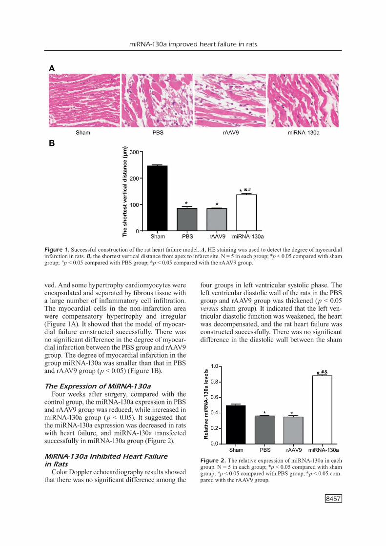
ved. And some hypertrophy cardiomyocytes were encapsulated and separated by fibrous tissue with a large number of inflammatory cell infiltration. The myocardial cells in the non-infarction area were compensatory hypertrophy and irregular (Figure 1A). It showed that the model of myocar-dial failure constructed successfully. There was no significant difference in the degree of myocar-dial infarction between the PBS group and rAAV9 group. The degree of myocardial infarction in the group miRNA-130a was smaller than that in PBS and rAAV9 group (p < 0.05) (Figure 1B).
The Expression of MiRNA-130aFour weeks after surgery, compared with the
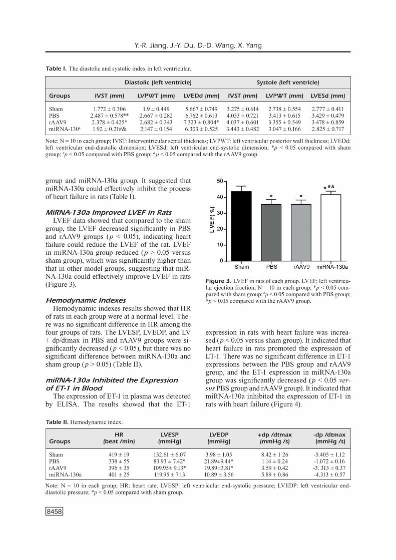
control group, the miRNA-130a expression in PBS and rAAV9 group was reduced, while increased in miRNA-130a group (p < 0.05). It suggested that the miRNA-130a expression was decreased in rats with heart failure, and miRNA-130a transfected successfully in miRNA-130a group (Figure 2).
MiRNA-130a Inhibited Heart Failure in Rats
Color Doppler echocardiography results showed that there was no significant difference among the
four groups in left ventricular systolic phase. The left ventricular diastolic wall of the rats in the PBS group and rAAV9 group was thickened (p < 0.05 versus sham group). It indicated that the left ven-tricular diastolic function was weakened, the heart was decompensated, and the rat heart failure was constructed successfully. There was no significant difference in the diastolic wall between the sham
Figure 1. Successful construction of the rat heart failure model. A, HE staining was used to detect the degree of myocardial infarction in rats. B, the shortest vertical distance from apex to infarct site. N = 5 in each group; *p < 0.05 compared with sham group; #p < 0.05 compared with PBS group; &p < 0.05 compared with the rAAV9 group.
Figure 2. The relative expression of miRNA-130a in each group. N = 5 in each group; *p < 0.05 compared with sham group; #p < 0.05 compared with PBS group; &p < 0.05 com-pared with the rAAV9 group.
Y.-R. Jiang, J.-Y. Du, D.-D. Wang, X. Yang
8458
group and miRNA-130a group. It suggested that miRNA-130a could effectively inhibit the process of heart failure in rats (Table I).
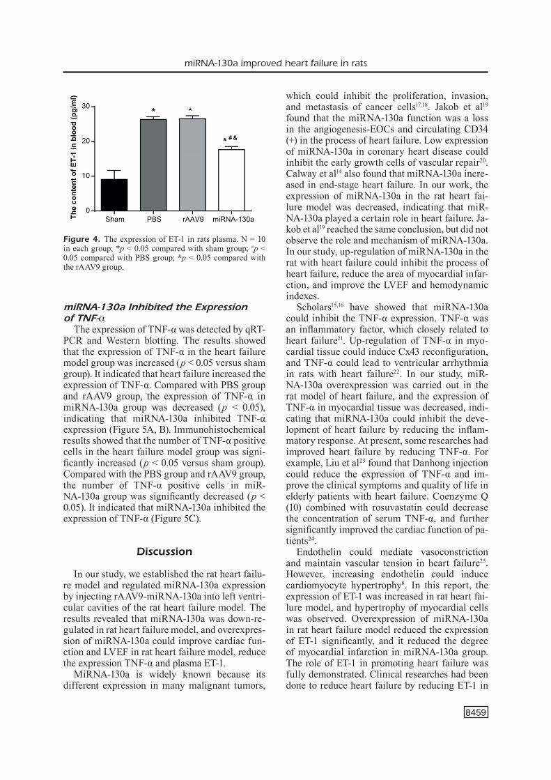
MiRNA-130a Improved LVEF in RatsLVEF data showed that compared to the sham
group, the LVEF decreased significantly in PBS and rAAV9 groups (p < 0.05), indicating heart failure could reduce the LVEF of the rat. LVEF in miRNA-130a group reduced (p > 0.05 versus sham group), which was significantly higher than that in other model groups, suggesting that miR-NA-130a could effectively improve LVEF in rats (Figure 3).
Hemodynamic IndexesHemodynamic indexes results showed that HR
of rats in each group were at a normal level. The-re was no significant difference in HR among the four groups of rats. The LVESP, LVEDP, and LV ± dp/dtmax in PBS and rAAV9 groups were si-gnificantly decreased (p < 0.05), but there was no significant difference between miRNA-130a and sham group (p > 0.05) (Table II).
miRNA-130a Inhibited the Expression of ET-1 in Blood
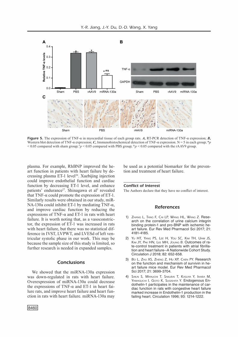
The expression of ET-1 in plasma was detected by ELISA. The results showed that the ET-1
expression in rats with heart failure was increa-sed (p < 0.05 versus sham group). It indicated that heart failure in rats promoted the expression of ET-1. There was no significant difference in ET-1 expressions between the PBS group and rAAV9 group, and the ET-1 expression in miRNA-130a group was significantly decreased (p < 0.05 ver-sus PBS group and rAAV9 group). It indicated that miRNA-130a inhibited the expression of ET-1 in rats with heart failure (Figure 4).
Table II. Hemodynamic index.
HR LVESP LVEDP +dp /dtmax -dp /dtmaxGroups (beat /min) (mmHg) (mmHg) (mmHg /s) (mmHg /s) Sham 419 ± 19 132.61 ± 6.07 3.98 ± 1.05 8.42 ± 1 26 -5.405 ± 1.12PBS 338 ± 55 83.93 ± 7.42* 21.89±9.44* 1.14 ± 0.24 -1.072 ± 0.16 rAAV9 396 ± 35 109.95± 9.13* 19.89±3.81* 3.59 ± 0.42 -3. 313 ± 0.37miRNA-130a 401 ± 25 119.95 ± 7.13 10.89 ± 3.56 5.89 ± 0.86 -4.313 ± 0.57
Note: N = 10 in each group; HR: heart rate; LVESP: left ventricular end-systolic pressure; LVEDP: left ventricular end-diastolic pressure; *p < 0.05 compared with sham group.
Table I. The diastolic and systolic index in left ventricular.
Diastolic (left ventricle) Systole (left ventricle)
Groups IVST (mm) LVPWT (mm) LVEDd (mm) IVST (mm) LVPWT (mm) LVESd (mm)
Sham 1.772 ± 0.306 1.9 ± 0.449 5.667 ± 0.749 3.275 ± 0.614 2.738 ± 0.554 2.777 ± 0.411PBS 2.487 ± 0.578** 2.667 ± 0.282 6.762 ± 0.613 4.033 ± 0.721 3.413 ± 0.615 3.429 ± 0.479rAAV9 2.378 ± 0.425* 2.682 ± 0.343 7.323 ± 0.804* 4.037 ± 0.601 3.355 ± 0.549 3.478 ± 0.859miRNA-130a 1.92 ± 0.21#& 2.147 ± 0.154 6.303 ± 0.525 3.443 ± 0.482 3.047 ± 0.166 2.825 ± 0.717
Note: N = 10 in each group; IVST: Interventricular septal thickness; LVPWT: left ventricular posterior wall thickness; LVEDd: left ventricular end-diastolic dimension; LVESd: left ventricular end-systolic dimension; *p < 0.05 compared with sham group; #p < 0.05 compared with PBS group; &p < 0.05 compared with the rAAV9 group.
Figure 3. LVEF in rats of each group. LVEF: left ventricu-lar ejection fraction; N = 10 in each group; *p < 0.05 com-pared with sham group; #p < 0.05 compared with PBS group; &p < 0.05 compared with the rAAV9 group.
miRNA-130a improved heart failure in rats
8459
miRNA-130a Inhibited the Expression of TNF-α
The expression of TNF-α was detected by qRT-PCR and Western blotting. The results showed that the expression of TNF-α in the heart failure model group was increased (p < 0.05 versus sham group). It indicated that heart failure increased the expression of TNF-α. Compared with PBS group and rAAV9 group, the expression of TNF-α in miRNA-130a group was decreased (p < 0.05), indicating that miRNA-130a inhibited TNF-α expression (Figure 5A, B). Immunohistochemical results showed that the number of TNF-α positive cells in the heart failure model group was signi-ficantly increased (p < 0.05 versus sham group). Compared with the PBS group and rAAV9 group, the number of TNF-α positive cells in miR-NA-130a group was significantly decreased (p < 0.05). It indicated that miRNA-130a inhibited the expression of TNF-α (Figure 5C).
Discussion
In our study, we established the rat heart failu-re model and regulated miRNA-130a expression by injecting rAAV9-miRNA-130a into left ventri-cular cavities of the rat heart failure model. The results revealed that miRNA-130a was down-re-gulated in rat heart failure model, and overexpres-sion of miRNA-130a could improve cardiac fun-ction and LVEF in rat heart failure model, reduce the expression TNF-α and plasma ET-1.
MiRNA-130a is widely known because its different expression in many malignant tumors,
which could inhibit the proliferation, invasion, and metastasis of cancer cells17,18. Jakob et al19 found that the miRNA-130a function was a loss in the angiogenesis-EOCs and circulating CD34 (+) in the process of heart failure. Low expression of miRNA-130a in coronary heart disease could inhibit the early growth cells of vascular repair20. Calway et al14 also found that miRNA-130a incre-ased in end-stage heart failure. In our work, the expression of miRNA-130a in the rat heart fai-lure model was decreased, indicating that miR-NA-130a played a certain role in heart failure. Ja-kob et al19 reached the same conclusion, but did not observe the role and mechanism of miRNA-130a. In our study, up-regulation of miRNA-130a in the rat with heart failure could inhibit the process of heart failure, reduce the area of myocardial infar-ction, and improve the LVEF and hemodynamic indexes.
Scholars15,16 have showed that miRNA-130a could inhibit the TNF-α expression. TNF-α was an inflammatory factor, which closely related to heart failure21. Up-regulation of TNF-α in myo-cardial tissue could induce Cx43 reconfiguration, and TNF-α could lead to ventricular arrhythmia in rats with heart failure22. In our study, miR-NA-130a overexpression was carried out in the rat model of heart failure, and the expression of TNF-α in myocardial tissue was decreased, indi-cating that miRNA-130a could inhibit the deve-lopment of heart failure by reducing the inflam-matory response. At present, some researches had improved heart failure by reducing TNF-α. For example, Liu et al23 found that Danhong injection could reduce the expression of TNF-α and im-prove the clinical symptoms and quality of life in elderly patients with heart failure. Coenzyme Q (10) combined with rosuvastatin could decrease the concentration of serum TNF-α, and further significantly improved the cardiac function of pa-tients24.
Endothelin could mediate vasoconstriction and maintain vascular tension in heart failure25. However, increasing endothelin could induce cardiomyocyte hypertrophy8. In this report, the expression of ET-1 was increased in rat heart fai-lure model, and hypertrophy of myocardial cells was observed. Overexpression of miRNA-130a in rat heart failure model reduced the expression of ET-1 significantly, and it reduced the degree of myocardial infarction in miRNA-130a group. The role of ET-1 in promoting heart failure was fully demonstrated. Clinical researches had been done to reduce heart failure by reducing ET-1 in
Figure 4. The expression of ET-1 in rats plasma. N = 10 in each group; *p < 0.05 compared with sham group; #p < 0.05 compared with PBS group; &p < 0.05 compared with the rAAV9 group.
Y.-R. Jiang, J.-Y. Du, D.-D. Wang, X. Yang
8460
plasma. For example, RhBNP improved the he-art function in patients with heart failure by de-creasing plasma ET-1 level26. Xuebijing injection could improve endothelial function and cardiac function by decreasing ET-1 level, and enhance patients’ endurance27. Shinagawa et al5 revealed that TNF-α could promote the expression of ET-1. Similarly results were obtained in our study, miR-NA-130a could inhibit ET-1 by mediating TNF-α, and improve cardiac function by reducing the expressions of TNF-α and ET-1 in rats with heart failure. It is worth noting that, as a vasoconstric-tor, the expression of ET-1 was increased in rats with heart failure, but there was no statistical dif-ference in IVST, LVPWT, and LVESd of left ven-tricular systolic phase in our work. This may be because the sample size of this study is limited, so further research is needed in expanded samples.
Conclusions
We showed that the miRNA-130a expression was down-regulated in rats with heart failure. Overexpression of miRNA-130a could decrease the expressions of TNF-α and ET-1 in heart fai-lure rats, and improve heart failure and heart fun-ction in rats with heart failure. miRNA-130a may
be used as a potential biomarker for the preven-tion and treatment of heart failure.
Conflict of InterestThe Authors declare that they have no conflict of interest.
References
1) Zhang L, Tang F, Cai LP, Wang hL, Wang Z. Rese-arch on the correlation of urine calcium integrin binding protein-1 and pro-BNP with ischemic he-art failure. Eur Rev Med Pharmacol Sci 2017; 21: 4181-4185.
2) Yu hT, Yang PS, Lee h, You SC, Kim Th, uhm JS, Kim JY, PaK hn, Lee mh, Joung B. Outcomes of ra-te-control treatment in patients with atrial fibrilla-tion and heart failure--A Nationwide Cohort Study. Circulation J 2018; 82: 652-658.
3) Bo L, Zhu XS, Zheng Z, hu XP, Chen PY. Research on the function and mechanism of survivin in he-art failure mice model. Eur Rev Med Pharmacol Sci 2017; 21: 3699-3704.
4) SaKai S, miYauChi T, SaKurai T, KaSuYa Y, ihara m, YamaguChi i, goTo K, SugiShiTa Y. Endogenous En-dothelin-1 participates in the maintenance of car-diac function in rats with congestive heart failure marked increase in Endothelin-1 production in the failing heart. Circulation 1996; 93: 1214-1222.
Figure 5. The expression of TNF-α in myocardial tissue of each group rats. A, RT-PCR detection of TNF-α expression; B, Western blot detection of TNF-α expression; C, Immunohistochemical detection of TNF-α expression. N = 5 in each group; *p < 0.05 compared with sham group; #p < 0.05 compared with PBS group; &p < 0.05 compared with the rAAV9 group.
miRNA-130a improved heart failure in rats
8461
5) ShinagaWa S, oKaZaKi T, iKeda m, Yudoh K, KiSanuKi YY, YanagiSaWa m, KaWahaTa K, oZaKi S. T cells upon activation promote endothelin 1 production in mo-nocytes via IFN-alpha and TNF-gamma. Sci Rep 2017; 7: 14500.
6) Wu CK, huang YT, Lin hh, Yang CY, Lien YC, Lee JK, huang JW, hung KY. Dissecting the mechanisms of left ventricular diastolic dysfunction and inflam-mation in peritoneal dialysis patients. PLoS One 2013; 8: e62722.
7) PaLaCín m, rodrigueZPaSCuaL F, reguero Jr, rodrígueZ i, avanZaS P, LoZano i, moríS C, aLvareZ v, CannaTa-andía JB, LamaS S, garCiaCaSTro m, CoTo E. Lack of association between endothelin-1 gene variants and myocardial infarction. J Atheroscler Thromb 2009; 16: 388-395.
8) gonCaLveS gK, SCaLZo S, aLveS aP, agero u, guaTi-moSim S, reiS am. Neonatal cardiomyocyte hyper-trophy induced by endothelin-1 is blocked by estradiol acting on GPER. Am J Physiol Cell Phy-siol 2018; 314: C310-C322.
9) Wu W. MicroRNA, noise, and gene expression re-gulation. Methods Mol Biol 2018; 1699: 91-96.
10) Lai L, Chen J, Wang n, Zhu g, duan X, Ling F. MiR-NA-30e mediated cardioprotection of ACE2 in rats with Doxorubicin-induced heart failure throu-gh inhibiting cardiomyocytes autophagy. Life Sci 2017; 169: 69-75.
11) Sang hQ, Jiang Zm, Zhao QP, Xin F. MicroR-NA-133a improves the cardiac function and fibro-sis through inhibiting Akt in heart failure rats. Bio-med Pharmacother 2015; 71: 185-189.
12) Zhang J, Xing Q, Zhou X, Li J, Li Y, Zhang L, Zhou Q, Tang B. Circulating miRNA-21 is a promising biomarker for heart failure. Mol Med Rep 2017; 16: 7766-7774.
13) PaniCo C, CondoreLLi g. microRNA-132: a new bio-marker of heart failure at last?. Eur J Heart Fail 2017; 20: 86-88.
14) CaLWaY T, BaK T, Kim g. Abstract 20568: increased microRNA-130a in patients with ventricular ta-chycardia and end-stage heart failure. Circulation 2014; 130: A20568-A20568.
15) Xu h, Li Y, Chen L, Wang C, Wang Q, Zhang h, Lin Y, Li Q, Pang T. SIRT2 mediates multidrug resistance in acute myelogenous leukemia cells via ERK1/2 signaling pathway. Int J Oncol 2015; 48: 613-623.
16) Zhang J, Wu h, Li P, Zhao Y, Liu m, Tang h. NF-κB-modulated miR-130a targets TNF-alpha in cervical cancer cells. J Transl Med 2014; 12: 155.
17) Wei h, Cui r, Bahr J, ZaneSi n, Luo Z, meng W, Liang g, CroCe Cm. miR-130a deregulates PTEN and
stimulates tumor growth. Cancer Res 2017; 77: 6168-6178.
18) Chen X, Zhao m, huang J, Li Y, Wang S, harringTon Ca, Qian dZ, Sun XX, dai mS. MicroRNA-130a suppresses breast cancer cell migration and inva-sion by targeting FOSL1 and upregulating ZO-1. J Cell Biochem 2018; 77: LB-322-LB-322.
19) JaKoB P, doerrieS C, Briand S, moCharLa P, KränKeL n, BeSLer C, mueLLer m, maneS C, TemPLin C, BaLTeS C, rudin m, adamS h, WoLFrum m, noLL g, ruSChiTZKa F, LüSCher TF, LandmeSSer u. Loss of angiomiR-126 and 130a in angiogenic early outgrowth cells from patients with chronic heart failure: role for impai-red in vivo neovascularization and cardiac repair capacity. Circulation 2012; 126: 2962-2975.
20) JaKoB P, Briand S, moCharLa P, KraenKeL n, mueLLer m, maneS C, noLL g, ruSChiTZKa F, LueSCher TF, Land-meSSer u. Reduced microRNA-130a expression in early outgrowth cells from patients with coronary disease: a novel mechanism limiting capacity of early outgrowth cells for vascular repair. Eur He-art J 2013; 34: P1463-P1463.
21) Barhoumi L, BaraKeT a, BeLLagamBi Fg, KaranaSiou gS, aLi mB, FoTiadiS di, BauSeLLS J, Zine n, Sigaud m, erraChid a. A novel chronoamperometric immuno-sensor for rapid detection of TNF-alpha in human saliva. Sens Actuators B Chem 2018; 3: 148-153.
22) FernandeZCoBo m, gingaLeWSKi C, druJan d, de ma. Downregulation of connexin 43 gene expression in rat heart during inflammation. The role of tu-mour necrosis factor. Cytokine 1999; 11: 216-224.
23) voLZ hC, SChieKoFer S, KaYa Z, KaTuS ha, andraSSY m. RAGE and NF-kB signaling in heart disease. For Immunopathol Dis Therap 2013; 4: 133-147.
24) neSSLer J, neSSLer B, KiTLińSKi m, gaCKoWSKi a, PiWoWar-SKa W, STePnieWSKi m. Concentration of BNP, endothelin 1, pro-inflammatory cytokines (TNF-alpha, IL-6) and exercise capacity in patients with heart failure treated with carvedilol. Kardiol Pol 2008; 66: 152-153.
25) BLiXT FW, haaneS Ka, ohLSSon L, ToLSTruP Chri-STianSen a, WarFvinge K, edvinSSon L. Increased endothelin-1-mediated vasoconstriction after or-gan culture in rat and pig ocular arteries can be suppressed with MEK/ERK1/2 inhibitors. Acta Ophthalmol 2018; 96: e619-e625.
26) hao Y, hua J, Wang Y, Zhang J, ZhaoChen ma, Car-dioLogY do. Influence of rhBNP on plasma Gal-3, ET-1, CysC levels and cardiac function in patients with acute heart failure. J Clin Emerg 2016; 3: 145.
27) KioWSKi W, SüTSCh g, hunZiKer P, müLLer P, Kim J, oe-ChSLin e, SChmiTT r, JoneS r, BerTeL o. Evidence of ET-1-mediated vasoconstriction in severe chronic heart failure. Lancet 1995; 346: 732-736.