MiR-21 regulates pulmonary hypertension in rats via TGF-β1 ... · study, a rat model of PH was...
9
3984 Abstract. – OBJECTIVE: To investigate the effects of micro ribonucleic acid (miR)-21 on pulmonary hypertension (PH) in rats via regu- lating tumor growth factor-β1 (TGF-β1)/moth- ers against decapentaplegic homolog 2 (Smad2) signaling pathway and the possible underlying mechanism. MATERIALS AND METHODS: MiR-21 inhi- bition vector (pLKO-anti-miR-21) was first con- structed. The rat model of PH was established by hypoxia feeding induction. A total of three groups were established, including: blank con- trol group, model group and miR-21 low-expres- sion group were set up, with 12 rats in each group. The expression level of miR-21 in lung tissues of rats in each group was detected via quantitative polymerase chain reaction (qPCR). The right ventricle systolic pressure (RVSP) and right ventricular hypertrophy index (RVHI) of rats in each group were measured. The pathological changes in lung tissues of rats were detected using hematoxylin and eosin (H&E) staining. Terminal deoxynucleotidyl transferase dUTP nick end labeling (TUNEL) staining was used to detect the level of apoptosis in lung tissues of rats in each group. Furthermore, Western blotting was adopted to detect the expression levels of TGF-β1/Smad2 signal pathway-related proteins and apoptosis-related proteins in lung tissues of rats in each group. RESULTS: Compared with blank control group, the expression level of miR-21 in lung tissues of rats in model group was significantly increased (p<0.01). Meanwhile, miR-21 expression in lung tissues of rats in miR-21 low-expression group was significantly decreased by transfection of miR-21 inhibition vector (p<0.01). The RVSP and RVHI of rats in model group were significantly higher than those of blank control group and miR- 21 low-expression group (p<0.01). H&E staining results indicated that the degree of lung tissue injury in model group was remarkably higher than blank control group and miR-21 low-ex- pression group (p<0.01). According to TUNEL staining results, the number of apoptotic cells in lung tissues of rats in model group was mark- edly smaller than that of miR-21 low-expression group (p<0.01). Moreover, the expression level of Caspase 3 in lung tissues of rats in model group was significantly lower than that of miR- 21 low-expression group, while the expression level of B-cell lymphoma 2 (Bcl-2)/Bcl-2-associ- ated X protein (BAX) was markedly higher. The expression levels of TGF-β1 and phosphorylated (p)-Smad2 in lung tissues of rats in model group were evidently higher than those of blank control group (p<0.01). In addition, lowly expressed miR- 21 could effectively reduce the expressions of TGF-β1 and p-Smad2 (p<0.01). CONCLUSIONS: MiR-21 regulates the symp- toms of PH in rats by activating TGF-β1/Smad2 signaling pathway. Key Words MiR-21, Pulmonary hypertension (PH), TGF- β 1/ Smad2 signaling pathway. Introduction Pulmonary hypertension (PH) is a common cardiovascular disease, which seriously threat- ens human life and health. Research evidence has shown that the average survival time of PH patients is only 1.9 years after the occurrence of significant clinical symptoms 1 . The main pathological fea- ture of PH is the abnormal increase in pulmonary vascular resistance. This may lead to continuous proliferation of pulmonary arteries and recon- struction of pulmonary vessels, eventually causing right heart failure and even death 2 . At present, the pathogenesis of PH remains unclear. Various fac- tors can lead to increased blood flow and resistance of pulmonary circulation, thus inducing the occur- rence of multiple diseases. Therefore, most current treatment methods cannot effectively improve the pathological state of patients with PH. Further- more, this may lead to poor prognosis of patients 3,4 . Therefore, the key to effective treatment of PH is to find new targets and discoveries through in-depth analysis of the molecular mechanism of PH. European Review for Medical and Pharmacological Sciences 2019; 23: 3984-3992 F. DING, T. YOU, X.-D. HOU, K. YI, X.-G. LIU, P. ZHANG, X.-K. WANG Department of Cardiovascular, Gansu Province Hospital, Lanzhou, China Corresponding Author: Xinkuan Wang, MM; e-mail: [email protected] MiR-21 regulates pulmonary hypertension in rats via TGF-β1/Smad2 signaling pathway
Transcript of MiR-21 regulates pulmonary hypertension in rats via TGF-β1 ... · study, a rat model of PH was...
3984
Abstract. – OBJECTIVE: To investigate the effects of micro ribonucleic acid (miR)-21 on pulmonary hypertension (PH) in rats via regu-lating tumor growth factor-β1 (TGF-β1)/moth-ers against decapentaplegic homolog 2 (Smad2) signaling pathway and the possible underlying mechanism.
MATERIALS AND METHODS: MiR-21 inhi-bition vector (pLKO-anti-miR-21) was first con-structed. The rat model of PH was established by hypoxia feeding induction. A total of three groups were established, including: blank con-trol group, model group and miR-21 low-expres-sion group were set up, with 12 rats in each group. The expression level of miR-21 in lung tissues of rats in each group was detected via quantitative polymerase chain reaction (qPCR). The right ventricle systolic pressure (RVSP) and right ventricular hypertrophy index (RVHI) of rats in each group were measured. The pathological changes in lung tissues of rats were detected using hematoxylin and eosin (H&E) staining. Terminal deoxynucleotidyl transferase dUTP nick end labeling (TUNEL) staining was used to detect the level of apoptosis in lung tissues of rats in each group. Furthermore, Western blotting was adopted to detect the expression levels of TGF-β1/Smad2 signal pathway-related proteins and apoptosis-related proteins in lung tissues of rats in each group.
RESULTS: Compared with blank control group, the expression level of miR-21 in lung tissues of rats in model group was significantly increased (p<0.01). Meanwhile, miR-21 expression in lung tissues of rats in miR-21 low-expression group was significantly decreased by transfection of miR-21 inhibition vector (p<0.01). The RVSP and RVHI of rats in model group were significantly higher than those of blank control group and miR-21 low-expression group (p<0.01). H&E staining results indicated that the degree of lung tissue injury in model group was remarkably higher than blank control group and miR-21 low-ex-pression group (p<0.01). According to TUNEL staining results, the number of apoptotic cells in lung tissues of rats in model group was mark-edly smaller than that of miR-21 low-expression group (p<0.01). Moreover, the expression level
of Caspase 3 in lung tissues of rats in model group was significantly lower than that of miR-21 low-expression group, while the expression level of B-cell lymphoma 2 (Bcl-2)/Bcl-2-associ-ated X protein (BAX) was markedly higher. The expression levels of TGF-β1 and phosphorylated (p)-Smad2 in lung tissues of rats in model group were evidently higher than those of blank control group (p<0.01). In addition, lowly expressed miR-21 could effectively reduce the expressions of TGF-β1 and p-Smad2 (p<0.01).
CONCLUSIONS: MiR-21 regulates the symp-toms of PH in rats by activating TGF-β1/Smad2 signaling pathway.
Key WordsMiR-21, Pulmonary hypertension (PH), TGF-β1/
Smad2 signaling pathway.
Introduction
Pulmonary hypertension (PH) is a common cardiovascular disease, which seriously threat-ens human life and health. Research evidence has shown that the average survival time of PH patients is only 1.9 years after the occurrence of significant clinical symptoms1. The main pathological fea-ture of PH is the abnormal increase in pulmonary vascular resistance. This may lead to continuous proliferation of pulmonary arteries and recon-struction of pulmonary vessels, eventually causing right heart failure and even death2. At present, the pathogenesis of PH remains unclear. Various fac-tors can lead to increased blood flow and resistance of pulmonary circulation, thus inducing the occur-rence of multiple diseases. Therefore, most current treatment methods cannot effectively improve the pathological state of patients with PH. Further-more, this may lead to poor prognosis of patients3,4. Therefore, the key to effective treatment of PH is to find new targets and discoveries through in-depth analysis of the molecular mechanism of PH.
European Review for Medical and Pharmacological Sciences 2019; 23: 3984-3992
F. DING, T. YOU, X.-D. HOU, K. YI, X.-G. LIU, P. ZHANG, X.-K. WANG
Department of Cardiovascular, Gansu Province Hospital, Lanzhou, China
Corresponding Author: Xinkuan Wang, MM; e-mail: [email protected]
MiR-21 regulates pulmonary hypertension in rats via TGF-β1/Smad2 signaling pathway
Role of MiR-21 in pulmonary hypertension
3985
A large number of studies5,6 have found that micro ribonucleic acids (miRNAs) participate in the physiological and pathological processes of various diseases, such as cell differentiation, proliferation and apoptosis. MiR-21 involves in vascular tension and relaxation, reduces the apop-tosis of vascular cells, and participates in vascular remodeling through multiple signaling pathways7. At the same time, over-expression of miR-21 in-hibits the apoptosis of myocardial cells. Pariskh et al8 have found that the expression level of miR-21 in myocardial cells of rats with myocardial ischemia is significantly increased. Meanwhile, over-expression of miR-21 can notably reduce the apoptosis of myocardial cells. Research evidence has suggested that the tumor growth factor-β1 (TGF-β1)/mothers against decapentaplegic homo-log 2 (Smad2) signaling pathway is involved in the process of pulmonary vascular remodeling. Bai et al9 have indicated that activation of the TGF-β1/Smad2 signaling pathway promotes fibrosis after myocardial injury, in which TGF-β1 plays a vital role. However, few studies have investigated the regulatory effect of miR-21 on TGF-β1/Smad2 as well as its role in pulmonary hypertension. In this study, a rat model of PH was established by hy-poxia feeding induction. Moreover, we evaluated the effect of miR-21 on PH in rats and further ex-plored the regulatory effect of miR-21 on TGF-β1/Smad2 signaling pathway.
Materials and Methods
Construction of the miR-21 Inhibition VectorThe sequence of mmu-miR-21a-5p was ob-
tained. MiR-21 inhibitor anti-miR-21 was syn-thesized by Shanghai GenePharm Co., Ltd. (Shanghai, China). SiRNA oligo sequence was 5-UCAACAUCAGUGAGAGAGCUA-3. After transfection of anti-miR-21 for 24 h, the transfec-tion efficiency was measured. Finally, successful-ly transfected plasmid was selected to prepare the miR-21 inhibition vector (pLKO-anti-miR-21).
Animal Grouping And Model Construction
Male Sprague-Dawley (SD) rats (250-280 g) were purchased from Guangdong Medical Lab-oratory Animal Center. All rats were raised in Specific-Pathogen-Free (SPF) animal houses with a temperature of (25±1)°C and a humidity of (45±2)%. According to the circadian rhythm, all animals had free access to food and water.
After one week’s adaptation to the environment, SD rats were randomly divided into blank con-trol group, model group and miR-21 low-expres-sion group. 12 rats were included in each group after ensuring the success of model construction. The rat model of PH was established by induc-tion via hypoxia feeding. Rats in blank control group were raised in normal environment, while those in model group and miR-21 low-expres-sion group were raised in hypoxia incubators. Besides, rats in miR-21 group were injected with pLKO-Anti-miR-21 into pulmonary arteries. This study was approved by the Laboratory An-imal Ethics Committee of our hospital. All ani-mal operations were carried out in accordance with the relevant provisions of the NIH Labora-tory Animal Guidelines.
Establishment of the PH model: rats were fed in a hypoxia incubator (mixed gas of 90% nitro-gen and 10% oxygen) for 8 h every day in hypoxic environment. After 3 weeks, the success of es-tablishment of the model was evaluated (hypoxia treatment time could be appropriately prolonged to ensure the success of the model establishment).
Measurement of the Right Ventricle Systolic Pressure (RVSP) and Right Ventricular Hypertrophy Index (RVHI) in Rats
The rats were first anesthetized by intraperito-neal injection of 5% chloral hydrate. Then the rats were fixed on the experimental operating table. RVSP of rats in each group was measured using a right cardiac catheter and recorded. After the rats were killed, heart samples were separated, and the right ventricle free wall (RV) and left ventricle + ventricular septum (LV+S) were removed. Final-ly, the RVHI was calculated after weighing based on the following formula: RVHI=RV/(LV+S).
Pathological Changes in Lung Tissues of Rats
Hematoxylin and eosin (H&E) staining (Boster, Wuhan, China) was used to detect the pathological changes of lung tissues in rats of each group. After killed, lung tissues of rats in each group were separated and rinsed with pre-cooled phosphate-buffered saline (PBS) to remove blood stains. After fixing with paraformaldehyde, the lung tissues were dehydrated with different con-centrations of ethanol, followed by transparency with xylene. Then the lung tissues were embed-ded with paraffin and sliced into 5 μm-thick sec-tions. After dewaxing, the sections were hydrated
F. Ding, T. You, X.-D. Hou, K. Yi, X.-G. Liu, P. Zhang, X.-K. Wang
3986
with 95%, 90%, 80%, 75% and 50% ethanol, re-spectively. Subsequently, they were stained with hematoxylin and eosin, respectively. After stain-ing, the sections were dehydrated with different concentrations of ethanol, followed by trans-parency with xylene and mounting with neutral resin. Finally, the morphological changes in lung tissues of rats in each group were observed under the bright field of microscope.
Detection of Apoptosis via Terminal Deoxynucleotidyl Transferase dUTP Nick End Labeling (TUNEL) Assay
Lung tissue sections of rats in each group were dewaxed into water, treated with prepared 3% hydrogen peroxide for 10 min and washed with PBS for 3 times. After that, proteinase K solution was added to the sections, followed by digestion at 37°C in a wet box for 10 min. Then, the sec-tions were washed with PBS for 3 times. Subse-quently, 40 μL mixed solution of TdT and DIG-d-UTP was added. The sections were labeled in a wet box at 4°C for 2 h and washed with PBS for 3 times. Next, the sections were incubated with 40 μL sealing solution at room temperature for 30 min. The antibody was added for incuba-tion in a wet box at 37°C for 40 min, followed by washing with PBS for 3 times. The sections were incubated with strept avidin-biotin complex-flu-orescein isothiocyanate (SABC-FITC) second-ary antibody (1:100) (Solarbio, Beijing, China) at 37°C in a wet box for 40 min. Anti-fluorescence quenching sealing liquid was added dropwise for mounting. Finally, the sections were observed and photographed under a confocal fluorescence mi-croscope. Cells with yellow-green fluorescence were TUNEL-positive cells, namely, apoptotic cells. TUNEL-positive cells were calculated from 10 fields of view.
Real-Time Quantitative Polymerase Chain Reaction (qPCR)
Lung tissues of rats in each group were col-lected, and added with TRIzol reagent (TRIzol kit, Thermo Fisher, Waltham, MA, USA) at the
ratio of 100 mg: 1 mL after weighing. After that, lung tissues were homogenized on an icebox un-til no tissue fragments were visible to naked eye. After standing for 5 min, 200 μL chloroforms was added, followed by centrifugation at 4°C and 12000 rpm for 10 min. Subsequently, the super-natant was taken, and equal volume of isopropyl alcohol was added for centrifugation at 4°C and 12000 rpm for 10 min. Then, the supernatant was discarded, and freshly prepared 75% ethanol was added for centrifugation at 4°C and 12000 rpm for 10 min. Finally, the supernatant was removed, and 30 μL diethyl pyrocarbonate (DEPC) (Beyotime, Shanghai, China) was added to solve precipitates. The product was total RNA sample. The purity and concentration of RNA were qualified through monitoring. Reverse transcription PCR (RT-PCR) was then conducted to obtain complementary deoxyribose nucleic acid (cDNA) strands. Subse-quently, they were taken as templates for qPCR in strict accordance with SYBR ExScriptTM RT-PCR Kit (TaKaRa, Otsu, Shiga, Japan). Reaction con-ditions were set as follows: 94°C for 4 min, 94°C for 20 s, 60°C for 30 s and 72°C for 30 s for a total of 35 cycles, and 72°C for 5 min. Primers were synthesized by Invitrogen (Carlsbad, CA, USA), and sequences were shown in Table I. Glyceral-dehyde 3-phosphate dehydrogenase (GAPDH) was used as the internal reference. The relative expression level of miR-21 in lung tissues of rats in each group was calculated by the 2-ΔΔCt method.
Western BlottingAfter rats in each group were euthanized, lung
tissues were separated and added with radio-im-munoprecipitation assay (RIPA) lysate (Beyo-time Biotechnology Co., Ltd., Shanghai, China) at a ratio of 100 mg: 1 mL after weighing. After adding 1% protease inhibitor and 1% phosphatase inhibitor, they were homogenized on an ice box by ultrasonic homogenizer. The concentration of extracted total protein in lung tissues of rats in each group was detected by the bicinchoninic acid (BCA) Protein Determination Kit (R&D Sys-tems, Minneapolis, MN, USA). A sample loading
Table I. PCR primers.
MRNA Sequence
MiR-21 Forward primer: 5’-CCGGTCAAGAGAGAGAGAGAGATA-3’ Reverse primer: 5’-GGTCTGTACAATCTACGGT-3’
GAPDH Forward primer: 5’-CAGTGCCAGCCTCGTCTCAT-3’ Reverse primer: 5’-AGGGCCATCCACAGTCTTC-3’
Role of MiR-21 in pulmonary hypertension
3987
system of equal concentration was prepared and boiled at 95°C. Subsequently, the proteins were separated by sodium dodecyl sulphate (SDS) gel at 80 V and transferred onto membranes at 100 V using the wet membrane transfer method. Then, the membranes were blocked with freshly pre-pared 5% skim milk powder at room temperature for 2 h. Target bands were cut according to the molecular weight of target proteins. The mem-branes were incubated with primary antibodies of b-cell lymphoma 2 (Bcl-2), Bcl-2-associated X protein (BAX), Bax, Caspase-3, TGF-β1, Smad2, phosphorylated (p)-Smad2 and GAPDH (anti-bodies were purchased from CST and diluted at 1:1000, Danvers, MA, USA) at 4°C overnight. After washing with Tris-buffered saline and Tween-20 (TBST) for 3 times, the membranes were incubated with secondary antibody conju-gated with horseradish peroxidase (HRP) (Shang-hai Yihyson Biotechnology Co., Ltd. Shanghai, China) at room temperature for 1 h. After wash-ing with TBST for 3 times, freshly prepared elec-trochemiluminescence (ECL) mixture was added (Thermo Fisher Scientific, Waltham, MA, USA). The bands were treated with software after color development in dark. Ultimately, the expression level of corresponding proteins in each group was calculated.
Statistical AnalysisStatistical Product and Service Solutions
(SPSS) 22.0 software (IBM, Armonk, NY, USA) was used for all statistical analysis. All data were all expressed by (x̅±SEM). One-way ANOVA was used to compare the difference among dif-ferent groups, followed by Post-Hoc Test LSD (Least Significant Difference). After homogene-ity of variance test, Bonferroni test was used in the case of homogeneity of variance. Meanwhile, Games-Howell test was adopted in the case of heterogeneity of variance. p<0.05 was considered statistically significant.
Results
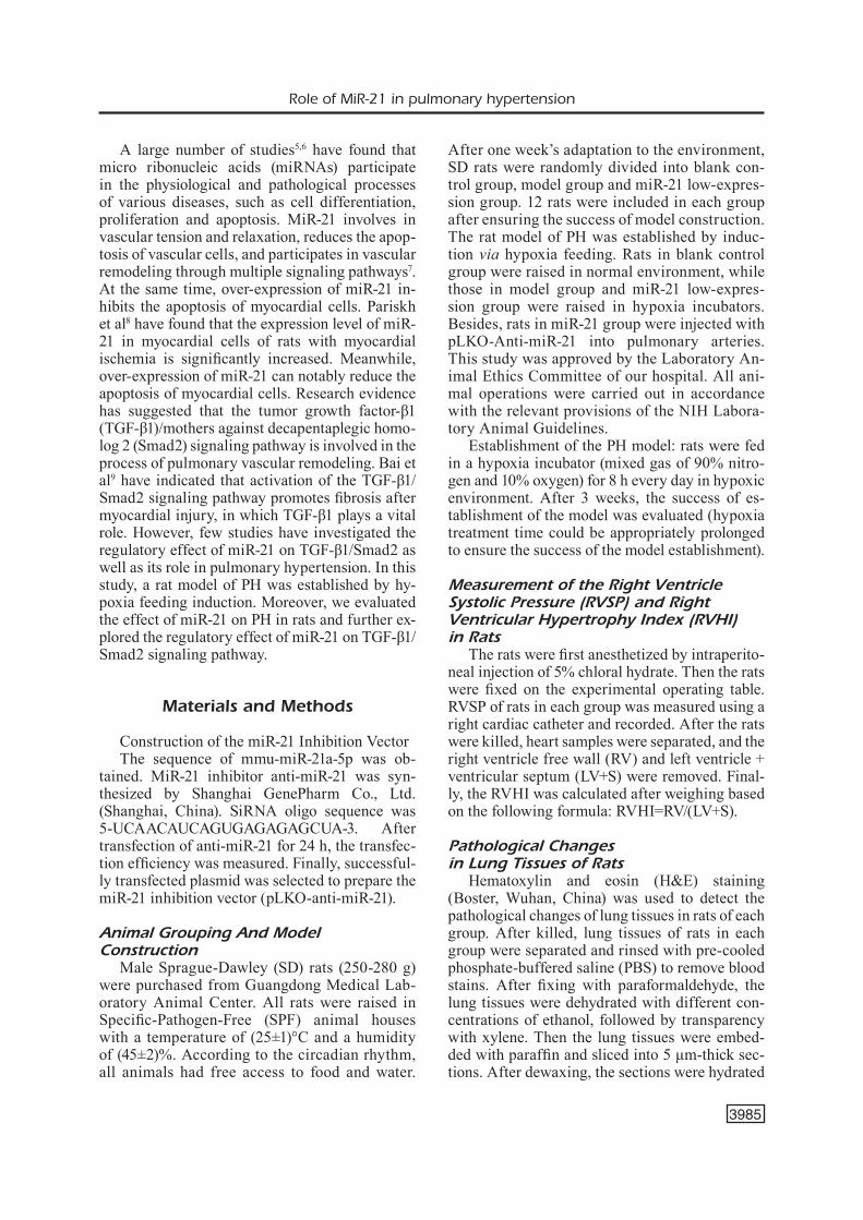
Expression Level of miR-21 in Lung Tissues of Rats
Real-time qPCR was performed to detect the expression level of miR-21 in lung tissues of rats in each group. The results revealed that compared with blank control group, the expression level of miR-21 in lung tissues of rats in model group was significantly increased (p<0.01). Meanwhile, miR-
21 expression in miR-21 low-expression group was significantly reduced after transfection of the miR-21 inhibition vector (p<0.01) (Figure 1).
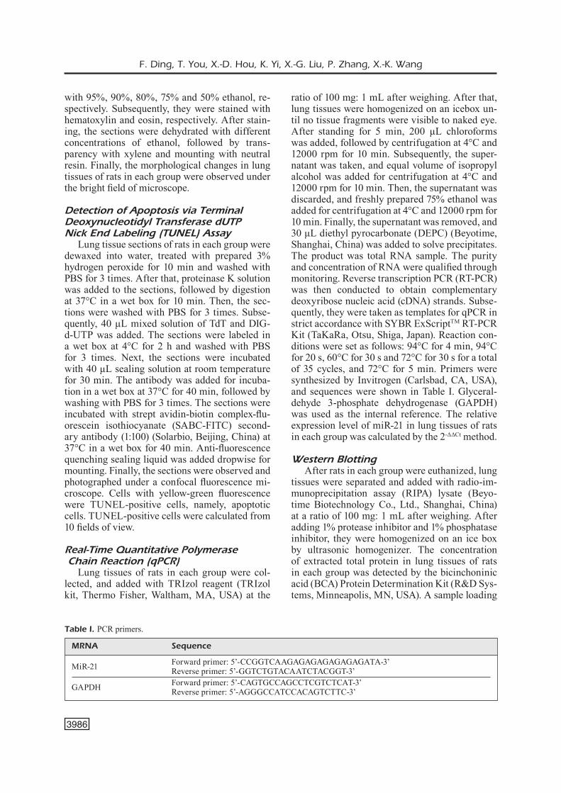
Detection of the RVSP and RVHI of Rats in Each Group
After anesthesia, the RVSP of rats in each group was measured by a right cardiac catheter. The right ventricle was separated to calculate the RVHI. As shown in Figure 2, the RVSP and RVHI of rats in model group were markedly higher than those of blank control group (p<0.01) and miR-21 low-expression group (p<0.01).
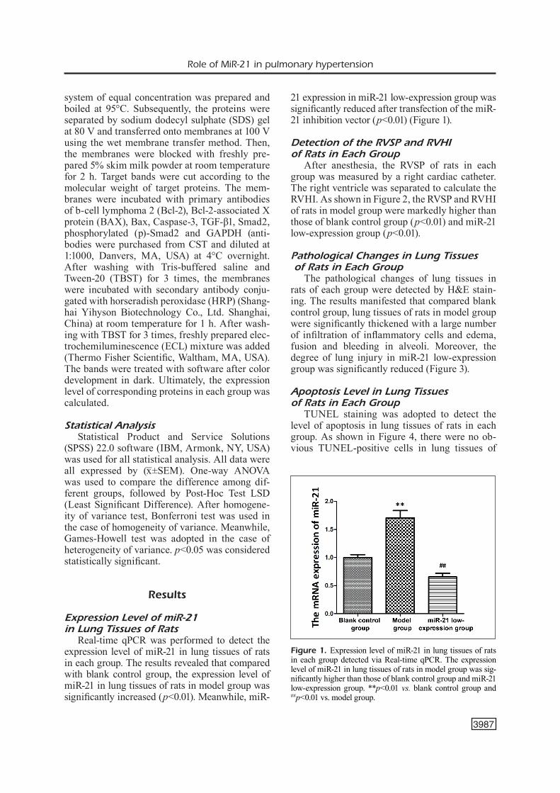
Pathological Changes in Lung Tissues of Rats in Each Group
The pathological changes of lung tissues in rats of each group were detected by H&E stain-ing. The results manifested that compared blank control group, lung tissues of rats in model group were significantly thickened with a large number of infiltration of inflammatory cells and edema, fusion and bleeding in alveoli. Moreover, the degree of lung injury in miR-21 low-expression group was significantly reduced (Figure 3).
Apoptosis Level in Lung Tissues of Rats in Each Group
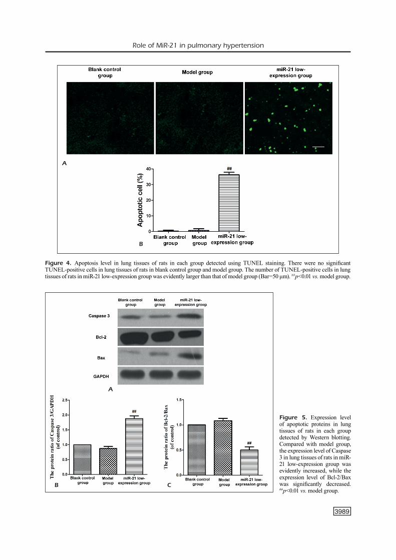
TUNEL staining was adopted to detect the level of apoptosis in lung tissues of rats in each group. As shown in Figure 4, there were no ob-vious TUNEL-positive cells in lung tissues of
Figure 1. Expression level of miR-21 in lung tissues of rats in each group detected via Real-time qPCR. The expression level of miR-21 in lung tissues of rats in model group was sig-nificantly higher than those of blank control group and miR-21 low-expression group. **p<0.01 vs. blank control group and ##p<0.01 vs. model group.
F. Ding, T. You, X.-D. Hou, K. Yi, X.-G. Liu, P. Zhang, X.-K. Wang
3988
rats in blank control group and model group. Fur-thermore, the number of TUNEL-positive cells in lung tissues of rats in miR-21 low-expression group was evidently larger than that of model group (p<0.01).
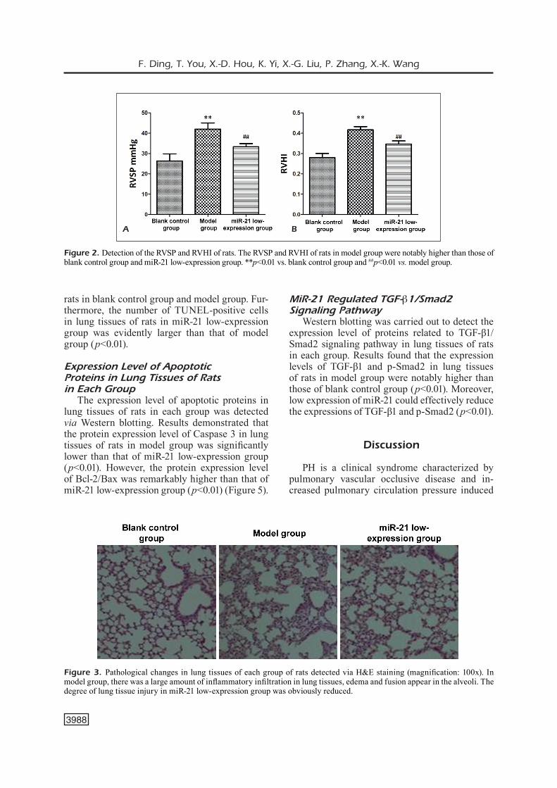
Expression Level of Apoptotic Proteins in Lung Tissues of Ratsin Each Group
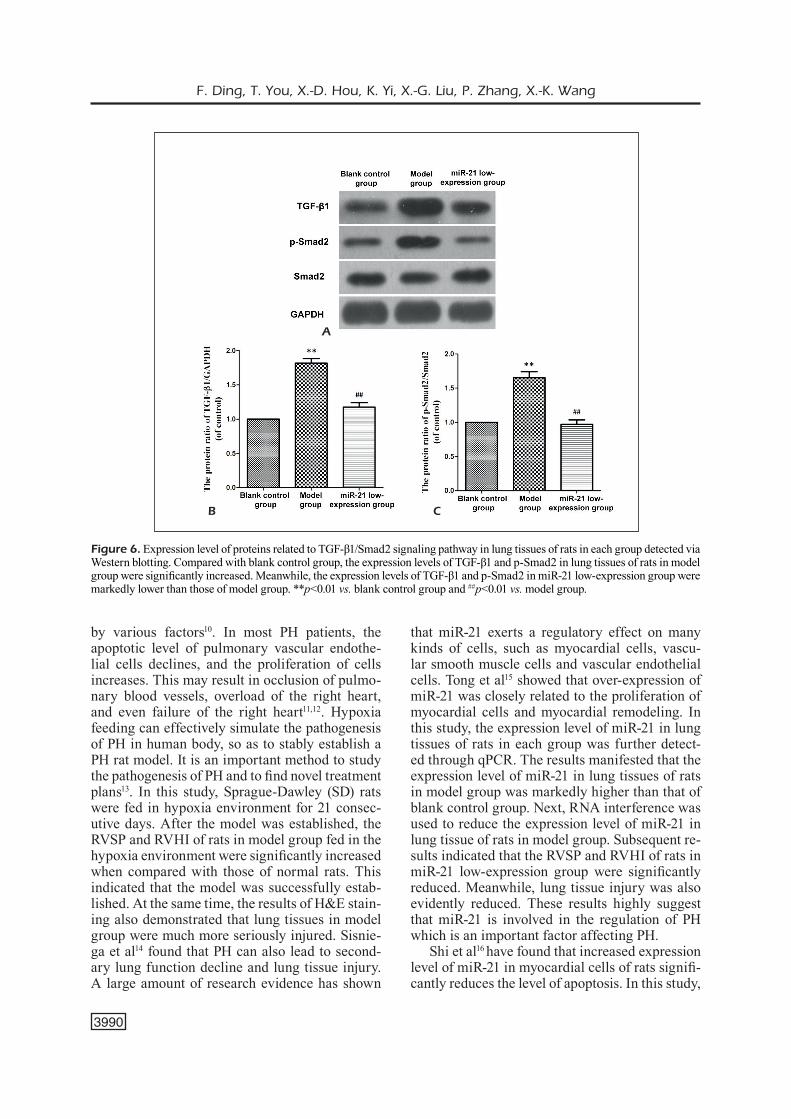
The expression level of apoptotic proteins in lung tissues of rats in each group was detected via Western blotting. Results demonstrated that the protein expression level of Caspase 3 in lung tissues of rats in model group was significantly lower than that of miR-21 low-expression group (p<0.01). However, the protein expression level of Bcl-2/Bax was remarkably higher than that of miR-21 low-expression group (p<0.01) (Figure 5).
MiR-21 Regulated TGF-β1/Smad2 Signaling Pathway
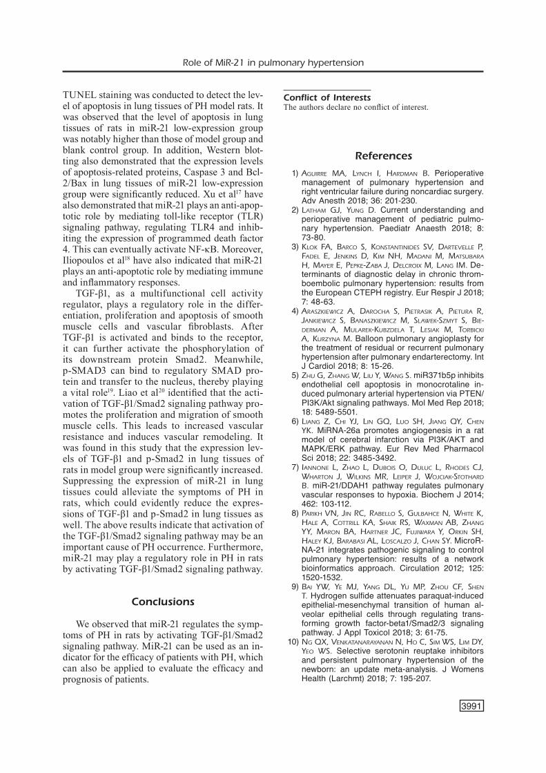
Western blotting was carried out to detect the expression level of proteins related to TGF-β1/Smad2 signaling pathway in lung tissues of rats in each group. Results found that the expression levels of TGF-β1 and p-Smad2 in lung tissues of rats in model group were notably higher than those of blank control group (p<0.01). Moreover, low expression of miR-21 could effectively reduce the expressions of TGF-β1 and p-Smad2 (p<0.01).
Discussion
PH is a clinical syndrome characterized by pulmonary vascular occlusive disease and in-creased pulmonary circulation pressure induced
Figure 2. Detection of the RVSP and RVHI of rats. The RVSP and RVHI of rats in model group were notably higher than those of blank control group and miR-21 low-expression group. **p<0.01 vs. blank control group and ##p<0.01 vs. model group.
A B
Figure 3. Pathological changes in lung tissues of each group of rats detected via H&E staining (magnification: 100x). In model group, there was a large amount of inflammatory infiltration in lung tissues, edema and fusion appear in the alveoli. The degree of lung tissue injury in miR-21 low-expression group was obviously reduced.
Role of MiR-21 in pulmonary hypertension
3989
Figure 4. Apoptosis level in lung tissues of rats in each group detected using TUNEL staining. There were no significant TUNEL-positive cells in lung tissues of rats in blank control group and model group. The number of TUNEL-positive cells in lung tissues of rats in miR-21 low-expression group was evidently larger than that of model group (Bar=50 μm). ##p<0.01 vs. model group.
A
B
Figure 5. Expression level of apoptotic proteins in lung tissues of rats in each group detected by Western blotting. Compared with model group, the expression level of Caspase 3 in lung tissues of rats in miR-21 low-expression group was evidently increased, while the expression level of Bcl-2/Bax was significantly decreased. ##p<0.01 vs. model group.
A
B C
F. Ding, T. You, X.-D. Hou, K. Yi, X.-G. Liu, P. Zhang, X.-K. Wang
3990
by various factors10. In most PH patients, the apoptotic level of pulmonary vascular endothe-lial cells declines, and the proliferation of cells increases. This may result in occlusion of pulmo-nary blood vessels, overload of the right heart, and even failure of the right heart11,12. Hypoxia feeding can effectively simulate the pathogenesis of PH in human body, so as to stably establish a PH rat model. It is an important method to study the pathogenesis of PH and to find novel treatment plans13. In this study, Sprague-Dawley (SD) rats were fed in hypoxia environment for 21 consec-utive days. After the model was established, the RVSP and RVHI of rats in model group fed in the hypoxia environment were significantly increased when compared with those of normal rats. This indicated that the model was successfully estab-lished. At the same time, the results of H&E stain-ing also demonstrated that lung tissues in model group were much more seriously injured. Sisnie-ga et al14 found that PH can also lead to second-ary lung function decline and lung tissue injury. A large amount of research evidence has shown
that miR-21 exerts a regulatory effect on many kinds of cells, such as myocardial cells, vascu-lar smooth muscle cells and vascular endothelial cells. Tong et al15 showed that over-expression of miR-21 was closely related to the proliferation of myocardial cells and myocardial remodeling. In this study, the expression level of miR-21 in lung tissues of rats in each group was further detect-ed through qPCR. The results manifested that the expression level of miR-21 in lung tissues of rats in model group was markedly higher than that of blank control group. Next, RNA interference was used to reduce the expression level of miR-21 in lung tissue of rats in model group. Subsequent re-sults indicated that the RVSP and RVHI of rats in miR-21 low-expression group were significantly reduced. Meanwhile, lung tissue injury was also evidently reduced. These results highly suggest that miR-21 is involved in the regulation of PH which is an important factor affecting PH.
Shi et al16 have found that increased expression level of miR-21 in myocardial cells of rats signifi-cantly reduces the level of apoptosis. In this study,
Figure 6. Expression level of proteins related to TGF-β1/Smad2 signaling pathway in lung tissues of rats in each group detected via Western blotting. Compared with blank control group, the expression levels of TGF-β1 and p-Smad2 in lung tissues of rats in model group were significantly increased. Meanwhile, the expression levels of TGF-β1 and p-Smad2 in miR-21 low-expression group were markedly lower than those of model group. **p<0.01 vs. blank control group and ##p<0.01 vs. model group.
A
B C
Role of MiR-21 in pulmonary hypertension
3991
TUNEL staining was conducted to detect the lev-el of apoptosis in lung tissues of PH model rats. It was observed that the level of apoptosis in lung tissues of rats in miR-21 low-expression group was notably higher than those of model group and blank control group. In addition, Western blot-ting also demonstrated that the expression levels of apoptosis-related proteins, Caspase 3 and Bcl-2/Bax in lung tissues of miR-21 low-expression group were significantly reduced. Xu et al17 have also demonstrated that miR-21 plays an anti-apop-totic role by mediating toll-like receptor (TLR) signaling pathway, regulating TLR4 and inhib-iting the expression of programmed death factor 4. This can eventually activate NF-κB. Moreover, Iliopoulos et al18 have also indicated that miR-21 plays an anti-apoptotic role by mediating immune and inflammatory responses.
TGF-β1, as a multifunctional cell activity regulator, plays a regulatory role in the differ-entiation, proliferation and apoptosis of smooth muscle cells and vascular fibroblasts. After TGF-β1 is activated and binds to the receptor, it can further activate the phosphorylation of its downstream protein Smad2. Meanwhile, p-SMAD3 can bind to regulatory SMAD pro-tein and transfer to the nucleus, thereby playing a vital role19. Liao et al20 identified that the acti-vation of TGF-β1/Smad2 signaling pathway pro-motes the proliferation and migration of smooth muscle cells. This leads to increased vascular resistance and induces vascular remodeling. It was found in this study that the expression lev-els of TGF-β1 and p-Smad2 in lung tissues of rats in model group were significantly increased. Suppressing the expression of miR-21 in lung tissues could alleviate the symptoms of PH in rats, which could evidently reduce the expres-sions of TGF-β1 and p-Smad2 in lung tissues as well. The above results indicate that activation of the TGF-β1/Smad2 signaling pathway may be an important cause of PH occurrence. Furthermore, miR-21 may play a regulatory role in PH in rats by activating TGF-β1/Smad2 signaling pathway.
Conclusions
We observed that miR-21 regulates the symp-toms of PH in rats by activating TGF-β1/Smad2 signaling pathway. MiR-21 can be used as an in-dicator for the efficacy of patients with PH, which can also be applied to evaluate the efficacy and prognosis of patients.
Conflict of InterestsThe authors declare no conflict of interest.
References
1) Aguirre MA, Lynch i, hArdMAn B. Perioperative management of pulmonary hypertension and right ventricular failure during noncardiac surgery. Adv Anesth 2018; 36: 201-230.
2) LAthAM gJ, yung d. Current understanding and perioperative management of pediatric pulmo-nary hypertension. Paediatr Anaesth 2018; 8: 73-80.
3) KLoK FA, BArco S, KonStAntinideS SV, dArteVeLLe P, FAdeL e, JenKinS d, KiM nh, MAdAni M, MAtSuBArA h, MAyer e, PePKe-ZABA J, deLcroix M, LAng iM. De-terminants of diagnostic delay in chronic throm-boembolic pulmonary hypertension: results from the European CTEPH registry. Eur Respir J 2018; 7: 48-63.
4) ArASZKiewicZ A, dArochA S, PietrASiK A, PieturA r, JAnKiewicZ S, BAnASZKiewicZ M, SLAweK-SZMyt S, Bie-derMAn A, MuLAreK-KuBZdeLA t, LeSiAK M, torBicKi A, KurZynA M. Balloon pulmonary angioplasty for the treatment of residual or recurrent pulmonary hypertension after pulmonary endarterectomy. Int J Cardiol 2018; 8: 15-26.
5) Zhu g, ZhAng w, Liu y, wAng S. miR371b5p inhibits endothelial cell apoptosis in monocrotaline in-duced pulmonary arterial hypertension via PTEN/PI3K/Akt signaling pathways. Mol Med Rep 2018; 18: 5489-5501.
6) LiAng Z, chi yJ, Lin gQ, Luo Sh, JiAng Qy, chen yK. MiRNA-26a promotes angiogenesis in a rat model of cerebral infarction via PI3K/AKT and MAPK/ERK pathway. Eur Rev Med Pharmacol Sci 2018; 22: 3485-3492.
7) iAnnone L, ZhAo L, duBoiS o, duLuc L, rhodeS cJ, whArton J, wiLKinS Mr, LeiPer J, woJciAK-StothArd B. miR-21/DDAH1 pathway regulates pulmonary vascular responses to hypoxia. Biochem J 2014; 462: 103-112.
8) PAriKh Vn, Jin rc, rABeLLo S, guLBAhce n, white K, hALe A, cottriLL KA, ShAiK rS, wAxMAn AB, ZhAng yy, MAron BA, hArtner Jc, FuJiwArA y, orKin Sh, hALey KJ, BArABASi AL, LoScALZo J, chAn Sy. MicroR-NA-21 integrates pathogenic signaling to control pulmonary hypertension: results of a network bioinformatics approach. Circulation 2012; 125: 1520-1532.
9) BAi yw, ye MJ, yAng dL, yu MP, Zhou cF, Shen t. Hydrogen sulfide attenuates paraquat-induced epithelial-mesenchymal transition of human al-veolar epithelial cells through regulating trans-forming growth factor-beta1/Smad2/3 signaling pathway. J Appl Toxicol 2018; 3: 61-75.
10) ng Qx, VenKAtAnArAyAnAn n, ho c, SiM wS, LiM dy, yeo wS. Selective serotonin reuptake inhibitors and persistent pulmonary hypertension of the newborn: an update meta-analysis. J Womens Health (Larchmt) 2018; 7: 195-207.
F. Ding, T. You, X.-D. Hou, K. Yi, X.-G. Liu, P. Zhang, X.-K. Wang
3992
11) Li d, Sun y, Kong x, LuAn c, yu y, chen F, chen P. Association between a single nucleotide poly-morphism in the 3'-UTR of ARHGEF18 and the risk of nonidiopathic pulmonary arterial hyper-tension in chinese population. Dis Markers 2018; 2018: 2461845.
12) yoon KL. New therapeutic target for pulmonary arterial hypertension. Korean Circ J 2018; 48: 1145-1147.
13) Lee dS, Jung yw. Protective effect of right ventric-ular mitochondrial damage by cyclosporine A in monocrotaline-induced pulmonary hypertension. Korean Circ J 2018; 48: 1135-1144.
14) SiSniegA c, ZAyAS n, PuLido t. Advances in medical therapy for pulmonary artery hypertension. Curr Opin Cardiol 2018; 5: 116-127.
15) tong Z, tAng y, JiAng B, wu y, Liu y, Li y, xiAo x. Phosphorylation of nucleolin is indispensable to upregulate miR-21 and inhibit apoptosis in cardio-myocytes. J Cell Physiol 2018; 5: 116-127.
16) Shi B, wAng y, ZhAo r, Long x, deng w, wAng Z. Bone marrow mesenchymal stem cell-derived
exosomal miR-21 protects C-kit+ cardiac stem cells from oxidative injury through the PTEN/PI3K/Akt axis. PLoS One 2018; 13: e191616.
17) xu r, huAng h, hAn Z, Li M, Zhou x. [Modulation of TLR-4/MyD88 signaling cascade by miR-21 is involved in airway immunologic dysfunction induced by cold air exposure]. Nan Fang Yi Ke Da Xue Xue Bao 2016; 36: 98-102.
18) iLioPouLoS d, KAVouSAnAKi M, ioAnnou M, BouMPAS d, VerginiS P. The negative costimulatory molecule PD-1 modulates the balance between immunity and toler-ance via miR-21. Eur J Immunol 2011; 41: 1754-1763.
19) Lin L, Li r, cAi M, huAng J, huAng w, guo y, yAng L, yAng g, LAn t, Zhu K. Andrographolide ameliorates liver fibrosis in mice: involvement of TLR4/NF-kappaB and TGF-beta1/Smad2 signal-ing pathways. Oxid Med Cell Longev 2018; 2018: 7808656.
20) LiAo K, yong cw, huA K. SB431542 inhibited ciga-rette smoke extract induced invasiveness of A549 cells via the TGF-beta1/Smad2/MMP3 pathway. Oncol Lett 2018; 15: 9681-9686.
.