Microglial responses to peripheral type 1 interferon
13
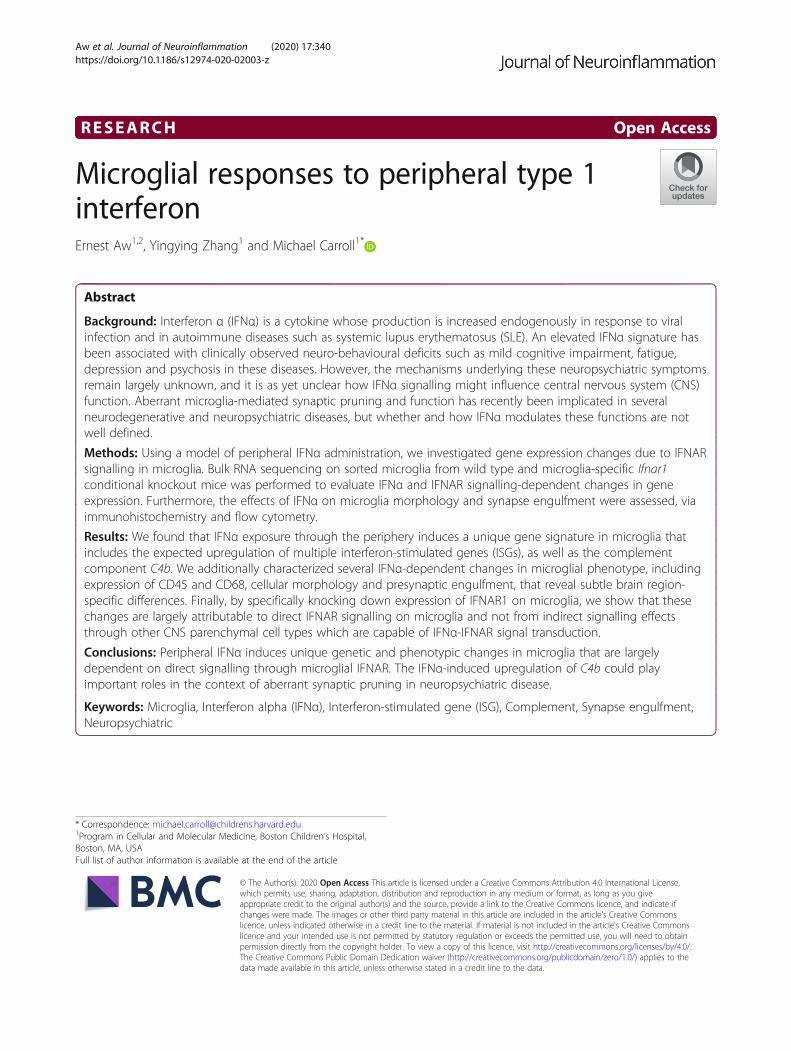
RESEARCH Open Access Microglial responses to peripheral type 1 interferon Ernest Aw 1,2 , Yingying Zhang 1 and Michael Carroll 1* Abstract Background: Interferon α (IFNα) is a cytokine whose production is increased endogenously in response to viral infection and in autoimmune diseases such as systemic lupus erythematosus (SLE). An elevated IFNα signature has been associated with clinically observed neuro-behavioural deficits such as mild cognitive impairment, fatigue, depression and psychosis in these diseases. However, the mechanisms underlying these neuropsychiatric symptoms remain largely unknown, and it is as yet unclear how IFNα signalling might influence central nervous system (CNS) function. Aberrant microglia-mediated synaptic pruning and function has recently been implicated in several neurodegenerative and neuropsychiatric diseases, but whether and how IFNα modulates these functions are not well defined. Methods: Using a model of peripheral IFNα administration, we investigated gene expression changes due to IFNAR signalling in microglia. Bulk RNA sequencing on sorted microglia from wild type and microglia-specific Ifnar1 conditional knockout mice was performed to evaluate IFNα and IFNAR signalling-dependent changes in gene expression. Furthermore, the effects of IFNα on microglia morphology and synapse engulfment were assessed, via immunohistochemistry and flow cytometry. Results: We found that IFNα exposure through the periphery induces a unique gene signature in microglia that includes the expected upregulation of multiple interferon-stimulated genes (ISGs), as well as the complement component C4b. We additionally characterized several IFNα-dependent changes in microglial phenotype, including expression of CD45 and CD68, cellular morphology and presynaptic engulfment, that reveal subtle brain region- specific differences. Finally, by specifically knocking down expression of IFNAR1 on microglia, we show that these changes are largely attributable to direct IFNAR signalling on microglia and not from indirect signalling effects through other CNS parenchymal cell types which are capable of IFNα-IFNAR signal transduction. Conclusions: Peripheral IFNα induces unique genetic and phenotypic changes in microglia that are largely dependent on direct signalling through microglial IFNAR. The IFNα-induced upregulation of C4b could play important roles in the context of aberrant synaptic pruning in neuropsychiatric disease. Keywords: Microglia, Interferon alpha (IFNα), Interferon-stimulated gene (ISG), Complement, Synapse engulfment, Neuropsychiatric © The Author(s). 2020 Open Access This article is licensed under a Creative Commons Attribution 4.0 International License, which permits use, sharing, adaptation, distribution and reproduction in any medium or format, as long as you give appropriate credit to the original author(s) and the source, provide a link to the Creative Commons licence, and indicate if changes were made. The images or other third party material in this article are included in the article's Creative Commons licence, unless indicated otherwise in a credit line to the material. If material is not included in the article's Creative Commons licence and your intended use is not permitted by statutory regulation or exceeds the permitted use, you will need to obtain permission directly from the copyright holder. To view a copy of this licence, visit http://creativecommons.org/licenses/by/4.0/. The Creative Commons Public Domain Dedication waiver (http://creativecommons.org/publicdomain/zero/1.0/) applies to the data made available in this article, unless otherwise stated in a credit line to the data. * Correspondence: [email protected] 1 Program in Cellular and Molecular Medicine, Boston Children’s Hospital, Boston, MA, USA Full list of author information is available at the end of the article Aw et al. Journal of Neuroinflammation (2020) 17:340 https://doi.org/10.1186/s12974-020-02003-z
Transcript of Microglial responses to peripheral type 1 interferon
RESEARCH Open Access
Microglial responses to peripheral type 1interferonErnest Aw1,2, Yingying Zhang1 and Michael Carroll1*
Abstract
Background: Interferon α (IFNα) is a cytokine whose production is increased endogenously in response to viralinfection and in autoimmune diseases such as systemic lupus erythematosus (SLE). An elevated IFNα signature hasbeen associated with clinically observed neuro-behavioural deficits such as mild cognitive impairment, fatigue,depression and psychosis in these diseases. However, the mechanisms underlying these neuropsychiatric symptomsremain largely unknown, and it is as yet unclear how IFNα signalling might influence central nervous system (CNS)function. Aberrant microglia-mediated synaptic pruning and function has recently been implicated in severalneurodegenerative and neuropsychiatric diseases, but whether and how IFNα modulates these functions are notwell defined.
Methods: Using a model of peripheral IFNα administration, we investigated gene expression changes due to IFNARsignalling in microglia. Bulk RNA sequencing on sorted microglia from wild type and microglia-specific Ifnar1conditional knockout mice was performed to evaluate IFNα and IFNAR signalling-dependent changes in geneexpression. Furthermore, the effects of IFNα on microglia morphology and synapse engulfment were assessed, viaimmunohistochemistry and flow cytometry.
Results: We found that IFNα exposure through the periphery induces a unique gene signature in microglia thatincludes the expected upregulation of multiple interferon-stimulated genes (ISGs), as well as the complementcomponent C4b. We additionally characterized several IFNα-dependent changes in microglial phenotype, includingexpression of CD45 and CD68, cellular morphology and presynaptic engulfment, that reveal subtle brain region-specific differences. Finally, by specifically knocking down expression of IFNAR1 on microglia, we show that thesechanges are largely attributable to direct IFNAR signalling on microglia and not from indirect signalling effectsthrough other CNS parenchymal cell types which are capable of IFNα-IFNAR signal transduction.
Conclusions: Peripheral IFNα induces unique genetic and phenotypic changes in microglia that are largelydependent on direct signalling through microglial IFNAR. The IFNα-induced upregulation of C4b could playimportant roles in the context of aberrant synaptic pruning in neuropsychiatric disease.
Keywords: Microglia, Interferon alpha (IFNα), Interferon-stimulated gene (ISG), Complement, Synapse engulfment,Neuropsychiatric
© The Author(s). 2020 Open Access This article is licensed under a Creative Commons Attribution 4.0 International License,which permits use, sharing, adaptation, distribution and reproduction in any medium or format, as long as you giveappropriate credit to the original author(s) and the source, provide a link to the Creative Commons licence, and indicate ifchanges were made. The images or other third party material in this article are included in the article's Creative Commonslicence, unless indicated otherwise in a credit line to the material. If material is not included in the article's Creative Commonslicence and your intended use is not permitted by statutory regulation or exceeds the permitted use, you will need to obtainpermission directly from the copyright holder. To view a copy of this licence, visit http://creativecommons.org/licenses/by/4.0/.The Creative Commons Public Domain Dedication waiver (http://creativecommons.org/publicdomain/zero/1.0/) applies to thedata made available in this article, unless otherwise stated in a credit line to the data.
* Correspondence: [email protected] in Cellular and Molecular Medicine, Boston Children’s Hospital,Boston, MA, USAFull list of author information is available at the end of the article
Aw et al. Journal of Neuroinflammation (2020) 17:340 https://doi.org/10.1186/s12974-020-02003-z
IntroductionType 1 interferons are a family of cytokines classicallyimplicated in antiviral defence, but also play importantpathological roles when their expression is dysregulated,such as in the autoimmune diseases Aicardi-Goutièressyndrome (AGS) and systemic lupus erythematosus(SLE) [1, 2]. Interferon α (IFNα) belongs to the type 1interferon family, which also include IFNβ and otherlesser studied IFNs; it comprises 14 different isoforms inmice and 13 in humans that are highly conserved ingene structure and amino acid sequence [3]. All type 1interferons signal through the type 1 interferon recep-tors IFNAR1 and IFNAR2, which dimerize upon ligandbinding and activate the JAK1 and TYK2 receptortyrosine kinases (RTKs). Canonically, these RTKs phos-phorylate the STAT1 and STAT2 transcription factorsresulting in their translocation into the nucleus, where,with IRF9 they form the transcription factor complexISGF3. ISGF3 binds to IFN-stimulated response elements(ISRE) on promoters, inducing gene expression pro-grammes involving interferon-stimulated genes (ISGs) thatare commonly antiviral and antiproliferative [3, 4]. Interest-ingly, the ISG expression landscape differs across cell typesin a manner that is dependent on signalling strength andchronicity, as well as the activation of other non-canonicalsignalling machinery, resulting in functionally distinct out-comes [3, 4].Importantly, IFNα has been closely associated with
numerous neuropsychiatric comorbidities in various clin-ical contexts, including recombinant IFNα treatment forchronic hepatitis C infection and melanoma [5–7], as wellas SLE associated neuropsychiatric disease [8, 9]. Theseneuropsychiatric deficiencies are heterogeneous in presen-tation, ranging in severity from mild cognitive dysfunctionand fatigue to overt psychosis. Despite its well-studied sig-nal transduction pathway in terms of antiviral function, itis as yet unclear how IFNα-IFNAR signalling within thebrain translates to such detrimental neuropsychiatricoutcomes. Furthermore, given the ubiquitous capacity ofmost nucleated cell types to respond to IFNAR signalling,it is unclear which cell types within the central nervoussystem (CNS) are critical, functional contributors toIFNα-associated neuropsychiatric disease.Microglia are long-lived tissue-resident macrophages
of the brain and play important roles in shaping neur-onal circuits during development via synaptic pruning[10, 11], while also constantly surveying the brain paren-chyma to detect injury or damage [12, 13]. Aberrantmicroglial function linked to dysregulated synaptic prun-ing has been implicated in the pathophysiology of severalneurodegenerative and neuropsychiatric diseases includ-ing Alzheimer’s disease (AD) [14, 15], schizophrenia [16]and autism spectrum disease [17, 18]. Furthermore, micro-glia have been shown to secrete pro-inflammatory cytokines
that negatively impact neurogenesis and influence depressivebehaviour in response to peripherally derived IFNα [19], anddysregulation of IFNAR signalling in white matter microgliavia genetic attenuation of its negative regulator Usp18 causesdestructive microgliopathy in the brain [20]. Interestingly,recent sequencing studies have identified a microglial type 1IFN gene signature in the contexts of ageing [21, 22], Alz-heimer’s disease [15, 23] and SLE [24], suggesting a potentialunifying role of type 1 IFN signalling in propagating theneurological and neuropsychiatric pathologies in these vari-able pathological contexts. However, despite the increasinglyappreciated role of IFNα in inflammation-associated CNSpathology, it is still unclear how IFNα regulates microglialfunction, and whether it might influence microglia-mediatedsynaptic pruning as a potential mechanism in driving IFNα-associated neuropsychiatric disease.In the present study, we report that peripherally derived
IFNα is able to transduce signalling across the blood-brainbarrier (BBB), resulting in a unique microglial genetic sig-nature that is primarily dependent on microglial IFNAR.Our results also demonstrate that in an acute model ofIFNα exposure, microglia adopt a unique activated stateand engulf synaptic material in a brain region-dependentmanner. In light of recent studies implicating the comple-ment system in both homeostatic and pathological synap-tic pruning [10, 14, 25], the IFNα-mediated upregulationof complement component C4b expression in microgliamay contribute to IFNα-related neuropsychiatric symp-toms by a similar mechanism.
Materials and methodsAnimalsEight-week-old male and female C57BL/6J (000664) micewere purchased from Jackson Laboratories. Injections ofPBS and murine recombinant IFNα-A (mIFNα) (BioLe-gend) were all performed intraperitoneally at 8 weeks ofage for C57BL/6 mice. Daily injections were performedaround between 0800 and 0900 h each day for 7 days, andmice were sacrificed 3 h post final injection. Cx3cr1-CreERT2 (020940) and R26-EyfpLSL (006148) mice werepurchased from Jackson Laboratories. R26-EyfpLSL micewere backcrossed to the C57BL/6 background in house forat least 10 generations. Ifnar1fl/fl (MGI:2655303) mice wereobtained from Ulrich Kalinke. Cx3cr1-CreERT2 mice werecrossed with Ifnar1fl/fl or R26-EyfpLSL mice to obtainCx3cr1-CreERT2+/−, Ifnarfl/fl and Cx3cr1-CreERT2+/−, R26-EyfpLSL mice respectively. CreERT2 activity was induced byoral gavage of either sunflower seed oil vehicle (SpectrumChemical) or 100mg/kg body weight tamoxifen (Sigma)daily for 5 days at 8 weeks of age. Recycling monocytic/macrophagic populations were allowed to replenish them-selves for 4 weeks [26] prior to sacrifice or mIFNα injec-tions. Male and female mice were used for all experiments,except for the determination of serum mIFNα levels, where
Aw et al. Journal of Neuroinflammation (2020) 17:340 Page 2 of 13
only male mice were used. All animal experiments wereapproved by the Boston Children’s Hospital and HarvardMedical School institutional animal use and care commit-tee in accordance with NIH guidelines for the humanetreatment of animals.
Serum ELISA for mIFNαSerum samples were assayed on the VeriPlex MouseCytokine 9-Plex ELISA Kit (PBL Assay Science) by con-tract service from the company (PBL Assay Science).
Microglia purificationMicroglia were purified using an adapted protocol [27].Briefly, mice were perfused with ice-cold PBS and brainregions were dissected into ice-cold HBSS withoutcalcium, magnesium and phenol red (Corning). Tissuewas Dounce homogenized using a 1-mL tissue grinder(Wheaton), with 12 slow strokes each on the loosefollowed by the tight pestle. The cell suspension wasthen spun down at 1200 rpm, 7 min at 4 °C and resus-pended in 70% Percoll (GE Healthcare). Thirty-sevenpercent Percoll was carefully layered on top and spundown at 800 g, 25 min at 23 °C with the acceleration setto 3 and deceleration set to 2 on a Sorvall Legend XTRcentrifuge (Thermo Fisher). The cloudy cellular layerwas then carefully pipetted and diluted in ice-cold FACSbuffer (0.1% BSA, 1 mM EDTA in PBS). Cells were thenprocessed for downstream staining for flow cytometry orfluorescence activated cell sorting (FACS). For flow cyto-metric assessment of presynaptic engulfment of synapticvesicle 2 (SV2), 10 μM cytochalasin D (Sigma) was addedto HBSS and all Percoll dilutions.
Flow cytometry and sortingCells were stained with primary antibodies againstCD11b (clone M1/70, Biolegend), CD45 (clone 30-F11,Biolegend), fixable viability dye eFluor-780 (eBioscience)for 30 min at 4 °C. For intracellular staining, cells werefixed using fixation buffer (Biolegend) and permeabilizedusing intracellular fixation and permeabilization buffer(Biolegend) according to manufacturer’s instructions.Fixed and permeabilized cells were stained with primaryantibodies against CD68 (clone FA-11, Biolegend) andSV2 (DSHB, UIowa). SV2 monoclonal antibodies werepurified and conjugated to fluorochromes in-house. Flowcytometry was performed on a FACSCanto (BD Biosci-ences), and microglial sorting was performed on a FACSAria II SORP (BD Biosciences) using a 70-μm nozzle.Data was analysed using FlowJo (Tree Star).
ImmunohistochemistryMice were perfused with ice-cold PBS, and brains weredissected and fixed in 4% PFA (Electron MicroscopySciences) overnight at 4 °C. Tissue was cryopreserved in
30% sucrose, followed by embedding in OCT (FisherHealthcare) and cryosectioned into 40-μm-thick floatingsections. Tissue sections were blocked for 1 h at RT withblocking buffer (5% BSA, 0.2% Triton-X in PBS) andstained with primary antibody against IBA1 (Wako, 1:400) overnight at RT. Tissue sections were then washed3× in PBS, stained with secondary antibody againstrabbit IgG (Invitrogen, 1:200) for 1 h at RT, washed 3×with PBS and then mounted onto a slide with mountingmedium with DAPI (Southern Biotech) and sealed withnail varnish. Primary and secondary antibodies were di-luted in blocking buffer.
Microglia morphology analysisConfocal images of the tissue sections were obtained at× 60 magnification using the FV1000 confocal system(Olympus). Three fields of view from 2 brain sectionseach of the frontal cortex, hippocampus and cerebellumper biological sample were imaged and analysed usingCell Profiler [28]. Briefly, microglia were identified andsegmented based on IBA1 staining, and various area andshape features were derived using the MeasureObjectSi-zeShape module of Cell Profiler. Pipeline used is avail-able upon request.
RNAscope in situ hybridization and analysisMouse brains were dissected and snap frozen on dry icefollowing terminal anaesthesia, without perfusion. OCTembedded brains were then cryosectioned into 16-μm-thick sections prior to RNAscope in situ hybridization.RNAscope in situ hybridization was performed using theRNAscope fluorescent multiplex assay kit (ACD Bio)using probes directed against Mx1 (474931, ACD Bio),Rsad2 (561251, ACD Bio) and Tmem119 (472901, ACDBio) according to manufacturer’s instructions. Confocalimages were obtained at × 40 magnification using theFV1000 confocal system (Olympus). Three fields of viewof the frontal cortex per biological sample were imagedand analysed using Cell Profiler [28]. Briefly, microgliawere classified as DAPI+ cells containing more than 2Tmem119 puncta, and cells were defined as ISG positiveif they expressed more than 1 identified puncta of Mx1or Rsad2. Pipeline used is available upon request.
Gene expression by qPCR and ddPCRBrain or spleen tissue was homogenized in TRIzol reagent(Ambion) using the TissueLyser homogenizer (Qiagen)and RNA was isolated by phenol-chloroform extraction.For microglia-specific gene expression assays, microgliawere directly sorted into TCL lysis buffer (Qiagen) supple-mented with 1% β-mercaptoethanol (Sigma). RNA was ex-tracted using 2.2X RNAclean XP beads (Beckman Coulter)following manufacturer’s instructions. cDNA was thensynthesized with gDNA depletion using the iScript gDNA
Aw et al. Journal of Neuroinflammation (2020) 17:340 Page 3 of 13
clear cDNA synthesis kit (BioRad) following manufac-turer’s instructions. qPCR reactions were assembled forgenes of interest (Actb, Ifnar1) using the iTaq universalSYBR Green supermix system (BioRad) and run on aCFX96 machine (BioRad). Digital Droplet PCR (ddPCR)(BioRad) reactions were assembled according to manufac-turer’s instructions, using manufacturer probes for genes ofinterest except for Eif4h, Rpp30 and C4b. For qPCR, geneexpression was normalized to Actb and compared usingthe ΔΔCt method. For ddPCR, gene expression was nor-malized to Eif4h for microglia or Rpp30 for spleen.qPCR primers are as follows: Actb (forward: 5′ AAGAGC
TATGAGCTGCCTGA reverse: 5′ TACGGATGTCAACGTCACAC) and Ifnar1 (forward: 5′TGTGCTTCCCAC-CACTCAAG, reverse: 5′ AGGCGCGTGCTTTACTTCTA).ddPCR primer/probe sets are as follows: Mx1 (dMmu
CPE5121908, BioRad), Ifit3 (dMmuCPE5197126, BioRad),Oas2 (dMmuCPE5099434, BioRad), Axl (dMmuCPE5090992, BioRad), Eif4h (forward: 5′ TGCAGCTTGCTTGGTAGC, reverse: 5′ GTAAATTGCCGAGACCTTGC, probe: 5′HEX-AGCCTACCCCTTGGCTCGGG), Rpp30 (forward: 5′TGACCCTATCAGAGGACTGC, reverse: 5′ CTCTGCAATTTGTGGACACG, probe: 5′HEX- TGGGCTTTCTGAAAATGATGGCAA) and C4b (forward: 5′ AGCCTGTTTCCAGCTCAAAG, reverse: 5′ GTCCTAAGGCCTCAC ACCTG, probe: 5′FAM- CCCCGGCTGCTGAACTCCAT).
Microglia bulk RNAseq and analysisOne thousand and two hundred microglia from anteriorcortex, spanning the frontal, motor and part of thesomatosensory cortices were sorted into TCL buffer sup-plemented with 1% β-mercaptoethanol, and RNA was ex-tracted using RNAclean XP beads as described. cDNAwas synthesized using the SMART-Seq v4 ultra-low inputkit for RNA sequencing (Takara Bio) according to manu-facturer instructions. Library preparation for sequencingwas performed using the Nextera XT DNA library prepkit (Illumina) according to the manufacturer’s instruc-tions. Sample quality was assayed as recommended usingthe high sensitivity DNA kit (Agilent). Samples werepooled equimolarly and sequencing was performed on aNextseq 500 sequencer (Illumina) with 1 nM input. Forbulk RNAseq analysis, 75 bp single-end reads were firsttrimmed of their adaptors and then aligned to the mm10mouse reference genome using STAR Aligner [29]. Allsamples had at least 20 million uniquely mapped reads.The raw read count matrix was then analysed in theEdgeR package [30] for differential gene expression usingthe QLF model. Gene ontology analysis was performedusing the gProfiler2 package [31].
Statistical analysisFor all statistical analysis, R and GraphPad Prism 8(GraphPad Software) were used. Error bars represent s.d.
in all figures. All replicate numbers, statistical tests, pvalues and q values used are indicated in the figure leg-ends where appropriate. Statistical tests were selectedbased on the normality of the data distribution, assessedby the Shapiro-Wilk test or quantile-quantile (QQ) plot,and also the equality of variance, assessed by the F test.
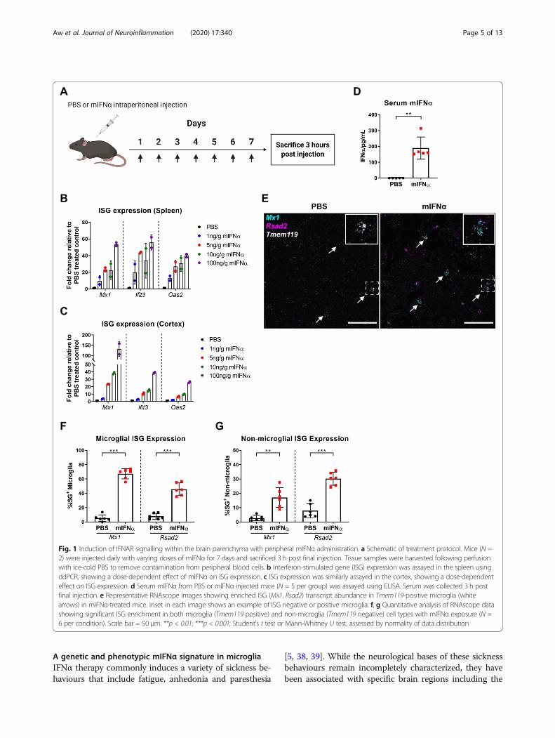
ResultsTitration of peripherally administered mIFNαIn order to determine whether peripherally administeredmurine recombinant interferon α (mIFNα) was able to in-duce a signalling effect within the central nervous system(CNS), mice were injected intraperitoneally with varyingdoses of mIFNα. To model acute type 1 interferon-mediated inflammation, mice were injected daily for 7 days(Fig. 1a), and a dose-dependent, titratable effect oninterferon-stimulated gene (ISG) expression both in thespleen (Fig. 1b) and in perfused cortical tissue from thebrain (Fig. 1c) was observed. A working dose of 10 ngmIFNα/g of body weight was selected and used for all fur-ther experiments, which is of similar dosage used in previ-ous studies and also within the relative dose range that hasbeen used therapeutically in patients with chronic hepatitisC infection or various cancers [19, 32–36]. Furthermore,serum concentrations of mIFNα averaged at around 200pg/mL 3 h post final injection using this dosing schedule(Fig. 1d), indicating a roughly 600-fold decrease in bioavail-ability at this time point, assuming a total blood volume of2mL for a 25-g male mouse. Given the induced ISG signa-ture, we observed in the spleen and brain cortex at thissame time point, it is reasonable to assume that at least partof this large decrease in bioavailability was due to cellularor tissue consumption and signal transduction of mIFNα asmanifested by their ISG signature.
Peripherally administered mIFNα induces ISG expressionin microgliaThe blood-brain barrier (BBB) tightly regulates themovement of a wide range of molecules, includingcytokines, to and from the CNS [37]. Given the recentappreciation that microglia play critical immunologicalroles in surveying and protecting the CNS from patho-genic threats and injury [12, 13, 25], it was next investi-gated whether peripherally administered mIFNα wouldbe able to induce IFNAR signalling in microglia acrossthe BBB. Indeed, using RNAscope in situ hybridization,peripherally administered mIFNα increased expressionof the ISGs Mx1 and Rsad2, in up to 75% of microglia(Tmem119-positive cells) within the frontal cortex (Fig.1e, f). We note that increased numbers of ISG punctawere also present in non-Tmem119 positive cells (Fig. 1g),suggesting that peripherally administered mIFNα is alsoable to induce functional IFNAR signalling in cell typesother than microglia within the CNS.
Aw et al. Journal of Neuroinflammation (2020) 17:340 Page 4 of 13
A genetic and phenotypic mIFNα signature in microgliaIFNα therapy commonly induces a variety of sickness be-haviours that include fatigue, anhedonia and paresthesia
[5, 38, 39]. While the neurological bases of these sicknessbehaviours remain incompletely characterized, they havebeen associated with specific brain regions including the
Fig. 1 Induction of IFNAR signalling within the brain parenchyma with peripheral mIFNα administration. a Schematic of treatment protocol. Mice (N =2) were injected daily with varying doses of mIFNα for 7 days and sacrificed 3 h post final injection. Tissue samples were harvested following perfusionwith ice-cold PBS to remove contamination from peripheral blood cells. b Interferon-stimulated gene (ISG) expression was assayed in the spleen usingddPCR, showing a dose-dependent effect of mIFNα on ISG expression. c ISG expression was similarly assayed in the cortex, showing a dose-dependenteffect on ISG expression. d Serum mIFNα from PBS or mIFNα injected mice (N = 5 per group) was assayed using ELISA. Serum was collected 3 h postfinal injection. e Representative RNAscope images showing enriched ISG (Mx1, Rsad2) transcript abundance in Tmem119-positive microglia (whitearrows) in mIFNα-treated mice. Inset in each image shows an example of ISG negative or positive microglia. f, g Quantitative analysis of RNAscope datashowing significant ISG enrichment in both microglia (Tmem119 positive) and non-microglia (Tmem119 negative) cell types with mIFNα exposure (N =6 per condition). Scale bar = 50 μm. **p < 0.01; ***p < 0.001; Student’s t test or Mann-Whitney U test, assessed by normality of data distribution
Aw et al. Journal of Neuroinflammation (2020) 17:340 Page 5 of 13
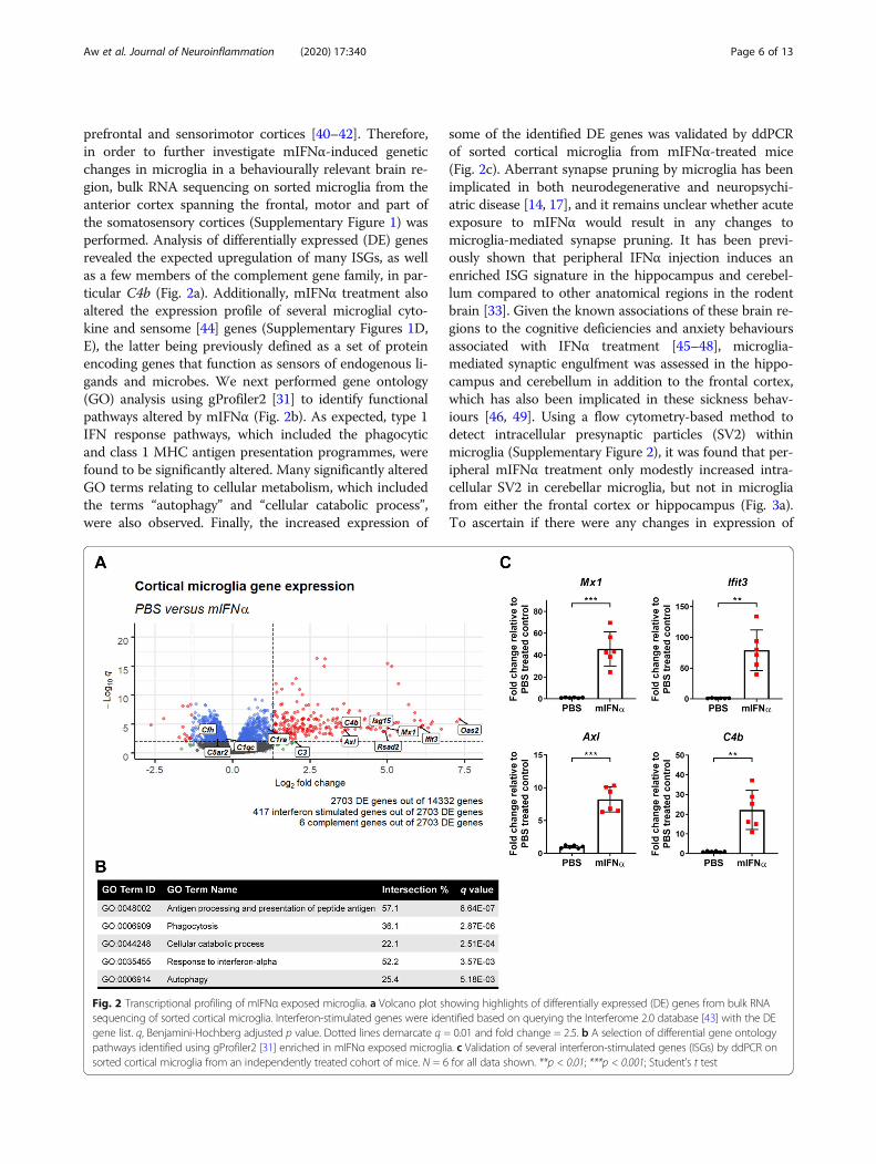
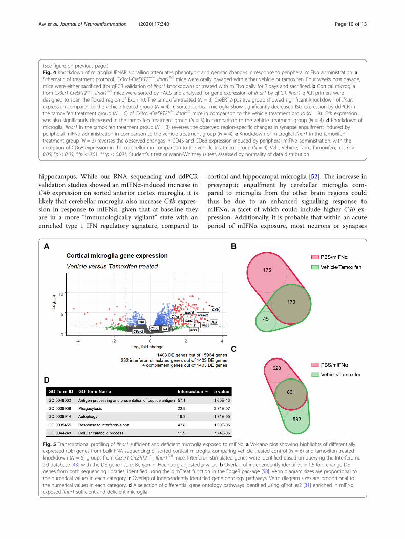
prefrontal and sensorimotor cortices [40–42]. Therefore,in order to further investigate mIFNα-induced geneticchanges in microglia in a behaviourally relevant brain re-gion, bulk RNA sequencing on sorted microglia from theanterior cortex spanning the frontal, motor and part ofthe somatosensory cortices (Supplementary Figure 1) wasperformed. Analysis of differentially expressed (DE) genesrevealed the expected upregulation of many ISGs, as wellas a few members of the complement gene family, in par-ticular C4b (Fig. 2a). Additionally, mIFNα treatment alsoaltered the expression profile of several microglial cyto-kine and sensome [44] genes (Supplementary Figures 1D,E), the latter being previously defined as a set of proteinencoding genes that function as sensors of endogenous li-gands and microbes. We next performed gene ontology(GO) analysis using gProfiler2 [31] to identify functionalpathways altered by mIFNα (Fig. 2b). As expected, type 1IFN response pathways, which included the phagocyticand class 1 MHC antigen presentation programmes, werefound to be significantly altered. Many significantly alteredGO terms relating to cellular metabolism, which includedthe terms “autophagy” and “cellular catabolic process”,were also observed. Finally, the increased expression of
some of the identified DE genes was validated by ddPCRof sorted cortical microglia from mIFNα-treated mice(Fig. 2c). Aberrant synapse pruning by microglia has beenimplicated in both neurodegenerative and neuropsychi-atric disease [14, 17], and it remains unclear whether acuteexposure to mIFNα would result in any changes tomicroglia-mediated synapse pruning. It has been previ-ously shown that peripheral IFNα injection induces anenriched ISG signature in the hippocampus and cerebel-lum compared to other anatomical regions in the rodentbrain [33]. Given the known associations of these brain re-gions to the cognitive deficiencies and anxiety behavioursassociated with IFNα treatment [45–48], microglia-mediated synaptic engulfment was assessed in the hippo-campus and cerebellum in addition to the frontal cortex,which has also been implicated in these sickness behav-iours [46, 49]. Using a flow cytometry-based method todetect intracellular presynaptic particles (SV2) withinmicroglia (Supplementary Figure 2), it was found that per-ipheral mIFNα treatment only modestly increased intra-cellular SV2 in cerebellar microglia, but not in microgliafrom either the frontal cortex or hippocampus (Fig. 3a).To ascertain if there were any changes in expression of
Fig. 2 Transcriptional profiling of mIFNα exposed microglia. a Volcano plot showing highlights of differentially expressed (DE) genes from bulk RNAsequencing of sorted cortical microglia. Interferon-stimulated genes were identified based on querying the Interferome 2.0 database [43] with the DEgene list. q, Benjamini-Hochberg adjusted p value. Dotted lines demarcate q = 0.01 and fold change = 2.5. b A selection of differential gene ontologypathways identified using gProfiler2 [31] enriched in mIFNα exposed microglia. c Validation of several interferon-stimulated genes (ISGs) by ddPCR onsorted cortical microglia from an independently treated cohort of mice. N = 6 for all data shown. **p < 0.01; ***p < 0.001; Student’s t test
Aw et al. Journal of Neuroinflammation (2020) 17:340 Page 6 of 13
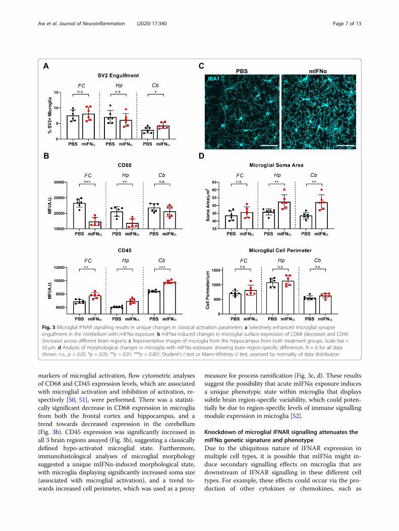
markers of microglial activation, flow cytometric analysesof CD68 and CD45 expression levels, which are associatedwith microglial activation and inhibition of activation, re-spectively [50, 51], were performed. There was a statisti-cally significant decrease in CD68 expression in microgliafrom both the frontal cortex and hippocampus, and atrend towards decreased expression in the cerebellum(Fig. 3b). CD45 expression was significantly increased inall 3 brain regions assayed (Fig. 3b), suggesting a classicallydefined hypo-activated microglial state. Furthermore,immunohistological analyses of microglial morphologysuggested a unique mIFNα-induced morphological state,with microglia displaying significantly increased soma size(associated with microglial activation), and a trend to-wards increased cell perimeter, which was used as a proxy
measure for process ramification (Fig. 3c, d). These resultssuggest the possibility that acute mIFNα exposure inducesa unique phenotypic state within microglia that displayssubtle brain region-specific variability, which could poten-tially be due to region-specific levels of immune signallingmodule expression in microglia [52].
Knockdown of microglial IFNAR signalling attenuates themIFNα genetic signature and phenotypeDue to the ubiquitous nature of IFNAR expression inmultiple cell types, it is possible that mIFNα might in-duce secondary signalling effects on microglia that aredownstream of IFNAR signalling in these different celltypes. For example, these effects could occur via the pro-duction of other cytokines or chemokines, such as
Fig. 3 Microglial IFNAR signalling results in unique changes in classical activation parameters. a Selectively enhanced microglial synapseengulfment in the cerebellum with mIFNα exposure. b mIFNα-induced changes in microglial surface expression of CD68 (decrease) and CD45(increase) across different brain regions. c Representative images of microglia from the hippocampus from both treatment groups. Scale bar =50 μm. d Analysis of morphological changes in microglia with mIFNα exposure, showing brain region-specific differences. N = 6 for all datashown. n.s., p > 0.05; *p < 0.05; **p < 0.01; ***p < 0.001; Student’s t test or Mann-Whitney U test, assessed by normality of data distribution
Aw et al. Journal of Neuroinflammation (2020) 17:340 Page 7 of 13
CXCL10 from brain endothelial cells [53], or even IL-6,IL-1β and TNFα from microglia [19] in response to type1 interferon signalling. Indeed, significant gene expres-sion upregulation of several of these cytokines and che-mokines in mIFNα-exposed microglia were observed(Supplementary Figure 1E), which could potentially con-tribute towards autocrine signalling effects. Furthermore,many ISGs can also be cross regulated by distinct signal-ling pathways downstream of other cytokines, such asIL-1β, IFNγ and TNFα [54–57]. Given the increased ex-pression of some of these cytokines in response to IFNα(Supplementary Figure 1E), it is possible that the ob-served canonical ISG signature is additionally derived inpart by other cytokine signalling pathways.In order to further investigate whether the IFNα-
induced phenotypic and genetic changes in microgliacan primarily be attributed to direct microglial IFNARsignalling, Ifnar1 expression on microglia was selectivelyknocked down using the inducible Cx3cr1-CreERT2driver [26] (Supplementary Figure 3; Fig. 4a, b). Indeed,mIFNα treatment of mice with microglial specificknockdown of Ifnar1 resulted in the attenuation of ISGexpression, as well as C4b expression, in microglia com-pared to control mice (Fig. 4c), suggesting that these genesare directly regulated by microglial IFNAR signalling. Fur-thermore, there was a reversal of the SV2 engulfmentphenotype in cerebellar microglia with knockdown ofmicroglial Ifnar1, with no corresponding significantchange in microglial SV2 engulfment in both the frontalcortex and hippocampus (Fig. 4d). Changes in microglialCD68 and CD45 expression levels were also largely re-versed with knockdown of microglial Ifnar1 (Fig, 4e).Finally, the possibility of any potential non-microglial
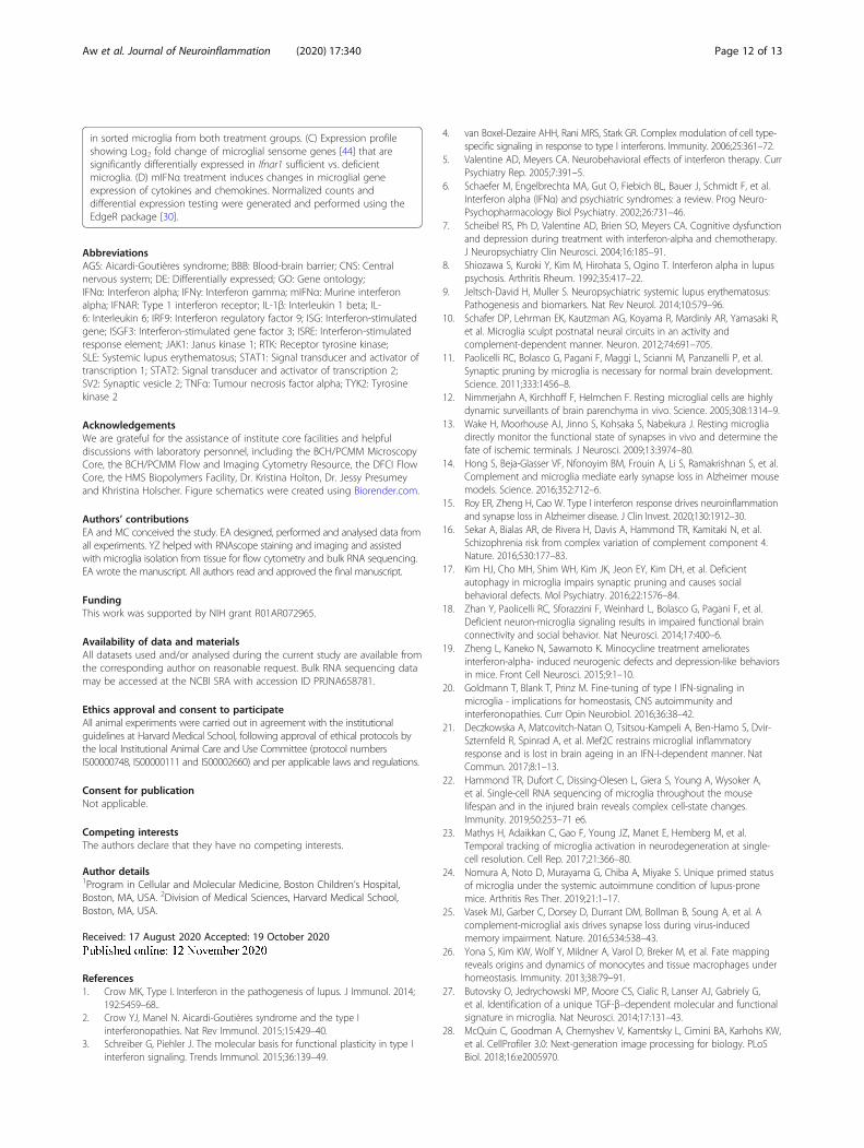
IFNAR signalling effects was investigated by bulk RNAsequencing of microglia sorted from the same anteriorcortex region of Ifnar1 sufficient and deficient micetreated with mIFNα (Supplementary Figure 4). As ex-pected, knockdown of Ifnar1 on microglia attenuatedmany of the genes induced by mIFNα as described earl-ier (Fig. 5a), with most of the DE genes displaying thehighest log2 fold change (> 1.5) overlapping in identitywith similarly high log2 fold change DE genes frommIFNα-exposed wild type microglia (Fig. 5b). Furthermore,the GO terms from both sets of experiments remainedsimilar (Fig. 5c, d). Finally, the panel of sensome and cyto-kine/chemokine genes that were differentially expressedremained similar as well (Supplementary Figures 4C, D).These data suggest that peripherally derived IFNα can
induce a functional signalling event through microgliaby upregulating a unique genetic and phenotypicprogramme. These genetic and phenotypic changes relyprimarily on microglial IFNAR signalling and are largelyindependent of any potential secondary signalling effectsthrough other cytokines or chemokines.
DiscussionTo investigate the interaction of peripheral IFNα withthe CNS, in particular microglia, we first show that per-ipherally administered mIFNα is able to exert directfunctional signalling effects across the BBB on microglia.This is in agreement with previous studies which haveshown that IFNα can enter the brain parenchyma fromthe periphery via a non-saturable mechanism [59, 60],and also elicit a transcriptional response within numer-ous cell types in the brain parenchyma [33]. Gene ex-pression differences were further characterized by RNAsequencing and showed an expected strong upregulationof genes associated with type 1 interferon signalling.Interestingly, we also found a strong increase in expres-sion of the complement component C4b, which has pre-viously been observed in the contexts of viral infection[61] and ageing [21, 23], in microglia that also concur-rently express a strong type 1 interferon signature. Inparticular, we also show that IFNAR signalling onmicroglia results in a unique phenotype characterized bysubtle morphological changes and changes in the clas-sical activation markers CD45 and CD68 in a region-specific manner. Furthermore, we also show an increasein microglial synaptic engulfment within the cerebellumbut not in the hippocampus or frontal cortex withmIFNα exposure.By specifically knocking down Ifnar1 expression on
microglia through the use of the inducible Cx3cr1-CreERT2 driver, we show that these genetic and pheno-typic changes are largely due to direct signalling throughmicroglial IFNAR. We acknowledge that direct compari-son between our RNAseq datasets is not possible due tothe experimental design and our resulting inability tocorrect the data for batch effects. We also cannot ruleout the likely possibility that incomplete knockdown ofIfnar1 on microglia diluted the significance and magni-tude of DE genes, and consequently, gene ontology path-ways identified. Nevertheless, we show that a sizeableproportion of highly DE genes and gene ontology path-ways overlap across both datasets, suggesting a majorcontribution of direct microglial IFNAR signalling des-pite the experimental caveats.It is increasingly appreciated that the complement
pathway might play a significant role in the pathogenesisof several neurodegenerative [14] and neuropsychiatric[16] diseases and that it is also a driver of synapse loss inviral infection-associated cognitive decline [25]. In par-ticular, recent studies have shown that overexpression ofmouse C4 within the mouse prefrontal cortex or humanC4A in the whole brain is sufficient to induce increasedmicroglial synaptic pruning that results in impairedsocial behaviour [62]. We observed a modest, but statis-tically significant, increase in microglial presynapticengulfment in the cerebellum, but not frontal cortex and
Aw et al. Journal of Neuroinflammation (2020) 17:340 Page 8 of 13
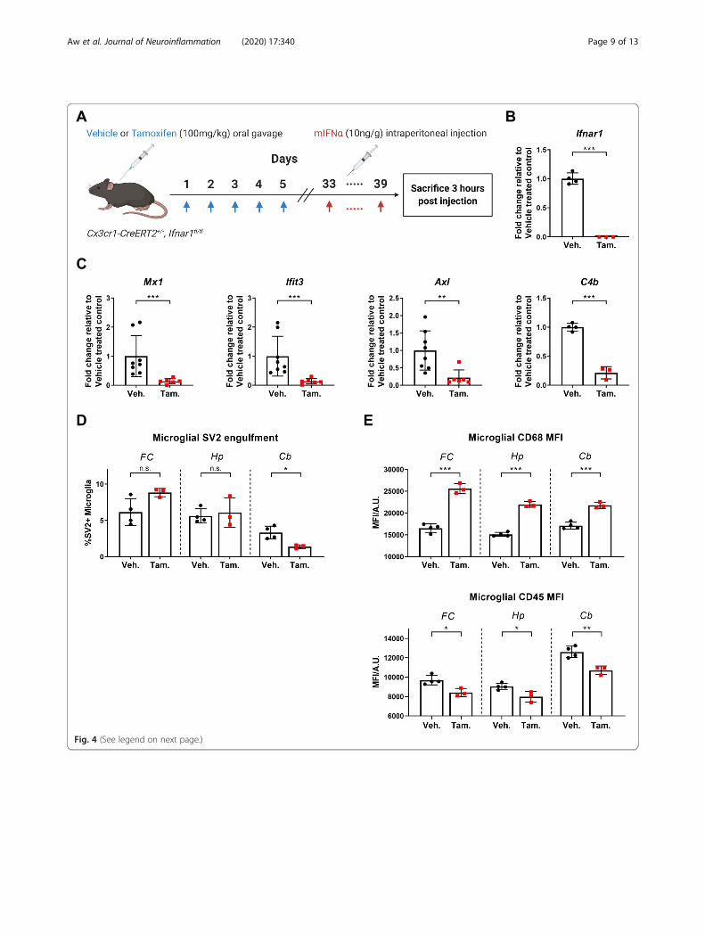
Fig. 4 (See legend on next page.)
Aw et al. Journal of Neuroinflammation (2020) 17:340 Page 9 of 13
hippocampus. While our RNA sequencing and ddPCRvalidation studies showed an mIFNα-induced increase inC4b expression on sorted anterior cortex microglia, it islikely that cerebellar microglia also increase C4b expres-sion in response to mIFNα, given that at baseline theyare in a more “immunologically vigilant” state with anenriched type 1 IFN regulatory signature, compared to
cortical and hippocampal microglia [52]. The increase inpresynaptic engulfment by cerebellar microglia com-pared to microglia from the other brain regions couldthus be due to an enhanced signalling response tomIFNα, a facet of which could include higher C4b ex-pression. Additionally, it is probable that within an acuteperiod of mIFNα exposure, most neurons or synapses
(See figure on previous page.)Fig. 4 Knockdown of microglial IFNAR signalling attenuates phenotypic and genetic changes in response to peripheral mIFNα administration. aSchematic of treatment protocol. Cx3cr1-CreERT2+/−, Ifnar1fl/fl mice were orally gavaged with either vehicle or tamoxifen. Four weeks post gavage,mice were either sacrificed (for qPCR validation of Ifnar1 knockdown) or treated with mIFNα daily for 7 days and sacrificed. b Cortical microgliafrom Cx3cr1-CreERT2+/−, Ifnar1fl/fl mice were sorted by FACS and analysed for gene expression of Ifnar1 by qPCR. Ifnar1 qPCR primers weredesigned to span the floxed region of Exon 10. The tamoxifen-treated (N = 3) CreERT2-positive group showed significant knockdown of Ifnar1expression compared to the vehicle-treated group (N = 4). c Sorted cortical microglia show significantly decreased ISG expression by ddPCR inthe tamoxifen treatment group (N = 6) of Cx3cr1-CreERT2+/−, Ifnarfl/fl mice in comparison to the vehicle treatment group (N = 8). C4b expressionwas also significantly decreased in the tamoxifen treatment group (N = 3) in comparison to the vehicle treatment group (N = 4). d Knockdown ofmicroglial Ifnar1 in the tamoxifen treatment group (N = 3) reverses the observed region-specific changes in synapse engulfment induced byperipheral mIFNα administration in comparison to the vehicle treatment group (N = 4). e Knockdown of microglial Ifnar1 in the tamoxifentreatment group (N = 3) reverses the observed changes in CD45 and CD68 expression induced by peripheral mIFNα administration, with theexception of CD68 expression in the cerebellum in comparison to the vehicle treatment group (N = 4). Veh., Vehicle; Tam., Tamoxifen; n.s., p >0.05; *p < 0.05; **p < 0.01; ***p < 0.001; Student’s t test or Mann-Whitney U test, assessed by normality of data distribution
Fig. 5 Transcriptional profiling of Ifnar1 sufficient and deficient microglia exposed to mIFNα. a Volcano plot showing highlights of differentiallyexpressed (DE) genes from bulk RNA sequencing of sorted cortical microglia, comparing vehicle-treated control (N = 6) and tamoxifen-treatedknockdown (N = 6) groups from Cx3cr1-CreERT2+/−, Ifnar1fl/fl mice. Interferon-stimulated genes were identified based on querying the Interferome2.0 database [43] with the DE gene list. q, Benjamini-Hochberg adjusted p value. b Overlap of independently identified > 1.5-fold change DEgenes from both sequencing libraries, identified using the glmTreat function in the EdgeR package [58]. Venn diagram sizes are proportional tothe numerical values in each category. c Overlap of independently identified gene ontology pathways. Venn diagram sizes are proportional tothe numerical values in each category. d A selection of differential gene ontology pathways identified using gProfiler2 [31] enriched in mIFNαexposed Ifnar1 sufficient and deficient microglia
Aw et al. Journal of Neuroinflammation (2020) 17:340 Page 10 of 13

still express sufficient signals to prohibit synaptic engulf-ment, such as CD47 [63] and other complement regula-tory factors. These regulatory signals and factors couldbe differentially distributed throughout different regionsof the murine brain, resulting in differing levels of inhib-ition of complement activation and deposition. It shouldalso be noted that our flow cytometric assay for synapseengulfment does not take into account any potential dif-ferences in the process of cargo degradation within thephagocytic pathway, although no significant differencesin SV2 signal were observed when wild type, corticalmicroglia were exposed to chloroquine, an inhibitor oflysosomal acidification and acid-sensitive lysosomal pro-teases (data not shown). Furthermore, a previous studydid not observe any differences in sensorimotor behav-iour of mice injected daily with mIFNα for up to 5 weeksat a similar dose [32], which suggests that the small in-crease in synaptic engulfment we observe in the cerebel-lum might be phenotypically insignificant. However, it ispossible that chronic IFNAR signalling within microgliaand other CNS cell types could result in elevated C4production and decreased complement and phagocyticinhibitory factors that over time could lead to the patho-logic pruning of synapses. Of note, it was shown in thesame study that chronic IFNα exposure (~ 4–5 weeks) inmice adversely affected neurogenesis and resulted in de-pressive behaviour [32]. Notably, endogenous IFNα canbiochemically, and presumably functionally, cross signalthrough μ-opioid receptors to exert anti-nociceptive ef-fects [64, 65]. It would therefore also be possible that thepathological effects of IFNα are mediated in part, or intandem, through these secondary, or non-IFNAR signal-ling related processes.Importantly, our study also shows a unique IFNα-
induced gene signature in microglia that bears similarityto other datasets presenting an enriched type 1 inter-feron signature in microglia within several pathologicaland biological contexts. GO analysis identified the ex-pected terms for interferon responses and, also interest-ingly, metabolic changes in catabolism. While cautionshould be borne in interpreting the directionality ofbiological pathways based solely on gene ontology terms,we note that IFNα and viral infection have been foundto generally induce a catabolic state in various cell typesincluding macrophages [66–68]. Deficiencies in micro-glial autophagy have also been recently linked to im-paired synaptic pruning and autism-like behaviouraldefects [17]. It would therefore be interesting to furtherprobe how IFNAR signalling in microglia might modu-late autophagic flux and whether increased autophagy inmicroglia might be a contributing mechanism for in-creased synaptic pruning in the context of chronic type1 interferon exposure during autoimmune disease suchas SLE.
ConclusionIn summary, we show that peripherally derived IFNα isable to signal directly across the BBB through microglialIFNAR. This results in a unique genetic and phenotypicprofile that is primarily dependent on IFNAR signallingwith minimal contribution from other secondary cyto-kine or chemokine signalling pathways. IFNα-inducedupregulation of C4b and other ISGs might regulate vari-ous cellular functions and processes such as synapticpruning in a brain region-dependent manner. Thesefindings would be of further investigative interest giventhe long-standing association of endogenous and thera-peutic IFNα with neuropsychiatric symptoms and theemerging therapeutic interest in IFNα use for prophylac-tic treatment of COVID19 [69–71].
Supplementary InformationSupplementary information accompanies this paper at https://doi.org/10.1186/s12974-020-02003-z.
Additional file 1: Figure S1. Microglia sorting and RNAseq dataanalysis. (A) Gating strategy for microglia cell sorting. (B) Principlecomponent analysis (PCA) plot showing unbiased clustering of samplesbased on treatment group (N = 6 per group). PC, Principal component.(C) Microglia specific genes were highly expressed in sorted microgliafrom both treatment groups. (D) Expression profile showing Log2 foldchange of microglial sensome genes [44] that are differentially expressedwith mIFNα treatment. (E) mIFNα treatment induces significant changesin microglial gene expression of cytokines and chemokines. Normalizedcounts and differential expression testing were generated and performedusing the EdgeR package [30].
Additional file 2: Figure S2. Flow cytometric assay of microglialsynapse engulfment. (A) Schematic of experimental procedure for flowcytometric assaying of microglial SV2+ presynaptic engulfment. 10 μMCytochalasin D was added in buffers used during douncehomogenization and Percoll gradient centrifugation. (B) Effect ofcytochalasin D treatment on the SV2 engulfment assay, showing theinhibition of active microglial phagocytosis of SV2 particles during tissueprocessing. Cortical tissue from opposing hemispheres in each mousewas perfused, dissected and processed with or without cytochalasin D,allowing for paired analysis of phagocytosis inhibition within the samemouse (N = 9). Veh., Vehicle; Cyto. D, Cytochalasin D; **, p < 0.01;Wilcoxon’s signed rank test. (C) Flow cytometric gating strategy for theengulfment assay. (D) Lack of neuronal debris adhering to microglia. Nonpermeabilized microglia show no fluorochrome signal compared topermeabilized microglia (N = 3 per treatment). P, Permeabilized; NP, Non-permeabilized; *, p < 0.05; Student’s t test. (E) Isotype controls showingabsence of non-epitope driven binding of SV2 and CD68 antibodies dur-ing intracellular staining.
Additional file 3: Figure S3. Validation of Cx3cr1-CreERT2 targeting ofmicroglia. (A) Schematic of treatment protocol. Cx3cr1-CreERT2+/−, R26-EYFPLSL mice were orally gavaged with either vehicle (N = 2) or tamoxifen(N = 3) and sacrificed 4 weeks post gavage. (B) Representative confocalimages of frontal cortex of Cx3cr1-CreERT2+/−, R26-EYFPLSL mice showingspecific expression of the Cx3cr1-CreERT2 transgene in IBA1 expressingmicroglia/myeloid lineage cells. Scale bars = 200 μm. (C) Flow cytometricanalysis of YFP expression in CD11b+, CD45lo microglia, showing elevatedYFP expression only in the tamoxifen treated group. Veh., Vehicle; Tam.,Tamoxifen.
Additional file 4: Figure S4. RNAseq analysis of anterior cortexmicroglia sorted from Ifnar1 sufficient and deficient mice treated withmIFNα. (A) Principle component analysis (PCA) plot showing unbiasedclustering of samples based on treatment group (N = 6 per group). PC,Principal component. (B) Microglia specific genes were highly expressed
Aw et al. Journal of Neuroinflammation (2020) 17:340 Page 11 of 13
in sorted microglia from both treatment groups. (C) Expression profileshowing Log2 fold change of microglial sensome genes [44] that aresignificantly differentially expressed in Ifnar1 sufficient vs. deficientmicroglia. (D) mIFNα treatment induces changes in microglial geneexpression of cytokines and chemokines. Normalized counts anddifferential expression testing were generated and performed using theEdgeR package [30].
AbbreviationsAGS: Aicardi-Goutières syndrome; BBB: Blood-brain barrier; CNS: Centralnervous system; DE: Differentially expressed; GO: Gene ontology;IFNα: Interferon alpha; IFNγ: Interferon gamma; mIFNα: Murine interferonalpha; IFNAR: Type 1 interferon receptor; IL-1β: Interleukin 1 beta; IL-6: Interleukin 6; IRF9: Interferon regulatory factor 9; ISG: Interferon-stimulatedgene; ISGF3: Interferon-stimulated gene factor 3; ISRE: Interferon-stimulatedresponse element; JAK1: Janus kinase 1; RTK: Receptor tyrosine kinase;SLE: Systemic lupus erythematosus; STAT1: Signal transducer and activator oftranscription 1; STAT2: Signal transducer and activator of transcription 2;SV2: Synaptic vesicle 2; TNFα: Tumour necrosis factor alpha; TYK2: Tyrosinekinase 2
AcknowledgementsWe are grateful for the assistance of institute core facilities and helpfuldiscussions with laboratory personnel, including the BCH/PCMM MicroscopyCore, the BCH/PCMM Flow and Imaging Cytometry Resource, the DFCI FlowCore, the HMS Biopolymers Facility, Dr. Kristina Holton, Dr. Jessy Presumeyand Khristina Holscher. Figure schematics were created using Biorender.com.
Authors’ contributionsEA and MC conceived the study. EA designed, performed and analysed data fromall experiments. YZ helped with RNAscope staining and imaging and assistedwith microglia isolation from tissue for flow cytometry and bulk RNA sequencing.EA wrote the manuscript. All authors read and approved the final manuscript.
FundingThis work was supported by NIH grant R01AR072965.
Availability of data and materialsAll datasets used and/or analysed during the current study are available fromthe corresponding author on reasonable request. Bulk RNA sequencing datamay be accessed at the NCBI SRA with accession ID PRJNA658781.
Ethics approval and consent to participateAll animal experiments were carried out in agreement with the institutionalguidelines at Harvard Medical School, following approval of ethical protocols bythe local Institutional Animal Care and Use Committee (protocol numbersIS00000748, IS00000111 and IS00002660) and per applicable laws and regulations.
Consent for publicationNot applicable.
Competing interestsThe authors declare that they have no competing interests.
Author details1Program in Cellular and Molecular Medicine, Boston Children’s Hospital,Boston, MA, USA. 2Division of Medical Sciences, Harvard Medical School,Boston, MA, USA.
Received: 17 August 2020 Accepted: 19 October 2020
References1. Crow MK, Type I. Interferon in the pathogenesis of lupus. J Immunol. 2014;
192:5459–68..2. Crow YJ, Manel N. Aicardi-Goutières syndrome and the type I
interferonopathies. Nat Rev Immunol. 2015;15:429–40.3. Schreiber G, Piehler J. The molecular basis for functional plasticity in type I
interferon signaling. Trends Immunol. 2015;36:139–49.
4. van Boxel-Dezaire AHH, Rani MRS, Stark GR. Complex modulation of cell type-specific signaling in response to type I interferons. Immunity. 2006;25:361–72.
5. Valentine AD, Meyers CA. Neurobehavioral effects of interferon therapy. CurrPsychiatry Rep. 2005;7:391–5.
6. Schaefer M, Engelbrechta MA, Gut O, Fiebich BL, Bauer J, Schmidt F, et al.Interferon alpha (IFNα) and psychiatric syndromes: a review. Prog Neuro-Psychopharmacology Biol Psychiatry. 2002;26:731–46.
7. Scheibel RS, Ph D, Valentine AD, Brien SO, Meyers CA. Cognitive dysfunctionand depression during treatment with interferon-alpha and chemotherapy.J Neuropsychiatry Clin Neurosci. 2004;16:185–91.
8. Shiozawa S, Kuroki Y, Kim M, Hirohata S, Ogino T. Interferon alpha in lupuspsychosis. Arthritis Rheum. 1992;35:417–22.
9. Jeltsch-David H, Muller S. Neuropsychiatric systemic lupus erythematosus:Pathogenesis and biomarkers. Nat Rev Neurol. 2014;10:579–96.
10. Schafer DP, Lehrman EK, Kautzman AG, Koyama R, Mardinly AR, Yamasaki R,et al. Microglia sculpt postnatal neural circuits in an activity andcomplement-dependent manner. Neuron. 2012;74:691–705.
11. Paolicelli RC, Bolasco G, Pagani F, Maggi L, Scianni M, Panzanelli P, et al.Synaptic pruning by microglia is necessary for normal brain development.Science. 2011;333:1456–8.
12. Nimmerjahn A, Kirchhoff F, Helmchen F. Resting microglial cells are highlydynamic surveillants of brain parenchyma in vivo. Science. 2005;308:1314–9.
13. Wake H, Moorhouse AJ, Jinno S, Kohsaka S, Nabekura J. Resting microgliadirectly monitor the functional state of synapses in vivo and determine thefate of ischemic terminals. J Neurosci. 2009;13:3974–80.
14. Hong S, Beja-Glasser VF, Nfonoyim BM, Frouin A, Li S, Ramakrishnan S, et al.Complement and microglia mediate early synapse loss in Alzheimer mousemodels. Science. 2016;352:712–6.
15. Roy ER, Zheng H, Cao W. Type I interferon response drives neuroinflammationand synapse loss in Alzheimer disease. J Clin Invest. 2020;130:1912–30.
16. Sekar A, Bialas AR, de Rivera H, Davis A, Hammond TR, Kamitaki N, et al.Schizophrenia risk from complex variation of complement component 4.Nature. 2016;530:177–83.
17. Kim HJ, Cho MH, Shim WH, Kim JK, Jeon EY, Kim DH, et al. Deficientautophagy in microglia impairs synaptic pruning and causes socialbehavioral defects. Mol Psychiatry. 2016;22:1576–84.
18. Zhan Y, Paolicelli RC, Sforazzini F, Weinhard L, Bolasco G, Pagani F, et al.Deficient neuron-microglia signaling results in impaired functional brainconnectivity and social behavior. Nat Neurosci. 2014;17:400–6.
19. Zheng L, Kaneko N, Sawamoto K. Minocycline treatment amelioratesinterferon-alpha- induced neurogenic defects and depression-like behaviorsin mice. Front Cell Neurosci. 2015;9:1–10.
20. Goldmann T, Blank T, Prinz M. Fine-tuning of type I IFN-signaling inmicroglia - implications for homeostasis, CNS autoimmunity andinterferonopathies. Curr Opin Neurobiol. 2016;36:38–42.
21. Deczkowska A, Matcovitch-Natan O, Tsitsou-Kampeli A, Ben-Hamo S, Dvir-Szternfeld R, Spinrad A, et al. Mef2C restrains microglial inflammatoryresponse and is lost in brain ageing in an IFN-I-dependent manner. NatCommun. 2017;8:1–13.
22. Hammond TR, Dufort C, Dissing-Olesen L, Giera S, Young A, Wysoker A,et al. Single-cell RNA sequencing of microglia throughout the mouselifespan and in the injured brain reveals complex cell-state changes.Immunity. 2019;50:253–71 e6.
23. Mathys H, Adaikkan C, Gao F, Young JZ, Manet E, Hemberg M, et al.Temporal tracking of microglia activation in neurodegeneration at single-cell resolution. Cell Rep. 2017;21:366–80.
24. Nomura A, Noto D, Murayama G, Chiba A, Miyake S. Unique primed statusof microglia under the systemic autoimmune condition of lupus-pronemice. Arthritis Res Ther. 2019;21:1–17.
25. Vasek MJ, Garber C, Dorsey D, Durrant DM, Bollman B, Soung A, et al. Acomplement-microglial axis drives synapse loss during virus-inducedmemory impairment. Nature. 2016;534:538–43.
26. Yona S, Kim KW, Wolf Y, Mildner A, Varol D, Breker M, et al. Fate mappingreveals origins and dynamics of monocytes and tissue macrophages underhomeostasis. Immunity. 2013;38:79–91.
27. Butovsky O, Jedrychowski MP, Moore CS, Cialic R, Lanser AJ, Gabriely G,et al. Identification of a unique TGF-β–dependent molecular and functionalsignature in microglia. Nat Neurosci. 2014;17:131–43.
28. McQuin C, Goodman A, Chernyshev V, Kamentsky L, Cimini BA, Karhohs KW,et al. CellProfiler 3.0: Next-generation image processing for biology. PLoSBiol. 2018;16:e2005970.
Aw et al. Journal of Neuroinflammation (2020) 17:340 Page 12 of 13

29. Dobin A, Davis CA, Schlesinger F, Drenkow J, Zaleski C, Jha S, et al. STAR:ultrafast universal RNA-seq aligner. Bioinformatics. 2013;29:15–21.
30. Robinson MD, McCarthy DJ, Smyth GK. edgeR: A Bioconductor package fordifferential expression analysis of digital gene expression data.Bioinformatics. 2009;26:139–40.
31. Raudvere U, Kolberg L, Kuzmin I, Arak T, Adler P, Peterson H, et al. g:Profiler:a web server for functional enrichment analysis and conversions of genelists (2019 update). Nucleic Acids Res. 2019;47:W191–8.
32. Zheng L, Hitoshi S, Kaneko N, Takao K, Miyakawa T. Mechanisms forinterferon-a-induced depression and neural stem cell dysfunction. Stem CellRep. 2014;3:73–84.
33. Wang J, Campbell IL, Zhang H. Systemic interferon-α regulates interferon-stimulated genes in the central nervous system. Mol Psychiatry. 2008;13:293–301.
34. Jonasch E. Interferon in oncological practice: review of interferon biology,clinical applications, and toxicities. Oncologist. 2001;6:34–55.
35. Moriyama M, Arakawa Y. Treatment of interferon-α for chronic hepatitis C.Expert Opin Pharmacother. 2006;7:1163–79.
36. Yu M-L, Dai C-Y, Chen S-C, Lee L-P, Hsieh M-Y, Lin Z-Y, et al. Highversus standard doses interferon-alpha in the treatment of naïvechronic hepatitis C patients in Taiwan: a 10-year cohort study. BMCInfect Dis. 2005;5:27.
37. Banks WA. From blood-brain barrier to blood-brain interface: newopportunities for CNS drug delivery. Nat Rev Drug Discov. 2016;15:275–92.
38. Raison CL, Demetrashvili M, Capuron L, Miller AH. Neuropsychiatric adverseeffects of interferon-α. CNS Drugs. 2005;19:105–23.
39. Mayr N, Zeitlhofer J, Deecke L, Fritz E, Ludwig H, Gisslinger H. Neurologicalfunction during long-term therapy with recombinant interferon alpha. JNeuropsychiatry Clin Neurosci. 1999;11:343–8.
40. Gorwood P. Neurobiological mechanisms of anhedonia. Dialogues ClinNeurosci. 2008;10:291–9.
41. Taylor JL, Amann M, Duchateau J, Meeusen R, Rice CL. Neural contributionsto muscle fatigue: from the brain to the muscle and back again. Med SciSports Exerc. 2016;48:2294–306.
42. Penner I-K, Paul F. Fatigue as a symptom or comorbidity of neurologicaldiseases. Nat Rev Neurol. 2017;13:662–75.
43. Rusinova I, Forster S, Yu S, Kannan A, Masse M, Cumming H, et al. INTERFEROME v2.0: an updated database of annotated interferon-regulated genes.Nucleic Acids Res. 2013;41:1040–6.
44. Hickman SE, Kingery ND, Ohsumi TK, Borowsky ML, Wang L, Means TK, et al.The microglial sensome revealed by direct RNA sequencing. Nat Neurosci.2013;16:1896–905.
45. Jimenez JC, Su K, Goldberg AR, Luna VM, Biane JS, Ordek G, et al. Anxietycells in a hippocampal-hypothalamic circuit. Neuron. 2018;97:670–83 e6.
46. Sigurdsson T, Duvarci S. Hippocampal-prefrontal interactions in cognition,behavior and psychiatric disease. Front Syst Neurosci. 2016;9:1–18.
47. Phillips JR, Hewedi DH, Eissa AM, Moustafa AA. The cerebellum andpsychiatric disorders. Front Public Heal. 2015;3:1–8.
48. Moreno-Rius J. The cerebellum in fear and anxiety-related disorders. ProgNeuro-Psychopharmacology Biol Psychiatry. 2018;85:23–32.
49. Hare BD, Duman RS. Prefrontal cortex circuits in depression and anxiety:contribution of discrete neuronal populations and target regions. MolPsychiatry. 2020;25:2742–58.
50. Korzhevskii DE, Kirik OV. Brain microglia and microglial markers. NeurosciBehav Physiol. 2016;46:284–90.
51. Salemi J, Obregon DF, Cobb A, Reed S, Sadic E, Jin J, et al. Flipping theswitches: CD40 and CD45 modulation of microglial activation states in HIVassociated dementia (HAD). Mol Neurodegener. 2011;6:1–8.
52. Grabert K, Michoel T, Karavolos MH, Clohisey S, Baillie JK, Stevens MP, et al.Microglial brain region-dependent diversity and selective regionalsensitivities to aging. Nat Neurosci. 2016;19:504–16.
53. Blank T, Detje CN, Spieß A, Hagemeyer N, Brendecke SM, Wolfart J, et al.Brain endothelial- and epithelial-specific interferon receptor chain 1 drivesvirus-induced sickness behavior and cognitive impairment. Immunity. 2016;44:901–12.
54. Orzalli MH, Smith A, Jurado KA, Iwasaki A, Garlick JA, Kagan JC. An antiviralbranch of the il-1 signaling pathway restricts immune-evasive virusreplication. Mol Cell. 2018;71:825–40 e6.
55. Wang W, Xu L, Brandsma JH, Wang Y, Hakim MS, Zhou X, et al. Convergenttranscription of interferon-stimulated genes by tnf-α and ifn-α augmentsantiviral Activity against HCV and HEV. Sci Rep. 2016;6:1–14.
56. Wang W, Xu L, Su J, Peppelenbosch MP, Pan Q. Transcriptional regulation ofantiviral interferon-stimulated genes. Trends Microbiol. 2017;25:573–84.
57. Schoggins JW. Interferon-stimulated genes: what do they all do? Annu RevVirol. 2019;6:567–84.
58. Chen Y, Lun ATL, Smyth GK. From reads to genes to pathways: differentialexpression analysis of RNA-Seq experiments using Rsubread and the edgeRquasi-likelihood pipeline. F1000Research. 2016;5:1438.
59. Pan W, Banks WA, Kastin AJ. Permeability of the blood-brain and blood-spinal cord barriers to interferons. J Neuroimmunol. 1997;76:105–11.
60. Smith RA, Norris F, Palmer D, Bernhardt L, Wills RJ. Distribution of alphainterferon in serum and cerebrospinal fluid after systemic administration.Clin Pharmacol Ther. 1985;37:85–8.
61. DePaula-Silva AB, Gorbea C, Doty DJ, Libbey JE, Sanchez JMS, Hanak TJ,et al. Differential transcriptional profiles identify microglial- andmacrophage-specific gene markers expressed during virus-inducedneuroinflammation. J Neuroinflammation. 2019;16:1–20.
62. Comer AL, Jinadasa T, Sriram B, Phadke RA, Kretsge LN, Nguyen TPH, et al.Increased expression of schizophrenia-associated gene C4 leads tohypoconnectivity of prefrontal cortex and reduced social interaction. PLOSBiol. 2020;18:1–36.
63. Lehrman EK, Wilton DK, Litvina EY, Welsh CA, Chang ST, Frouin A, et al.CD47 protects synapses from excess microglia-mediated pruning duringdevelopment. Neuron. 2018;100:120–34.
64. Jiang C, Song L, Lu C, You Z, Wang Y, Sun L, et al. Analgesic effect of interferon-alpha via mu opioid receptor in the rat. Neurochem Int. 2000;36:193–6.
65. Liu C-C, Gao Y-J, Luo H, Berta T, Xu Z-Z, Ji R-R, et al. Interferon alpha inhibitsspinal cord synaptic and nociceptive transmission via neuronal-glialinteractions. Sci Rep. 2016;6:34356.
66. Schmeisser H, Bekisz J, Zoon KC. New function of type I IFN: induction ofautophagy. J Interf Cytokine Res. 2014;34:71–8.
67. Fritsch SD, Weichhart T. Effects of interferons and viruses on metabolism.Front Immunol. 2016;7:1–13.
68. Ahmed D, Jaworski A, Roy D, Willmore W, Golshani A, Cassol E.Transcriptional profiling suggests extensive metabolic rewiring of humanand mouse macrophages during early interferon alpha responses. MediatorsInflamm. 2018;2018:1–15.
69. Park A, Iwasaki A. Type I and type III interferons – induction, signaling, evasion,and application to combat COVID-19. Cell Host Microbe. 2020;27:870–8.
70. Sallard E, Lescure F-X, Yazdanpanah Y, Mentre F, Peiffer-Smadja N. Type 1interferons as a potential treatment against COVID-19. Antiviral Res. 2020;178:1–4.
71. Zhou Q, Chen V, Shannon CP, Wei X-S, Xiang X, Wang X, et al. Interferon-α2b treatment for COVID-19. Front Immunol. 2020;11:1–6.
Publisher’s NoteSpringer Nature remains neutral with regard to jurisdictional claims inpublished maps and institutional affiliations.
Aw et al. Journal of Neuroinflammation (2020) 17:340 Page 13 of 13