Mice Deficient in Endothelial α5 Integrin are Profoundly ...
14
University of Kentucky UKnowledge Sanders-Brown Center on Aging Faculty Publications Aging 11-13-2015 Mice Deficient in Endothelial α5 Integrin are Profoundly Resistant to Experimental Ischemic Stroke Jill Roberts University of Kentucky, [email protected] Leon de Hoog University of Kentucky Gregory J. Bix University of Kentucky, [email protected] Right click to open a feedback form in a new tab to let us know how this document benefits you. Follow this and additional works at: hps://uknowledge.uky.edu/sbcoa_facpub Part of the Biochemical Phenomena, Metabolism, and Nutrition Commons , Family, Life Course, and Society Commons , Geriatrics Commons , Medical Neurobiology Commons , and the Neurology Commons is Article is brought to you for free and open access by the Aging at UKnowledge. It has been accepted for inclusion in Sanders-Brown Center on Aging Faculty Publications by an authorized administrator of UKnowledge. For more information, please contact [email protected]. Repository Citation Roberts, Jill; de Hoog, Leon; and Bix, Gregory J., "Mice Deficient in Endothelial α5 Integrin are Profoundly Resistant to Experimental Ischemic Stroke" (2015). Sanders-Brown Center on Aging Faculty Publications. 84. hps://uknowledge.uky.edu/sbcoa_facpub/84
Transcript of Mice Deficient in Endothelial α5 Integrin are Profoundly ...

University of KentuckyUKnowledge
Sanders-Brown Center on Aging FacultyPublications Aging
11-13-2015
Mice Deficient in Endothelial α5 Integrin areProfoundly Resistant to Experimental IschemicStrokeJill RobertsUniversity of Kentucky, [email protected]
Leon de HoogUniversity of Kentucky
Gregory J. BixUniversity of Kentucky, [email protected]
Right click to open a feedback form in a new tab to let us know how this document benefits you.
Follow this and additional works at: https://uknowledge.uky.edu/sbcoa_facpub
Part of the Biochemical Phenomena, Metabolism, and Nutrition Commons, Family, Life Course,and Society Commons, Geriatrics Commons, Medical Neurobiology Commons, and the NeurologyCommons
This Article is brought to you for free and open access by the Aging at UKnowledge. It has been accepted for inclusion in Sanders-Brown Center onAging Faculty Publications by an authorized administrator of UKnowledge. For more information, please contact [email protected].
Repository CitationRoberts, Jill; de Hoog, Leon; and Bix, Gregory J., "Mice Deficient in Endothelial α5 Integrin are Profoundly Resistant to ExperimentalIschemic Stroke" (2015). Sanders-Brown Center on Aging Faculty Publications. 84.https://uknowledge.uky.edu/sbcoa_facpub/84

Mice Deficient in Endothelial α5 Integrin are Profoundly Resistant to Experimental Ischemic Stroke
Notes/Citation InformationPublished in Journal of Cerebral Blood Flow & Metabolism, v. 37, issue 1, p. 85-96.
© Author(s) 2015
This article is distributed under the terms of the Creative Commons Attribution-NonCommercial 3.0 License(http://www.creativecommons.org/licenses/by-nc/3.0/) which permits non-commercial use, reproductionand distribution of the work without further permission provided the original work is attributed as specifiedon the SAGE and Open Access page(https://us.sagepub.com/en-us/nam/open-access-at-sage).
Digital Object Identifier (DOI)https://doi.org/10.1177/0271678X15616979
This article is available at UKnowledge: https://uknowledge.uky.edu/sbcoa_facpub/84
Original Article
Mice deficient in endothelial a5 integrinare profoundly resistant to experimentalischemic stroke
Jill Roberts1,2, Leon de Hoog1 and Gregory J Bix1,2,3
Abstract
Stroke is a disease in dire need of better therapies. We have previously shown that a fragment of the extracellular matrix
proteoglycan, perlecan, has beneficial effects following cerebral ischemia via the a5b1 integrin receptor. We now report
that endothelial cell selective a5 integrin deficient mice (a5 KO) are profoundly resistant to ischemic infarct after transient
middle cerebral artery occlusion. Specifically, a5 KOs had little to no infarct 2–3 days post-stroke, whereas controls had an
increase in mean infarct volume over the same time period as expected. Functional outcome is also improved in the a5 KOs
compared with controls. Importantly, no differences in cerebrovascular anatomy or collateral blood flow were noted that
could account for this difference in ischemic injury. Rather, we demonstrate that a5 KOs have increased blood-brain barrier
integrity (increased expression of claudin-5, and absent brain parenchymal IgG extravasation) after stroke compared with
controls, which could explain their resistance to ischemic injury. Additionally, inhibition of a5 integrin in vitro leads to
decreased permeability of brain endothelial cells following oxygen-glucose deprivation. Together, these findings indicate
endothelial cell a5 integrin plays an important role in stroke outcome and blood-brain barrier integrity, suggesting that a5
integrin could be a novel therapeutic target for stroke.
Keywords
a5b1, blood-brain barrier, extracellular matrix, middle cerebral artery occlusion, transgenic
Received 7 August 2015; Revised 27 September 2015; Accepted 27 September 2015
Introduction
In ischemic stroke, a severe reduction in the bloodsupply to the brain leads to cell death in a region referredto as the ischemic core, and hypoxia and metabolicchanges in a surrounding penumbral region that is vul-nerable to injury and death over time. One strategy totreat ischemic stroke is to limit the expansion of injuryand death from the initially formed core into the pen-umbra. Unfortunately, this experimental neuroprotec-tive approach has stumbled in translation to humanstroke patients (see Kahle and Bix1 for a review) in nosmall part due to an evolving understanding of whatdefines the ischemic core and penumbra. Indeed, ratherthan being the discrete, homogenously defined regionsdescribed above, the core and penumbra appear to beheterogeneous, each containing islands of ‘‘mini-cores’’and ‘‘mini-penumbras’’ with differing spatiotemporalsusceptibilities to injury and death after stroke.2
Understanding this complex stroke pathophysiologymay be critical to developing effective therapies.
Integrins are cell surface transmembrane glycopro-tein receptors for the extracellular matrix (ECM) con-sisting of non-covalently linked a and b subunits that,in addition to playing important roles in cell survival,proliferation, and differentiation throughout the body,play an essential role in stroke pathophysiology.3–5
In particular, integrins are critical to the endothelialcell-astrocyte configuration of the blood-brain barrier(BBB), whose rapid breakdown after stroke leads toedema, inflammation, and ultimately worsening strokeinjury.6 In this context, integrins ‘‘integrate’’ the ECM,
1Sanders-Brown Center on Aging, University of Kentucky, Lexington,
KY, USA2Department of Anatomy and Neurobiology, University of Kentucky,
Lexington, KY, USA3Department of Neurology, University of Kentucky, Lexington, KY, USA
Corresponding author:
Gregory J. Bix, University of Kentucky, Sanders-Brown Building 430, 800
S. Limestone, Lexington, KY 40536, USA.
Email: [email protected]
Journal of Cerebral Blood Flow &
Metabolism
2017, Vol. 37(1) 85–96
! Author(s) 2015
Reprints and permissions:
sagepub.co.uk/journalsPermissions.nav
DOI: 10.1177/0271678X15616979
jcbfm.sagepub.com
endothelial cells, and astrocyte end-feet for proper pos-itioning and adhesion, which is disrupted afterstroke.7–9 Under normal physiologic conditions, theBBB is made up of several layers of protection all work-ing together to restrict the passage of substancesbetween the blood and brain parenchyma. Tight junc-tions (TJ), composed predominantly of occludin, zonaoccludins, claudin-5, -3, and -12 and junctional adhe-sion molecules (JAM),10 exist between adjacent endo-thelial cells and are important for maintaining barrierfunction as they prevent paracellular transport andeven restrict the passage of ions.11 Disruption of integ-rin function not only leads to a breakdown in the BBBvia ECM dysfunction but also has been recently shownto impact TJ protein expression and BBB permeability.Specifically, antibody blockade of the b1 family ofintegrins (containing several different a subunit pair-ings) has been shown to decrease expression of clau-din-5 in brain endothelial cell (BEC) monolayersin vitro and increase BBB permeability in vivo.12 Thissuggests that the b1 integrin family could play animportant role in regulating BBB integrity undernormal and stroke conditions. However, because integ-rin subunit (a4, a5, a8, b1)-null mice result in embry-onic lethality, the exact role of specific b1 integrins inregulating BBB integrity is unknown.13–15
We have previously shown that a protein fragmentof the ECM proteoglycan perlecan, known as domainV (DV), is rapidly, persistently and consequently gen-erated in the stroked brain and when systemicallyadministered in multiple animal models of cerebralischemia is neuroprotective and pro-angiogenic.16,17
This may be due, in part, to DV’s interaction with itsreceptor a5b1 integrin on BECs and subsequent gener-ation and release of VEGF.17,18 Furthermore, DVappears to inhibit chronic post-stroke astrogliosis viainteraction with astrocyte a5b1 integrin.19 These stu-dies, and the fact that a5b1 integrin is conditionallyupregulated in brain blood vessels after stroke,20 sug-gest that the a5b1 integrin, in particular, plays animportant role in stroke pathophysiology.
In light of the fact that a5 integrin-null mice areembryonic lethal (�E10.5) due in part to defects intheir vasculature,13–15 an endothelial-specific a5 integrinknockout mouse (a5 KO) was created to study the roleof a5 integrin in vascular biology.21 This mouse, which isviable and has no obvious phenotype,21 exhibits signifi-cantly delayed brain angiogenesis in response to chroniccerebral hypoxia22 (8% O2). As angiogenesis is animportant component of post-stroke brain repair but ispreceded by endothelial cell activation and decreasedinter-endothelial cell adhesion, we here used the a5 KOmouse in a model of middle cerebral artery (MCA)occlusion to determine the significance of the a5 integrinin stroke pathophysiology and functional outcome.
Materials and methods
Animals
The experimental protocol was approved by theInstitutional Animal Care and Use Committee of theUniversity of Kentucky and experiments were performedin accordance with the Guide for the Care and Use ofLaboratory Animals of the National Institutes of Healthas well as the ARRIVE guidelines. All experiments wereperformed in a blinded fashion using randomized selec-tion. Adult (3 months, male) mice which lack expressionof the a5 integrin specifically in endothelial cells (a5f/�;Tie2-Cre; referred to here as a5 KO) were generated asdescribed previously15,21 and generously provided byRichard Hynes (M.I.T., MA, USA). Littermate micewhich do express a5 integrin in endothelial cells and arenegative for Tie2-Cre were used as controls (a5þ/f;referred to here as Ctrl). Genotyping was performedusing previously described protocols.15,21 All mice werehoused in a climate-controlled room on a 12-h light/dark cycle and food and water were provided ad libitum.
Stroke model
Ctrl and a5 KO mice were subjected to transient tandemipsilateral common carotid artery (CCA)/MCA occlu-sion for 60min, followed by reperfusion of both arteriesfor 1–7 days. Briefly, a small burr hole was made in theskull to expose the MCA and a metal wire with a diam-eter of 0.005 inch was placed under the artery. Slightelevation of the metal wire causes visible occlusion ofthe MCA. The CCA was then isolated and occludedusing an aneurysm clip. Diminished blood flow was con-firmed with Laser Doppler Perfusion Monitor (Perimed,Ardmore, PA, USA) and only those animals witha diminished blood flow of at least 80% and re-establishment of at least 75% of baseline levels wereincluded in subsequent experimentation. Animal physio-logic measurements before and after stroke were madeusing MouseOx (Starr Life Science, Massachusetts, MA,USA). Blood was collected on post-stroke day (PSD) 3for blood gas and ion analysis; done within 30min afterthe samples were collected. To confirm stroke sizeand location, brains were cut into 2-mm thick coronalsections and stained with 2,3-triphenyltetrazolium chlor-ide (TTC; BD, Sparks, MD, USA) on PSDs 1–3.Alternatively, brains were flash frozen and sectioned(20mm) to undergo cresyl violet (Sigma) staining.Infarct size was analyzed using Image J (NIH) andinfarct volume was calculated as,
Infarct Volume ¼ Apparent Infarct
� ðContralateral Hemisphere=Ipsilateral HemisphereÞ
86 Journal of Cerebral Blood Flow & Metabolism 37(1)

Animals were excluded from the study if the middlecerebral or CCA was punctured during wire and clampinsertion or removal, died following surgery in recovery orwere euthanized before the end of the study due to poorhealth. Overall, there is <5% death rate for our strokemodel both following surgery and during behavioral testing.
Behavioral testing
Animals were tested on the Rotor Rod to examine forcedmotor coordination. Behavioral testing took place for 3days prior to stroke surgery (training and baseline meas-urement) and on PSDs 1 and 7. The mice were placed onthe Rotor Rod for 5min with an increasing accelerationfrom 0 to 40 rpm for three trials and the parameters wereset to measure latency in seconds.
Vessel staining and vascular territory
Cerebrovascular anatomy of the a5 KO and Ctrl micewas measured by intravenous injection of a combin-ation of two carbon ink dyes as previously described.23
Briefly, papaverine hydrochloride (50mg/kg; Sigma, St.Louis, MO, USA) was injected intravenously for vesseldilation just prior to the injection of carbon ink. Amixture of ink 1 (Fount India, Pelikan, Germany)and ink 2 (Super Black, Speedball, USA) was intraven-ously injected at a ratio of 1:9, respectively. After10min, the brain was removed and fixed in 4% paraf-ormaldehyde prior to images being taken with aMoticam 2 (Motic, British Columbia, Canada)camera and software. Points of anastomoses betweenthe MCA and the anterior cerebral artery (ACA) weredetermined and the total area of the MCA and ACAwas measured using Image J software (NIH).
Blood perfusion imaging
Cortical blood flow was monitored using a blood perfusionimager (PeriCam PSI System, Perimed) based on LaserSpeckle Contrast Analysis technology. Briefly, the skinwas retracted to expose the skull of the animal and theimager was positioned above the head. The laser detectsmovement in tissue, such as red blood cells, and createsspeckle contrast. Measurement in the contrast fluctuationsprovides information about blood perfusion in the brain.Baseline (5min) measurements of blood flow were firstobtained and the animal was then subjected to the strokemodel. Immediately following occlusion of the CCA/MCA, blood flow measurements were taken for another5min. Measurements were also recorded following reper-fusion of the CCA/MCA, confirming the re-establishmentof blood flow. Only animals with at least a 30% decrease intotal blood perfusion following occlusion were included insubsequent analysis. Regions of interest equal in size
(encompassing the visible region of the MCA) were usedto determine the perfusion in the ipsilateral and contralat-eral hemispheres of the Ctrl and a5 KO mice. Blood per-fusion is expressed in arbitrary units (Perfusion Units).
Tissue histology and immunohistochemistry
On PSDs 1–3, brains were removed, flash frozen andstored at �80�C until use. Brains were cut into 20 -mmsections using a cryostat and mounted onto slides.Sections were fixed with ice cold acetone prior to incubat-ing in blocking buffer (5% BSA in phosphate bufferedsaline (PBS) with 0.1% Triton X-100) for 1h at roomtemperature. The sections were then incubated overnightat 4�C in the primary antibody against IgG (1:1000; LifeTechnologies). Sections were washed and incubated with asecondary antibody (HRP anti-mouse, Life Technologies)for 1h at room temperature. Sections were washed andincubated with DAB (Vector Labs, Burlingame, CA,USA) for 1h prior to counterstaining with hematoxylin(Fisher Scientific, USA). Alternatively, sections werestained with primary antibodies against fluorescein isothio-cyanate (FITC)-conjugated-tomato lectin (1:200; VectorLabs), glial fibrillary acidic protein (GFAP) (1:500,Sigma), terminal deoxynucleotidyl transferase dUTP nickend labeling (TUNEL) (Apoptag Fluorescien kit,Millipore), or cresyl violet (Sigma) overnight at 4�C.Slides were then coverslipped with fluorescent mountingmedia (Vector Labs) or xylene-based mounting media(Sigma) and images were captured using a Nikon EclipseTi microscope and software (Nikon, Melville, NY, USA).
Gene expression
On PSDs 1–3, brains were removed and cut into 2-mmthick sections. The sections were cut to separate the twohemispheres and the ipsilateral hemisphere wastrimmed to remove striatum (leaving cortical ischemiccore and penumbra). All ipsilateral sections/animalwere combined, and tissue was homogenized in Trizol(Life Technologies). RNA was extracted according tothe instructions of the RNA extraction kit manufac-turer (Life Technologies). RNA was converted tocDNA using a high capacity cDNA reverse transcrip-tion kit (Applied Bioscience, Grand Island, NY, USA).Claudin-5 and 18S (control) genes were analyzed usingReal-time PCR (ViiA7; Life Technologies). Data wereanalyzed comparing a5 KO to Ctrl mice and are pre-sented as a % of control (sham mice).
Oxygen-glucose deprivation (OGD)
Stroke was modeled in vitro via OGD. C57/Bl6 mouseBECs17 (immortalized; Ctrl) were plated and once con-fluent incubated in either glucose-depleted Dulbecco’s
Roberts et al. 87

Modified Eagle Medium supplemented with 1% FBS,1� antibiotic/antimycotic and 1% L-glutamine (LifeTechnologies) or control medium which contained4.5 g/l glucose Dulbecco’s Modified Eagle Mediumidentically supplemented with the aforementioned com-pounds. Cells undergoing OGD were then placed in aModular Incubator Chamber (Billups-Rothenberg Inc.,CA, USA) and flushed with N2 for 5min to displace O2
levels, whereas the control plate (normoxic conditions)was not flushed with N2, but left in O2 for 5min.Subsequently, the chamber was moved to an incubatorat 37�C and 5% CO2 for 8 h. The control plate wasplaced in the same incubator. At the end of the OGDperiod, cells were re-oxygenated and re-gluconated for24 h by exchanging the glucose-depleted medium (orcontrol medium) back for the normal cell mediumand placed into the incubator.
Permeability assays
BECs (as above) were plated on Transwell Permeableplates (Corning, Tewksbury, MA, USA) at a density of50,000 cells/insert and incubated at 37�C until a confluentmonolayer formed. Cells were then exposed to OGD for8h and allowed to re-oxygenate/re-gluconate for 24h.Following 4h of re-oxygenation, cells were treated withthe small peptide a5b1 integrin inhibitor ATN-161(10mM, MedKoo Biosciences, Chapel Hill, NC) or vehi-cle for the remaining re-oxygenation period. Inserts werethen treated with FITC-dextran (4kDa, 10mg/ml, Sigma-Aldrich, St. Louis, MO) and incubated at 37�C for60min. Samples (200ml) from the wells were collected at30 and 60min and transferred to a 96-well plate. Thevolume collected from the well was replaced with anequal volume of cell medium. Subsequently, a fluorescentplate reader was used to measure fluorescence of thesesamples at 528nm. Here, increased fluorescence indicatesan increase in permeability. Additionally, permeabilitywas indirectly determined by measuring trans-endothelialelectrical resistance (TEER) across the cell monolayerusing an Epithelial Volthommeter (World PrecisionInstruments, Sarasota, FL, USA). Here, decreased resist-ance indicates an increase in permeability.
Statistical analysis
Experiments were conducted in accordance to the STAIRrecommendations24 and, where applicable, were per-formed in a blinded and randomized fashion. All mea-sured variables are presented as mean� SEM from aminimum of three independent experiments. We con-ducted a power analysis to ensure adequate subject num-bers as detailed in the figure legends for each study.Analysis of results for comparison between Ctrl and a5KO groups was performed using a Student’s t-test. For
time course comparisons, a two-way repeated measuresanalysis of variance (ANOVA) was used. Significance isdefined as a *p� 0.05, **p� 0.01, and ***p� 0.001.
Results
�5 KO mice have smaller infarct volumesfollowing stroke
Ctrl and a5 KO mice underwent tandem transient ipsilat-eral CCA/MCA occlusion surgery with reperfusion of 1–3days. On PSDs 1, 2, or 3, brains were analyzed using TTCstain and the volume of white, TTC negative area in thecortex was measured (Figure 1(a) and (b)). As expected,the infarct volume in the Ctrl animals increased with time,reaching the peak infarct volume by PSDs 2–3.17 Whilethe infarct volume in a5 KO animals appeared to be smalland of similar size to the Ctrls on PSD 1, to our surprise,the infarct remained similarly sized on PSD 2 and wasvirtually non-detectable on PSD 3. A significant(p< 0.05) difference between the Ctrl and the a5 KO ani-mals is observed on PSD 2 (37.9� 2.6 vs. 5.2� 1.3 mm3,respectively) and PSD 3 (38.3� 2.6 vs. 0.1� 0.6 mm3,respectively) (Figure 1(b)). Of note, no significant differ-ences in vital signs (heart rate, temperature) immediatelybefore or after stroke were noted, nor were there anysignificant differences in pre-stroke or PSD body weight(data not shown). General physiological parameters(blood gas analysis) were also not different between Ctrland a5 KO animals (data not shown).
�5 KO animals have better post-stroke function
To determine whether functional differences exist betweenCtrl and a5 KO animals after stroke, forced motor coord-ination was studied using a Rotor Rod. Baseline measure-ments were obtained prior to MCA/CCA occlusion model(no significant differences were noted in raw baseline per-formance on the rotor rod between the two groups) andthe animals were again tested on PSDs 1 and 7. By PSD 7,the a5 KO animals had shown a significant (p< 0.01)improvement over their own baseline levels (152.4� 10.9vs. 100.0� 6.7% baseline, respectively), but more import-antly had a significantly (p< 0.05) longer latency on theRotor Rod when compared with the Ctrls (152.4� 10.9 vs.114.5� 10.4% baseline, respectively), but not when com-pared with naı̈ve animals (data not shown), at PSD 7(Figure 1(c)), indicating better post-stroke functional out-come with a return to standard levels.
Cerebral vasculature does not appear differentbetween Ctrls and �5 KOs
We next used several approaches to determine whetheralterations in cerebral vasculature between Ctrl and
88 Journal of Cerebral Blood Flow & Metabolism 37(1)

a5 KO mice could contribute to the differences weobserved in the infarct volumes. First, we observedblood vessels in the brain tissue via tomato lectin(T-Lectin) immunohistochemistry and found no signifi-cant difference in cerebrovascular density between thegroups (100.0� 3.8 vs. 92.8� 6.5% Ctrl) (Figure 2(a)).We then examined the architecture and cerebrovascularterritory of the MCA and ACA. Following injection ofthe blood vessel dilator, papaverine, we intravenouslyinjected carbon black ink to visualize the cerebral bloodvessels.23 Figure 2(b) shows the territory of the MCA(shaded pink) and when quantified demonstrates thatthere is no significant difference in the MCA territoryarea between the Ctrl and a5 KO (21.5� 0.8 vs.19.9� 0.7 mm2, respectively) animals. There was alsono difference in the territory area of the ACA (data notshown). Therefore, no differences in brain blood vesseldensity or cerebrovascular anatomy exist that couldotherwise explain the differences observed in infarctvolume between the Ctrl and a5 KO animals.
Likewise, to determine whether potential differencesin collateral blood flow to the MCA brain territory
could exist between the Ctrl and a5 KO mice, whichmight also explain the infarct volume differences, weobserved cortical blood flow in real time using aPeriCam PSI System imager. This system measuresthe movement of red blood cells in the cortex using aspeckle laser, providing information about blood per-fusion over time. Baseline measurements were recordedover a 5-min period prior to subjecting the animals tothe MCA/CCA occlusion model. Immediately follow-ing vessel occlusion, perfusion measurements were rec-orded for another 5-min period. After 60min ofocclusion, the animals were re-perfused. Figure 2(c)shows representative images of cortical perfusion inCtrl and a5 KO animals. A significant (p< 0.05)decrease in blood flow within the ipsilateral hemi-sphere is observed following MCA/CCA occlusion inboth Ctrl (33.99% decrease from baseline) and a5 KO(33.97% decrease from baseline) animals (Figure 2(c)).Although blood perfusion is slightly higher in the a5KO mice, it is important to note that no significantdifference in overall perfusion (baseline levels, post-occlusion levels, or reperfusion levels) is observed
Figure 1. Infarct volume in a5 KOs is significantly smaller than Ctrls following transient MCA occlusion. (a) Representative images of
Ctrl and a5 KO brain sections stained with TTC on PSD 1–3. (b) Quantification of TTC negative (white) area in sections from Ctrl and
a5 KO mice on PSDs 1–3. *p< 0.05 compared to Ctrl animals at same time point. N¼ 12. (c) a5 KO animals have improved functional
behavior compared to Ctrls. Graph shows latency (% of baseline) on Rotor Rod functional test prior to stroke and on PSDs 1 and 7.
The a5 KO animals have a significant improvement in latency compared to the Ctrl animals. *p< 0.05 compared to Ctrls at same time
point and **p< 0.01 compared to a5 KO baseline values. N¼ 6.
MCA: middle cerebral artery; TTC: triphenyltetrazolium chloride; PSD: post-stroke day.
Roberts et al. 89
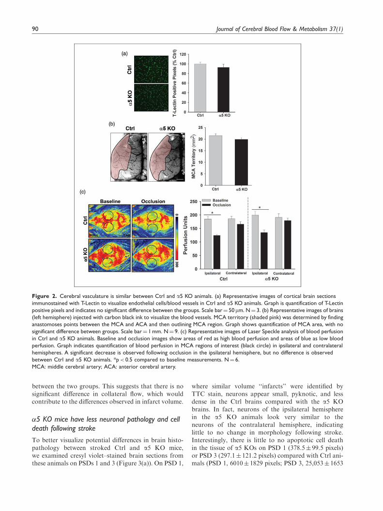
between the two groups. This suggests that there is nosignificant difference in collateral flow, which wouldcontribute to the differences observed in infarct volume.
�5 KO mice have less neuronal pathology and celldeath following stroke
To better visualize potential differences in brain histo-pathology between stroked Ctrl and a5 KO mice,we examined cresyl violet–stained brain sections fromthese animals on PSDs 1 and 3 (Figure 3(a)). On PSD 1,
where similar volume ‘‘infarcts’’ were identified byTTC stain, neurons appear small, pyknotic, and lessdense in the Ctrl brains compared with the a5 KObrains. In fact, neurons of the ipsilateral hemispherein the a5 KO animals look very similar to theneurons of the contralateral hemisphere, indicatinglittle to no change in morphology following stroke.Interestingly, there is little to no apoptotic cell deathin the tissue of a5 KOs on PSD 1 (378.5� 99.5 pixels)or PSD 3 (297.1� 121.2 pixels) compared with Ctrl ani-mals (PSD 1, 6010� 1829 pixels; PSD 3, 25,053� 1653
Figure 2. Cerebral vasculature is similar between Ctrl and a5 KO animals. (a) Representative images of cortical brain sections
immunostained with T-Lectin to visualize endothelial cells/blood vessels in Ctrl and a5 KO animals. Graph is quantification of T-Lectin
positive pixels and indicates no significant difference between the groups. Scale bar¼ 50mm. N¼ 3. (b) Representative images of brains
(left hemisphere) injected with carbon black ink to visualize the blood vessels. MCA territory (shaded pink) was determined by finding
anastomoses points between the MCA and ACA and then outlining MCA region. Graph shows quantification of MCA area, with no
significant difference between groups. Scale bar¼ 1 mm. N¼ 9. (c) Representative images of Laser Speckle analysis of blood perfusion
in Ctrl and a5 KO animals. Baseline and occlusion images show areas of red as high blood perfusion and areas of blue as low blood
perfusion. Graph indicates quantification of blood perfusion in MCA regions of interest (black circle) in ipsilateral and contralateral
hemispheres. A significant decrease is observed following occlusion in the ipsilateral hemisphere, but no difference is observed
between Ctrl and a5 KO animals. *p< 0.5 compared to baseline measurements. N¼ 6.
MCA: middle cerebral artery; ACA: anterior cerebral artery.
90 Journal of Cerebral Blood Flow & Metabolism 37(1)

pixels), as observed by TUNEL staining (Figure 3(b)).Taken together, a5 KO animals appear to be protectedagainst cell death following a stroke, unlike the Ctrlanimals.
To examine whether the activation of astrocytesplays a role in the infarct size differences observedbetween these animals, we performed immunohisto-chemistry for GFAP. On PSD 3, Ctrl animals showan apparent lack of GFAP staining in the infarct coreregion (Figure 3(c)), while tissue surrounding the coremaintains a normal (non-activated) expression level.In comparison, the a5 KO animals have an even dis-tribution of GFAP staining throughout the cortex,including the ‘‘infarcted’’ region (Figure 3(c)).Therefore, while there are differences in GFAP expres-sion between WT and a5 KO mice, there does notappear to be an increase in activated astrocytes atthis time point, suggesting the differences in infarctvolume between these mice is not due to astrocyteactivation and subsequent reduction in TTC negativestaining.
Changes in BBB function
The ECM and its integrin receptors contribute to theintegrity of the BBB. After stroke, disruption/degrad-ation of the ECM and alteration of its interaction withintegrins contributes to the breakdown of the BBBand subsequent edema, inflammatory cell infiltration,and neuronal death. This cascade of events is a majorcontributor to the expansion of ischemic injury andcell death that occurs over time after stroke. As ourresults suggest that ischemic injury is minimal anddoes not expand over time in a5 KO mice, wehypothesized that this could be due to differences inBBB integrity in these mice after stroke. To test thishypothesis, we performed IgG immunohistochemistryon brain sections from Ctrl and a5 KO animals onPSDs 1–3. IgG (approximately 150 kDa) will penetratebrain parenchyma only if the BBB is disrupted. Wewere unable to detect any IgG in the brain paren-chyma of the a5 KO mice at any PSD, while it wasabundant in the Ctrl brains at PSDs 1–3 (Figure 4(a)).This suggests that the BBB remains relatively intact in
Figure 3. Significantly less cell death occurs in the a5 KO animals compared with the Ctrls. (a) Representative images of cresyl violet
staining in cortical regions of brains from Ctrl and a5 KO animals following stroke. Neurons within infarct region of Ctrl mice are
small, pyknotic, and fewer in number compared with a5 KOs, which appear to have normal looking neurons, when compared with the
contralateral hemisphere. Scale bar¼ 50 mm. N¼ 6. (b) TUNEL (green) staining of infarct region on PSDs 1 and 3 and quantification of
TUNEL-positive pixel staining indicate an increase in cell death overtime in the Ctrl animals with very little cell death observed in the
a5 KOs. DAPI (blue) counterstain. Scale bar¼ 50 mm. *p< 0.05, **p< 0.01, N¼ 6. (c) GFAP staining of infarct region ( Þ�ð ) on PSD 3
shows lack of astrocytes in infarct core of Ctrl tissue and normal distribution of astrocytes in a5 KO tissue. Scale bar¼ 50 mm. N¼ 3.
PSD: post-stroke day.
Roberts et al. 91

the a5 KO mice after stroke compared with Ctrlanimals.
Next, as pan-b1 integrin inhibition has been directlylinked to decreased TJ protein claudin-5 expression anddecreased BBB integrity,12 we hypothesized that endo-thelial cell selective KO of a5(b1) integrin couldincrease BBB integrity by affecting post-stroke TJ pro-tein expression. Using real-time PCR, we examined theTJ protein claudin-5 after stroke and determined thatwhile baseline claudin-5 gene expression levels are simi-lar in sham animals between Ctrl and a5 KO mice,stroke causes a significantly smaller drop in claudin-5gene expression in a5 KO mice on PSD 1 comparedwith Ctrl levels (0.81� 0.03 vs. 0.52� 0.08 fold, respect-ively) (Figure 4(b)). These data suggest that a5 KOmice have differently regulated BBB components after
stroke that may contribute to its increased post-strokeintegrity.
Inhibition of �5 integrin maintains barrier integrityin vitro after OGD
To further investigate the effects of a5 integrin on brainendothelial barrier function, we examined in vitro BECmonolayer permeability using a small peptide inhibitorof a5b1 integrin, ATN-161.25 Employing a TEER assay(an indirect measure of cell monolayer permeabilitywhere greater TEER is indicative of lower permeability,etc.), we found that BECs treated with ATN-161 undernormoxic conditions had similar TEER values as PBSvehicle (Ctrl) treated cells (Figure 5(a)). However, fol-lowing OGD (8 h) and 24 h of re-oxygenation to mimic
Figure 4. BBB integrity is preserved in a5 KO animals compared with Ctrls following cerebral ischemia. (a) Representative images of
brain tissue (infarcted region) stained for IgG (brown) and counterstained with hematoxylin (purple) in Ctrl and a5 KO animals on
PSDs 1–3. Scale bar¼ 50 mm. N¼ 3. (b) Gene expression of Claudin 5 in Ctrl and a5 KO tissue (ipsilateral hemisphere) following
cerebral ischemia. A significant decrease is observed on PSD 1, but the decrease in a5 KO tissue is not as great as in Ctrls. *p< 0.05
compared to Ctrl sham and #p< 0.05 compared to Ctrl animals at same time point. N¼ 3.
BBB: blood-brain barrier; PSD: post-stroke day.
92 Journal of Cerebral Blood Flow & Metabolism 37(1)

the ischemia/reperfusion of stroke, Ctrl cells, but notATN-161-treated cells, had significantly reducedTEER (greater permeability) compared with normoxicconditions (Figure 5(a)). Next, BEC monolayer perme-ability was directly measured with FITC-dextran(Figure 5(b)). Under normoxic conditions, the ATN-161-treated BECs showed lower levels of fluorescence(lower permeability) compared with Ctrl BECs.Following OGD, Ctrl cells significantly increased theirpermeability while cells treated with ATN-161 main-tained levels consistent with normoxic conditions(Figure 5(b)). Taken together, blockade of a5 integrinon endothelial cells helps maintain a tighter barrierunder both normoxic and OGD conditions.
Discussion
In this study, we demonstrate that mice with an endo-thelial cell selective deletion of a5 integrin have pro-foundly smaller infarcts and functional deficit aftertransient MCA/CCA occlusion. Importantly, thisapparent resistance to ischemic injury cannot beexplained by differences in vital statistics, blood gas,serum electrolytes, cerebral vasculature (macrovesselson the brain surface or smaller blood vessels withinthe brain parenchyma in agreement with the angiogenicand vasculogenic analysis in the original report of theseanimals21) blood flow, or infiltration or activation ofGFAP cells into the stroke affected area, as none ofthese variables significantly differ between Ctrl and a5KO animals. Rather, our results suggest that a5 KOmice resist ischemic stroke injury due to greater post-stroke BBB integrity, resulting in less BBB leakage andresultant expansion of ischemic injury. This is demon-strated by absent IgG extravasation into the brain par-enchyma in a5 KO mice after stroke, relativelyincreased levels of the TJ protein claudin-5 comparedwith Ctrls, as well as decreased permeability in cellstreated with the a5b1 integrin inhibitor ATN-161under both normoxic and OGD (stroke-like) condi-tions. To the best of our knowledge, this is the firststudy to link endothelial cell a5b1 integrin to resistanceto ischemic injury via increased BBB integrity.
The importance of the BBB for maintainingthe homeostasis of the brain is well known.Cerebrovascular permeability is controlled by the endo-thelial cells and their TJs, proteins, and integrins of theECM, as well as pericytes and astrocytes. During ische-mia, permeability of the BBB increases due to dysfunc-tion, disassembly (e.g. of TJ proteins), and differentialregulation (e.g. decreases in TJ expression and increasesin ECM-processing proteases) in one or all of thesefactors. As described by Sandoval and Witt,26 threephases of paracellular permeability may occur follow-ing post-stroke reperfusion; an initial reperfusion per-meability associated with re-establishment of bloodflow, and a ‘‘bi-phasic’’ response, which may occurhours to days following injury. This increase in perme-ability may contribute to the continuously evolvinginfarct, which may occur over a period of days depend-ing on the form of animal stroke model used for assess-ment. For example, we show here, as well as in ourprevious work17 and the work of others27,28 that thesize of the infarct gradually increases in size followingtransient MCA/CCA occlusion, becoming maximal byPSD 3. In contrast, a5 KO mice in this stroke modelshow limited early injury (TTC-negative area) that bothfails to expand and appears to rapidly regress, possiblydue to a tightening of the BBB and a decrease in thenumber of phasic events. Importantly, while TTC ana-lysis suggests that the size of the infarct between Ctrls
Figure 5. a5 integrin inhibition maintains barrier integrity. (a)
TEER permeability assay of PBS vehicle-treated (Ctrl) and ATN-
161-treated endothelial cells under normoxic or OGD condi-
tions. Graph presented in % change from Ctrl normoxic condi-
tions. **p< 0.01 and ***p< 0.001 compared to Ctrl. N¼ 3. (b)
FITC-dextran permeability assay of Ctrl and ATN-161-treated
endothelial cells under normoxic or OGD conditions. Graph
presented in % change from Ctrl normoxic conditions. **p< 0.01
and ***p< 0.001 compared to Ctrl. N¼ 3.
TEER: trans-endothelial electrical resistance; OGD: oxygen-glu-
cose deprivation.
Roberts et al. 93
and a5 KOs appears to be similar on PSD 1, our cresylviolet and TUNEL stain analysis (Figure 3) demon-strates that the TTC-negative area on PSD 1 in thea5 KO mice actually contains few pyknotic and apop-totic cells, which persists out to PSD 3. These seeminglydiscrepant results may be due to the fact that whileTTC staining is traditionally used as a measure ofstroke infarct area, i.e. dead cells, it is actually a meas-urement of mitochondrial functional activity. Likewise,we did not detect any significant increase in other celltypes, such as GFAP-positive astrocytes, into the ipsi-lateral brain on PSDs 1–3 that could restore TTC-posi-tive signal to this area and thereby otherwise explainthis result. Therefore, we conclude that this TTC-nega-tive area contains stressed, but surviving cells whichproduce an insufficient level of formazan (TTC stain)to be detected macroscopically on PSD 1,29 but, in theabsence of continued insult secondary to a more stableBBB, gradually recover and are again TTC positive. Infact, the ischemic core and penumbra is a heteroge-neous region, with pockets of both dead and livecells.2 Therefore, the permeability of the BBB overtime may play a large role in the survival of injuredtissue following stroke, particularly so in our transientstroke model with a slowly evolving infarct. For thisreason, studies employing stroke models with a morerapidly evolving infarct size, including permanentocclusion models, should shed further light on thenature of a5 KO mice resistance to ischemic injury.Likewise, serial imaging experiments (ongoing in ourlab) are necessary to further evaluate the temporalnature of the infarct (or lack thereof) within each indi-vidual stroked a5 KO mouse.
A potential link between the b1 family of integrinsand BBB permeability has recently been suggested.12 Inthese studies, treatment with the pan-b1 integrin func-tion blocking antibody Ha2/5 increased primary BECmonolayer permeability in vitro while simultaneouslydecreasing their claudin-5 expression at the cell surface.Likewise, in vivo stereotactic injection of Ha2/5 into thestriatum of mice resulted in increased IgG extravasa-tion into the brain parenchyma as compared withstereotactically injected IgM control. However, theuse of a pan-b1 integrin functioning blocking antibodyin these studies by design made it impossible to deter-mine the relative contribution of any particular a(x)b1integrin to this BBB permeability effect. Furthermore,the potential for this antibody to interact with andblock the function of b1 integrin expressed on add-itional cell types in addition to endothelial cells whenstereotactically injected into the brain striatum, andthereby also impact BBB permeability, could not becompletely ruled out. For these reasons, we chose tofocus on a specific b1 integrin whose deletion was lim-ited to endothelial cells.
Of the various b1 integrin receptors known to beexpressed in BECs, we chose to focus on a5b1 for anumber of reasons. Although highly expressed inbrain microvasculature during development, a5b1integrin is virtually absent in these cells in the maturebrain. However, a5b1 integrin expression is signifi-cantly upregulated in BECs following global hypoxiaor ischemic stroke22,30 (in peri-infarct regions). In bothcases, it has been shown to have an important role inpost-injury angiogenesis22 and endothelial cell selectivea5 KO mice (the same as used in our study) have asignificantly delayed brain angiogenic response follow-ing global cerebral hypoxia.22 Additionally, we haveidentified a5b1 integrin as a key receptor for the bene-ficial angiogenic effects of perlecan domain V in treat-ing experimental ischemic stroke.17 Therefore, deletionof a5 integrin on endothelial cells would be predicted tohave a negative impact on angiogenesis following cere-bral ischemia, leading to deficient post-stroke neurore-pair and worse functional outcomes. Indeed, it was ourexpectation to demonstrate this when we initially sub-jected the a5 KO mice to MCA occlusion rather than todiscover any potential effects on the extent of ischemicinjury in these mice. However, endothelial cell activa-tion and associated ECM proteolysis (via MMP2 andother proteases) that occurs after ischemic stroke andas an early stage of angiogenesis all contribute toincreased BBB permeability. Therefore, it stands toreason that if post-stroke endothelial cell upregulationof a5b1 integrin is a component of this endothelial cellactivation, ECM proteolysis and subsequent angiogen-esis, it also has the potential to significantly contributeto the resultant decreased integrity of the BBB andexpansion of injury that occurs. In this light, our resultssupport the novel conclusion that BEC a5b1 integrinupregulation after ischemic stroke is a component ofendothelial cell activation that directly contributes topost-stroke breakdown of the BBB and resultant wor-sening of ischemic injury. Here, we report that inhib-ition of a5b1 on BECs show decreased permeabilityunder OGD conditions compared with Ctrl cells.These results were also observed under normoxic con-ditions when measured by FITC-dextran (4 kDa) versusTEER. Discrepancies in the normoxic conditionbetween these methods may possibly be due to differ-ences in detection sensitivity. Ultimately, these resultsindicate that inhibition of a5 integrin on endothelialcells lead to a tighter barrier in vitro. In the a5 KOmouse, BECs may be resistant to activation afterstroke as a result of their inability to upregulate a5b1integrin, resulting in less of a decrease in claudin-5,contributing to or resulting in retained BBB integrityand ultimately minimal ischemic injury. Likewise, thedelayed, but ultimately not absent brain angiogenesisthat was observed by Li et al.22 in the a5 KO mice after
94 Journal of Cerebral Blood Flow & Metabolism 37(1)

continuous exposure to global hypoxia might indicate asimilar resistance to hypoxia-induced endothelial cellactivation that is only gradually overcome by the con-tinually present (over several days) hypoxic conditions.Ultimately, a variety of BBB components (other TJproteins, markers of permeability, etc.) must be exam-ined to better understand the link between a5b1 integ-rin and BBB integrity.
Our results with the a5 KO mouse suggest that brainendothelial a5b1 integrin could represent a novel thera-peutic target in the acute or subacute phase after ischemicstroke. However, the timing of such a therapy (i.e. tostabilize the BBB and minimize injury in the short termwithout impeding more chronic angiogenic neurorepair)as well as the ability to selectively target endothelial cellsmay represent challenges to such a treatment strategy.Indeed, in our own previous work, intravenous treatmentwith a5b1 integrin function blocking antibody on PSDs1–3 after transient MCA occlusion in wild-type miceresulted in slightly (but significantly) larger ischemicinfarcts on PSDs 2 and 3 and diminished peri-infarctangiogenesis on PSDs 5–7.17 This seemingly contradictoryeffect of a5b1 integrin blockade on ischemic infarct size islikely due to the experimental design used in the previousstudy—a systemic route of administration in combinationwith an antibody inhibitor that would non-cell selectivelybind to and inhibit the a5b1 integrin wherever it encoun-tered it throughout the body (i.e. not limited to endothe-lial cells), an especially important consideration as a5b1 isexpressed in many different cell types throughout thebody such as platelets and chondrocytes.31,32 In supportof this conclusion, preliminary studies in our lab withsystemic administration of ATN-161, a peptide a5b1integrin inhibitor that specifically targets and inhibits acti-vated a5b1 (i.e. not the vast majority of quiescent a5b1integrin throughout the body) integrin25,33 24h afterMCA occlusion in Ctrl mice mimics the results of dimin-ished ischemic injury seen in the a5 KO mouse in thisreport. This suggests that selective targeting of activateda5b1 integrin as occurs in peri-infarct vasculature may bea valid therapeutic approach for stroke worthy of furtherinvestigation.
In conclusion, we report for the first time that micewith selective endothelial cell knockout of a5 integrinare resistant to stroke injury. The lack of infarctobserved by PSD 3 may be due to maintenance of theBBB integrity, unlike the Ctrl animals, which experi-ence increased BBB permeability. It is possible thatthe absence of a5 integrin decreases the number ofBBB phasic opening events, therefore decreasing thepotential for secondary injury by infiltrating cells andmolecules, limiting injury and affording rapid recovery.Further studies will expand our knowledge of the rolethis integrin plays in BBB integrity and its potential as anovel stroke therapeutic target.
Funding
The author(s) disclosed receipt of the following financial sup-
port for the research, authorship, and/or publication of thisarticle: NIH 2R01NS065842-08 to G.J.B.
Acknowledgments
We would like to acknowledge Dr. Richard Hynes (M.I.T.,MA) for kindly providing the a5 KO mice used in this study
and Michael Maniskas for his assistance with the behavioraltesting.
Declaration of conflicting interests
The author(s) declared no potential conflicts of interest withrespect to the research, authorship, and/or publication of this
article.
Authors’ contributions
JR and GJB conceptualized and designed the study. JR per-formed the in vivo experiments/analyzed the data and LdHperformed the in vitro experiments/analyzed the data while
GJB provided overall oversight of the research. All authorscritically reviewed the manuscript and approved the finalmanuscript as submitted.
References
1. Kahle MP and Bix GJ. Successfully climbing the
‘‘STAIRs’’: Surmounting failed translation of experimen-
tal ischemic stroke treatments. Stroke Res Treat 2012;
2012: 374098.2. del Zoppo GJ, Sharp FR, Heiss WD, et al. Heterogeneity
in the penumbra. J Cereb Blood Flow Metab 2011; 31:
1836–1851.
3. del Zoppo GJ and Milner R. Integrin-matrix interactions
in the cerebral microvasculature. Arterioscler Thromb
Vasc Biol 2006; 26: 1966–1975.4. Huveneers S, Truong H and Danen HJ. Integrins: signal-
ing, disease, and therapy. Int J Radiat Biol 2007; 83:
743–751.5. Hynes RO. Integrins: versatility, modulation, and signal-
ing in cell adhesion. Cell 1992; 69: 11–25.6. Yang Y and Rosenberg GA. Blood-brain barrier break-
down in acute and chronic cerebrovascular disease.
Stroke 2011; 42: 3323–3328.
7. Bolton SJ, Anthony DC and Perry VH. Loss of the tight
junction proteins occludin and zonula occludens-1 from
cerebral vascular endothelium during neutrophil-induced
blood-brain barrier breakdown in vivo. Neuroscience
1998; 86: 1245–1257.
8. Dore-Duffy P. Pericytes: pluripotent cells of the blood
brain barrier. Curr Pharm Des 2008; 14: 1581–1593.9. Willis CL, Nolan CC, Reith SN, et al. Focal astrocyte
loss is followed by microvascular damage, with subse-
quent repair of the blood-brain barrier in the apparent
absence of direct astrocytic contact. Glia 2004; 45:
325–337.
Roberts et al. 95

10. Baeten KM and Akassoglou K. Extracellular matrix andmatrix receptors in blood-brain barrier formation andstroke. Dev Neurobiol 2011; 71: 1018–1039.
11. Huber JD, Egleton RD and Davis TP. Molecular physi-ology and pathophysiology of tight junctions in theblood-brain barrier. Trends Neurosci 2001; 24: 719–725.
12. Osada T, Gu YH, Kanazawa M, et al. Interendothelial
claudin-5 expression depends on cerebral endothelial cell-matrix adhesion by beta(1)-integrins. J Cereb Blood FlowMetab 2011; 31: 1972–1985.
13. Francis SE, Goh KL, Hodivala-Dilke K, et al. Centralroles of alpha5beta1 integrin and fibronectin in vasculardevelopment in mouse embryos and embryoid bodies.
Arterioscler Thromb Vasc Biol 2002; 22: 927–933.14. Goh KL, Yang JT and Hynes RO. Mesodermal defects
and cranial neural crest apoptosis in alpha5 integrin-null
embryos. Development 1997; 124: 4309–4319.15. Yang JT, Rayburn H and Hynes RO. Embryonic meso-
dermal defects in alpha 5 integrin-deficient mice.Development 1993; 119: 1093–1105.
16. Bix GJ. Perlecan domain V therapy for stroke: a beaconof hope? ACS Chem Neurosci 2013; 4: 370–374.
17. Lee B, Clarke D, Al Ahmad A, et al. Perlecan domain V
is neuroprotective and proangiogenic following ischemicstroke in rodents. J Clin Invest 2011; 121: 3005–3023.
18. Clarke DN, Al Ahmad A, Lee B, et al. Perlecan domain
V induces VEGf secretion in brain endothelial cellsthrough integrin alpha5beta1 and ERK-dependent sig-naling pathways. PLoS One 2012; 7: e45257.
19. Al-Ahmad AJ, Lee B, Saini M, et al. Perlecan domain V
modulates astrogliosis in vitro and after focal cerebralischemia through multiple receptors and increased nervegrowth factor release. Glia 2011; 59: 1822–1840.
20. Li L, Liu F, Welser-Alves JV, et al. Upregulation offibronectin and the alpha5beta1 and alphavbeta3 integ-rins on blood vessels within the cerebral ischemic penum-
bra. Exp Neurol 2012; 233: 283–291.21. van der Flier A, Badu-Nkansah K, Whittaker CA, et al.
Endothelial alpha5 and alphav integrins cooperate in
remodeling of the vasculature during development.Development 2010; 137: 2439–2449.
22. Li L, Welser-Alves J, van der Flier A, et al. An angiogenicrole for the alpha5beta1 integrin in promoting endothelial
cell proliferation during cerebral hypoxia. Exp Neurol2012; 237: 46–54.
23. Hasan MR, Herz J, Hermann DM, et al. Visualization of
macroscopic cerebral vessel anatomy—a new and reliable
technique in mice. J Neurosci Methods 2012; 204:
249–253.24. Stroke Therapy Academic Industry R. Recommendations
for standards regarding preclinical neuroprotective and
restorative drug development. Stroke 1999; 30:
2752–2758.25. Stoeltzing O, Liu W, Reinmuth N, et al. Inhibition of
integrin alpha5beta1 function with a small peptide
(ATN-161) plus continuous 5-FU infusion reduces colo-
rectal liver metastases and improves survival in mice. Int
J Cancer 2003; 104: 496–503.
26. Sandoval KE and Witt KA. Blood-brain barrier tight
junction permeability and ischemic stroke. Neurobiol
Dis 2008; 32: 200–219.27. Du C, Hu R, Csernansky CA, et al. Very delayed infarc-
tion after mild focal cerebral ischemia: a role for apop-
tosis? J Cereb Blood Flow Metab 1996; 16: 195–201.28. Weston RM, Jones NM, Jarrott B, et al. Inflammatory
cell infiltration after endothelin-1-induced cerebral ische-
mia: histochemical and myeloperoxidase correlation with
temporal changes in brain injury. J Cereb Blood Flow
Metab 2007; 27: 100–114.29. Benedek A, Moricz K, Juranyi Z, et al. Use of TTC
staining for the evaluation of tissue injury in the early
phases of reperfusion after focal cerebral ischemia in
rats. Brain Res 2006; 1116: 159–165.30. Milner R, Hung S, Erokwu B, et al. Increased expression
of fibronectin and the alpha 5 beta 1 integrin in angio-
genic cerebral blood vessels of mice subject to hypobaric
hypoxia. Mol Cell Neurosci 2008; 38: 43–52.31. Kasirer-Friede A, Kahn ML and Shattil SJ. Platelet
integrins and immunoreceptors. Immunol Rev 2007; 218:
247–264.
32. Loeser RF. Integrins and cell signaling in chondrocytes.
Biorheology 2002; 39: 119–124.
33. Veine DM, Yao H, Stafford DR, et al. A D-amino acid
containing peptide as a potent, noncovalent inhibitor of
alpha5beta1 integrin in human prostate cancer invasion
and lung colonization. Clin Exp Metastasis. Epub
ahead of print 25 January 2014. DOI: 10.1007/s10585-
013-9634-1.
96 Journal of Cerebral Blood Flow & Metabolism 37(1)

![Soluble αKlotho downregulates Orai1-mediated store ......Klotho is an aging-suppressor gene that encodes type 1 transmembrane glycoprotein called αKlotho [22, 23]. Klotho-deficient](https://static.fdocument.org/doc/165x107/613b4592f8f21c0c8268e811/soluble-klotho-downregulates-orai1-mediated-store-klotho-is-an-aging-suppressor.jpg)