Methyl 4-O-(2-chlorobenzoyl)-α-L-rhamnopyranosides ...
9
Orbital: Electron. J. Chem. 2021, 13(1), 19-27 Bioorganic and Medicinal Chemistry Laboratory, Department of Chemistry, Faculty of Science, University of Chittagong, Chattogram, 4331, Bangladesh. *Corresponding author. E-mail: [email protected] Tel: +880 1716 839689 Published by Federal University of Mato Grosso do Sul | www.orbital.ufms.br 19 Full Paper | http://dx.doi.org/10.17807/orbital.v13i1.1532 Methyl 4-O-(2-chlorobenzoyl)-α-L-rhamnopyranosides: Synthesis, Characterization, and Thermodynamic Studies Mohammed Mahbubul Matin* , Md. Zahid Iqbal Sugar esters (SEs) with promising antimicrobial functionality were found to be a better choice to solve the multidrug resistant (MDR) pathogens due to their improved antimicrobial efficacy, and drug-likeness properties. In this context, 2-chlorobenzoyl ester group at C-4 position of methyl α-L-rhamnopyranoside was prepared via 2,3-O-acetonide protection followed by unimolar 2-chlorobenzoylation, and acetonide deprotection. The selective 4-O-(2-chlorobenzoyl)-α-L-rhamnopyranoside, thus formed, was converted into five 2,3-di-O-acyl esters with different aliphatic, and sulphonyl chains to obtain biologically important novel rhamnopyranoside-based SEs. All the synthesized compounds were optimized employing density functional theory (DFT). Thermodynamic calculations including frontier molecular orbital, and molecular electrostatic potential (MEP) were calculated and discussed. Attachment of multiple ester groups enhanced their stability, reactivity, and softness indicating their more polar and reactive nature than the non-ester sugars. Corroboration of all these properties might be helpful for their interactions with several enzymes (proteins) during different biological activities. The present study also revealed that incorporation of 2-chlorobenzoyl and mesyl groups in rhamnopyranoside skeleton increased better thermodynamic properties. Graphical abstract 1. Introduction Carbohydrate molecules participate in numerous biological processes due to their highly specific interaction with the physiological receptors. Their esters, especially monosaccharide-based sugar esters (SEs), are involved in many diverse biological events in organisms from all kind of life [1, 2]. SEs are generally non-toxic, biodegradable, non- allergic, and non-irritating as they are composed of a carbohydrate moiety (hydrophilic) and one or more fatty acid Keywords DFT calculations MEP Methyl α-L-rhamnopyranoside Selective esterification Sugar esters (SEs) Thermodynamic calculations Article history Received 13 August 2020 Revised 07 January 2021 Accepted 11 January 2021 Available online 06 March 2021 Editor: Jamal Rafique
Transcript of Methyl 4-O-(2-chlorobenzoyl)-α-L-rhamnopyranosides ...
Orbital: Electron. J. Chem. 2021, 13(1), 19-27
Bioorganic and Medicinal Chemistry Laboratory, Department of Chemistry, Faculty of Science, University of Chittagong, Chattogram, 4331, Bangladesh. *Corresponding author. E-mail: [email protected] Tel: +880 1716 839689
Published by Federal University of Mato Grosso do Sul | www.orbital.ufms.br 19
Full Paper | http://dx.doi.org/10.17807/orbital.v13i1.1532
Methyl 4-O-(2-chlorobenzoyl)-α-L-rhamnopyranosides: Synthesis, Characterization, and Thermodynamic Studies
Mohammed Mahbubul Matin* , Md. Zahid Iqbal
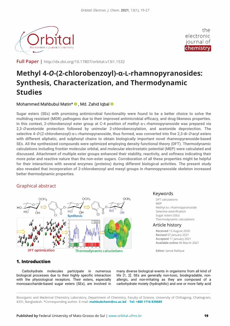
Sugar esters (SEs) with promising antimicrobial functionality were found to be a better choice to solve the multidrug resistant (MDR) pathogens due to their improved antimicrobial efficacy, and drug-likeness properties. In this context, 2-chlorobenzoyl ester group at C-4 position of methyl α-L-rhamnopyranoside was prepared via 2,3-O-acetonide protection followed by unimolar 2-chlorobenzoylation, and acetonide deprotection. The selective 4-O-(2-chlorobenzoyl)-α-L-rhamnopyranoside, thus formed, was converted into five 2,3-di-O-acyl esters with different aliphatic, and sulphonyl chains to obtain biologically important novel rhamnopyranoside-based SEs. All the synthesized compounds were optimized employing density functional theory (DFT). Thermodynamic calculations including frontier molecular orbital, and molecular electrostatic potential (MEP) were calculated and discussed. Attachment of multiple ester groups enhanced their stability, reactivity, and softness indicating their more polar and reactive nature than the non-ester sugars. Corroboration of all these properties might be helpful for their interactions with several enzymes (proteins) during different biological activities. The present study also revealed that incorporation of 2-chlorobenzoyl and mesyl groups in rhamnopyranoside skeleton increased better thermodynamic properties.
Graphical abstract
1. Introduction
Carbohydrate molecules participate in numerous biological processes due to their highly specific interaction with the physiological receptors. Their esters, especially monosaccharide-based sugar esters (SEs), are involved in
many diverse biological events in organisms from all kind of life [1, 2]. SEs are generally non-toxic, biodegradable, non-allergic, and non-irritating as they are composed of a carbohydrate moiety (hydrophilic) and one or more fatty acid
Keywords
DFT calculations MEP Methyl α-L-rhamnopyranoside Selective esterification Sugar esters (SEs) Thermodynamic calculations
Article history
Received 13 August 2020 Revised 07 January 2021 Accepted 11 January 2021 Available online 06 March 2021 Editor: Jamal Rafique
Orbital: Electron. J. Chem. 2021, 13(1), 19-27
Published by Federal University of Mato Grosso do Sul | www.orbital.ufms.br 20
parts (lipophilic moieties) [3, 4]. SEs have attracted considerable research interest and wide range of application in industry, and medicine mainly due to their considerable insecticidal, and antimicrobial activities [5-8]. SEs and related drugs have also opened a new gateway in the medical, and pharmaceutical field which are generally rely on (i) linkage of sugars, (ii) esterified/protected hydroxyl group(s), and (iii) unesterified/unprotected hydroxyl group(s) [9]. These factors are known to improve the water-solubility, stability, and biocompatibility leading to better absorption aiding delivery of drugs to the targeted site [10]. For example, the anticancer activity, and water solubility of glucose-aspirin ester (1, Figure 1) was improved eight to nine-fold and seven-fold, respectively, in comparison with aspirin alone [11]. Encouragingly some of the SEs were found highly active against multiple drug resistant (MDR) pathogenic organisms [7, 8].
Of the SEs, rhamnopyranoside derived esters are found to be of great importance due to their antigenic, anticarcinogenic [12], antimicrobial [13-19], and pharmacological properties [20]. For example, four new rhamnopyranose esters (e.g. 2a-d), isolated from the stem of P. odorata, were found to be good scaffolds for developing new anti-breast cancer, and antioxidant agents [21]. In addition, naturally occurring 2-O-acetyl-3,4-di-O-(E)-p-methoxycinnamoyl-α-L-rhamnopyranoside(s) (3, Figure 1) exhibited significant protective effects against glutamate-induced neurodegeneration in primary cultures of cortical neurons [22]. All these observations indicated that the esterification of the rhamnose moiety in natural products is essential for high affinity binding, selectivity, and development of effective anticancer agents (RSK inhibitors) [12, 21].
Fig. 1. Bioactive sugar esters 1, 2a-d and 3.
However, selective esterification of these sugar
molecules is very difficult due to the presence of multiple secondary hydroxyl groups of similar reactivity [23], and tends to produce a mixture of mono-, di-, and polyesters. Several esterification methods have so far been developed, and employed successfully for monosaccharide including direct esterification [24-26], organotin technique [18, 19, 27], microwave (MW) irradiation [28], enzyme catalyzed method [29], and protection-deprotection technique [16, 17, 30]. All the methods and/or strategies have some shortcomings such as tedious, increase the number of steps, expensive, and hence generally reduce the overall yield. For rhamnopyranoside 4, organotin (dibutyltin oxide) method was found to furnish 3-O-acyl esters [18, 19]. As many bioactive products bear substituents at C-4 position of rhamnopyranoside 4 we adopted protection-deprotection technique for esterification of this position, and reported herein.
Despite the broad-spectrum applications of sugar esters (SEs) as mentioned above, variable methods, and results were reported for synthesis, and different microbial species [31, 32]. Also, efficient synthetic strategy with thermodynamic studies is very less reported. Many of our previous encouraging results [4, 6-8] prompted us to design, and synthesize some new rhamnopyranoside-based SEs containing various alkyl, sulphonyl and aryl moieties in a single molecular framework. Thermodynamic properties of the synthesized SEs, as obtained from DFT analyses, are also reported herein.
2. Results and Discussion
2.1. Synthesis of 4-O-(2-chlorobenzoyl)rhamnopyranoside 7 The attachment of aglycon (non-sugar) group at C-4
position of L-rhamnose mimics many natural products of biological importance [21, 22]. Keeping this in mind, the present work mainly describes the selective 4-O-(2-chlorobenzoylation) of methyl α-L-rhamnopyranoside (4) using protection-deprotection technique [30] followed by 2,3-di-O-derivatization with their thermodynamic properties.
Initially, methyl α-L-rhamnopyranoside (4), prepared from L-rhamnose [13], was subjected for acetonide protection by the treatment of triol 4 with 2,2-dimethoxypropane using literature procedure [16, 17]. The acetonide selectively formed at the favorable cis-vicinal glycol system between C-2 and C-3 hydroxyl groups, and gave 2,3-O-isopropylidene-α-L-rhamnopyranoside 5. Having acetonide protected compound 5 in hand; we conducted its 2-chlorobenzoylation with the unimolar amount of 2-chlorobenzoyl chloride in pyridine (Scheme 1), and obtained a clear syrup in excellent yield.
The FT-IR spectrum of this syrup showed the presence of a carbonyl-stretching band at 1692 cm-1 with the disappearance of hydroxyl stretching band which indicated the attachment of benzoyl group in the molecule. In the 1H NMR spectrum, appearance of two one-proton multiplets at δ 7.83 and 7.30, and a two-proton multiplet at δ 7.38-7.45, totaling four aromatic protons were indicative of the incorporation of one chlorobenzoyloxy group in the molecule. Also, the considerable downfield shift of its H-4 to δ 5.10 ppm from its precursor compound 5 (δ 3.28-3.33 ppm [16, 17]) confirmed the attachment of the chlorobenzoyloxy group at C-4 position. This was finally supported by the analysis of its 13C NMR spectrum, where related characteristic carbonyl, and aromatic signals were observed
Orbital: Electron. J. Chem. 2021, 13(1), 19-27
Published by Federal University of Mato Grosso do Sul | www.orbital.ufms.br 21
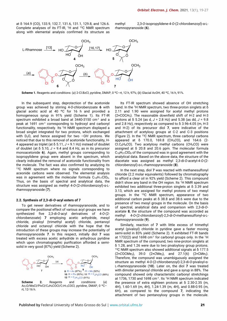
at δ 164.9 (CO), 133.9, 132.7, 131.6, 131.1, 129.8, and 126.6. Complete analyses of its FT-IR, 1H and 13C NMR spectrum along with elemental analysis confirmed its structure as
methyl 2,3-O-isopropylidene-4-O-(2-chlorobenzoyl)-α-L-rhamnopyranoside (6).
O
OCH3
HOHO OH
L-Rhamnose [13]
4
[16,17] O
OCH3
HOO O
5
(a) (b)O
OCH3
OO O
6OCl
O
OCH3
OHO OHOCl
7
Scheme 1. Reagents and conditions: (a) 2-Cl.BzCl, pyridine, DMAP, 0 ºC–rt, 12 h, 97%; (b) Glacial AcOH, 40 ºC, 16 h, 91%.
In the subsequent step, deprotection of the acetonide
group was achieved by stirring 4-O-chlorobenzoate 6 with glacial acetic acid at 40 ºC for 16 h and provided a homogeneous syrup in 91% yield (Scheme 1). Its FT-IR spectrum exhibited a broad band at 3440-3150 cm-1 and a peak at 1691 cm-1 corresponding to hydroxyl and carbonyl functionality, respectively. Its 1H NMR spectrum displayed a broad singlet integrated for two protons, which exchanged with D2O, and hence assigned for two –OH protons. We noticed that due to this removal of acetonide functionality, H-4 appeared as triplet (at δ 5.11, J = 9.1 Hz) instead of doublet of doublet (at δ 5.10, J = 9.4 and 8.4 Hz, as in its precursor monoacetonide 6). Again, methyl groups corresponding to isopropylidene group were absent in the spectrum, which clearly indicated the removal of acetonide functionality from the molecule. The fact was also confirmed by analyzing its 13C NMR spectrum where no signals corresponding to aceonide carbons were observed. The elemental analysis was in agreement with the molecular formula C14H17ClO6. Thus, on the basis of spectral and analytical data, the structure was assigned as methyl 4-O-(2-chlorobenzoyl)-α-L-rhamnopyranoside (7).
2.2. Synthesis of 2,3-di-O-acyl esters of 7
To get newer derivatives of rhamnopyranoside, and to compare the positional effect of various acyl groups we have synthesized five 2,3-di-O-acyl derivatives of 4-O-(2-chlorobenzoate) 7 employing acetic anhydride, mesyl chloride, pivaloyl (trimethyl acetyl) chloride, pentanoyl chloride and octanoyl chloride with the hope that the introduction of these groups may increase the potentiality of rhamnopyranoside 7. In this respect, initially diol 7 was treated with excess acetic anhydride in anhydrous pyridine which upon chromatographic purification afforded a semi-solid in very good (87%) yield (Scheme 2).
(a)O
OCH3
OHO OHOCl
7
O
OCH3
ORO OROCl
8: R = Ac (87%); 9: R = Ms (92%)10: R = Piv (83%); 11: R = C4H9CO (88%)12: R = C7H15CO (91%)
23
Scheme 2. Reagents and conditions: (a) Ac2O/MsCl/PivCl/C4H9COCl/C7H15COCl, pyridine, DMAP, 0 ºC–rt, 12-16 h.
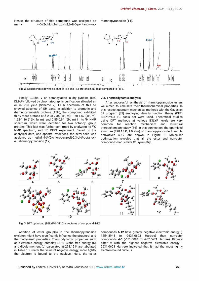
Its FT-IR spectrum showed absence of OH stretching band. In the 1H NMR spectrum, two three-proton singlets at δ 2.11 and 1.90 were assigned for acetyl methyl protons (2×COCH3). The reasonable downfield shift of H-2 and H-3 protons at δ 5.24 (as d, J = 2.8 Hz) and 5.38 (as dd, J = 9.8 and 2.8 Hz), respectively as compared to δ 3.96-4.05 (m, H-3 and H-2) of its precursor diol 7, were indicative of the attachment of acetyloxy groups at C-2 and C-3 positions (Figure 2). In the 13C NMR spectrum, three carbonyl carbons appeared at δ 170.0, 169.8 (CH3CO), and 164.6 (2-Cl.C6H4CO). Two acetyloxy methyl carbons (CH3CO) were assigned at δ 20.8 and 20.6 ppm. The molecular formula C18H21ClO8 of the compound was in good agreement with the analytical data. Based on the above data, the structure of the diacetate was assigned as methyl 2,3-di-O-acetyl-4-O-(2-chlorobenzoyl)-α-L-rhamnopyranoside (8).
In the next step, diol 7 was reacted with methanesulfonyl chloride (2.2 molar equivalents) followed by chromatography to afford a clear oil in 92% yield (Scheme 2). This compound didn’t show any band in the OH region. Its 1H NMR spectrum exhibited two additional three-proton singlets at δ 3.39 and 3.13, which are assigned for methyl protons of two mesyl groups. In the 13C NMR spectrum, appearance of two additional carbon peaks at δ 38.8 and 38.6 were due to the presence of two mesyl groups in the molecule. On the basis of spectral, analytical data and comparison with 2,3-di-O-acetate 8, the structure of the compound was accorded as methyl 4-O-(2-chlorobenzoyl)-2,3-di-O-methanesulfonyl-α-L-rhamnopyranoside (9).
Similarly, reaction of 7 with little excess of trimethyl acetyl (pivaloyl) chloride in pyridine gave a faster moving semi-solid in 83% yield (Scheme 2). It exhibited FT-IR bands at 1732(2) and 1698 cm-1 for carbonyl groups only. In the 1H NMR spectrum of the compound, two nine-proton singlets at δ 1.28, and 1.26 were due to two pivaloyloxy group protons. 13C NMR spectrum also showed additional signals at δ 177.5 (2×COCMe3), 39.0 (2×CMe3), and 27.1(6) (2×CMe3). Therefore, the compound was unambiguously assigned the structure as methyl 4-O-(2-chlorobenzoyl)-2,3-di-O-pivaloyl-α-L-rhamnopyranoside (10). Later on, the diol 7 was treated with dimolar pentanoyl chloride and gave a syrup in 88%. The compound showed only characteristic carbonyl stretchings at 1736, 1730 and 1698 cm-1. Its 1H NMR spectrum indicated the presence of extra eighteen protons at δ 2.30-2.35 (m, 4H), 1.60-1.69 (m, 4H), 1.24-1.39 (m, 4H), and 0.88-0.95 (m, 6H), as compared to the compound 7, indicating the attachment of two pentanoyloxy groups in the molecule.
Orbital: Electron. J. Chem. 2021, 13(1), 19-27
Published by Federal University of Mato Grosso do Sul | www.orbital.ufms.br 22
Hence, the structure of this compound was assigned as methyl 4-O-(2-chlorobenzoyl)-2,3-di-O-pentanoyl-α-L-
rhamnopyranoside (11).
Fig. 2. Considerable downfield shift of H-2 and H-3 protons in (a) 8 as compared to (b) 7.
Finally, 2,3-diol 7 on octanoylation in dry pyridine (cat.
DMAP) followed by chromatographic purification afforded an oil in 91% yield (Scheme 2). FT-IR spectrum of this oil showed absence of OH band. In addition to aromatic and rhamnopyranoside protons (15H), the compound exhibited thirty more protons at δ 2.28-2.35 (4H, m), 1.60-1.67 (4H, m), 1.22-1.36 (16H, br m), and 0.85-0.94 (6H, m) in its 1H NMR spectrum, which were identified for two octanoyl group protons. This fact was further confirmed by analyzing its 13C NMR spectrum, and 13C DEPT experiment. Based on the analytical data, and spectral evidences, the semi-solid was assigned as methyl 4-O-(2-chlorobenzoyl)-2,3-di-O-octanoyl-α-L-rhamnopyranoside (12).
2.3. Thermodynamic analysis After successful synthesis of rhamnopyranoside esters
we aimed to calculate their thermochemical properties. In this respect quantum mechanical methods with the Gaussian 09 program [33] employing density function theory (DFT) B3LYP/6-311G basis set were used. Theoretical studies using DFT methods at various B3LYP levels are very common for reaction mechanism and structural stereochemistry study [34]. In this connection, the optimized structure (298.15 K, 1.0 atm) of rhamnopyranoside 4 and its derivatives 5-12 are shown in Figure 3. Molecular optimization revealed that all the ester and non-ester compounds had similar C1 symmetry.
Fig. 3. DFT optimized (B3LYP/6-311G) structures of compound 4-12.
Addition of ester group(s) in the rhamnopyranoside skeleton might have significantly influence the structural and thermodynamic properties. Thermodynamic properties such as electronic energy, enthalpy (ΔH), Gibbs free energy (G) and dipole moment (μ) calculated at 298.15 K are tabulated in Table 1. Greater the value of negative energy, more tightly the electron is bound to the nucleus. Here, the ester
compounds 6-12 have greater negative electronic energy (-1454.8944 to -2631.0603 Hartree) than non-ester compounds 4-5 (-651.0084 to -767.6671 Hartree). Dimesyl ester 9 with the highest negative electronic energy -2631.0603 Hartree) indicated that it had the most tightly electron bound nucleus.
Orbital: Electron. J. Chem. 2021, 13(1), 19-27
Published by Federal University of Mato Grosso do Sul | www.orbital.ufms.br 23
Table 1. Molecular formula (MF), molecular weight (MW, g/mol), electronic energy (EE), enthalpy, Gibbs free energy (GFE) and dipole moment (DM) of 4-12.
Compound No. MF MW EE (Hartree)
Enthalpy (Hartree)
GFE (Hartree)
DM (Debye)
4 C7H14O5 178.184 -651.0084 -651.0075 -651.0598 2.6783 5 C10H18O5 218.249 -767.6671 -767.6661 -767.7251 1.2699 6 C17H21ClO6 356.799 -1571.5523 -1571.5514 -1571.6308 1.6004 7 C14H17ClO6 316.734 -1454.8944 -1454.8934 -1454.9662 4.2468 8 C18H21ClO8 400.808 -1760.0882 -1760.0872 -1760.1769 4.3815 9 C16H21ClO10S2 472.904 -2631.0603 -2631.0594 -2631.1545 5.8948
10 C24H33ClO8 484.186 -1995.8072 -1995.8063 -1995.9145 5.3127 11 C24H33ClO8 484.970 -1995.7805 -1995.7796 -1995.8900 4.6619 12 C30H45ClO8 569.132 -2231.4708 -2231.4699 -2231.6024 4.6513
Another important parameter is ΔH, and the rhamnopyranoside esters 6-12 possess lower ΔH values (-1454 to -2631 Hartree) than non-ester sugars 4-5 (-651 to -7.67 Hartree). This clearly indicated the more stability of esters 6-12 than non-ester 4-5. These values are in consistent with the exothermic esterification reaction of sugar molecules. Gibbs free energy (G) combines enthalpy and entropy into a single value. It signifies spontaneity of a reaction when G < 0. As shown in Table 1, with the attachment of acyl groups and increase of chain length G (negative value) were increased. Thus, these SEs are suitable for the spontaneous binding, and interaction with other substrates. Furthermore, dipole moment (μ) is the measure of net molecular polarity, and improved μ can enhance hydrogen bond, and non-bonded interactions in drug receptor complexes which can play an important role to increase binding affinity [35, 36]. Increased μ as in rhamnoside-based SEs 7-12 (4.2-5.8 Debye, Table 1) as compared to non-ester 4-5 (1.2-2.6 Debye) clearly demonstrated the higher binding affinity with target enzyme during antimicrobial activities and more polar nature of these molecules [6-8]. Amongst the synthesized esters, dimesylate 9 was found to possess the highest thermodynamic properties (Table 1).
2.4. Molecular orbitals (MO) analysis
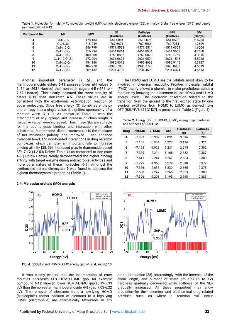
The HOMO and LUMO are the orbitals most likely to be involved in chemical reactivity. Frontier molecular orbital (FMO) theory allows a chemist to make predictions about a reaction by knowing the placement of the HOMO and LUMO energy levels. The electronic absorption related to the transition from the ground to the first excited state by one electron excitation from HOMO to LUMO, as derived from DFT (B3LYP/6-311G) [37], is presented in Table 2 (Figure 4).
Table 2. Energy (eV) of HOMO, LUMO, energy gap, hardness, and softness of SEs 4-12.
Drug 𝜀𝜀HOMO 𝜀𝜀LUMO Gap Hardness (η)
Softness (S)
4 -7.333 -0.302 7.031 3.516 0.284 5 -7.161 -0.934 6.227 3.114 0.321
6 -7.153 -1.922 5.231 2.615 0.382 7 -7.379 -2.214 5.165 2.582 0.387
8 -7.511 -2.244 5.267 2.633 0.380
9 -7.229 -1.953 5.279 2.640 0.379 10 -7.366 -2.036 5.330 2.665 0.375 11 -7.508 -2.242 5.266 2.633 0.380 12 -7.396 -2.201 5.195 2.598 0.385
Fig. 4. DOS plot and HOMO-LUMO energy gap of (a) 4, and (b) 10.
It was clearly evident that the incorporation of ester
moieties decreases SEs HOMO-LUMO gap, for example compound 6-12 showed lower HOMO-LUMO gap (5.19-5.33 eV) than the non-ester rhamnopyranoside 4-5 (gap 7.03-6.22 eV). The removal of electrons from a low-lying HOMO (nucleophile) and/or addition of electrons to a high-lying LUMO (electrophile) are energetically favourable in any
potential reaction [38]. Interestingly, with the increase of the chain length, and number of ester group(s) (6 to 12) hardness gradually decreased while softness of the SEs gradually increased. All these properties may allow prediction for their chemical and biochemical drug related activities such as where a reaction will occur
Orbital: Electron. J. Chem. 2021, 13(1), 19-27
Published by Federal University of Mato Grosso do Sul | www.orbital.ufms.br 24
(regioselectivity), and direction of approach (stereochemistry).
2.5. Molecular electrostatic potential (MEP) analysis
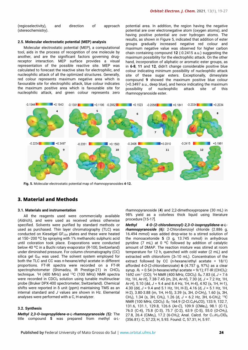
Molecular electrostatic potential (MEP), a computational tool, aids in the process of recognition of one molecule by another, and are the significant factors governing drug-receptor interaction. MEP surface provides a visual representation of the possible reactive site. MEP was calculated to forecast the reactive sites for electrophilic, and nucleophilic attack of all the optimized structures. Generally, red colour represents maximum negative area which is favourable site for electrophilic attack, blue colour indicates the maximum positive area which is favourable site for nucleophilic attack, and green colour represents zero
potential area. In addition, the region having the negative potential are over electronegative atom (oxygen atoms), and having positive potential are over hydrogen atoms. The results, as shown in Figure 5, indicated that addition of ester groups gradually increased negative red colour and maximum negative value was observed for higher carbon chain containing compound 12 (-0.2415 a.u.) suggesting the maximum possibility for the electrophilic attack. On the other hand, incorporation of aliphatic or aromatic ester groups, as in 6-8, 11 and 12, didn’t change considerable positive blue colour indicating minimum possibility of nucleophilic attack site of these sugar esters. Exceptionally, dimesylate compound 9 showed the maximum positive blue colour (+0.3497 a.u., deep blue), and hence indicating the maximum possibility of nucleophilic attack site of this rhamnopyranoside ester.
Fig. 5. Molecular electrostatic potential map of rhamnopyranosides 4-12.
3. Material and Methods
3.1. Materials and instrumentation All the reagents used were commercially available
(Aldrich), and were used as received unless otherwise specified. Solvents were purified by standard methods or used as purchased. Thin layer chromatography (TLC) was conducted on Kieselgel GF254 plates and these were heated at 150–200 ºC by spraying with 1% methanolic sulphuric acid until coloration took place. Evaporations were conducted below 40 ºC in a Buchi rotary evaporator (R-100, Switzerland) under diminished pressure. For column chromatography (CC) silica gel G60 was used. The solvent system employed for both the TLC and CC was n-hexane/ethyl acetate in different proportions. FT-IR spectra were recorded on a FT-IR spectrophotometer (Shimadzu, IR Prestige-21) in CHCl3 technique. 1H (400 MHz) and 13C (100 MHz) NMR spectra were recorded in CDCl3 solution using tunable multinuclear probe (Bruker DPX-400 spectrometer, Switzerland). Chemical shifts were reported in δ unit (ppm) maintaining TMS as an internal standard and J values are shown in Hz. Elemental analyses were performed with a C, H-analyzer.
3.2. Synthesis Methyl 2,3-O-isopropylidene-α-L-rhamnopyranoside (5): The title compound 5 was prepared from methyl α-L-
rhamnopyranoside (4) and 2,2-dimethoxypropane (30 mL) in 98% yield as a colorless thick liquid using literature procedure [15-17]. Methyl 4-O-(2-chlorobenzoyl)-2,3-O-isopropylidene-α-L-rhamnopyranoside (6): 2-Chlorobenzoyl chloride (2.886 g, 16.494 mmol) was added drop-wise to a stirred solution of the monoacetonide 5 (3 g, 13.745 mmol) in anhydrous pyridine (7 mL) at 0 ºC followed by addition of catalytic amount of DMAP. The reaction mixture was stirred at room temperature for 12 h, quenched with cold water (2 mL) and extracted with chloroform (3×10 mL). Concentration of the extract followed by CC (n-hexane/ethyl acetate = 18/1) afforded 4-O-(2-chlorobenzoate) 6 (4.757 g, 97%) as a clear syrup. Rf = 0.54 (n-hexane/ethyl acetate = 9/1); FT-IR (CHCl3): 1692 cm-1 (CO); 1H NMR (400 MHz, CDCl3): δH 7.83 (d, J = 7.6 Hz, 1H, Ar-H), 7.38-7.45 (m, 2H, Ar-H), 7.30 (d, J = 7.2 Hz, 1H, Ar-H), 5.10 (dd, J = 9.4 and 8.4 Hz, 1H, H-4), 4.92 (s, 1H, H-1), 4.30 (dd, J = 9.4 and 5.1 Hz, 1H, H-3), 4.16 (d, J = 5.1 Hz, 1H, H-2), 3.80-3.88 (m, 1H, H-5), 3.39 (s, 3H, O-CH3), 1.60 (s, 3H, CH3), 1.34 (s, 3H, CH3), 1.26 (d, J = 6.2 Hz, 3H, 6-CH3); 13C NMR (100 MHz, CDCl3): δC 164.9 (2-Cl.C6H4CO), 133.9, 132.7, 131.6, 131.1, 129.8, 126.6 (Ar-C), 109.9 (CMe2), 98.1 (C-1), 76.0 (C-4), 75.8 (C-3), 75.7 (C-2), 63.9 (C-5), 55.0 (O-CH3), 27.8, 26.4 (CMe2), 17.2 (6-CH3); Anal. Calcd. for C17H21ClO6 (356.81): C, 57.23; H, 5.93. Found: C, 57.31; H, 5.97.
Orbital: Electron. J. Chem. 2021, 13(1), 19-27
Published by Federal University of Mato Grosso do Sul | www.orbital.ufms.br 25
Methyl 4-O-(2-chlorobenzoyl)-α-L-rhamnopyranoside (7): A solution of 4-O-(2-chlorobenzoate) 6 (4 g, 11.21 mmol) in glacial acetic acid (96%) (25 mL) was stirred at 40 ºC for 16 h. Formation of a slower moving product (Rf = 0.48) was evident from TLC examination. The mixture was concentrated in vacuum and co-evaporated with toluene (3×3 mL) to remove traces of acetic acid. The residue, thus obtained, on chromatography with n-hexane/ethyl acetate (3/2) afforded 2,3-diol 7 (3.231 g, 91%) as a chromatographically homogeneous syrup. Rf = 0.48 (n-hexane/ethyl acetate = 1/1); FT-IR (CHCl3): 3440-3150 (OH), 1691 cm-1 (CO); 1H NMR (400 MHz, CDCl3): δH 7.83 (d, J = 7.2 Hz, 1H, Ar-H), 7.40-7.47 (m, 2H, Ar-H), 7.28 (d, J = 7.1 Hz, 1H, Ar-H), 5.11 (t, J = 9.1 Hz, 1H, H-4), 4.74 (s, 1H, H-1), 3.96-4.05 (2H, m, H-3 and H-2), 3.87-3.93 (m, 1H, H-5), 3.39 (s, 3H, O-CH3), 2.63-3.82 (br s, exchange with D2O, 2H, 2OH), 1.27 (d, J = 6.0 Hz, 3H, 6-CH3); 13C NMR (100 MHz, CDCl3): δH 166.3 (2-Cl.C6H4CO), 133.7, 132.9, 131.6, 131.1, 129.7, 126.7 (Ar-C), 100.5 (C-1), 76.4 (C-4), 71.0 (C-3), 70.1 (C-2), 65.6 (C-5), 55.1 (O-CH3), 17.6 (6-CH3); Anal. Calcd. for C14H17ClO6 (316.74): C, 53.09; H, 5.41. Found: C, 53.12; H, 5.44.
3.2.1. General procedure for 2,3-di-O-acylation of compound 7
To a solution of the 2,3-diol 7 (0.5 g, 1.578 mmol) in dry pyridine (2 mL) was added acetic anhydride or acyl halides (~3.47 mmol) at an ice cooled temperature (0 ºC) followed by the addition of catalytic amount of 4-dimethylamino pyridine (DMAP). For compound 9, the reaction mixture was initially stirred 1 h at -5 ºC. The reaction mixture was then allowed to attain room temperature and stirred at this temperature for 12-16 h, treated with cold water (2 mL), and extracted with dichloromethane (DCM, 5×3 mL). The DCM layer was washed successively with 5% hydrochloric acid, saturated aqueous sodium hydrogen carbonate solution and brine. The DCM layer was dried and concentrated under reduced pressure. The obtained residue was then purified by CC (gradient elution from n-hexane to n-hexane/ethyl acetate = 12/1) afforded the corresponding 2,3-di-O-substituted rhamnopyranoside esters 8-12. Methyl 2,3-di-O-acetyl-4-O-(2-chlorobenzoyl)-α-L-rhamnopyranoside (8): White semi-solid; Yield 87%; Rf = 0.53 (n-hexane/ethyl acetate = 7/1); FT-IR (CHCl3): 1729(2), 1696 cm-1 (CO); 1H NMR (400 MHz, CDCl3): δH 7.66 (d, J = 7.5 Hz, 1H, Ar-H), 7.36-7.42 (m, 2H, Ar-H), 7.25 (d, J = 7.9 Hz, 1H, Ar-H), 5.38 (dd, J = 9.8 and 2.8 Hz, 1H, H-3), 5.30 (t, J = 9.8 Hz, 1H, H-4), 5.24 (d, J = 2.8 Hz, 1H, H-2), 4.62 (s, 1H, H-1), 3.90-3.97 (m, 1H, H-5), 3.35 (s, 3H, O-CH3), 2.11 (s, 3H, COCH3), 1.90 (s, 3H, COCH3), 1.28 (d, J = 6.0 Hz, 3H, 6-CH3); 13C NMR (100 MHz, CDCl3): δC 170.0, 169.8 (2×CH3CO), 164.6 (2-Cl.C6H4CO), 133.5, 132.7, 131.0, 130.9, 129.6, 126.6 (Ar-C), 98.4 (C-1), 71.9, 69.7, 69.0 (C-2/C-3/C-4), 66.1 (C-5), 55.0 (O-CH3), 20.8, 20.6 (2×CH3CO), 17.4 (6-CH3); Anal. Calcd. for C18H21ClO8 (400.82): C, 53.94; H, 5.28. Found: C, 54.03; H, 5.31. Methyl 4-O-(2-chlorobenzoyl)-2,3-di-O-methanesulfonyl-α-L-rhamnopyranoside (9): Clear oil (turned pale-yellow after a couple of days); Yield 92%; Rf = 0.50 (n-hexane/ethyl acetate = 8/1); FT-IR (CHCl3): 1690 (CO), 1462, 1459 cm-1 (SO2); 1H NMR (400 MHz, CDCl3): δH 7.69 (d, J = 7.8 Hz, 1H, Ar-H), 7.38-7.44 (m, 2H, Ar-H), 7.28 (d, J = 7.8 Hz, 1H, Ar-H), 5.41 (dd, J = 9.6 and 3.0 Hz, 1H, H-3), 5.36 (d, J = 3.0 Hz, 1H, H-2), 5.33 (t, J = 9.6 Hz, 1H, H-4), 4.83 (s, 1H, H-1), 3.92-4.01 (m, 1H, H-5), 3.40 (s, 3H, O-CH3), 3.39 (s, 3H, SO2CH3), 3.13 (s, 3H, SO2CH3), 1.25 (d, J = 6.3 Hz, 3H, 6-CH3); 13C NMR (100 MHz,
CDCl3): δC 164.5 (2-Cl.C6H4CO), 133.5, 132.9, 131.1, 131.0, 129.5, 126.7 (Ar-C), 98.6 (C-1), 75.3, 71.4, 70.3 (C-2/C-3/C-4), 66.5 (C-5), 55.3 (O-CH3), 38.8, 38.6 (SO2CH3), 17.5 (6-CH3); Anal. Calcd. for C16H21ClO10S2 (472.92): C, 40.64; H, 4.48. Found: C, 40.70; H, 4.44. Methyl 4-O-(2-chlorobenzoyl)-2,3-di-O-pivaloyl-α-L-rhamnopyranoside (10): Semi-solid; Yield 83%; Rf = 0.53 (n-hexane/ethyl acetate = 9/1); FT-IR (CHCl3): 1732(2), 1698 cm-
1 (CO); 1H NMR (400 MHz, CDCl3): δH 7.70 (d, J = 7.7 Hz, 1H, Ar-H), 7.38-7.45 (m, 2H, Ar-H), 7.30 (d, J = 7.7 Hz, 1H, Ar-H), 5.42 (dd, J = 10.0 and 3.1 Hz, 1H, H-3), 5.33 (t, J = 9.9 Hz, 1H, H-4), 5.28 (d, J = 3.1 Hz, 1H, H-2), 4.64 (s, 1H, H-1), 3.95-4.03 (m, 1H, H-5), 3.40 (s, 3H, O-CH3), 1.32 (d, J = 6.1 Hz, 3H, 6-CH3), 1.28 (s, 9H, CMe3), 1.26 (s, 9H, CMe3); 13C NMR (100 MHz, CDCl3): δC 177.5 (2×COCMe3), 164.7 (2-Cl.C6H4CO), 133.7, 132.8, 131.1(2), 129.7, 126.7 (Ar-C), 98.6 (C-1), 72.2, 69.5, 69.4 (C-2/C-3/C-4), 66.2 (C-5), 55.2 (O-CH3), 39.1, 39.0 (2×CMe3), 27.1 (6) (2×CMe3), 17.7 (6-CH3); Anal. Calcd. for C24H33ClO8 (484.98): C, 59.44; H, 6.86. Found: C, 59.48; H, 6.96. Methyl 4-O-(2-chlorobenzoyl)-2,3-di-O-pentanoyl-α-L-rhamnopyranoside (11): Syrup; Yield 88%; Rf = 0.54 (n-hexane/ethyl acetate = 9/1); FT-IR (CHCl3): 1736, 1730, 1698 cm-1 (CO); 1H NMR (400 MHz, CDCl3): δH 7.63 (d, J = 7.6 Hz, 1H, Ar-H), 7.31-7.35 (m, 2H, Ar-H), 7.28 (d, J = 7.6 Hz, 1H, Ar-H), 5.30 (t, J = 9.6 Hz, 1H, H-4), 5.28 (dd, J = 9.6 and 3.1 Hz, 1H, H-3), 5.22 (d, J = 3.0 Hz, 1H, H-2), 4.55 (s, 1H, H-1), 3.86-3.91 (m, 1H, H-5), 3.36 (s, 3H, O-CH3), 2.30-2.35 [m, 4H, 2×CH3(CH2)2CH2CO], 1.60-1.69 [m, 4H, 2×CH3CH2CH2CH2CO], 1.24-1.39 [m, 4H, 2×CH3CH2(CH2)2CO], 1.20 (d, J = 6.4 Hz, 3H, 6-CH3), 0.88-0.95 [m, 6H, 2×CH3(CH2)3CO]; Anal. Calcd. for C24H33ClO8 (484.97): C, 59.44; H, 6.86. Found: C, 59.52; H, 6.88. Methyl 4-O-(2-chlorobenzoyl)-2,3-di-O-octanoyl-α-L-rhamnopyranoside (12): Oil; Yield 91%; Rf = 0.56 (n-hexane/ethyl acetate = 9/1); FT-IR (CHCl3): 1739, 1736, 1701, cm-1 (CO); 1H NMR (400 MHz, CDCl3): δH 7.65 (d, J = 7.5 Hz, 1H, Ar-H), 7.30-7.38 (m, 2H, Ar-H), 7.28 (d, J = 7.5 Hz, 1H, Ar-H), 5.32 (t, J = 9.6 Hz, 1H, H-4), 5.27 (dd, J = 9.6 and 3.1 Hz, 1H, H-3), 5.20 (d, J = 3.1 Hz, 1H, H-2), 4.58 (s, 1H, H-1), 3.85-3.91 (m, 1H, H-5), 3.37 (s, 3H, O-CH3), 2.28-2.35 [m, 4H, 2×CH3(CH2)5CH2CO], 1.60-1.67 [m, 4H, 2×CH3(CH2)4CH2CH2CO], 1.22-1.36 [br m, 16H, 2×CH3(CH2)4CH2CH2CO], 1.19 (d, J = 6.5 Hz, 3H, 6-CH3), 0.85-0.94 [m, 6H, 2×CH3(CH2)6CO]; 13C NMR (100 MHz, CDCl3): δC 172.5, 172.0 [2×CH3(CH2)6CO], 164.6 (2-Cl.C6 H4CO), 98.8 (C-1), 71.8, 69.6, 69.5 (C-2/C-3/C-4), 66.5 (C-5), 55.3 (O-CH3), 34.2, 31.8, 31.6 (3), 29.4, 29.1, 29.0, 25.9, 25.7, 24.7, 22.6 [2×CH3(CH2)6CO], 17.2 (6-CH3), 14.1, 14.0 [2×CH3(CH2)6CO]; Anal. Calcd. for C30H45ClO8 (569.14): C, 63.31; H, 7.97. Found: C, 63.38; H, 8.05.
3.3. Computational calculations
In computational chemistry, quantum mechanical methods are widely used to predict thermal energies, molecular orbital (MO), and molecular electrostatic potential (MEP) properties [39, 40]. Initial geometry of the basic methyl α-L-rhamnopyranoside (4) was taken from the online structure database named ChemSpider. The other sugar ester structures (6-12) were built with the GaussView (5.0) program [33]. All the compounds structures were optimized, and further modified with Gaussian 09 program [33, 41] employing density function theory (DFT) B3LYP/6-311G basis set. For molecular orbital energy (FMO) such as highest occupied molecular orbital (HOMO), lowest
Orbital: Electron. J. Chem. 2021, 13(1), 19-27
Published by Federal University of Mato Grosso do Sul | www.orbital.ufms.br 26
unoccupied molecular orbital (LUMO), HOMO-LUMO gap, hardness (η), and softness (S) were calculated at the same level of theory using the following equations:
Gap = [εLUMO - εHOMO]; 𝜂𝜂 = 𝜀𝜀𝜀𝜀𝜀𝜀𝜀𝜀𝜀𝜀−𝜀𝜀𝜀𝜀𝜀𝜀𝜀𝜀𝜀𝜀2
; 𝑆𝑆 = 1𝜂𝜂
DOS plot was obtained from GaussSum 3.0. To visualize MEP online WebMO demo server was used for all the compounds.
4. Conclusions
Thus, selective 2-chlorobenzoylation of methyl α-L-rhamnopyranoside (4) at C-4 position was achieved via acetonide protection-deprotection technique. The chlorobonzoate 7, thus obtained, was further converted into five 2,3-di-O-acyl esters with aliphatic and sulphonyl acylating agents reasonably in good yield. Density functional theory (DFT) studies of the synthesized compounds indicated several important parameters of rhamnopyranoside-based SEs. Density function theory (DFT) based thermodynamic studies like electronic energy, enthalpy, Gibbs free energy, and dipole moment of all the synthesized compounds were studied. Frontier molecular orbital (FMO) calculation showed that with the increase of the chain length, and number of ester group(s) (6 to 12) hardness gradually decreased while softness of the SEs gradually increased. Molecular electrostatic potential (MEP) indicated that these SEs (except 9) had maximum possibility of electrophilic attack than the precursor sugar 4. Considering the suitable polarity, stability, and electrophilic mode of reaction the study might be helpful for designing monosaccharide-based drug molecules.
Acknowledgments
Financial support from the Research and Publication Cell, University of Chittagong, Bangladesh (2018-2019) is gratefully acknowledged.
Author Contributions
Both MM Matin and MZ Iqbal contributed in conceptualization, methodology, investigation, synthesis, formal analysis, validation and writing. MM Matin especially contributed in funding acquisition, resources, software, supervision, review & editing of the manuscript.
References and Notes
[1] Pöhnlein, M.; Slomka, C.; Kukharenko, O.; Gärtner, T.; Wiemann, L. O.; Sieber, V.; Syldatk, C.; Hausmann, R. Eur. J. Lipid Sci. Technol. 2014, 116, 423. [Crossref]
[2] Matin, M. M.; Sharma, T.; Sabharwal, S. G.; Dhavale, D. D. Org. Biomol. Chem. 2005, 3, 1702. [Crossref]
[3] Lawandi, J.; Rocheleau, S.; Moitessier, N. Tetrahedron 2016, 72, 6283. [Crossref]
[4] Matin, M. M.; Bhattacharjee, S. C.; Chakraborty, P.; Alam, M. S.; Carbohydr. Res. 2019, 485, 107812. [Crossref]
[5] Kazmi, I.; Rahman, M.; Afzal, M.; Gupta, G.; Saleem, S.; Afzal, O.; Shaharyar, M. A.; Nautiyal, U.; Ahmed S.; Anwar, F. Fitoterapia 2012, 83, 142. [Crossref]
[6] Matin, M. M.; Bhuiyan, M. M. H.; Kabir, E.; Sanaullah, A. F. M.; Rahman, M. A.; Hossain, M. E.; Uzzaman, M. J. Mol. Struct. 2019, 1195, 189. [Crossref]
[7] Matin, M. M., Chakraborty, P.; Alam, M. S.; Islam, M. M.; Hanee, U. Carbohydr. Res. 2020, 496, 108130. [Crossref]
[8] Matin, M. M.; Hasan, M. S.; Uzzaman, M.; Bhuiyan, M. M. H.; Kibria, S. M.; Hossain, M. E.; Roshid, M. H. O. J. Mol. Struct. 2020, 1222, 128821. [Crossref]
[9] Quan, J.; Chen, Z.; Han, C.; Lin, X. Bioorg. Med. Chem. 2007, 15, 1741. [Crossref]
[10] Mizuma, T.; Ohta, K.; Hayashi, M.; Awazu, S. Biochem. Pharmacol. 1992, 43, 2037. [Crossref]
[11] Jacob, J. N.; Tazawa, M. J. Bioorg. Med. Chem. Lett. 2012, 22, 3168. [Crossref]
[12] Mihoub, M.; Pichette, A.; Sylla, B.; Gauthier, C.; Legault, J. PLoS ONE 2018, 13, 0193386. [Crossref]
[13] Matin, M. M.; Nath, A. R.; Saad, O.; Bhuiyan, M. M. H.; Kadir, F. A.; Hamid, S. B. A.; Alhadi, A. A.; Ali, M. E.; Yehye, W. A. Int. J. Mol. Sci. 2016, 17, 1412. [Crossref]
[14] Kabir, A. K. M. S.; Matin, M. M.; Hossain, A.; Sattar, M. A. J. Bangladesh Chem. Soc. 2003, 16, 85. [Link]
[15] Matin, M. M.; Iqbal, M. Z. Proc. Bangladesh Chem. Congress 2008, 2008, 254. [Crossref]
[16] Matin, M. M.; Ibrahim, M. J. Appl. Sci. Res. 2010, 6, 1527. [Link]
[17] Matin, M. M. Orbital: Electron. J. Chem. 2014, 6, 20. [Link]
[18] Kabir, A. K. M. S.; Matin, M. M. J. Bangladesh Chem. Soc. 1994, 7, 73. [Link]
[19] Kabir, A. K. M. S.; Matin, M. M. J. Bangladesh Acad. Sci. 1997, 21, 83. [Link]
[20] Matin, M. M.; Roshid, M. H. O.; Bhattacharjee, S. C.; Azad, A. K. M. S. Med. Res. Arch., 2020, 8, ID: 2165. [Crossref]
[21] Elmaidomy, A. H.; Mohammed, R.; Owis A. I.; et al. RSC Adv. 2020, 10, 10584. [Crossref]
[22] Kim, S. R.; Kim, Y. C. Phytochem. 2000, 54, 503. [Crossref]
[23] Wang, C. -C.; Lee, J. -C.; Luo, S. -Y.; Kulkarni, S. S.; Huang, Y. -W.; Lee, C. -C.; Chang, K. -L.; Hung, S. -C. Nature 2007, 446, 896. [Crossref]
[24] Matin, M. M.; Bhuiyan, M. H.; Hossain, M. M.; Roshid, M. H. O. Orbital: Electron. J. Chem. 2015, 7, 160. [Crossref]
[25] Matin, M. M.; Bhuiyan, M. M. H.; Hossain, M. M.; Roshid, M. H. O. J. Turkish Chem. Soc. Sect. A: Chem. 2015, 2, 12. [Link]
[26] Matin, M. M.; Bhuiyan, M. M. H.; Azad, A. K. M. S.; Roshid, M. H. O. J. Physical Sci. 2015, 26, 1. [Link]
[27] Kabir, A. K. M. S.; Matin, M. M.; Ali, M.; Anwar, M. N. J. Bangladesh Aca. Sci. 2003, 27, 43. [Link]
[28] Kondamudi, N.; McDougal, O. M. J. Surfactants Deterg. 2019, 22, 721. [Crossref]
[29] Siebenhaller, S.; Gentes, J.; Infantes, A.; Muhle-Goll, C.; Kirschhöfer, F.; Brenner-Weiß, G.; Ochsenreither, K.; Syldatk, C. Front. Chem. 2018, 6, ID: 24. [Crossref]
Orbital: Electron. J. Chem. 2021, 13(1), 19-27
Published by Federal University of Mato Grosso do Sul | www.orbital.ufms.br 27
[30] Matin, M. M.; Bhuiyan, M. M. H.; Azad, A. K. M. S.; Akther, N. Current Chem. Lett. 2017, 6, 31. [Crossref]
[31] Xiao, D.; Ye, R.; Davidson, P. M.; Hayes, D. G.; Golden, D. A.; Zhong, Q. Int. J. Food Microb. 2011, 145, 64. [Crossref]
[32] Awual, M. R. Chem. Eng. J. 2017, 307, 85. [Crossref] [33] Frisch, M. J.; Trucks, G. W.; Schlegel, H. B.; Scuseria,
G. E.; Robb, M. A.; Cheeseman, J. R.; Scalmani, G.; Barone, V.; Petersson, G. A.; Nakatsuji, H. Gaussian 09, 2013, Gaussian, Inc. (Wallingford CT).
[34] Ghaleb, A.; Aouidate, A.; Sbai, A.; Lakhlifi, T.; Maghat, H.; Bouachrine, M. Orbital: Electron. J. Chem. 2017, 9, 337. [Crossref]
[35] Bastos, C. C.; Leite, B. S. Orbital: Electron. J. Chem. 2017, 9, 360. [Crossref]
[36] Matin, M. M.; Uzzaman, M.; Chowdhury, S. A.; Bhuiyan, M. M. H. J. Biomol. Struct. Dyn. 2020, in press. [Crossref]
[37] Santos, C. B. R.; Lobato, C. C.; Braga, F. S.; Morais, S. S. S.; Santos, C. F.; Fernandes, C. P.; Brasil, D. S. B.; Hage-Melim, L. I. S.; Macêdo, W. J. C.; Carvalho, J. C. T. Comput. Mol. Biosci. 2014, 4, 1. [Crossref]
[38] Parr, R. G.; Zhou, Z. Acc. Chem. Res. 1993, 26, 256. [Crossref]
[39] Matin, M. M.; Chakraborty, P. J. Appl. Sci. Process Eng. 2020, 7, 572. [Crossref]
[40] Loukhovitski, B. I.; Sharipov, A. S.; Alexander M. Starik, A. M. J. Phys. Chem. A 2015, 119, 1369. [Crossref]
[41] Matin, M. M.; Islam, N.; Siddika, A.; Bhattacharjee, S. C. J. Turkish Chem. Soc. Sec. A: Chem. 2021, 8, 363. [Crossref]
How to cite this article
Matin, M. M.; Iqbal, M. Z. Orbital: Electron. J. Chem. 2021, 13, 19. http://dx.doi.org/10.17807/orbital.v13i1.1532


![Inclusion of the insecticide fenitrothion in dimethylated ... · Fenitrothion [O,O-dimethyl O-(3-methyl-4-nitrophenyl)phos-phorothioate] (1, Figure€1) is an organophosphorus insecticide](https://static.fdocument.org/doc/165x107/5e5a05ae27941506fe4e0c19/inclusion-of-the-insecticide-fenitrothion-in-dimethylated-fenitrothion-oo-dimethyl.jpg)