M&M 8-1 220705
24
more Vol 8 – 1/2004 MA CS & HIV MAGNETIZED • DIRECT ISOLATION OF HIV1 FROM PATIENT PLASMA • NEW μMACS GFP ISOLATION KIT CELLS’ PARADISE • A FULL RANGE OF MEDIA FOR HEMATOPOIETIC AND NONHEMATOPOIETIC STEM CELLS • DENDRITIC CELLS FROM PEYER‘S PATCHES • HUMAN STEM CELL HOMING RARE CELL ANALYSIS • NOVEL MOUSE PDCA1 ANTIBODIES • ISOLATION OF T CELL SUBSETS • FASER KITS: ENHANCE YOUR FLUORESCENCE SIGNALS! • DEAD CELL DISCRIMINATION WITH FIXED CELLS • BUFFERS FOR CONVENIENT CELL SORTING • CD16 + MONOCYTE PURIFICATION MADE EASY PERSPECTIVE: DISPLAY TECHNOLOGIES AND MAGNETIC SELECTION NEW PRODUCTS
Transcript of M&M 8-1 220705

more Vol 8 – 1/2004
MACS & HIV MAGNETIZED
• DIRECT ISOLATION OF HIV1 FROM PATIENT PLASMA• NEW μMACS GFP ISOLATION KIT
CELLS’ PARADISE
• A FULL RANGE OF MEDIA FOR HEMATOPOIETIC
AND NONHEMATOPOIETIC STEM CELLS
• DENDRITIC CELLS FROM PEYER‘S PATCHES
• HUMAN STEM CELL HOMING
RARE CELL ANALYSIS
• NOVEL MOUSE PDCA1 ANTIBODIES
• ISOLATION OF T CELL SUBSETS
• FASER KITS: ENHANCE YOUR FLUORESCENCE SIGNALS!
• DEAD CELL DISCRIMINATION WITH FIXED CELLS
• BUFFERS FOR CONVENIENT CELL SORTING
• CD16+ MONOCYTE PURIFICATION MADE EASY
PERSPECTIVE: DISPLAY TECHNOLOGIES AND MAGNETIC SELECTION
NEW PRODUCTS
Vol 8 1/2004 EDITORIAL2
MACS&more (ISSN 1610-4994) is published by Miltenyi Biotec GmbH and printed in Germany.
Editor: Prof. Dr. Uwe A. O. Heinlein ([email protected])
Editorial board: Dr. Kirt Braun, Dr. Susan Donath, Elisabeth Hennig, Dr. Alexander Horst, Dr. Betina Marquardt, Dr. Mariette Mohaupt, Dr. Kerstin Otterbach, Dr. Katrin Pauls, Dr. Thorsten Peters-Regehr, Dr. Katja Petry
Graphics & Layout: Miltenyi Biotec GmbH, © 2004 Miltenyi Biotec GmbH, Friedrich-Ebert-Str. 68, 51429 Bergisch Gladbach, Germany, Phone: +49 2204 8306-0 E-mail: [email protected]
Unless otherwise indicated, all Miltenyi Biotec products are for research use only and not for diagnostic or therapeutic use.
MACS® and CliniMACS® are registered trademarks of Miltenyi Biotec GmbH.
New productsFixation and Dead Cell Discrimination Kit 3
HSC-CFU and NH media: products for
hematopoietic and nonhematopoietic stem cells 4
MACS® buffers for convenient cell sorting 6
µMACS™ GFP Isolation Kit and Anti-GFP-HRP 6
Purification and analysis of B cells 7
New CD16+ Monocyte Isolation Kit 8
FASER Kits – Beam up your cells ! 9
Novel mPDCA-1 antibodies 13
New kits for the isolation of T cell subsets 22
Test yourself Regulatory T cells 15 The answers 19
Customer reports K. A. Kadaoui and B. Corthésy
Isolation of dendritic cells from
mouse Peyer’s patches using magnetic cell sorting 10
T. Lapidot and O. Kollet
Mechanism of human CD34+ stem cell homing and mobilization
in transplanted immune-deficient NOD/SCID mice 14
L. D. Lupo and S. T. Butera
Application of µMACS Streptavidin MicroBeads
for the analysis of HIV-1 directly from patient plasma 16
I. Johnston
Efficient target molecule identification by
combining display technology with magnetic selection 20
MACS paper highlight 23
MACS&more online: www.miltenyibiotec.com/MACS&more
CONTENTS
The portfolio of MACS® products is steadily
growing. In this issue you will find information
about exiting new products as well as scientific
reports on findings obtained with MACS®
Technology.
A stimulating example is provided on pages 14
and 15 by Lupo and Butera who demonstrate the
analytical power of magnetic cell separation by
their report on direct analysis of HIV particles
Dear customer, and their cellular origin, while Kadaoui and
Corthésy provide further proof that MACS®
Technology is the method of choice for
isolating rare cells (p.10). On page 20, Johnston
examines the perspectives of how to discover
new protein-ligand interactions by combined
display technology and magnetic selection.Last
but not least, Lapidot and Kollet review the
action of activating factors on the properties of
translanted CD34+ stem cells.
Specially formulated cell culture media for
hematopoietic and nonhematopoietic stem
cells are presented on pages 4 and 5. These
media ensure optimal conditions during cell
isolation, cultivation, and differentiation.
New kits for purification of CD16+ monocytes
(p. 8) and of T cell subsets (p.20) are now
available, providing further options to isolate
the cells of your choice even more
conveniently.
Much more product information is available
online – we look forward to your visit!
Your MACS&more team
NEW PRODUCTS
Fixation and Dead Cell Discrimination Kit
The Dead Cell Discriminator selectively stains dead cells.
© by Miltenyi Biotec
Incubation under a light source causes irreversible staining of the dead cells.
Cells can be fixed with the Fix Solution. Discriminator Stop Reagent allows prolonged storage of the cell sample.
Dead cells are detected by flow cytometric analysis by their fluorescent staining.
Dead cells are positively stained. Cells which were viable prior to fixation have not been stained.
The End
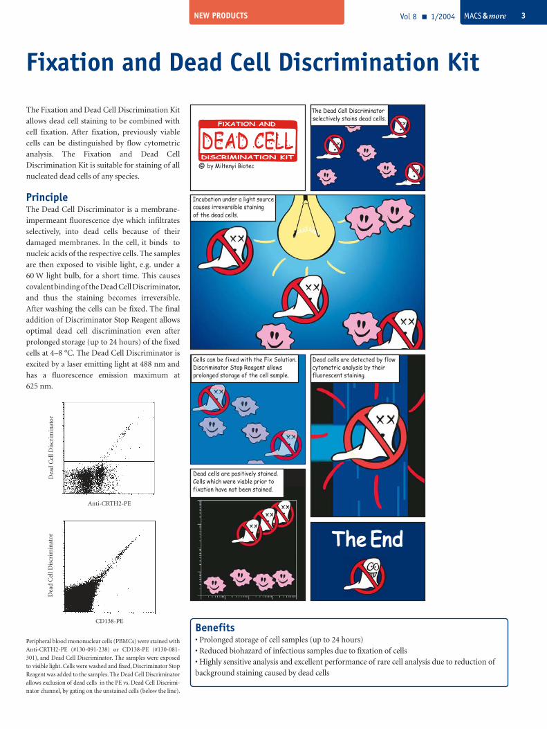
The Fixation and Dead Cell Discrimination Kit
allows dead cell staining to be combined with
cell fixation. After fixation, previously viable
cells can be distinguished by flow cytometric
analysis. The Fixation and Dead Cell
Discrimination Kit is suitable for staining of all
nucleated dead cells of any species.
PrincipleThe Dead Cell Discriminator is a membrane-
impermeant fluorescence dye which infiltrates
selectively, into dead cells because of their
damaged membranes. In the cell, it binds to
nucleic acids of the respective cells. The samples
are then exposed to visible light, e.g. under a
60 W light bulb, for a short time. This causes
covalent binding of the Dead Cell Discriminator,
and thus the staining becomes irreversible.
After washing the cells can be fixed. The final
addition of Discriminator Stop Reagent allows
optimal dead cell discrimination even after
prolonged storage (up to 24 hours) of the fixed
cells at 4–8 °C. The Dead Cell Discriminator is
excited by a laser emitting light at 488 nm and
has a fluorescence emission maximum at
625 nm.
CD138-PE
Dea
d C
ell D
iscr
imin
ator
Anti-CRTH2-PE
Dea
d C
ell D
iscr
imin
ator
Peripheral blood mononuclear cells (PBMCs) were stained with Anti-CRTH2-PE (#130-091-238) or CD138-PE (#130-081-301), and Dead Cell Discriminator. The samples were exposed to visible light. Cells were washed and fixed, Discriminator Stop Reagent was added to the samples. The Dead Cell Discriminator allows exclusion of dead cells in the PE vs. Dead Cell Discrimi-nator channel, by gating on the unstained cells (below the line).
Vol 8 1/2004 MACS & more 3
Benefits• Prolonged storage of cell samples (up to 24 hours)
• Reduced biohazard of infectious samples due to fixation of cells
• Highly sensitive analysis and excellent performance of rare cell analysis due to reduction of
background staining caused by dead cells
Vol 8 1/2004 MACS & more4 NEW PRODUCTS
Throughout the whole life cycle, the human
body preserves adult stem cells as natural
resources for the regeneration of a wide range
of tissues. Therefore, these stem cells are a
promising source to alleviate or cure
hematopoietic diseases and tissue injuries
(tissue replacement therapies).
HSC-CFU mediaThe hematopoietic system comprises constantly
self-renewing stem cells, capable of differen-
tiating into progenitor cells and mature blood
cells of all hematopoietic lineages. Thus, it
contains cells at various stages of maturation.
Primitive hematopoietic stem cells (HSCs) with
multi-lineage differentiation capacity give rise
to identical daughter cells, whereas progenitor
cells have restricted differentiation potential
and lack self-renewal capacity. Stem cell
transplants can help restore bone marrow
function and rebuild the immune system for
patients with inherited immunodeficiency or
autoimmune diseases, or after immuno-
suppressive treatment.
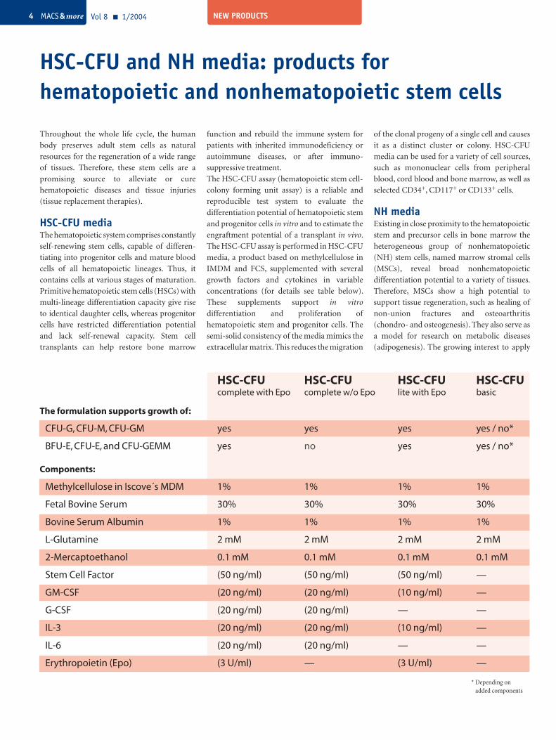
The HSC-CFU assay (hematopoietic stem cell-
colony forming unit assay) is a reliable and
reproducible test system to evaluate the
differentiation potential of hematopoietic stem
and progenitor cells in vitro and to estimate the
engraftment potential of a transplant in vivo.
The HSC-CFU assay is performed in HSC-CFU
media, a product based on methylcellulose in
IMDM and FCS, supplemented with several
growth factors and cytokines in variable
concentrations (for details see table below).
These supplements support in vitro
differentiation and proliferation of
hematopoietic stem and progenitor cells. The
semi-solid consistency of the media mimics the
extracellular matrix. This reduces the migration
HSC-CFU and NH media: products for hematopoietic and nonhematopoietic stem cells
of the clonal progeny of a single cell and causes
it as a distinct cluster or colony. HSC-CFU
media can be used for a variety of cell sources,
such as mononuclear cells from peripheral
blood, cord blood and bone marrow, as well as
selected CD34+, CD117+ or CD133+ cells.
NH mediaExisting in close proximity to the hematopoietic
stem and precursor cells in bone marrow the
heterogeneous group of nonhematopoietic
(NH) stem cells, named marrow stromal cells
(MSCs), reveal broad nonhematopoietic
differentiation potential to a variety of tissues.
Therefore, MSCs show a high potential to
support tissue regeneration, such as healing of
non-union fractures and osteoarthritis
(chondro- and osteogenesis). They also serve as
a model for research on metabolic diseases
(adipogenesis). The growing interest to apply
HSC-CFU HSC-CFU HSC-CFU HSC-CFU complete with Epo complete w/o Epo lite with Epo basic
The formulation supports growth of:
CFU-G, CFU-M, CFU-GM yes yes yes yes / no*
BFU-E, CFU-E, and CFU-GEMM yes no yes yes / no*
Components:
Methylcellulose in Iscove´s MDM 1% 1% 1% 1%
Fetal Bovine Serum 30% 30% 30% 30%
Bovine Serum Albumin 1% 1% 1% 1%
L-Glutamine 2 mM 2 mM 2 mM 2 mM
2-Mercaptoethanol 0.1 mM 0.1 mM 0.1 mM 0.1 mM
Stem Cell Factor (50 ng/ml) (50 ng/ml) (50 ng/ml) —
GM-CSF (20 ng/ml) (20 ng/ml) (10 ng/ml) —
G-CSF (20 ng/ml) (20 ng/ml) — —
IL-3 (20 ng/ml) (20 ng/ml) (10 ng/ml) —
IL-6 (20 ng/ml) (20 ng/ml) — —
Erythropoietin (Epo) (3 U/ml) — (3 U/ml) —
* Depending on added components
NEW PRODUCTS Vol 8 1/2004 MACS & more 5
MACS® productsHSC-CFU complete with Epo
100 mL #130-091-280
24 × 3 mL #130-091-278
HSC-CFU complete w/o Epo
100 mL #130-091-277
24 × 3 mL #130-091-276
HSC-CFU lite with Epo
100 mL #130-091-281
24 × 3 mL #130-091-282
HSC-CFU basic
80 mL #130-091-275
NH CFU-F Medium
24 × 3 mL #130-091-676
NH Expansion Medium
500 mL #130-091-680
NH AdipoDiff Medium
100 mL #130-091-677
NH ChondroDiff Medium
100 mL #130-091-679
NH OsteoDiff Medium
100 mL #130-091-678
RPMI 1640
500 mL #130-091-440
RPMI 1640 with stable Glutamine
500 mL #130-091-439
DMEM
500 mL #130-091-437
DMEM with stable Glutamine
500 mL #130-091-438
MSCs for clinical applications increases the
necessity to better understand the fundamental
processes that mediate the differentiation of
MSCs into functional nonhematopoietic cell
types and to identify the factors involved.
Therefore, it is essential to establish efficient and
reproducible procedures for the isolation,
cultivation and differentiation of target cells.
The optimization and standardization of
experimental conditions, such as in vitro culture
systems is an important first step. NH media are
optimized for most reproducible and convenient
quantification, quality control, expansion and
differentiation of MSCs from human bone
marrow.
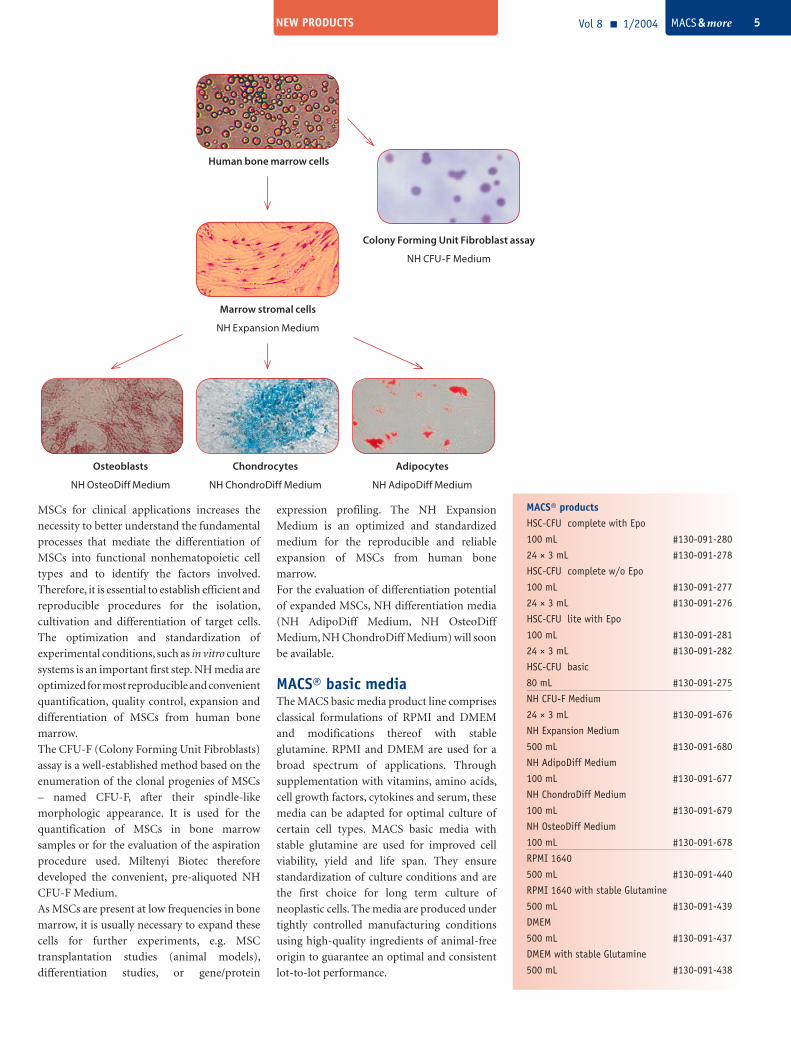
The CFU-F (Colony Forming Unit Fibroblasts)
assay is a well-established method based on the
enumeration of the clonal progenies of MSCs
– named CFU-F, after their spindle-like
morphologic appearance. It is used for the
quantification of MSCs in bone marrow
samples or for the evaluation of the aspiration
procedure used. Miltenyi Biotec therefore
developed the convenient, pre-aliquoted NH
CFU-F Medium.
As MSCs are present at low frequencies in bone
marrow, it is usually necessary to expand these
cells for further experiments, e.g. MSC
transplantation studies (animal models),
differentiation studies, or gene/protein
expression profiling. The NH Expansion
Medium is an optimized and standardized
medium for the reproducible and reliable
expansion of MSCs from human bone
marrow.
For the evaluation of differentiation potential
of expanded MSCs, NH differentiation media
(NH AdipoDiff Medium, NH OsteoDiff
Medium, NH ChondroDiff Medium) will soon
be available.
MACS® basic mediaThe MACS basic media product line comprises
classical formulations of RPMI and DMEM
and modifications thereof with stable
glutamine. RPMI and DMEM are used for a
broad spectrum of applications. Through
supplementation with vitamins, amino acids,
cell growth factors, cytokines and serum, these
media can be adapted for optimal culture of
certain cell types. MACS basic media with
stable glutamine are used for improved cell
viability, yield and life span. They ensure
standardization of culture conditions and are
the first choice for long term culture of
neoplastic cells. The media are produced under
tightly controlled manufacturing conditions
using high-quality ingredients of animal-free
origin to guarantee an optimal and consistent
lot-to-lot performance.
Human bone marrow cells
Colony Forming Unit Fibroblast assay
NH CFU-F Medium
Marrow stromal cells
NH Expansion Medium
Chondrocytes
NH ChondroDiff Medium
Adipocytes
NH AdipoDiff Medium
Osteoblasts
NH OsteoDiff Medium
NEW PRODUCTS
µMACS GFP Isolation Kit and Anti-GFP-HRPIsolation of tagged proteins from eukaryotes
made easy – with the second generation of the
µMACS™ GFP Isolation Kit for even better
results. They enable the isolation of epitope-
tagged proteins whenever high sensitivity and
minimum background are important, such as
expression in eukaryotic cells and purification
of unknown proteins or interaction partners.
The Anti-GFP MicroBeads coupled to new
high-affinity monoclonal antibodies together
with MACS® Column Technology allow the
sensitive purification of GFP-tagged proteins in
less than two hours. The kit contains all buffers
for convenient working.
As a special feature, MACS® Column Tech-
nology allows enzymatic reactions with native
bound proteins to be performed directly on the
column. For this special application the new
temperature-controlled thermoMACS® Sepa-
ration Unit is ideal.
Aliquots of recombinant GFP fusion protein (20, 10, 5, and 1 ng) were subjected to gradient SDS-PAGE and transfered to a PDVF membrane. Bands were detected with Anti-GFP-HRP (1:5000, 1 hr, RT) and chemiluminescent signal capture.
MACS® productsµMACS GFP Isolation Kit # 130-091-125
Anti-GFP-HRP # 130-091-833
New developments for B cell research
MACS® Technology offers a wide range of
MicroBeads, Cell Isolation Kits, MultiSort
Kits, manual and automated separators and
antibodies for the purification and detection
of B cells from any kind of cell source and
species. Optimized and ready-to-use prod-
ucts are available for human, non-human
primate, mouse and rat B cells.
MACS® buffers for convenient manual and automated cell separation
MACS® productsautoMACS Running Buffer
6 × 1.5 L # 130-091-221
autoMACS Rinsing Solution
6 × 1.45 L # 130-091-222
MACS BSA Stock Solution
6 × 75 mL # 130-091-376
MACS® products
Untouched B cells
B Cell Isolation Kit II # 130-091-151
Naive B Cell Isolation Kit II # 130-091-150
CD43 MicroBeads # 130-091-333
B cells directly from whole bloodWhole Blood CD19 MicroBeads # 130-090-880
Analysis of human B cells CD19-FITC # 130-091-328
CD19-PE # 130-091-247
CD19-APC # 130-091-248
Analysis of mouse B cells CD45R (B220)-FITC # 130-091-829
CD45R (B220)-PE # 130-091-828
CD45R (B220)-APC # 130-091-843
CD43-FITC # 130-091-580
CD43-PE # 130-091-585
CD43-APC # 130-091-581
New MACS® cell separation buffers are sterile-
filtered solutions prepared from high-quality
ingredients and manufactured under tightly
controlled conditions. MACS buffers are
optimized for automated or manual magnetic
cell sorting approaches using MACS Tech-
nology.
The autoMACS™ Separator is an automated
bench-top magnetic cell sorter capable of
sorting up to 10 million cells per second. It is
compatible with any MACS cell separation
reagent. The specially designed autoMACS™
Running Buffer and autoMACS Rinsing
Solution bottles connect directly to the
autoMACS Separator allowing standardiza-
tion in automated high-throughput cell sort-
ing. The combination of MACS® BSA Stock
Solution and autoMACS Rinsing Solution
allows the preparation of an optimal and
preservative-free MACS Separation Buffer.
Vol 8 1/2004 MACS & more6
The new Anti-GFP antibody is also available as
a horseradish peroxidase conjugate and can be
used for the single-step detection of wildtype
and mutant GFP and GFP fusion proteins in
Western blots and ELISA.
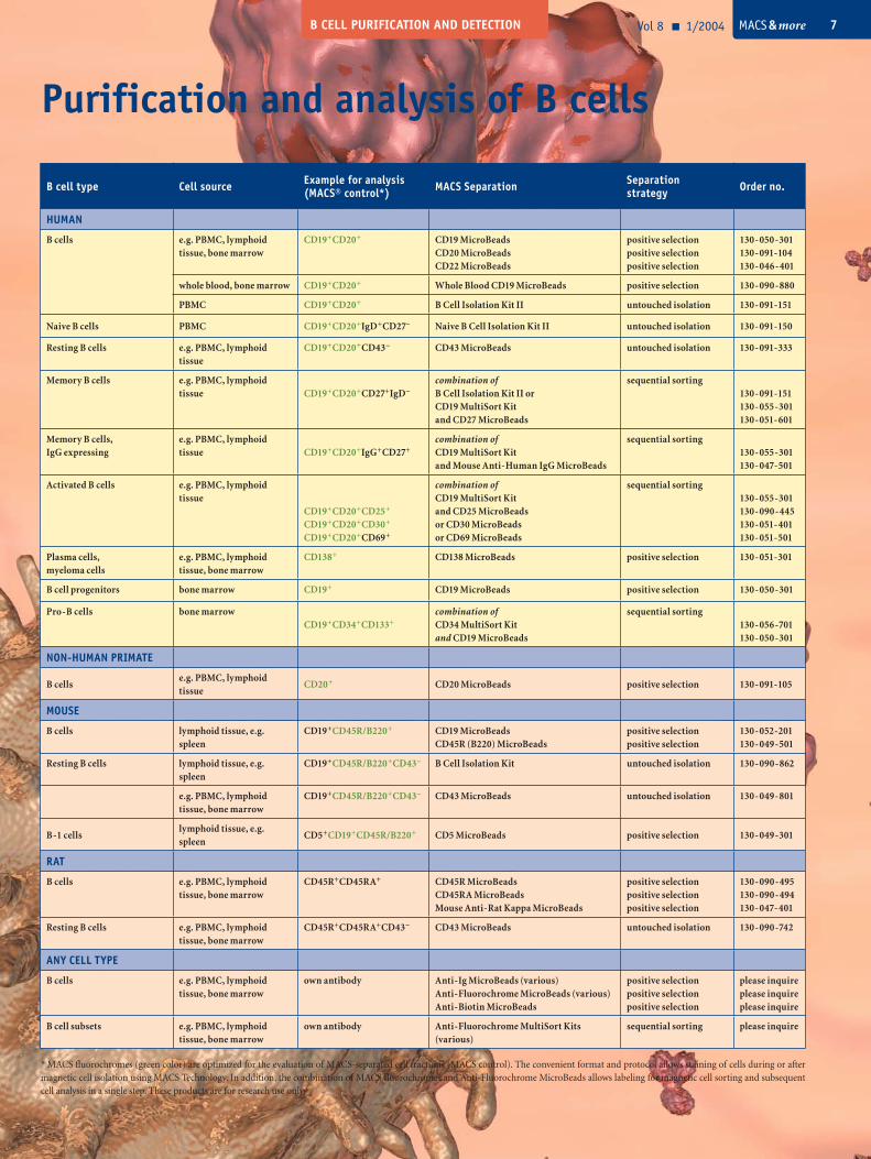
B cell type Cell source Example for analysis(MACS® control*) MACS Separation Separation
strategy Order no.
HUMAN
B cells e.g. PBMC, lymphoid tissue, bone marrow
CD19+CD20+ CD19 MicroBeadsCD20 MicroBeadsCD22 MicroBeads
positive selectionpositive selectionpositive selection
130-050-301130-091-104130-046-401
whole blood, bone marrow CD19+CD20+ Whole Blood CD19 MicroBeads positive selection 130-090-880
PBMC CD19+CD20+ B Cell Isolation Kit II untouched isolation 130-091-151
Naive B cells PBMC CD19+CD20+IgD+CD27– Naive B Cell Isolation Kit II untouched isolation 130-091-150
Resting B cells e.g. PBMC, lymphoid tissue
CD19+CD20+CD43– CD43 MicroBeads untouched isolation 130-091-333
Memory B cells e.g. PBMC, lymphoid tissue CD19+CD20+CD27+IgD–
combination of B Cell Isolation Kit II orCD19 MultiSort Kitand CD27 MicroBeads
sequential sorting130-091-151130-055-301130-051-601
Memory B cells,IgG expressing
e.g. PBMC, lymphoid tissue CD19+CD20+IgG+CD27+
combination of CD19 MultiSort Kitand Mouse Anti-Human IgG MicroBeads
sequential sorting130-055-301130-047-501
Activated B cells e.g. PBMC, lymphoid tissue
CD19+CD20+CD25+
CD19+CD20+CD30+
CD19+CD20+CD69+
combination of CD19 MultiSort Kitand CD25 MicroBeadsor CD30 MicroBeadsor CD69 MicroBeads
sequential sorting130-055-301130-090-445130-051-401130-051-501
Plasma cells,myeloma cells
e.g. PBMC, lymphoid tissue, bone marrow
CD138+ CD138 MicroBeads positive selection 130-051-301
B cell progenitors bone marrow CD19+ CD19 MicroBeads positive selection 130-050-301
Pro-B cells bone marrowCD19+CD34+CD133+
combination of CD34 MultiSort Kitand CD19 MicroBeads
sequential sorting130-056-701130-050-301
NON-HUMAN PRIMATE
B cellse.g. PBMC, lymphoid tissue
CD20+ CD20 MicroBeads positive selection 130-091-105
MOUSE
B cells lymphoid tissue, e.g. spleen
CD19+CD45R/B220+ CD19 MicroBeadsCD45R (B220) MicroBeads
positive selectionpositive selection
130-052-201130-049-501
Resting B cells lymphoid tissue, e.g. spleen
CD19+CD45R/B220+CD43– B Cell Isolation Kit untouched isolation 130-090-862
e.g. PBMC, lymphoid tissue, bone marrow
CD19+CD45R/B220+CD43– CD43 MicroBeads untouched isolation 130-049-801
B-1 cellslymphoid tissue, e.g. spleen
CD5+CD19+CD45R/B220+ CD5 MicroBeads positive selection 130-049-301
RAT
B cells e.g. PBMC, lymphoid tissue, bone marrow
CD45R+CD45RA+ CD45R MicroBeadsCD45RA MicroBeadsMouse Anti-Rat Kappa MicroBeads
positive selectionpositive selectionpositive selection
130-090-495130-090-494130-047-401
Resting B cells e.g. PBMC, lymphoid tissue, bone marrow
CD45R+CD45RA+CD43– CD43 MicroBeads untouched isolation 130-090-742
ANY CELL TYPE
B cells e.g. PBMC, lymphoid tissue, bone marrow
own antibody Anti-Ig MicroBeads (various)Anti-Fluorochrome MicroBeads (various)Anti-Biotin MicroBeads
positive selectionpositive selectionpositive selection
please inquireplease inquireplease inquire
B cell subsets e.g. PBMC, lymphoid tissue, bone marrow
own antibody Anti-Fluorochrome MultiSort Kits (various)
sequential sorting please inquire
B CELL PURIFICATION AND DETECTION
Purification and analysis of B cells
Vol 8 1/2004 MACS & more 7
* MACS fluorochromes (green color) are optimized for the evaluation of MACS-separated cell fractions (MACS control). The convenient format and protocol allows staining of cells during or after magnetic cell isolation using MACS Technology. In addition, the combination of MACS fluorochromes and Anti-Fluorochrome MicroBeads allows labeling for magnetic cell sorting and subsequent cell analysis in a single step. These products are for research use only.
NEW PRODUCTS
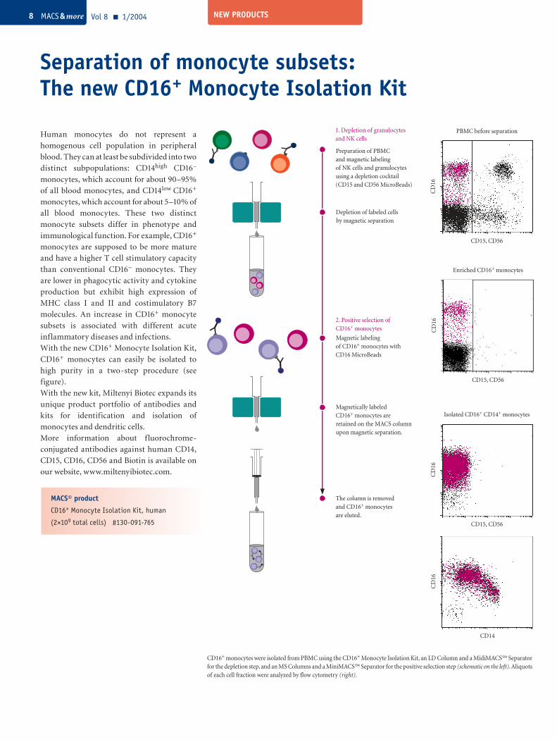
Separation of monocyte subsets: The new CD16+ Monocyte Isolation Kit
Vol 8 1/2004 MACS & more8
Magnetic labeling of CD16+ monocytes with CD16 MicroBeads
The column is removed and CD16+ monocytes are eluted.
2. Positive selection of CD16+ monocytes
Magnetically labeled CD16+ monocytes are retained on the MACS column upon magnetic separation.
Preparation of PBMC and magnetic labeling of NK cells and granulocytesusing a depletion cocktail (CD15 and CD56 MicroBeads)
Depletion of labeled cells by magnetic separation
1. Depletion of granulocytes and NK cells
CD16+ monocytes were isolated from PBMC using the CD16+ Monocyte Isolation Kit, an LD Column and a MidiMACS™ Separator for the depletion step, and an MS Columns and a MiniMACS™ Separator for the positive selection step (schematic on the left). Aliquots of each cell fraction were analyzed by flow cytometry (right).
Human monocytes do not represent a
homogenous cell population in peripheral
blood. They can at least be subdivided into two
distinct subpopulations: CD14high CD16–
monocytes, which account for about 90–95%
of all blood monocytes, and CD14low CD16+
monocytes, which account for about 5–10% of
all blood monocytes. These two distinct
monocyte subsets differ in phenotype and
immunological function. For example, CD16+
monocytes are supposed to be more mature
and have a higher T cell stimulatory capacity
than conventional CD16– monocytes. They
are lower in phagocytic activity and cytokine
production but exhibit high expression of
MHC class I and II and costimulatory B7
molecules. An increase in CD16+ monocyte
subsets is associated with different acute
inflammatory diseases and infections.
With the new CD16+ Monocyte Isolation Kit,
CD16+ monocytes can easily be isolated to
high purity in a two-step procedure (see
figure).
With the new kit, Miltenyi Biotec expands its
unique product portfolio of antibodies and
kits for identification and isolation of
monocytes and dendritic cells.
More information about f luorochrome-
conjugated antibodies against human CD14,
CD15, CD16, CD56 and Biotin is available on
our website, www.miltenyibiotec.com.
CD15, CD56
PBMC before separation
CD
16C
D16
CD15, CD56
Enriched CD16+ monocytes
CD
16
CD15, CD56
Isolated CD16+ CD14+ monocytes
CD
16
CD14
MACS® product
CD16+ Monocyte Isolation Kit, human
(2×10⁹ total cells) #130-091-765
NEW PRODUCTS
FASER Kits – Beam up your cells!
MACS® productsFASER Kit – FITC #130-091-763
FASER Kit – PE #130-091-764
FASER Kit – APC #130-091-762
Vol 8 1/2004 MACS & more 9
Weak fluorescence signals?Difficult-to-define cell populations?Beam up your cells !With the new FASER Kits, f luorescence
intensity of cells labeled with virtually any
FITC-, PE- or APC-conjugated antibody can
be amplified. The Kits are suitable for fresh or
formaldehyde-fixed cells in suspension of any
type and species. Analysis is performed by flow
cytometry. The higher sensitivity provided by
FASER Kits makes f low cytometric analysis
easy and more reliable.
Applications• for amplification of fluorescence signals
caused by weakly expressed antigens, low
antibody affinity, or immunomagnetic and
immunofluorescent labeling of one target
epitope,
• for more clearly defined cell populations,
• for amplification of magnetic labeling in
combination with MACS® Anti-FITC, -PE,
or -APC MicroBeads.
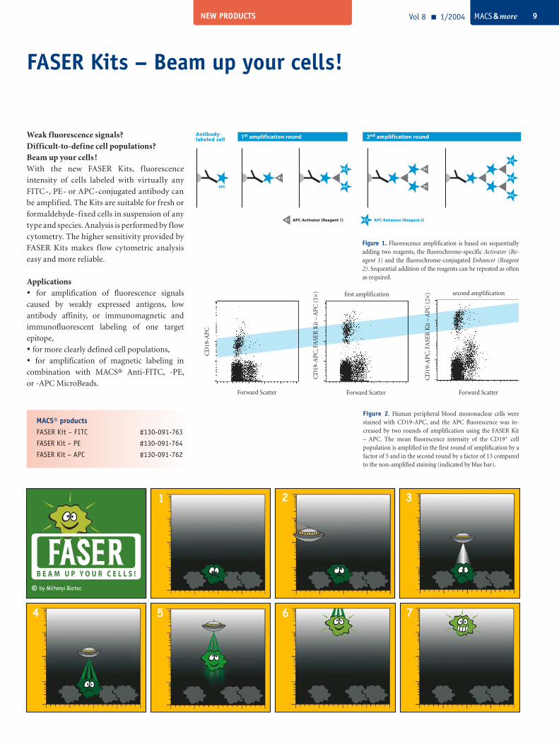
Figure 2. Human peripheral blood mononuclear cells were stained with CD19-APC, and the APC fluorescence was in-creased by two rounds of amplification using the FASER Kit – APC. The mean fluorescence intensity of the CD19+ cell population is amplified in the first round of amplification by a factor of 5 and in the second round by a factor of 13 compared to the non-amplified staining (indicated by blue bar).
Forward Scatter
CD
19-A
PC, F
ASE
R K
it –
APC
(1×
)
Forward Scatter
CD
19-A
PC, F
ASE
R K
it –
APC
(2×
)
Figure 1. Fluorescence amplification is based on sequentially adding two reagents, the fluorochrome-specific Activator (Re-agent 1) and the fluorochrome-conjugated Enhancer (Reagent 2). Sequential addition of the reagents can be repeated as often as required.
Forward Scatter
CD
19-A
PC
1 2 3
4 5 6 7
© by Miltenyi Biotec
R2 APC-Enhancer (Reagent 2)R1 APC-Activator (Reagent 1)
1st amplification round 2nd amplification round
R2
R2
R2
R1
R1APC
R1
R2
R2
Antibody- labeled cell
first amplification second amplification
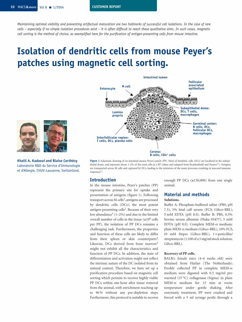
IntroductionIn the mouse intestine, Peyer’s patches (PP)
represent the primary site for uptake and
presentation of antigens (figure 1). Following
transport across M cells1, antigens are processed
by dendritic cells (DCs), the most potent
antigen-presenting cells2. Because of their very
low abundance3 (1–2%) and due to the limited
overall number of cells in the tissue (≥106 cells
per PP), the isolation of PP DCs remains a
challenging task. Furthermore, the properties
and function of these cells are likely to differ
from their spleen or skin counterparts4.
Likewise, DCs derived from bone marrow5
might not exhibit all the characteristics and
function of PP DCs. In addition, the state of
differentiation and activation might not reflect
the intrinsic nature of the DC isolated from its
natural context. Therefore, we have set up a
purification procedure based on magnetic cell
sorting which permits to recover highly viable
PP DCs within one hour after tissue removal
from the animal, with enrichment reaching up
to 96 % without any pre-depletion steps.
Furthermore, this protocol is suitable to recover
Maintaining optimal viability and preventing artifactual maturation are two hallmarks of successful cell isolations. In the case of rare cells – especially if no simple isolation procedures exist – it is often difficult to reach these qualitative aims. In such cases, magnetic cell sorting is the method of choice, as exemplified here for the purification of antigen-presenting cells from mouse intestine.
Isolation of dendritic cells from mouse Peyer’s patches using magnetic cell sorting.
Khalil A. Kadaoui and Blaise Corthésy Laboratoire R&D du Service d’Immunologie et d’Allergie, CHUV-Lausanne, Switzerland.
enough PP DCs (≥150,000) from one single
animal.
Material and methodsSolutions. Buffer A: Phosphate-buffered saline (PBS; pH
7.3), 5% fetal calf serum (FCS; Gibco-BRL),
5 mM EDTA (pH 8.0). Buffer B: PBS, 0.5%
bovine serum albumin (Fluka 05477), 5 mM
EDTA (pH 8.0). Complete MEM-α medium:
plain MEM-α medium (Gibco-BRL), 10% FCS,
10 mM Hepes (Gibco-BRL), 1 × penicillin/
streptomycin (1:100 of a 5 mg/ml stock solution;
Gibco-BRL).
Recovery of PP cells. BALB/c female mice (4–6 weeks old) were
obtained from Harlan (The Netherlands).
Freshly collected PP in complete MEM-α
medium were digested with 0.5 mg/ml pre-
warmed (37 °C) collagenase (Sigma) in plain
MEM-α medium for 15 min at room
temperature under gentle shaking. After
enzymatic treatment, PP were crushed and
forced with a 5 ml syringe pestle through a
CUSTOMER REPORT
Figure 1 Schematic drawing of an intestinal mouse Peyer’s patch (PP). Most of dendritic cells (DCs) are localized in the subepi-thelial dome, and represent about 1–2% of the total cells in a PP (taken and adapted from Kraehenbuhl and Neutra11). Antigens are transported across M cells and captured by DCs, leading to the initiation of the many processes resulting in mucosal immune responses12.
Interfollicular region:T cells, DCs, plasma cells
Corona:B cells, CD4+ cells
Germinal center:B cells, DCs,follicular DCs,macrophages
Subepithelial dome:DCs, T cells,macrophages
Follicularassociatedepithelium
Intestinal lumen
M cellEnterocyte
Laminapropria
Vol 8 1/2004 MACS & more10
70 µm-pore nylon mesh cell strainer (Falcon;
BD Biosciences). 5×2 ml of complete MEM-α medium were used to guarantee the passage of
most cells through the strainer. Cells were then
passed through a 40 µm-pore nylon mesh cell
strainer (Falcon; BD Biosciences) with two
subsequent washings with 2×2.5 ml of complete
MEM-α medium. Cells were recovered by
centrifugation for 5 min at 400×g at 4 °C. The
supernatant was discarded, and the pellet was
suspended in cold buffer A at a concentration of
5×10⁶ cells/100 µl. To avoid non-specific
labeling, Fcγ II/III receptors were blocked using
rat anti-mouse CD16/CD32 antibodies (Ab) (1:
100 dilution; Pharmingen) for 15 min at 4 °C.
The excess of blocking Ab was washed away
using buffer A, and the cells were pelleted by
centrifugation for 5 min at 400×g at 4 °C prior
to suspension in 100 µl of buffer A.
Magnetic separation. 10–25×106 cells were incubated with MACS®
CD11c MicroBeads (Miltenyi Biotec), using
10 µl beads per 107 cells. The tube containing
the mixture of cells and beads was kept on ice
for 15 min, with gentle tapping performed every
5 min. In the meantime, an MS Separation
Column (Miltenyi Biotec) was mounted on a
magnet, and equilibrated with 500 µl of buffer
A (one MS column fits up to 2×10⁸ total cells).
The mixture of cells and beads was then applied
to the column, the non-binding cells were
allowed to flow through, and the column was
washed twice with 500 µl of buffer B. To recover
bound cells, the column was removed from the
magnet and placed on a 5 ml polystyrene round-
bottom tube (Falcon; BD Biosciences). Elution
was carried out with 500 µl of buffer B using the
plunger supplied with the column. To ensure
enrichment values shown in figure 3A–C (see
Results and Discussion), the magnetic
separation had to be repeated using a second
column under the same conditions with the
exception that the column was washed twice
with 250 µl of buffer B instead of 500 µl as
indicated just above. A schematic representation
of the procedure is depicted in figure 2.
CUSTOMER REPORT
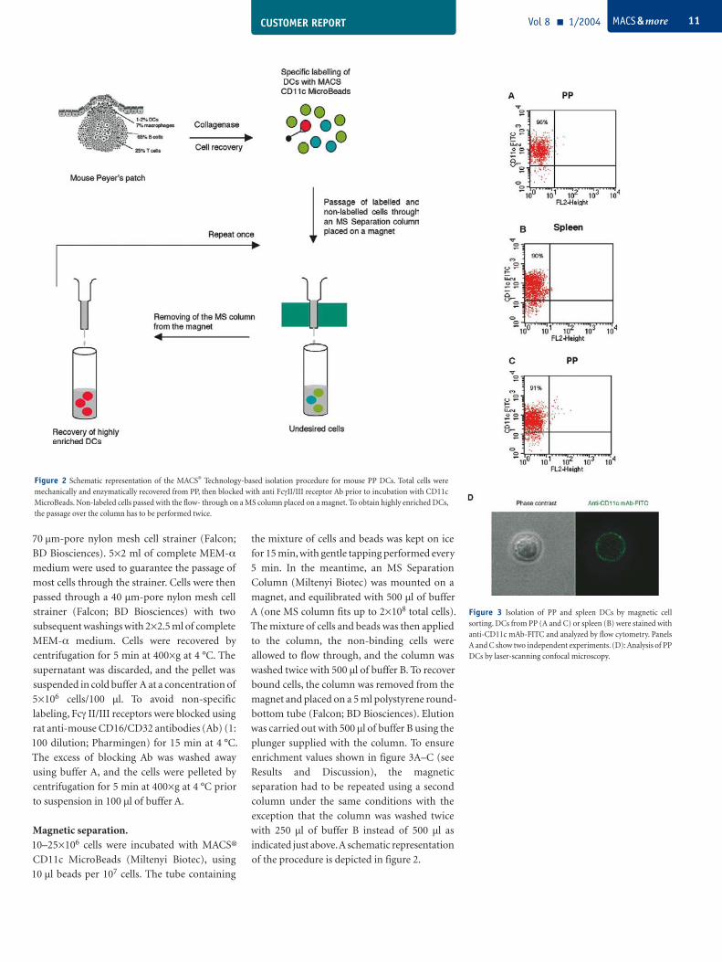
Figure 2 Schematic representation of the MACS® Technology-based isolation procedure for mouse PP DCs. Total cells were mechanically and enzymatically recovered from PP, then blocked with anti FcγII/III receptor Ab prior to incubation with CD11c MicroBeads. Non-labeled cells passed with the flow- through on a MS column placed on a magnet. To obtain highly enriched DCs, the passage over the column has to be performed twice.
Vol 8 1/2004 MACS & more 11
Figure 3 Isolation of PP and spleen DCs by magnetic cell sorting. DCs from PP (A and C) or spleen (B) were stained with anti-CD11c mAb-FITC and analyzed by flow cytometry. Panels A and C show two independent experiments. (D): Analysis of PP DCs by laser-scanning confocal microscopy.
CUSTOMER REPORT
Results and DiscussionIn order to prevent artifactual maturation and
ensure optimal viability, procedures to isolate
cells must guarantee appropriate yield and
speed. This holds true for DCs, and particularly
for PP DCs whose abundance is very low, and
for which no simple isolation procedure exists.
The tissue from which the cells originate
represents a sensitive issue as it is highly likely
that PP DCs exhibit functional properties and
degree of maturation different to those from
spleen for instance7.
We have thus developed a two-step protocol to
isolate DCs from PP using magnetic cell sorting.
The isolation procedure relies on incubation
with CD11c MicroBeads, and sorting using two
successive MS columns. When analyzed by flow
cytometry on the basis of CD11c expression, we
demonstrate that the purity of sorted DCs
reached up to 96% (figure 3A). The high purity
obtained is close to the enrichment we and
others could obtain using fluorescence-
activated cell sorting8,9, yet the FACS procedure
is burdensome, often based on negative
selection, and prone to trigger DC activation. In
addition, the protocol described herein is
adapted to the isolation from one single mouse,
and provides the investigator with as many as
150,000 cells for pilot experiments. Similar
mean percentages of enrichment were achieved
when the procedure was applied to spleen (90%,
figure 3B) used as a control in comparison with
91% for PP (figure 3C). Interestingly, purified
DCs can be easily stained with anti-CD11c
fluorescent mAb to permit visualization by
confocal microscopy (figure 3D). Ongoing
experiments in the laboratory indicate that PP
DCs are suitable to study the possible
internalization of antigens and track internal
processing ex vivo.
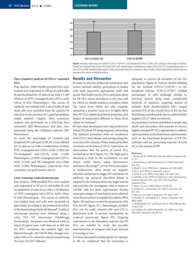
For those interested in studying the DC subtype
in PP, we confirmed that the procedure is
adequate to recover all members of the DC
population (figure 4). Indeed, double-labeling
for the myeloid (CD11c+/CD11b+) or the
lymphoid subtype (CD11c+/CD8+) yielded
percentages of cells strikingly similar to
previous reports using more complicated
methods of isolation, requiring dozens of
animals. Both double-labeled DCs ranged
around 35% of the overall DCs in PP, the last
third being contributed by the so-called double-
negative DCs10 (data not shown).
In conclusion, we have established a simple, fast,
small-scale procedure that permits to recover
highly enriched PP DCs appropriate to address
open questions in the field of mucosal immunity
such as the functional properties of PP DC
subtypes and the processing capacity of such
DC in the context of PP.
References1. Neutra, M. R. (1999) Curr. Top. Microbiol. Immunol. 236: 17–32.2. Banchereau, J., and Steinman, R. M. (1998) Nature 392: 245–252.3. Ruedl, C. et al. (1996) Eur. J. Immunol. 26: 1801–1806.4. Rey, J. et al. (2004) J. Immunol., in press.5. Inaba, K. et al. (1992) J. Exp. Med. 176: 1693–1702.6. Griffiths, G. M. (1997) in Lefkovits, I. (ed.): Immunology Methods Manual, Vol. 3, pp. 1251–1257. San Diego, Academic Press.7. Mora, J. R. et al. (2003) Nature 424: 88–93.8. Kelsall, B. L., and Strober, W. (1996) J. Exp. Med. 183: 237–247.9. Sato, A. et al. (2003) J. Immunol. 171: 3684–3690. 10. Iwasaki, A., and Kelsall, B. L. (2001) J. Immunol. 166: 4884–4890.11. Kraehenbuhl, J. P., and Neutra, M. R. (1992) Physiol. Rev. 72: 853–879.12. Corthésy, B., and Kraehenbuhl, J. P. (1999) Curr. Top. Microbiol. Immunol. 236: 93–111.
Figure 4 Surface phenotype of myeloid (CD11c+/CD11b+) and lymphoid (CD11c+/CD8+) DC subtypes. Percentage of double-labeled DCs isolated from mouse PP. Sorted DCs were stained with a combination of anti-CD11c/CD11b (myeloid phenotype) or anti-CD11c/CD8 (lymphoid phenotype) mAbs, and analyzed by flow cytometry. Mean results of four independent experiments are presented. Bars show standard deviation.
Vol 8 1/2004 MACS & more12
Flow cytometry analysis of CD11c+-enriched DCs. Post-elution, 5,000–30,000 purified DCs were
washed and suspended in 200 µl of cold buffer
B, and incubated for 25 min on ice with a 1:100
dilution of FITC-conjugated anti-CD11c mAb
(clone N-418; Pharmingen). The excess of
antibody was washed with 1 ml of buffer B and
dead cells were excluded from the analysis by
selection in the presence of 1 µg/ml propidium
iodide solution (Sigma). Flow cytometry
analysis was performed on a FACScan flow
cytometer (BD Biosciences) and data were
processed using the CellQuest software (BD
Biosciences).
To track the percentage of myeloid and
lymphoid DC subtypes in PP, DCs were labeled
for 25 min on ice with a combination of either
FITC-conjugated anti-CD11c mAb (1:100) and
PE-conjugated anti-CD11b mAb (1:200;
Pharmingen), or FITC-conjugated anti-CD11c
mAb (1:100) and PE-conjugated anti-CD8α
mAb (1:200; Pharmingen), respectively. Flow
cytometry was performed as above.
Laser-scanning confocal microscopy. Post-elution, 5,000 purified DCs were washed
and suspended in 50 µl of cold buffer B, and
incubated for 25 min on ice with a 1:50 dilution
of FITC-conjugated anti-CD11c mAb (clone
N-418; Pharmingen). The excess of antibody
was washed away, and cells were mounted on
glass slides according to the protocol described
in the Immunology Methods Manual6. Confocal
microscopy pictures were obtained using a
Leica TCS NT microscope (Glattbrugg,
Switzerland). Excitation was obtained with an
Argon-Krypton laser, with lines set at 488 nm
for FITC excitation, the emitted light was
filtered through a BF 530/30 filter. Images were
taken with a 63× objective and processed using
the Leica TCS NT software.
NEW
CUSTOMER REPORT Vol 8 1/2004 MACS & more 13
In mice as well as in humans, dendritic cells
(DC) do not represent a homogenous cell
population but rather a mixture of distinct DC
subsets differing in function and phenotype.
In mouse spleen, CD8a+ CD11b– DC were
originally distinguished from CD8– CD11b+
DC. Later, splenic CD8– CD11b+ DC were
further subdivided into a CD4+ and CD4–
subset (Shortman, 2000). All these splenic DC
populations express MHC II and high levels of
CD11c. More recently, murine plasmacytoid
DC (mPDC) have been identified in mouse
lymphoid organs as CD45R (B220)+, Ly-6C+,
CD11c+, CD8a+/–, CD11b– cells (Asselin-
Paturel, C. et al., 2001). Like their human
counterpart, murine plasmacytoid DC exhibit
a plasmacytoid morphology and are able to
produce large amounts of type I interferon
(IFN-α and IFN-β) in response to bacterial
DNA containing particular unmethylated CpG
motifs (CpG-DNA) or upon viral challenge. In
humans, PDC have been shown to specifically
express BDCA-2 and BDCA-4 (Dzionek et
al., 2000). In mouse, no such specific markers
are available to date. New markers specific
for mPDC would, however, be of great
benefit, in order to monitor, characterize and
isolate mPDC and also to study their in vivo
function by mAb-mediated in vivo depletion
of mPDC. We have generated a panel of
monoclonal antibodies (mAb) recognizing a
single, presumably novel antigen, which we
have termed murine plasmacytoid dendritic
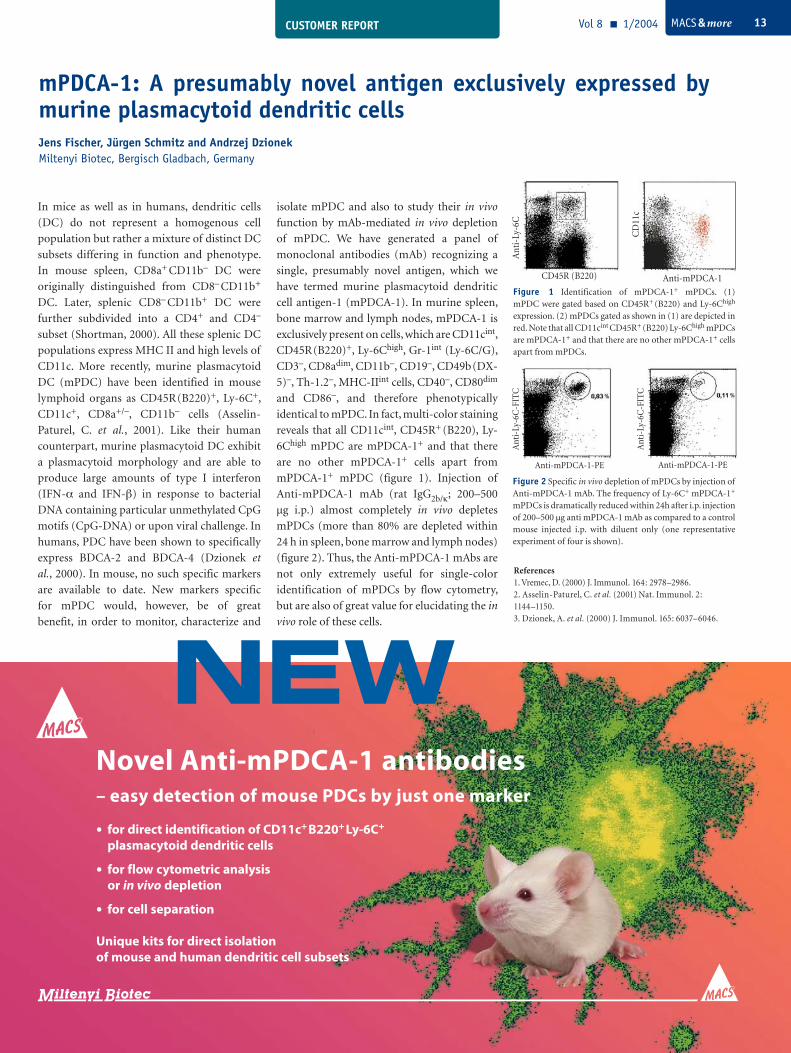
cell antigen-1 (mPDCA-1). In murine spleen,
bone marrow and lymph nodes, mPDCA-1 is
exclusively present on cells, which are CD11cint,
CD45R (B220)+, Ly-6Chigh, Gr-1int (Ly-6C/G),
CD3–, CD8adim, CD11b–, CD19–, CD49b (DX-
5)–, Th-1.2–, MHC-IIint cells, CD40–, CD80dim
and CD86–, and therefore phenotypically
identical to mPDC. In fact, multi-color staining
reveals that all CD11cint, CD45R+ (B220), Ly-
6Chigh mPDC are mPDCA-1+ and that there
are no other mPDCA-1+ cells apart from
mPDCA-1+ mPDC (figure 1). Injection of
Anti-mPDCA-1 mAb (rat IgG2b/κ; 200–500
µg i.p.) almost completely in vivo depletes
mPDCs (more than 80% are depleted within
24 h in spleen, bone marrow and lymph nodes)
(figure 2). Thus, the Anti-mPDCA-1 mAbs are
not only extremely useful for single-color
identification of mPDCs by flow cytometry,
but are also of great value for elucidating the in
vivo role of these cells.
References1. Vremec, D. (2000) J. Immunol. 164: 2978–2986.2. Asselin-Paturel, C. et al. (2001) Nat. Immunol. 2: 1144–1150.3. Dzionek, A. et al. (2000) J. Immunol. 165: 6037–6046.
Novel Anti-mPDCA-1 antibodies– easy detection of mouse PDCs by just one marker
· for direct identification of CD11c+ B220+ Ly-6C+
plasmacytoid dendritic cells
· for flow cytometric analysis or in vivo depletion
· for cell separation
Unique kits for direct isolation of mouse and human dendritic cell subsets
mPDCA-1: A presumably novel antigen exclusively expressed by murine plasmacytoid dendritic cells Jens Fischer, Jürgen Schmitz and Andrzej DzionekMiltenyi Biotec, Bergisch Gladbach, Germany
Figure 2 Specific in vivo depletion of mPDCs by injection of Anti-mPDCA-1 mAb. The frequency of Ly-6C+ mPDCA-1+ mPDCs is dramatically reduced within 24h after i.p. injection of 200–500 µg anti mPDCA-1 mAb as compared to a control mouse injected i.p. with diluent only (one representative experiment of four is shown).
Figure 1 Identification of mPDCA-1+ mPDCs. (1) mPDC were gated based on CD45R+ (B220) and Ly-6Chigh expression. (2) mPDCs gated as shown in (1) are depicted in red. Note that all CD11cint CD45R+ (B220) Ly-6Chigh mPDCs are mPDCA-1+ and that there are no other mPDCA-1+ cells apart from mPDCs.
Anti-mPDCA-1-PE
Ant
i-Ly
-6C
-FIT
C
Anti-mPDCA-1-PE
Ant
i-Ly
-6C
-FIT
C
CD45R (B220) Anti-mPDCA-1
Ant
i-Ly
-6C
CD
11c
CUSTOMER REPORTVol 8 1/2004 MACS & more14
murine bone marrow2 and high-level multi-
lineage repopulation in NOD/SCID and
serially transplanted B2mnull NOD/SCID
mice transplanted with MACS-enriched
human CD34+ stem and progenitor cells,
isolated from cord blood, adult bone marrow
and G-CSF induced mobilized peripheral
blood from healthy donors3. These
interactions, mediated activation of the major
adhesion molecules expressed by immature
human CD34+/CXCR4+ cells, leading to firm
adhesion to their ligands expressed by SDF-1+
human endothelial cells, under shear flow in
vitro, mimicking in vivo interactions between
transplanted human progenitors migrating in
the blood circulation and the bone marrow
endothelium, which is an essential first step in
the multi-step homing and repopulation
process4.
In addition, SDF-1 mediated activation of the
major adhesion molecules LFA-1, VLA-4,
VLA-5 5 and CD44 on MACS-enriched human
CD34+ /CXCR4+ cells which are essential for
human stem and progenitor cell migration and
in vivo homing, retention and repopulation of
the BM in transplanted NOD/SCID mice.
MACS-enriched human cord blood CD34+
cells which do not express CXCR4 by cell
sorting harbor low levels of intracellular
CXCR4 which can rapidly oscillate in vitro and
in vivo and mediate SDF-1-dependent homing
and repopulation in vivo6. Short-term in vitro
stimulation with the cytokines SCF and IL-6
for 24–48 hr increase surface CXCR4
expression, in vitro directional migration in
response to a gradient of SDF-1, and in vivo
homing and repopulation2,3,6.
Since all clinically transplanted patients have
only low levels of immature long-term culture-
initiating cells (LTC-IC), increasing CXCR4
expression before transplantation and/or
increasing levels of SDF-1 in the bone marrow1
could improve the outcome of clinical stem
In their review, Lapidot and Kollet discuss the properties of transplanted CD34+ stem cells and the action of various factors mediating their activity, especially Stromal Derived Factor 1 and its receptor, CXCR4.
Mechanism of human CD34+ stem cell homing and mobilization in transplanted immune-deficient NOD/SCID mice.
Tsvee Lapidot and Orit KolletDept. of Immunology, The Weizmann Institute, Rehovot, Israel.
Transplanted human CD34+ stem cells migrate
via the circulation, home to and engraft the
host bone marrow (BM) and continuously
release maturing blood cells into the
circulation. This process is essential for
successful clinical transplantations of blood
forming stem cells. In addition, G-CSF-
induced CD34+ stem cell mobilization from
the BM into the blood circulation is the major
source of stem cells for transplantation.
During late embryonic development hema-
topoietic stem cells migrate from the liver to
the BM. Murine embryos which lack the
chemokine Stromal Derived Factor 1 (SDF-1)
or its receptor CXCR4 have multiple lethal
defects, including lack of bone marrow
hematopoiesis, suggesting a major role for
SDF-1/CXCR4 interactions during stem cell
homing and/or BM engraftment.
SDF-1 is highly expressed in human bone
marrow endothelium and the stem cell-rich
endosteum region1. CXCR4 is expressed by
many cell types, including primitive human
CD34+/CD38– and murine Sca-1+/c-kit+/lin–
stem cells. Human and murine SDF-1 differ in
one amino acid and are cross reactive, enabling
murine SDF-1 in the BM of immune-deficient
NOD/SCID mice transplanted with enriched,
human CD34+ progenitors, to signal via
CXCR4. We demonstrated that SDF-1/CXCR4
interactions are essential for homing to the
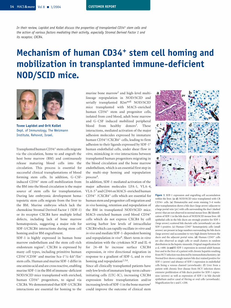
Figure 1 SDF-1 expression and engrafting cell accumulation within the liver. (a–d) NOD/SCID mice transplanted with CB CD34+ cells. (a) Hematoxylin and eosin staining 5–6 weeks after transplantation shows a bile duct (large arrow) adjacent to a large portal vein (pv) with cells surrounding the duct (dashed arrow) that are not observed in normal mouse liver. (b) Identifi-cation of SDF-1 in the bile ducts of NOD/SCID mouse liver. All epithelial cells in the bile ducts are strongly positive for SDF-1 (large arrow); scattered bile ductule cells (arrowheads) are also SDF-1-positive. (c) Human CD45+ hematopoietic cells (small arrows) are present in large numbers surrounding the bile ducts (large arrows) and accumulate to very high density between the ducts and the adjacent portal veins. (d) Human CD45+ cells are also observed as single cells or small clusters in random distribution in the hepatic sinusoids. Original magnification for a–d, ×400. (e and f) SDF-1 expression in normal adult human liver and in the liver of a patient with chronic hepatitis resulting from HCV infection was detected by immunohistochemistry. (e) Normal liver shows a single mature bile duct stained positive for SDF-1 (arrow) and absence of SDF-1 expression in endothelial cells lining a venous channel (arrowheads). (f) Liver from a patient with chronic liver disease from HCV infection shows extensive proliferation of bile ducts positive for SDF-1 expres-sion (arrows) as well as expression of SDF-1 in bile ductule epithelium and/or canal of Hering or oval cells (arrowheads). Magnification for e and f, ×200.
CCUSTOMER REPORT Vol 8 1/2004 MACS & more 15
cell transplantation and SDF-1/CXCR4
interactions could also be used to navigate cells
in vivo for a wide variety of clinical protocols.
Stem cell mobilization in response to daily
stimulations with G-CSF mimics stress-
induced release and recruitment of stem and
progenitor cells from the bone marrow
reservoir in response to stress signals during
inflammation and injury.
This process involves a transient increase in
SDF-1 levels within the bone marrow after
each injection which is followed by a profound
decrease, mediated by activated myeloid cells
such as neutrophils and osteoclasts which
secrete the proteolytic enzymes elastase,
cathepsin G, CD26 and MMP-2/9 reaching
lowest levels during the time of mobilization.
In parallel, after each injection there is a slight
decrease of surface CXCR4 on BM
hematopoietic cells including CD34 cells,
followed by an increase, reaching peak levels of
surface CXCR4 expression during the
mobilization period, demonstrating for
CXCR4 signaling during stress induced stem
and progenitor cell egress7,8. Recruitment of
MACS-enriched immature human cord blood
or mobilized PBL CD34+ cells to the irradiated
NOD/SCID mouse liver or to the inflamed
human liver of transplanted patients due to
infection with hepatitis virus also involve
increase in SDF-1 levels in this organ and a
shift in membrane-bound SDF-1. Liver injury
in previously transplanted NOD/SCID mice
leads to stress-induced recruitment of human
stem and progenitor cells also by shifting
surface SDF-1 expression, to the release of the
metalloprotei-nases MMP-2 and MMP-9
which increase surface CXCR4 expression on
human progenitors in the murine BM of
established chimeras, and to the release of
cytokines such as HGF, SCF and IL-6 which
also increase surface CXCR4 expression and
SDF-1-mediated directional migration of
human progenitors which are recruited to the
damaged liver as part of organ repair9.
In summary, human CD34+ stem and proge-
nitor cells migrate in response to stress signals
which are mediated by an interplay between
cytokines, chemokines, proteolytic enzymes
and adhesion molecules, demonstrating a
central role for SDF-1/CXCR4 interactions in
both stem cell homing to the bone marrow and
stress-induced mobilization, or recruitment
to the irradiated or injured liver.
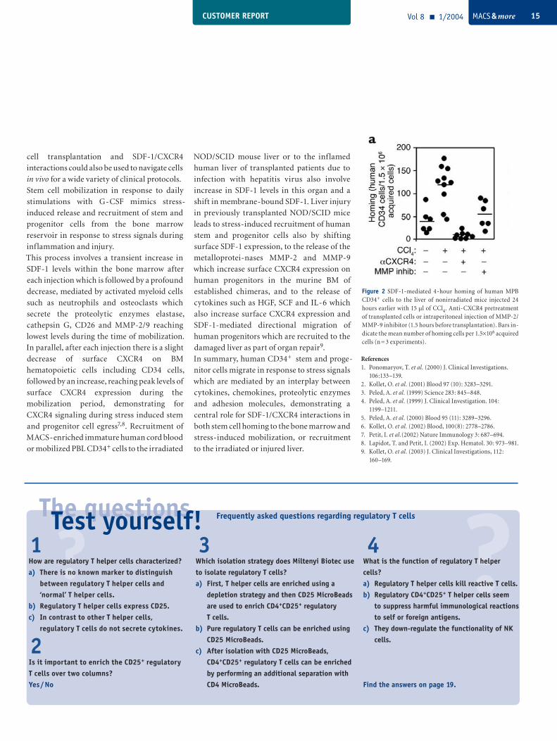
Figure 2: SDF-1 Four-hour homing of human MPB CD34+ cells to the liver of nonirradiated mice injected 24 hours earlier with 15 µl of CCl4. Anti-CXCR4 pretreatment of transplanted cells or intraperitoneal injection of MMP-2/MMP-9 inhibitor (1.5 hours before transplantation). Bars indicate the mean number of homing cells per 1.5×10⁶ acquired cells (n = 3 experiments).
References1. Ponomaryov, T. et al. (2000) J. Clinical Investigations. 106:133–139.2. Kollet, O. et al. (2001) Blood 97 (10): 3283–3291.3. Peled, A. et al. (1999) Science 283: 845–848.4. Peled, A. et al. (1999) J. Clinical Investigation. 104: 1199–1211.5. Peled, A. et al. (2000) Blood 95 (11): 3289–3296.6. Kollet, O. et al. (2002) Blood, 100(8): 2778–2786.7. Petit, I. et al.(2002) Nature Immunology 3: 687–694.8. Lapidot, T. and Petit, I. (2002) Exp. Hematol. 30: 973–981.9. Kollet, O. et al. (2003) J. Clinical Investigations, 112: 160–169.
?The questions
1 3 4 ?How are regulatory T helper cells characterized?a) There is no known marker to distinguish between regulatory T helper cells and ‘normal’ T helper cells.b) Regulatory T helper cells express CD25. c) In contrast to other T helper cells, regulatory T cells do not secrete cytokines.
Test yourself!Which isolation strategy does Miltenyi Biotec use to isolate regulatory T cells?a) First, T helper cells are enriched using a depletion strategy and then CD25 MicroBeads are used to enrich CD4+CD25+ regulatory T cells.b) Pure regulatory T cells can be enriched using CD25 MicroBeads.c) After isolation with CD25 MicroBeads, CD4+CD25+ regulatory T cells can be enriched by performing an additional separation with CD4 MicroBeads.
Frequently asked questions regarding regulatory T cells
What is the function of regulatory T helper cells?a) Regulatory T helper cells kill reactive T cells.b) Regulatory CD4+CD25+ T helper cells seem to suppress harmful immunological reactions to self or foreign antigens.c) They down-regulate the functionality of NK cells.
Find the answers on page 19.
2Is it important to enrich the CD25+ regulatory T cells over two columns?Yes / No
Figure 2 SDF-1-mediated 4-hour homing of human MPB CD34+ cells to the liver of nonirradiated mice injected 24 hours earlier with 15 µl of CCl4. Anti-CXCR4 pretreatment of transplanted cells or intraperitoneal injection of MMP-2/MMP-9 inhibitor (1.5 hours before transplantation). Bars in-dicate the mean number of homing cells per 1.5×10⁶ acquired cells (n = 3 experiments).
CUSTOMER REPORTVol 8 1/2004 MACS & more16
Application of μMACS™ Streptavidin MicroBeads for the analysis of HIV-1 directly from patient plasma
L. Davis Lupo and Salvatore T. ButeraHIV and Retrovirology Branch, Division of AIDS, STD, and TB Laboratory Research, National Center for HIV, STD, and TB Prevention, Centers for Disease Control and Prevention, Atlanta, Georgia, USA
When cells are infected by viruses and are forced to synthesize viral progeny, the budding particles take with them components of the host cell membrane. It is therefore possible to identify susceptible cell types by purification using appropriate markers. With MACS Technology, this can be achieved directly – without prior blood plasma processing.
Introduction
It is established that host membrane proteins
are incorporated into the retroviral envelope as
HIV-1 virions bud from human cells (for a
review see Tremblay et al.1). Direct or indirect
mechanisms of host protein inclusion or
exclusion from the retroviral envelope may be
involved. This fact has permitted the ability to
discriminate host cell types supporting viral
replication by a targeted capture of virions
directly from HIV-infected patient plasma via a
previously described method2–5. However, this
method required extensive processing of patient
plasma to permit immunomagnetic capture of
HIV-1 with high efficiency. In this report, we
now describe marked improvements in the
immunomagnetic capture protocol that permits
analysis of virions directly from patient plasma
without prior processing. These improvements
not only allow direct analysis of patient plasma
but they also open new approaches to the
characterization of virions from HIV-infected
individuals.
Materials and MethodsPreparation of samples. Laboratory stocks of HIV-1 (strain Ba-L,
subtype B), grown in either purified
macrophages or CD4+ T lymphocytes, were
spiked into tissue culture medium or plasma at
known viral particle numbers2. Either a
commercial source of normal human plasma or
a subtype B HIV-1-infected patient plasma with
high anti-HIV antibody titers but undetectable
viral load was used. Where indicated, HIV-1
spiked into specific antibody-containing plasma
was processed by ultracentrifugation, salt
dissociation, and spin column filtration, as
described2–5. HIV-1-infected plasma samples
with detectable viral loads were obtained from
patients enrolled in a longitudinal TB therapy
cohort under study in Kampala, Uganda, via
collaboration with Dr. Zahra Toossi at Case
Western Reserve University (Cleveland, Ohio).
Appropriate informed consent was obtained by
the Case Western Reserve Makerere Research
Collaboration.
Immunomagnetic capture of HIV-1.HIV-1 spiked into medium or plasma
(processed or unprocessed) was first incubated
with 0.5 μg of various antibodies (all obtained
biotinylated from commercial sources) for 30
min at room temperature. To each sample, 20 µl
µMACS™ Streptavidin MicroBeads (Miltenyi
Biotec, Auburn, CA) were added and the
binding reaction was incubated an additional 10
min at room temperature. Antibody-bound
virus was then captured by magnetic separation
with slight modifications from the
manufacturer’s protocols. Briefly, μ columns
were placed in the µMACS™ magnetic separator
attached to a MultiStand under a biological
safety cabinet. Each column was prepared by
prewetting with 100 µl protein equilibration
buffer and rinsing twice with 100 µl PBS
containing 2% fetal bovine serum (PBS/FBS).
The entire volume of the virus capture reaction
mixture (approximately 200 μl) was then
applied to the column and allowed to drain
completely. The columns were washed four
times with 200 µl volumes of PBS/FBS. Fifty
microliters of an appropriate lysis buffer (see
below) were then added to the column, allowed
to stand for 5 minutes at room temperature, and
then followed by an additional 150 μl of the
same buffer. All lysate elution fractions were
collected as a pool. When capture efficiencies
using MACS™ Technology were compared to
the previous protocol using Dynal magnetic
beads (Great Neck, NY), 2×10⁷ streptavidin-
conjugated Dynabeads (M-280) were used and
the capture reactions were performed as
previously described2–5.
Analysis of captured virus.For quantitation of captured HIV-1, two
commercially available kits were used and assays
were performed as directed. For the HIV-1 p24
antigen enzyme immunoassay (Coulter/
Immunotech Inc., Westbrook, Maine), the lysis
buffer consisted of PBS containing 0.5% NP-40.
Whereas, for the more sensitive HIV-1 RNA
quantitation assay, the lysis buffer was obtained
from the commercial source of the NucliSens
QT viral load assay (Organon Teknika/
CUSTOMER REPORT Vol 8 1/2004 MACS & more 17
bioMérieux, Inc., Durham, NC). Viral RNA
isolation and RT-PCR quantitation were
performed using manufacturer’s reagents and
protocols. In some experiments, intact captured
virus was recovered by removing the column
from the magnetic separator and eluting with
complete tissue culture medium. The infectivity
of captured virus was then determined by direct
inoculation onto purified CD4+ T lymphocyte
cultures that had been stimulated with PHA for
3 days. Cultures were fed with fresh media on
day 7 and HIV-1 replication was monitored in
cell-free culture supernatants by virion-
associated, magnesium-dependent reverse
transcriptase (RT) activity, as described6.
Results and DiscussionComparison of HIV-1 capture efficiency from processed and unprocessed plasma. The ability to capture HIV-1 from plasma of
infected persons has novel applications for
understanding cellular compartments of viral
replication (figure 1) and the impact of
opportunistic infections. However, as we
developed this technique2–5, the need for
extensive processing of plasma to overcome
inhibition of capture by anti-HIV antibodies
and serum reactive proteins was laborious,
time-consuming, and damaging to the virus.
Furthermore, plasma processing reduced the
overall sensitivity of the technique due to
aggregation or loss of virus. Because of their
small size and behavior in suspension, we hoped
that µMACS™ MicroBeads would show
improved efficiency of virus capture directly
from unprocessed plasma as compared to the
much larger magnetic beads (Dynabeads) on
which the original technique was developed.
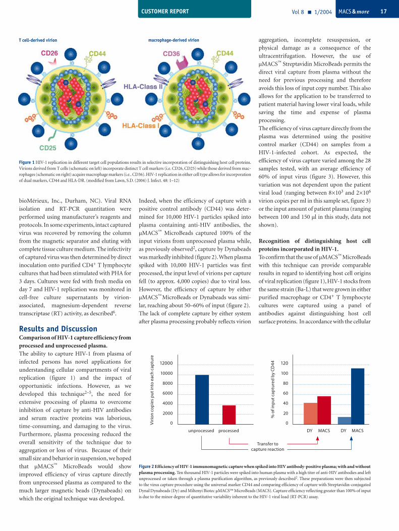
Indeed, when the efficiency of capture with a
positive control antibody (CD44) was deter-
mined for 10,000 HIV-1 particles spiked into
plasma containing anti-HIV antibodies, the
µMACS™ MicroBeads captured 100% of the
input virions from unprocessed plasma while,
as previously observed2, capture by Dynabeads
was markedly inhibited (figure 2). When plasma
spiked with 10,000 HIV-1 particles was first
processed, the input level of virions per capture
fell (to approx. 4,000 copies) due to viral loss.
However, the efficiency of capture by either
µMACS™ MicroBeads or Dynabeads was simi-
lar, reaching about 50–60% of input (figure 2).
The lack of complete capture by either system
after plasma processing probably reflects virion
aggregation, incomplete resuspension, or
physical damage as a consequence of the
ultracentrifugation. However, the use of
µMACS™ Streptavidin MicroBeads permits the
direct viral capture from plasma without the
need for previous processing and therefore
avoids this loss of input copy number. This also
allows for the application to be transferred to
patient material having lower viral loads, while
saving the time and expense of plasma
processing.
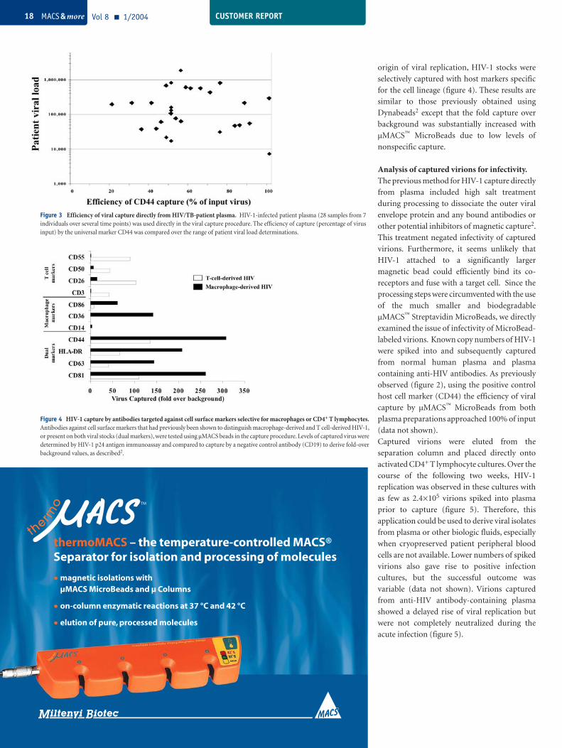
The efficiency of virus capture directly from the
plasma was determined using the positive
control marker (CD44) on samples from a
HIV-1-infected cohort. As expected, the
efficiency of virus capture varied among the 28
samples tested, with an average efficiency of
60% of input virus (figure 3). However, this
variation was not dependent upon the patient
viral load (ranging between 8×10³ and 2×10⁶
virion copies per ml in this sample set, figure 3)
or the input amount of patient plasma (ranging
between 100 and 150 µl in this study, data not
shown).
Recognition of distinguishing host cell proteins incorporated in HIV-1.To confirm that the use of µMACS™ MicroBeads
with this technique can provide comparable
results in regard to identifying host cell origins
of viral replication (figure 1), HIV-1 stocks from
the same strain (Ba-L) that were grown in either
purified macrophage or CD4+ T lymphocyte
cultures were captured using a panel of
antibodies against distinguishing host cell
surface proteins. In accordance with the cellular
Figure 2 Efficiency of HIV-1 immunomagnetic capture when spiked into HIV antibody-positive plasma; with and without plasma processing. Ten thousand HIV-1 particles were spiked into human plasma with a high titer of anti-HIV antibodies and left unprocessed or taken through a plasma purification algorithm, as previously described2. These preparations were then subjected to the virus capture procedure using the universal marker CD44 and comparing efficiency of capture with Streptavidin-conjugated Dynal Dynabeads (Dy) and Miltenyi Biotec µMACS™ MicroBeads (MACS). Capture efficiency reflecting greater than 100% of input is due to the minor degree of quantitative variability inherent to the HIV-1 viral load (RT-PCR) assay.
Figure 1 HIV-1 replication in different target cell populations results in selective incorporation of distinguishing host cell proteins. Virions derived from T cells (schematic on left) incorporate distinct T cell markers (i.e. CD26, CD25) while those derived from mac-rophages (schematic on right) acquire macrophage markers (i.e.. CD36). HIV-1 replication in either cell type allows for incorporation of dual markers, CD44 and HLA-DR. (modified from Lawn, S.D. (2004) J. Infect. 48: 1–12)
T cell-derived virion macrophage-derived virion
12000
Vir
ion
cop
ies
put
into
eac
h ca
ptu
re
10000
8000
6000
4000
2000
0
unprocessed processed
120
% o
f in
put
cap
ture
d b
y C
D44
100
80
60
40
20
0
DY MACS DY MACS
Transfer to capture reaction
CUSTOMER REPORTVol 8 1/2004 MACS & more18
origin of viral replication, HIV-1 stocks were
selectively captured with host markers specific
for the cell lineage (figure 4). These results are
similar to those previously obtained using
Dynabeads2 except that the fold capture over
background was substantially increased with
µMACS™ MicroBeads due to low levels of
nonspecific capture.
Analysis of captured virions for infectivity.The previous method for HIV-1 capture directly
from plasma included high salt treatment
during processing to dissociate the outer viral
envelope protein and any bound antibodies or
other potential inhibitors of magnetic capture2.
This treatment negated infectivity of captured
virions. Furthermore, it seems unlikely that
HIV-1 attached to a significantly larger
magnetic bead could efficiently bind its co-
receptors and fuse with a target cell. Since the
processing steps were circumvented with the use
of the much smaller and biodegradable
µMACS™ Streptavidin MicroBeads, we directly
examined the issue of infectivity of MicroBead-
labeled virions. Known copy numbers of HIV-1
were spiked into and subsequently captured
from normal human plasma and plasma
containing anti-HIV antibodies. As previously
observed (figure 2), using the positive control
host cell marker (CD44) the efficiency of viral
capture by µMACS™ MicroBeads from both
plasma preparations approached 100% of input
(data not shown).
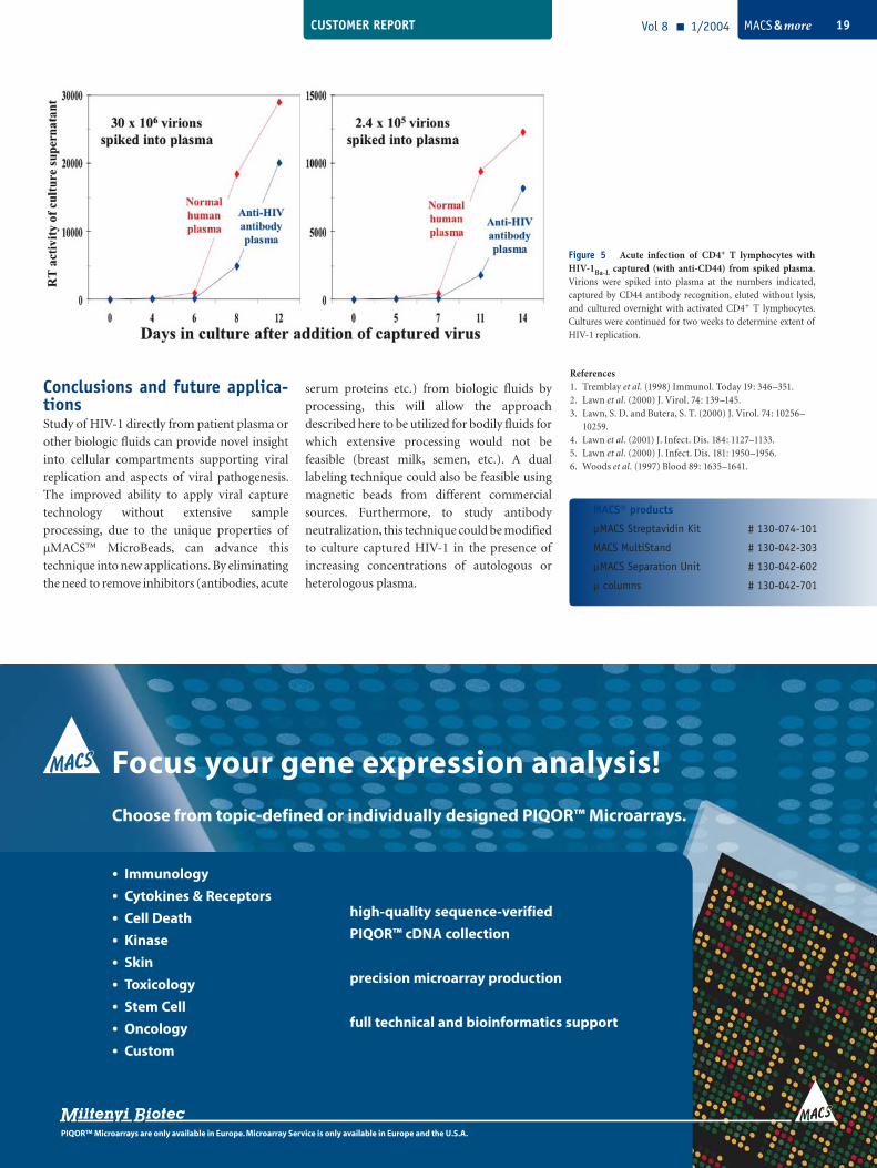
Captured virions were eluted from the
separation column and placed directly onto
activated CD4+ T lymphocyte cultures. Over the
course of the following two weeks, HIV-1
replication was observed in these cultures with
as few as 2.4×10⁵ virions spiked into plasma
prior to capture (figure 5). Therefore, this
application could be used to derive viral isolates
from plasma or other biologic fluids, especially
when cryopreserved patient peripheral blood
cells are not available. Lower numbers of spiked
virions also gave rise to positive infection
cultures, but the successful outcome was
variable (data not shown). Virions captured
from anti-HIV antibody-containing plasma
showed a delayed rise of viral replication but
were not completely neutralized during the
acute infection (figure 5).
Figure 3 Efficiency of viral capture directly from HIV/TB-patient plasma. HIV-1-infected patient plasma (28 samples from 7 individuals over several time points) was used directly in the viral capture procedure. The efficiency of capture (percentage of virus input) by the universal marker CD44 was compared over the range of patient viral load determinations.
TM
• magnetic isolations with µMACS MicroBeads and µ Columns
• on-column enzymatic reactions at 37 °C and 42 °C
• elution of pure, processed molecules
thermoMACS – the temperature-controlled MACS® Separator for isolation and processing of molecules
Figure 4 HIV-1 capture by antibodies targeted against cell surface markers selective for macrophages or CD4+ T lymphocytes. Antibodies against cell surface markers that had previously been shown to distinguish macrophage-derived and T cell-derived HIV-1, or present on both viral stocks (dual markers), were tested using µMACS beads in the capture procedure. Levels of captured virus were determined by HIV-1 p24 antigen immunoassay and compared to capture by a negative control antibody (CD19) to derive fold-over background values, as described2.
CUSTOMER REPORT Vol 8 1/2004 MACS & more 19
Figure 5 Acute infection of CD4+ T lymphocytes with HIV-1Ba-L captured (with anti-CD44) from spiked plasma. Virions were spiked into plasma at the numbers indicated, captured by CD44 antibody recognition, eluted without lysis, and cultured overnight with activated CD4+ T lymphocytes. Cultures were continued for two weeks to determine extent of HIV-1 replication.
References1. Tremblay et al. (1998) Immunol. Today 19: 346–351.2. Lawn et al. (2000) J. Virol. 74: 139–145.3. Lawn, S. D. and Butera, S. T. (2000) J. Virol. 74: 10256– 10259.4. Lawn et al. (2001) J. Infect. Dis. 184: 1127–1133.5. Lawn et al. (2000) J. Infect. Dis. 181: 1950–1956.6. Woods et al. (1997) Blood 89: 1635–1641.
Conclusions and future applica-tionsStudy of HIV-1 directly from patient plasma or
other biologic fluids can provide novel insight
into cellular compartments supporting viral
replication and aspects of viral pathogenesis.
The improved ability to apply viral capture
technology without extensive sample
processing, due to the unique properties of
µMACS™ MicroBeads, can advance this
technique into new applications. By eliminating
the need to remove inhibitors (antibodies, acute
serum proteins etc.) from biologic fluids by
processing, this will allow the approach
described here to be utilized for bodily fluids for
which extensive processing would not be
feasible (breast milk, semen, etc.). A dual
labeling technique could also be feasible using
magnetic beads from different commercial
sources. Furthermore, to study antibody
neutralization, this technique could be modified
to culture captured HIV-1 in the presence of
increasing concentrations of autologous or
heterologous plasma.
Focus your gene expression analysis!
Choose from topic-defined or individually designed PIQOR™ Microarrays.
· Immunology
· Cytokines & Receptors
· Cell Death
· Kinase
· Skin
· Toxicology
· Stem Cell
· Oncology
· Custom
high-quality sequence-verified
PIQOR™ cDNA collection
precision microarray production
full technical and bioinformatics support
PIQOR™ Microarrays are only available in Europe. Microarray Service is only available in Europe and the U.S.A.
MACS® products
µMACS Streptavidin Kit # 130-074-101
MACS MultiStand # 130-042-303
µMACS Separation Unit # 130-042-602
μ columns # 130-042-701
PERSPECTIVEVol 8 1/2004 Vol 8 MACS & more20
Efficient target molecule identification by combining display technology with magnetic selection
Display technologyDisplay technology1 classically refers to the
generation and screening of a large library of
expressed proteins or peptides using an
immobilized ligand to characterize or discover
new protein-ligand interactions.
There are a number of characteristics that have
ensured that display technology is the method
of choice when a new biomolecular interaction
is to be identified. Firstly, the structure of the
expressed protein (phenotype) is linked to the
genetic information encoding it (genotype).
This allows immediate identification and
further characterization of the novel binding
partner once it has been isolated from the
library. Secondly, very large libraries can be
generated and displayed to the ligand of
interest. The larger the library, the greater is the
chance of finding a binding partner. Thirdly,
most established display systems enable
amplification of the library without the
introduction of a bias for a particular type of
DNA fragment or protein. This allows
successive unbiased rounds of screening to take
place against a particular ligand, increasing the
sensitivity. Finally, the amplification of the
library can be used to introduce random errors
into the encoding DNA sequence to enable
mutant proteins with altered binding affinities
or binding kinetics to be isolated (affinity
maturation).
The extraordinary efficiency of MACS Technology offers new perspectives for research using phage, ribosome, or yeast display techniques.
Popular display systemsPhage displayThe display of foreign peptides and proteins on
the surface of filamentous bacteriophages,
“phage display”, is now a widely-used technique
to investigate molecular interactions. Normally
the protein library to be screened is expressed as
a fusion with the gene 3 protein product at one
end of the bacteriophage particle. Infection of
bacteria with this phage library allows very
efficient library amplification. However, as the
gene 3 protein is also required for bacteriophage
assembly and infectivity, there are some limits to
the size of protein that can be successfully
expressed and displayed without introducing a
bias during bacteriophage replication.
Ribosome displayRibosome display2 is a technology that enables
the selection and evolution of large protein
libraries completely in vitro. The only biological
component required is a bacterial cell extract
that contains the factors required for the
translation of in vitro-generated transcripts
encoding the protein sequences. Genotype and
phenotype are linked through ribosomal
complexes, consisting of messenger RNA
(mRNA), ribosome, and encoded protein. After
selection via the encoded protein, the isolated
mRNAs are amplified by RT-PCR and can be
freshly transcribed and translated for another
round of selection.
Yeast displayYeast display is a powerful new technique to
study protein-protein interactions that has been
developed by Prof. Dane Wittrup of the
Massachussetts Institute of Technology3. This
surface expression system can be used to express
recombinant proteins on the surface of S.
cerevisiae as a fusion with the a-agglutinin yeast
adhesion receptor. Due to correct post-
translational modification, processing and
folding of mammalian proteins, this system
provides an extremely attractive alternative to
phage display for the discovery, characterization
and affinity maturation of protein-ligand
interactions, especially where full-length
proteins are to be studied and not isolated
protein fragments.
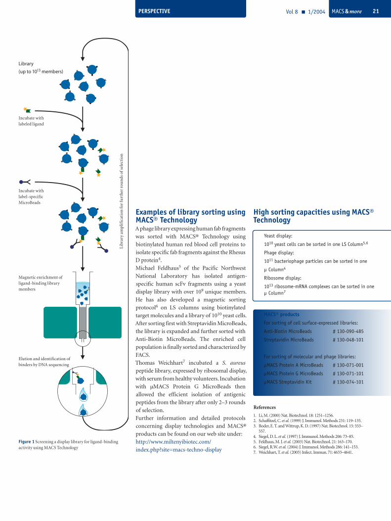
Display technology and magnetic sortingIn all of the display systems described, magnetic
sorting with MACS® Technology enables an
efficient and rapid isolation of interacting
proteins. Orders of magnitude more events can
be sorted than with other technologies and a
very low non-specific enrichment is obtained
compared to conventional panning techniques
due to the highly efficient washing steps that are
possible on the column. As a general strategy,
the ligand can be labeled (for example
biotinylated) and then after incubation with the
target library, the interacting complex can be
isolated using Streptavidin or Anti-Biotin
MicroBeads as shown in figure 1. Alternatively,
if the ligand is an antibody, µMACS Protein A or
Protein G MicroBeads can be used.
Ian Johnston Miltenyi Biotec GmbH, Bergisch Gladbach, Germany
PERSPECTIVE Vol 8 1/2004 MACS & more 21
Libr
ary
ampl
ific
atio
n fo
r fu
rthe
r ro
und
s of
sel
ecti
on
Examples of library sorting using MACS® TechnologyA phage library expressing human fab fragments
was sorted with MACS® Technology using
biotinylated human red blood cell proteins to
isolate specific fab fragments against the Rhesus
D protein4.
Michael Feldhaus5 of the Pacific Northwest
National Laboratory has isolated antigen-
specific human scFv fragments using a yeast
display library with over 109 unique members.
He has also developed a magnetic sorting
protocol6 on LS columns using biotinylated
target molecules and a library of 1010 yeast cells.
After sorting first with Streptavidin MicroBeads,
the library is expanded and further sorted with
Anti-Biotin MicroBeads. The enriched cell
population is finally sorted and characterized by
FACS.
Thomas Weichhart7 incubated a S. aureus
peptide library, expressed by ribosomal display,
with serum from healthy volunteers. Incubation
with µMACS Protein G MicroBeads then
allowed the efficient isolation of antigenic
peptides from the library after only 2–3 rounds
of selection.
Further information and detailed protocols
concerning display technologies and MACS®
products can be found on our web site under:
http://www.miltenyibiotec.com/
index.php?site=macs-techno-display
References1. Li, M. (2000) Nat. Biotechnol. 18: 1251–1256.2. Schaffitzel, C. et al. (1999) J. Immunol. Methods 231: 119–135.3. Boder, E. T. and Wittrup, K. D. (1997) Nat. Biotechnol. 15: 553– 557.4. Siegel, D. L. et al. (1997) J. Immunol. Methods 206: 73–85.5. Feldhaus, M. J. et al. (2003) Nat. Biotechnol. 21: 163–170.6. Siegel, R.W. et al. (2004) J. Immunol. Methods 286: 141–153.7. Weichhart, T. et al. (2003) Infect. Immun. 71: 4633–4641.
High sorting capacities using MACS® Technology
Library
(up to 1013 members)
Incubate withlabeled ligand
Incubate withlabel-specific MicroBeads
Magnetic enrichment of ligand-binding library members
Elution and identification of binders by DNA sequencing
Figure 1 Screening a display library for ligand-binding activity using MACS Technology
Yeast display:
1010 yeast cells can be sorted in one LS Column5,6
Phage display:
1011 bacteriophage particles can be sorted in one
µ Column4
Ribosome display:
1013 ribosome-mRNA complexes can be sorted in one µ Column7
MACS® products
For sorting of cell surface-expressed libraries:
Anti-Biotin MicroBeads # 130-090-485
Streptavidin MicroBeads # 130-048-101
For sorting of molecular and phage libraries:
µMACS Protein A MicroBeads # 130-071-001
µMACS Protein G MicroBeads # 130-071-101
µMACS Streptavidin Kit # 130-074-101
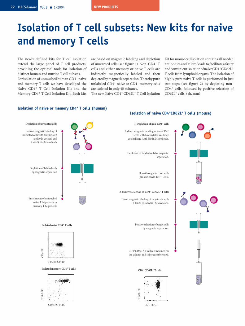
NEW PRODUCTSVol 8 1/2004 MACS & more22
The newly defined kits for T cell isolation
extend the large panel of T cell products,
providing the optimal tools for isolation of
distinct human and murine T cell subsets.
For isolation of untouched human CD4+ naive
and memory T cells we have developed the
Naive CD4+ T Cell Isolation Kit and the
Memory CD4+ T Cell Isolation Kit. Both kits
Isolation of T cell subsets: New kits for naive and memory T cells
are based on magnetic labeling and depletion
of unwanted cells (see figure 1). Non-CD4+ T
cells and either memory or naive T cells are
indirectly magnetically labeled and then
depleted by magnetic separation. Thereby pure
unlabeled CD4+ naive or CD4+ memory cells
are isolated in only 45 minutes.
The new Naive CD4+ CD62L+ T Cell Isolation
Kit for mouse cell isolation contains all needed
antibodies and MicroBeads to facilitate a faster
and convenient isolation of naive CD4+CD62L+
T cells from lymphoid organs. The isolation of
highly pure naive T cells is performed in just
two steps (see figure 2) by depleting non-
CD4+ cells, followed by positive selection of
CD62L+ cells. (eh, mm)
Isolation of naive or memory CD4+ T cells (human)Isolation of naive CD4+CD62L+ T cells (mouse)
Enrichment of untouched naive T helper cells or memory T helper cells
Depletion of labeled cells by magnetic separation
Depletion of unwanted cells
Indirect magnetic labeling of unwanted cells with biotinylated
antibody cocktail and Anti-Biotin MicroBeads
2. Positive selection of CD4+ CD62L+ T cells
Direct magnetic labeling of target cells with CD62L (L-selectin) MicroBeads.
1. Depletion of non-CD4+ cells
Indirect magnetic labeling of non-CD4+ T cells with biotinylated antibody
cocktail and Anti-Biotin MicroBeads.
Depletion of labeled cells by magnetic separation.
Flow-through fraction with pre-enriched CD4+ T cells.
Positive selection of target cells by magnetic separation.
CD4+CD62L+ T cells are retained on the column and subsequently eluted.
CD45RA-FITC
CD
4-PE
Isolated naive CD4+ T cells
CD45RO-FITC
CD
4-A
PC
Isolated memory CD4+ T cells
CD4-FITC
CD
62L-
PE
CD4+CD62L+ T cells
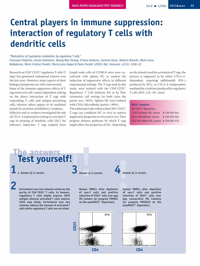
MACS PAPER HIGHLIGHT/TEST YOURSELF Vol 8 1/2004 MACS & more 23
!The answers
!!Test yourself!Answer a) is correct.3 4
Central players in immune suppression: interaction of regulatory T cells with dendritic cells
MACS® productsCD4+CD25+ Regulatory
T Cell Isolation Kit, mouse # 130-091-041
CD11c MicroBeads, mouse # 130-052-001
CD25 MicroBead Kit, mouse # 130-091-072
Research on CD4+CD25+ regulatory T cells (T
regs) has generated widespread interest over
the last years. However, many aspects of their
biological properties are still controversial.
Some of the immune suppressive effects of T
regs seem to be cell-contact dependent, relying
on the direct interaction of T regs with
responding T cells and antigen-presenting
cells, whereas others appear to be mediated
mainly by secretion of inhibitory cytokines.
Fallarino and co-workers investigated the role
of CTLA-4 expressed on resting or activated T
regs in priming of dendritic cells (DC) for
tolerance induction. T regs isolated from
lymph node cells of C57BL/6 mice were co-
cultured with splenic DC to analyse the
induction of suppressive effects in different
experimental settings. The T regs used in this
study, were isolated with the CD4+CD25+
Regulatory T Cell Isolation Kit or by f low
cytometric cell sorting (in both cases the
purity was: >90%). Splenic DC were isolated
with CD11c MicroBeads (purity: >98%).
The authors provide evidence that CD4+CD25+
T regs can condition DC in vitro to express
suppressive properties in vitro and in vivo. They
propose distinct pathways by which T regs
might affect the properties of DC. Depending
on the stimuli used for activation of T regs, the
process is supposed to be either CTLA-4-
dependent, requiring additionally IFN-γ
produced by DCs, or CTLA-4-independent,
mediated by cytokines produced by regulatory
T cells (IFN-γ, IL−10). (mm)
“Modulation of tryptophan catabolism by regulatory T cells”Francesca Fallarino, Ursula Grohmann, Kwang Woo Hwang, Ciriana Orabona, Carmine Vacca, Roberta Bianchi, Maria Laura Belladonna, Maria Cristina Fioretti, Maria-Luisa Alegre & Paolo Pucetti (2003) Nat. Immunol. 4(12): 1206–12.
2 Enrichment over two columns enhances the purity of CD4+CD25+ T cells. In humans, regulatory T cells highly express CD25 antigen whereas activated T cells express CD25 only dimly. Enrichment over two columns reduces the amount of activated T cells while regulatory T cells are enriched.
Human PBMCs after depletion of non-T cells and positive selection of CD25+ cells over one MS Column (or program POSSEL on the autoMACS™ Separator).
Human PBMCs after depletion of non-T cells and positive selection of CD25+ cells over two consecutive MS Columns (or program POSSELD on the autoMACS™ Separator).
CD4 CD4
CD25
81% 93%
1 1. Answer b) is correct. Answer b) is correct.
ArgentinaLab Systems S.A.Phone: +54 11 4572 8458BelgiumTEBU-BIO nvPhone: +32 3 454 00 66BrazilAmbriex S/APhone: +55 11 3826 6722BulgariaANTI-SEL Selidis Bros. BG Ltd.Phone +359 2 953 12 24China / Hong KongMiltenyi Biotec Shanghai OfficePhone: +86 21 623 510 [email protected] RepublicLV-BiomedPhone: +420 603 417 315DenmarkBiotech Line ASPhone: +45 7027 9920FinlandNuppulinnan laboratoriopalvelu OyPhone: +358 9 27940200Oriola OY ProlabPhone: +358 10 42 94 927GreeceEpsilon & Epsilon Medical S.A.Phone: +30 210 6996191HungaryFrank DiagnosztikaPhone: +361 388 3114IndiaLabmate Asia PvT. Ltd. Phone: +91 44 220 0166IsraelAlmog DiagnosticPhone: +972 3 9773390
JapanDaiichi Pure Chemicals Co., Ltd.Phone: +81 3 5820 9408MalaysiaBiomarketing Services Sdn BhdPhone: +603 627 33068MexicoUniparts S.A.Phone: +52 55 5281 4718NetherlandsSanquin ReagentsPhone: +31 20 512 3739NorwayAH diagnostics asPhone: +47 23 23 32 60PolandMEDianus Biuro HandlowePhone: +48 12 665 31 31PortugalCitomed Lda.Phone: +351 21 842 14 30SingaporeBiomed Diagnostics Pte LtdPhone: +65 2984347SlovakiaBDSR s.r.o.Phone: +421 95 625 5765South KoreaFine Life Science Co., Ltd.Phone: +82 2 744 7859SwedenGTFPhone: +46 31 689400TaiwanInterlab Co., Ltd.Phone: +886 2 2736 7100TurkeyLab Laboratuar Malzemeleri Ith ve Tic. Ltd. Sti.Phone: +90 212 283 6915
Argentinasee leftBrazilsee leftBulgariasee leftChinaKirin Pharmaceuticals (China) Co., Ltd.Phone: +86 10 64 10 7210Czech Republic see leftGreecesee leftHong KongKirin Pharmaceuticals Phone: +852 29 56 08 28Hungarysee leftIsrael see leftJapanKirin Brewery Co., Ltd.Pharmaceutical Division Phone: +81 3 5485 6259MexicoInterlab de MéxicoPhone: +52 55 5355 7818South KoreaJeil-Kirin Pharmaceutical Inc.Phone: +82 2 3471 4321Slovakiasee leftTaiwanKirin Pharmaceuticals Co., Ltd.Phone: +886 2 25 01 80 67TurkeyMAK Saglik Urunleri Ltd. Sti.Phone: +90 312 4302960
All MACS® products Distributors for research products Distributors for clinical products
Germany/Austria/SwitzerlandMiltenyi Biotec GmbHFriedrich-Ebert-Straße 6851429 Bergisch GladbachPhone: +49 2204 8306-0Fax: +49 2204 85197 [email protected]/CanadaMiltenyi Biotec Inc.12740 Earhart AvenueAuburn, CA 95602Phone: 800 FOR MACSPhone: 530 888 8871Fax: 530 888 [email protected] Biotec Australia Pty. Ltd.Unit 16A, 2 Eden Park DriveNorth Ryde NSW 2113Phone: 02 8877 7400Fax: 02 9889 [email protected] Biotec18 Avenue Parmentier75011 Paris Phone: 01 56 98 16 16Fax: 01 56 98 16 [email protected] Biotec S.r.l.Via Persicetana, 2/d40012 Calderara di Reno (BO)Phone: 051 6 460 411 Fax: 051 6 460 [email protected] Biotec S.L.C/Luis Buñuel 2, Ciudad de la Imagen28223 Pozuelo de Alarcón (Madrid)Phone: 91 512 12 90Fax: 91 512 12 [email protected] Kingdom Miltenyi Biotec Ltd. Almac House, Church Lane, BisleySurrey GU24 9DRPhone: 01483 799 800Fax: 01483 799 [email protected]
Where to find us For additional address details and further product information check www.miltenyibiotec.com
With compliments from:
130-
091-
976
MACSelect LNGFRThe smart selection after each transfection
· select transfected cells in 2–3 hours · no antibiotics required· flexible system for all primary cells and cell lines· new LNGFR surface marker