
Magnoliae Officinalis Flos - cmd.gov.hk · PDF file335 Magnoliae Officinalis Flos Powder...
12
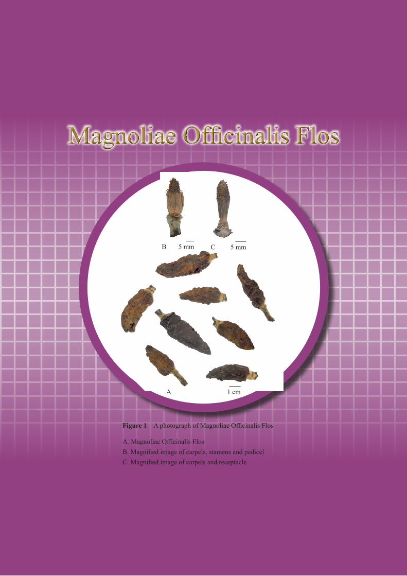
Figure 1 A photograph of Magnoliae Officinalis Flos A. Magnoliae Officinalis Flos B. Magnified image of carpels, stamens and pedicel C. Magnified image of carpels and receptacle A B C 1 cm 5 mm 5 mm
Transcript of Magnoliae Officinalis Flos - cmd.gov.hk · PDF file335 Magnoliae Officinalis Flos Powder...
A 1 cm B 0.5 cm C 0.5 cm
A 1 cm B 0.5 cm C 0.5 cm
A 1 cm B 0.5 cm C 0.5 cm
Magnoliae Officinalis Flos
Figure 1 A photograph of Magnoliae Officinalis Flos
A. Magnoliae Officinalis Flos B. Magnified image of carpels, stamens and pedicel C. Magnified image of carpels and receptacle
A
B C
1 cm
5 mm 5 mm
334
Magnoliae Officinalis Flos
1. NAMES
Official Name: Magnoliae Officinalis Flos
Chinese Name: 厚朴花
Chinese Phonetic Name: Houpohua
2. SOURCE
Magnoliae Officinalis Flos is the dried flower bud of Magnolia officinalis Rehd. et Wils.
(Magnoliaceae). The flower bud is collected in spring before flowering, steamed briefly, then dried
under the sun to obtain Magnoliae Officinalis Flos.
3. DESCRIPTION
Flower buds long-conical or oblong, 3.0-6.8 cm long, 1.1-3.2 cm in diameter. Tepals fleshy, surface
scabrous, outermost whorl of tepals relatively large and thin, reddish-brown to blackish-brown; inner
whorls relatively small and thick, reddish-brown to dark brown. Stamens numerous, spirally arranged
on elongated receptacles, anthers flat linear, filaments short and board; carpels numerous, separated,
spirally arranged on elongated receptacles. Pedicels long, densely covered with yellowish-white or
greyish-white villi. Texture fragile, easily broken. Odour fragrant; taste bland (Fig. 1).
4. IDENTIFICATION
4.1 MicroscopicIdentification(Appendix III)
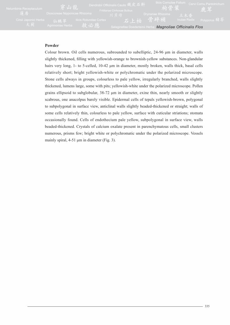
TransversesectionPedicel: Epidermis consists of 1 layer of cells, covered with cuticle, with non-glandular hairs.
Cortex consists of parenchymatous cells, scattered with collateral or amphicribral vascular
bundles, varying in size. Inner vascular bundles fusiform or oblong, collateral, arranged in
an interrupted ring. Pith consists of parenchymatous cells. Oil cells and groups of stone cells
scattered in parenchymatous cells (Fig. 2).
335
Magnoliae Officinalis Flos
PowderColour brown. Oil cells numerous, subrounded to subelliptic, 24-96 μm in diameter, walls
slightly thickened, filling with yellowish-orange to brownish-yellow substances. Non-glandular
hairs very long, 1- to 5-celled, 10-42 μm in diameter, mostly broken, walls thick, basal cells
relatively short; bright yellowish-white or polychromatic under the polarized microscope.
Stone cells always in groups, colourless to pale yellow, irregularly branched, walls slightly
thickened, lumens large, some with pits; yellowish-white under the polarized microscope. Pollen
grains ellipsoid to subglobular, 38-72 μm in diameter, exine thin, nearly smooth or slightly
scabrous, one anacolpus barely visible. Epidermal cells of tepals yellowish-brown, polygonal
to subpolygonal in surface view, anticlinal walls slightly beaded-thickened or straight; walls of
some cells relatively thin, colourless to pale yellow, surface with cuticular striations; stomata
occasionally found. Cells of endothecium pale yellow, subpolygonal in surface view, walls
beaded-thickened. Crystals of calcium oxalate present in parenchymatous cells, small clusters
numerous, prisms few; bright white or polychromatic under the polarized microscope. Vessels
mainly spiral, 4-51 μm in diameter (Fig. 3).
336
Magnoliae Officinalis Flos
2
C
1
100 µm
6
D 100 µm
1 2 3
4 5 6 7 8
9 10
B
1 2
3
5
6
4
7
9
10
8
200 µm
2
C
1
100 µm
6
D 100 µm
1 2 3
4 5 6 7 8
9 10
B
1 2
3
5
6
4
7
9
10
8
200 µm
2
C
1
100 µm
6
D 100 µm
1 2 3
4 5 6 7 8
9 10
B
1 2
3
5
6
4
7
9
10
8
200 µm
2
C
1
100 µm
6
D 100 µm
1 2 3
4 5 6 7 8
9 10
B
1 2
3
5
6
4
7
9
10
8
200 µm
Figure 2 Microscopic features of transverse section of pedicel of Magnoliae Officinalis Flos
A. Sketch B. Section illustration C. Epidermis and non-glandular hairs D. Oil cell
1. Epidermis 2. Non-glandular hair 3. Cortex 4. Amphicribral vascular bundle 5. Collateral vascular bundle 6. Oil cell 7. Phloem 8. Xylem 9. Stone cells 10. Pith
C
D
12
3
6
4
5
7
8
9
10
3
645
6
7
910
8
B 200 μm
1
2
2
1
100 μm
100 μm
A
337
Magnoliae Officinalis Flos
1a
4a
6a 7a 7b 8a
2a-1
3a-1 3b-1
5a-1
5a-2
3b-23a-2
2a-2
2b-1
2b-2
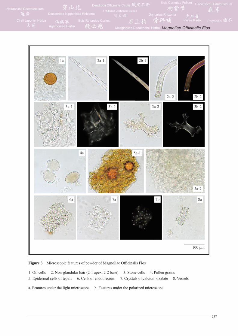
Figure 3 Microscopic features of powder of Magnoliae Officinalis Flos
1. Oil cells 2. Non-glandular hair (2-1 apex, 2-2 base) 3. Stone cells 4. Pollen grains 5. Epidermal cells of tepals 6. Cells of endothecium 7. Crystals of calcium oxalate 8. Vessels
a. Features under the light microscope b. Features under the polarized microscope
100 μm
338
Magnoliae Officinalis Flos
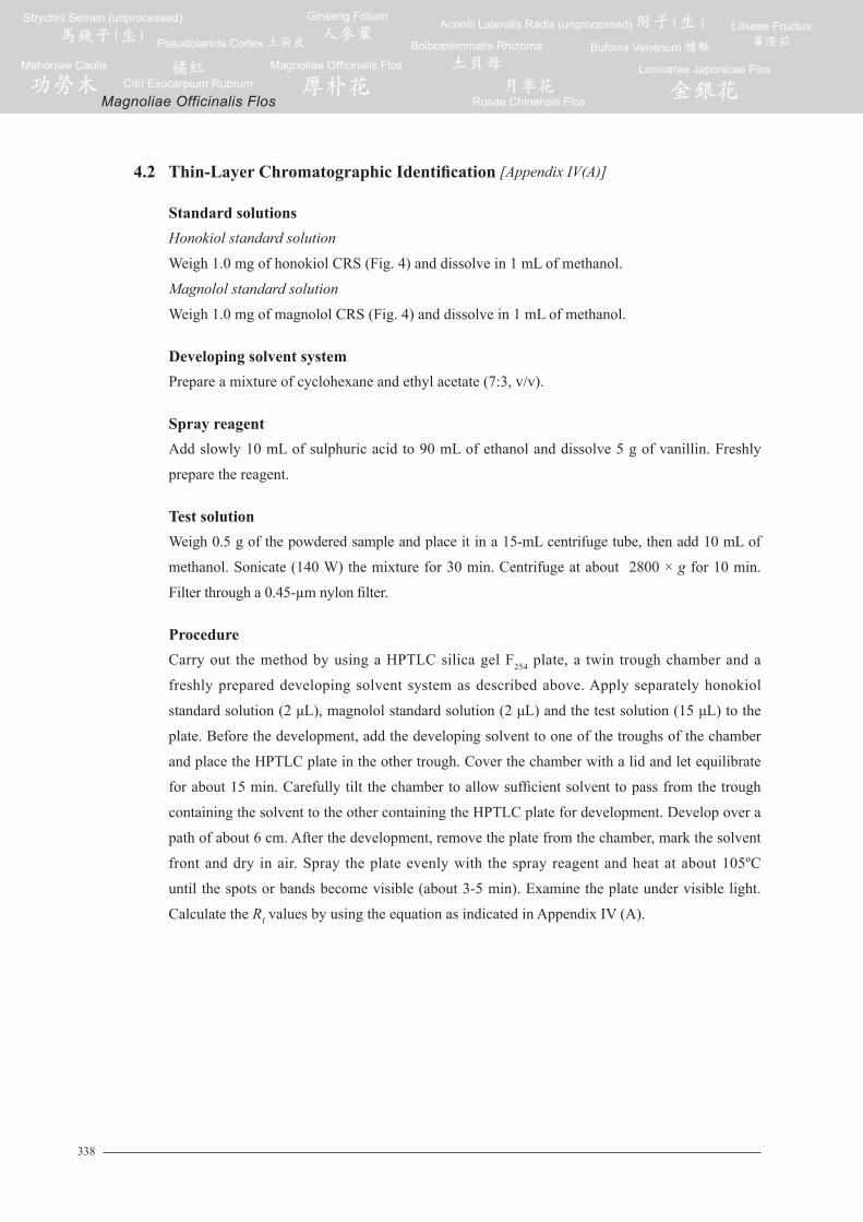
4.2 Thin-LayerChromatographicIdentification [Appendix IV(A)]
StandardsolutionsHonokiol standard solution
Weigh 1.0 mg of honokiol CRS (Fig. 4) and dissolve in 1 mL of methanol.
Magnolol standard solution
Weigh 1.0 mg of magnolol CRS (Fig. 4) and dissolve in 1 mL of methanol.
DevelopingsolventsystemPrepare a mixture of cyclohexane and ethyl acetate (7:3, v/v).
SprayreagentAdd slowly 10 mL of sulphuric acid to 90 mL of ethanol and dissolve 5 g of vanillin. Freshly
prepare the reagent.
Test solutionWeigh 0.5 g of the powdered sample and place it in a 15-mL centrifuge tube, then add 10 mL of
methanol. Sonicate (140 W) the mixture for 30 min. Centrifuge at about 2800 × g for 10 min.
Filter through a 0.45-µm nylon filter.
ProcedureCarry out the method by using a HPTLC silica gel F254 plate, a twin trough chamber and a
freshly prepared developing solvent system as described above. Apply separately honokiol
standard solution (2 μL), magnolol standard solution (2 μL) and the test solution (15 μL) to the
plate. Before the development, add the developing solvent to one of the troughs of the chamber
and place the HPTLC plate in the other trough. Cover the chamber with a lid and let equilibrate
for about 15 min. Carefully tilt the chamber to allow sufficient solvent to pass from the trough
containing the solvent to the other containing the HPTLC plate for development. Develop over a
path of about 6 cm. After the development, remove the plate from the chamber, mark the solvent
front and dry in air. Spray the plate evenly with the spray reagent and heat at about 105ºC
until the spots or bands become visible (about 3-5 min). Examine the plate under visible light.
Calculate the Rf values by using the equation as indicated in Appendix IV (A).
339
Magnoliae Officinalis Flos
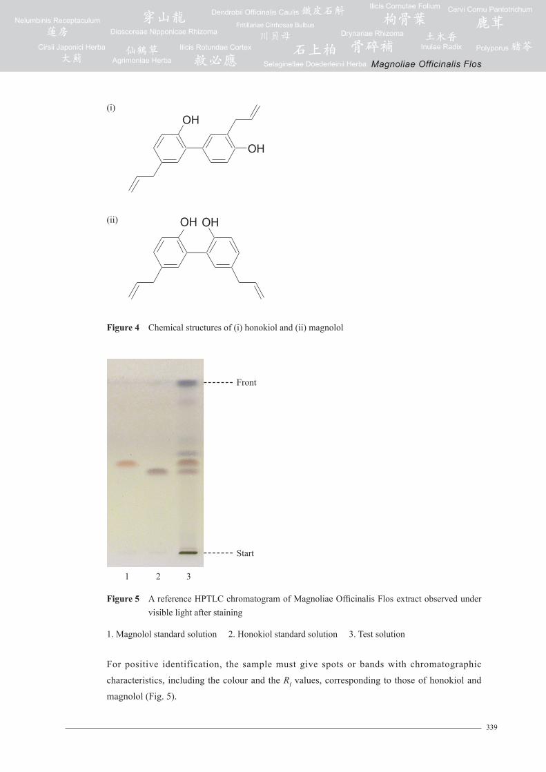
Figure 5 A reference HPTLC chromatogram of Magnoliae Officinalis Flos extract observed under visible light after staining
1. Magnolol standard solution 2. Honokiol standard solution 3. Test solution
For positive identification, the sample must give spots or bands with chromatographic
characteristics, including the colour and the Rf values, corresponding to those of honokiol and
magnolol (Fig. 5).
Figure 4 Chemical structures of (i) honokiol and (ii) magnolol
(i)
(ii)
OH
OH
Front
Start
21 3
1 2 3
Front
Start
OH OH
340
Magnoliae Officinalis Flos
4.3High-PerformanceLiquidChromatographicFingerprinting(Appendix XII)
StandardsolutionsHonokiol standard solution for fingerprinting, Std-FP (200 mg/L)
Weigh 0.2 mg of honokiol CRS and dissolve in 1 mL of methanol (70%).
Magnolol standard solution for fingerprinting, Std-FP (200 mg/L)
Weigh 0.2 mg of magnolol CRS and dissolve in 1 mL of methanol (70%).
Test solutionWeigh 1.0 g of the powdered sample and place it in a 50-mL centrifuge tube, then add 40 mL of
methanol (70%). Sonicate (270 W) the mixture for 1 h. Centrifuge at about 2800 × g for 5 min.
Transfer the supernatant to a 250-mL round-bottomed flask. Repeat the extraction for one more
time. Combine the supernatants. Evaporate the solvent to dryness at reduced pressure in a rotary
evaporator. Dissolve the residue in methanol (70%). Transfer the solution to a 25-mL volumetric
flask and make up to the mark with methanol (70%). Filter through a 0.45-µm PTFE filter.
ChromatographicsystemThe liquid chromatograph is equipped with a DAD (320 nm) and a column (4.6 × 250 mm)
packed with ODS bonded silica gel (5 µm particle size). The flow rate is about 0.8 mL/min.
Programme the chromatographic system as follows (Table 1) –
Table1 Chromatographic system conditions
Time(min)
0.4%Formicacid(%,v/v)
Methanol(%,v/v)
Elution
0 – 5 50 50 isocratic
5 – 40 50→ 0 50→ 100 linear gradient
40 – 45 0 100 isocratic
SystemsuitabilityrequirementsPerform at least five replicate injections, each using 20 µL of honokiol Std-FP and magnolol
Std-FP. The requirements of the system suitability parameters are as follows: the RSD of the
peak areas of honokiol and magnolol should not be more than 5.0%; the RSD of the retention
times of honokiol and magnolol peaks should not be more than 2.0%; the column efficiencies
determined from honokiol and magnolol peaks should not be less than 50000 theoretical plates.
The R value between peak 2 and the closest peak; and the R value between peak 3 and the closest
peak in the chromatogram of the test solution should not be less than 1.0 (Fig. 6).
341
Magnoliae Officinalis Flos
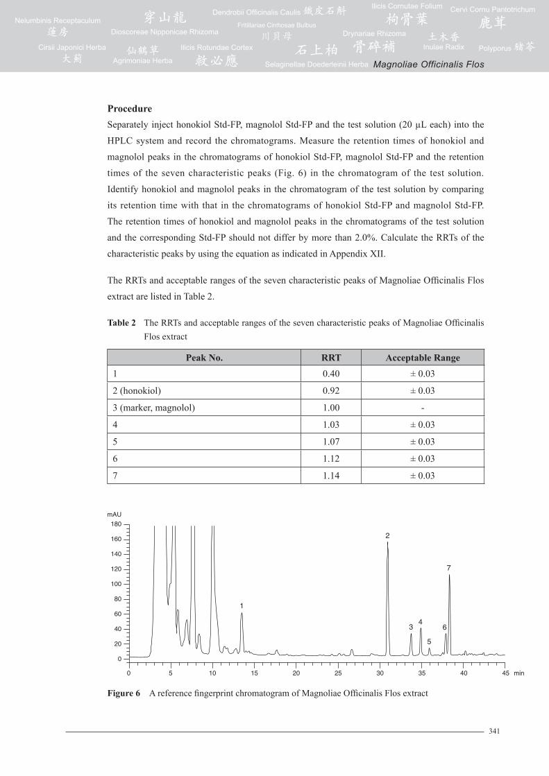
Figure 6 A reference fingerprint chromatogram of Magnoliae Officinalis Flos extract
ProcedureSeparately inject honokiol Std-FP, magnolol Std-FP and the test solution (20 µL each) into the
HPLC system and record the chromatograms. Measure the retention times of honokiol and
magnolol peaks in the chromatograms of honokiol Std-FP, magnolol Std-FP and the retention
times of the seven characteristic peaks (Fig. 6) in the chromatogram of the test solution.
Identify honokiol and magnolol peaks in the chromatogram of the test solution by comparing
its retention time with that in the chromatograms of honokiol Std-FP and magnolol Std-FP.
The retention times of honokiol and magnolol peaks in the chromatograms of the test solution
and the corresponding Std-FP should not differ by more than 2.0%. Calculate the RRTs of the
characteristic peaks by using the equation as indicated in Appendix XII.
The RRTs and acceptable ranges of the seven characteristic peaks of Magnoliae Officinalis Flos
extract are listed in Table 2.
Table2 The RRTs and acceptable ranges of the seven characteristic peaks of Magnoliae Officinalis Flos extract
PeakNo. RRT AcceptableRange1 0.40 ± 0.03
2 (honokiol) 0.92 ± 0.03
3 (marker, magnolol) 1.00 -
4 1.03 ± 0.03
5 1.07 ± 0.03
6 1.12 ± 0.03
7 1.14 ± 0.03
0 5 10 15 20 25 30 35 40 45
0
20
40
60
80
100
120
140
160
180
min
mAU
1
2
3 4
5
6
7
342
Magnoliae Officinalis Flos
For positive identification, the sample must give the above seven characteristic peaks with
RRTs falling within the acceptable range of the corresponding peaks in the reference fingerprint
chromatogram (Fig. 6).
5. TESTS
5.1 HeavyMetals(Appendix V): meet the requirements.
5.2 Pesticide Residues (Appendix VI): meet the requirements.
5.3 Mycotoxins(Appendix VII): meet the requirements.
5.4 ForeignMatter(Appendix VIII): not more than 1.0%.
5.5 Ash (Appendix IX)
Total ash: not more than 8.0%.
Acid-insoluble ash: not more than 0.5%.
5.6 WaterContent(Appendix X)
Oven dried method: not more than 10.0%.
6. EXTRACTIVES (Appendix XI)
Water-soluble extractives (cold extraction method): not less than 24.0%.
Ethanol-soluble extractives (hot extraction method): not less than 25.0%.
7. ASSAY
Carry out the method as directed in Appendix IV (B).
StandardsolutionMixed honokiol and magnolol standard stock solution, Std-Stock (1000 mg/L each)
Weigh accurately 1.0 mg of honokiol CRS and 1.0 mg of magnolol CRS, and dissolve in 1 mL of
methanol (70%).
Mixed honokiol and magnolol standard solution for assay, Std-AS
Measure accurately the volume of the mixed honokiol and magnolol Std-Stock, dilute with methanol
(70%) to produce a series of solutions of 10, 25, 50, 100, 125 mg/L for both honokiol and magnolol.

343
Magnoliae Officinalis Flos
Test solutionWeigh accurately 1.0 g of the powdered sample and place it in a 50-mL centrifuge tube, then
add 40 mL of methanol (70%). Sonicate (270 W) the mixture for 30 min. Centrifuge at about
2800 × g for 10 min. Transfer the supernatant to a 100-mL volumetric flask. Repeat the extraction
for one more time. Wash the residue with methanol (70%). Centrifuge at about 2800 × g for
10 min. Combine the supernatants and make up to the mark with methanol (70%). Filter through a
0.45-µm nylon filter.
ChromatographicsystemThe liquid chromatograph is equipped with a DAD (294 nm) and a column (4.6 × 250 mm) packed
with ODS bonded silica gel (5 µm particle size). The flow rate is about 1.0 mL/min. The mobile phase
is a mixture of 0.4% formic acid and acetonitrile (35:65, v/v). The elution time is about 25 min.
SystemsuitabilityrequirementsPerform at least five replicate injections, each using 10 µL of the mixed honokiol and magnolol
Std-AS (50 mg/L each). The requirements of the system suitability parameters are as follows: the
RSD of the peak areas of honokiol and magnolol should not be more than 5.0%; the RSD of the
retention times of honokiol and magnolol peaks should not be more than 2.0%; the column efficiencies
determined from honokiol and magnolol peaks should not be less than 8000 theoretical plates.
The R value between honokiol peak and the closest peak; and the R value between magnolol peak and
the closest peak in the chromatogram of the test solution should not be less than 1.5.
CalibrationcurvesInject a series of the mixed honokiol and magnolol Std-AS (10 µL each) into the HPLC system and
record the chromatograms. Plot the peak areas of honokiol and magnolol against the corresponding
concentrations of the mixed honokiol and magnolol Std-AS. Obtain the slopes, y-intercepts and the
r2 values from the corresponding 5-point calibration curves.
ProcedureInject 10 µL of the test solution into the HPLC system and record the chromatogram. Identify honokiol
and magnolol peaks in the chromatogram of the test solution by comparing their retention times
with those in the chromatogram of the mixed honokiol and magnolol Std-AS. The retention times of
honokiol and magnolol peaks in the chromatograms of the test solution and the Std-AS should not
differ by more than 5.0%. Measure the peak areas and calculate the concentrations (in milligram per
litre) of honokiol and magnolol in the test solution, and calculate the percentage contents of honokiol
and magnolol in the sample by using the equations as indicated in Appendix IV (B).
344
Magnoliae Officinalis Flos
LimitsThe sample contains not less than 1.0% of the total content of honokiol (C18H18O2) and
magnolol (C18H18O2), calculated with reference to the dried substance.


![Original Article Interleukin-1β induces metabolic and ...excessive apoptosis of disc cells is also pivotal in DDD, which frequently contributes to neck or low back pain [11]. Numerous](https://static.fdocument.org/doc/165x107/5e8e95c68742d36e0b68f874/original-article-interleukin-1-induces-metabolic-and-excessive-apoptosis-of.jpg)