Macroautophagy-generated increase of lysosomal...
48
Macroautophagy-generated increase of lysosomal amyloid β-protein mediates oxidant- induced apoptosis of cultured neuroblastoma cells Lin Zheng, Alexi Terman, Martin Hallbeck, Nodi Dehvari, Richard F. Cowburn, Eirikur Benedikz, Katarina Kågedal, Angel Cedazo-Minguez and Jan Marcusson Linköping University Post Print N.B.: When citing this work, cite the original article. Original Publication: Lin Zheng, Alexi Terman, Martin Hallbeck, Nodi Dehvari, Richard F. Cowburn, Eirikur Benedikz, Katarina Kågedal, Angel Cedazo-Minguez and Jan Marcusson, Macroautophagy- generated increase of lysosomal amyloid β-protein mediates oxidant-induced apoptosis of cultured neuroblastoma cells, 2011, Autophagy, (7), 12, 1528-1545. http://dx.doi.org/10.4161/auto.7.12.18051 Copyright: Landes Bioscience http://www.landesbioscience.com/index.php Postprint available at: Linköping University Electronic Press http://urn.kb.se/resolve?urn=urn:nbn:se:liu:diva-72778
Transcript of Macroautophagy-generated increase of lysosomal...
Macroautophagy-generated increase of
lysosomal amyloid β-protein mediates oxidant-
induced apoptosis of cultured neuroblastoma
cells
Lin Zheng, Alexi Terman, Martin Hallbeck, Nodi Dehvari, Richard F. Cowburn, Eirikur
Benedikz, Katarina Kågedal, Angel Cedazo-Minguez and Jan Marcusson
Linköping University Post Print
N.B.: When citing this work, cite the original article.
Original Publication:
Lin Zheng, Alexi Terman, Martin Hallbeck, Nodi Dehvari, Richard F. Cowburn, Eirikur
Benedikz, Katarina Kågedal, Angel Cedazo-Minguez and Jan Marcusson, Macroautophagy-
generated increase of lysosomal amyloid β-protein mediates oxidant-induced apoptosis of
cultured neuroblastoma cells, 2011, Autophagy, (7), 12, 1528-1545.
http://dx.doi.org/10.4161/auto.7.12.18051
Copyright: Landes Bioscience
http://www.landesbioscience.com/index.php
Postprint available at: Linköping University Electronic Press
http://urn.kb.se/resolve?urn=urn:nbn:se:liu:diva-72778
1
Macroautophagy-generated increase of lysosomal amyloid β-protein
mediates oxidant-induced apoptosis of cultured neuroblastoma cells
Lin Zheng1*, Alexei Terman
2, Martin Hallbeck
3,4, Nodi Dehvari
5, Richard F. Cowburn
7,
Eirikur Benedikz8,9
, Katarina Kågedal6, Angel Cedazo-Minguez
5, Jan Marcusson
1
1Division of Geriatric Medicine, Department of Clinical and Experimental Medicine, IKE,
Faculty of Health Sciences, Linköping University, SE-581 85 Linköping, Sweden;
2Department of Clinical Pathology and Cytology, Karolinska University Hospital, Huddinge,
SE-141 86 Stockholm, Sweden; 3Division of Pathology, Department of Clinical and
Experimental Medicine, IKE, Faculty of Health Science, Linköping University, 4Department
of Pathology, Linköping University Hospital, SE-581 85 Linköping, Sweden; 5KI-Alzheimer
Disease Research Center, NVS, Novum, Karolinska Institutet, SE-141 57, Stockholm,
Sweden; 6Experimental Pathology, Department of Clinical and Experimental Medicine, IKE,
Faculty of Health Science, Linköping University; 7AstraZeneca R&D, SE-151 85 Södertälje,
Sweden; 8Division of Neurodegeneration, NVS, Novum, Karolinska Institutet, SE-141 86,
Stockholm, Sweden; 9Department Neurobiology Research, Institute for Molecular Medicine,
University of Southern Denmark, 5000 Odense, Denmark
*Corresponding author: Lin Zheng, Division of Geriatric Medicine, Faculty of Health
Sciences, Linköping University, SE-581 85 Linköping, Sweden.
Telephone: +46 10 1032271, Fax: +46 13 221529, e-mail: [email protected]
Running title: Autophagy and cytotoxicity of amyloid β-protein

2
Keywords: Alzheimer disease, Amyloid β-protein, Amyloid precursor protein, Apoptosis,
Autophagy, Lysosomes, Oxidative stress
3
List of abbreviations
3MA, 3-methyladenine; AD, Alzheimer disease; ATG5, autophagy related protein 5; Aβ,
amyloid β-protein; APP, amyloid precursor protein; NH4Cl , Ammonium chloride; APPwt,
wild-type APP695; APPswe, Swedish KM670/671NL double mutation; α-APP, α-secretase
processed APP; DAPT, LY-374973, N-[N-(3,5-Difluorophenacetyl)-L-alanyl]-S-
phenylglycine t-butyl ester; DCF, carboxy-H2DCFDA; DAPI, 4’ 6-diamidino-2-phenylindole;
E64d, (2S,3S)-trans-Epoxysuccinyl-L-leucylamido-3-methylbutane ethyl ester, EST; HEK,
human embryonic kidney; LAMP-2, lysosomal associated membrane protein-2; LC3,
microtubule-associated protein light chain 3; NFT, neurofibrillary tangles; RA, retinoic acid;
ROS, reactive oxygen species.
4
Abstract
Increasing evidence suggests the toxicity of intracellular amyloid β-protein (Aβ) to neurons,
as well as the involvement of oxidative stress in Alzheimer disease (AD). Here we show that
normobaric hyperoxia (exposure of cells to 40% oxygen for five days), and consequent
activation of macroautophagy and accumulation of Aβ within lysosomes, induced apoptosis in
differentiated SH-SY5Y neuroblastoma cells. Cells under hyperoxia showed: 1) increased
numbers of autophagic vacuoles that contained amyloid precursor protein (APP) as well as
Aβ monomers and oligomers, 2) increased reactive oxygen species production, and 3)
enhanced apoptosis. Oxidant-induced apoptosis positively correlated with cellular Aβ
production, being the highest in cells that were stably transfected with APP Swedish
KM670/671NL double mutation. Inhibition of -secretase, prior and/or in parallel to
hyperoxia, suggested that the increase of lysosomal Aβ resulted mainly from its autophagic
uptake, but also from APP processing within autophagic vacuoles. The oxidative stress
mediated effects were prevented by macroautophagy inhibition using 3-methyladenine or
ATG5 downregulation. Our results suggest that upregulation of macroautophagy and resulting
lysosomal Aβ accumulation are essential for oxidant-induced apoptosis in cultured
neuroblastoma cells and provide additional support for the interactive role of oxidative stress
and the lysosomal system in AD-related neurodegeneration.
5
Introduction
Alzheimer disease (AD), the most common age-related neurodegenerative disorder, is
characterized by extracellular senile plaques, intracellular neurofibrillary tangles and
progressive neurodegeneration.1 Senile plaques are mainly composed of amyloid β-protein
(Aβ), a 39-43 amino acid peptide formed due to sequential cleavage of the amyloid precursor
protein (APP) by β- and γ-secretases.2 Aβ1-40 and Aβ1-42 are the two most common forms of
the peptide, of which the latter is more prone to aggregate, more resistant to degradation and
more toxic as compared to Aβ1-40.3 Aβ monomers can bind to each other forming oligomers
and eventually fibrils.4 Soluble Aβ oligomers are more toxic than monomeric or fibrillar Aβ,
being able to induce cognitive deficits and synaptic loss, and thus contribute to the
development of AD.5, 6
Extracellular Aβ plaques are believed to originate from intracellular Aβ, the level of
which may be associated with cognitive deficits that precede the development of plaques.6, 7
Intraneuronal Aβ accumulation at early stages of AD occurs before senile plaque formation
and can promote neuronal death.8 There is accumulating evidence that the autophagic-
lysosomal system, the principal self-clearance machinery,9-11
plays an important role in this
process. Three types of autophagy are described in mammalian cells: macroautopagy,
microautophagy and chaperone-mediated autophagy. In macroautophagy, portions of the
cytoplasm that can include various macromolecules and also organelles, such as mitochondria,
are sequestered within non-acidic double membrane vacuoles called autophagosomes. The
latter then fuse with lysosomes – acidic vacuoles containing a variety of hydrolytic enzymes –
forming autolysosomes (also called secondary lysosomes) where the sequestered material is
degraded. Lysosomes that have not yet received material for degradation are sometimes called
primary lysosomes. Autophagosomes and autolysosomes are together called autophagic
vacuoles. In microautophagy, macromolecules and probably small organelles enter lysosomes
6
through invagination of the membrane, while in chaperone-mediated autophagy specific
proteins are delivered to lysosomes by molecular chaperones, such as Hsp73 (reviewed in12-
14).
The involvement of autophagic-lysosomal system in AD follows from several
observations. First, neurons from AD patients contain increased numbers of autophagosomes
and lysosomes15
and show increased expression of lysosomal hydrolases,16
indicating
activation of the autophagic-lysosomal system in this disorder. Second, Aβ generation has
been detected within autophagic vacuoles following activation of macroautophagy.17
Third,
Aβ shows partial accumulation within neuronal lysosomes in transgenic mice expressing both
human mutant APP and mutant presenilin-1.18
Fourth, exogenous Aβ1-42 is internalized by
cultured cells and accumulates within lysosomes, causing lysosomal membrane
permeabilization and ensuing apoptotic cell death,19, 20
in accordance with the previously
demonstrated involvement of lysosomes in apoptosis.21, 22
Less than 5% of AD cases are familial early onset AD (FAD) associated with mutations
that alter APP processing resulting in enhanced Aβ generation and/or aggregation. The
majority of all AD cases belong to late-onset, sporadic AD (SAD).23
Oxidative stress is
associated with both normal aging and AD.24, 25
It is reported that oxidative damage is one of
the earliest changes in AD and plays an important role in the development of the disease.26
Furthermore, Aβ has been shown to exert neurotoxicity by increasing neuronal sensitivity to
oxidative stress.27-29
In AD, levels of oxidative stress and protein oxidation increase
predominantly in cognition-associated Aβ-rich regions, such as the cortex and hippocampus.30
Evidence indicates that a long, gradual accumulation of oxidative damage precedes the
appearance of clinical and pathological AD symptoms, including Aβ deposition,
neurofibrillary tangle formation, metabolic dysfunction, and cognitive decline.31
Consistent
with the role of oxidative stress in AD pathogenesis, some studies report beneficial effects of
7
antioxidant intake on the risk for AD.32
The relationship between oxidative stress and the autophagic-lysosomal system in AD is
not well understood. We have previously shown that mild chronic oxidative stress
(normobaric hyperoxia) results in increased numbers of autophagic vacuoles and
intralysosomal accumulation of Aβ in retinoic acid (RA) differentiated neuroblastoma cells.33
Furthermore, using human embryonal kidney (HEK) cells, we demonstrated that increased
cellular Aβ production is associated with enhanced oxidant-induced intralysosomal Aβ
accumulation, causing apoptotic cell death through lysosomal destabilization.34
In this study,
we investigated the effects of normobaric hyperoxia and APP overexpression on lysosomal Aβ
accumulation and cell viability, using RA-differentiated SH-SY5Y neuroblastoma cells. We
show that SH-SY5Y cells overexpressing APP are characterized by both enhanced oxidative
stress and enhanced macroautophagy, resulting in increased intralysosomal accumulation of
monomers and oligomers of Aβ and consequent apoptosis. Moreover, specific inhibition of
the lysosomal degradation and -secretase suggest that intralysosomal accumulation of Aβ
resulted to a large extent from its macroautophagic uptake, although APP processing within
autophagic vacuoles can occur as well.17
The deleterious effects of normobaric hyperoxia
were more pronounced in cells overexpressing the Swedish FAD mutation (APPswe)
compared to those with wild type APP (APPwt), and were prevented by the inhibition of
macroautophagy using 3-methyladenine (3MA) or siRNA against the autophagy-related
protein ATG5. These data provide further support for the interactive roles of oxidative stress
and autophagy in AD.
8
Results
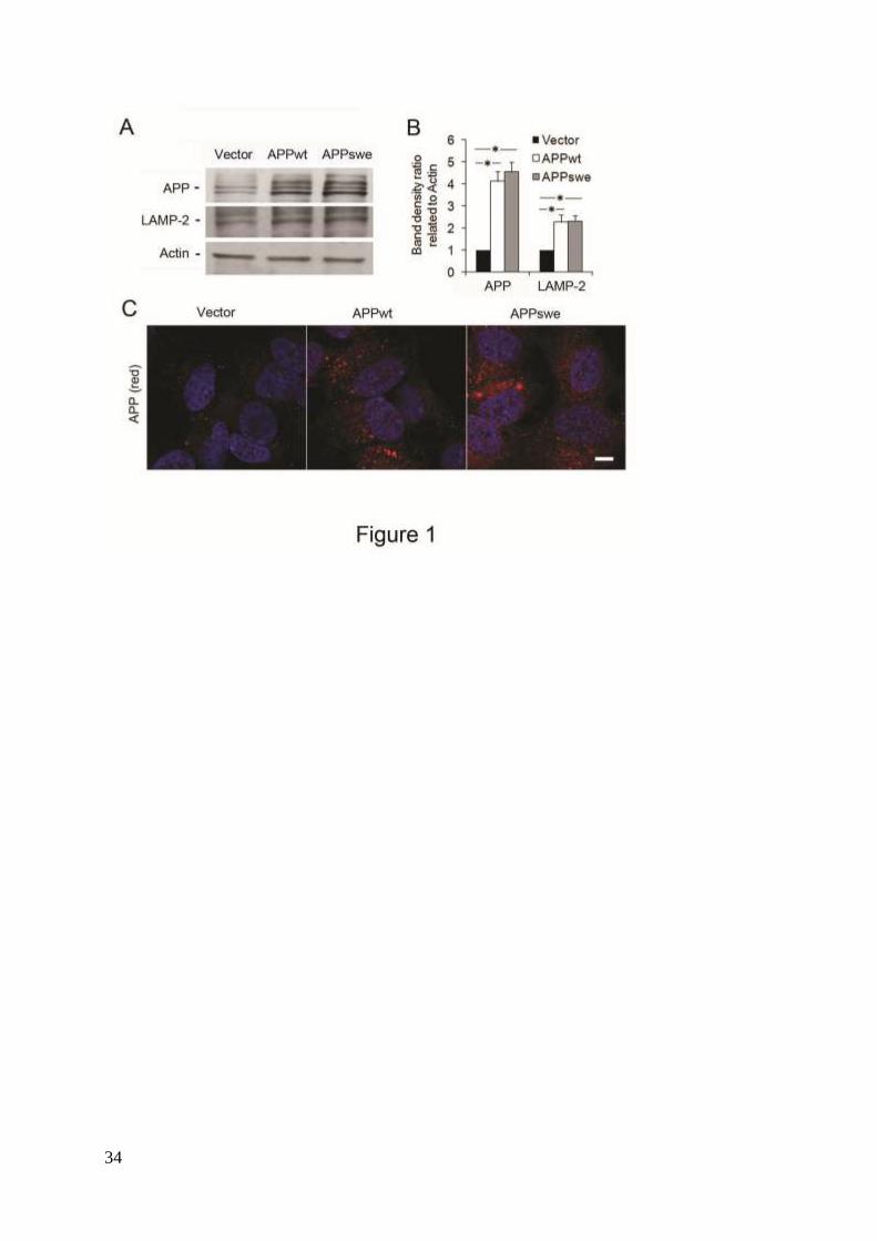
Intracellular APP and the lysosomal system in SH-SY5Y neuroblastoma cells stably
transfected with APPwt and APPswe. Western blotting of lysates from SH-SY5Y cells
transfected with APPwt or APPswe revealed that, under normal conditions, both APPwt and
APPswe produced approximately four times more APP than cells transfected with an empty
vector (P<0.05, Figs. 1A and B). This was confirmed by immunofluorescence studies (Fig.
1C).
Levels of LAMP-2 were approximately two-fold increased in APPwt and APPswe cells
compared to vector-transfected cells, (p<0.05, Figs. 1A and B), suggesting an upregulation of
the lysosomal system in APP-overexpressing cells.
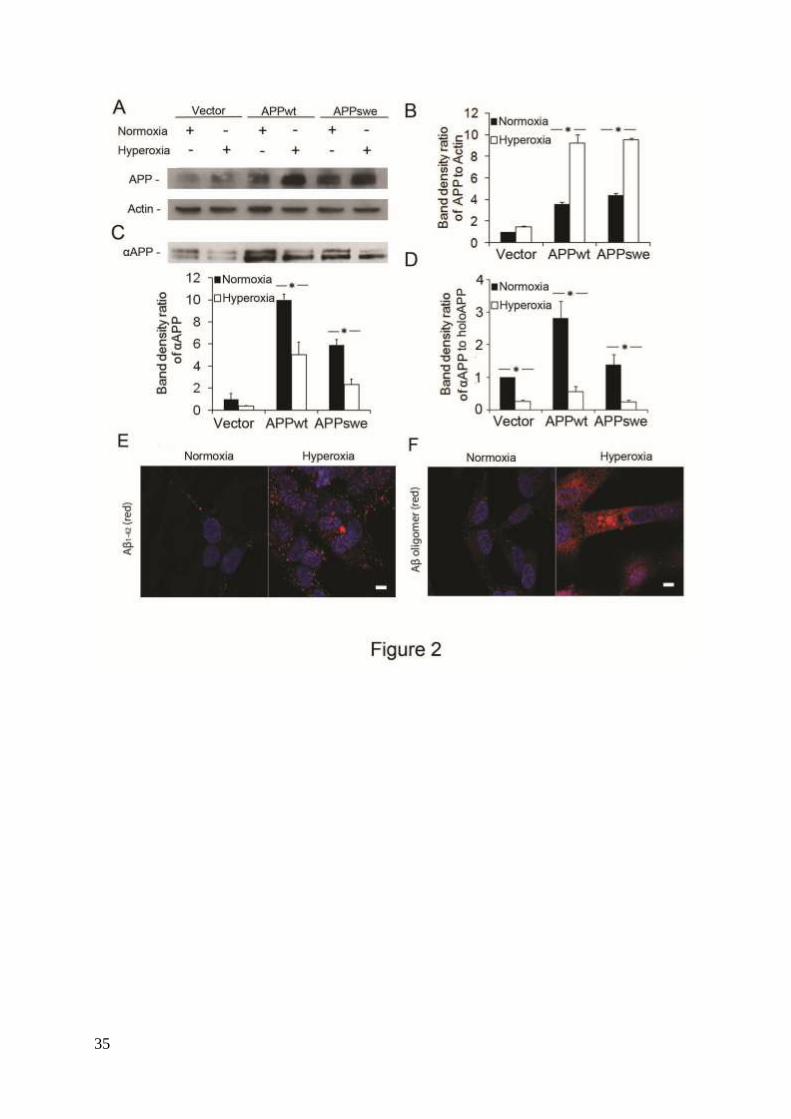
Increased production of APP and Aβ under hyperoxia. When SH-SY5Y cells were
exposed to hyperoxia (40% oxygen) for 5 days, the levels of intracellular APP increased as
compared to those observed under normoxic (8% oxygen) conditions. This was the case for
all cell lines studied, with significantly higher levels seen in APPwt and APPswe cells, P<0.05.
(Figs. 2A and B). As indication of non-amyloidogenic APP cleavage, 35
the ratio of αAPP
fragments in conditioned media against full intracellular APP (holoAPP) was calculated. After
cultivation of cells in either normoxia or hyperoxia for 5 days, the media was collected, and
both secreted αAPP and holoAPP were detected by western blotting using 6E10 antibodies.
Under normoxia, the ratio αAPP /holoAPP was considerably higher in APP-overexpressing
cells than in vector-transfected cells. For APPwt cells, αAPP levels and αAPP /holoAPP ratio
were twice as high as for APPswe cells (Figs. 2C and D). Following exposure to hyperoxia,
αAPP levels and the αAPP/ holoAPP ratio decreased significantly in all cell types (P<0.05,
Figs. 2C and D), suggesting that oxidative stress decreased α-secretase activity.
Immunofluorescence microscopy using antibodies against Aβ1-42 or A11 (anti-Aβ
9
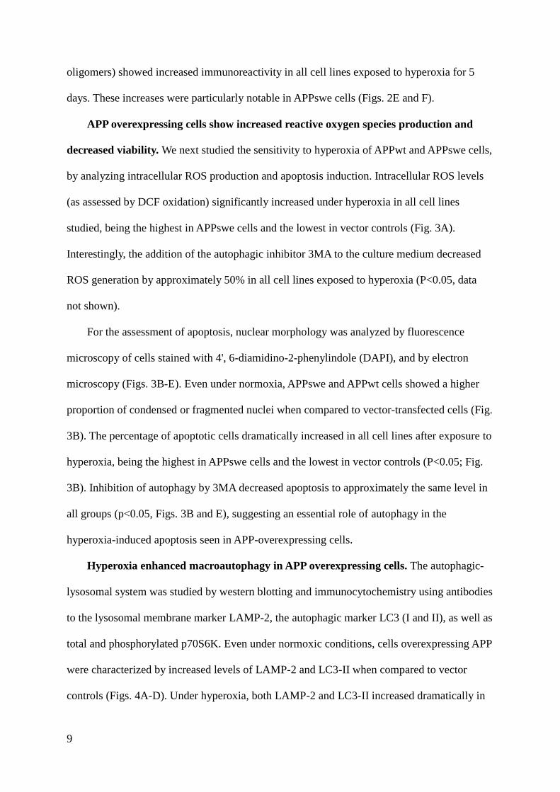
oligomers) showed increased immunoreactivity in all cell lines exposed to hyperoxia for 5
days. These increases were particularly notable in APPswe cells (Figs. 2E and F).
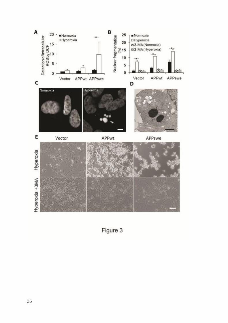
APP overexpressing cells show increased reactive oxygen species production and
decreased viability. We next studied the sensitivity to hyperoxia of APPwt and APPswe cells,
by analyzing intracellular ROS production and apoptosis induction. Intracellular ROS levels
(as assessed by DCF oxidation) significantly increased under hyperoxia in all cell lines
studied, being the highest in APPswe cells and the lowest in vector controls (Fig. 3A).
Interestingly, the addition of the autophagic inhibitor 3MA to the culture medium decreased
ROS generation by approximately 50% in all cell lines exposed to hyperoxia (P<0.05, data
not shown).
For the assessment of apoptosis, nuclear morphology was analyzed by fluorescence
microscopy of cells stained with 4', 6-diamidino-2-phenylindole (DAPI), and by electron
microscopy (Figs. 3B-E). Even under normoxia, APPswe and APPwt cells showed a higher
proportion of condensed or fragmented nuclei when compared to vector-transfected cells (Fig.
3B). The percentage of apoptotic cells dramatically increased in all cell lines after exposure to
hyperoxia, being the highest in APPswe cells and the lowest in vector controls (P<0.05; Fig.
3B). Inhibition of autophagy by 3MA decreased apoptosis to approximately the same level in
all groups (p<0.05, Figs. 3B and E), suggesting an essential role of autophagy in the
hyperoxia-induced apoptosis seen in APP-overexpressing cells.
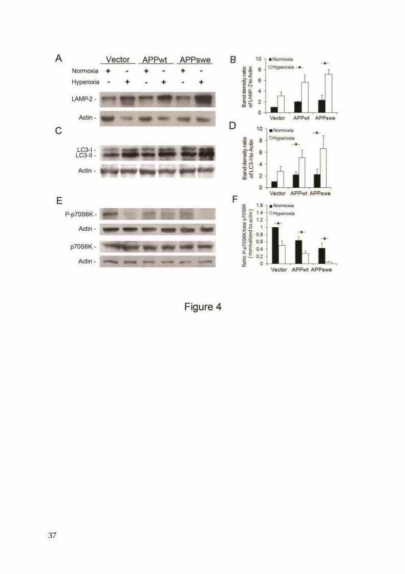
Hyperoxia enhanced macroautophagy in APP overexpressing cells. The autophagic-
lysosomal system was studied by western blotting and immunocytochemistry using antibodies
to the lysosomal membrane marker LAMP-2, the autophagic marker LC3 (I and II), as well as
total and phosphorylated p70S6K. Even under normoxic conditions, cells overexpressing APP
were characterized by increased levels of LAMP-2 and LC3-II when compared to vector
controls (Figs. 4A-D). Under hyperoxia, both LAMP-2 and LC3-II increased dramatically in
10
all cell lines, reaching the highest levels in APPswe cells (Figs. 4 A-D). In agreement with the
western blotting data, immunofluorescence microscopy showed an increased number and size
of LAMP-2 and LC3-positive vacuoles, respectively, in all cell lines exposed to hyperoxia,
but especially in APPswe cells (Supplementary Fig. 1). Moreover, the ratio of phosphorylated
p70S6K (P-p70S6K) to total p70S6K decreased in all cells lines after exposure to hyperoxia,
being the lowest in APPswe cells (Figs. 4E and F). These findings suggest that both APP
overexpression and hyperoxia enhanced macroautophagy, resulting in the upregulation of the
lysosomal system.
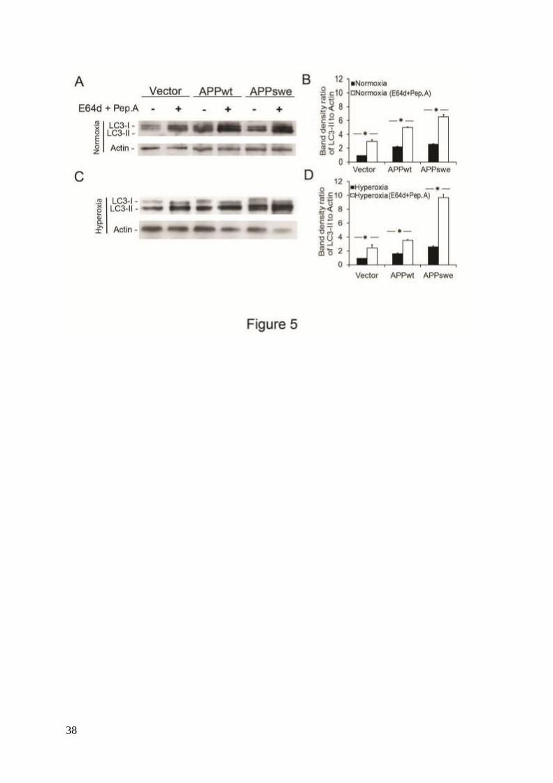
We next investigated the possibility that the increase of LC3-II seen in APP-
overexpressing and hyperoxia-exposed cells was associated with decreased autophagy flux
due to impaired lysosomal degradation. Cells were exposed to the lysosomal protease
inhibitors E64d and Pepstatin A, both inhibiting LC3-II degradation.14, 36
This resulted in an
even higher increase of LC3-II, suggesting a true upregulation of macroautophagic activity
(Figs. 5A-D).
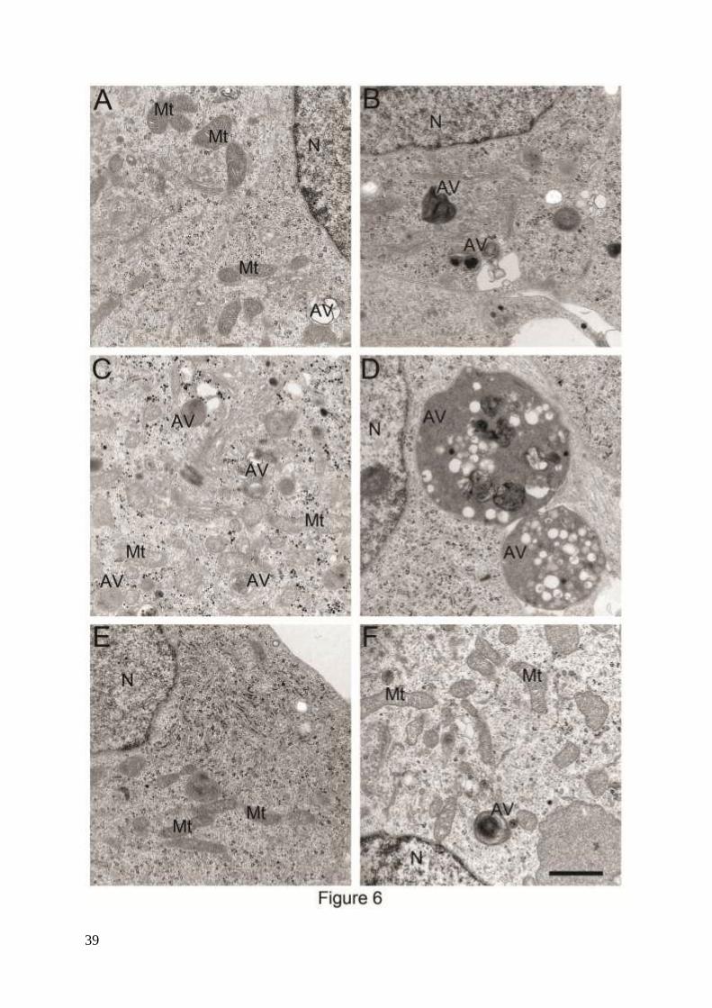
Furthermore, activation of macroautophagy by APP overexpression and hyperoxia was
demonstrated by electron microscopy (Fig. 6). In both empty vector and APPswe transfected
cells an increase in the number and size of autophagic vacuoles after exposure to hyperoxia
was detected (Figs. 6A-D). This was reversed by the treatment of cells with 3MA (Figs. 6E
and F). Various forms of autophagic vacuoles induced by hyperoxia in SH-SY5Y cells are
shown in supplementary Fig. 2. Similar types of vacuoles (reflecting different stages of
autophagic degradation) have also been described in AD neurons with an upregulated
lysosomal system.37
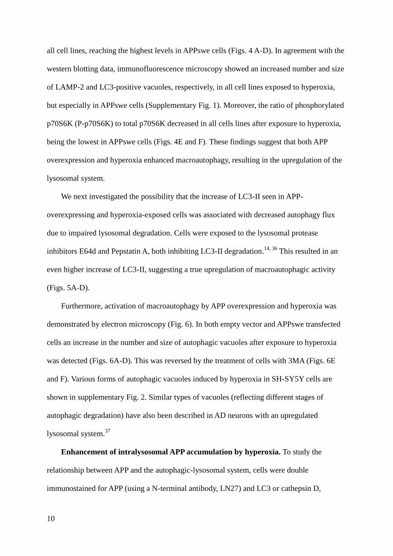
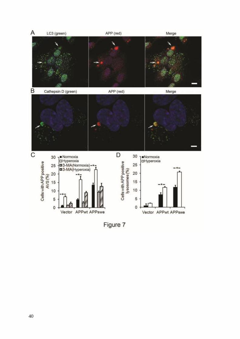
Enhancement of intralysosomal APP accumulation by hyperoxia. To study the
relationship between APP and the autophagic-lysosomal system, cells were double
immunostained for APP (using a N-terminal antibody, LN27) and LC3 or cathepsin D,
11
respectively. Co-localization of APP with LC3 or cathepsin D positive vacuoles was
significantly higher in APP-overexpressing cells compared to vector controls, with the highest
levels seen in APPswe cells. Hyperoxia greatly increased the number of APP-positive
autophagic vacuoles in all cell lines studied (Fig. 7). Exposure of cells to 3MA, (either in
normoxia or hyperoxia) dramatically reduced the number of APP-positive autophagic
vacuoles (Fig. 7C), suggesting that APP accumulates intralysosomally through
macroautophagy.
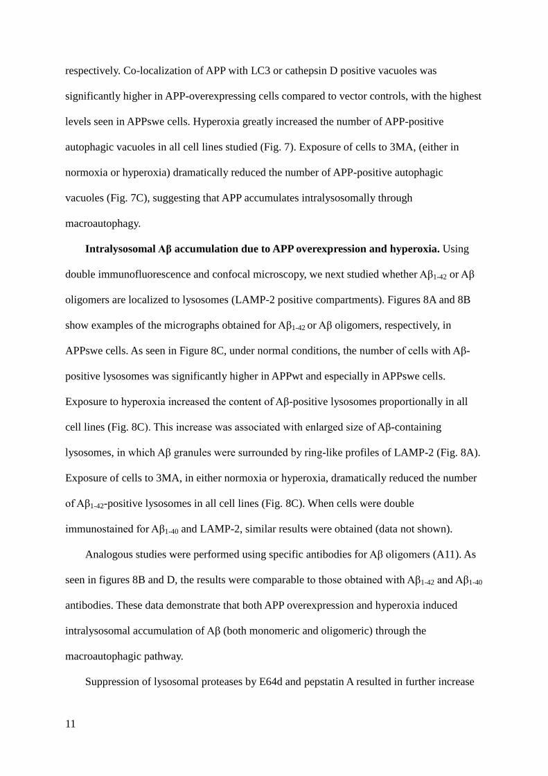
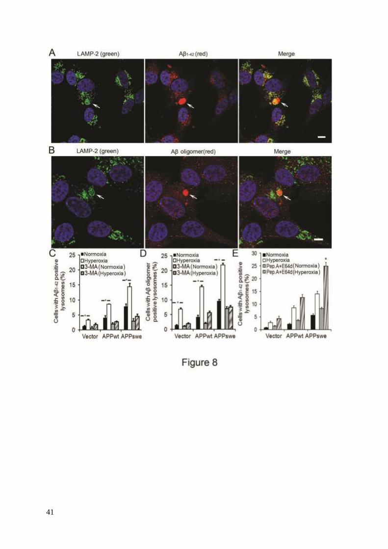
Intralysosomal Aβ accumulation due to APP overexpression and hyperoxia. Using
double immunofluorescence and confocal microscopy, we next studied whether Aβ1-42 or Aβ
oligomers are localized to lysosomes (LAMP-2 positive compartments). Figures 8A and 8B
show examples of the micrographs obtained for Aβ1-42 or Aβ oligomers, respectively, in
APPswe cells. As seen in Figure 8C, under normal conditions, the number of cells with Aβ-
positive lysosomes was significantly higher in APPwt and especially in APPswe cells.
Exposure to hyperoxia increased the content of Aβ-positive lysosomes proportionally in all
cell lines (Fig. 8C). This increase was associated with enlarged size of Aβ-containing
lysosomes, in which Aβ granules were surrounded by ring-like profiles of LAMP-2 (Fig. 8A).
Exposure of cells to 3MA, in either normoxia or hyperoxia, dramatically reduced the number
of Aβ1-42-positive lysosomes in all cell lines (Fig. 8C). When cells were double
immunostained for Aβ1-40 and LAMP-2, similar results were obtained (data not shown).
Analogous studies were performed using specific antibodies for Aβ oligomers (A11). As
seen in figures 8B and D, the results were comparable to those obtained with Aβ1-42 and Aβ1-40
antibodies. These data demonstrate that both APP overexpression and hyperoxia induced
intralysosomal accumulation of Aβ (both monomeric and oligomeric) through the
macroautophagic pathway.
Suppression of lysosomal proteases by E64d and pepstatin A resulted in further increase
12
of intralysosomal Aβ1-42 (Fig. 8E). This suggests that intralysosomal Aβ accumulation
resulting from APP overexpression + hyperoxia is likely associated with enhanced autophagic
uptake of APP and/or Aβ, rather than with the inhibition of lysosomal degradation.
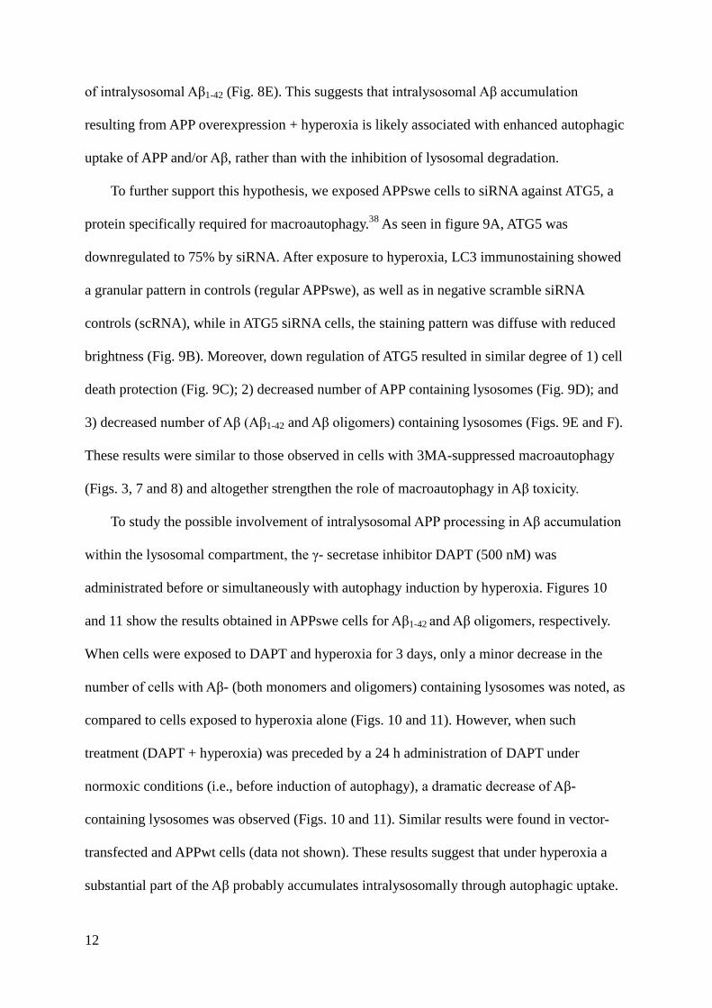
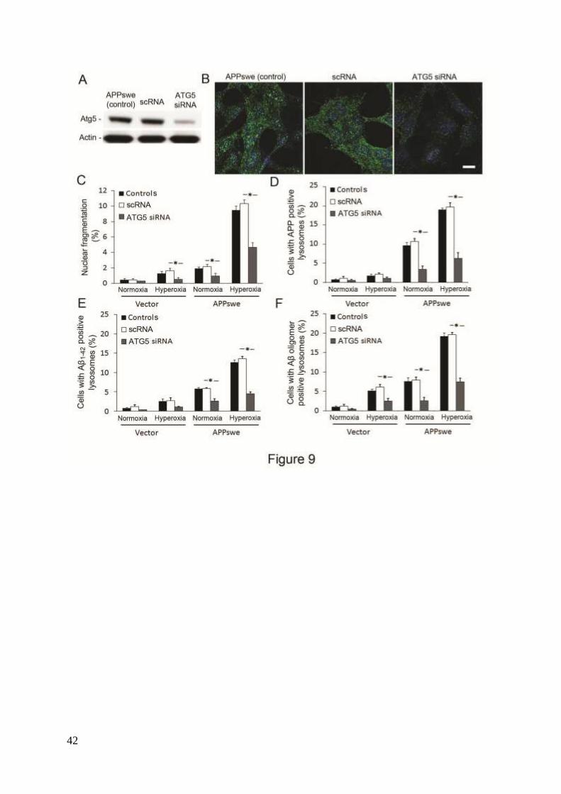
To further support this hypothesis, we exposed APPswe cells to siRNA against ATG5, a
protein specifically required for macroautophagy.38
As seen in figure 9A, ATG5 was
downregulated to 75% by siRNA. After exposure to hyperoxia, LC3 immunostaining showed
a granular pattern in controls (regular APPswe), as well as in negative scramble siRNA
controls (scRNA), while in ATG5 siRNA cells, the staining pattern was diffuse with reduced
brightness (Fig. 9B). Moreover, down regulation of ATG5 resulted in similar degree of 1) cell
death protection (Fig. 9C); 2) decreased number of APP containing lysosomes (Fig. 9D); and
3) decreased number of Aβ (Aβ1-42 and Aβ oligomers) containing lysosomes (Figs. 9E and F).
These results were similar to those observed in cells with 3MA-suppressed macroautophagy
(Figs. 3, 7 and 8) and altogether strengthen the role of macroautophagy in Aβ toxicity.
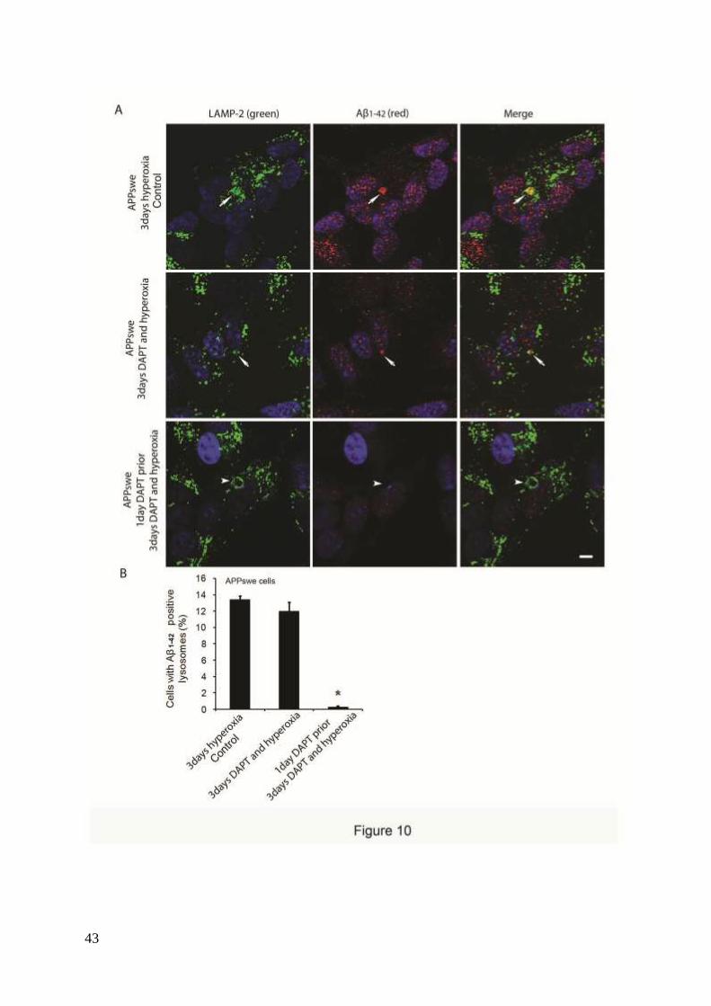
To study the possible involvement of intralysosomal APP processing in Aβ accumulation
within the lysosomal compartment, the γ- secretase inhibitor DAPT (500 nM) was
administrated before or simultaneously with autophagy induction by hyperoxia. Figures 10
and 11 show the results obtained in APPswe cells for Aβ1-42 and Aβ oligomers, respectively.
When cells were exposed to DAPT and hyperoxia for 3 days, only a minor decrease in the
number of cells with Aβ- (both monomers and oligomers) containing lysosomes was noted, as
compared to cells exposed to hyperoxia alone (Figs. 10 and 11). However, when such
treatment (DAPT + hyperoxia) was preceded by a 24 h administration of DAPT under
normoxic conditions (i.e., before induction of autophagy), a dramatic decrease of Aβ-
containing lysosomes was observed (Figs. 10 and 11). Similar results were found in vector-
transfected and APPwt cells (data not shown). These results suggest that under hyperoxia a
substantial part of the Aβ probably accumulates intralysosomally through autophagic uptake.
13
However, since we also noted an overall decrease of Aβ levels (both cytosolic and lysosomal)
after simultaneous inductions of autophagy and γ- secretase inhibition, the co- existence of
intralysosomal APP processing is also plausible.
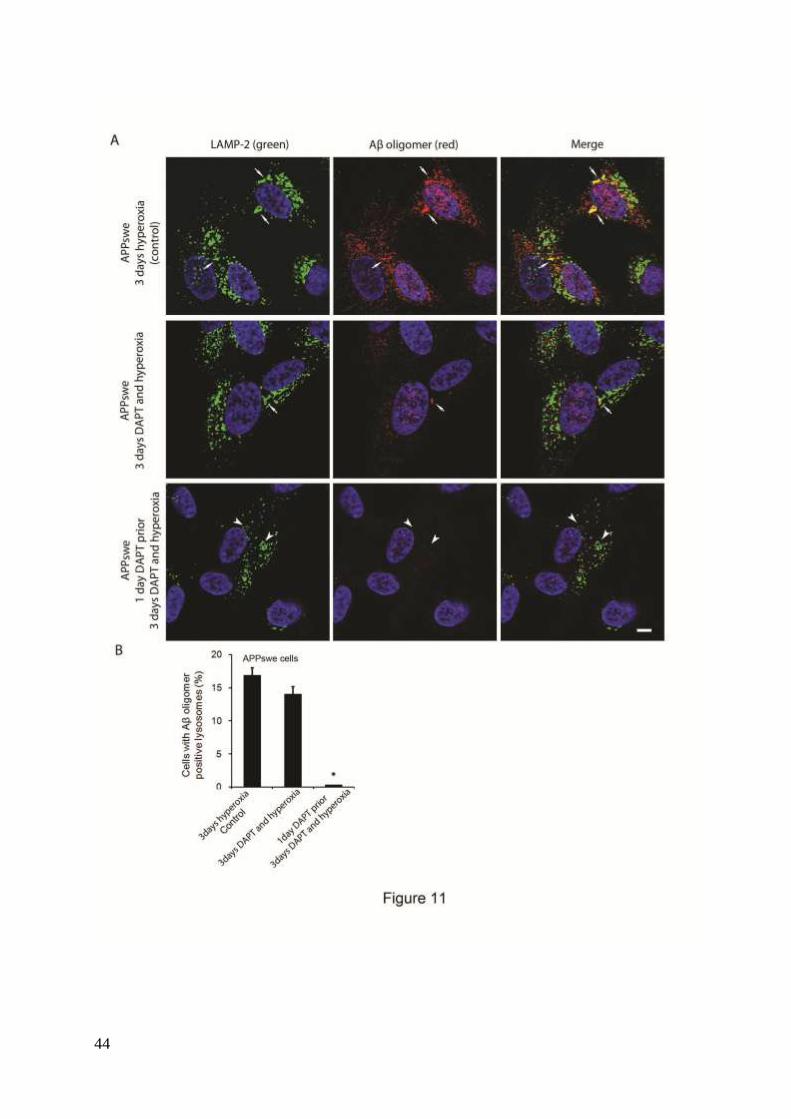
Aβ aggregation is pH-dependent, and oligomerization seems to be more rapid at a low
pH.39
To investigate if Aβ oligomerization takes place inside the lysosomes, or if Aβ
oligomers are delivered through autophagy, we increased lysosomal pH with NH4Cl and
exposed cells to hyperoxia for 3 days. No significant changes in the number of cells with Aβ
oligomer-containing lysosomes were found after NH4Cl treatment, although a slight decrease
(approximately 10%) was noted. These results suggest that, under hyperoxia, the majority of
intralysosomal Aβ oligomers were taken up through autophagic pathway.
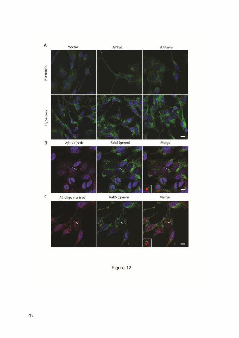
Finally, a possible involvement of endocytosis in the intralysosomal Aβ accumulation
was assessed by double immunostaining for Aβ1-42 and Rab5. Although hyperoxia was
associated with increased endocytosis, as suggested by increased number and size of Rab5-
positive granules, suggesting membrane distribution of Rab5 (Fig. 12A), no apparent
colocalization of Rab5 and Aβ (Aβ1-42 and Aβ oligomers) was noted (Figs. 12B and C).
14
Discussion
According to the free radical theory of aging, normal aerobic metabolism results in
unavoidable ROS-induced molecular damage leading to aging and age-associated disease,
including AD.24, 40
In our study, RA-differentiated SH-SY5Y human neuroblastoma cells that
overexpress APP (APPwt or APPswe) were exposed to chronic mild oxidative stress by
normobaric hyperoxia (40% ambient oxygen). These cells produced increased quantities of
APP and Aβ and showed decreased viability under oxidative stress conditions, as compared to
vector control cells. Both APPwt and APPswe cells, especially the latter, also showed
enhanced intracellular ROS production and increased macroautophagy. These effects were
particularly pronounced under hyperoxia, but some minor oxidative damage was also
observed under normal oxygen conditions. The enhanced sensitivity of cells to oxidant-
induced apoptosis was paralleled by increased intralysosomal accumulation of Aβ, both
monomeric and in form of oligomers. Moreover, inhibition of macroautophagy by either 3MA
treatment or down regulation of ATG5 prevented ROS production, intralysosomal
accumulation of Aβ and cell death. These results together suggest the important role of
macroautophagy in Aβ toxicity.
Our results raised the question to what extent the intralysosomal Aβ accumulation was a
result of increased autophagic uptake of Aβ or enhanced intralysosomal processing of APP.
Indeed, increased macroautophagy resulted in the intralysosomal accumulation of APP, some
of which is probably processed to Aβ as previously described.17
We showed that inhibition of
-secretase during autophagic induction by hyperoxia caused only a minor decrease in the
number of lysosomes containing Aβ, either monomers or oligomers. In contrast, inhibition of
-secretase prior to autophagy induction resulted in a substantial decrease of Aβ in the
lysosomes, suggesting that, in our paradigm, a large part of intralysosomal Aβ was delivered
15
through macroautophagy. However, since simultaneous inductions of autophagy and γ-
secretase inhibition also caused some decrease of Aβ levels in the lysosomes and cytosol,
intralysosomal production of A from APP (previously demonstrated by Yu et al.17
) probably
occurs as well. Such possibility is supported by our finding of enhanced intralysosomal APP
accumulation due to hyperoxia.
The lack of colocalization of Aβ and Rab5 suggests that the intralysosomal Aβ did not
originate from previously secreted Aβ that could enter cells via endocytosis. Nevertheless, Aβ
has been found in many intracellular sites, such as mitochondria, lysosomes, endosomes,
cytosol, ER and Golgi complexes.6 Any of these intracellular sites could be the source for
macroautophagic delivery of Aβ to lysosomes seen in our system.
It has been suggested that the acidic lysosomal environment can promote formation of
Aβ oligomers and protofibrils39
and that intralysosomally accumulated Aβ damages lysosomal
membranes, resulting in the leak of acid hydrolases into the cytosol and apoptotic death as
described in our previous study.34
In support of these results, we show that the prevention of
intralysosomal Aβ (Aβ1-42 and oligomeric Aβ) accumulation by macroautophagy inhibition
rescued the cells. Surprisingly, the increase of lysosomal pH using NH4Cl resulted only in a
slight decrease in intralysosomal oligomeric Aβ. It is possible that NH4Cl treatment did not
affect Aβ oligomerization outside the lysosomal compartment, and these oligomers would
have been delivered to the lysosomes through autophagy. Also, it has been demonstrated that
membranes of different cellular compartments, such as the lysosomal membrane, can initiate
Aβ aggregation regardless of pH.41
Thus, when Aβ gradually accumulates intralysosomally,
its concentrations can finally reach levels resulting in aggregation.42
Ling et al43
demonstrated that the lysosomal system in Aβ expressing Drosophila flies
loose degradative capacity with aging and that autophagy induction in the aged flies decreased
their survival. In the human brain, there is a massive accumulation of undegraded material
16
within autophagic vacuoles in dystrophic neurons, indicating an impaired lysosomal
function.17, 44
Thus, it is possible that the increased intralysosomal Aβ seen in hyperoxia could
be the consequence of deficient lysosomal degradation. To explore this possibility, we
inhibited lysosomal proteases with E64d and pepstatin A, which resulted in enhanced
accumulation of intralysosomal Aβ. Since E64d and pepstatin A are also and -secretase
inhibitors, respectively,45, 46
we found an overall decrease in cytosolic A (data not shown).
These results are in agreement with the conception that at least some lysosomal degradative
capacity was still present under hyperoxia.
Why do APP-overexpressing cells show elevated ROS production and increased
macroautophagy? The most plausible explanation is that Aβ acts as a pro-oxidant. 27, 28, 47
Because APPwt and APPswe cells produce more Aβ than vector controls, they would also
generate more ROS under the same environmental conditions. Oxidative stress, in turn, can
increase APP and Aβ formation, as shown in this report and our earlier observation,34
giving
rise to an amplifying oxidative stress - Aβ loop. As a consequence, the APPwt and APPswe
cells would also show higher autophagic activity, which upregulates in an attempt to repair
ROS-induced molecular damage. Hence, APP-overexpressing cells experience triple
pathogenic effects of increased Aβ production, increased oxidative stress, and increased
macroautophagy – all contributing to intralysosomal Aβ accumulation and consequent
apoptotic cell death.
An inhibitor of autophagic sequestration, 3MA, decreased the content of APP- and Aβ-
containing lysosomes in APPwt and APPswe cells approximately to the level seen in vector
control cells, indicating that APP and Aβ (including toxic Aβ oligomers) accumulate within
the lysosomal compartment through macroautophagy. Also, 3MA decreased intracellular
levels of ROS in all three cell lines exposed to hyperoxia, probably suggesting the
involvement of substances delivered to lysosomes through autophagy, for example, transition
17
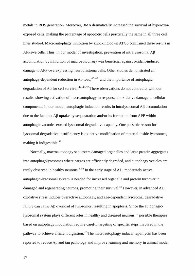
metals in ROS generation. Moreover, 3MA dramatically increased the survival of hyperoxia-
exposed cells, making the percentage of apoptotic cells practically the same in all three cell
lines studied. Macroautophagy inhibition by knocking down ATG5 confirmed these results in
APPswe cells. Thus, in our model of investigation, prevention of intralysosomal Aβ
accumulation by inhibition of macroautophagy was beneficial against oxidant-induced
damage to APP-overexpressing neuroblastoma cells. Other studies demonstrated an
autophagy-dependent reduction in Aβ load,42, 48
and the importance of autophagic
degradation of Aβ for cell survival.42, 49-52
These observations do not contradict with our
results, showing activation of macroautophagy in response to oxidative damage to cellular
components. In our model, autophagic induction results in intralysosomal Aβ accumulation
due to the fact that Aβ uptake by sequestration and/or its formation from APP within
autophagic vacuoles exceed lysosomal degradative capacity. One possible reason for
lysosomal degradative insufficiency is oxidative modification of material inside lysosomes,
making it indigestible.53
Normally, macroautophagy sequesters damaged organelles and large protein aggregates
into autophagolysosomes where cargos are efficiently degraded, and autophagy vesicles are
rarely observed in healthy neurons.9, 54
In the early stage of AD, moderately active
autophagic-lysosomal system is needed for increased organelle and protein turnover in
damaged and regenerating neurons, promoting their survival.55
However, in advanced AD,
oxidative stress induces overactive autophagy, and age-dependent lysosomal degradative
failure can cause Aβ overload of lysosomes, resulting in apoptosis. Since the autophagic-
lysosomal system plays different roles in healthy and diseased neurons,10
possible therapies
based on autophagy modulation require careful targeting of specific steps involved in the
pathway to achieve efficient digestion.37
The macroautophagy inducer rapamycin has been
reported to reduce Aβ and tau pathology and improve learning and memory in animal model
18
of AD, when lysosomes maintained good digestive function.51
However, when lysosomal
digestive ability is suppressed, stimulation of autophagy may accelerate the course of the
disease, while its inhibition may prevent the intralysosomal accumulation of Aβ and other
toxic materials, promoting cell survival.
In summary, our results indicate that macroautophagy-generated intralysosomal increase
of Aβ is essential for oxidant-induced apoptosis, providing additional support for the
interactive role of oxidative stress and the lysosomal system in AD-related neurodegeneration.
19
Materials and methods
Cells and culture conditions. Human SH-SY5Y neuroblastoma cells were obtained from the
American Type Culture Collection (ATCC, CRL-2266™) and stably transfected with an
empty pcDNA 3.1 vector containing a cytomegalovirus promoter, or wild-type APP695
(APPwt), or APP Swedish KM670/671NL double mutation (APPswe) using Lipofectamine
TM2000 according to the manufacturer’s (Invitrogen, 11668027) instructions. Overexpression
of APP was confirmed by western blotting and fluorescence microscopy (Fig. 1).
Transfected cells were cultured in Minimum Essential Medium with Glutamax
(Invitrogen, 32561-037) containing 10% fetal bovine serum and 200 μl/ml geneticin (for
selection of transfected cells, Invitrogen, 10131035) in the atmosphere of 8% O2, 87% N2 and
5% CO2 at 37oC, (normal conditions) in 75 cm
2 plastic culture flasks (Corning, 430641). For
differentiation, neuroblastoma cells were exposed to 10 µM all-trans RA (Sigma, R-2625) for
14 days. The medium was changed every second day.
Differentiated cells were plated in plastic Petri dishes at a density of 104 cells per cm
2.
After 24 hours, the culture medium was changed to the serum-free OptiMEM1 (Invitrogen,
51985-026) supplemented with 10 µM RA and 200 μg/ml geneticin. The cells were divided
into two groups: 1) cells cultured under normoxia, i.e., 8% O2, 87% N2 and 5% CO2; 2) cells
cultured under hyperoxia, i.e., 40% O2, 55% N2 and 5% CO2 (chronic oxidative stress).
In some experiments, for the increase of lysosomal pH, cultures were treated with 10 mM
ammonium chloride (NH4Cl, Sigma, 443093) for 3 days.
Inhibition of macroautophagy. 3-methyladenine (3MA; Sigma, P0899) and ATG5
siRNA (Qiagen, 1027416) were used to inhibit autophagic sequestration. For 3MA treatment
(1 mM, 5 days), differentiated cells were plated on coverslips in plastic Petri dishes at a
density of 104 cells per cm
2. After 24 hours, the culture medium was changed to the serum-
20
free OptiMEM1 supplemented with 10 µM RA and 200 μg/ml geneticin and 1 mM 3MA.
After 5 days, cells and culture medium were used for analysis.
For macroautophagy inhibition by RNA interference, siRNA against ATG5 (Quiagen,
1027416) or negative scramble siRNA controls, scRNA (Qiagen, 1022076) were used. Cells
were seeded on coverslips in 12 well plates in serum medium and transfected with 25 nM
ATG5 siRNA for 48 h by using HiPerFect Transfection Reagent (Qiagen, 301704) following
the manusfacturer’s protocol. Before hyperoxia exposure, the medium was changed to serum
free OptiMEM1 containing 25 nM ATG5 siRNA. The efficacy of RNA interference was
assessed by western blot analysis of Atg5 and LC3.
Detection of macroautophagy. Detection of macroautophagy was performed by several
methods: 14
1) Western blotting of LC3 (performed on 15% SDS page gels). The density of
LC3-II band (16 KDa) indicated autophagosome formation (detail in western blot section
below). 2) Immunocytochemistry of LC3 for visualization autophagic vacuoles (details in
immunocytochemistry section). 3) The ratio of P-p70S6K to p70S6K, representing induction
of autophagy (detail in western blot section below). 4) Autophagy flux was evaluated by
western blotting of LC3 after exposure of cells to 10 µM E64d (an inhibitor of cysteine
proteases, Sigma, E8640) and 10 µg/ml Pepstatin A (an inhibitor of aspartic peptidases, Sigma,
P5318) during normoxia or hyperoxia. 5) Transmission electron microscopy: cells in 100×20
mm Petri dishes were fixed in 2% glutaraldehyde in 0.1 M sodium cacodylate buffer
containing 0.1 M sucrose and 3 mM CaCl2, pH 7.4 at room temperature for 30 min then
overnight at 4°C. After fixation, cells were rinsed in 0.1 M phosphate buffer, pH 7.4 and
centrifuged. Pellets were then post-fixed in 2% osmium tetroxide in 0.1 M phosphate buffer,
pH 7.4 at 4°C for 2 hours, dehydrated in ethanol followed by acetone and embedded in LX-
112 (Ladd, Burlington, Vermont, USA). Sections were contrasted with uranyl acetate followed
by lead citrate and examined in a Tecnai 12 transmission electron microscope (FEI,
21
Eindhoven, The Netherlands) at 80 kV. Digital images were taken using a Veleta digital
camera (Soft Imaging System, GmbH, Münster, Germany).
Western blot analysis. Detection of α-secretase processed APP (α-APP) was performed
as previously described. 56
75 µl medium of each sample was loaded to the gels.
Immunoblotting was performed as described elsewhere 57
using primary antibodies at 1:1000
dilution: anti-Aβ1-17 (6E10, Signet Laboratories, SIG-39320), anti-lysosomal associated
membrane protein-2 (CD107b/LAMP-2, Southern Biotechnology, 9840-01), anti-autophagy
related protein LC3B (LC3, Novus Biologicals, NB600-1384B), anti-p70 S6 Kinase (Cell
Signaling, 9202S), anti-Phospho-p70 S6 Kinase (Cell signaling, 9206S), or anti-actin (Sigma-
Aldrich, A 2668). They were followed by anti-rabbit (Amersham, 384927) or anti-mouse
(Amersham, 380199) horseradish peroxidase-linked secondary antibodies at 1:2000 dilution.
Immunoreactivity was detected by the ECL detection system (Amersham, RPN2209) and
exposure to Hyper film MP (Amersham, 28906837). Some immunoblots were stripped using
RestoreTM
Western Blot Stripping buffer (Pierce, 21059) at room temperature for 15 minutes,
and then re-blotted with other antibodies. The films were scanned and the quantification of
immunoblots was performed using Image J program (available at http://rsbweb.nih.gov/ij/).
The relative amount of protein corresponding to an immunoreactive band was calculated as a
product of average optical density of the band by its area and expressed in arbitrary units.
Immunofluorescence microscopy. Cells were prepared for immunocytochemistry as
described earlier 33
. Formaldehyde-fixed cells were incubated with primary antibodies against
Aβ1-42 (1:100 dilution, Chemicon, AB5078P), Aβ1-40 (1:100 dilution, Chemicon, AB5074B),
amyloid oligomers (A11, 1:100 dilution, Invitrogen, AHB0052), amino terminus of APP
(LN27, 1:100 dilution, Zymed Laboratories, 13-0200), CD107b/LAMP-2 (1:400 dilution,
Southern Biotechnology, 9840-01), LC3 (1:100 dilution, Novus Biologicals, NB600-1384B),
Rab5 (1:200 dilution, BD Biosciences Pharmingen, sc 46692), or cathepsin D (1:100, Upstate
22
Biotechnology, 06-467). Alexa Fluor 488 conjugated goat anti mouse IgG or Alexa Fluor 594
conjugated goat anti rabbit IgG (both diluted 1:400, Molecular Probes, A11029 or A11037,
respectively) were used as secondary antibodies. For double immunofluorescence, a
combination of two different primary antibodies was followed by a combination of two
different secondary antibodies. The specimens were mounted in Vectashield containing DAPI
(Vector Laboratories, H-1200) and inspected with a Nikon Eclipse E600 W confocal
microscope using a 488 nm argon laser and 543 nm helium-neon laser. Cells with one or more
Aβ (including Aβ1-40, Aβ1-42 and Aβ oligomers) or APP positive autophagic
vacuoles/lysosomes (usually exceeding 1 µm in diameter) were randomly selected and
counted under a Nikon Microphot-SA microscope using both phase contrast and fluorescence
illuminations. The percentage of these cells was calculated for each specimen and averaged
within each experimental group (n=3). At least 300 randomly selected cells in each specimen
(900 cells in one group) were counted.
Detection of cell death. As previously described, 33
formaldehyde-fixed cells were
mounted in Vectashield containing DAPI. Cells were randomly selected, and those with
condensed and/or fragmented nuclei (considered as apoptotic cells) were counted under a
Nikon Microphot-SA microscope using both phase contrast and fluorescence illuminations
(330–380/420 nm excitation/barrier filter). The percentage of these cells was calculated for
each specimen and averaged within each experimental group (n=3). At least 300 cells in each
specimen (900 cells in one group) were counted.
Inhibition of γ-secretase. . LY-374973, N-[N-(3,5-Difluorophenacetyl)-L-alanyl]-S-
phenylglycine t-butyl ester (DAPT), a γ- secretase inhibitor (Sigma, D5942) was used at a
concentration of 500 nM to inhibit Aβ production. Three different conditions were used: 1)
cells cultured under hyperoxia for 3 days. 2) Cells exposed to hyperoxia and DAPT for 3 days.
3) Cells pretreated with DAPT for 1 day, and then exposed to hyperoxia and DAPT for 3 days.
23
Measurement of intracellular reactive oxygen species production. Intracellular
reactive oxygen species (ROS) production was detected by carboxy-H2DCFDA (DCF,
Invitrogen, C-400) oxidation that was assessed by flow cytometry. Cells cultured in 12 well
plates were washed, incubated with 10 µM DCF in serum-free medium at 37oC for 15 min.
The fluorescence of 10,000 cells was analyzed in an LSR flow cytometer (Becton-Dickinson)
using a 488 nm argon laser and CellQuest software.
Statistical analysis. Values are given as mean ± SD. The results were analyzed for
statistical significance using the Mann-Whitney U test for two-group comparisons and
Kruskal-Wallis test for multi-group comparisons. P values ≤ 0.05 were considered significant.

24
Acknowledgments
This work was supported by the Gustav V and Queen Victoria Foundation (JM), County
Council of Östergötland (JM, LZ, MH), Stiftelsen Olle Engkvist Byggmästare (LZ, KK),
Stiftelsen för Gamla Tjänarinnor (LZ, AC-M), Gun och Bertil Stohnes Stiftelse (LZ, AC-M),
Lions forskningsfond (LZ), Svenska Lundbeckstiftelsen (LZ), Karolinska Institute Fund for
Geriatric Research (AC-M), Alice och Knut Wallenberg Stiftelse (AC-M), Swedish Alzheimer
Foundation (KK, MH) and The Swedish Brain Power (AC-M). The authors thank Lisbeth
Hjälle and Åsa-Lena Dackland for excellent technical and flow cytometry assistance,
respectively, and Kjell Hultenby’s group for transmission electron microscopy.
25
References
1. Mattson MP. Pathways towards and away from Alzheimer's disease. Nature 2004; 430:631-9.
2. Zheng H, Koo EH. The amyloid precursor protein: beyond amyloid. Mol Neurodegener 2006; 1:5.
3. Glabe C. Intracellular mechanisms of amyloid accumulation and pathogenesis in Alzheimer's disease. J Mol
Neurosci 2001; 17:137-45.
4. Irvine GB, El-Agnaf OM, Shankar GM, Walsh DM. Protein aggregation in the brain: the molecular basis for
Alzheimer's and Parkinson's diseases. Mol Med 2008; 14:451-64.
5. Haass C, Selkoe DJ. Soluble protein oligomers in neurodegeneration: lessons from the Alzheimer's amyloid
beta-peptide. Nat Rev Mol Cell Biol 2007; 8:101-12.
6. LaFerla FM, Green KN, Oddo S. Intracellular amyloid-beta in Alzheimer's disease. Nat Rev Neurosci 2007;
8:499-509.
7. Koistinaho M, Ort M, Cimadevilla JM, Vondrous R, Cordell B, Koistinaho J, et al. Specific spatial learning
deficits become severe with age in beta -amyloid precursor protein transgenic mice that harbor diffuse beta -
amyloid deposits but do not form plaques. Proc Natl Acad Sci U S A 2001; 98:14675-80.
8. Gouras GK, Tsai J, Naslund J, Vincent B, Edgar M, Checler F, et al. Intraneuronal Abeta42 accumulation in
human brain. The American journal of pathology 2000; 156:15-20.
9. Kim J, Klionsky DJ. Autophagy, cytoplasm-to-vacuole targeting pathway, and pexophagy in yeast and
mammalian cells. Annu Rev Biochem 2000; 69:303-42.
10. Shintani T, Klionsky DJ. Autophagy in health and disease: a double-edged sword. Science 2004; 306:990-5.
11. Yorimitsu T, Klionsky DJ. Autophagy: molecular machinery for self-eating. Cell Death Differ 2005; 12
Suppl 2:1542-52.
12. Cuervo AM. Autophagy and aging: keeping that old broom working. Trends Genet 2008; 24:604-12.
13. Luzio JP, Poupon V, Lindsay MR, Mullock BM, Piper RC, Pryor PR. Membrane dynamics and the
biogenesis of lysosomes. Mol Membr Biol 2003; 20:141-54.
14. Klionsky DJ, Abeliovich H, Agostinis P, Agrawal DK, Aliev G, Askew DS, et al. Guidelines for the use and
interpretation of assays for monitoring autophagy in higher eukaryotes. Autophagy 2008; 4:151-75.
15. Yu WH, Kumar A, Peterhoff C, Shapiro Kulnane L, Uchiyama Y, Lamb BT, et al. Autophagic vacuoles are
enriched in amyloid precursor protein-secretase activities: implications for beta-amyloid peptide over-production
and localization in Alzheimer's disease. Int J Biochem Cell Biol 2004; 36:2531-40.
16. Adamec E, Mohan PS, Cataldo AM, Vonsattel JP, Nixon RA. Up-regulation of the lysosomal system in
experimental models of neuronal injury: implications for Alzheimer's disease. Neurosci 2000; 100:663-75.
17. Yu WH, Cuervo AM, Kumar A, Peterhoff CM, Schmidt SD, Lee JH, et al. Macroautophagy--a novel Beta-
amyloid peptide-generating pathway activated in Alzheimer's disease. J Cell Biol 2005; 171:87-98.
18. Langui D, Girardot N, El Hachimi KH, Allinquant B, Blanchard V, Pradier L, et al. Subcellular topography
of neuronal Abeta peptide in APPxPS1 transgenic mice. The American journal of pathology 2004; 165:1465-77.
19. Ditaranto K, Tekirian TL, Yang AJ. Lysosomal membrane damage in soluble Abeta-mediated cell death in
Alzheimer's disease. Neurobiol Dis 2001; 8:19-31.
20. Yang AJ, Chandswangbhuvana D, Margol L, Glabe CG. Loss of endosomal/lysosomal membrane
impermeability is an early event in amyloid Abeta1-42 pathogenesis. J Neurosci Res 1998; 52:691-8.
21. Terman A, Neuzil J, Kagedal K, Ollinger K, Brunk UT. Decreased apoptotic response of inclusion-cell
disease fibroblasts: a consequence of lysosomal enzyme missorting? Exp Cell Res 2002; 274:9-15.
22. Brunk UT, Dalen H, Roberg K, Hellquist HB. Photo-oxidative disruption of lysosomal membranes causes
apoptosis of cultured human fibroblasts. Free Radic Biol Med 1997; 23:616-26.
23. Hardy J, Selkoe DJ. The amyloid hypothesis of Alzheimer's disease: progress and problems on the road to
therapeutics. Science 2002; 297:353-6.
24. Harman D. Aging: a theory based on free radical and radiation chemistry. J Gerontol 1956; 11:298-300.
25. Sohal RS, Weindruch R. Oxidative stress, caloric restriction, and aging. Science 1996; 273:59-63.
26. Nunomura A, Castellani RJ, Zhu X, Moreira PI, Perry G, Smith MA. Involvement of oxidative stress in
Alzheimer disease. J Neuropathol Exp Neurol 2006; 65:631-41.
27. Cedazo-Minguez A, Huttinger M, Cowburn RF. Beta-VLDL protects against A beta(1-42) and apoE toxicity
in human SH-SY5Y neuroblastoma cells. Neuroreport 2001; 12:201-6.
28. Akterin S, Cowburn RF, Miranda-Vizuete A, Jimenez A, Bogdanovic N, Winblad B, et al. Involvement of
glutaredoxin-1 and thioredoxin-1 in beta-amyloid toxicity and Alzheimer's disease. Cell Death Differ 2006;
13:1454-65.
29. Zhu X, Su B, Wang X, Smith MA, Perry G. Causes of oxidative stress in Alzheimer disease. Cell Mol Life
Sci 2007; 64:2202-10.
30. Ding Q, Dimayuga E, Keller JN. Oxidative damage, protein synthesis, and protein degradation in
Alzheimer's disease. Current Alzheimer research 2007; 4:73-9.
31. Bonda DJ, Wang X, Perry G, Nunomura A, Tabaton M, Zhu X, et al. Oxidative stress in Alzheimer disease:
26
A possibility for prevention. Neuropharmacology 2010.
32. Engelhart MJ, Geerlings MI, Ruitenberg A, van Swieten JC, Hofman A, Witteman JC, et al. Dietary intake
of antioxidants and risk of Alzheimer disease. JAMA 2002; 287:3223-9.
33. Zheng L, Roberg K, Jerhammar F, Marcusson J, Terman A. Autophagy of amyloid beta-protein in
differentiated neuroblastoma cells exposed to oxidative stress. Neurosci Lett 2006; 394:184-9.
34. Zheng L, Kagedal K, Dehvari N, Benedikz E, Cowburn R, Marcusson J, et al. Oxidative stress induces
macroautophagy of amyloid beta-protein and ensuing apoptosis. Free Radic Biol Med 2009; 46:422-9.
35. Hooper NM, Turner AJ. The search for alpha-secretase and its potential as a therapeutic approach to
Alzheimer s disease. Curr Med Chem 2002; 9:1107-19.
36. Mizushima N, Yoshimori T. How to interpret LC3 immunoblotting. Autophagy 2007; 3:542-5.
37. Nixon RA. Autophagy, amyloidogenesis and Alzheimer disease. J Cell Sci 2007; 120:4081-91.
38. Kuma A, Hatano M, Matsui M, Yamamoto A, Nakaya H, Yoshimori T, et al. The role of autophagy during
the early neonatal starvation period. Nature 2004; 432:1032-6.
39. Su Y, Chang PT. Acidic pH promotes the formation of toxic fibrils from beta-amyloid peptide. Brain Res
2001; 893:287-91.
40. Harman D. Free radical theory of aging: dietary implications. Am J Clin Nutr 1972; 25:839-43.
41. Waschuk SA, Elton EA, Darabie AA, Fraser PE, McLaurin JA. Cellular membrane composition defines A
beta-lipid interactions. J Biol Chem 2001; 276:33561-8.
42. Tian Y, Bustos V, Flajolet M, Greengard P. A small-molecule enhancer of autophagy decreases levels of
A{beta} and APP-CTF via Atg5-dependent autophagy pathway. FASEB J 2011; 25:1934-42.
43. Ling D, Song HJ, Garza D, Neufeld TP, Salvaterra PM. Abeta42-induced neurodegeneration via an age-
dependent autophagic-lysosomal injury in Drosophila. PLoS One 2009; 4:e4201.
44. Nixon RA, Wegiel J, Kumar A, Yu WH, Peterhoff C, Cataldo A, et al. Extensive involvement of autophagy
in Alzheimer disease: an immuno-electron microscopy study. J Neuropathol Exp Neurol 2005; 64:113-22.
45. Hook VY, Kindy M, Hook G. Inhibitors of cathepsin B improve memory and reduce beta-amyloid in
transgenic Alzheimer disease mice expressing the wild-type, but not the Swedish mutant, beta-secretase site of
the amyloid precursor protein. J Biol Chem 2008; 283:7745-53.
46. Tian G, Sobotka-Briner CD, Zysk J, Liu X, Birr C, Sylvester MA, et al. Linear non-competitive inhibition of
solubilized human gamma-secretase by pepstatin A methylester, L685458, sulfonamides, and benzodiazepines. J
Biol Chem 2002; 277:31499-505.
47. Yatin SM, Varadarajan S, Butterfield DA. Vitamin E Prevents Alzheimer's Amyloid beta-Peptide (1-42)-
Induced Neuronal Protein Oxidation and Reactive Oxygen Species Production. J Alzheimers Dis 2000; 2:123-31.
48. Hung SY, Huang WP, Liou HC, Fu WM. Autophagy protects neuron from Abeta-induced cytotoxicity.
Autophagy 2009; 5:502-10.
49. Yang DS, Stavrides P, Mohan PS, Kaushik S, Kumar A, Ohno M, et al. Reversal of autophagy dysfunction
in the TgCRND8 mouse model of Alzheimer's disease ameliorates amyloid pathologies and memory deficits.
Brain 2011; 134:258-77.
50. Sun B, Zhou Y, Halabisky B, Lo I, Cho SH, Mueller-Steiner S, et al. Cystatin C-cathepsin B axis regulates
amyloid beta levels and associated neuronal deficits in an animal model of Alzheimer's disease. Neuron 2008;
60:247-57.
51. Caccamo A, Majumder S, Richardson A, Strong R, Oddo S. Molecular interplay between mammalian target
of rapamycin (mTOR), amyloid-beta, and Tau: effects on cognitive impairments. J Biol Chem 2010; 285:13107-
20.
52. Pickford F, Masliah E, Britschgi M, Lucin K, Narasimhan R, Jaeger PA, et al. The autophagy-related protein
beclin 1 shows reduced expression in early Alzheimer disease and regulates amyloid beta accumulation in mice.
J Clin Invest 2008; 118:2190-9.
53. Terman A, Gustafsson B, Brunk UT. Autophagy, organelles and ageing. J Pathol 2007; 211:134-43.
54. Ling D, Salvaterra PM. A central role for autophagy in Alzheimer-type neurodegeneration. Autophagy 2009;
5:738-40.
55. Boya P, Gonzalez-Polo RA, Casares N, Perfettini JL, Dessen P, Larochette N, et al. Inhibition of
macroautophagy triggers apoptosis. Molecular and cellular biology 2005; 25:1025-40.
56. Cedazo-Minguez A, Bonecchi L, Winblad B, Post C, Wong EH, Cowburn RF, et al. Nicergoline stimulates
protein kinase C mediated alpha-secretase processing of the amyloid precursor protein in cultured human
neuroblastoma SH-SY5Y cells. Neurochem Int 1999; 35:307-15.
57. Sandebring A, Dehvari N, Perez-Manso M, Thomas KJ, Karpilovski E, Cookson MR, et al. Parkin
deficiency disrupts calcium homeostasis by modulating phospholipase C signalling. FEBS J 2009; 276:5041-52.
27
FIGURE LEGENDS
Figure 1. Levels of APP and LAMP-2 in RA-differentiated SH-SY5Y neuroblastoma cells
stably transfected with vector, APPwt or APPswe. (A) Representative western blots for APP
(using the 6E10 antibody), LAMP-2 and actin (loading control). (B) Quantification of APP
and LAMP-2 band optical densities normalized against actin levels. The intracellular content
of both APP and LAMP-2 were significantly increased in APPwt and APPswe cells as
compared to vector controls (p<0.05, asterisks; n=3). (C) Confocal microscopy images of SH-
SY5Y cells immunostained for APP (red fluorescence). Nuclei were stained by DAPI (blue
fluorescence). APP immunoreactivity was apparently higher in APPwt and APPswe cells as
compared to vector controls (n=3). Bar, 20 μm.
Figure 2. Increased amounts of intracellular APP and Aβ, and decreased amounts of secreted
αAPP in RA- differentiated SH-SY5Y cells exposed to hyperoxia or normoxia (40% O2
versus 8% O2) for 5 days. (A) Representative western blots for holoAPP (probes with 6E10
antibodies) and actin (loading control). (B) Quantification of APP bands normalized against
actin levels. The content of APP significantly increased in APPwt and APPswe cells after
exposure to hyperoxia (p<0.05, asterisks; n=4.) (C) Representative western blot and
quantification of αAPP (probed with 6E10 antibodies) in conditioned media from cell cultures
(n=4; p<0.05, asterisks). (D) The ratio of secreted αAPP levels to holoAPP significantly
decreased in APPwt and APPswe cells (n=4; p<0.05, asterisks), suggesting a decreased α-
secretase activity after exposure to hyperoxia. Confocal microscopy images of APPswe cells
exposed to normoxia or hyperoxia and immunostained for (E) Aβ1-42 (red fluorescence) or (F)
Aβ oligomers (using A11 antibody, red fluorescence). Nuclei were stained by DAPI (blue
fluorescence). Note the increased immunoreactivity in hyperoxia-exposed cells for both Aβ1-42
28
and Aβ oligomers (n=3). Bars, 30 μm.
Figure 3. Inhibition of macroautophagy prevented apoptosis induced by APP overexpression
and hyperoxia. RA-differentiated SH-SY5Y cells transfected with vector, APPwt and APPswe
were investigated under hyperoxia and normoxia (40% O2 versus 8% O2) for 5 days, with or
without the autophagic sequestration inhibitor 3MA (1 mM). (A) Intracellular amounts of
ROS were measured by DCF oxidation using flow cytometry. ROS levels increased after
exposure to hyperoxia, most significantly in APPswe cells. (P <0.05, asterisks; n = 4). (B)
Percentage of cells with condensed or fragmented nuclei (assessed by DAPI staining)
increased in all three cell lines after hyperoxia exposure for 5 days (p<0.05, asterisks; n = 3),
being the highest in APPswe cells (Vector<APPwt<APPswe). This effect of hyperoxia was
prevented by 3MA. (C) Images of DAPI-stained nuclei show normal and fragmented
(apoptotic) nuclei in APPswe cells cultured in normoxia and hyperoxia, respectively. Arrow
indicates nuclear fragmentation. Bar, 5 μm. (D) Transmission electron microscopy of APPswe
cells shows nuclear fragmentation after exposure to hyperoxia. Bar, 2 μm. (E) Phase contrast
images of vector-transfected, APPwt and APPswe cells cultured in hyperoxia, with or without
3MA for 7 days. Hyperoxia-exposed cultures show increased numbers of dead cells. This
effect was inhibited by 3MA in all cell lines (compared to Fig. 3B). Experiments were
performed 3 times. Bar, 100 μm.
Figure 4. Enhancement of macroautophagy by overexpression of APP and hyperoxia. Vector-
transfected, APPwt, and APPswe SH-SY5Y cells were exposed to hyperoxia or normoxia
(40% O2 versus 8% O2) for 5 days. (A) Western blotting of LAMP-2 and actin (loading
control). (B) Densitometry of LAMP-2 (normalized to actin levels) indicates significant
upregulation of the lysosomal system in all cell lines exposed to hyperoxia, particularly in
29
APPwt and APPswe cells (P<0.05, n=3, asterisks). (C) Western blotting of LC3 and actin
(loading control). (D) Densitometry of LC3-II (versus actin) showed significant activation of
macroautophagy by hyperoxia in all cell lines, particularly in APPwt and APPswe cells
(P<0.05, n=5, asterisks). (E) Western blotting of total and phosphorylated p70S6K (pThr389).
Actin was used as loading control. (F) P-p70S6K to total p70S6K densitometry ratio was
decreased in APPwt and APPswe cells in both normoxia and hyperoxia (P<0.05, n=4,
asterisks).
Figure 5. Autophagy flux measurements suggest enhancement of macroautophagy. (A)
Western blotting of LC3 in cells cultured with or without lysosomal enzyme inhibitors E64d
(10 M) and Pepstatin A (Pep.A; 10 g/ml) in normoxic conditions. (B) Increased LC3-II
levels (normalized to actin) after lysosomal enzyme inhibition (P<0.05, n=4, asterisks). (C)
Western blotting of LC3 in cells cultured with or without lysosomal enzyme inhibitors E64d
(10 M) and Pepstatin A (10 g/ml) in hyperoxic conditions. (D) Increased LC3-II levels
(normalized to actin) after lysosomal enzyme inhibition (P<0.05, n=4, asterisks). The elevated
levels of LC3-II after lysosomal enzyme inhibition indicate an increased autophagic flux.
Figure 6. Electron micrograghs of autophagic vacuoles in RA-differentiated SH-SY5Y cells.
Vector-transfected (A, C and E) and APPswe (B, D and F) cells were cultured in normoxia (A
and B), hyperoxia (C and D), or hyperoxia combined with 3MA exposure (E and F),
respectively. Exposure to hyperoxia dramatically increased the number and size of autophagic
vacuoles in all cell lines, especially in APPswe cells. This effect of hyperoxia was prevented
by 3MA (1 mM). AV, autophagic vacuoles; Mt, mitochondria; N, nucleus. Bar, 1 μm. n=3.
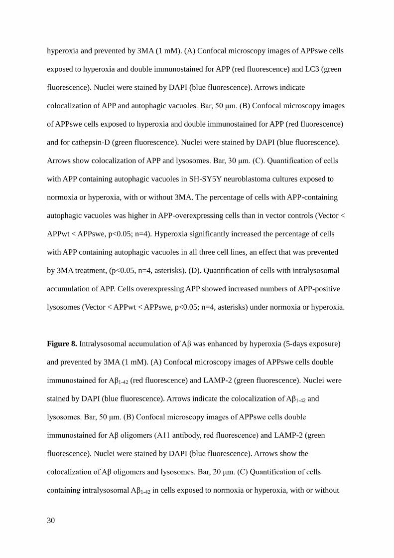
Figure 7. APP accumulation in autophagic-lysosomal compartment was enhanced by
30
hyperoxia and prevented by 3MA (1 mM). (A) Confocal microscopy images of APPswe cells
exposed to hyperoxia and double immunostained for APP (red fluorescence) and LC3 (green
fluorescence). Nuclei were stained by DAPI (blue fluorescence). Arrows indicate
colocalization of APP and autophagic vacuoles. Bar, 50 μm. (B) Confocal microscopy images
of APPswe cells exposed to hyperoxia and double immunostained for APP (red fluorescence)
and for cathepsin-D (green fluorescence). Nuclei were stained by DAPI (blue fluorescence).
Arrows show colocalization of APP and lysosomes. Bar, 30 μm. (C). Quantification of cells
with APP containing autophagic vacuoles in SH-SY5Y neuroblastoma cultures exposed to
normoxia or hyperoxia, with or without 3MA. The percentage of cells with APP-containing
autophagic vacuoles was higher in APP-overexpressing cells than in vector controls (Vector <
APPwt < APPswe, p<0.05; n=4). Hyperoxia significantly increased the percentage of cells
with APP containing autophagic vacuoles in all three cell lines, an effect that was prevented
by 3MA treatment, (p<0.05, n=4, asterisks). (D). Quantification of cells with intralysosomal
accumulation of APP. Cells overexpressing APP showed increased numbers of APP-positive
lysosomes (Vector < APPwt < APPswe, p<0.05; n=4, asterisks) under normoxia or hyperoxia.
Figure 8. Intralysosomal accumulation of Aβ was enhanced by hyperoxia (5-days exposure)
and prevented by 3MA (1 mM). (A) Confocal microscopy images of APPswe cells double
immunostained for Aβ1-42 (red fluorescence) and LAMP-2 (green fluorescence). Nuclei were
stained by DAPI (blue fluorescence). Arrows indicate the colocalization of Aβ1-42 and
lysosomes. Bar, 50 μm. (B) Confocal microscopy images of APPswe cells double
immunostained for Aβ oligomers (A11 antibody, red fluorescence) and LAMP-2 (green
fluorescence). Nuclei were stained by DAPI (blue fluorescence). Arrows show the
colocalization of Aβ oligomers and lysosomes. Bar, 20 μm. (C) Quantification of cells
containing intralysosomal Aβ1-42 in cells exposed to normoxia or hyperoxia, with or without
31
3MA. The percentage of cells with Aβ-containing lysosomes was higher in APP-
overexpressing cells than in vector controls (Vector < APPwt < APPswe, p<0.05). Hyperoxia
significantly increases the percentage of cells with Aβ-positive lysosomes in all three cell
lines and this increase was prevented by 3MA. (p<0.05, n=4, asterisks). (D) Quantification of
cells with intralysosomal accumulation of Aβ oligomers. Cells overexpressing APP showed
increased numbers of Aβ-positive lysosomes (Vector < APPwt < APPswe). Hyperoxia
significantly increased the percentage of cells with lysosomes containing Aβ oligomers in all
three cell lines and this increase was prevented by 3MA. (p<0.05, n=4, asterisks). (E)
Quantification of cells containing intralysosomal Aβ1-42 in cultures exposed to normoxia or
hyperoxia, with or without lysosomal inhibitors (E64d and Pepstatin A) (P<0.05, n=3,
asterisks).
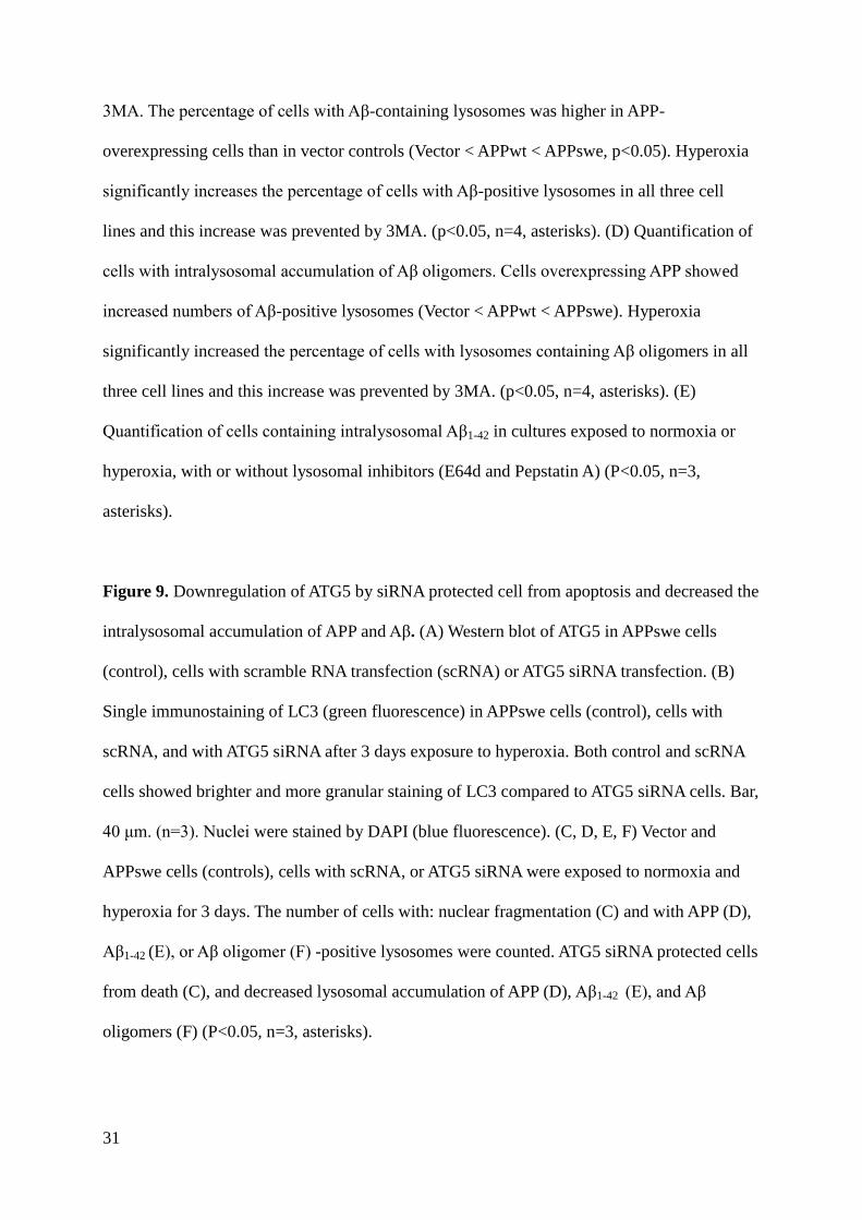
Figure 9. Downregulation of ATG5 by siRNA protected cell from apoptosis and decreased the
intralysosomal accumulation of APP and Aβ. (A) Western blot of ATG5 in APPswe cells
(control), cells with scramble RNA transfection (scRNA) or ATG5 siRNA transfection. (B)
Single immunostaining of LC3 (green fluorescence) in APPswe cells (control), cells with
scRNA, and with ATG5 siRNA after 3 days exposure to hyperoxia. Both control and scRNA
cells showed brighter and more granular staining of LC3 compared to ATG5 siRNA cells. Bar,
40 μm. (n=3). Nuclei were stained by DAPI (blue fluorescence). (C, D, E, F) Vector and
APPswe cells (controls), cells with scRNA, or ATG5 siRNA were exposed to normoxia and
hyperoxia for 3 days. The number of cells with: nuclear fragmentation (C) and with APP (D),
Aβ1-42 (E), or Aβ oligomer (F) -positive lysosomes were counted. ATG5 siRNA protected cells
from death (C), and decreased lysosomal accumulation of APP (D), Aβ1-42 (E), and Aβ
oligomers (F) (P<0.05, n=3, asterisks).
32
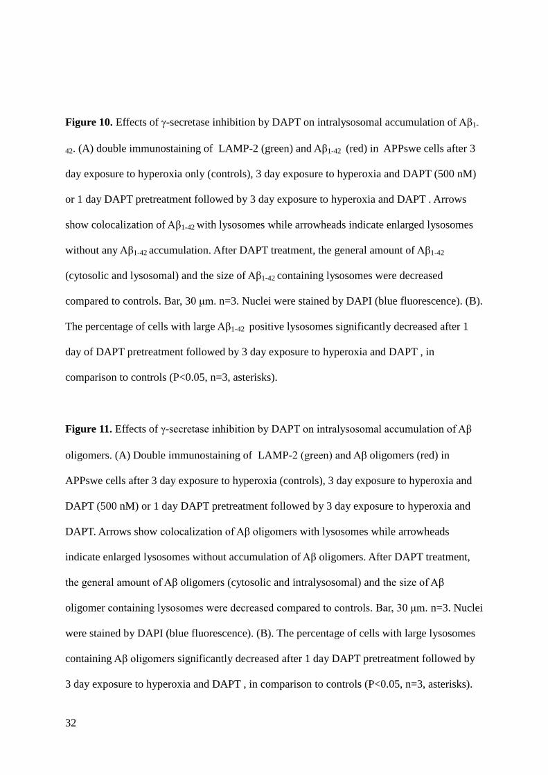
Figure 10. Effects of -secretase inhibition by DAPT on intralysosomal accumulation of Aβ1-
42. (A) double immunostaining of LAMP-2 (green) and Aβ1-42 (red) in APPswe cells after 3
day exposure to hyperoxia only (controls), 3 day exposure to hyperoxia and DAPT (500 nM)
or 1 day DAPT pretreatment followed by 3 day exposure to hyperoxia and DAPT . Arrows
show colocalization of Aβ1-42 with lysosomes while arrowheads indicate enlarged lysosomes
without any Aβ1-42 accumulation. After DAPT treatment, the general amount of Aβ1-42
(cytosolic and lysosomal) and the size of Aβ1-42 containing lysosomes were decreased
compared to controls. Bar, 30 μm. n=3. Nuclei were stained by DAPI (blue fluorescence). (B).
The percentage of cells with large Aβ1-42 positive lysosomes significantly decreased after 1
day of DAPT pretreatment followed by 3 day exposure to hyperoxia and DAPT , in
comparison to controls (P<0.05, n=3, asterisks).
Figure 11. Effects of -secretase inhibition by DAPT on intralysosomal accumulation of Aβ
oligomers. (A) Double immunostaining of LAMP-2 (green) and Aβ oligomers (red) in
APPswe cells after 3 day exposure to hyperoxia (controls), 3 day exposure to hyperoxia and
DAPT (500 nM) or 1 day DAPT pretreatment followed by 3 day exposure to hyperoxia and
DAPT. Arrows show colocalization of Aβ oligomers with lysosomes while arrowheads
indicate enlarged lysosomes without accumulation of Aβ oligomers. After DAPT treatment,
the general amount of Aβ oligomers (cytosolic and intralysosomal) and the size of Aβ
oligomer containing lysosomes were decreased compared to controls. Bar, 30 μm. n=3. Nuclei
were stained by DAPI (blue fluorescence). (B). The percentage of cells with large lysosomes
containing Aβ oligomers significantly decreased after 1 day DAPT pretreatment followed by
3 day exposure to hyperoxia and DAPT , in comparison to controls (P<0.05, n=3, asterisks).
33
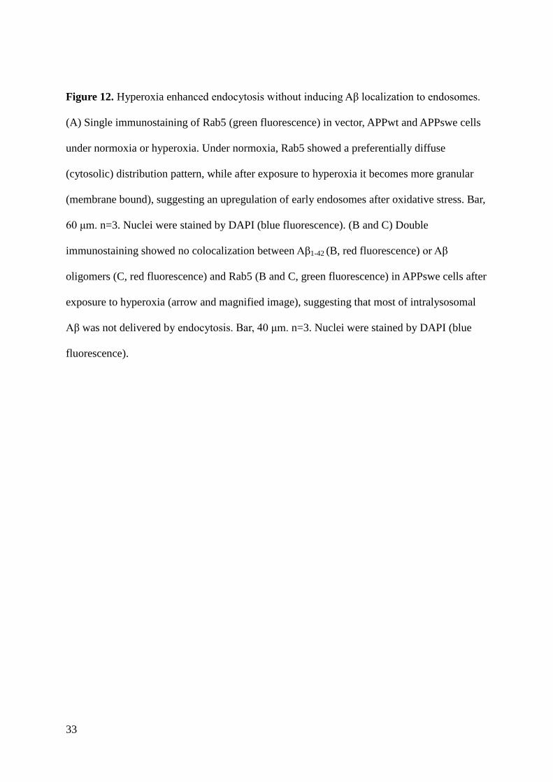
Figure 12. Hyperoxia enhanced endocytosis without inducing Aβ localization to endosomes.
(A) Single immunostaining of Rab5 (green fluorescence) in vector, APPwt and APPswe cells
under normoxia or hyperoxia. Under normoxia, Rab5 showed a preferentially diffuse
(cytosolic) distribution pattern, while after exposure to hyperoxia it becomes more granular
(membrane bound), suggesting an upregulation of early endosomes after oxidative stress. Bar,
60 μm. n=3. Nuclei were stained by DAPI (blue fluorescence). (B and C) Double
immunostaining showed no colocalization between Aβ1-42 (B, red fluorescence) or Aβ
oligomers (C, red fluorescence) and Rab5 (B and C, green fluorescence) in APPswe cells after
exposure to hyperoxia (arrow and magnified image), suggesting that most of intralysosomal
Aβ was not delivered by endocytosis. Bar, 40 μm. n=3. Nuclei were stained by DAPI (blue
fluorescence).
34
35
36
37
38
39
40
41
42
43
44
45
Supplementary figures and figure legends
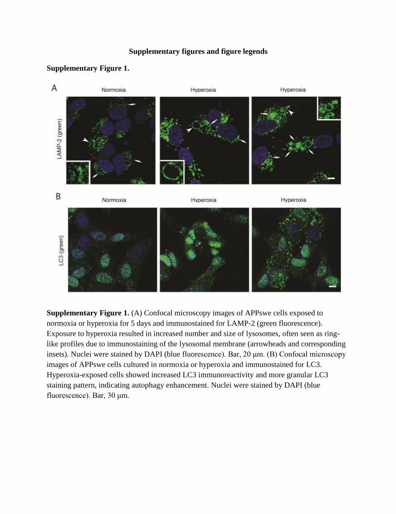
Supplementary Figure 1.
Supplementary Figure 1. (A) Confocal microscopy images of APPswe cells exposed to
normoxia or hyperoxia for 5 days and immunostained for LAMP-2 (green fluorescence).
Exposure to hyperoxia resulted in increased number and size of lysosomes, often seen as ring-
like profiles due to immunostaining of the lysosomal membrane (arrowheads and corresponding
insets). Nuclei were stained by DAPI (blue fluorescence). Bar, 20 μm. (B) Confocal microscopy
images of APPswe cells cultured in normoxia or hyperoxia and immunostained for LC3.
Hyperoxia-exposed cells showed increased LC3 immunoreactivity and more granular LC3
staining pattern, indicating autophagy enhancement. Nuclei were stained by DAPI (blue
fluorescence). Bar, 30 μm.
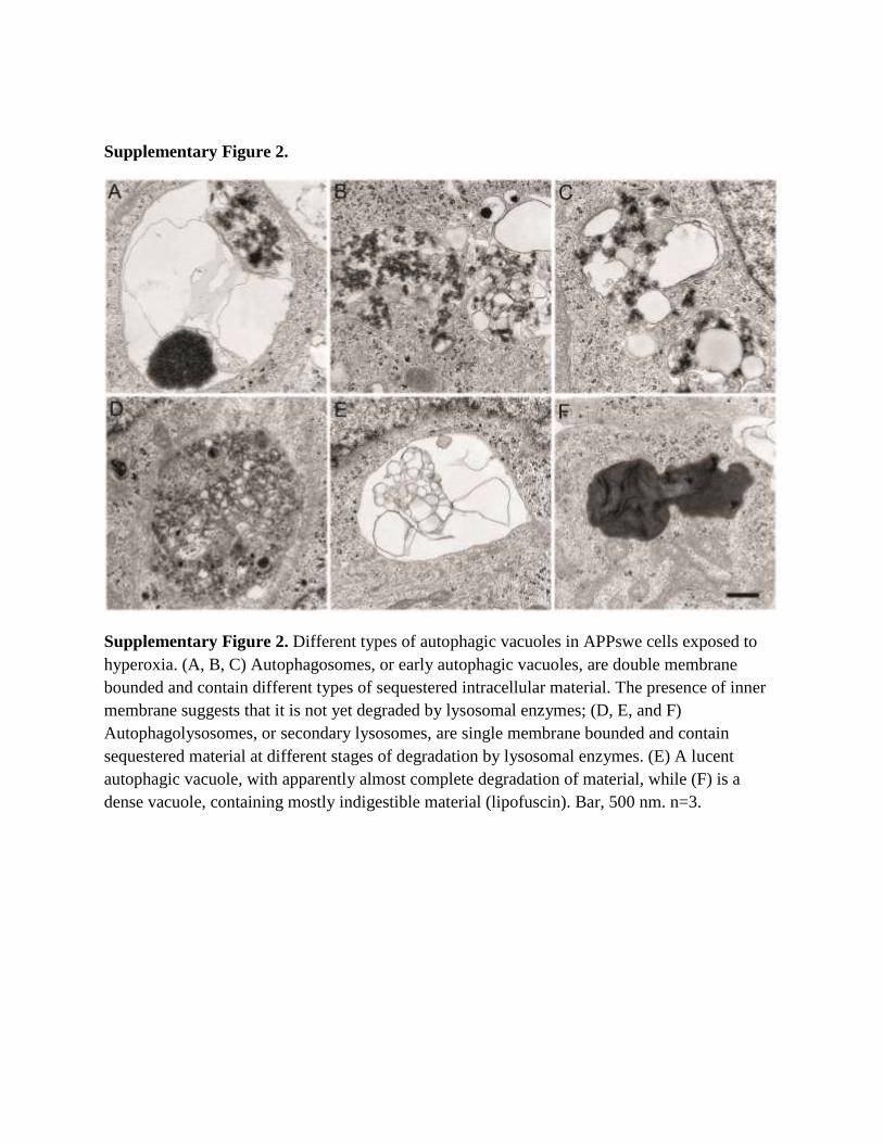
Supplementary Figure 2.
Supplementary Figure 2. Different types of autophagic vacuoles in APPswe cells exposed to
hyperoxia. (A, B, C) Autophagosomes, or early autophagic vacuoles, are double membrane
bounded and contain different types of sequestered intracellular material. The presence of inner
membrane suggests that it is not yet degraded by lysosomal enzymes; (D, E, and F)
Autophagolysosomes, or secondary lysosomes, are single membrane bounded and contain
sequestered material at different stages of degradation by lysosomal enzymes. (E) A lucent
autophagic vacuole, with apparently almost complete degradation of material, while (F) is a
dense vacuole, containing mostly indigestible material (lipofuscin). Bar, 500 nm. n=3.