Low temperature preparation of α-tricalcium phosphate and ... 36 04.pdf · 1.5) are heated above...
6
Processing and Application of Ceramics 11 [2] (2017) 100–105 https://doi.org/10.2298/PAC1702100W Low temperature preparation of α-tricalcium phosphate and its mechanical properties Song Wang 1,2 , Yaping Wang 1,2 , Kangning Sun 1,2,* , Xiaoning Sun 1,2,* 1 Key Laboratory for Liquid-Solid Structural Evolution and Processing of Materials (Ministry of Education), Shandong University, Ji’nan 250061, People’s Republic of China 2 Key Laboratory of Engineering Ceramics, Shandong University, Ji’nan 250061, People’s Republic of China Received 25 November 2016; Received in revised form 23 February 2017; Accepted 24 April 2017 Abstract In this work, α-tricalcium phosphate (α-TCP) was successfully prepared by the thermal transformation of amorphous calcium phosphate (ACP) precursor. β-cyclodextrin (β-CD) was used for preparation of ACP pre- cursor and played an important role in designing its special structure. The phase composition and microstruc- tures of the obtained α-TCP at different annealing temperature were analysed by X-ray diffraction and scan- ning electron microscope, and confirmed that α-TCP can be prepared at 650 °C for 3 h using ACP as precursor, which is much lower than the phase transition temperature of α-TCP. Mechanical properties were tested 24h after mixing the obtained α-TCP with 30wt.% of deionised water. The compressive strength and the flexu- ral strength were 26.4MPa and 12.0MPa, respectively. The flexural strength was higher than that of α-TCP prepared by other methods. Keywords: α-tricalcium phosphate, thermal transformation, amorphous calcium phosphate, β-cyclodextrin I. Introduction The past 40 years have witnessed a wide applica- tion of calcium phosphate as bioactive ceramic mate- rial in bone substitutes [1–3]. α-tricalcium phosphate (α-TCP), a high-temperature polymorph of tricalcium phosphate, is stable at 1125–1430°C and can be re- tained at room temperature by sudden cooling in air [4]. Typically, α-TCP can be converted to hydroxyapatite (HAp) in aqueous solutions at room temperature, which is more active than HAp ceramics [5–8]. Therefore, α- TCP has been widely used in the research of bone ce- ment. Nowadays, there are only a few commercial products of α-TCP in the market even though it has been studied for a long time. Complex preparation technique is one of important factors that hinders its commercial applica- tions. Solid state reaction is a preferred method to syn- thesize α-TCP presented in many literature reports [3,9– 14]. Solid precursors (mixture with molar ratio Ca/P ≈ 1.5) are heated above the transformation temperature and then quenched to room temperature after a period * Corresponding author: tel/fax: +86 53188392914, e-mail: [email protected], [email protected] of dwell time. This method has some risks in quench- ing process and can cause an adverse impact on the sin- tering furnace because of high thermal shock. Further- more, the obtained products are often a mixture of α- TCP and some amounts of β-TCP or HAp [15,16]. As a result, scientists have been searching for new strategies to improve the preparation process and obtain the pure α-TCP. Subsequently, it was found that α-TCP can be synthesized at temperatures much lower than the transi- tion temperature to avoid the reversion of α-phase [17– 20]. This synthesis method is often accomplished by thermal transformation of a tailor-made amorphous cal- cium phosphate (ACP). However, the α-TCP prepared by these methods also had a small amount of β-TCP and the hydrolytic and mechanical properties were not anal- ysed and compared with the α-TCP prepared by tradi- tional methods. Therefore, scientists have to find a new approach for preparing the pure α-TCP with excellent hydrolytic and mechanical properties due to their com- mercial importance. β-cyclodextrin (β-CD) can be used as building block of ACP because it can be linked both covalently and noncovalently in a regioselective manner [21]. There- fore, the targeted compounds can be synthesized with 100
Transcript of Low temperature preparation of α-tricalcium phosphate and ... 36 04.pdf · 1.5) are heated above...
Processing and Application of Ceramics 11 [2] (2017) 100–105
https://doi.org/10.2298/PAC1702100W
Low temperature preparation of α-tricalcium phosphate and its
mechanical properties
Song Wang1,2, Yaping Wang1,2, Kangning Sun1,2,∗, Xiaoning Sun1,2,∗
1Key Laboratory for Liquid-Solid Structural Evolution and Processing of Materials (Ministry of Education),
Shandong University, Ji’nan 250061, People’s Republic of China2Key Laboratory of Engineering Ceramics, Shandong University, Ji’nan 250061, People’s Republic of China
Received 25 November 2016; Received in revised form 23 February 2017; Accepted 24 April 2017
Abstract
In this work, α-tricalcium phosphate (α-TCP) was successfully prepared by the thermal transformation ofamorphous calcium phosphate (ACP) precursor. β-cyclodextrin (β-CD) was used for preparation of ACP pre-cursor and played an important role in designing its special structure. The phase composition and microstruc-tures of the obtained α-TCP at different annealing temperature were analysed by X-ray diffraction and scan-ning electron microscope, and confirmed that α-TCP can be prepared at 650 °C for 3 h using ACP as precursor,which is much lower than the phase transition temperature of α-TCP. Mechanical properties were tested 24 hafter mixing the obtained α-TCP with 30 wt.% of deionised water. The compressive strength and the flexu-ral strength were 26.4 MPa and 12.0 MPa, respectively. The flexural strength was higher than that of α-TCPprepared by other methods.
Keywords: α-tricalcium phosphate, thermal transformation, amorphous calcium phosphate, β-cyclodextrin
I. Introduction
The past 40 years have witnessed a wide applica-
tion of calcium phosphate as bioactive ceramic mate-
rial in bone substitutes [1–3]. α-tricalcium phosphate
(α-TCP), a high-temperature polymorph of tricalcium
phosphate, is stable at 1125–1430 °C and can be re-
tained at room temperature by sudden cooling in air [4].
Typically, α-TCP can be converted to hydroxyapatite
(HAp) in aqueous solutions at room temperature, which
is more active than HAp ceramics [5–8]. Therefore, α-
TCP has been widely used in the research of bone ce-
ment.
Nowadays, there are only a few commercial products
of α-TCP in the market even though it has been studied
for a long time. Complex preparation technique is one
of important factors that hinders its commercial applica-
tions. Solid state reaction is a preferred method to syn-
thesize α-TCP presented in many literature reports [3,9–
14]. Solid precursors (mixture with molar ratio Ca/P ≈
1.5) are heated above the transformation temperature
and then quenched to room temperature after a period
∗Corresponding author: tel/fax: +86 53188392914,
e-mail: [email protected], [email protected]
of dwell time. This method has some risks in quench-
ing process and can cause an adverse impact on the sin-
tering furnace because of high thermal shock. Further-
more, the obtained products are often a mixture of α-
TCP and some amounts of β-TCP or HAp [15,16]. As a
result, scientists have been searching for new strategies
to improve the preparation process and obtain the pure
α-TCP. Subsequently, it was found that α-TCP can be
synthesized at temperatures much lower than the transi-
tion temperature to avoid the reversion of α-phase [17–
20]. This synthesis method is often accomplished by
thermal transformation of a tailor-made amorphous cal-
cium phosphate (ACP). However, the α-TCP prepared
by these methods also had a small amount of β-TCP and
the hydrolytic and mechanical properties were not anal-
ysed and compared with the α-TCP prepared by tradi-
tional methods. Therefore, scientists have to find a new
approach for preparing the pure α-TCP with excellent
hydrolytic and mechanical properties due to their com-
mercial importance.
β-cyclodextrin (β-CD) can be used as building block
of ACP because it can be linked both covalently and
noncovalently in a regioselective manner [21]. There-
fore, the targeted compounds can be synthesized with
100
S. Wang et al. / Processing and Application of Ceramics 11 [2] (2017) 100–105
special structure using β-CD. For example, Li et al. [22]
synthesized ACP in aqueous solution at room tempera-
ture using cyclodextrins. Xiao et al. [23] prepared spher-
ical HAp crystals successfully with β-CD as template
by biomimetic method. In this paper, the precursor ACP
was also prepared in aqueous solution using β-CD as
template and optimal heating conditions for preparation
of α-TCP were studied. The hydrolysis, compressive
strength and flexural strength were used to evaluate the
properties of the obtained α-TCP.
II. Experimental procedure
2.1. Preparation of α-TCP
Amorphous calcium phosphate (ACP) was prepared
by chemical precipitation method. Calcium nitrate
tetrahydrate (Ca(NO3)2·4 H2O) and β-CD were dis-
solved in deionised water to form 0.2 mol/l calcium ni-
trate saturated β-CD solution. The solution was kept in
water bath at 25 °C for 24 h to ensure complete reaction
of Ca2+ with β-CD. The same volume of dibasic ammo-
nium phosphate ((NH4)2HPO4) solution, with the Ca/P
molar ratio of 1.5 to Ca(NO3)2, was added to the above
solution dropwise. The reaction was kept in water bath
at 25 °C for half an hour, meanwhile, the pH value of
the solution was adjusted to 10–11 by the addition of
ammonia. The precipitates were washed with deionised
water and absolute ethanol for several times, then dried
at room temperature for 24 h until white loose powders
were obtained, which is ACP precursor. This precursor
was heated at different temperature (550, 600, 650, 700
and 800 °C) for 2–4 h to form α-TCP.
All used chemicals were analytical grade. Ca(NO3)2·
4 H2O, (NH4)2HPO4, ammonia and absolute ethanol
were purchased from Sinopharm Chemical Reagent
CO., LTD (Shanghai, China) and β-CD was purchased
from Kemiou Chemical Reagent Co., LTD (Tianjin,
China).
2.2. Hydrolysis of α-TCP
α-TCP was ground for 5 h at 400 r/min in a ball mill
and passed through 300 mesh screen to form calcium
phosphate cement (CPC) powder. The CPC powder was
mixed with 30 wt.% of deionised water to form CPC
paste. This CPC paste was quickly injected in a mould
and air bubbles were eliminated by oscillation. After
that, the paste was kept at 37 °C and 100% humidity
environment for 24 h.
2.3. Mechanical test
The CPC specimens after hydrolysis were made to
�6 mm × 12 mm cylinder for compressive strength test-
ing and 3 mm × 4 mm × 25 mm cuboid for flexural
strength testing. Compressive strength was calculated
by the following equation:
σC =4Pm
π · d2(1)
where Pm is the maximum load and d is the diameter
of the cylinder specimen. A three-point test was em-
ployed for flexural strength which was calculated by
equation (2):
σ f =3 · P · L
2 · b · h2(2)
where P is the fracturing load, L is the span, b is the
width of sample and h is the height of the sample. At
least 5 samples for flexural strength and compressive
strength were tested to get the average values.
2.4. Characterization
Differential scanning calorimetry (DSC) was used to
analyse the ACP precursor in the temperature range
from room temperature to 800 °C with the heating rate
of 5 °C/min to confirm the phase-transition temperature.
The polymorphism of specimens was determined by X-
ray diffraction (XRD) patterns which were conducted
on a Rigaku Dmax-rc diffractometer with Ni-filtered
Cu Kα radiation (V = 50 kV, I = 80 mA). Step scan
(2°/min) was used for calculation of the crystallite sizes
by Debye-Scherrer equation:
D =K · γ
B cos θ(3)
where D is the average thickness of crystalline grain in
vertical direction of lattice plane, K is the Scherrer con-
stant, γ is the X-ray wavelength, B is the full width at
half maximum (FWHM) of diffraction peaks and θ is
the X-ray diffraction angle. Fourier transform infrared
spectroscopy (FTIR) was conducted on a Bruker Vertex
70 spectrometer over the range of 400–4000 cm-1 with
resolution of 4 cm-1. The morphologies of α-TCP and
hydrolysis products were analysed on a Hitachi SU-70
scanning electron microscope (SEM).
III. Results and discussion
3.1. Heating conditions for α-TCP formation
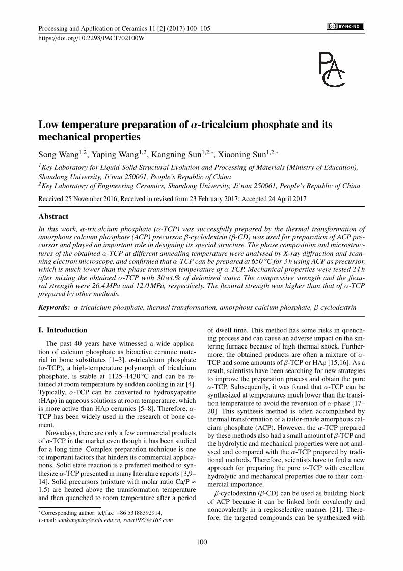
Figure 1 shows the DSC curve of ACP precursor.
A crystallization peak starts to form at 650.8 °C and
reaches its maximum at 657.1 °C, which indicates that
Figure 1. DSC curve of ACP precursor
101
S. Wang et al. / Processing and Application of Ceramics 11 [2] (2017) 100–105
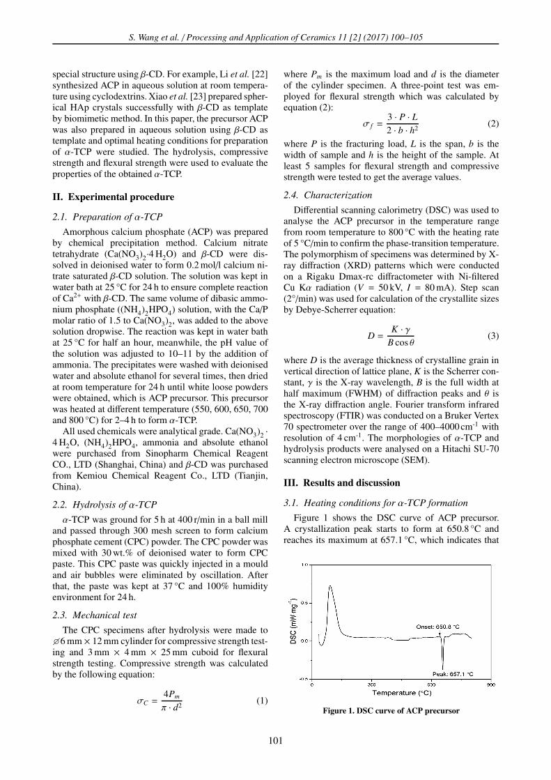
Figure 2. XRD patterns of ACP precursors heated atdifferent temperature: a) 550 °C, b) 600 °C, c) 650 °C,
d) 700 °C and e) 800 °C for 3 h
the transition temperature of α-TCP is around 650 °C.
The XRD patterns of ACP precursor heated at dif-
ferent temperature for 3 h are shown in Fig. 2. All the
diffraction peaks in Figs. 2b and 2c match well with α-
TCP (JCPDS NO.29-0359). However, when the temper-
ature was increased to 700 °C, the peaks of β-TCP ap-
peared. After being heated at 800 °C, the products are
typical β-TCP (JCPDS NO.70-2065) without any other
diffraction peaks. Therefore, the phase transition tem-
perature of α-TCP was 600–650 °C, and α-TCP would
be transformed to β-TCP when the temperature reached
700 °C.
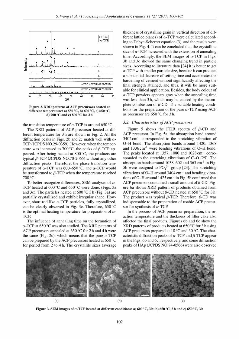
To better recognize differences, SEM analyses of α-
TCP heated at 600 °C and 650 °C were done, (Figs. 3a
and 3c). The particles heated at 600 °C 3 h (Fig. 3a) are
partially crystallized and exhibit irregular shape. How-
ever, short rod-like α-TCP particles, fully crystallized,
can be clearly observed in Fig. 3c. Therefore, 650 °C
is the optimal heating temperature for preparation of α-
TCP.
The influence of annealing time on the formation of
α-TCP at 650 °C was also studied. The XRD patterns of
ACP precursors annealed at 650 °C for 2 h and 4 h were
the same (Fig. 2c), which means that the pure α-TCP
can be prepared by the ACP precursors heated at 650 °C
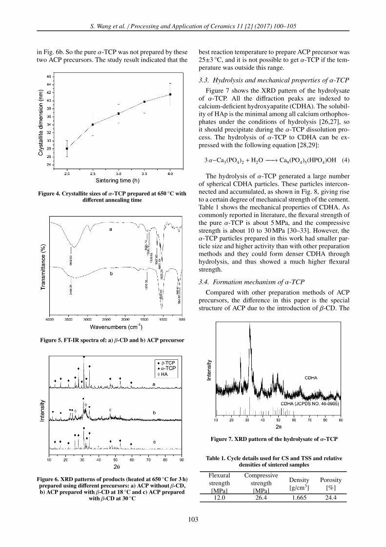
for period from 2 to 4 h. The crystallite sizes (average
thickness of crystalline grain in vertical direction of dif-
ferent lattice planes) of α-TCP were calculated accord-
ing to Debye-Scherrer equation (3), and the results were
shown in Fig. 4. It can be concluded that the crystalline
size of α-TCP increased with the extension of annealing
time. Accordingly, the SEM images of α-TCP in Figs.
3b and 3c showed the same changing trend in particle
sizes. According to literature data [24] it is better to get
α-TCP with smaller particle size, because it can produce
a substantial decrease of setting time and accelerates the
hardening of cement without significantly affecting the
final strength attained, and thus, it will be more suit-
able for clinical application. Besides, the body colour of
α-TCP powders appears gray when the annealing time
was less than 3 h, which may be caused by the incom-
plete combustion of β-CD. The suitable heating condi-
tions for the preparation of the pure α-TCP using ACP
as precursor are 650 °C for 3 h.
3.2. Characteristics of ACP precursors
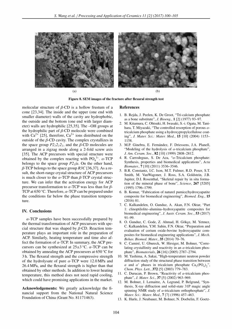
Figure 5 shows the FTIR spectra of β-CD and
ACP precursor. In Fig. 5a, the absorption band around
3402 cm-1 corresponded to the stretching vibration of
O–H bond. The absorption bands around 1420, 1368
and 1336 cm-1 were bending vibrations of O–H bond.
The peaks located at 1357, 1080 and 1028 cm-1 corre-
sponded to the stretching vibrations of C–O [25]. The
absorption bands around 1038, 602 and 563 cm-1 in Fig.
5b were assigned to PO43– group [23]. The stretching
vibrations of O–H around 3404 cm-1 and bending vibra-
tions of O–H around 1425 cm-1 in Fig. 5b confirmed that
ACP precursors contained a small amount of β-CD. Fig-
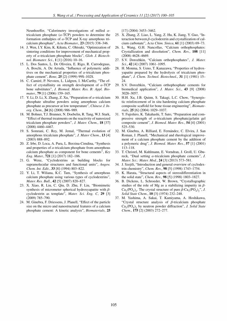
ure 6a shows XRD pattern of products obtained from
ACP precursors without β-CD heated at 650 °C for 3 h.
The product was typical β-TCP. Therefore, β-CD was
indispensable to the preparation of usable ACP precur-
sor for synthesis of α-TCP.
In the process of ACP precursor preparation, the re-
action temperature and the thickness of filter cake also
affected the final products. Figures 6b and 6c show the
XRD patterns of products heated at 650 °C for 3 h using
ACP precursors prepared at 18 °C and 30 °C. The char-
acteristic diffraction peaks of α-TCP and β-TCP appear
in the Figs. 6b and 6c, respectively, and some diffraction
peaks of HAp (JCPDS NO.74-0566) were also observed
(a) (b) (c)
Figure 3. SEM images of α-TCP heated at different conditions: a) 600 °C, 3 h; b) 650 °C, 2 h and c) 650 °C, 3 h
102
S. Wang et al. / Processing and Application of Ceramics 11 [2] (2017) 100–105
in Fig. 6b. So the pure α-TCP was not prepared by these
two ACP precursors. The study result indicated that the
Figure 4. Crystallite sizes of α-TCP prepared at 650 °C withdifferent annealing time
Figure 5. FT-IR spectra of: a) β-CD and b) ACP precursor
Figure 6. XRD patterns of products (heated at 650 °C for 3 h)prepared using different precursors: a) ACP without β-CD,b) ACP prepared with β-CD at 18 °C and c) ACP prepared
with β-CD at 30 °C
best reaction temperature to prepare ACP precursor was
25±3 °C, and it is not possible to get α-TCP if the tem-
perature was outside this range.
3.3. Hydrolysis and mechanical properties of α-TCP
Figure 7 shows the XRD pattern of the hydrolysate
of α-TCP. All the diffraction peaks are indexed to
calcium-deficient hydroxyapatite (CDHA). The solubil-
ity of HAp is the minimal among all calcium orthophos-
phates under the conditions of hydrolysis [26,27], so
it should precipitate during the α-TCP dissolution pro-
cess. The hydrolysis of α-TCP to CDHA can be ex-
pressed with the following equation [28,29]:
3α−Ca3(PO4)2 + H2O −−−→ Ca9(PO4)5(HPO4)OH (4)
The hydrolysis of α-TCP generated a large number
of spherical CDHA particles. These particles intercon-
nected and accumulated, as shown in Fig. 8, giving rise
to a certain degree of mechanical strength of the cement.
Table 1 shows the mechanical properties of CDHA. As
commonly reported in literature, the flexural strength of
the pure α-TCP is about 5 MPa, and the compressive
strength is about 10 to 30 MPa [30–33]. However, the
α-TCP particles prepared in this work had smaller par-
ticle size and higher activity than with other preparation
methods and they could form denser CDHA through
hydrolysis, and thus showed a much higher flexural
strength.
3.4. Formation mechanism of α-TCP
Compared with other preparation methods of ACP
precursors, the difference in this paper is the special
structure of ACP due to the introduction of β-CD. The
Figure 7. XRD pattern of the hydrolysate of α-TCP
Table 1. Cycle details used for CS and TSS and relativedensities of sintered samples
Flexural
strength
[MPa]
Compressive
strength
[MPa]
Density
[g/cm3]
Porosity
[%]
12.0 26.4 1.665 24.4
103
S. Wang et al. / Processing and Application of Ceramics 11 [2] (2017) 100–105
(a) (b)
Figure 8. SEM images of the fracture after flexural strength test
molecular structure of β-CD is a hollow frustum of a
cone [23,34]. The inside and the upper (one end with
smaller diameter) walls of the cavity are hydrophobic,
the outside and the bottom (one end with larger diam-
eter) walls are hydrophilic [25,35]. The –OH groups at
the hydrophilic part of β-CD molecule were combined
with Ca2+ [25], therefore, Ca2+ ions distributed on the
outside of the β-CD cavity. The complex crystallizes in
the space group P212121, and the β-CD molecules are
arranged in a zigzag mode along a 2-fold screw axis
[35]. The ACP precursors with special structure were
obtained by the complex reacting with PO43–. α-TCP
belongs to the space group P21/a. On the other hand,
β-TCP belongs to the space group R3C [36,37]. As a re-
sult, the short-range crystal structure of ACP precursors
is much closer to the α-TCP than β-TCP crystal struc-
ture. We can infer that the activation energy for ACP
precursor transformation to α-TCP was less than for β-
TCP at 650 °C. Therefore, α-TCP can be prepared under
the conditions far below the phase transition tempera-
ture.
IV. Conclusions
α-TCP samples have been successfully prepared by
the thermal transformation of ACP precursors with spe-
cial structure that was shaped by β-CD. Reaction tem-
perature plays an important role in the preparation of
ACP. Similarly, heating temperature and time also af-
fect the formation of α-TCP. In summary, the ACP pre-
cursors can be synthesized at 25±3 °C. α-TCP can be
obtained by annealing the ACP precursors at 650 °C for
3 h. The flexural strength and the compressive strength
of the hydrolysate of pure α-TCP were 12.0 MPa and
26.4 MPa, and the flexural strength is higher than that
obtained by other methods. In addition to lower heating
temperature, this method does not need rapid cooling,
which could have promising applications in the market.
Acknowledgements: We greatly acknowledge the fi-
nancial support from the National Natural Science
Foundation of China (Grant No. 81171463).
References
1. B. Rejda, J. Peelen, K. De Groot, “Tri-calcium phosphate
as a bone substitute”, J. Bioeng., 1 [2] (1977) 93–97.
2. M. Kitamura, C. Ohtsuki, H. Iwasaki, S.-i. Ogata, M. Tani-
hara, T. Miyazaki, “The controlled resorption of porous α-
tricalcium phosphate using a hydroxypropylcellulose coat-
ing”, J. Mater. Sci.: Mater. Med., 15 [10] (2004) 1153–
1158.
3. M.P. Ginebra, E. Fernández, F. Driessens, J.A. Planell,
“Modeling of the hydrolysis of α-tricalcium phosphate”,
J. Am. Ceram. Soc., 82 [10] (1999) 2808–2812.
4. R. Carrodeguas, S. De Aza, “α-Tricalcium phosphate:
Synthesis, properties and biomedical applications”, Acta
Biomater., 7 [10] (2011) 3536–3546.
5. B.R. Constantz, I.C. Ison, M.T. Fulmer, R.D. Poser, S.T.
Smith, M. VanWagoner, J. Ross, S.A. Goldstein, J.B.
Jupiter, D.I. Rosenthal, “Skeletal repair by in situ forma-
tion of the mineral phase of bone”, Science, 267 [5205]
(1995) 1796–1799.
6. B. Komur, “Fabrication of naturel pumice/hydroxyapatite
composite for biomedical engineering”, Biomed. Eng., 15
(2016) 81.
7. C. Kalkandelen, O. Gunduz, A. Akan, F.N. Oktar, “Part
1: clinoptilolite–alumina–hydroxyapatite composites for
biomedical engineering”, J. Austr. Ceram. Soc., 53 (2017)
91–99.
8. O. Gunduz, C. Gode, Z. Ahmad, H. Gökçe, M. Yetmez,
C. Kalkandelen, Y.M. Sahin, F.N. Oktar, “Preparation and
evaluation of cerium oxide-bovine hydroxyapatite com-
posites for biomedical engineering applications”, J. Mech.
Behav. Biomed. Mater., 35 (2014) 70–76.
9. C. Camiré, U. Gbureck, W. Hirsiger, M. Bohner, “Corre-
lating crystallinity and reactivity in an α-tricalcium phos-
phate”, Biomaterials, 26 [16] (2005) 2787–2794.
10. M. Yashima, A. Sakai, “High-temperature neutron powder
diffraction study of the structural phase transition between
α and α’ phases in tricalcium phosphate Ca3(PO4)2”,
Chem. Phys. Lett., 372 [5] (2003) 779–783.
11. C. Durucan, P. Brown, “Reactivity of α-tricalcium phos-
phate”, J. Mater. Sci., 37 [5] (2002) 963–969.
12. M. Bohner, J. Lemaitre, A. Legrand, P. Belgrand, “Syn-
thesis, X-ray diffraction and solid-state 31P magic angle
spinning NMR study of α-tricalcium orthophosphate”, J.
Mater. Sci.: Mater. Med., 7 [7] (1996) 457–463.
13. K. Hurle, J. Neubauer, M. Bohner, N. Doebelin, F. Goetz-
104
S. Wang et al. / Processing and Application of Ceramics 11 [2] (2017) 100–105
Neunhoeffer, “Calorimetry investigations of milled α-
tricalcium phosphate (α-TCP) powders to determine the
formation enthalpies of α-TCP and X-ray amorphous tri-
calcium phosphate”, Acta Biomater., 23 (2015) 338–346.
14. J. Wen, I.Y. Kim, K. Kikuta, C. Ohtsuki, “Optimization of
sintering conditions for improvement of mechanical prop-
erty of α-tricalcium phosphate blocks”, Glob. J. Biotech-
nol. Biomater. Sci., 1 [1] (2016) 10–16.
15. L. Dos Santos, L. De Oliveira, E. Rigo, R. Carrodeguas,
A. Boschi, A. De Arruda, “Influence of polymeric addi-
tives on the mechanical properties of α-tricalcium phos-
phate cement”, Bone, 25 [2] (1999) 99S–102S.
16. C. Camiré, P. Nevsten, L. Lidgren, I. McCarthy, “The ef-
fect of crystallinity on strength development of α-TCP
bone substitutes”, J. Biomed. Mater. Res. B: Appl. Bio-
mater., 79 [1] (2006) 159–165.
17. Y. Li, D. Li, X. Zhang, Z. Xu, “Preparation of α-tricalcium
phosphate ultrafine powders using amorphous calcium
phosphate as precursor at low temperature”, Chinese J. In-
org. Chem., 24 [6] (2008) 986–989.
18. M. Bohner, T.J. Brunner, N. Doebelin, R. Tang, W.J. Stark,
“Effect of thermal treatments on the reactivity of nanosized
tricalcium phosphate powders”, J. Mater. Chem., 18 [37]
(2008) 4460–4467.
19. S. Somrani, C. Rey, M. Jemal, “Thermal evolution of
amorphous tricalcium phosphate”, J. Mater Chem., 13 [4]
(2003) 888–892.
20. Z. Irbe, D. Loca, A. Pura, L. Berzina-Cimdina, “Synthesis
and properties of α-tricalcium phosphate from amorphous
calcium phosphate as component for bone cements”, Key
Eng. Mater., 721 [1] (2017) 182–186.
21. G. Wenz, “Cyclodextrins as building blocks for
supramolecular structures and functional units”, Angew.
Chem. Int. Edit., 33 [8] (1994) 803–822.
22. Y. Li, T. Wiliana, K.C. Tam, “Synthesis of amorphous
calcium phosphate using various types of cyclodextrins”,
Mater. Res. Bull., 42 [5] (2007) 820–827.
23. X. Xiao, R. Liu, C. Qiu, D. Zhu, F. Liu, “Biomimetic
synthesis of micrometer spherical hydroxyapatite with β-
cyclodextrin as template”, Mater. Sci. Eng. C, 29 [3]
(2009) 785–790.
24. M. Ginebra, F. Driessens, J. Planell, “Effect of the particle
size on the micro and nanostructural features of a calcium
phosphate cement: A kinetic analysis”, Biomaterials, 25
[17] (2004) 3453–3462.
25. X. Zhang, Z. Liao, L. Yang, Z. Hu, K. Jiang, Y. Guo, “In-
teraction between β-cyclodextrin and crystallization of cal-
cium carbonate”, Acta Chim. Sinica, 61 [1] (2003) 69–73.
26. L. Wang, G.H. Nancollas, “Calcium orthophosphates:
Crystallization and dissolution”, Chem. Rev., 108 [11]
(2008) 4628–4669.
27. S.V. Dorozhkin, “Calcium orthophosphates”, J. Mater.
Sci., 42 [4] (2007) 1061–1095.
28. H. Monma, S. Ueno, T. Kanazawa, “Properties of hydrox-
yapatite prepared by the hydrolysis of tricalcium phos-
phate”, J. Chem. Technol. Biotechnol., 31 [1] (1981) 15–
24.
29. S.V. Dorozhkin, “Calcium orthophosphate cements for
biomedical application”, J. Mater. Sci., 43 [9] (2008)
3028–3057.
30. H.H. Xu, J.B. Quinn, S. Takagi, L.C. Chow, “Synergis-
tic reinforcement of in situ hardening calcium phosphate
composite scaffold for bone tissue engineering”, Biomate-
rials, 25 [6] (2004) 1029–1037.
31. Y. Fujishiro, K. Takahashi, T. Sato, “Preparation and com-
pressive strength of α-tricalcium phosphate/gelatin gel
composite cement”, J. Biomed. Mater. Res., 54 [4] (2001)
525–530.
32. M. Ginebra, A. Rilliard, E. Fernández, C. Elvira, J. San
Roman, J. Planell, “Mechanical and rheological improve-
ment of a calcium phosphate cement by the addition of
a polymeric drug”, J. Biomed. Mater. Res., 57 [1] (2001)
113–118.
33. T. Christel, M. Kuhlmann, E. Vorndran, J. Groll, U. Gbu-
reck, “Dual setting α-tricalcium phosphate cements”, J.
Mater. Sci.: Mater. Med., 24 [3] (2013) 573–581.
34. J. Szejtli, “Introduction and general overview of cyclodex-
trin chemistry”, Chem. Rev., 98 [5] (1998) 1743–1754.
35. K. Harata, “Structural aspects of stereodifferentiation in
the solid state”, Chem. Rev., 98 [5] (1998) 1803–1827.
36. B. Dickens, L. Schroeder, W. Brown, “Crystallographic
studies of the role of Mg as a stabilizing impurity in β-
Ca3(PO4)2. The crystal structure of pure β-Ca3(PO4)
2”, J.
Solid State Chem., 10 [3] (1974) 232–248.
37. M. Yashima, A. Sakai, T. Kamiyama, A. Hoshikawa,
“Crystal structure analysis of β-tricalcium phosphate
Ca3(PO4)2
by neutron powder diffraction”, J. Solid State
Chem., 175 [2] (2003) 272–277.
105