Loveday 2012 Food Hydrocoll 27-242 .pdf
20
PUBLIC ACCESS AUTHOR MANUSCRIPT 1 of 19 Published in final form as: Loveday, S. M., X. L. Wang, et al. (2012). "β-Lactoglobulin Nanofibrils: effect of temperature on fibril formation kinetics, fibril morphology, and the rheological properties of fibril dispersions." Food Hydrocolloids 27(242-249). β-Lactoglobulin Nanofibrils: Effect of Temperature on Fibril Formation Kinetics, Fibril Morphology and the Rheological Properties of Fibril Dispersions S.M. Loveday Riddet Institute, New Zealand 1 X.L. Wang Riddet Institute, New Zealand 1 M.A. Rao Cornell University, USA 2 S.G. Anema Fonterra Research Centre, New Zealand 3 H. Singh* Riddet Institute, New Zealand 1 1: Riddet Institute Massey University Private Bag 11 222 Palmerston North NEW ZEALAND 2: Department of Food Science Cornell University Geneva NY 14456 USA 3: Fonterra Research Centre Dairy Farm Road Private Bag 11029 Palmerston North NEW ZEALAND * Corresponding author
Transcript of Loveday 2012 Food Hydrocoll 27-242 .pdf
PUBLIC ACCESS AUTHOR MANUSCRIPT
1 of 19
Published in final form as:
Loveday, S. M., X. L. Wang, et al. (2012). "β-Lactoglobulin Nanofibrils: effect of temperature on fibril formation kinetics, fibril morphology, and the rheological properties of fibril dispersions." Food Hydrocolloids 27(242-249).
β-Lactoglobulin Nanofibrils: Effect of Temperature on Fibril Formation Kinetics, Fibril Morphology and the Rheological Properties of Fibril Dispersions
S.M. Loveday Riddet Institute, New Zealand1 X.L. Wang Riddet Institute, New Zealand1 M.A. Rao Cornell University, USA2 S.G. Anema Fonterra Research Centre, New Zealand3 H. Singh* Riddet Institute, New Zealand1 1: Riddet Institute Massey University Private Bag 11 222 Palmerston North NEW ZEALAND 2: Department of Food Science Cornell University Geneva NY 14456 USA 3: Fonterra Research Centre Dairy Farm Road Private Bag 11029 Palmerston North NEW ZEALAND * Corresponding author
Food Hydrocolloids 27:242-249 2012
2 of 19
Abstract Almost all published studies of heat-induced β-lactoglobulin self-assembly into amyloid-like fibrils at low pH and low ionic strength have involved heating at 80°C, and the effect of heating temperature on self-assembly has received little attention. Here we heated β-lactoglobulin at pH 2 and 75°C, 80°C, 90°C, 100°C, 110°C or 120°C and investigated the kinetics of self-assembly (using Thioflavin T fluorescence), the morphology of fibrils, and the rheological properties of fibril dispersions.
Self-assembly occurred at all temperatures tested. Thioflavin T fluorescence increased sigmoidally at all temperatures, however it decreased sharply with >3.3 h heating at 110°C and with >5 h heating at 120°C. The sharp decreases were attributed partly to local gelation, but destruction of fibrils may have occurred at 120°C. Thioflavin T fluorescence results indicated that maximal rates of fibril formation increased with increasing temperature, especially above 100°C, but fibril yield (maximum Thioflavin T fluorescence) was not affected by temperature.
At 100ºC and 110ºC, fibrils were slightly shorter than at 80°C, but otherwise they looked very similar. Fibrils made by heating at 120°C for 1 h were also similar, but heating at 120°C for 8 h gave predominantly short fibrils, apparently the products of larger fibrils fragmenting. Heating at 100°C gave consistently higher viscosity than at 80°C, and heating for >2 h at 120°C decreased viscosity, which may have been linked with fibril fragmentation seen in micrographs.
Key words
β-lactoglobulin, whey protein, milk, heat, self-assembly, protein, amyloid, fibril, acid, denaturation
Introduction Amyloid fibrils consisting of stacked protein β-sheet structures are seen in vivo in association with certain degenerative diseases called amyloidoses. Structurally similar ‘amyloid-like’ fibrils form in vitro under certain denaturing conditions, such as high temperature with low pH and low ionic strength, or in high concentrations of alcohols (Gosal, Clark, & Ross-Murphy, 2004). A wide range of proteins form amyloid fibrils, including a number that are not associated with amyloidoses, which has lead to the suggestion that amyloid formation is a generic property of polypeptide chains (Dobson, 1999). Amyloid-like fibrils may be useful as scaffolds for enzyme immobilisation (Pilkington, Roberts, Meade, & Gerrard, 2009) or tissue engineering (Yan, et al., 2008). The food industry is also interested in amyloid-like protein fibrils, which enhance viscosity and form gels at relatively low protein concentration (Loveday, Rao, Creamer, & Singh, 2009).
β-Lactoglobulin comprises approximately 50% of whey proteins in bovine milk, and forms amyloid-like fibrils within a few hours on heating (typically at 80°C) at low pH and low ionic strength. Heat-induced fibril formation by β-lactoglobulin usually shows an initial lag phase when followed with the Thioflavin T (ThT) fluorescence assay (Loveday, et al., 2010), in contrast to egg proteins and bovine serum albumin (Pearce, Mackintosh, & Gerrard, 2007). The limiting process during the lag phase is thought to be the formation of fibril nuclei, since the lag phase is reduced or abolished by shearing and/or adding fibril seeds (Bolder, Sagis,
Food Hydrocolloids 27:242-249 2012
3 of 19
Venema, & van der Linden, 2007a; Hill, Krebs, Goodall, Howlett, & Dunstan, 2006). However the details of the nucleation process remain elusive. Although fibrils formed after heating for 20 h at 80-85°C are comprised of peptides (Akkermans, Venema, van der Goot, Gruppen, et al., 2008; Oboroceanu, Wang, Brodkorb, Magner, & Auty, 2010), it has not been shown whether hydrolysis is a necessary precursor to the nucleation or growth of fibrils.
It is clear that there is a common requirement among amyloid-forming globular proteins for some degree of unfolding before self-assembly into amyloid fibrils will occur (Chiti & Dobson, 2006). However, Chiti and Dobson (2006) suggested that, rather than a single assembly pathway leading to amyloid formation, “each protein sequence can form a spectrum of structurally distinct fibrillar aggregates and that kinetic factors can dictate which of these alternatives is dominant under given circumstances.” The circumstances in our case are low pH, low ionic strength and high temperature, and the kinetic factor of interest in the present work is the effect of heating temperature.
Changing the temperature of an aqueous solution of a globular protein changes the balance of forces determining the position of the equilibrium between folded and unfolded states. In the folded state, there are internal hydrogen bonds, van der Waals interactions and often disulphide bonds stabilising the structure, and most hydrophobic regions are buried in the interior, minimising the entropically unfavourable interaction (at ambient temperature) between hydrophobic residues and water molecules (Privalov & Makhatadze, 1993). The transition to a completely unfolded state involves breakage of internal bonds, unfolding of the peptide chain, and hydration of previously buried residues, all of which contribute in a temperature-dependent way to the Gibbs free energy of unfolding (Makhatadze & Privalov, 1993; Privalov, et al., 1993).
The net result is that the Gibbs free energy of unfolding for globular proteins has a parabolic dependence on temperature (Franks, 2002; Privalov, et al., 1993), and becomes negative (spontaneous unfolding) above the thermal denaturation temperature and below the cold denaturation temperature. Above the thermal denaturation temperature, the Gibbs free energy of unfolding becomes more negative with increasing temperature (Privalov, et al., 1993), and this translates to an increased rate of denaturation. For β-lactoglobulin at pH 2.5 and low ionic strength, the pseudo-first order rate of unfolding was minimal below 75°C and increased dramatically between 85°C and 94°C (Harwalkar, 1980).
Once unfolded, a variety of protein-protein crosslinks can form (Gerrard, 2002). During heating at low pH, disulphide bonding between β-lactoglobulin molecules does not occur to any significant extent (Alting, de Jongh, Visschers, & Simons, 2002; Otte, Zakora, & Qvist, 2000) because cysteine residues are predominantly protonated. Even with 20 h heating at 85°C and pH 2, Akkermans et al. (2008) reported several suspected chemical modifications to amino acid side chains, but no covalent crosslinking.
Besides crosslinking, another chemical reaction that can occur at low pH and high temperature is hydrolysis of peptide bonds. Acid-catalysed hydrolysis of amides, such as the peptide bond, proceeds via protonation of the carbonyl oxygen followed by rate-limiting nucleophilic attack on the amide carbon by a water molecule oxygen (Brown, Bennet, & Slebocka-Tilk, 1992). Steric effects of amino acid side chains on either side of a peptide bond affect the susceptibility of the amide carbon to attack by nucleophiles, so the rate of cleavage of a given peptide bond depends on both the identity of the amino acids involved and their order (Harris, Cole, & Pon, 1956). Kinetic studies with dipeptides indicated that peptide bonds involving C-terminal aspartic acid (Asp) residues were particularly labile in hot acid (Harris, et al., 1956). This was confirmed by the appearance of peptides with N-terminal Asp in protein acid hydrolysis reactions (Akkermans, Venema, van der Goot, Gruppen, et al., 2008;
Food Hydrocolloids 27:242-249 2012
4 of 19
Frare, de Laureto, Zurdo, Dobson, & Fontana, 2004; Inglis, 1983; Schultz, 1967). Peptides with C-terminal Asp residues were also common products in these studies, indicating that both Asp-X and X-Asp peptide bonds (where ‘X’ is any other amino acid) are easily cleaved in hot acid. Increasing the temperature of an acidic peptide solution increases the rate of peptide bond cleavage, but apparently does not change the relative susceptibilities of peptide bonds between different amino acid pairs (Harris, et al., 1956; Lawrence & Moore, 1951).
Amyloid-like fibril formation by β-lactoglobulin is preceded by the formation of oligomeric ‘seeds’, typically 28 nm wide and 4-5 nm high (Hill, et al., 2006). Addition of an extra building block onto a nucleus or fibril requires the two to diffuse together in solution, so the viscosity of the solution will have a non-specific effect on fibril nucleation and growth. The viscosity of water decreases from 374 µPa.s at 75°C to 230 µPa.s at 120°C (Cooper & LeFevre, 1969), so diffusion of monomers and growing species is expected to be faster at higher temperatures. Increased thermal motion at higher temperature may also speed up alignment/rearrangement and attachment of the extra building block, but this effect is hard to quantify.
Previous reports of heat-induced fibril formation experiments with β-lactoglobulin have been carried out almost exclusively at pH 2 and 80°C. We have not found systematic studies of how heating temperature affects the heat-induced self-assembly of β-lactoglobulin at low pH and low ionic strength. In an earlier paper (Loveday, et al., 2010) we explored the effect of pH, NaCl and CaCl2 concentration on fibril formation kinetics and fibril morphology. Here we examine the effect of temperature on the kinetics of fibril formation and morphology of fibrils. We also report the effect of heating time and temperature on the viscosity of β-lactoglobulin nanofibril dispersions.
Materials and Methods Chemicals Thioflavin T powder and β-Lactoglobulin (90% pure), containing a mixture of genetic variants A and B, were purchased from Sigma-Aldrich (St. Louis, MO). Milli-Q® water was used throughout the study.
Preparation of β-lactoglobulin solutions Milli-Q water was adjusted to pH 2 ± 0.05, and β-lactoglobulin was added to make a 1.2% w/v dispersion. The solution was stirred overnight at 4°C then centrifuged at 22,600 x g for 30 min (Himac CR22G II super speed centrifuge, Hitachi Koki Co., Japan) and filtered (0.2 μm syringe filter, Millex-GS®, Millipore, Billerica, MA).
Residual salts were removed by ultrafiltration using a centrifugal filter (10 kDa cutoff, Amicon® Ultra-15, Millipore) by centrifuging at 3,000 x g for 15 min. Filters were rinsed with pH 2 water prior to use. The retentate was topped up to the original volume with pH 2 water, and filtering was repeated two more times. After three filtration steps, the conductivity of the protein solution was close to that of the pH 2 water.
Protein concentration in the desalted β-lactoglobulin solution was determined by absorption at 278 nm (Ultrospec 2000 UV spectrophotometer, Pharmacia Biotech, Cambridge, UK), using a β-lactoglobulin standard curve and assuming 90% purity, as stated by the supplier. A small proportion of protein was lost during centrifuging and filtering, and an initial concentration of
Food Hydrocolloids 27:242-249 2012
5 of 19
1.2% w/v gave a final concentration close to 1% w/v. Solutions of β-lactoglobulin were stored at 4°C and used within two days of preparation.
Heating of β-lactoglobulin solutions Aliquots of β-lactoglobulin solution in screw-capped glass tubes (16 mm diameter Kimax® glass, Schott, Elmsford, NY) were heated in a dry block heater (Techne DB-3D, Bibby Scientific Ltd, Staffordshire, UK). Following the requisite heating time, a tube was cooled in ice water for 5-10 min. Aliquots were 2 mL for the ThT assay and 6 mL for rheometry.
To check for water loss during heating, tubes containing only water were heated for 4 h at 80-120°C and weighed every hour. Mass losses were converted to percentages, and rates calculated by linear regression.
Thioflavin T (ThT) fluorescence assay A stock solution of 3.0 mM ThT in phosphate-NaCl buffer (10 mM phosphate and 150 mM NaCl, pH 7.0) was filtered through a 0.2 μm syringe filter (Millex-GS, Millipore). The stock solution was stored at 4°C in a brown glass bottle covered with aluminium foil. Working solution was prepared by diluting the stock solution 50-fold in phosphate-NaCl buffer (final ThT concentration 60 μM).
In the assay, 48 μL of sample was added to 4 mL of working solution; the mixture was vortexed briefly and held at room temperature for 1 min before measuring fluorescence (RF-1501 spectrofluorimeter, Shimadzu, Kyoto, Japan). Excitation and emission wavelengths were 440 nm and 482 nm, respectively. The fluorescence of unheated protein solution was subtracted from all measurements. ThT fluorescence readings were corrected for water loss using the measured rates of mass loss.
Nonlinear regression of Thioflavin T fluorescence Thioflavin T fluorescence data were fitted with the Eq. (1), originally given by Morris, Watzky, Agar, and Finke (2008). Note that we use this equation as an empirical curve-fitting function, in which ft is fluorescence at time t and α, β, and γ are arbitrary constants.
(1)
( )[ ]αγβαγβ
αγβ
α++
+−=
tft
exp1
Food Hydrocolloids 27:242-249 2012
6 of 19
The lag time, tlag, time for fluorescence to increase to half of its maximal value, t1/2 max, and maximum rate of increase in fluorescence, (df/dt)max, were calculated with the analytical expressions in Eq. (2)-(4), which were derived from Eq. (1) in our earlier work (Loveday, et al., 2010).
( )αγββ
αγ
+
+
=2ln
max1/2 t (2)
(3)
(4)
Rheology Continuous rotational flow data were collected at 20°C using an AR-G2 rheometer (TA Instruments, New Castle, DE) fitted with a 60 mm diameter stainless steel cone with angle 4° and truncation length 112 µm. Every two hours during heating, sample tubes were withdrawn from the heating block and placed in ice water for 5-10 min. Sample was transferred from the tube onto the rheometer platform with a disposable plastic transfer pipette.
Prior to measurement, each sample was pre-sheared at 200 s-1 for 2 min to erase its shear history. Following the pre-shear, shear rate was varied from 0.01 s-1 to 200 s-1 in steps that gave five points per decade, evenly spaced on a logarithmic scale. Each shear step lasted 10 s, and data were collected only during the last 5 s of the shear step. This avoided making measurements during the first few seconds after the shear rate was changed, when instabilities sometimes occurred. Each rheometry experiment was repeated twice, with duplicate samples in each case.
Negative stain transmission electron microscopy (TEM) The ultrafiltration method of Bolder, Vasbinder, Sagis, & van der Linden (2007) was used to purify fibrils and reduce the background in TEM images. 100 µL of heated protein solution was added to 2 mL pH 2 water in a centrifuge filter (100 kDa cutoff, Amicon Ultra-4, Millipore), which had been previously washed with 2 mL pH 2 water. The filter was centrifuged at 3,000 x g for 15 min, the filtrate was discarded and 2 mL pH 2 water was added to the retentate. The sample was centrifuged again, retentate topped up with 2 mL pH 2 water, centrifuged a third time, then 1 mL pH 2 water was added to the retentate, mixed by inversion and transferred to a 1.5 mL plastic tube (Eppendorf, Hamburg, Germany). The final dilution of heated protein was approximately 10-fold.
A copper grid coated with Formvar was placed on a droplet of sample for 5 min. The grid was removed, touched against filter paper to soak away excess sample then placed on a drop of 2% uranyl acetate in water for 5 min. Excess stain was soaked away with filter paper. The negatively-stained grid was examined with a Philips CM10 electron microscope (Eindhoven, The Netherlands).
( )
4
αγβαγβ
+
+
=
maxdtdf
+
+−
+
= 24ln1αγβ
αγβ
αγαγβlagt
Food Hydrocolloids 27:242-249 2012
7 of 19
In some cases the sample did not spread properly on the grid. To improve spreading, 0.5% bovine serum albumin (Sigma-Aldrich) solution was added to the sample in a volume ratio of 1 to 9.
Image contrast was improved by contrast-stretching with Adobe Photoshop Elements 2.0 (Adobe Systems Inc., San Jose, CA).
Image analysis The software used to measure persistence length and helical pitch was ImageJ 1.42, available from http://rsb.info.nih/gov/ij. Persistence length was calculated with Eq. (5), where Lp is persistence length, l is segment length, and φ is the angle made by three adjacent points along a fibril.
(5)
Details of data acquisition and calculations for Lp measurements are given in Loveday et al. (2010).
The helical pitch of twisted fibrils was calculated by measuring the length over which 2 to 6 twists occurred (‘measure’ function in ImageJ) and dividing by half the number of twists. At least 5 measurements were made on each fibril.
Data Analysis SigmaPlot 10.0 (Systat software, Chicago, IL) was used for nonlinear regression. Persistence length calculations were done in Excel 2007 (Microsoft, Redmond, WA), and Minitab 15 (Minitab Inc., State College, PA) was used for statistical analysis.
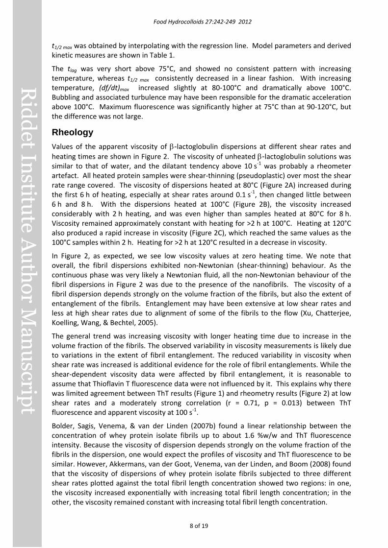
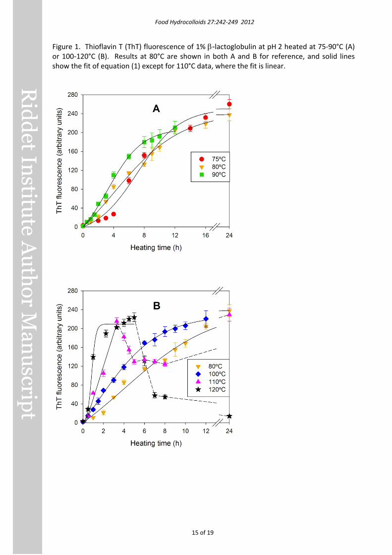
Results and Discussion Thioflavin T fluorescence ThT fluorescence increased with heating time in a way that depended on temperature (Figure 1). At 75°C and 80°C (Figure 1A) there was a distinct lag phase with duration of approximately 4 and 2 hours respectively, during which fluorescence remained low. The lag phase was less distinct at higher temperatures. At 90°C (Figure 1A) and 100°C (Figure 1B), fluorescence increased during the first 8-10 hours’ heating and then plateaued at just over 200 fluorescence units (FU).
During heating at 110°C, fluorescence increased to a maximum of 215.3 ± 7.7 FU at 3.3 hours (Figure 1B), then decreased to a plateau around 130 FU, finally increasing to 229.3 ± 13.8 FU after 24 hours’ heating. Fluorescence increased even faster at 120°C (Figure 1B), plateaued at 200-220 FU between 3.3 and 5 hours, then decreased dramatically in the next two hours and more gradually over the subsequent 17 hours.
The decreases in ThT fluorescence with longer heating at 110°C and 120°C could have been due to local gelation of the sample and/or breakdown of fibril structure. It was not uncommon to see small gel particles in samples, so the former possibility is quite reasonable, and the latter possibility was investigated with TEM (see section 3.3).
The mathematical model in Eq. (1) fitted fluorescence data well at 75-100°C, and fitted the first 5 hours’ data at 120°C, but did not fit 110°C data at all. Values for tlag and (df/dt)max at 110°C were instead derived by linear regression of data between 0.5 and 3.3h (r2 = 0.97), and
)cos1/( ><−>=< φlLp
Food Hydrocolloids 27:242-249 2012
8 of 19
t1/2 max was obtained by interpolating with the regression line. Model parameters and derived kinetic measures are shown in Table 1.
The tlag was very short above 75°C, and showed no consistent pattern with increasing temperature, whereas t1/2 max consistently decreased in a linear fashion. With increasing temperature, (df/dt)max increased slightly at 80-100°C and dramatically above 100°C. Bubbling and associated turbulence may have been responsible for the dramatic acceleration above 100°C. Maximum fluorescence was significantly higher at 75°C than at 90-120°C, but the difference was not large.
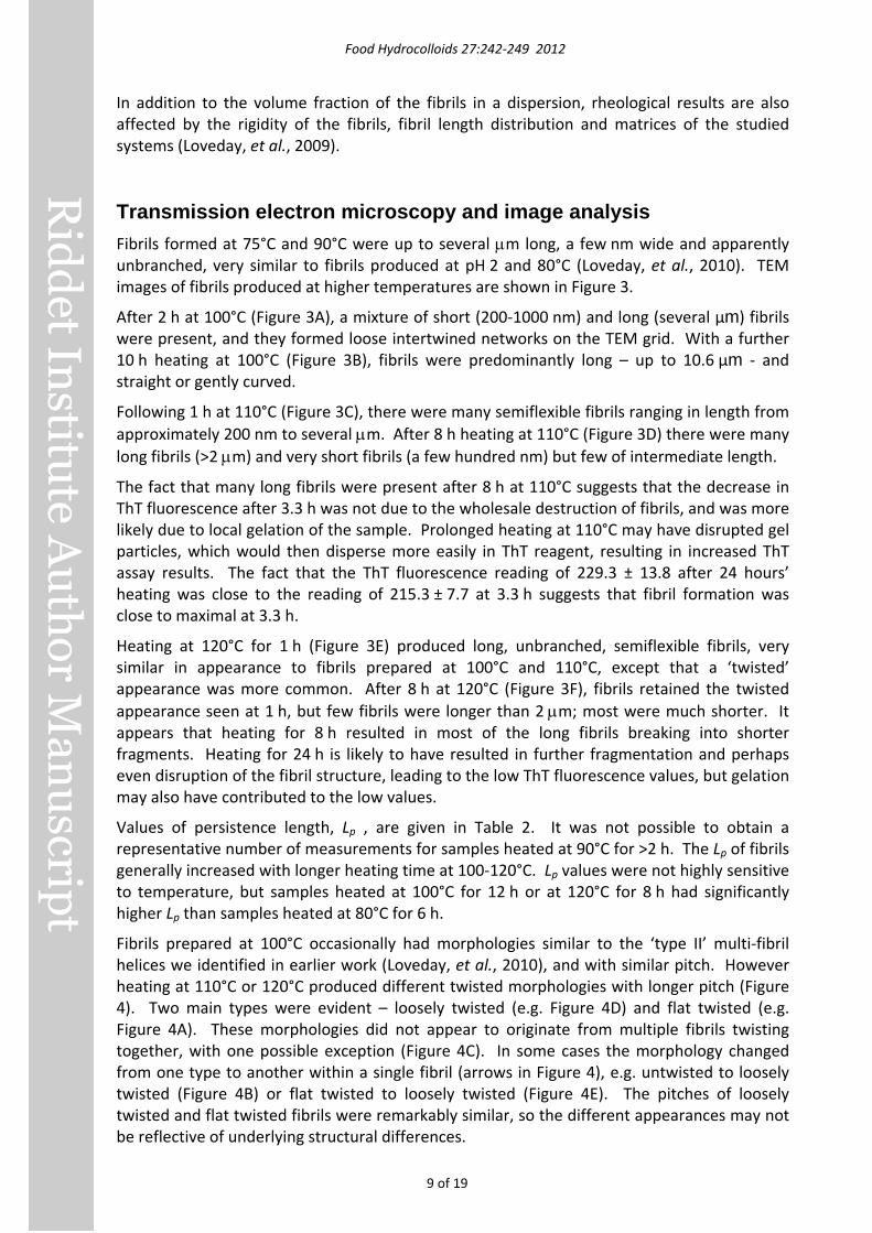
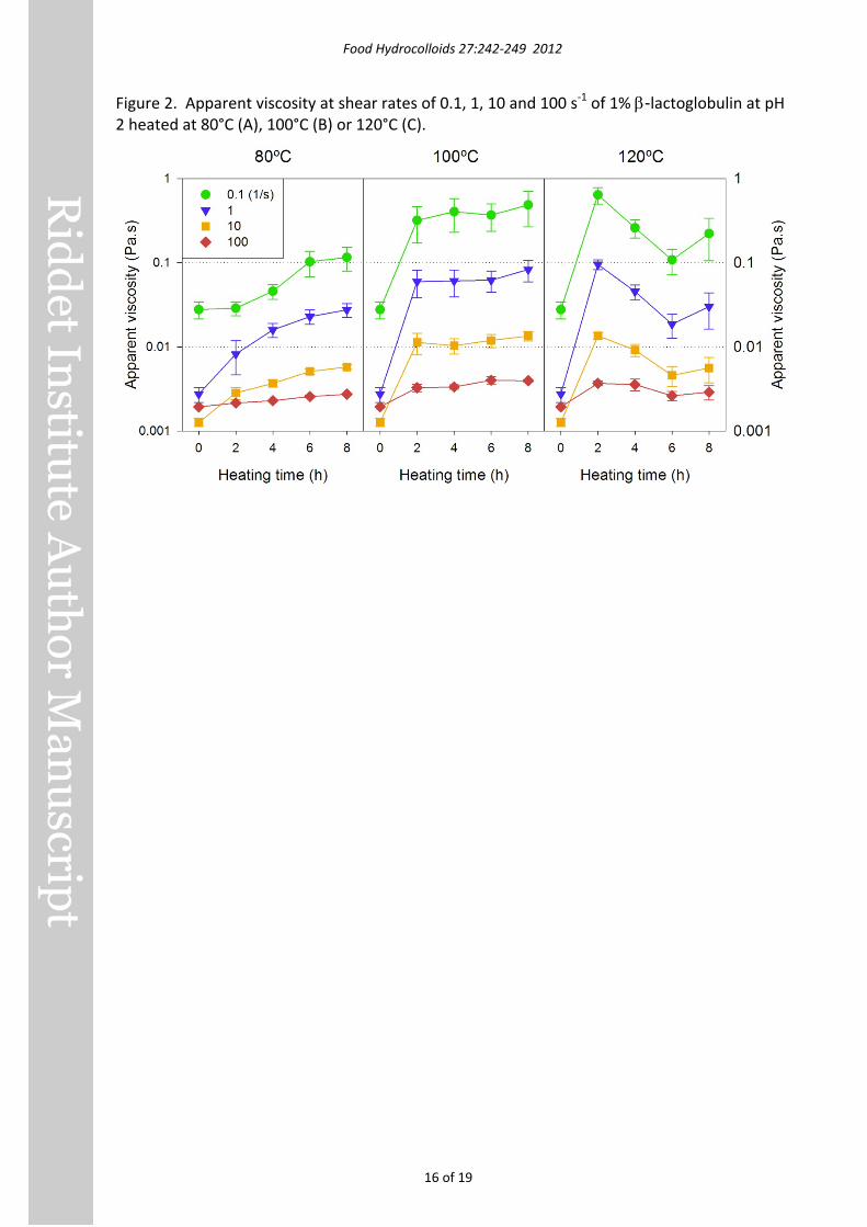
Rheology Values of the apparent viscosity of β-lactoglobulin dispersions at different shear rates and heating times are shown in Figure 2. The viscosity of unheated β-lactoglobulin solutions was similar to that of water, and the dilatant tendency above 10 s-1 was probably a rheometer artefact. All heated protein samples were shear-thinning (pseudoplastic) over most the shear rate range covered. The viscosity of dispersions heated at 80°C (Figure 2A) increased during the first 6 h of heating, especially at shear rates around 0.1 s-1, then changed little between 6 h and 8 h. With the dispersions heated at 100°C (Figure 2B), the viscosity increased considerably with 2 h heating, and was even higher than samples heated at 80°C for 8 h. Viscosity remained approximately constant with heating for >2 h at 100°C. Heating at 120°C also produced a rapid increase in viscosity (Figure 2C), which reached the same values as the 100°C samples within 2 h. Heating for >2 h at 120°C resulted in a decrease in viscosity.
In Figure 2, as expected, we see low viscosity values at zero heating time. We note that overall, the fibril dispersions exhibited non-Newtonian (shear-thinning) behaviour. As the continuous phase was very likely a Newtonian fluid, all the non-Newtonian behaviour of the fibril dispersions in Figure 2 was due to the presence of the nanofibrils. The viscosity of a fibril dispersion depends strongly on the volume fraction of the fibrils, but also the extent of entanglement of the fibrils. Entanglement may have been extensive at low shear rates and less at high shear rates due to alignment of some of the fibrils to the flow (Xu, Chatterjee, Koelling, Wang, & Bechtel, 2005).
The general trend was increasing viscosity with longer heating time due to increase in the volume fraction of the fibrils. The observed variability in viscosity measurements is likely due to variations in the extent of fibril entanglement. The reduced variability in viscosity when shear rate was increased is additional evidence for the role of fibril entanglements. While the shear-dependent viscosity data were affected by fibril entanglement, it is reasonable to assume that Thioflavin T fluorescence data were not influenced by it. This explains why there was limited agreement between ThT results (Figure 1) and rheometry results (Figure 2) at low shear rates and a moderately strong correlation (r = 0.71, p = 0.013) between ThT fluorescence and apparent viscosity at 100 s-1.
Bolder, Sagis, Venema, & van der Linden (2007b) found a linear relationship between the concentration of whey protein isolate fibrils up to about 1.6 %w/w and ThT fluorescence intensity. Because the viscosity of dispersion depends strongly on the volume fraction of the fibrils in the dispersion, one would expect the profiles of viscosity and ThT fluorescence to be similar. However, Akkermans, van der Goot, Venema, van der Linden, and Boom (2008) found that the viscosity of dispersions of whey protein isolate fibrils subjected to three different shear rates plotted against the total fibril length concentration showed two regions: in one, the viscosity increased exponentially with increasing total fibril length concentration; in the other, the viscosity remained constant with increasing total fibril length concentration.
Food Hydrocolloids 27:242-249 2012
9 of 19
In addition to the volume fraction of the fibrils in a dispersion, rheological results are also affected by the rigidity of the fibrils, fibril length distribution and matrices of the studied systems (Loveday, et al., 2009).
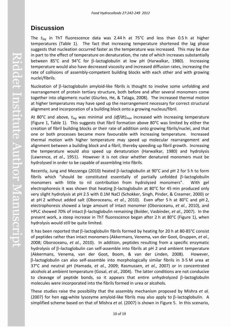
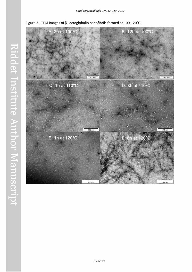
Transmission electron microscopy and image analysis Fibrils formed at 75°C and 90°C were up to several µm long, a few nm wide and apparently unbranched, very similar to fibrils produced at pH 2 and 80°C (Loveday, et al., 2010). TEM images of fibrils produced at higher temperatures are shown in Figure 3.
After 2 h at 100°C (Figure 3A), a mixture of short (200-1000 nm) and long (several μm) fibrils were present, and they formed loose intertwined networks on the TEM grid. With a further 10 h heating at 100°C (Figure 3B), fibrils were predominantly long – up to 10.6 μm - and straight or gently curved.
Following 1 h at 110°C (Figure 3C), there were many semiflexible fibrils ranging in length from approximately 200 nm to several µm. After 8 h heating at 110°C (Figure 3D) there were many long fibrils (>2 µm) and very short fibrils (a few hundred nm) but few of intermediate length.
The fact that many long fibrils were present after 8 h at 110°C suggests that the decrease in ThT fluorescence after 3.3 h was not due to the wholesale destruction of fibrils, and was more likely due to local gelation of the sample. Prolonged heating at 110°C may have disrupted gel particles, which would then disperse more easily in ThT reagent, resulting in increased ThT assay results. The fact that the ThT fluorescence reading of 229.3 ± 13.8 after 24 hours’ heating was close to the reading of 215.3 ± 7.7 at 3.3 h suggests that fibril formation was close to maximal at 3.3 h.
Heating at 120°C for 1 h (Figure 3E) produced long, unbranched, semiflexible fibrils, very similar in appearance to fibrils prepared at 100°C and 110°C, except that a ‘twisted’ appearance was more common. After 8 h at 120°C (Figure 3F), fibrils retained the twisted appearance seen at 1 h, but few fibrils were longer than 2 µm; most were much shorter. It appears that heating for 8 h resulted in most of the long fibrils breaking into shorter fragments. Heating for 24 h is likely to have resulted in further fragmentation and perhaps even disruption of the fibril structure, leading to the low ThT fluorescence values, but gelation may also have contributed to the low values.
Values of persistence length, Lp , are given in Table 2. It was not possible to obtain a representative number of measurements for samples heated at 90°C for >2 h. The Lp of fibrils generally increased with longer heating time at 100-120°C. Lp values were not highly sensitive to temperature, but samples heated at 100°C for 12 h or at 120°C for 8 h had significantly higher Lp than samples heated at 80°C for 6 h.
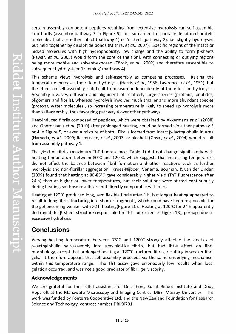
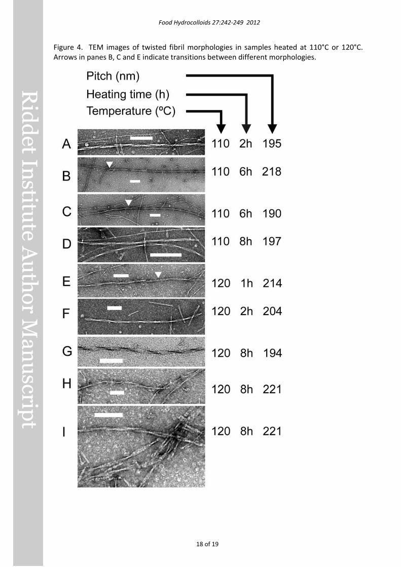
Fibrils prepared at 100°C occasionally had morphologies similar to the ‘type II’ multi-fibril helices we identified in earlier work (Loveday, et al., 2010), and with similar pitch. However heating at 110°C or 120°C produced different twisted morphologies with longer pitch (Figure 4). Two main types were evident – loosely twisted (e.g. Figure 4D) and flat twisted (e.g. Figure 4A). These morphologies did not appear to originate from multiple fibrils twisting together, with one possible exception (Figure 4C). In some cases the morphology changed from one type to another within a single fibril (arrows in Figure 4), e.g. untwisted to loosely twisted (Figure 4B) or flat twisted to loosely twisted (Figure 4E). The pitches of loosely twisted and flat twisted fibrils were remarkably similar, so the different appearances may not be reflective of underlying structural differences.
Food Hydrocolloids 27:242-249 2012
10 of 19
Discussion The tlag in ThT fluorescence data was 2.44 h at 75°C and less than 0.5 h at higher temperatures (Table 1). The fact that increasing temperature shortened the lag phase suggests that nucleation occurred faster as the temperature was increased. This may be due in part to the effect of temperature on denaturation, the rate of which increases substantially between 85°C and 94°C for β-lactoglobulin at low pH (Harwalkar, 1980). Increasing temperature would also have decreased viscosity and increased diffusion rates, increasing the rate of collisions of assembly-competent building blocks with each other and with growing nuclei/fibrils.
Nucleation of β-lactoglobulin amyloid-like fibrils is thought to involve some unfolding and rearrangement of protein tertiary structure, both before and after several monomers come together into oligomeric nuclei (Giurleo, He, & Talaga, 2008). The increased thermal motion at higher temperatures may have sped up the rearrangement necessary for correct structural alignment and incorporation of a building block onto a growing nucleus/fibril.
At 80°C and above, tlag was minimal and (df/dt)max increased with increasing temperature (Figure 1, Table 1). This suggests that fibril formation above 80°C was limited by either the creation of fibril building blocks or their rate of addition onto growing fibrils/nuclei, and that one or both processes became more favourable with increasing temperature. Increased thermal motion with higher temperature may speed up molecular rearrangement and alignment between a building block and a fibril, thereby speeding up fibril growth. Increasing the temperature would also speed up denaturation (Harwalkar, 1980) and hydrolysis (Lawrence, et al., 1951). However it is not clear whether denatured monomers must be hydrolysed in order to be capable of assembling into fibrils.
Recently, Jung and Mezzenga (2010) heated β-lactoglobulin at 90°C and pH 2 for 5 h to form fibrils which “should be constituted essentially of partially unfolded β-lactoglobulin monomers with little to nil contribution from hydrolyzed monomers”. With gel electrophoresis it was shown that heating β-lactoglobulin at 80°C for 45 min produced only very slight hydrolysis at pH 2.5 with 0.1M NaCl (Schokker, Singh, Pinder, & Creamer, 2000) or at pH 2 without added salt (Oboroceanu, et al., 2010). Even after 5 h at 80°C and pH 2, electrophoresis showed a large amount of intact monomer (Oboroceanu, et al., 2010), and HPLC showed 70% of intact β-lactoglobulin remaining (Bolder, Vasbinder, et al., 2007). In the present work, a steep increase in ThT fluorescence began after 2 h at 80°C (Figure 1), when hydrolysis would still be quite limited.
It has been reported that β-lactoglobulin fibrils formed by heating for 20 h at 80-85°C consist of peptides rather than intact monomers (Akkermans, Venema, van der Goot, Gruppen, et al., 2008; Oboroceanu, et al., 2010). In addition, peptides resulting from a specific enzymatic hydrolysis of β-lactoglobulin can self-assemble into fibrils at pH 2 and ambient temperature (Akkermans, Venema, van der Goot, Boom, & van der Linden, 2008). However, β-lactoglobulin can also self-assemble into morphologically similar fibrils in 3-5 M urea at 37°C and neutral pH (Hamada, et al., 2009; Rasmussen, et al., 2007) or in concentrated alcohols at ambient temperature (Gosal, et al., 2004). The latter conditions are not conducive to cleavage of peptide bonds, so it appears that entire unhydrolyzed β-lactoglobulin molecules were incorporated into the fibrils formed in urea or alcohols.
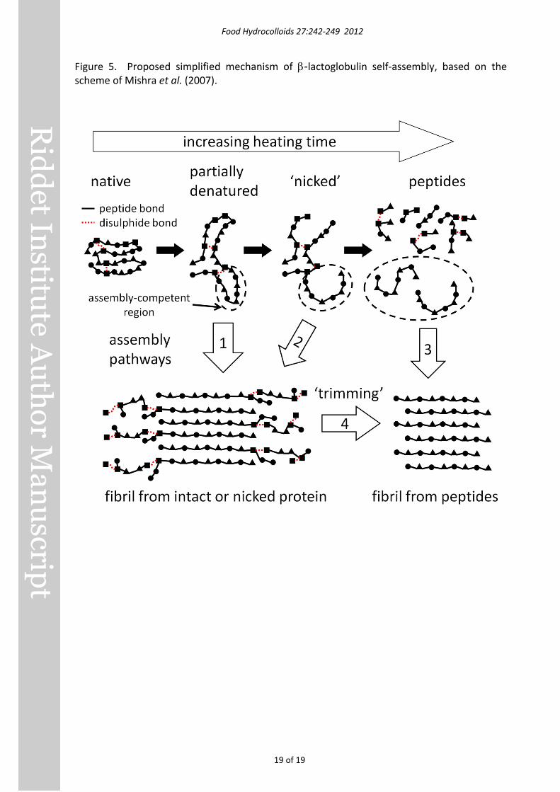
These studies raise the possibility that the assembly mechanism proposed by Mishra et al. (2007) for hen egg-white lysozyme amyloid-like fibrils may also apply to β-lactoglobulin. A simplified scheme based on that of Mishra et al. (2007) is shown in Figure 5. In this scenario,
Food Hydrocolloids 27:242-249 2012
11 of 19
certain assembly-competent peptides resulting from extensive hydrolysis can self-assemble into fibrils (assembly pathway 3 in Figure 5), but so can entire partially-denatured protein molecules that are either intact (pathway 1) or ‘nicked’ (pathway 2), i.e. slightly hydrolysed but held together by disulphide bonds (Mishra, et al., 2007). Specific regions of the intact or nicked molecules with high hydrophobicity, low charge and the ability to form β-sheets (Pawar, et al., 2005) would form the core of the fibril, with connecting or outlying regions being more mobile and solvent-exposed (Török, et al., 2002) and therefore susceptible to subsequent hydrolysis or ‘trimming’ (pathway 4).
This scheme views hydrolysis and self-assembly as competing processes. Raising the temperature increases the rate of hydrolysis (Harris, et al., 1956; Lawrence, et al., 1951), but the effect on self-assembly is difficult to measure independently of the effect on hydrolysis. Assembly involves diffusion and alignment of relatively large species (proteins, peptides, oligomers and fibrils), whereas hydrolysis involves much smaller and more abundant species (protons, water molecules), so increasing temperature is likely to speed up hydrolysis more than self-assembly, thus favouring pathway 4 over other pathways.
Heat-induced fibrils composed of peptides, which were obtained by Akkermans et al. (2008) and Oboroceanu et al. (2010) after prolonged heating, could be formed via either pathway 3 or 4 in Figure 5, or even a mixture of both. Fibrils formed from intact β-lactoglobulin in urea (Hamada, et al., 2009; Rasmussen, et al., 2007) or alcohols (Gosal, et al., 2004) would result from assembly pathway 1.
The yield of fibrils (maximum ThT fluorescence, Table 1) did not change significantly with heating temperature between 80°C and 120°C, which suggests that increasing temperature did not affect the balance between fibril formation and other reactions such as further hydrolysis and non-fibrillar aggregation. Kroes-Nijboer, Venema, Bouman, & van der Linden (2009) found that heating at 80-85°C gave considerably higher yield (ThT fluorescence after 24 h) than at higher or lower temperatures, but their solutions were stirred continuously during heating, so those results are not directly comparable with ours.
Heating at 120°C produced long, semiflexible fibrils after 1 h, but longer heating appeared to result in long fibrils fracturing into shorter fragments, which could have been responsible for the gel becoming weaker with >2 h heating(Figure 2C). Heating at 120°C for 24 h apparently destroyed the β-sheet structure responsible for ThT fluorescence (Figure 1B), perhaps due to excessive hydrolysis.
Conclusions Varying heating temperature between 75°C and 120°C strongly affected the kinetics of β-lactoglobulin self-assembly into amyloid-like fibrils, but had little effect on fibril morphology, except that prolonged heating at 120°C fractured fibrils, resulting in weaker fibril gels. It therefore appears that self-assembly proceeds via the same underlying mechanism within this temperature range. The ThT assay gave erroneously low results when local gelation occurred, and was not a good predictor of fibril gel viscosity.
Acknowledgements
We are grateful for the skilful assistance of Dr Jiahong Su at Riddet Institute and Doug Hopcroft at the Manawatu Microscopy and Imaging Centre, IMBS, Massey University. This work was funded by Fonterra Cooperative Ltd. and the New Zealand Foundation for Research Science and Technology, contract number DRIX0701.
Food Hydrocolloids 27:242-249 2012
12 of 19
References Akkermans, C., van der Goot, A. J., Venema, P., van der Linden, E., & Boom, R. M. (2008).
Formation of fibrillar whey protein aggregates: Influence of heat and shear treatment, and resulting rheology. Food Hydrocolloids, 22(7), 1315-1325.
Akkermans, C., Venema, P., van der Goot, A. J., Boom, R. M., & van der Linden, E. (2008). Enzyme-induced formation of β-lactoglobulin fibrils by AspN endoproteinase. Food Biophys., 3(4), 390-394.
Akkermans, C., Venema, P., van der Goot, A. J., Gruppen, H., Bakx, E. J., Boom, R. M., & van der Linden, E. (2008). Peptides are building blocks of heat-induced fibrillar protein aggregates of β-lactoglobulin formed at pH 2. Biomacromolecules, 9, 1474-1479.
Alting, A. C., de Jongh, H. H. J., Visschers, R. W., & Simons, J. W. F. A. (2002). Physical and chemical interactions in cold gelation of food proteins. J. Agric. Food Chem., 50(16), 4682-4689.
Bolder, S. G., Sagis, L. M. C., Venema, P., & van der Linden, E. (2007a). Effect of stirring and seeding on whey protein fibril formation. J. Agric. Food Chem., 55(14), 5661-5669.
Bolder, S. G., Sagis, L. M. C., Venema, P., & van der Linden, E. (2007b). Thioflavin T and birefringence assays to determine the conversion of proteins into fibrils. Langmuir, 23(8), 4144-4147.
Bolder, S. G., Vasbinder, A. J., Sagis, L. M. C., & van der Linden, E. (2007). Heat-induced whey protein isolate fibrils: Conversion, hydrolysis, and disulphide bond formation. Int. Dairy J., 17(7), 846-853.
Brown, R. S., Bennet, A. J., & Slebocka-Tilk, H. (1992). Recent perspectives concerning the mechanism of H3O+- and OH--promoted amide hydrolysis. Accounts of Chemical Research, 25(11), 481-488.
Chiti, F. & Dobson, C. M. (2006). Protein misfolding, functional amyloid, and human disease. Annu. Rev. Biochem., 75(1), 333-366.
Cooper, J. & LeFevre, E. (1969). Thermophysical properties of water substance. Oxford, UK: Butterworth-Heinemann.
Dobson, C. M. (1999). Protein misfolding, evolution and disease. Trends Biochem. Sci., 24(9), 329-332.
Franks, F. (2002). Protein stability: the value of 'old literature'. Biophys. Chem., 96(2-3), 117-127.
Frare, E., de Laureto, P. P., Zurdo, J., Dobson, C. M., & Fontana, A. (2004). A highly amyloidogenic region of hen lysozyme. J. Mol. Biol., 340(5), 1153-1165.
Gerrard, J. A. (2002). Protein-protein crosslinking in food: methods, consequences, applications. Trends in Food Science and Technology, 13(12), 391-399.
Giurleo, J. T., He, X., & Talaga, D. S. (2008). β-Lactoglobulin assembles into amyloid through sequential aggregated intermediates. J. Mol. Biol., 381(5), 1332-1348.
Gosal, W. S., Clark, A. H., & Ross-Murphy, S. B. (2004). Fibrillar β-lactoglobulin gels: Part 1. Fibril formation and structure. Biomacromolecules, 5(6), 2408-2419.
Hamada, D., Tanaka, T., Tartaglia, G. G., Pawar, A., Vendruscolo, M., Kawamura, M., Tamura, A., Tanaka, N., & Dobson, C. M. (2009). Competition between folding, native-state dimerisation and amyloid aggregation in β−lactoglobulin. J. Mol. Biol., 386(3), 878-890.
Harris, J. I., Cole, R. D., & Pon, N. G. (1956). The kinetics of acid hydrolysis of dipeptides. Biochem. J., 62(1), 154-159.
Harwalkar, V. R. (1980). Kinetics of thermal-denaturation of β-lactoglobulin at pH 2.5. J. Dairy Sci., 63(7), 1052-1057.
Food Hydrocolloids 27:242-249 2012
13 of 19
Hill, E. K., Krebs, B., Goodall, D. G., Howlett, G. J., & Dunstan, D. E. (2006). Shear flow induces amyloid fibril formation. Biomacromolecules, 7(1), 10-13.
Inglis, A. S. (1983). [28] Cleavage at aspartic acid. Methods Enzymol., 91(C), 324-332. Jung, J. M. & Mezzenga, R. (2010). Liquid crystalline phase behavior of protein fibers in water:
Experiments versus theory. Langmuir, 26(1), 504-514. Kroes-Nijboer, A., Venema, P., Bouman, J., & van der Linden, E. (2009). The critical
aggregation concentration of β-lactoglobulin-based fibril formation. Food Biophysics, 4(2), 1-5.
Lawrence, L. & Moore, W. J. (1951). Kinetics of the hydrolysis of simple glycine peptides. J. Am. Chem. Soc., 73(8), 3973-3977.
Loveday, S. M., Rao, M. A., Creamer, L. K., & Singh, H. (2009). Factors affecting rheological characteristics of fibril gels: The case of β-lactoglobulin and α-lactalbumin. J. Food Sci., 74(3), R47-R55.
Loveday, S. M., Wang, X. L., Rao, M. A., Anema, S. G., Creamer, L. K., & Singh, H. (2010). Tuning the properties of β-lactoglobulin nanofibrils with pH, NaCl and CaCl2. Int. Dairy J., 20, 571-579.
Makhatadze, G. I. & Privalov, P. L. (1993). Contribution of hydration to protein folding thermodynamics. I. The enthalpy of hydration. J. Mol. Biol., 232(2), 639-659.
Mishra, R., Sorgjerd, K., Nystrom, S., Nordigarden, A., Yu, Y.-C., & Hammarstrom, P. (2007). Lysozyme amyloidogenesis is accelerated by specific nicking and fragmentation but decelerated by intact protein binding and conversion. J. Mol. Biol., 366(3), 1029-1044.
Morris, A. M., Watzky, M. A., Agar, J. N., & Finke, R. G. (2008). Fitting neurological protein aggregation kinetic data via a 2-step, minimal/"Ockham's razor" Model: The finke-watzky mechanism of nucleation followed by autocatalytic surface growth. Biochemistry, 47(8), 2413-2427.
Oboroceanu, D., Wang, L., Brodkorb, A., Magner, E., & Auty, M. A. E. (2010). Characterization of β-lactoglobulin fibrillar assembly using atomic force microscopy, polyacrylamide gel electrophoresis, and in situ Fourier transform infrared spectroscopy. J. Agric. Food Chem., 58, 3667-3673.
Otte, J., Zakora, M., & Qvist, K. B. (2000). Involvement of disulfide bands in bovine β-lactoglobulin B gels set thermally at various pH. J. Food Sci., 65(3), 384-389.
Pawar, A. P., DuBay, K. F., Zurdo, J., Chiti, F., Vendruscolo, M., & Dobson, C. M. (2005). Prediction of "aggregation-prone" and "aggregation-susceptible" regions in proteins associated with neurodegenerative diseases. J. Mol. Biol., 350(2), 379-392.
Pearce, F. G., Mackintosh, S. H., & Gerrard, J. A. (2007). Formation of amyloid-like fibrils by ovalbumin and related proteins under conditions relevant to food processing. J. Agric. Food Chem., 55(2), 318-322.
Pilkington, S. M., Roberts, S. J., Meade, S. J., & Gerrard, J. A. (2009). Amyloid fibrils as a nanoscaffold for enzyme immobilization. Biotechnol. Prog., 26(1), 93-100.
Privalov, P. L. & Makhatadze, G. I. (1993). Contribution of hydration to protein folding thermodynamics. II. The entropy and gibbs energy of hydration. J. Mol. Biol., 232(2), 660-679.
Rasmussen, P., Barbiroli, A., Bonomi, F., Faoro, F., Ferranti, P., Iriti, M., Picariello, G., & Iametti, S. (2007). Formation of structured polymers upon controlled denaturation of β-lactoglobulin with different chaotropes. Biopolymers, 86(1), 57-72.
Schokker, E. P., Singh, H., Pinder, D. N., & Creamer, L. K. (2000). Heat-induced aggregation of β-lactoglobulin AB at pH 2.5 as influenced by ionic strength and protein concentration. Int. Dairy J., 10(4), 233-240.
Schultz, J. (1967). [28] Cleavage at aspartic acid. Methods Enzymol., 11(C), 255-263.
Food Hydrocolloids 27:242-249 2012
14 of 19
Török, M., Milton, S., Kayed, R., Wu, P., McIntire, T., Glabe, C. G., & Langen, R. (2002). Structural and dynamic features of alzheimer's Aβ peptide in amyloid fibrils studied by site-directed spin labeling. J. Biol. Chem., 277(43), 40810-40815.
Xu, J., Chatterjee, S., Koelling, K. W., Wang, Y., & Bechtel, S. E. (2005). Shear and extensional rheology of carbon nanofiber suspensions. Rheologica Acta, 44(6), 537-562.
Yan, H., Nykanen, A., Ruokolainen, J., Farrar, D., Gough, J. E., Saiani, A., & Miller, A. F. (2008). Thermo-reversible protein fibrillar hydrogels as cell scaffolds. Faraday Discussions, 139, 71-84.
Food Hydrocolloids 27:242-249 2012
15 of 19
Figure 1. Thioflavin T (ThT) fluorescence of 1% β-lactoglobulin at pH 2 heated at 75-90°C (A) or 100-120°C (B). Results at 80°C are shown in both A and B for reference, and solid lines show the fit of equation (1) except for 110°C data, where the fit is linear.
Food Hydrocolloids 27:242-249 2012
16 of 19
Figure 2. Apparent viscosity at shear rates of 0.1, 1, 10 and 100 s-1 of 1% β-lactoglobulin at pH 2 heated at 80°C (A), 100°C (B) or 120°C (C).
Food Hydrocolloids 27:242-249 2012
17 of 19
Figure 3. TEM images of β-lactoglobulin nanofibrils formed at 100-120°C.
Food Hydrocolloids 27:242-249 2012
18 of 19
Figure 4. TEM images of twisted fibril morphologies in samples heated at 110°C or 120°C. Arrows in panes B, C and E indicate transitions between different morphologies.
Food Hydrocolloids 27:242-249 2012
19 of 19
Figure 5. Proposed simplified mechanism of β-lactoglobulin self-assembly, based on the scheme of Mishra et al. (2007).
MASSEY UNIVERSITY
MASSEY RESEARCH ONLINE http://mro.massey.ac.nz/
Massey Documents by Type Journal Articles
β-Lactoglobulin nanofibrils: Effect oftemperature on fibril formation kinetics,fibril morphology and the rheologicalproperties of fibril dispersions
Loveday, SM2012-05
19/12/2018 - Downloaded from MASSEY RESEARCH ONLINE